Semiconductor Quantum Dots for Bioimaging
24
Semiconductor Quantum Dots for Bioimaging and Biodiagnostic Applications Brad A. Kairdolf 1 , Andrew M. Smith 1 , Todd H. Stokes 2 , May D. Wang 2 , Andrew N. Young 3 , and Shuming Nie 1 Shuming Nie: [email protected] 1 Department of Biomedical Engineering, Emory University and Georgia Institute of Technology, Atlanta, Georgia 30322 2 Departments of Biomedical Engineering and Electrical and Computer Engineering, Georgia Institute of Technology, Atlanta, Georgia 30332 3 Department of Pathology and Laboratory Medicine, Emory University School of Medicine, Atlanta, Georgia 30322 Abstract Semiconductor quantum dots (QDs) are light-emitting particles on the nanometer scale that have emerged as a new class of fluorescent labels for chemical analysis, molecular imaging, and biomedical diagnostics. Compared with traditional fluorescent probes, QDs have unique optical and electronic properties such as size-tunable light emission, narrow and symmetric emission spectra, and broad absorption spectra that enable the simultaneous excitation of multiple fluorescence colors. QDs are also considerably brighter and more resistant to photobleaching than are organic dyes and fluorescent proteins. These properties are well suited for dynamic imaging at the single-molecule level and for multiplexed biomedical diagnostics at ultrahigh sensitivity. Here, we discuss the fundamental properties of QDs; the development of next-generation QDs; and their applications in bioanalytical chemistry, dynamic cellular imaging, and medical diagnostics. For in vivo and clinical imaging, the potential toxicity of QDs remains a major concern. However, the toxic nature of cadmium-containing QDs is no longer a factor for in vitro diagnostics, so the use of multicolor QDs for molecular diagnostics and pathology is probably the most important and clinically relevant application for semiconductor QDs in the immediate future. Keywords nanotechnology; fluorescence imaging; cellular dynamics; multiplexing; cancer detection; single- cell analysis 1. INTRODUCTION The development of nanoparticle probes for biomolecular imaging and diagnostics is currently an area of considerable interest (1–10). The basic concept is that nanometer-sized particles have functional and structural properties that are not available from either discrete molecules or bulk materials (1–3). When conjugated with biomolecular affinity ligands, Copyright © 2013 by Annual Reviews. All rights reserved DISCLOSURE STATEMENT B.A.K., A.M.S., and S.M. hold patents related to QD synthesis, coating, and use for diagnostics. The other authors are not aware of any other affiliations, memberships, funding, or financial holdings that might be perceived as affecting the objectivity of this review. NIH Public Access Author Manuscript Annu Rev Anal Chem (Palo Alto Calif). Author manuscript; available in PMC 2013 August 05. Published in final edited form as: Annu Rev Anal Chem (Palo Alto Calif). 2013 June 12; 6(1): 143–162. doi:10.1146/annurev- anchem-060908-155136. NIH-PA Author Manuscript NIH-PA Author Manuscript NIH-PA Author Manuscript
description
Review article on QDs
Transcript of Semiconductor Quantum Dots for Bioimaging

Semiconductor Quantum Dots for Bioimaging and BiodiagnosticApplications
Brad A. Kairdolf1, Andrew M. Smith1, Todd H. Stokes2, May D. Wang2, Andrew N. Young3,and Shuming Nie1
Shuming Nie: [email protected] of Biomedical Engineering, Emory University and Georgia Institute of Technology,Atlanta, Georgia 303222Departments of Biomedical Engineering and Electrical and Computer Engineering, GeorgiaInstitute of Technology, Atlanta, Georgia 303323Department of Pathology and Laboratory Medicine, Emory University School of Medicine,Atlanta, Georgia 30322
AbstractSemiconductor quantum dots (QDs) are light-emitting particles on the nanometer scale that haveemerged as a new class of fluorescent labels for chemical analysis, molecular imaging, andbiomedical diagnostics. Compared with traditional fluorescent probes, QDs have unique opticaland electronic properties such as size-tunable light emission, narrow and symmetric emissionspectra, and broad absorption spectra that enable the simultaneous excitation of multiplefluorescence colors. QDs are also considerably brighter and more resistant to photobleaching thanare organic dyes and fluorescent proteins. These properties are well suited for dynamic imaging atthe single-molecule level and for multiplexed biomedical diagnostics at ultrahigh sensitivity. Here,we discuss the fundamental properties of QDs; the development of next-generation QDs; and theirapplications in bioanalytical chemistry, dynamic cellular imaging, and medical diagnostics. For invivo and clinical imaging, the potential toxicity of QDs remains a major concern. However, thetoxic nature of cadmium-containing QDs is no longer a factor for in vitro diagnostics, so the use ofmulticolor QDs for molecular diagnostics and pathology is probably the most important andclinically relevant application for semiconductor QDs in the immediate future.
Keywordsnanotechnology; fluorescence imaging; cellular dynamics; multiplexing; cancer detection; single-cell analysis
1. INTRODUCTIONThe development of nanoparticle probes for biomolecular imaging and diagnostics iscurrently an area of considerable interest (1–10). The basic concept is that nanometer-sizedparticles have functional and structural properties that are not available from either discretemolecules or bulk materials (1–3). When conjugated with biomolecular affinity ligands,
Copyright © 2013 by Annual Reviews. All rights reserved
DISCLOSURE STATEMENTB.A.K., A.M.S., and S.M. hold patents related to QD synthesis, coating, and use for diagnostics. The other authors are not aware ofany other affiliations, memberships, funding, or financial holdings that might be perceived as affecting the objectivity of this review.
NIH Public AccessAuthor ManuscriptAnnu Rev Anal Chem (Palo Alto Calif). Author manuscript; available in PMC 2013 August 05.
Published in final edited form as:Annu Rev Anal Chem (Palo Alto Calif). 2013 June 12; 6(1): 143–162. doi:10.1146/annurev-anchem-060908-155136.
NIH
-PA Author Manuscript
NIH
-PA Author Manuscript
NIH
-PA Author Manuscript

such as antibodies, peptides, or small molecules, these nanoparticles can be used to detectmolecular biomarkers and tumor cells at high sensitivity and specificity (11–13).Nanoparticles also have large surface areas for the attachment of multiple diagnostic (e.g.,optical, radioisotopic, or magnetic) and therapeutic (e.g., anticancer) agents. Recentadvances have led to the development of biodegradable nanostructures for drug delivery(14–18), iron oxide nanocrystals for magnetic resonance imaging (19, 20), and luminescentquantum dots (QDs) for multiplexed molecular diagnosis and in vivo imaging (21–27).
Semiconductor QDs exhibit novel optical and electronic properties and are emerging as anew class of nanoparticle probes for bioimaging and biodiagnostics (Figure 1). Recentresearch has generated monodispersed QDs encapsulated in stable polymers with versatilesurface chemistries. These nanocrystals are brightly fluorescent, enabling their use asimaging probes both in vitro and in vivo (21–27). Here, we discuss recent developments inthe synthesis and modification of QD nanocrystals and their use in dynamic cellularimaging. We also discuss the use of multiplexedQD–antibody conjugates for mapping themolecular, cellular, and glandular heterogeneity of human cancer specimens. For clinicaldiagnostics, multiplexed QD mapping can provide new molecular and morphologicalinformation that is not available from traditional H&E (hematoxylin and eosin) andimmunohistochemistry (IHC), especially at complex and suspicious disease foci (25, 26). Asdiscussed in more detail below, these results have raised exciting possibilities for theintegration of morphological and molecular biomarker information for cancer diagnosis andtreatment selection.
2. QUANTUM DOT PROBE DEVELOPMENTExtensive research during the past 20 years has led to the development of high-quality andwater-soluble QD probes for a broad range of applications in biology and medicine (1–10,21–27). However, these QDs are still not perfect, mainly because of their largehydrodynamic sizes, a propensity for nonspecific binding to proteins and cellularmembranes, poorly controlled conjugation chemistry, and diminished brightness when thecrystalline core size is reduced. Often in the size range of 15 to 30 nm, these QDs tend tononspecifically adhere to cellular membranes and proteins. These interactions cause theadsorption of a protein layer on the nanocrystal surface, which further increases the particlesize and induces nonspecific cellular uptake. To mitigate this problem, researchers havecoated QDs with neutral hydrophilic polymers such as polyethylene glycol (PEG), yieldingreduced nonspecific binding but at the expense of a large increase in the hydrodynamic size(28). Researchers have also used small zwitterionic ligands (such as cysteine) to overcomethis problem, resulting in particles that are both small and resistant to nonspecific binding(29–32). However, the resulting QDs frequently suffer from low colloidal stability,photobleaching, or low quantum yields. Bioaffinity ligands are usually attached to QDsthrough chemical schemes that are inherently stochastic, such that the number and geometricorientation of conjugated molecules vary widely across the nanoparticle population (6).Consequently, some QD bioconjugates have numerous active ligands that can cross-linkmultiple target molecules (Figure 2). In an attempt to overcome this heterogeneity problem,Ting and colleagues (33, 34) have used monovalent streptavidin to prepare monovalent QDprobes, resulting in reduced cross-linking of target proteins. A further problem is that thebrightness of QDs quickly diminishes when the crystalline core size is reduced. The reasonis that, with the same fluorescence quantum yield, the brightness of single QDs isproportionally related to their molar absorbance, which is scaled approximately to the thirdpower of the particle size (1, 2). Thus, smaller QDs are not as efficiently excited as largerdots and, therefore, are dimmer under the same photon-flux conditions.
Kairdolf et al. Page 2
Annu Rev Anal Chem (Palo Alto Calif). Author manuscript; available in PMC 2013 August 05.
NIH
-PA Author Manuscript
NIH
-PA Author Manuscript
NIH
-PA Author Manuscript

Also, note that current QDs exhibit rapid on-and-off light emission (known as blinking)when observed individually under a fluorescence microscope (35). This attribute is a mixedblessing because it can be detrimental for single-molecule imaging due to a frequent loss ofsignal from the molecule being monitored. It can also be beneficial because it is largely asingle-particle behavior and can be used to differentiate single probes from aggregates.Recent work by Krauss and colleagues (36) indicates that it is possible to completelyeliminate blinking by preparing core/shell particles in which there is a smooth compositiongradient from the core to the shell. However, this research is still controversial, and theresults have not been independently reproduced.
2.1. Next-Generation Quantum DotsThere has been considerable interest in developing new and improved QDs with optimizedbrightness, minimized hydrodynamic size, resistance to nonspecific interactions, and site-specific ligand conjugation. In this section, we discuss recent advances both in engineeringnovel crystalline nanostructures and in developing new surface coatings and moleculartagging strategies. If the hydrodynamic size of QDs is reduced to that of green fluorescentproteins (GFPs), investigators believe that QD-tagged proteins will behave similarly asGFP-tagged proteins inside living cells (6–8). However, the task of developing such protein-sized dots is challenging because small dots often have low optical absorbance and must becoated with a thin polymer layer. As discussed below, novel insights and related results haveraised new possibilities in developing the next generation of QDs for molecular andbiodiagnostic applications.
2.2. Alloyed NanocrystalsThe most common nanocrystalline cores are composed of CdSe, which allows one to tunethe wavelength of fluorescence emission between ~500 and 650 nm by altering the core size(1, 2). However, a major disadvantage of this tuning methodology is that each different colorhas a different hydrodynamic size and fluorescence brightness, making multicolorcomparisons difficult. Recently, several groups have explored the use of ternary alloys inplace of CdSe. With alloys such as CdSexS1−x, CdTexSe1−x, HgxCd1−xTe, and HgxCd1−xSe,the core nanocrystal size can be held constant while the fluorescence wavelength is tunedthrough chemical composition (37, 38), which normalizes the brightness and size to similarvalues and widens the spectral range for fluorescence tuning. In particular, Smith & Nie (39)have used cation exchange to partially replace cadmium ions in CdTe nanocrystals withmercury ions, yielding HgxCd1−xTe alloyed nanocrystals in which the size did not changedue to a similar bond length between CdTe and HgTe (Figure 3). Because of the largedifference in bandgap energy between CdTe and HgTe, these particles can be widely tunedin fluorescence across the near-IR spectrum while maintaining a similarly compact size.Fluorescence emission in the near-IR spectral range is very important for high-sensitivitybioassays due to both the greater penetration of near-IR light in scattering media and thelower autofluorescence background. In contrast to organic dyes and fluorescent proteins thathave limited fluorescence emission efficiencies at wavelengths longer than ~700 nm,quantum yields for QDs can approach unity at longer wavelengths, and stable probescomposed of nanocrystals such as HgxCd1−xTe, InAs, and PbxCd1−xSe can be produced inwater with fluorescence emission that is tunable from 700 to 2,000 nm (39–41).
Other nanocrystal architectures under development include heterostructures with mixeddimensionality through seeded growth. A single-component nanocrystal can be used as aseed for the overgrowth of a shell with a different composition and dimensionality (42). Forexample, the Alivisatos group (43) and the Manna group (44) simultaneously reported theuse of CdSe seeds for overgrowth of CdS rods. Thus, the quasi-spherical seeds are buriedwithin the CdS rod. Moreover, Alivisatos and colleagues have reported that tuning the
Kairdolf et al. Page 3
Annu Rev Anal Chem (Palo Alto Calif). Author manuscript; available in PMC 2013 August 05.
NIH
-PA Author Manuscript
NIH
-PA Author Manuscript
NIH
-PA Author Manuscript
crystalline structure of the core between hexagonal and cubic causes the rods to growoutward from four facets of the core, yielding a tetrapod structure. Furthermore, a widevariety of compositions have been explored for these materials, opening the door to uniquecharge-carrier confinement regimes and complex wave function engineering strategies (45).The unique optoelectronic characteristics of these multidimensionality particles has beeninvestigated by the Weller group (46), who demonstrated that an electric field applied alongthe long axis of a CdSe (coreQD)/CdS (shell rod) nanocrystal alters the fluorescenceintensity and wavelength, allowing for electronic modulation of fluorescence resonanceenergy transfer (FRET) between the nanocrystals and the dye molecules.
2.3. New Surface Coatings to Minimize Hydrodynamic Size and Nonspecific BindingRecently, Smith & Nie (10, 47) have developed a multifunctional, multidentate polymerligand for generating highly compact QDs with ultrasmall sizes that preserves the excellentoptical properties of the nanocrystals (Figure 4). These multidentate polymers can displacethe existing ligands on the QD and tightly bind to the nanocrystal surface in a closed “loops-and-trains” conformation. This unique design eliminates the hydrophobic barrier layer andcauses the polymer ligand to tightly conform to the nanocrystal surface, resulting in anexceptionally thin polymer shell and small overall particle size. In contrast to water-solubleQDs with small-molecule ligands, the multidentate binding of the polymer providesexcellent colloidal stability, resistance to photo-bleaching, and high quantum yield. Usingthis strategy, investigators have prepared high-quality QDs with a hydrodynamic size of 4 to6 nm (47). Importantly, Frangioni and colleagues (31) have demonstrated that QDs with ahydrodynamic diameter of <5.5 nm undergo rapid renal clearance, making these size-minimized nanoparticles ideal for in vivo imaging, wherein the potential toxicity of theheavy metal–containing QD has been a significant impediment to clinical adoption.
In addition to size, another problem is that current QDs are often “sticky” because they havea tendency to bind nonspecifically to proteins, cellular membranes, or extracellular matrixes(48–51). Nonspecific binding reduces the signal-to-noise ratio and limits immunostainingspecificity and detection sensitivity. Nonspecific binding can also lead to false-positivestaining for biomarkers in fluids, cells, and tissues. In particular, QDs with highly negativeor positive surface charges, such as surface coatings containing carboxylic acids or amines,can exhibit strong nonspecific binding to cells and tissues (48–50) as well as to proteins inserum and blood. Because most biomolecules are charged or have charged domains (52),QDs could interact electrostatically with many soluble proteins in solution or withbiomolecules on the cell surface and in the cytoplasm, resulting in the commonly observednonspecific binding.
To reduce nonspecific binding, PEGs are often attached to the organic coating layer of QDs(23, 48). PEGylated QDs have a nearly neutral surface charge and can maintain colloidalstability through steric repulsion between the PEG chains. The reduced charge of a PEG, aswell as its conformational flexibility, provides a stable surface coating that can reducenonspecific binding (Figure 5) in biological environments. Rosenthal and colleagues (48)have shown that PEG chains as short as 550 Da can significantly reduce the nonspecificbinding of QDs to cells. As a result, PEGylated QDs have been successfully used for both invitro (23, 48) and in vivo (27–29) applications. However, PEG-coated QDs havesignificantly larger hydrodynamic sizes than those of comparable non-PEGylatednanoparticles, often more than doubling the particles’ hydrodynamic size. This increasedsize can prevent the probes from accessing biological targets deep within complex tissue orcellular structures. Kairdolf et al. (51) reported an alternative approach, in which the QDsurface is modified with a small hydroxyl-containing molecule (such as 1,3-diamino-2-propanol), yielding hydroxylated QDs. These particles show a dramatically reduced surface
Kairdolf et al. Page 4
Annu Rev Anal Chem (Palo Alto Calif). Author manuscript; available in PMC 2013 August 05.
NIH
-PA Author Manuscript
NIH
-PA Author Manuscript
NIH
-PA Author Manuscript

charge with virtually no nonspecific binding to cells or tissues, while maintaining excellentcolloidal stability.
2.4. Tagging StrategiesThe most widely used bioconjugation strategy is covalent attachment of a molecule to theQD coating surface via a functional group. This approach typically involves the formation ofan amide bond between a carboxylic acid group on the nanocrystal and an amine group onthe affinity molecule by use of carbodiimide chemistry. Other common functional groups forthe covalent attachment include a thiol group coupling to maleimide to form a thioetherbond (53, 54). This chemistry is particularly useful for conjugating QDs to antibodies, whichhave thiol groups that can be exposed following reduction of the interchain disulfide bondsand do not disrupt the affinity site. These covalent techniques can also be used to conjugatestreptavidin and biotin to QDs and biomolecules (23, 55) to produce versatile reagents thatcan simply be mixed prior to use to form a targeted probe. Noncovalent interactions havealso been employed to attach molecules to the QD surface. Mattoussi et al. (56) havedemonstrated that QDs with a highly negative surface charge can be bound to biomoleculesthrough electrostatic interactions by use of a chimeric fusion protein with a positivelycharged attachment domain. Gao and colleagues (57) have further demonstrated thisprinciple by using positively charged QDs for noncovalent binding of negatively chargedsmall interfering RNA (siRNA). Furthermore, histidine tags have been used to directlycouple molecules to the QD surface in a defined orientation for optimal function (58, 59).These tags consist of a polyhistidine peptide, which has strong affinity to charged metalatoms such as Ni2+ and Zn2+ and can be fused to the termini of recombinant proteins. Size-minimized QDs with thin coatings have accessible surfaces, allowing the polyhistidinepeptide to bind directly to the nanocrystal through coordination with the surface metalatoms. Rao and colleagues (60) have described another strategy for site-specific conjugationthat uses the covalent coupling of a chloroalkane to Halo Tag proteins. These proteins arehaloalkane dehalogenases that have been adapted to cleave the carbon–halogen bond in achloroalkane molecule to form a stable ester bond. Such a protein, through fusion to abiomolecule of interest, can be coupled to a chloroalkane-containing QD in a highlycontrolled manner.
3. DYNAMIC CELLULAR IMAGINGRecent research using QD–ligand or QD–antibody conjugates has revealed the complexworkings of membrane receptors at high sensitivity and temporal resolution (61, 62). Newreceptor behaviors, such as motor-driven transport of the epidermal growth factor receptoralong cellular outgrowths toward the cell body (63), have been reported. In particular,neurons are known to have richly complex plasma membranes with multiple types ofmicrodomains that form intracellular signaling complexes termed synapses, which exhibitdynamic receptor exchanges. The diffusion of several types of neurotransmitter receptors inand out of the synapse has been studied by use of QDs attached to glycine neurotransmitterand AMPA glutamate receptors; these investigations have revealed rapid fluctuations indiffusion rates in different membrane domains (61, 63). Some receptors, such as theneuronal growth factor (NGF) receptor, become internalized into the cell once they bind to aspecific ligand, a process that can now be studied in great detail due to the photostability ofQDs (64). QDs conjugated to NGF bind to the NGF receptor in the terminals of neuronalaxons (long neuronite outgrowths involved in signal transduction), inducing endocytosis ofthe receptor–ligand pair within vesicular structures. QD imaging revealed that these vesiclesusually contain only a single NGF receptor and that they are shuttled great lengths to the cellbody along multiple molecular tracks that behave as a multilane highway within the axons.However, these studies were carried out by using conventional QDs with large sizes andmultivalent ligand presentation. As discussed above, the large size of conventional QD
Kairdolf et al. Page 5
Annu Rev Anal Chem (Palo Alto Calif). Author manuscript; available in PMC 2013 August 05.
NIH
-PA Author Manuscript
NIH
-PA Author Manuscript
NIH
-PA Author Manuscript
probes is a major problem for their application in crowded cell-surface domains, such as thesynaptic cleft, an intracellular junction between neurons that is typically only 20 nm wide.Larger QD conjugates have limited access to this region compared with smaller antibody–fluorophore conjugates, which adds some uncertainty to the QD neuronal diffusion studiesreported to date (65, 66).
For intracellular transport, Ruan et al. (67) have used peptide-conjugated QDs to examinethe complex behavior of nanoparticles in live cells. Dynamic confocal imaging revealed thatthe peptide-conjugated QDs were internalized by macropinocytosis, a fluid-phaseendocytosis process triggered by QD binding to cell membranes. The internalized QDs weretethered to the inner vesicle surfaces and trapped in cytoplasmic organelles. The QD-loadedvesicles were actively transported by molecular machines (such as dyneins) alongmicrotubule tracks (Figure 6). The destination of this active transport process was anasymmetric perinuclear region (outside the cell nucleus) known as the MTOC (microtubuleorganizing center). Indeed, motor protein translocation proceeds in discrete steps and with avelocity indicative of specific motor protein–filament pairs. QD–kinesin and QD–myosinconjugates delivered to the cellular cytoplasm through osmotic pinosome lysis undergodirected motion that was remarkably similar to that observed in purified filaments (68,69).The molecularmotors were tracked for extended periods of time without loss of signal.
Currently, a major challenge is to deliver freely diffusing and monodispersed QD probesinto the cytoplasm of living cells. One effective technique is to directly inject QDs intoliving cells by using a microneedle. However, this process is rather low throughput becausethe individual cells must be injected one at a time (70). To achieve higher-throughputdelivery of QDs to cell populations, investigators have attempted to temporarilypermeabilize the cellular plasma membrane through the formation of microscopic pores,either through the use of bacterial toxins (e.g., streptolysin O) that form well-definedmembrane pores or through brief exposure to a pulsed electric field. These mechanisms arepromising but have yet to demonstrate homogeneous delivery of free QDs in cells.
An alternative and promising approach is the controlled disruption of endosomal vesicles.Cells naturally engulf their surrounding environment through various processes that yieldintracellular vesicles containing extracellular fluid. This mechanism is a convenient way toenable entry of QD probes into cells, but the particles remain trapped and therefore are notfree to interact with target molecules, so it is necessary to have a strategy for QD release orendosomal escape. One method is to use osmosis for swelling and bursting the endosomes(68). This process can be performed by allowing cells to engulf QDs during a brief exposureto a hypertonic medium (prepared by adding sucrose or other solutes), which leads to therapid formation of pinocytic vesicles that bud off of the plasmamembrane due to watermoving out of the cells (efflux). In the second step, a brief and well-controlled exposure ofthese cells to a hypotonic solution containing a low solute concentration causes water to rushinto the solute-rich vesicles, inducing osmotic lysis and allowing the QDs to be dispersedinto the cytoplasm.
Recent research has further shown that QDs coated with proton-sponge polymers can escapefrom endosomes after cellular internalization (56).The proton sponge effect arises fromnumerous weak conjugate bases (such as carboxylic acid and tertiary amine, with bufferingcapabilities at pH 5–6), leading to proton absorption in acid organelles and an osmoticpressure buildup across the organelle membrane (71). This osmotic pressure causes swellingand/or rupture of the acidic endosomes and a release of the trapped QDs into the cytoplasm.Alternatively, QDs can be encapsulated in proton-sponge polymer beads, which are brokendown into proton-absorbing units in the lysosomes, thereby releasing the QD cargo into thecytoplasm (72).
Kairdolf et al. Page 6
Annu Rev Anal Chem (Palo Alto Calif). Author manuscript; available in PMC 2013 August 05.
NIH
-PA Author Manuscript
NIH
-PA Author Manuscript
NIH
-PA Author Manuscript
4. BIOMEDICAL DIAGNOSTICSIn contrast to in vivo imaging, in which the potential toxicity of QDs remains amajorconcern (73–75), analyses of cells and tissues as well as solution-based biomarkers areperformed on in vitro or ex vivo clinical patient samples. Because toxicity is of no concernwhen analyzing these specimens, the use of multiplexed QDs as ultrasensitive probes for invitro biodiagnostics is probably the most important and clinically relevant application ofQDs (22–26). The unique optical properties of QDs can significantly enhance the sensitivityof biodiagnostic assays such as IHC, fluorescence in situ hybridization (FISH), flowcytometry, and biochips and can provide new capabilities to extend the utility ofbiodiagnostic assays in the clinic. In particular, the multiplexing capability of QDs can beused to quantitatively measure a panel of molecular biomarkers, enabling personalizeddiagnostics and treatment.
4.1. Multiplexed ImmunostainingOne of the most widely explored clinical applications for QDs is in multiplexedimmunostaining of formalin-fixed paraffin-embedded (FFPE) tissue specimens (Figure 7a).IHC was first reported for protein marker detection and localization in tissue specimens 70years ago (76–78) and has been extensively used in anatomic pathology since thedevelopment of robust staining methods (79–81). IHC is especially useful for clinicalbiomarker detection because it preserves the morphology of the tissue, which is critical formany diagnoses. Despite its ubiquitous use, IHC for diagnostics has seen only minorimprovements in the past 50 years; the most notable innovations have involved thedevelopment of companion diagnostics, such as PATHWAY (http://www.ventana.com/product/98?type=93) and HercepTest™ (82), to predict response to a specific therapy. Nieand colleagues (26) have recently described detailed methods for the preparation, staining,and analysis of clinical tissue specimens by using QDs in both direct and indirectprocedures, laying the foundation for multiplexed and quantitative QD-based clinical IHCassays. Research by several other groups (83–87) has shown that multiplexed QD stainingfor biomarkers in clinical tissue specimens enhances the diagnostic potential of IHC andenables the detection of multiple disease markers within a single slide. Paired with analyticalhardware and software tools, QD-based methods have transformed immunostaining into apowerful diagnostic tool for high-throughput analysis of disease markers in clinical samples,including minute specimens such as needle aspirates. These methods are expected to play akey role in medical diagnostics as pathology continues to progress, particularly with thetransition to digital pathology (88).
4.2. Cancer DiagnosticsOne of the most common biomarker panels employed by oncologists and pathologists is ER/PR/Her2. This biomarker panel is used to diagnose breast cancer and to determine the mosteffective treatment strategy for breast cancer patients (89–91). These markers are currentlymeasured individually with immunoassays such as HercepTest and traditional IHCtechniques (92, 93), all of which rely on the subjective assessment of protein markerexpression visualized by standard chromagens. Yezhelyev et al. (25) have demonstrated thesimultaneous staining and measurement of these biomarkers in both cultured human breastcancer cells and in fixed (FFPE) clinical tissue specimens by using multiplexed QDs. Theseauthors have shown that the QD-based methods for the quantification of ER/PR/Her2proteins on a single slide correlate closely with the results achieved from traditional IHC,Western blot analysis, and FISH. Also, they used five QD colors simultaneously on a singleclinical tissue specimen to detect five unique markers (ER/mTOR/PR/EGFR/Her2), furtherdemonstrating the molecular profiling potential of these nanoparticles in complex tissuesamples.
Kairdolf et al. Page 7
Annu Rev Anal Chem (Palo Alto Calif). Author manuscript; available in PMC 2013 August 05.
NIH
-PA Author Manuscript
NIH
-PA Author Manuscript
NIH
-PA Author Manuscript
Investigations into the effectiveness of QD immunostaining and comparisons with currentclinical methods have been reported by Chen et al. (94), who used lung cancertissuemicroarrays to detect caveolin-1 and PCNA (proliferating cell nuclear antigen).Theseauthors reported that QD-based immunostaining methods have a higher detection sensitivityin comparison to conventional clinical techniques. Increased accuracy and sensitivity wereindependently demonstrated by Li and colleagues (85), who performed a detailedexamination of QD staining for the Her2 protein. Marker detection using QDs, comparedwith conventional IHC and FISH, was more sensitive and accurate than the standardtechniques, particularly in cases with moderate marker expression, in which subjectiveassessment using conventional methods are often problematic and can introduce error orbias.
On the basis of these findings, further studies were conducted to determine the fulldiagnostic potential of QD-based methods. By combining the increased accuracy andsensitivity of QD-based immunostaining with another key parameter (total tumor size), Liand colleagues (95) introduced a new indicator (total Her2 load) to assess prognosis inbreast cancer patients. This indicator identified more patients in the poor prognosis groupthan did Her2 gene amplification. The total Her2 load parameter identified a distinctsubgroup of patients with particularly poor 5-year disease-free survival who were notdifferentiated with other methods. These results are especially promising for improvingbreast cancer diagnosis, and demonstrate the potential for individualized diagnostics andpatient classification using QD-based immunostaining assays.
4.3. Single-Cell AnalysisBecause of the high sensitivity and multiplexing capabilities of QD probes, biodiagnosticassays with these nanomaterials can even be used to analyze single rare cells, such ascirculating tumor cells or isolated malignant cells, within the complex microenvironments ofheterogeneous tumor tissue or in biological fluids (Figure 7 b). Such biodiagnostic analysisalso leaves the specimens structurally intact, preserving important morphologicalinformation to correlate with the molecular profiling data. Morphology and biomarkerexpression data cannot be integrated with conventional molecular profiling or analyticalmethods such as gene chips, protein microarrays, or polymerase chain reaction (PCR). Liu etal. (26) have demonstrated that a panel of four biomarkers (E-cadherin/HML cytokeratin/p63/AMACR) can be simultaneously measured with QD probes and used to detect andcharacterize individual cells on prostatectomy and needle aspirate specimens. The QD-basedmolecular profiling technique enabled the mapping of molecular and cellular variationswithin a heterogeneous tissue specimen and enabled the identification of isolated malignantcells within predominantly benign prostate glands. This technique is a major improvementover other methods to analyze distinct regions (such as laser capture microdissection)because it allows molecular and morphological data to be digitally extracted from singlecells, cell clusters, or glands without physically removing the regions of interest from thesection. Using this method, Liu et al. identified prostate glands with only a single cancerouscell as the gland began its malignant transformation.
The rapid identification of low-abundance cancer cells has also been reported for thediagnosis of Hodgkin’s lymphoma (96). The presence of Reed–Sternberg (RS) cells is ahallmark of the disease, but these cells constitute only ~1% of the total infiltrating cells inthe lymph node specimens. Using multiplexed QD probes, Liu et al. (96) characterizedindividual RS cells with a panel of four biomarkers (CD15/CD30/CD45/Pax5) anddistinguished them from other immune cells in clinical specimens. To further evaluate thediagnostic potential of QDs, they also compared QD staining analysis with previouslydetermined pathological examination results. The QD-based methods rapidly identified all
Kairdolf et al. Page 8
Annu Rev Anal Chem (Palo Alto Calif). Author manuscript; available in PMC 2013 August 05.
NIH
-PA Author Manuscript
NIH
-PA Author Manuscript
NIH
-PA Author Manuscript

patients with confirmed disease and showed the presence of disease in two patients whowere classified as suspected of having the disease. The abundance of RS cells in these“suspicious” specimens was extremely low, which probably caused the ambiguous resultsoriginally obtained from standard pathological examination. Specimens from patients withreactive lymph nodes (but not Hodgkin’s lymphoma) showed a complete absence of RScells. The results from these studies clearly illustrate the detection sensitivity of QD probesand show that biodiagnostic assays with multiplexed QDs can be used to diagnose patientsat a much earlier stage than is achievable with conventional diagnostic methods, possiblyimproving the therapeutic success rate for patients.
4.4. Solution-Based DiagnosticsAlthough the use of QDs for immunostaining in tissue has been the focus of recent research,solution-based biodiagnostic assays are another area in which the unique properties of QDscan be exploited. Numerous assays using QDs as ultrasensitive and multiplexed probes foranalyte detection have been developed. In particular, polychromatic flow cytometry is atechnique that dramatically benefits from the superior signal brightness and multiplexingcapabilities of these nanomaterials. Immunophenotyping using flow cytometry is a powerfultool for the detection, identification, and characterization of many cell types and has broadapplications in diagnostic medicine. Using a combination of organic fluorophores andmultiplexed QDs, Roederer and coworkers (97) demonstrated the simultaneousquantification of 17 unique markers with flow cytometry; this result is a dramatic increaseover techniques using organic fluorophores alone. The increase in multiplexing capabilityhas significant implications for the use of flow cytometry in the characterization of cellularimmune responses; the diagnosis of complex diseases such as cancer; and the identificationof T cells for HIV characterization, which exhibit a surprisingly high degree ofheterogeneity.
QDs have also been used in a microfluidic instrument for the detection of single intactviruses in solution. Agrawal et al. (98) developed a dual-color method using red and greenQD probes for identifying respiratory syncytial virus, a primary cause of lower respiratorytract disease in infants and young children and an important pathogen of the elderly andimmune-compromised individuals. By targeting the probes to two different antigens on thevirus surface (F and G proteins), these authors used photon coincidence to distinguish thesignals of QDs bound to the virus particles from unbound QDs in the solution. QDs are idealfor this application because of their large Stokes shift and broad excitation profiles, whichallow multiple colors to be excited simultaneously using a single high-energy excitationsource. Strong coincidence signals were observed from samples containing the virus,whereas control samples showed little to no signal. This method also distinguishedvariations in the relative expression of viral surface proteins to determine virulence in asensitive manner in real time (99).
In addition to protein analysis, the analysis of genes and genetic defects is a vital tool fordisease diagnosis and is the major application of many molecular profiling tools such asgene chips and PCR. The exceptional optical properties of QDs make them ideal probes foruse in these applications and provide unique capabilities that are not available with existingtechnologies. Han et al. (100) were the first to report a novel bar-coding technology usingQD-tagged microbeads for the optical coding of biomolecules. With the use of six differentQD colors with 10 intensity levels, one million unique combinations can theoretically beobtained. By coupling the microbeads to a unique DNA-recognition sequence, the authorseasily detected and identified the target molecules. Hybridization studies (100) have shownthat coding and target signals can be read at the single-bead level, demonstrating the utilityof QD bar coding in the rapid analysis of DNA. Single-QD nanoparticles are also useful for
Kairdolf et al. Page 9
Annu Rev Anal Chem (Palo Alto Calif). Author manuscript; available in PMC 2013 August 05.
NIH
-PA Author Manuscript
NIH
-PA Author Manuscript
NIH
-PA Author Manuscript

DNA analysis, and probes have been developed for the ultrasensitive detection of DNA andgenetic mutations (101, 102).
Gerion et al. (101) have reported QD–DNA conjugates for the detection of single-nucleotidepolymorphisms (SNPs), in which a sequence varies by a single base. These probes candetect both SNPs and single-base deletions in minutes at room temperature with highspecificity. More recently, Wang and colleagues (102) developed a DNA nanosensor systemby using single QDs with a bioconjugated capture sequence and a separate dye-conjugatedreporter sequence (Figure 8). Following binding of the target DNA sequence to the QDsensor, the reporter sequence binds the target in a sandwich assay, bringing the reporter dyein close proximity to the nanocrystal and forming a FRET donor–acceptor pair for targetdetection at femtomolar (10−15 M) sensitivity. This process enables analyte detectionwithout amplification, dramatically reducing the time and cost of gene analysis, whichtypically requires amplification with currently used technologies.
5. CONCLUDING REMARKSLooking into the future, we expect major advances in both fundamental studies and practicalapplications for semiconductor nanocrystals. For fundamental research, the synthesis of newnanocrystals with unusual structures and properties is a boundless frontier and will continueto yield surprises such as doped and strain-tuned QDs. There are a wide variety of newnanocrystalline materials available with a diverse range of chemical, electronic, and opticalproperties. In particular, oxide materials such as ZnO would be an exceptional shell materialfor nanocrystal capping because of their wide bandgap and resistance to oxidativedegradation; IV–VI semiconductors have uniquely positive deformation potentials; andmercury-based II–VI materials may allow for the continuous tuning of bandgaps throughspontaneous cation-exchange reactions. For biomedical applications, it is important tominimize the overall size of bioconjugated nanocrystals, to reduce steric hindrance andnonspecific protein adsorption, to develop chemically activatable or photoswitchablenanocrystals for multicolor superresolution optical microscopy, and to understand thepotential toxic effects of semiconductor materials (6). A major long-term goal is thedevelopment of QD probes that are simultaneously monovalent, free from nonspecificadsorption, compact in size, and still bright for single-molecule imaging. Reaching this goalwill require innovations not only in developing novel crystalline nanostructures but also indeveloping new surface-coating, molecular tagging, and cellular delivery strategies.
AcknowledgmentsThe authors acknowledge the National Institutes of Health for financial support (grants R01CA163256,RC2CA148265, and HHSN268201000043C). S.N. and M.D.W. are Distinguished Cancer Scholars of the GeorgiaCancer Coalition. A.M.S. acknowledges the National Cancer Institute Nano-Alliance Program for a Pathway toIndependence Award (1K99CA154006). A.M.S.’s current affiliation is: Department of Bioengineering, Universityof Illinois at Urbana-Champaign, Urbana, Illinois 61801.
Glossary
IHC immunohistochemistry
PEG polyethylene glycol
GFP green fluorescent protein
FRET fluorescence resonance energy transfer
FISH fluorescence in situ hybridization
Kairdolf et al. Page 10
Annu Rev Anal Chem (Palo Alto Calif). Author manuscript; available in PMC 2013 August 05.
NIH
-PA Author Manuscript
NIH
-PA Author Manuscript
NIH
-PA Author Manuscript

FFPE formalin-fixed paraffin-embedded
ER/mTOR/PR/EGFR/Her2
a breast cancer protein biomarker panel consisting of estrogenreceptor (ER), mammalian target of rapamycin (mTOR),progesterone receptor (PR), epidermal growth factor receptor(EGFR), and human epidermal growth factor receptor 2 (Her2)
Caveolin-1 scaffolding protein located in the caveolae of the cell membrane
E-cadherin/HMLcytokeratin/p63/AMACR
a prostate cancer protein biomarker panel consisting of epithelial(E-)cadherin, high–molecular weight (HML) cytokeratin,transformation-related protein 63 (p63), and α-methylacyl-CoAracemase (AMACR)
RS cell Reed–Sternberg cell
CD15/CD30/CD45/Pax5
a Hodgkin’s lymphoma biomarker panel consisting of cluster ofdifferentiation molecules (CDs) and a paired box protein (PAX)
LITERATURE CITED1. Alivisatos P. The use of nanocrystals in biological detection. Nat. Biotechnol. 2004; 22:47–52.
[PubMed: 14704706]
2. Smith AM, Nie S. Semiconductor nanocrystals: structure, properties, and band gap engineering.Acc. Chem. Res. 2010; 43:190–200. [PubMed: 19827808]
3. Michalet X, Pinaud FF, Bentolila LA, Tsay JM, Doose S, et al. Quantum dots for live cells, in vivoimaging, and diagnostics. Science. 2005; 307:538–544. [PubMed: 15681376]
4. Medintz IL, Uyeda HT, Goldman ER, Mattoussi H. Quantum dot bioconjugates for imaging,labelling and sensing. Nat. Mater. 2005; 4:435–446. [PubMed: 15928695]
5. Gao X, Yang L, Petros JA, Marshall FF, Simons JW, Nie S. In vivo molecular and cellular imagingwith quantum dots. Curr. Opin. Biotechnol. 2005; 16:63–72. [PubMed: 15722017]
6. Smith AM, Duan H, Mohs AM, Nie S. Bioconjugated quantum dots for in vivo molecular andcellular imaging. Adv. Drug Deliv. Rev. 2008; 60:1226–1240. [PubMed: 18495291]
7. Baker M. Nanotechnology imaging probes: smaller and more stable. Nat. Methods. 2010; 7:957–962.
8. Smith AM, Wen MM, Nie S. Imaging dynamic cellular events with quantum dots—the brightfuture. Biochemist. 2010; 32:12–17. [PubMed: 20824157]
9. Nie S, Xing Y, Kim GJ, Simons JW. Nanotechnology applications in cancer. Annu. Rev. Biomed.Eng. 2007; 9:257–288. [PubMed: 17439359]
10. Smith AM, Nie S. Next-generation quantum dots. Nat. Biotechnol. 2009; 27:732–733. [PubMed:19668181]
11. Liu Z, Cai W, He L, Nakayama N, Chen K, et al. In vivo biodistribution and highly efficienttumour targeting of carbon nanotubes in mice. Nat. Nanotechnol. 2007; 2:47–52. [PubMed:18654207]
12. Weissleder R, Kelly K, Sun EY, Shtatland T, Josephson L. Cell-specific targeting of nanoparticlesby multivalent attachment of small molecules. Nat. Biotechnol. 2005; 23:1418–1423. [PubMed:16244656]
13. Lee ES, Na K, Bae YH. Polymeric micelle for tumor pH and folate-mediated targeting. J. Control.Release. 2003; 91:103–113. [PubMed: 12932642]
14. Hood JD, Bednarski M, Frausto R, Guccione S, Reisfeld RA, et al. Tumor regression by targetedgene delivery to the neovasculature. Science. 2002; 296:2404–2407. [PubMed: 12089446]
15. Duncan R. Polymer conjugates as anticancer nanomedicines. Nat. Rev. Cancer. 2006; 6:688–701.[PubMed: 16900224]
16. Couvreur P, Vauthier C. Nanotechnology: intelligent design to treat complex disease. Pharm. Res.2006; 23:1417–1450. [PubMed: 16779701]
Kairdolf et al. Page 11
Annu Rev Anal Chem (Palo Alto Calif). Author manuscript; available in PMC 2013 August 05.
NIH
-PA Author Manuscript
NIH
-PA Author Manuscript
NIH
-PA Author Manuscript

17. Moghimi SM, Hunter AC, Murray JC. Long-circulating and target-specific nanoparticles: theory topractice. Pharmacol. Rev. 2001; 53:283–318. [PubMed: 11356986]
18. Torchilin VP. Micellar nanocarriers: pharmaceutical perspectives. Pharm. Res. 2007; 24:1–16.[PubMed: 17109211]
19. McCarthy JR, Kelly KA, Sun EY, Weissleder R. Targeted delivery of multifunctional magneticnanoparticles. Nanomedicine. 2007; 2:153–167. [PubMed: 17716118]
20. Harisinghani MG, Barentsz J, Hahn PF, Deserno WM, Tabatabaei S, et al. Noninvasive detectionof clinically occult lymph-node metastases in prostate cancer. N. Engl. J. Med. 2003; 348:2491–2499. [PubMed: 12815134]
21. Rhyner MN, Smith AM, Gao XH, Mao H, Yang L, Nie SM. Quantum dots and multifunctionalnanoparticles: new contrast agents for tumor imaging. Nanomedicine. 2006; 1:209–217. [PubMed:17716110]
22. Xing Y, Chaudry Q, Shen C, Kong KY, Zhau HE, et al. Bioconjugated quantum dots formultiplexed and quantitative immunohistochemistry. Nat. Protoc. 2007; 2:1152–1165. [PubMed:17546006]
23. Wu X, Liu H, Liu J, Haley KN, Treadway JA, et al. Immunofluorescent labeling of cancer markerHer2 and other cellular targets with semiconductor quantum dots. Nat. Biotechnol. 2003; 21:41–46. [PubMed: 12459735]
24. Kim S, Lim YT, Soltesz EG, DeGrand AM, Lee J, et al. Near-infrared fluorescent type II quantumdots for sentinel lymph node mapping. Nat. Biotechnol. 2004; 22:93–97. [PubMed: 14661026]
25. Yezhelyev MV, Al-Hajj A, Morris C, Marcus AI, Liu T, et al. In situ molecular profiling of breastcancer biomarkers with multicolor quantum dots. Adv. Mater. 2007; 19:3146–3151.
26. Liu J, Lau S, Varma V, Moffitt R, Caldwell M, et al. Molecular mapping of tumor heterogeneityon clinical tissue specimens with multiplexed quantum dots. Am. Chem. Soc. Nano. 2010;4:2755–2765.
27. Gao X, Cui Y, Levenson RM, Chung LW, Nie S. In vivo cancer targeting and imaging withsemiconductor quantum dots. Nat. Biotechnol. 2004; 22:969–976. [PubMed: 15258594]
28. Ballou B, Lagerholm BC, Ernst LA, Bruchez MP, Waggoner AS. Noninvasive imaging ofquantum dots in mice. Bioconjug. Chem. 2004; 15:79–86. [PubMed: 14733586]
29. Liu WH, Choi HS, Zimmer JP, Tanaka E, Frangioni JV, Bawendi MG. Compact cysteine-coatedCdSe (ZnCdS) quantum dots for in vivo applications. J. Am. Chem. Soc. 2007; 129:14530–14531.[PubMed: 17983223]
30. Liang GX, Gu MM, Zhang JR, Zhu JJ. Preparation and bioapplication of high-quality, water-soluble, biocompatible, and near-infrared-emitting CdSeTe alloyed quantum dots.Nanotechnology. 2009; 20:415103. [PubMed: 19762946]
31. Choi HS, Liu W, Misra P, Tanaka E, Zimmer JP, et al. Renal clearance of quantum dots. Nat.Biotechnol. 2007; 25:1165–1170. [PubMed: 17891134]
32. Liu XF, Gao Y, Wang XM, Wu SJ, Tang ZY. Preparation of stable, water-soluble, highlyluminescence quantum dots with small hydrodynamic sizes. J. Nanosci. Nanotechnol. 2011;11:1941–1949. [PubMed: 21449332]
33. Howarth M, Liu WH, Puthenveetil S, Zheng Y, Marshall LF, et al. Monovalent, reduced-sizequantum dots for imaging receptors on living cells. Nat. Methods. 2008; 5:397–399. [PubMed:18425138]
34. Howarth M, Takao K, Hayashi Y, Ting AY. Targeting quantum dots to surface proteins in livingcells with biotin ligase. Proc. Natl. Acad. Sci. USA. 2005; 102:7583–7588. [PubMed: 15897449]
35. Nirmal M, Dabbousi BO, Bawendi MG, Macklin JJ, Trautman JK, et al. Fluorescenceintermittency in single cadmium selenide nanocrystals. Nature. 1996; 383:802–804.
36. Wang X, Ren X, Kahen K, Hahn MA, Rajeswaran M, et al. Non-blinking semiconductornanocrystals. Nature. 2009; 459:686–689. [PubMed: 19430463]
37. Bailey RE, Nie S. Alloyed semiconductor quantum dots: tuning the optical properties withoutchanging the particle size. J. Am. Chem. Soc. 2003; 125:7100–7106. [PubMed: 12783563]
38. Regulacio MD, Han MY. Composition-tunable alloyed semiconductor nanocrystals. Acc. Chem.Res. 2010; 43:621–630. [PubMed: 20214405]
Kairdolf et al. Page 12
Annu Rev Anal Chem (Palo Alto Calif). Author manuscript; available in PMC 2013 August 05.
NIH
-PA Author Manuscript
NIH
-PA Author Manuscript
NIH
-PA Author Manuscript
39. Smith AM, Nie S. Bright and compact alloyed quantum dots with broadly tunable near-infraredabsorption and fluorescence spectra through mercury cation exchange. J. Am. Chem. Soc. 2011;133:24–26. [PubMed: 21142154]
40. Aharoni A, Mokari T, Popov I, Banin U. Synthesis of InAs/CdSe/ZnSe core/shell 1/shell 2structures with bright and stable near-infrared fluorescence. J. Am. Chem. Soc. 2006; 128:257–264. [PubMed: 16390155]
41. Pietryga JM, Werder DJ, Williams DJ, Casson JL, Schaller RD, et al. Utilizing the lability of leadselenide to produce heterostructured nanocrystals with bright, stable infrared emission. J. Am.Chem. Soc. 2008; 130:4879–4885. [PubMed: 18341344]
42. Talapin DV, Koeppe R, Götzinger S, Kornowski A, Lupton JM, et al. Highly emissive colloidalCdSe/CdS heterostructures of mixed dimensionality. Nano Lett. 2003; 3:1677–1681.
43. Talapin DV, Nelson JH, Shevchenko EV, Aloni S, Sadtler B, Alivisatos AP. Seeded growth ofhighly luminescent CdSe/CdS nanoheterostructures with rod and tetrapod morphologies. NanoLett. 2007; 7:2951–2959. [PubMed: 17845068]
44. Carbone L, Nobile C, De Giorgi M, Della Sala F, Morello G, et al. Synthesis and micrometer-scaleassembly of colloidal CdSe/CdS nanorods prepared by a seeded growth approach. Nano Lett.2007; 7:2942–2950. [PubMed: 17845067]
45. Fiore A, Mastria R, Lupo MG, Lanzani G, Giannini C, et al. Tetrapod-shaped colloidalnanocrystals of II–VI semiconductors prepared by seeded growth. J. Am. Chem. Soc. 2009;131:2274–2282. [PubMed: 19170630]
46. Becker K, Lupton JM, Müller J, Rogach AL, Talapin DV, et al. Electrical control of Förster energytransfer. Nat. Mater. 2006; 5:777–781. [PubMed: 16998470]
47. Smith AM, Nie S. Minimizing the hydrodynamic size of quantum dots with multifunctionalmultidentate polymer ligands. J. Am. Chem. Soc. 2008; 130:11278–11279. [PubMed: 18680294]
48. Bentzen EL, Tomlinson ID, Mason J, Gresch P, Warnement MR, et al. Surface modification toreduce nonspecific binding of quantum dots in live cell assays. Bioconjug. Chem. 2005; 16:1488–1494. [PubMed: 16287246]
49. Gerion D, Parak WJ, Williams SC, Zanchet D, Micheel CM, Alivisatos AP. Sorting fluorescentnanocrystals with DNA. J. Am. Chem. Soc. 2001; 124:7070–7074. [PubMed: 12059231]
50. Pathak S, Choi SK, Arnheim N, Thompson ME. Hydroxylated quantum dots as luminescent probesfor in situ hybridization. J. Am. Chem. Soc. 2001; 123:4103–4104. [PubMed: 11457171]
51. Kairdolf BA, Mancini MC, Smith AM, Nie S. Minimizing nonspecific cellular binding of quantumdots with hydroxyl-derivatized surface coatings. Anal. Chem. 2008; 80:3029–3034. [PubMed:18324840]
52. Sheinerman FB, Norel R, Honig B. Electrostatic aspects of protein–protein interactions. Curr.Opin. Struct. Biol. 2000; 10:153–159. [PubMed: 10753808]
53. Zhou M, Nakatani E, Gronenberg LS, Tokimoto T, Wirth MJ, et al. Peptide-labeled quantum dotsfor imaging GPCRs in whole cells and as single molecules. Bioconjug. Chem. 2007; 18:323–332.[PubMed: 17373766]
54. Parak WJ, Gerion D, Zanchet D, Woerz AS, Pellegrino T, et al. Conjugation of DNA to silanizedcolloidal semiconductor nanocrystalline quantum dots. Chem. Mater. 2002; 14:2113–2119.
55. Willard DM, Carillo LL, Jung J, Van Orden A. CdSe-ZnS quantum dots as resonance energytransfer donors in a model protein–protein binding assay. Nano Lett. 2001; 1:469–474.
56. Mattoussi H, Mauro JM, Goldman ER, Anderson GP, Sundar VC, et al. Self-assembly of CdS–ZnSquantum dot bioconjugates using an engineered recombinant protein. J. Am. Chem. Soc. 2000;122:12142–12150.
57. Yezhelyev MV, Qi L, O’Regan RM, Nie S, Gao X. Proton-sponge coated quantum dots for siRNAdelivery and intracellular imaging. J. Am. Chem. Soc. 2008; 130:9006–9012. [PubMed:18570415]
58. Medintz IL, Clapp AR, Mattoussi H, Goldman ER, Fisher B, Mauro JM. Self-assembled nanoscalebiosensors based on quantum dot FRET donors. Nat. Mater. 2003; 2:630–638. [PubMed:12942071]
Kairdolf et al. Page 13
Annu Rev Anal Chem (Palo Alto Calif). Author manuscript; available in PMC 2013 August 05.
NIH
-PA Author Manuscript
NIH
-PA Author Manuscript
NIH
-PA Author Manuscript

59. Sapsford KE, Pons T, Medintz IL, Higashiya S, Brunel FM, et al. Kinetics of metal-affinity drivenself-assembly between proteins or peptides and CdSe–ZnS quantum dots. J. Phys. Chem. C. 2007;111:11528–11538.
60. Zhang Y, So M, Loening AM, Yao H, Gambhir SS, Rao J. HaloTag protein–mediated site-specificconjugation of bioluminescent proteins to quantum dots. Angew. Chem. 2006; 118:5058–5062.
61. Dahan M, Levi S, Luccardini C, Rostaing P, Riveau B, Triller A. Diffusion dynamics of glycinereceptors revealed by single–quantum dot tracking. Science. 2003; 302:442–445. [PubMed:14564008]
62. Lidke DS, Nagy P, Heintzmann R, Arndt-Jovin DJ, Post JN, et al. Quantum dot ligands providenew insights into erbB/HER receptor–mediated signal transduction. Nat. Biotechnol. 2004;22:198–203. [PubMed: 14704683]
63. So M, Yao H, Rao J. HaloTag protein–mediated specific labeling of living cells with quantumdots. Biochem. Biophys. Res. Commun. 2008; 374:419–423. [PubMed: 18621022]
64. Cui B, Wu C, Chen L, Ramirez A, Bearer EL, et al. One at a time, live tracking of NGF axonaltransport using quantum dots. Proc. Natl. Acad. Sci. USA. 2007; 104:13666–13671. [PubMed:17698956]
65. Liu W, Howarth M, Greytak AB, Zheng Y, Nocera DG, et al. Compact biocompatible quantumdots functionalized for cellular imaging. J. Am. Chem. Soc. 2008; 130:1274–1284. [PubMed:18177042]
66. Groc L, Lafourcade M, Heine M, Renner M, Racine V, et al. Surface trafficking ofneurotransmitter receptor: comparison between single-molecule/quantum dot strategies. J.Neurosci. 2007; 27:12433–12437. [PubMed: 18003820]
67. Ruan G, Agrawal A, Marcus AI, Nie S. Imaging and tracking of Tat peptide–conjugated quantumdots in living cells: new insights into nanoparticle uptake, intracellular transport, and vesicleshedding. J. Am. Chem. Soc. 2007; 129:14759–14766. [PubMed: 17983227]
68. Courty S, Luccardini C, Bellaiche Y, Cappello G, Dahan M. Tracking individual kinesin motors inliving cells using single quantum-dot imaging. Nano Lett. 2006; 6:1491–1495. [PubMed:16834436]
69. Pierobon P, Achouri S, Courty S, Dunn AR, Spudich JA, et al. Velocity, processivity, andindividual steps of single myosin V molecules in live cells. Biophys. J. 2009; 96:4268–4275.[PubMed: 19450497]
70. Derfus AM, Chan WCW, Bhatia SN. Intracellular delivery of quantum dots for live cell labelingand organelle tracking. Adv. Mater. 2004; 16:961–966.
71. Boussif O, Lezoualc’h F, Zanta MA, Mergny MD, Scherman D, et al. A versatile vector for geneand oligonucleotide transfer into cells in culture and in vivo: polyethylenimine. Proc. Natl. Acad.Sci. USA. 1995; 92:7297–7301. [PubMed: 7638184]
72. Kim BYS, Jiang W, Oreopoulos J, Yip CM, Rutka JT, Chan WCW. Biodegradable quantum dotnanocomposites enable live cell labeling and imaging of cytoplasmic targets. Nano Lett. 2008;8:3887–3892. [PubMed: 18816147]
73. Derfus AM, Chan WCW, Bhatia SN. Probing the cytotoxicity of semiconductor quantum dots.Nano Lett. 2004; 4:11–18.
74. Hardman R. A toxicologic review of quantum dots: Toxicity depends on physicochemical andenvironmental factors. Environ. Health Perspect. 2006; 114:165–172. [PubMed: 16451849]
75. Mancini MC, Kairdolf BA, Smith AM, Nie S. Oxidative quenching and degradation of polymer-encapsulated quantum dots: new insights into the long-term fate and toxicity of nanocrystals invivo. J. Am. Chem. Soc. 2008; 130:10836–10837. [PubMed: 18652463]
76. Coons AH, Creech HJ, Jones RN. Immunological properties of an antibody containing afluorescent group. Proc. Soc. Exp. Biol. Med. 1941; 47:200–202.
77. Coons AH, Leduc EH, Connolly JM. Studies on antibody production. I. A method for thehistochemical demonstration of specific antibody and its application to a study of thehyperimmune rabbit. J. Exp. Med. 1955; 102:49–60. [PubMed: 14392240]
78. Coons AH, Kaplan MH. Localization of antigen in tissue cells. II. Improvements in a method forthe detection of antigen by means of fluorescent antibody. J. Exp. Med. 1950; 91:1–13. [PubMed:15395569]
Kairdolf et al. Page 14
Annu Rev Anal Chem (Palo Alto Calif). Author manuscript; available in PMC 2013 August 05.
NIH
-PA Author Manuscript
NIH
-PA Author Manuscript
NIH
-PA Author Manuscript

79. Nakane PK, Pierce GB Jr. Enzyme-labeled antibodies: preparation and application for thelocalization of antigens. J. Histochem. Cytochem. 1966; 14:929–931. [PubMed: 17121392]
80. Avrameas S. Coupling of enzymes to proteins with glutaraldehyde: use of the conjugates for thedetection of antigens and antibodies. Immunochemistry. 1969; 6:43–48. [PubMed: 4975324]
81. Mason DY, Sammons R. Alkaline phosphatase and peroxidase for double immunoenzymaticlabelling of cellular constituents. J. Clin. Pathol. 1978; 31:454–460. [PubMed: 77279]
82. Jacobs TW, Gown AM, Yaziji H, Barnes MJ, Schnitt SJ. Specificity of HercepTest in determiningHER-2/neu status of breast cancers using the United States Food and Drug Administration–approved scoring system. J. Clin. Oncol. 1999; 17:1983–1987. [PubMed: 10561248]
83. Ghazani AA, Lee JA, Klostranec J, Xiang Q, Dacosta RS, et al. High throughput quantification ofprotein expression of cancer antigens in tissue microarray using quantum dot nanocrystals. NanoLett. 2006; 6:2881–2886. [PubMed: 17163724]
84. Tholouli E, Sweeney E, Barrow E, Clay V, Hoyland JA, Byers RJ. Quantum dots light uppathology. J. Pathol. 2008; 216:275–285. [PubMed: 18814189]
85. Chen C, Peng J, Xia HS, Yang GF, Wu QS, et al. Quantum dots–based immunofluorescencetechnology for the quantitative determination of HER2 expression in breast cancer. Biomaterials.2009; 30:2912–2918. [PubMed: 19251316]
86. Fountaine TJ, Wincovitch SM, Geho DH, Garfield SH, Pittaluga S. Multispectral imaging ofclinically relevant cellular targets in tonsil and lymphoid tissue using semiconductor quantum dots.Mod. Pathol. 2006; 19:1181–1191. [PubMed: 16778828]
87. Peng CW, Liu XL, Chen C, Liu X, Yang XQ, et al. Patterns of cancer invasion revealed by QDs-based quantitative multiplexed imaging of tumor microenvironment. Biomaterials. 2011; 32:2907–2917. [PubMed: 21262536]
88. Al-Janabi S, Huisman A, Van Diest PJ. Digital pathology: current status and future perspectives.Histopathology. 2011; 61:1–9. [PubMed: 21477260]
89. Carey LA, Dees EC, Sawyer L, Gatti L, Moore DT, et al. The triple negative paradox: primarytumor chemosensitivity of breast cancer subtypes. Clin. Cancer Res. 2007; 13:2329. [PubMed:17438091]
90. Cleator S, Heller W, Coombes RC. Triple-negative breast cancer: therapeutic options. LancetOncol. 2007; 8:235–244. [PubMed: 17329194]
91. Rakha EA, El-Sayed ME, Green AR, Lee AHS, Robertson JF, Ellis IO. Prognostic markers intriple-negative breast cancer. Cancer. 2007; 109:25–32. [PubMed: 17146782]
92. Jacobs TW, Gown AM, Yaziji H, Barnes MJ, Schnitt SJ. Specificity of HercepTest in determiningHER-2/neu status of breast cancers using the United States Food and Drug Administration-approved scoring system. J. Clin. Oncol. 1999; 17:1983–1987. [PubMed: 10561248]
93. Masood S, Bui MM. Assessment of Her-2/neu overexpression in primary breast cancers and theirmetastatic lesions: an immunohistochemical study. Ann. Clin. Lab. Sci. 2000; 30:259–265.[PubMed: 10945565]
94. Chen H, Xue J, Zhang Y, Zhu X, Gao J, Yu B. Comparison of quantum dots immunofluorescencehistochemistry and conventional immunohistochemistry for the detection of caveolin-1 and PCNAin the lung cancer tissue microarray. J. Mol. Histol. 2009; 40:261–268. [PubMed: 19908148]
95. Chen C, Xia HS, Gong YP, Peng J, Peng CW, et al. The quantitative detection of totalHER2 loadby quantum dots and the identification of a new subtype of breast cancer with different 5-yearprognosis. Biomaterials. 2010; 31:8818–8825. [PubMed: 20723971]
96. Liu J, Lau S, Varma V, Kairdolf B, Nie S. Multiplexed detection and characterization of rare tumorcells in Hodgkin’s lymphoma with multicolor quantum dots. Anal. Chem. 2010; 82:6237–6243.[PubMed: 20565106]
97. Chattopadhyay PK, Price DA, Harper TF, Betts MR, Yu J, et al. Quantum dot semiconductornanocrystals for immunophenotyping by polychromatic flow cytometry. Nat. Med. 2006; 12:972–977. [PubMed: 16862156]
98. Agrawal A, Zhang C, Byassee T, Tripp RA, Nie S. Counting single native biomolecules and intactviruses with color-coded nanoparticles. Anal. Chem. 2006; 78:1061–1070. [PubMed: 16478096]
Kairdolf et al. Page 15
Annu Rev Anal Chem (Palo Alto Calif). Author manuscript; available in PMC 2013 August 05.
NIH
-PA Author Manuscript
NIH
-PA Author Manuscript
NIH
-PA Author Manuscript

99. Agrawal A, Tripp RA, Anderson LJ, Nie S. Real-time detection of virus particles and viral proteinexpression with two-color nanoparticle probes. J. Virol. 2005; 79:8625–8628. [PubMed:15956604]
100. Han M, Gao X, Su JZ, Nie S. Quantum-dot-tagged microbeads for multiplexed optical coding ofbiomolecules. Nat. Biotechnol. 2001; 19:631–635. [PubMed: 11433273]
101. Gerion D, Chen F, Kannan B, Fu A, Parak WJ, et al. Room-temperature single-nucleotidepolymorphism and multiallele DNA detection using fluorescent nanocrystals and microarrays.Anal. Chem. 2003; 75:4766–4772. [PubMed: 14674452]
102. Zhang CY, Yeh HC, Kuroki MT, Wang TH. Single-quantum-dot-based DNA nanosensor. Nat.Mater. 2005; 4:826–831. [PubMed: 16379073]
Kairdolf et al. Page 16
Annu Rev Anal Chem (Palo Alto Calif). Author manuscript; available in PMC 2013 August 05.
NIH
-PA Author Manuscript
NIH
-PA Author Manuscript
NIH
-PA Author Manuscript

Figure 1.Unique optical properties of quantum dots (QDs). (a) Fluorescence image of vials containingQDs of increasing size (left to right). The size-dependent properties of nanocrystals allowfor the synthesis of fluorescent probes with emissions covering the entire visible–to–near-IRwavelength range. (b) (Top) Fluorescence and (bottom) absorbance spectra of green and redQDs. Narrow and symmetric fluorescence spectra enable accurate modeling fordeconvolution and spectral unmixing to differentiate probes with significant emissionoverlaps. The absorbance spectra show a broad absorption profile, which enables a widewavelength range for excitation and a single excitation source for multiple QD colors.
Kairdolf et al. Page 17
Annu Rev Anal Chem (Palo Alto Calif). Author manuscript; available in PMC 2013 August 05.
NIH
-PA Author Manuscript
NIH
-PA Author Manuscript
NIH
-PA Author Manuscript

Figure 2.Current and next-generation quantum dots (QDs.) (a) Current QDs are often large andelongated nanocrystals with a thick and sticky micellar bilayer and randomly linkedbioaffinity molecules. (b) Next-generation QDs will have compact and spherical cores witha thin, inert monolayer coating conjugated to a single biomolecule through a site-specific,high-affinity attachment. Adapted with permission from Reference 10.
Kairdolf et al. Page 18
Annu Rev Anal Chem (Palo Alto Calif). Author manuscript; available in PMC 2013 August 05.
NIH
-PA Author Manuscript
NIH
-PA Author Manuscript
NIH
-PA Author Manuscript

Figure 3.Structure and energy-band diagrams of CdTe and cation-exchanged HgxCd1−xTe quantumdots (QDs). (a) Cd2+ replacement by Hg2+. (b) The potential energy wells (gray lines),quantum-confined kinetic energy levels (blue lines), and wave functions (red) of electronsand holes in CdTe and HgxCd1−xTe QDs, as calculated using the effective massapproximation. Reproduced with permission from Reference 39.
Kairdolf et al. Page 19
Annu Rev Anal Chem (Palo Alto Calif). Author manuscript; available in PMC 2013 August 05.
NIH
-PA Author Manuscript
NIH
-PA Author Manuscript
NIH
-PA Author Manuscript

Figure 4.Quantum dot (QD) size minimization by use of multidentate ligands that contain a mixtureof amine and thiol functional groups. (a) The multidentate ligands can wrap around the QDin a closed monolayer conformation, in contrast to the bulky bilayer structure of amphiphilicpolymers. (b) A balance of thiol (−SH) and amine (−NH2) functional groups is needed forstable multidentate ligand binding. Abbreviations: DIC, diisopropylcarbodiimide; DMSO,dimethylsulfoxide; FMOC, fluorenylmethyloxycarbonyl; NHS, N-hydroxysuccinimide.Adapted with permission from Reference 47.
Kairdolf et al. Page 20
Annu Rev Anal Chem (Palo Alto Calif). Author manuscript; available in PMC 2013 August 05.
NIH
-PA Author Manuscript
NIH
-PA Author Manuscript
NIH
-PA Author Manuscript

Figure 5.Quantum dot (QD) coatings and their effects on particle size and nonspecific binding. (a)QD size strongly depends on the surface coating; PEGylation (left) and amphiphilicpolymers (middle) add considerably to the overall size. Size minimization is possible withthe use of multidentate polymer ligands (right), which can wrap around the QD in a “loops-and-trains” conformation. (b) Charge reduction through the incorporation of PEG or −OHfunctional groups dramatically decreases nonspecific binding of QDs in cells and tissues.Abbreviation: PEG, polyethylene glycol.
Kairdolf et al. Page 21
Annu Rev Anal Chem (Palo Alto Calif). Author manuscript; available in PMC 2013 August 05.
NIH
-PA Author Manuscript
NIH
-PA Author Manuscript
NIH
-PA Author Manuscript

Figure 6.Direct observation of active transport of endocytosed Tat quantum dots (Tat-QDs) insideliving cells. (a,b) Directed motion from the cell periphery to an intracellular region adjacentto the cell nucleus. The boxed red area in panel a is magnified in panel b. The white linerepresents the trajectory of the Tat-QD vesicle indicated by the red arrow. The green lineshows the plasma membrane boundary of the cell. (c,d) Directed motion along cell-peripheral tracks. The boxed red area of panel c is magnified in panel d. The green lineindicates the plasma membrane of the cell. Reproduced with permission from Reference 67.
Kairdolf et al. Page 22
Annu Rev Anal Chem (Palo Alto Calif). Author manuscript; available in PMC 2013 August 05.
NIH
-PA Author Manuscript
NIH
-PA Author Manuscript
NIH
-PA Author Manuscript

Figure 7.Immunoprofiling in complex tissues for the identification and characterization of rare cells.(a) Multiplexed quantum dot (QD) imaging in prostate biopsy specimens allows for thedifferentiation of a benign prostate gland (left) from a gland with a single malignant cell(right), as determined by positive AMACR staining (arrowhead). (b) A four-biomarker panelwas used to identify (i) low-abundance Reed–Sternberg cells, (ii) B cells, and (iii) T cells ina heterogeneous lymph node specimen for the diagnosis of Hodgkin’s lymphoma (left). Byuse of wavelength-resolved imaging, the QD staining pattern can be analyzed to determinethe biomarker expression profile of single cells within the specimen (right), enablingaccurate differentiation. Adapted with permission from References 26 and 96.
Kairdolf et al. Page 23
Annu Rev Anal Chem (Palo Alto Calif). Author manuscript; available in PMC 2013 August 05.
NIH
-PA Author Manuscript
NIH
-PA Author Manuscript
NIH
-PA Author Manuscript
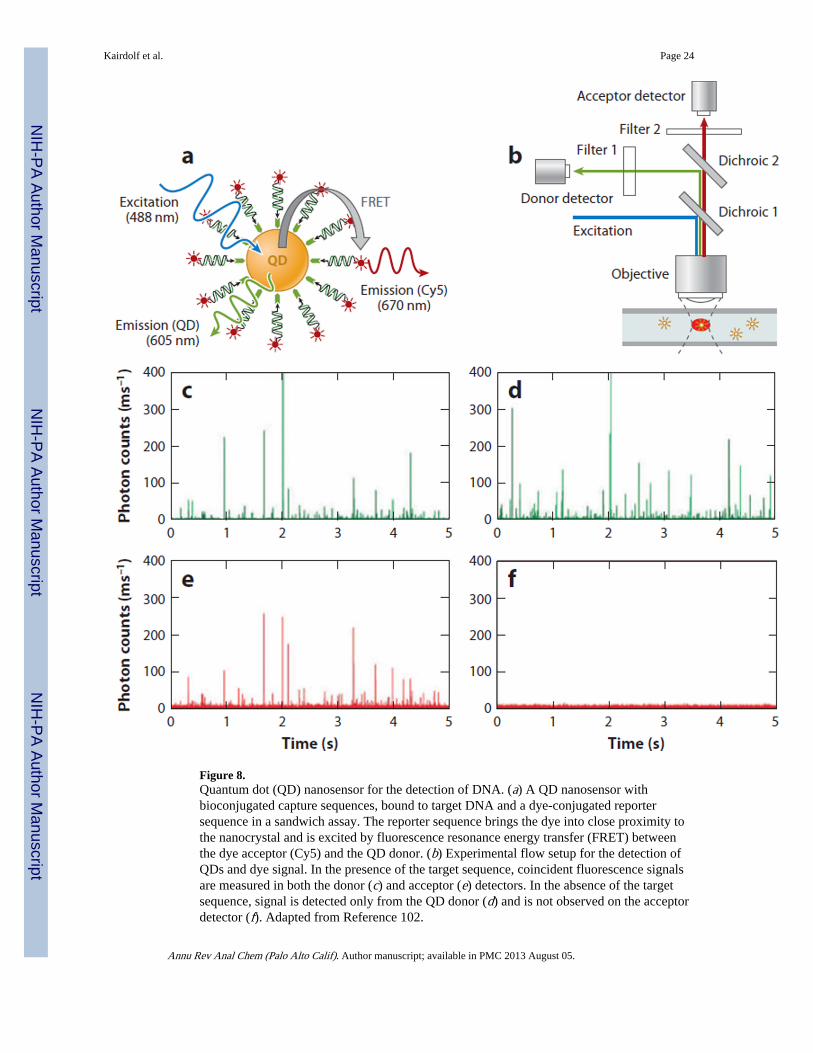
Figure 8.Quantum dot (QD) nanosensor for the detection of DNA. (a) A QD nanosensor withbioconjugated capture sequences, bound to target DNA and a dye-conjugated reportersequence in a sandwich assay. The reporter sequence brings the dye into close proximity tothe nanocrystal and is excited by fluorescence resonance energy transfer (FRET) betweenthe dye acceptor (Cy5) and the QD donor. (b) Experimental flow setup for the detection ofQDs and dye signal. In the presence of the target sequence, coincident fluorescence signalsare measured in both the donor (c) and acceptor (e) detectors. In the absence of the targetsequence, signal is detected only from the QD donor (d) and is not observed on the acceptordetector (f). Adapted from Reference 102.
Kairdolf et al. Page 24
Annu Rev Anal Chem (Palo Alto Calif). Author manuscript; available in PMC 2013 August 05.
NIH
-PA Author Manuscript
NIH
-PA Author Manuscript
NIH
-PA Author Manuscript