Semaphorin signaling in cancer cells and in cells of the tumor...
14
1723 Commentary Introduction Semaphorins were initially identified as evolutionarily conserved axon-guidance cues in the assembly of the neural circuitry (Kolodkin et al., 1993; Luo et al., 1993). However, it is now clear that they are widely expressed outside the nervous system, and consist of more than 20 family members (in vertebrates), which are implicated in a range of processes, including the regulation of cell survival, apoptosis, cell-substrate adhesion and directional cell migration (Tamagnone and Comoglio, 2004; Zhou et al., 2008). Semaphorins are secreted or membrane-associated glycoproteins that have been grouped into eight classes on the basis of their structural elements and amino-acid-sequence similarity (Fig. 1A). Invertebrate semaphorins mainly fall into classes 1 and 2, whereas classes 3 to 7 comprise vertebrate semaphorins and the final group (class V) contains semaphorins that are encoded by viral genomes. In the current nomenclature, the abbreviation SEMA is followed by a number indicating the subclass, and a capital letter designating the individual member – for example, SEMA3A and SEMA4D (Goodman et al., 1999). Semaphorins are characterized by an N-terminal 500-amino-acid sema domain that is essential for semaphorin function. The structure of the sema domain is a seven-blade β-propeller fold that has overall structural similarity to the extracellular domain of α-integrins (Gherardi et al., 2004; Koppel et al., 1997). Next to the sema domain, semaphorins contain the plexin-semaphorin- integrin (PSI) domain, which is a cysteine-rich motif that is also referred to as a MET-related sequence (MRS) (Bork et al., 1999). Semaphorins are further distinguished by other distinct protein domains, including immunoglobulin-like, thrombospondin and basic C-terminal domains. Class-3 semaphorins are the only secreted vertebrate semaphorins and are distinguished by a conserved, basic-charged domain at the C-terminus. Class 4-7 semaphorins are cell-membrane-anchored proteins that are distinguished by their unique structural elements, such as thrombospondin repeats (in the case of class-5 semaphorins) or a glycophosphatidylinositol (GPI) anchor (class-7 semaphorins). Membrane-anchored semaphorins can be further processed into soluble forms through proteolytic degradation, as seen for SEMA4D (Basile et al., 2007b; Elhabazi et al., 2001). High-affinity receptors for semaphorins include plexins and neuropilins (NPs) (He and Tessier-Lavigne, 1997; Kolodkin and Ginty, 1997; Tamagnone et al., 1999) (Fig. 1B,C). Whereas the genomes of invertebrates contain only two plexin genes, nine plexins have been identified in vertebrates, which are divided into four subfamilies: plexin-A(1-4), plexin-B(1-3), plexin-C1 and plexin-D1. Similar to their ligands, plexins contain a sema domain in the extracellular portion; in addition, they have two to three repeated PSI domains and three IPT (Ig-like fold shared by plexins and transcription factors) domains. By contrast, the cytoplasmic moiety of plexins lacks homology to known proteins or functional motifs, although it contains two amino acid stretches that are weakly similar to GTPase-activating proteins (GAPs), which catalyze the inactivation of R-Ras monomeric GTPase (Oinuma et al., 2004). NPs (NP1 and NP2), which are found only in vertebrates, are single- span transmembrane glycoproteins that share a similar domain structure and were initially characterized as co-receptors for class-3 semaphorins and for members of the vascular endothelial growth factor (VEGF) family (He and Tessier-Lavigne, 1997; Kolodkin et al., 1997; Soker et al., 1998). Membrane-bound vertebrate semaphorins bind directly to plexins, whereas secreted semaphorins (class 3) also require NPs as obligate co-receptors; SEMA3E is one known exception (Gu et al., 2005) (Table 1). Additional transmembrane molecules are found in semaphorin-receptor complexes in association with plexins and NPs, Semaphorins are a large family of secreted and membrane- bound molecules that were initially implicated in the development of the nervous system and in axon guidance. More recently, they have been found to regulate cell adhesion and motility, angiogenesis, immune responses, and tumor progression. Semaphorin receptors, the neuropilins and the plexins, are expressed by a wide variety of cell types, including endothelial cells, bone-marrow-derived cells and cancer cells. Interestingly, a growing body of evidence indicates that semaphorins also have an important role in cancer. It is now known that cancer progression, invasion and metastasis involve not only genetic changes in the tumor cells but also crosstalk between tumor cells and their surrounding non-tumor cells. Through the recruitment of endothelial cells, leukocytes, pericytes and fibroblasts, and the local release of growth factors and cytokines, the tumor microenvironment can mediate tumor- cell survival, tumor proliferation and regulation of the immune response. Moreover, by conferring cancer cells with an enhanced ability to migrate and invade adjacent tissues, extracellular regulatory signals can play a major role in the metastatic process. In this Commentary, we focus on the emerging role of semaphorins in mediating the crosstalk between tumor cells and multiple stromal cell types in the surrounding microenvironment. Key words: Semaphorins, Plexins, Tumor, Tumor microenvironment Summary Semaphorin signaling in cancer cells and in cells of the tumor microenvironment – two sides of a coin Lorena Capparuccia and Luca Tamagnone* Institute for Cancer Research and Treatment (IRCC), University of Turin, S.P. 142, 10060, Candiolo (TO), Italy *Author for correspondence (e-mail: [email protected]) Journal of Cell Science 122, 1723-1736 Published by The Company of Biologists 2009 doi:10.1242/jcs.030197 Journal of Cell Science
-
Upload
truongngoc -
Category
Documents
-
view
227 -
download
0
Transcript of Semaphorin signaling in cancer cells and in cells of the tumor...
1723Commentary
IntroductionSemaphorins were initially identified as evolutionarily conservedaxon-guidance cues in the assembly of the neural circuitry (Kolodkinet al., 1993; Luo et al., 1993). However, it is now clear that theyare widely expressed outside the nervous system, and consist ofmore than 20 family members (in vertebrates), which are implicatedin a range of processes, including the regulation of cell survival,apoptosis, cell-substrate adhesion and directional cell migration(Tamagnone and Comoglio, 2004; Zhou et al., 2008).
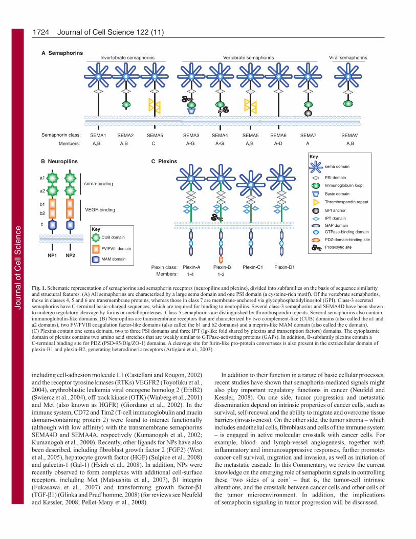
Semaphorins are secreted or membrane-associatedglycoproteins that have been grouped into eight classes on thebasis of their structural elements and amino-acid-sequencesimilarity (Fig. 1A). Invertebrate semaphorins mainly fall intoclasses 1 and 2, whereas classes 3 to 7 comprise vertebratesemaphorins and the final group (class V) contains semaphorinsthat are encoded by viral genomes. In the current nomenclature,the abbreviation SEMA is followed by a number indicating thesubclass, and a capital letter designating the individual member– for example, SEMA3A and SEMA4D (Goodman et al., 1999).Semaphorins are characterized by an N-terminal 500-amino-acidsema domain that is essential for semaphorin function. Thestructure of the sema domain is a seven-blade β-propeller foldthat has overall structural similarity to the extracellular domainof α-integrins (Gherardi et al., 2004; Koppel et al., 1997). Nextto the sema domain, semaphorins contain the plexin-semaphorin-integrin (PSI) domain, which is a cysteine-rich motif that is alsoreferred to as a MET-related sequence (MRS) (Bork et al., 1999).Semaphorins are further distinguished by other distinct proteindomains, including immunoglobulin-like, thrombospondin andbasic C-terminal domains. Class-3 semaphorins are the onlysecreted vertebrate semaphorins and are distinguished by aconserved, basic-charged domain at the C-terminus. Class 4-7
semaphorins are cell-membrane-anchored proteins that aredistinguished by their unique structural elements, such asthrombospondin repeats (in the case of class-5 semaphorins) or aglycophosphatidylinositol (GPI) anchor (class-7 semaphorins).Membrane-anchored semaphorins can be further processed intosoluble forms through proteolytic degradation, as seen forSEMA4D (Basile et al., 2007b; Elhabazi et al., 2001).
High-affinity receptors for semaphorins include plexins andneuropilins (NPs) (He and Tessier-Lavigne, 1997; Kolodkinand Ginty, 1997; Tamagnone et al., 1999) (Fig. 1B,C). Whereas thegenomes of invertebrates contain only two plexin genes, nine plexinshave been identified in vertebrates, which are divided intofour subfamilies: plexin-A(1-4), plexin-B(1-3), plexin-C1 andplexin-D1. Similar to their ligands, plexins contain a sema domainin the extracellular portion; in addition, they have two to threerepeated PSI domains and three IPT (Ig-like fold shared by plexinsand transcription factors) domains. By contrast, the cytoplasmicmoiety of plexins lacks homology to known proteins or functionalmotifs, although it contains two amino acid stretches that are weaklysimilar to GTPase-activating proteins (GAPs), which catalyze theinactivation of R-Ras monomeric GTPase (Oinuma et al., 2004).NPs (NP1 and NP2), which are found only in vertebrates, are single-span transmembrane glycoproteins that share a similar domainstructure and were initially characterized as co-receptors for class-3semaphorins and for members of the vascular endothelial growthfactor (VEGF) family (He and Tessier-Lavigne, 1997; Kolodkinet al., 1997; Soker et al., 1998).
Membrane-bound vertebrate semaphorins bind directly to plexins,whereas secreted semaphorins (class 3) also require NPs as obligateco-receptors; SEMA3E is one known exception (Gu et al., 2005)(Table 1). Additional transmembrane molecules are found insemaphorin-receptor complexes in association with plexins and NPs,
Semaphorins are a large family of secreted and membrane-bound molecules that were initially implicated in thedevelopment of the nervous system and in axon guidance. Morerecently, they have been found to regulate cell adhesion andmotility, angiogenesis, immune responses, and tumorprogression. Semaphorin receptors, the neuropilins and theplexins, are expressed by a wide variety of cell types, includingendothelial cells, bone-marrow-derived cells and cancer cells.Interestingly, a growing body of evidence indicates thatsemaphorins also have an important role in cancer. It is nowknown that cancer progression, invasion and metastasis involvenot only genetic changes in the tumor cells but also crosstalkbetween tumor cells and their surrounding non-tumor cells.
Through the recruitment of endothelial cells, leukocytes,pericytes and fibroblasts, and the local release of growth factorsand cytokines, the tumor microenvironment can mediate tumor-cell survival, tumor proliferation and regulation of the immuneresponse. Moreover, by conferring cancer cells with an enhancedability to migrate and invade adjacent tissues, extracellularregulatory signals can play a major role in the metastaticprocess. In this Commentary, we focus on the emerging role ofsemaphorins in mediating the crosstalk between tumor cellsand multiple stromal cell types in the surroundingmicroenvironment.
Key words: Semaphorins, Plexins, Tumor, Tumor microenvironment
Summary
Semaphorin signaling in cancer cells and in cells ofthe tumor microenvironment – two sides of a coinLorena Capparuccia and Luca Tamagnone*Institute for Cancer Research and Treatment (IRCC), University of Turin, S.P. 142, 10060, Candiolo (TO), Italy*Author for correspondence (e-mail: [email protected])
Journal of Cell Science 122, 1723-1736 Published by The Company of Biologists 2009doi:10.1242/jcs.030197
Jour
nal o
f Cel
l Sci
ence
1724
including cell-adhesion molecule L1 (Castellani and Rougon, 2002)and the receptor tyrosine kinases (RTKs) VEGFR2 (Toyofuku et al.,2004), erythroblastic leukemia viral oncogene homolog 2 (ErbB2)(Swiercz et al., 2004), off-track kinase (OTK) (Winberg et al., 2001)and Met (also known as HGFR) (Giordano et al., 2002). In theimmune system, CD72 and Tim2 (T-cell immunoglobulin and mucindomain-containing protein 2) were found to interact functionally(although with low affinity) with the transmembrane semaphorinsSEMA4D and SEMA4A, respectively (Kumanogoh et al., 2002;Kumanogoh et al., 2000). Recently, other ligands for NPs have alsobeen described, including fibroblast growth factor 2 (FGF2) (Westet al., 2005), hepatocyte growth factor (HGF) (Sulpice et al., 2008)and galectin-1 (Gal-1) (Hsieh et al., 2008). In addition, NPs wererecently observed to form complexes with additional cell-surfacereceptors, including Met (Matsushita et al., 2007), β1 integrin(Fukasawa et al., 2007) and transforming growth factor-β1(TGF-β1) (Glinka and Prud’homme, 2008) (for reviews see Neufeldand Kessler, 2008; Pellet-Many et al., 2008).
In addition to their function in a range of basic cellular processes,recent studies have shown that semaphorin-mediated signals mightalso play important regulatory functions in cancer (Neufeld andKessler, 2008). On one side, tumor progression and metastaticdissemination depend on intrinsic properties of cancer cells, such assurvival, self-renewal and the ability to migrate and overcome tissuebarriers (invasiveness). On the other side, the tumor stroma – whichincludes endothelial cells, fibroblasts and cells of the immune system– is engaged in active molecular crosstalk with cancer cells. Forexample, blood- and lymph-vessel angiogenesis, together withinflammatory and immunosuppressive responses, further promotescancer-cell survival, migration and invasion, as well as initiation ofthe metastatic cascade. In this Commentary, we review the currentknowledge on the emerging role of semaphorin signals in controllingthese ‘two sides of a coin’ – that is, the tumor-cell intrinsicalterations, and the crosstalk between cancer cells and other cells ofthe tumor microenvironment. In addition, the implicationsof semaphorin signaling in tumor progression will be discussed.
Journal of Cell Science 122 (11)
CUB domain
FV/FVIII domain
MAM domain
B Neuropilins
NP1 NP2
sema-binding
VEGF-binding
a1
a2
b1
b2
c
SEMA1 SEMA2 SEMA5 SEMA4 SEMA5 SEMA6 SEMA7 SEMAVSemaphorin class:
Members: A,B A,B C A-G A-G A,B A-D A A,B
Invertebrate semaphorins Vertebrate semaphorins Viral semaphorinsA Semaphorins
SEMA3
C Plexins
Plexin-A Plexin-B Plexin-C1 Plexin-D1Plexin class:Members: 1-4 1-3
Thrombospondin repeat
Immunoglobulin loop
Basic domain
GPI anchor
IPT domain
GAP domain
GTPase-binding domain
PDZ-domain-binding site
sema domain
PSI domain
Proteolytic site
Key
Key
Fig. 1. Schematic representation of semaphorins and semaphorin receptors (neuropilins and plexins), divided into subfamilies on the basis of sequence similarityand structural features. (A) All semaphorins are characterized by a large sema domain and one PSI domain (a cysteine-rich motif). Of the vertebrate semaphorins,those in classes 4, 5 and 6 are transmembrane proteins, whereas those in class 7 are membrane-anchored via glycophosphatidylinositol (GPI). Class-3 secretedsemaphorins have C-terminal basic-charged sequences, which are required for binding to neuropilins. Several class-3 semaphorins and SEMA4D have been shownto undergo regulatory cleavage by furins or metalloproteases. Class-5 semaphorins are distinguished by thrombospondin repeats. Several semaphorins also containimmunoglobulin-like domains. (B) Neuropilins are transmembrane receptors that are characterized by two complement-like (CUB) domains (also called the a1 anda2 domains), two FV/FVIII coagulation factor-like domains (also called the b1 and b2 domains) and a meprin-like MAM domain (also called the c domain).(C) Plexins contain one sema domain, two to three PSI domains and three IPT (Ig-like fold shared by plexins and transcription factors) domains. The cytoplasmicdomain of plexins contains two amino acid stretches that are weakly similar to GTPase-activating proteins (GAPs). In addition, B-subfamily plexins contain aC-terminal binding site for PDZ (PSD-95/Dlg/ZO-1) domains. A cleavage site for furin-like pro-protein convertases is also present in the extracellular domain ofplexin-B1 and plexin-B2, generating heterodimeric receptors (Artigiani et al., 2003).
Jour
nal o
f Cel
l Sci
ence
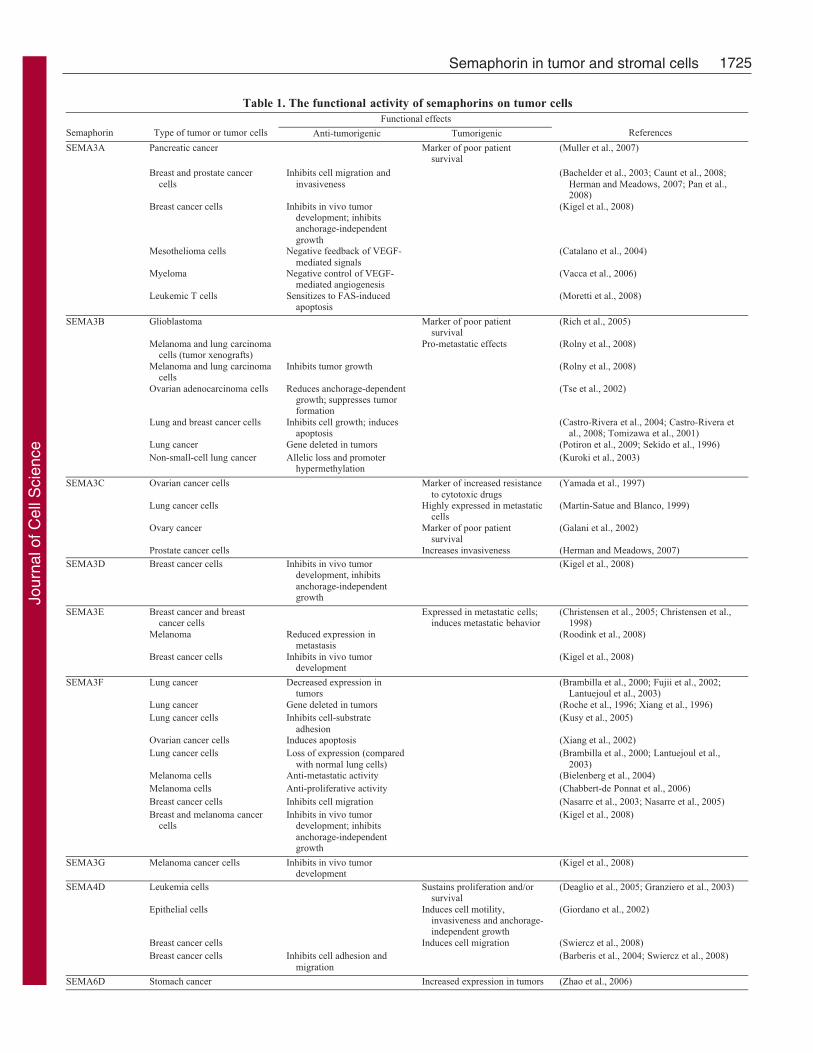
1725Semaphorin in tumor and stromal cells
Table 1. The functional activity of semaphorins on tumor cellsFunctional effects
Semaphorin Type of tumor or tumor cells Anti-tumorigenic Tumorigenic References
Pancreatic cancer Marker of poor patientsurvival
(Muller et al., 2007)
Breast and prostate cancercells
Inhibits cell migration andinvasiveness
(Bachelder et al., 2003; Caunt et al., 2008;Herman and Meadows, 2007; Pan et al.,2008)
Breast cancer cells Inhibits in vivo tumordevelopment; inhibitsanchorage-independentgrowth
(Kigel et al., 2008)
Mesothelioma cells Negative feedback of VEGF-mediated signals
(Catalano et al., 2004)
Myeloma Negative control of VEGF-mediated angiogenesis
(Vacca et al., 2006)
SEMA3A
Leukemic T cells Sensitizes to FAS-inducedapoptosis
(Moretti et al., 2008)
Glioblastoma Marker of poor patientsurvival
(Rich et al., 2005)
Melanoma and lung carcinomacells (tumor xenografts)
Pro-metastatic effects (Rolny et al., 2008)
Melanoma and lung carcinomacells
Inhibits tumor growth (Rolny et al., 2008)
Ovarian adenocarcinoma cells Reduces anchorage-dependentgrowth; suppresses tumorformation
(Tse et al., 2002)
Lung and breast cancer cells Inhibits cell growth; inducesapoptosis
(Castro-Rivera et al., 2004; Castro-Rivera etal., 2008; Tomizawa et al., 2001)
Lung cancer Gene deleted in tumors (Potiron et al., 2009; Sekido et al., 1996)
SEMA3B
Non-small-cell lung cancer Allelic loss and promoterhypermethylation
(Kuroki et al., 2003)
Ovarian cancer cells Marker of increased resistanceto cytotoxic drugs
(Yamada et al., 1997)
Lung cancer cells Highly expressed in metastaticcells
(Martin-Satue and Blanco, 1999)
Ovary cancer Marker of poor patientsurvival
(Galani et al., 2002)
SEMA3C
Prostate cancer cells Increases invasiveness (Herman and Meadows, 2007)SEMA3D Breast cancer cells Inhibits in vivo tumor
development, inhibitsanchorage-independentgrowth
(Kigel et al., 2008)
Breast cancer and breastcancer cells
Expressed in metastatic cells;induces metastatic behavior
(Christensen et al., 2005; Christensen et al.,1998)
Melanoma Reduced expression inmetastasis
(Roodink et al., 2008)
SEMA3E
Breast cancer cells Inhibits in vivo tumordevelopment
(Kigel et al., 2008)
Lung cancer Decreased expression intumors
(Brambilla et al., 2000; Fujii et al., 2002;Lantuejoul et al., 2003)
Lung cancer Gene deleted in tumors (Roche et al., 1996; Xiang et al., 1996)Lung cancer cells Inhibits cell-substrate
adhesion(Kusy et al., 2005)
Ovarian cancer cells Induces apoptosis (Xiang et al., 2002)Lung cancer cells Loss of expression (compared
with normal lung cells)(Brambilla et al., 2000; Lantuejoul et al.,
2003)Melanoma cells Anti-metastatic activity (Bielenberg et al., 2004)Melanoma cells Anti-proliferative activity (Chabbert-de Ponnat et al., 2006)Breast cancer cells Inhibits cell migration (Nasarre et al., 2003; Nasarre et al., 2005)
SEMA3F
Breast and melanoma cancercells
Inhibits in vivo tumordevelopment; inhibitsanchorage-independentgrowth
(Kigel et al., 2008)
SEMA3G Melanoma cancer cells Inhibits in vivo tumordevelopment
(Kigel et al., 2008)
Leukemia cells Sustains proliferation and/orsurvival
(Deaglio et al., 2005; Granziero et al., 2003)
Epithelial cells Induces cell motility,invasiveness and anchorage-independent growth
(Giordano et al., 2002)
Breast cancer cells Induces cell migration (Swiercz et al., 2008)
SEMA4D
Breast cancer cells Inhibits cell adhesion andmigration
(Barberis et al., 2004; Swiercz et al., 2008)
SEMA6D Stomach cancer Increased expression in tumors (Zhao et al., 2006)
Jour
nal o
f Cel
l Sci
ence
1726
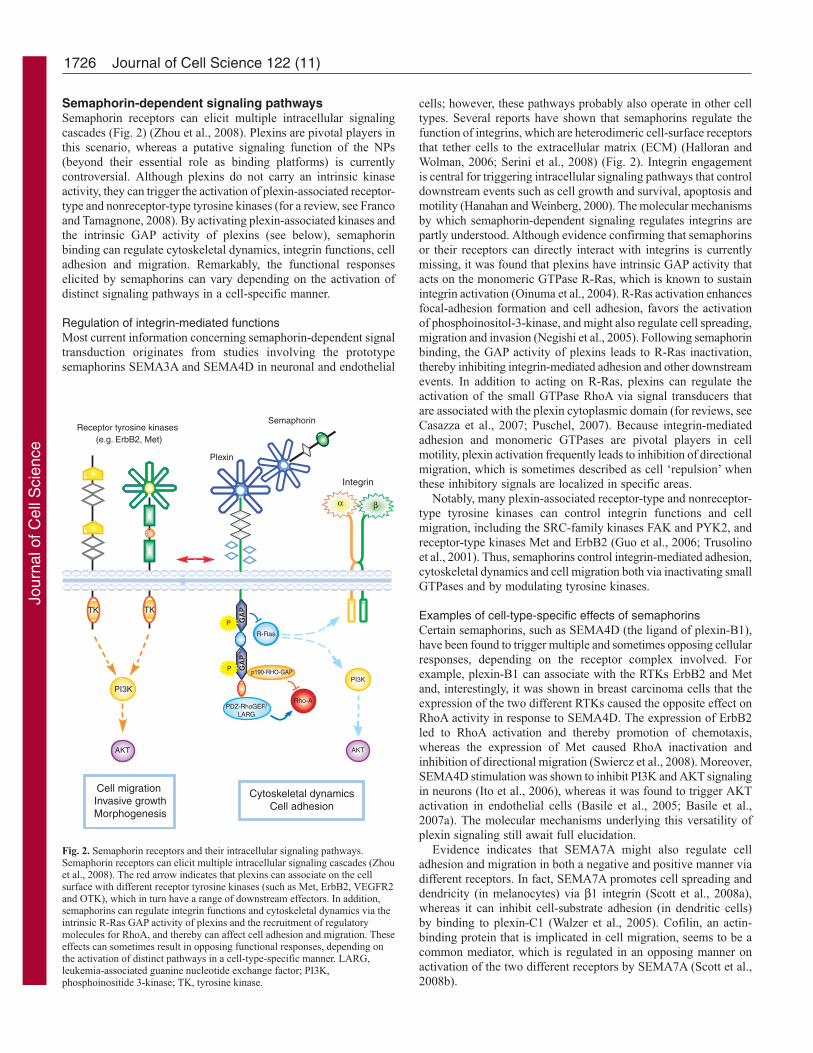
Semaphorin-dependent signaling pathwaysSemaphorin receptors can elicit multiple intracellular signalingcascades (Fig. 2) (Zhou et al., 2008). Plexins are pivotal players inthis scenario, whereas a putative signaling function of the NPs(beyond their essential role as binding platforms) is currentlycontroversial. Although plexins do not carry an intrinsic kinaseactivity, they can trigger the activation of plexin-associated receptor-type and nonreceptor-type tyrosine kinases (for a review, see Francoand Tamagnone, 2008). By activating plexin-associated kinases andthe intrinsic GAP activity of plexins (see below), semaphorinbinding can regulate cytoskeletal dynamics, integrin functions, celladhesion and migration. Remarkably, the functional responseselicited by semaphorins can vary depending on the activation ofdistinct signaling pathways in a cell-specific manner.
Regulation of integrin-mediated functionsMost current information concerning semaphorin-dependent signaltransduction originates from studies involving the prototypesemaphorins SEMA3A and SEMA4D in neuronal and endothelial
cells; however, these pathways probably also operate in other celltypes. Several reports have shown that semaphorins regulate thefunction of integrins, which are heterodimeric cell-surface receptorsthat tether cells to the extracellular matrix (ECM) (Halloran andWolman, 2006; Serini et al., 2008) (Fig. 2). Integrin engagementis central for triggering intracellular signaling pathways that controldownstream events such as cell growth and survival, apoptosis andmotility (Hanahan and Weinberg, 2000). The molecular mechanismsby which semaphorin-dependent signaling regulates integrins arepartly understood. Although evidence confirming that semaphorinsor their receptors can directly interact with integrins is currentlymissing, it was found that plexins have intrinsic GAP activity thatacts on the monomeric GTPase R-Ras, which is known to sustainintegrin activation (Oinuma et al., 2004). R-Ras activation enhancesfocal-adhesion formation and cell adhesion, favors the activationof phosphoinositol-3-kinase, and might also regulate cell spreading,migration and invasion (Negishi et al., 2005). Following semaphorinbinding, the GAP activity of plexins leads to R-Ras inactivation,thereby inhibiting integrin-mediated adhesion and other downstreamevents. In addition to acting on R-Ras, plexins can regulate theactivation of the small GTPase RhoA via signal transducers thatare associated with the plexin cytoplasmic domain (for reviews, seeCasazza et al., 2007; Puschel, 2007). Because integrin-mediatedadhesion and monomeric GTPases are pivotal players in cellmotility, plexin activation frequently leads to inhibition of directionalmigration, which is sometimes described as cell ‘repulsion’ whenthese inhibitory signals are localized in specific areas.
Notably, many plexin-associated receptor-type and nonreceptor-type tyrosine kinases can control integrin functions and cellmigration, including the SRC-family kinases FAK and PYK2, andreceptor-type kinases Met and ErbB2 (Guo et al., 2006; Trusolinoet al., 2001). Thus, semaphorins control integrin-mediated adhesion,cytoskeletal dynamics and cell migration both via inactivating smallGTPases and by modulating tyrosine kinases.
Examples of cell-type-specific effects of semaphorinsCertain semaphorins, such as SEMA4D (the ligand of plexin-B1),have been found to trigger multiple and sometimes opposing cellularresponses, depending on the receptor complex involved. Forexample, plexin-B1 can associate with the RTKs ErbB2 and Metand, interestingly, it was shown in breast carcinoma cells that theexpression of the two different RTKs caused the opposite effect onRhoA activity in response to SEMA4D. The expression of ErbB2led to RhoA activation and thereby promotion of chemotaxis,whereas the expression of Met caused RhoA inactivation andinhibition of directional migration (Swiercz et al., 2008). Moreover,SEMA4D stimulation was shown to inhibit PI3K and AKT signalingin neurons (Ito et al., 2006), whereas it was found to trigger AKTactivation in endothelial cells (Basile et al., 2005; Basile et al.,2007a). The molecular mechanisms underlying this versatility ofplexin signaling still await full elucidation.
Evidence indicates that SEMA7A might also regulate celladhesion and migration in both a negative and positive manner viadifferent receptors. In fact, SEMA7A promotes cell spreading anddendricity (in melanocytes) via β1 integrin (Scott et al., 2008a),whereas it can inhibit cell-substrate adhesion (in dendritic cells)by binding to plexin-C1 (Walzer et al., 2005). Cofilin, an actin-binding protein that is implicated in cell migration, seems to be acommon mediator, which is regulated in an opposing manner onactivation of the two different receptors by SEMA7A (Scott et al.,2008b).
Journal of Cell Science 122 (11)
Cell migration Invasive growthMorphogenesis
Cytoskeletal dynamicsCell adhesion
Receptor tyrosine kinases
TK
AKT
TK
AKT
PI3K
(e.g. ErbB2, Met)
Semaphorin
p190-RHO-GAP
P
α βα β
Integrin
PI3K
AKT
GA
P
Plexin
PDZ-RhoGEF/LARG
P GA
P
R-Ras
Rho-A
Fig. 2. Semaphorin receptors and their intracellular signaling pathways.Semaphorin receptors can elicit multiple intracellular signaling cascades (Zhouet al., 2008). The red arrow indicates that plexins can associate on the cellsurface with different receptor tyrosine kinases (such as Met, ErbB2, VEGFR2and OTK), which in turn have a range of downstream effectors. In addition,semaphorins can regulate integrin functions and cytoskeletal dynamics via theintrinsic R-Ras GAP activity of plexins and the recruitment of regulatorymolecules for RhoA, and thereby can affect cell adhesion and migration. Theseeffects can sometimes result in opposing functional responses, depending onthe activation of distinct pathways in a cell-type-specific manner. LARG,leukemia-associated guanine nucleotide exchange factor; PI3K,phosphoinositide 3-kinase; TK, tyrosine kinase.
Jour
nal o
f Cel
l Sci
ence

1727Semaphorin in tumor and stromal cells
Together, these studies highlight not only the common featuresof semaphorin signaling pathways but also support the notion thatsemaphorin signaling events are cell-type specific. Therefore, it willbe crucial to thoroughly assess whether the semaphorin signalingthat is observed in one particular cell type or tissue also occurs inother cell types.
Expression of semaphorin receptors by tumor cellsThe expression of plexins has been reported in a wide variety oftumors. For example, plexin-A expression has been observed inovarian (Syed et al., 2005) and gastric (Zhao et al., 2007) carcinoma,as well as in tumor-derived cells of glioma (Rieger et al., 2003),melanoma, and breast (Castro-Rivera et al., 2008; Castro-Riveraet al., 2004; Kigel et al., 2008) and colon (Nguyen et al., 2006)cancer. The expression of plexin-B1 has been reported in ovarianand prostate cancers (Syed et al., 2005; Wong et al., 2007). Inaddition, a high frequency of somatic missense mutationsin plexin-B1, and its overexpression at the protein level, seems tocorrelate with increased invasiveness and metastatic progression ofprostate tumors (Wong et al., 2007). In contrast to these reports, amarked downregulation of plexin-B1 expression has been reportedin clear-cell renal carcinoma (Gomez Roman et al., 2008), and lossof plexin-B1 gene expression was found to correlate with poorprognosis in estrogen-receptor-positive breast cancer (Rody et al.,2007). These discrepancies might be explained by the co-expressionof distinctive signaling partners in the different tumors. Plexin-C1,which is expressed by melanocytes, is reduced or absent in humanmelanoma cells (Scott et al., 2008b); in tissue microarrays thatcompared nevi and melanoma samples, an inverse correlationbetween plexin-C1 levels and the depth of skin invasion, and aremarkable decrease in the expression of plexin-C1, was found inmetastatic samples (Scott et al., 2008b). Finally, plexin-D1 isexpressed in a range of tumor cells, including melanoma and breast-cancer cell lines (Kigel et al., 2008; Roodink et al., 2005; Roodinket al., 2008). A positive correlation between plexin-D1 expressionand tumor grading has been reported.
NPs are also expressed by a wide variety of human tumor celllines and neoplasms (reviewed by Pellet-Many et al., 2008). NP1is prevalently expressed in carcinomas, whereas NP2 is frequentlyexpressed in non-epithelial tumors such as melanomas, leukemiasand neuroblastomas (Pellet-Many et al., 2008). This expressionpattern is not without exceptions and, in fact, the two receptorsare often co-expressed (Marcus et al., 2005; Rieger et al., 2003).Clinical studies suggest that NPs play a role in tumor growth anddisease progression because they are involved mediatingsemaphorin- and VEGF-mediated effects on cancer-cellproliferation, survival and migration (reviewed by Barr et al., 2005;Bielenberg et al., 2006; Ellis, 2006; Geretti et al., 2008; Guttmann-Raviv et al., 2006; Pan et al., 2007). High levels of NP1 weresignificantly associated with a poor outcome in patients with breastcancer (Ghosh et al., 2008), and correlated with invasive behaviorand metastatic potential in gastrointestinal carcinomas (Hanselet al., 2004), glioma (Osada et al., 2004) and prostate carcinoma(Latil et al., 2000). These observations are further supported by invitro studies. Indeed, NP1 overexpression in prostate carcinomacells promoted tumor growth in vivo (Miao et al., 2000) and,consistent with this result, knockdown of NP1 expression usingRNA interference inhibited tumor-cell migration (Bachelder et al.,2003). Although most studies have indicated a tumorigenic rolefor NPs, other reports suggest that NP1 might have the oppositefunction in tumors. For example, NP1 overexpression in pancreatic
carcinoma cells has been reported to inhibit cell migration,anchorage-independent growth and tumor incidence in vivo (Grayet al., 2005; Kamiya et al., 2006), and elevated expression of thegene encoding NP1 is associated with a more favorable prognosisin individuals with colon cancer (Kamiya et al., 2006). Thesediscrepancies are currently unresolved. They might be explainedby the non-physiological effects that are induced by the ectopicoverexpression of NP1, or they might reflect cell-type-specificresponses and/or the involvement of different signaling pathways.Furthermore, an elevated expression of NP2 correlated withadvanced tumor progression and poor prognosis in osteosarcomas(Handa et al., 2000) and colorectal carcinoma cells (Gray et al.,2008), and the silencing of NP2 expression by short-hairpin RNAreduced the development of tumors. Notably, treatment with anNP2-specific blocking antibody reduced metastatic disseminationof murine mammary carcinoma and rat glioma cell lines to lymphnodes and distant organs (Caunt et al., 2008).
The expression patterns of plexins and NPs in different tumortypes is consistent with the putative regulatory role of semaphorinsin cancer. However, current evidence does not associate anyindividual semaphorin receptor with a univocal function in tumordevelopment. This is probably due to the fact that semaphorinsinduce multiple signaling cascades in different tumor cells and incells of the tumor microenvironment, an aspect that deserves furtherinvestigation.
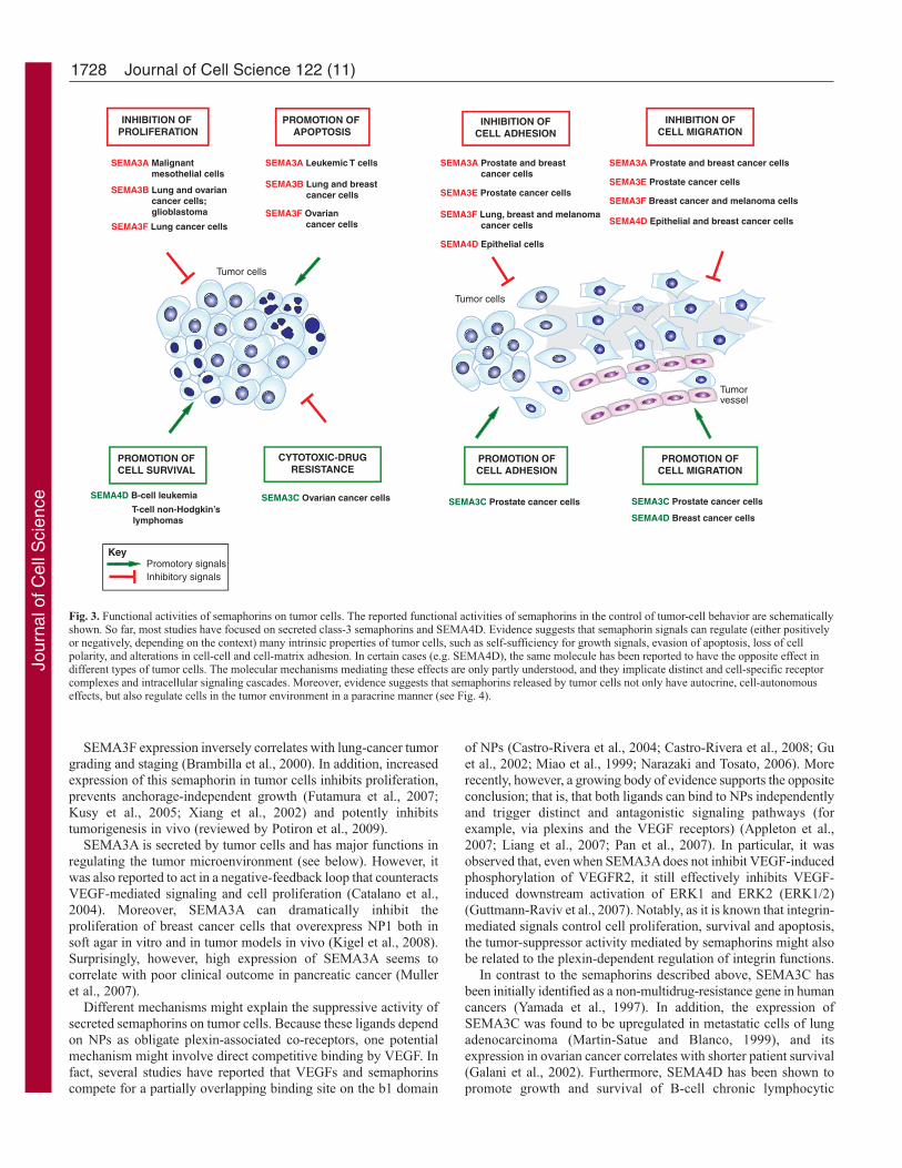
Semaphorins in the control of tumor-cell behaviorSemaphorins act as pleiotropic signals that are able to controlmultiple functions in tumor cells, ranging from survival,proliferation and apoptosis to cell adhesion and migration (Fig. 3;Table 1). Semaphorins might be released by cells in the tumormicroenvironment (such as infiltrating leukocytes) or by cancercells, which can establish their own autocrine regulatory loops.
Cell survival, proliferation and apoptosisEssential alterations in cell behavior that collectively dictate cancerprogression include self-sufficiency in growth signals, limitlessreplicative potential and evasion of apoptosis (Hanahan andWeinberg, 2000). Increasing evidence suggests that semaphorinsignals regulate many of these properties. For example, it has beenproposed that SEMA3B and SEMA3F act as tumor suppressors,because they undergo loss of heterozygosity, promoterhypermethylation and downregulation of expression in humantumors (reviewed by Potiron et al., 2009). Consistently, SEMA3Boverexpression in tumor cell lines induced apoptosis (Castro-Rivera et al., 2004; Castro-Rivera et al., 2008), and inhibited cellproliferation and colony formation in soft agar (Tomizawa et al.,2001; Tse et al., 2002). However, data obtained from a large numberof human tumor samples do not show a statistically significantcorrelation between SEMA3B expression and patient survival(Rolny et al., 2008). In fact, SEMA3B expression has beencorrelated with poor prognosis in glioblastoma cases (Rich et al.,2005), and tumor subsets that have an increased level of SEMA3Bexpression are associated with metastatic progression (Rolny et al.,2008). Notably, SEMA3B expression was found to inhibit tumorgrowth while sustaining metastatic dissemination in a number oftumorigenic models, which is consistent with p38-MAPK-dependentactivation of p21, a cell-cycle inhibitor, and the induction ofinterleukin-8 (IL-8) cytokine secretion from tumor and stromal cells,which promotes cancer progression and metastasis (Rolny et al.,2008).
Jour
nal o
f Cel
l Sci
ence
1728
SEMA3F expression inversely correlates with lung-cancer tumorgrading and staging (Brambilla et al., 2000). In addition, increasedexpression of this semaphorin in tumor cells inhibits proliferation,prevents anchorage-independent growth (Futamura et al., 2007;Kusy et al., 2005; Xiang et al., 2002) and potently inhibitstumorigenesis in vivo (reviewed by Potiron et al., 2009).
SEMA3A is secreted by tumor cells and has major functions inregulating the tumor microenvironment (see below). However, itwas also reported to act in a negative-feedback loop that counteractsVEGF-mediated signaling and cell proliferation (Catalano et al.,2004). Moreover, SEMA3A can dramatically inhibit theproliferation of breast cancer cells that overexpress NP1 both insoft agar in vitro and in tumor models in vivo (Kigel et al., 2008).Surprisingly, however, high expression of SEMA3A seems tocorrelate with poor clinical outcome in pancreatic cancer (Mulleret al., 2007).
Different mechanisms might explain the suppressive activity ofsecreted semaphorins on tumor cells. Because these ligands dependon NPs as obligate plexin-associated co-receptors, one potentialmechanism might involve direct competitive binding by VEGF. Infact, several studies have reported that VEGFs and semaphorinscompete for a partially overlapping binding site on the b1 domain
of NPs (Castro-Rivera et al., 2004; Castro-Rivera et al., 2008; Guet al., 2002; Miao et al., 1999; Narazaki and Tosato, 2006). Morerecently, however, a growing body of evidence supports the oppositeconclusion; that is, that both ligands can bind to NPs independentlyand trigger distinct and antagonistic signaling pathways (forexample, via plexins and the VEGF receptors) (Appleton et al.,2007; Liang et al., 2007; Pan et al., 2007). In particular, it wasobserved that, even when SEMA3A does not inhibit VEGF-inducedphosphorylation of VEGFR2, it still effectively inhibits VEGF-induced downstream activation of ERK1 and ERK2 (ERK1/2)(Guttmann-Raviv et al., 2007). Notably, as it is known that integrin-mediated signals control cell proliferation, survival and apoptosis,the tumor-suppressor activity mediated by semaphorins might alsobe related to the plexin-dependent regulation of integrin functions.
In contrast to the semaphorins described above, SEMA3C hasbeen initially identified as a non-multidrug-resistance gene in humancancers (Yamada et al., 1997). In addition, the expression ofSEMA3C was found to be upregulated in metastatic cells of lungadenocarcinoma (Martin-Satue and Blanco, 1999), and itsexpression in ovarian cancer correlates with shorter patient survival(Galani et al., 2002). Furthermore, SEMA4D has been shown topromote growth and survival of B-cell chronic lymphocytic
Journal of Cell Science 122 (11)
Promotory signalsInhibitory signals
INHIBITION OFCELL ADHESION
SEMA3E Prostate cancer cells
SEMA3F Lung, breast and melanoma cancer cells
SEMA3A Prostate and breast cancer cells
SEMA4D Epithelial cells
SEMA3E Prostate cancer cells
SEMA3F Breast cancer and melanoma cells
SEMA3A Prostate and breast cancer cells
SEMA4D Epithelial and breast cancer cells
INHIBITION OFCELL MIGRATION
SEMA3C Prostate cancer cells
SEMA4D Breast cancer cells
PROMOTION OFCELL MIGRATION
PROMOTION OFCELL ADHESION
SEMA3C Prostate cancer cells
Tumor cells
Tumorvessel
Tumor cells
SEMA3B Lung and breast cancer cells
SEMA3A Leukemic T cells
SEMA3F Ovarian cancer cells
PROMOTION OF APOPTOSIS
INHIBITION OF PROLIFERATION
SEMA3B Lung and ovarian cancer cells; glioblastoma
SEMA3F Lung cancer cells
SEMA3A Malignant mesothelial cells
CYTOTOXIC-DRUGRESISTANCE
SEMA3C Ovarian cancer cells
PROMOTION OF CELL SURVIVAL
SEMA4D B-cell leukemia
T-cell non-Hodgkin’s lymphomas
Key
Fig. 3. Functional activities of semaphorins on tumor cells. The reported functional activities of semaphorins in the control of tumor-cell behavior are schematicallyshown. So far, most studies have focused on secreted class-3 semaphorins and SEMA4D. Evidence suggests that semaphorin signals can regulate (either positivelyor negatively, depending on the context) many intrinsic properties of tumor cells, such as self-sufficiency for growth signals, evasion of apoptosis, loss of cellpolarity, and alterations in cell-cell and cell-matrix adhesion. In certain cases (e.g. SEMA4D), the same molecule has been reported to have the opposite effect indifferent types of tumor cells. The molecular mechanisms mediating these effects are only partly understood, and they implicate distinct and cell-specific receptorcomplexes and intracellular signaling cascades. Moreover, evidence suggests that semaphorins released by tumor cells not only have autocrine, cell-autonomouseffects, but also regulate cells in the tumor environment in a paracrine manner (see Fig. 4).
Jour
nal o
f Cel
l Sci
ence
1729Semaphorin in tumor and stromal cells
leukemia (B-CLL) (Deaglio et al., 2005; Granziero et al., 2003),and anchorage-independent growth of epithelial cells (Giordanoet al., 2002).
Cell adhesionA proper balance between adhesion to and dissociation fromadjacent cells and the ECM is essential for morphogenesis, and playsa central role in cancer (Nishimura and Sasaki, 2008). Notably,signaling crosstalk coordinates the function of integrins with thestability of cadherin-based cell-cell-adhesion complexes. Severalsignals, including semaphorins, are known to control these cell-adhesion complexes through complex intracellular signalingpathways (Halloran and Wolman, 2006). Notably, it was shown thatthe expression of adhesion receptors in cancer cells is differentiallymodulated by class-3 semaphorins (Herman and Meadows, 2007;Pan et al., 2008). In particular, SEMA3F anti-tumor activity in lungcarcinoma cells was associated with loss of activated α5β3 integrin(Brambilla et al., 2000), downregulation of integrin-linked kinase(ILK) activity and decreased adhesion to the ECM (Kusy et al.,2005; Potiron et al., 2007). Consistent with these observations, theforced re-expression of SEMA3F (which is low in highly metastaticmelanoma cells) inhibited adhesion, migration and the expressionof β1 integrin in melanoma cells (Bielenberg et al., 2004).
So far, experimental evidence implicating class-3 semaphorinsas direct regulators of cadherin-based cell-cell adhesion is relativelyweak. In fact, weakening of cell-cell junctions might also be asecondary effect of cytoskeletal remodeling and ‘collapse’ that iselicited by semaphorins. Nevertheless, recent findings suggestingthat SEMA3A and SEMA3C regulate E-cadherin and integrinexpression at the transcriptional level (Herman and Meadows, 2007)deserve further investigation owing to their potential implicationsin cancer progression. The downregulation of E-cadherin and thedisassembly of cell-cell junctions, together with changes in integrinactivity and the acquisition of a migratory phenotype, are key eventsin a process known as epithelial-mesenchymal transition (EMT)(Thiery and Sleeman, 2006). Dysregulated activation of EMT istypically observed in carcinomas and it is often associated withprogression to malignancy (Nishimura and Sasaki, 2008; Turleyet al., 2008). Notably, the gene encoding E-cadherin is one of themain targets of transcriptional regulators of EMT, and might bedownregulated at both the mRNA and the protein level (as a resultof changes in subcellular distribution, internalization anddegradation). In this case, it is tempting to speculate that R-Rasinactivation that is mediated by plexins might repress EMT byinhibiting cell-substrate adhesion (and by increasing the expressionof cell-adhesion molecules). However, it is also possible thatsemaphorin-mediated activation of alternative pathways (such asthose that involve tyrosine kinases) leads to increased integrin-dependent adhesion, loss of cell-cell-adhesion complexes and theacquisition of a migratory and/or invasive phenotype. Thus, thereported effects of semaphorins on E-cadherin and integrins mightmean that semaphorins are multi-functional regulators of EMT,which could explain some of their described anti-tumorigenic, pro-invasive and/or pro-metastatic activities.
Cell migrationDuring cancer progression, tumor cells can migrate and invadesurrounding tissues. For this to occur, strong cell-matrix adhesionsneed to be dissolved, whereas transient adhesions have to be formedat the leading edge. Integrins act at the ‘feet’ of migrating cells bymediating adhesion to the ECM (or to other cells) and by linking
it to the intracellular actin cytoskeleton (Ridley et al., 2003). Thus,by regulating integrin-mediated adhesion (or affecting integrinexpression), semaphorins can regulate tumor-cell migration. Indeed,following plexin activation by semaphorins, integrin-based focal-adhesive structures are disassembled within minutes, followed byactin depolymerization and cytoskeletal remodeling, which canresult in the extreme effect of cellular collapse (Barberis et al., 2004).In addition, plexin activation can hinder the formation of newadhesive complexes and block lamellipodia formation anddirectional migration by uncoupling cell-substrate adhesion fromthe cytoskeletal dynamics that are required for cell migration(Barberis et al., 2004).
Autocrine SEMA3A was found to reduce both the migratory andinvasive behavior of breast tumor cells (Bachelder et al., 2003),possibly by stimulating the expression of α2β1 integrin, a suppressorof metastatic breast-tumor growth (Pan et al., 2008). Similarly, thetumor-suppressor gene SEMA3F inhibits cell-spreading andmigration both in breast carcinoma (Nasarre et al., 2003) andmelanoma cells (Bielenberg et al., 2004), which correlates withreduced metastatic dissemination. Conversely, SEMA3C expressionincreases the motility and invasion of prostate cancer cells (Hermanand Meadows, 2007).
A further mechanism through which class-3 semaphorins canregulate cell migration is by interfering with VEGF-mediatedsignaling. In human tumor cells, it was shown that cell migrationis finely regulated by a balance between autocrine loops ofSEMA3A and VEGF expression (Bachelder et al., 2003). Asdiscussed above, it is still controversial whether both factorscompete for a partly overlapping binding site on NP1 (Gu et al.,2002; Miao et al., 1999) or whether they trigger antagonisticdownstream signaling pathways in tumor cells (Appleton et al.,2007; Liang et al., 2007; Pan et al., 2007).
Experimental evidence indicates that SEMA4D can inhibit orpromote cell migration, depending on the receptor complexes thatare expressed by the tumor cells. For instance, whereas plexin-B1activation by SEMA4D can inhibit cell migration (Barberis et al.,2004; Swiercz et al., 2008), it was shown that the same receptor-ligand interaction can promote chemotaxis and sustain invasivegrowth, depending on whether plexin-B1 was associated with theoncogenic RTKs Met and ErbB2 (Conrotto et al., 2004; Giordanoet al., 2002; Swiercz et al., 2008).
In summary, the behavior of cancer cells might be regulated bymultiple semaphorins that are either produced by tumor cells in anautocrine manner or provided by non-cancerous cells that arerecruited to the tumor microenvironment. Some semaphorin-mediated signals might inhibit rather than promote tumor growthand invasion, as many studies have shown that their expression isoften lost during advanced cancer. Other semaphorins mightpromote tumor-cell survival, invasion and metastasis, and theirexpression might be upregulated to sustain tumor progression.
Semaphorins as regulators of cells in the tumormicroenvironmentTumors are a complex network of different cell types, and the fullmanifestation of their malignant potential depends on supportingsignals from adjacent non-cancerous cells. In fact, tumor cellssecrete many factors to regulate their own microenvironment,including multiple semaphorins that have the potential to act ondifferent stromal cells. Importantly, the tumor microenvironment iscomplex and dynamic, and varies depending on the type of cancer.It can comprise resident fibroblasts, blood vessels and lymphatic
Jour
nal o
f Cel
l Sci
ence
1730
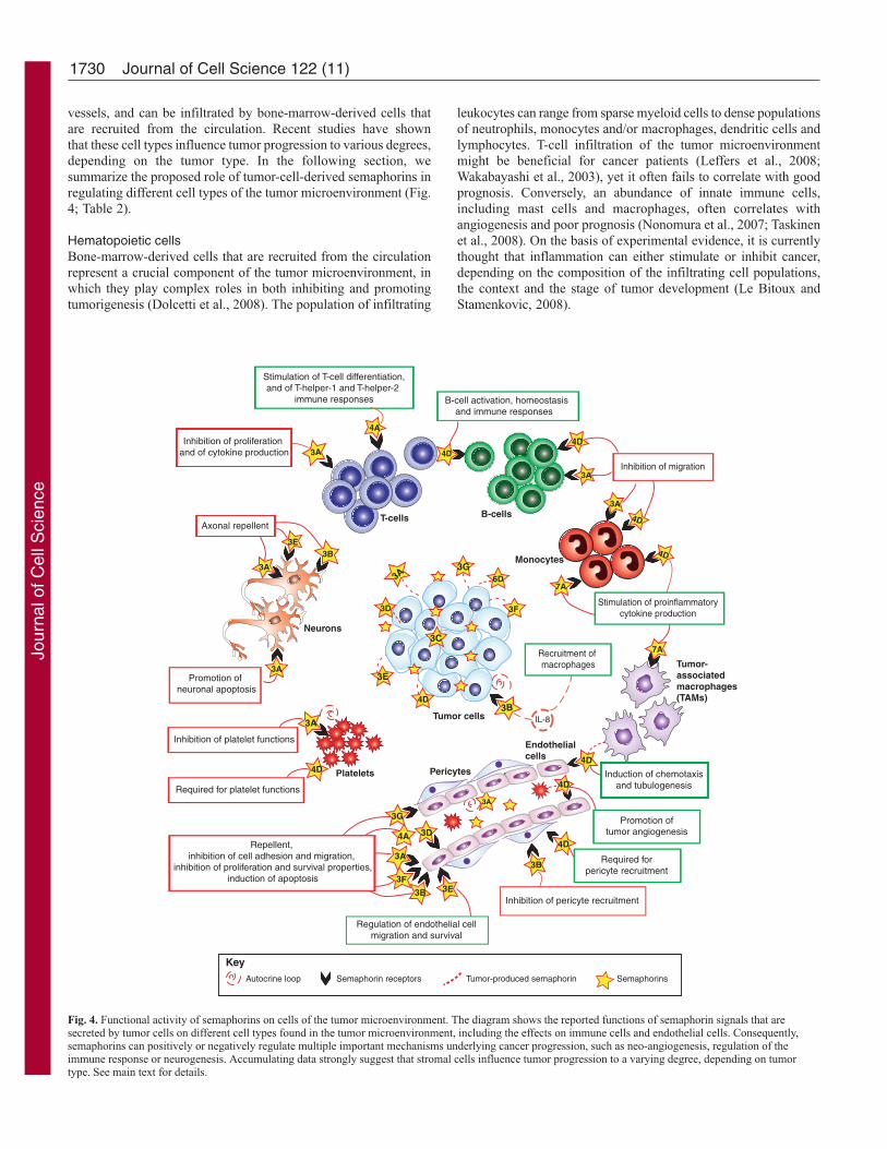
vessels, and can be infiltrated by bone-marrow-derived cells thatare recruited from the circulation. Recent studies have shownthat these cell types influence tumor progression to various degrees,depending on the tumor type. In the following section, wesummarize the proposed role of tumor-cell-derived semaphorins inregulating different cell types of the tumor microenvironment (Fig.4; Table 2).
Hematopoietic cellsBone-marrow-derived cells that are recruited from the circulationrepresent a crucial component of the tumor microenvironment, inwhich they play complex roles in both inhibiting and promotingtumorigenesis (Dolcetti et al., 2008). The population of infiltrating
leukocytes can range from sparse myeloid cells to dense populationsof neutrophils, monocytes and/or macrophages, dendritic cells andlymphocytes. T-cell infiltration of the tumor microenvironmentmight be beneficial for cancer patients (Leffers et al., 2008;Wakabayashi et al., 2003), yet it often fails to correlate with goodprognosis. Conversely, an abundance of innate immune cells,including mast cells and macrophages, often correlates withangiogenesis and poor prognosis (Nonomura et al., 2007; Taskinenet al., 2008). On the basis of experimental evidence, it is currentlythought that inflammation can either stimulate or inhibit cancer,depending on the composition of the infiltrating cell populations,the context and the stage of tumor development (Le Bitoux andStamenkovic, 2008).
Journal of Cell Science 122 (11)
3E
Inhibition of proliferation and of cytokine production
Inhibition of migration
Induction of chemotaxisand tubulogenesis
Stimulation of T-cell differentiation,and of T-helper-1 and T-helper-2
immune responses
T-cells B-cells
Endothelial cells
Monocytes
3A
3F
3A
4D
Tumor-associatedmacrophages(TAMs)
4D
3G
Tumor cells
3A
4D
4D
3B
4D
3A
7A
7A
3E3B
3A
3G
3D4A4D
Stimulation of proinflammatorycytokine production
IL-8
Recruitment of macrophages
Inhibition of pericyte recruitment
3F
3ARepellent,
inhibition of cell adhesion and migration, inhibition of proliferation and survival properties,
induction of apoptosis
Regulation of endothelial cellmigration and survival
4D
Pericytes
B-cell activation, homeostasis and immune responses
Inhibition of platelet functions
3A
Platelets
4A
3E3B
3APromotion of
neuronal apoptosis
3A
Axonal repellent
Neurons
Autocrine loop Semaphorin receptors Tumor-produced semaphorin
3C
3D
6D
Required forpericyte recruitment
Promotion oftumor angiogenesis
4D
4D
Required for platelet functions
Semaphorins
Key
3B
Fig. 4. Functional activity of semaphorins on cells of the tumor microenvironment. The diagram shows the reported functions of semaphorin signals that aresecreted by tumor cells on different cell types found in the tumor microenvironment, including the effects on immune cells and endothelial cells. Consequently,semaphorins can positively or negatively regulate multiple important mechanisms underlying cancer progression, such as neo-angiogenesis, regulation of theimmune response or neurogenesis. Accumulating data strongly suggest that stromal cells influence tumor progression to a varying degree, depending on tumortype. See main text for details.
Jour
nal o
f Cel
l Sci
ence
1731Semaphorin in tumor and stromal cells
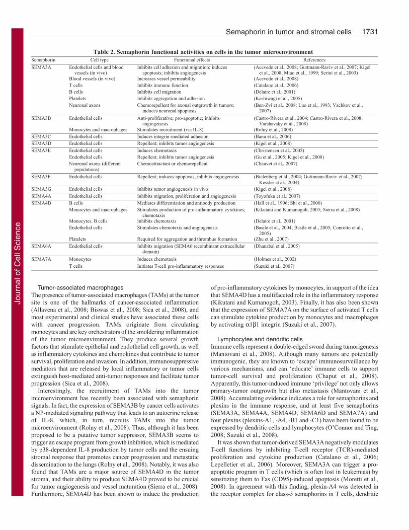
Tumor-associated macrophagesThe presence of tumor-associated macrophages (TAMs) at the tumorsite is one of the hallmarks of cancer-associated inflammation(Allavena et al., 2008; Biswas et al., 2008; Sica et al., 2008), andmost experimental and clinical studies have associated these cellswith cancer progression. TAMs originate from circulatingmonocytes and are key orchestrators of the smoldering inflammationof the tumor microenvironment. They produce several growthfactors that stimulate epithelial and endothelial cell growth, as wellas inflammatory cytokines and chemokines that contribute to tumorsurvival, proliferation and invasion. In addition, immunosuppressivemediators that are released by local inflammatory or tumor cellsextinguish host-mediated anti-tumor responses and facilitate tumorprogression (Sica et al., 2008).
Interestingly, the recruitment of TAMs into the tumormicroenvironment has recently been associated with semaphorinsignals. In fact, the expression of SEMA3B by cancer cells activatesa NP-mediated signaling pathway that leads to an autocrine releaseof IL-8, which, in turn, recruits TAMs into the tumormicroenvironment (Rolny et al., 2008). Thus, although it has beenproposed to be a putative tumor suppressor, SEMA3B seems totrigger an escape program from growth inhibition, which is mediatedby p38-dependent IL-8 production by tumor cells and the ensuingstromal response that promotes cancer progression and metastaticdissemination to the lungs (Rolny et al., 2008). Notably, it was alsofound that TAMs are a major source of SEMA4D in the tumorstroma, and their ability to produce SEMA4D proved to be crucialfor tumor angiogenesis and vessel maturation (Sierra et al., 2008).Furthermore, SEMA4D has been shown to induce the production
of pro-inflammatory cytokines by monocytes, in support of the ideathat SEMA4D has a multifaceted role in the inflammatory response(Kikutani and Kumanogoh, 2003). Finally, it has also been shownthat the expression of SEMA7A on the surface of activated T cellscan stimulate cytokine production by monocytes and macrophagesby activating α1β1 integrin (Suzuki et al., 2007).
Lymphocytes and dendritic cellsImmune cells represent a double-edged sword during tumorigenesis(Mantovani et al., 2008). Although many tumors are potentiallyimmunogenic, they are known to ‘escape’ immunosurveillance byvarious mechanisms, and can ‘educate’ immune cells to supporttumor-cell survival and proliferation (Chaput et al., 2008).Apparently, this tumor-induced immune ‘privilege’ not only allowsprimary-tumor outgrowth but also metastasis (Mantovani et al.,2008). Accumulating evidence indicates a role for semaphorins andplexins in the immune response, and at least five semaphorins(SEMA3A, SEMA4A, SEMA4D, SEMA6D and SEMA7A) andfour plexins (plexins-A1, -A4, -B1 and -C1) have been found to beexpressed by dendritic cells and lymphocytes (O’Connor and Ting,2008; Suzuki et al., 2008).
It was shown that tumor-derived SEMA3A negatively modulatesT-cell functions by inhibiting T-cell receptor (TCR)-mediatedproliferation and cytokine production (Catalano et al., 2006;Lepelletier et al., 2006). Moreover, SEMA3A can trigger a pro-apoptotic program in T cells (which is often lost in leukemias) bysensitizing them to Fas (CD95)-induced apoptosis (Moretti et al.,2008). In agreement with this finding, plexin-A4 was detected inthe receptor complex for class-3 semaphorins in T cells, dendritic
Table 2. Semaphorin functional activities on cells in the tumor microenvironmentSemaphorin Cell type Functional effects References
Endothelial cells and bloodvessels (in vivo)
Inhibits cell adhesion and migration; inducesapoptosis; inhibits angiogenesis
(Acevedo et al., 2008; Guttmann-Raviv et al., 2007; Kigelet al., 2008; Miao et al., 1999; Serini et al., 2003)
Blood vessels (in vivo) Increases vessel permeability (Acevedo et al., 2008)T cells Inhibits immune function (Catalano et al., 2006)B cells Inhibits cell migration (Delaire et al., 2001)Platelets Inhibits aggregation and adhesion (Kashiwagi et al., 2005)
SEMA3A
Neuronal axons Chemorepellent for axonal outgrowth in tumors;induces neuronal apoptosis
(Ben-Zvi et al., 2008; Luo et al., 1993; Vachkov et al.,2007)
Endothelial cells Anti-proliferative; pro-apoptotic; inhibitsangiogenesis
(Castro-Rivera et al., 2004; Castro-Rivera et al., 2008;Varshavsky et al., 2008)
SEMA3B
Monocytes and macrophages Stimulates recruitment (via IL-8) (Rolny et al., 2008)SEMA3C Endothelial cells Induces integrin-mediated adhesion (Banu et al., 2006)SEMA3D Endothelial cells Repellent; inhibits tumor angiogenesis (Kigel et al., 2008)
Endothelial cells Induces chemotaxis (Christensen et al., 2005)Endothelial cells Repellent; inhibits tumor angiogenesis (Gu et al., 2005; Kigel et al., 2008)
SEMA3E
Neuronal axons (differentpopulations)
Chemoattractant or chemorepellent (Chauvet et al., 2007)
SEMA3F Endothelial cells Repellent; induces apoptosis; inhibits angiogenesis (Bielenberg et al., 2004; Guttmann-Raviv et al., 2007;Kessler et al., 2004)
SEMA3G Endothelial cells Inhibits tumor angiogenesis in vivo (Kigel et al., 2008)
SEMA4A Endothelial cells Inhibits migration, proliferation and angiogenesis (Toyofuku et al., 2007)B cells Mediates differentiation and antibody production (Hall et al., 1996; Shi et al., 2000)Monocytes and macrophages Stimulates production of pro-inflammatory cytokines;
chemotaxis(Kikutani and Kumanogoh, 2003; Sierra et al., 2008)
Monocytes, B cells Inhibits chemotaxis (Delaire et al., 2001)Endothelial cells Stimulates chemotaxis and angiogenesis (Basile et al., 2004; Basile et al., 2005; Conrotto et al.,
2005)
SEMA4D
Platelets Required for aggregation and thrombus formation (Zhu et al., 2007)
SEMA6A Endothelial cells Inhibits migration (SEMA6 recombinant extracellulardomain)
(Dhanabal et al., 2005)
Monocytes Induces chemotaxis (Holmes et al., 2002)SEMA7A
T cells Initiates T-cell pro-inflammatory responses (Suzuki et al., 2007)
Jour
nal o
f Cel
l Sci
ence

1732
cells and macrophages, and was found to negatively regulate T-cell-mediated immune responses (Yamamoto et al., 2008). Moreover,SEMA3A and SEMA4D inhibit spontaneous and chemokine-induced migration of monocytes and B cells (Delaire et al., 2001).Conversely, the transmembrane semaphorin SEMA4A, throughinteractions with Tim2, appears to be involved both in T-cell priming(Kumanogoh et al., 2002), and T-helper-1 and T-helper-2 immuneresponses (Kumanogoh et al., 2005). Finally, the immune regulatoryactivity of GPI-anchored SEMA7A is still debated (Czopik et al.,2006; Holmes et al., 2002). As noted above, SEMA7A expressionby activated T cells stimulates monocytes and/or macrophages toproduce proinflammatory cytokines, thereby suggesting thatSEMA7A is an important cue for the effector phase of theinflammatory immune response (Suzuki et al., 2007).
Collectively, these observations indicate that there are importantroles for semaphorin signaling in modulating the activity of theseimmune-cell types during cancer progression. By inhibiting T-cell-mediated responses, semaphorins might help cancer cells to escapeimmune detection and clearance; moreover, by sustaining innateinflammatory responses, they might favor immune dysfunction inthe tumor microenvironment and support cancer progression.
PlateletsPlatelets are highly reactive components of the blood-circulatorysystem that exert not only hemostatic activity but also contributeto inflammation, cancer progression and metastasis, through severaldifferent mechanisms (Gupta and Massague, 2004). Notably, anincreasing amount of evidence suggests that platelets play importantroles in tumor angiogenesis; they are a rich source both of pro-angiogenic factors and of inhibitors of angiogenesis, and, byadhering to the endothelium, they can also facilitate thetransmigration of pro-angiogenic cells to the extracellular space(Sierko and Wojtukiewicz, 2007). Furthermore, platelets tend toaggregate with circulating tumor cells, thereby facilitating immuneevasion and the binding of tumor cells to microvascular endothelium(followed by extravasation and metastasis) (Borsig, 2008). Plateletsmight also contribute to the formation of fibrin clots that surroundtumor cells, supporting the survival of these cells (Boccaccio andComoglio, 2005).
Interestingly, semaphorins might be able to modulate plateletfunction. It has been shown that SEMA3A inhibitsplatelet aggregation, allowing speculation that, by keeping plateletsin the resting state, endothelial-derived SEMA3A may contributeto maintaining blood flow in newly synthesized vessels (Kashiwagiet al., 2005). Conversely, SEMA4D is expressed by platelets andis required for their function (Zhu et al., 2007). Interestingly,following platelet aggregation, membrane-bound SEMA4D isproteolytically cleaved and shed (Zhu et al., 2007); it could thencontribute to tumor-cell survival and tumor angiogenesis in themicroenvironment by acting on adjacent endothelial cells.
Endothelial cellsNeo-angiogenesis, defined as the growth and development of newblood vessels, is a crucial step in cancer progression, as tumors haveto establish a sufficient blood supply in order to grow and metastasize.New blood vessels in tumors can sprout from pre-existing vessels orgrow by recruitment of rare, circulating bone-marrow-derivedendothelial progenitor cells (Avraamides et al., 2008). Accumulatingreports suggest that tumor cells, as well as macrophages andfibroblasts in the microenvironment, can secrete a wide range ofsemaphorins that can modulate blood-vessel development.
Many secreted semaphorins act as inhibitory signals for tumorangiogenesis. SEMA3A suppresses the adhesion and migration ofendothelial cells (Miao et al., 1999; Serini et al., 2003), induces thecollapse of the actin cytoskeleton, promotes apoptosis and inhibitsangiogenesis in vitro (Guttmann-Raviv et al., 2007). Furthermore,SEMA3A can inhibit angiogenesis in vivo (Acevedo et al., 2008;Guttmann-Raviv et al., 2006; Guttmann-Raviv et al., 2007;Varshavsky et al., 2008) and induce microvascular permeability(Acevedo et al., 2008). Similarly, SEMA3F repels endothelial cellsand inhibits their adhesion, migration, proliferation and survival;moreover, the SEMA3F expression pattern correlates with largeareas of diminished vascularity and apoptosis in experimentaltumors (Bielenberg et al., 2008; Guttmann-Raviv et al., 2007;Kessler et al., 2004). Moreover, melanoma-cell-derived SEMA3Fhas chemorepulsive activity for lymphatic endothelial cells andinhibits lymphangiogenesis and lymphatic metastasization in vivo(Bielenberg et al., 2004).
An emerging concept suggests that VEGF might physiologicallyinduce the autocrine secretion of anti-angiogenic semaphorins inendothelial cells, as a negative-feedback loop, to restrain its ownsignaling and prevent unwarranted angiogenesis. For instance, ithas been shown that the increased aggressiveness of multiplemyeloma is associated with an angiogenic switch driven by animbalanced secretion of VEGF165 and SEMA3A by endothelialcells (Vacca et al., 2006).
Similarly to SEMA3A and SEMA3F, three other class-3semaphorins – SEMA3B, SEMA3D and SEMA3G – were alsoreported to repel endothelial cells and/or inhibit tumorangiogenesis (Kigel et al., 2008). Moreover, notable defects inthe recruitment of pericytes to blood vessels have been observedin SEMA3B-expressing tumors (Charlotte Rolny and L.T.,unpublished data). The anti-angiogenic function of SEMA3Bmight be partly balanced in tumors by its ability to increase theproduction of the angiogenic factor IL-8 (Rolny et al., 2008).Interestingly, it was also demonstrated that the production ofSEMA3B by tumor cells fails to repel endothelial cells if they aresubjected to the proteolytic activity of furin-like convertases (pro-protein convertases; PPCs) (Varshavsky et al., 2008). Thus, itseems that cleavage by PPCs that are produced by cancer cellsmight be an additional mechanism by which to curb the anti-angiogenic effects of SEMA3B.
Other class-3 semaphorins have been reported to regulate tumorangiogenesis in a different manner. For instance, SEMA3C wasfound to induce endothelial-cell growth and migration (Banu et al.,2006). Moreover, highly metastatic cancer cells often secreteSEMA3E, and its specific receptor, plexin-D1, is well expressed invascular endothelium both during development and in tumor vessels(Christensen et al., 2005; Gu et al., 2005; Roodink et al., 2008).However, the function of SEMA3E in regulating angiogenesis isnot clear, as different studies have reported either pro-migratory orinhibitory activities for this semaphorin in different types ofendothelial cells (Christensen et al., 2005; Gu et al., 2005; Kigelet al., 2008). These discrepancies might be explained by theinvolvement of different receptor complexes. Interestingly,the generation of a smaller isoform of SEMA3E by furin-dependentproteolytic processing is required to reveal its chemotactic activityfor endothelial cells (Christensen et al., 2005). Therefore, cancercells might be able to convert this secreted inhibitory signal into apro-angiogenic and pro-metastatic factor.
Of the membrane-bound semaphorins, SEMA4D has beenreported to exert potent pro-angiogenic activity in vitro and in
Journal of Cell Science 122 (11)
Jour
nal o
f Cel
l Sci
ence
1733Semaphorin in tumor and stromal cells
vivo by binding to the high-affinity receptors plexin-B1 (Basileet al., 2004; Basile et al., 2005; Conrotto et al., 2005) and possiblyplexin-B2 (Fazzari et al., 2007), which are expressed byendothelial cells. Notably, SEMA4D is not crucial fordevelopmental angiogenesis. However, many human cancersexpress this semaphorin, which can be processed and released ina soluble form by the activity of membrane type 1 matrixmetalloproteinase (MMP-1) and thereby induce endothelial-cellchemotaxis and blood-vessel growth in vivo (Sierra et al., 2008).Moreover, because recruited macrophages are a main source ofSEMA4D in the microenvironment, experimental tumors grownin sema4D–/– mice were found to contain smaller and aberrantvessels that were poorly lined with pericytes, and both cancergrowth and metastases were strikingly reduced (Toyofuku et al.,2007). Conversely, SEMA4A was shown to suppress VEGF-dependent endothelial-cell migration, proliferation andangiogenesis by binding to the plexin-D1 receptor (Toyofuku et al.,2007); in addition, the recombinant extracellular domain ofSEMA6A could inhibit tumor-induced angiogenesis in vivo(Dhanabal et al., 2005).
Nerve fibersSemaphorins are well-known axon-guidance molecules that havebeen further implicated in axon branching, axon pruning and axondegeneration (He et al., 2002; Tran et al., 2007). In fact, althoughthey have mainly been described as inhibitory signals, they can alsopromote neurite outgrowth and/or attraction. Examples are thesoluble SEMA4D, a neurotrophic molecule that can enhance neuriteoutgrowth (Masuda et al., 2004), and SEMA3E, which can mediateattractant or repellent signals through the plexin-D1 receptor,depending on the ‘gating’ function played by the extracellulardomain of NP1 (Chauvet et al., 2007). Interestingly, plexin-A3 hasbeen recently described to mediate semaphorin-induced neuronalcell death (Ben-Zvi et al., 2008), supporting the idea that naturallyoccurring neuronal cell death might involve pro-apoptotic signalingmediated by semaphorins.
Interestingly, new evidence supports a crucial role for theneuroendocrine system in cancer progression; innervation ofthe tumor mass and the local release of neurotransmitters andhormones can potentially act as regulatory signals for multiple celltypes in the tumor microenvironment (Entschladen et al., 2006).This process is bidirectional, as tumor cells can also releaseattracting, repelling and neurotrophic factors. Notably, whereasmany class-3 semaphorins seem to inhibit axonal growth in thetumor microenvironment (Vachkov et al., 2007), SEMA4F hasrecently been implicated in prostate-cancer neurogenesis (Ayalaet al., 2008). Thus, during tumor progression, the balance betweenthe neurotrophic and restrictive cues that are mediated bysemaphorins could impinge on tumor innervation. In this scenario,trophic signals that accompany neurogenesis might represent ametastatic impetus to tumor cells. It will be intriguing to explorefurther the suggested role of semaphorins in the process known as‘perineural invasion’ (Entschladen et al., 2007). This is a commonoccurrence in some tumors (such as those in the prostate andpancreas) whereby cancer cells seem to migrate along the route ofnerve fibers; this is putatively linked with invasion and cancerprogression.
Concluding remarks and future perspectivesWe have summarized here the current evidence indicating thatsemaphorins, plexins and NPs are important regulators of tumor
progression. Semaphorins are multifaceted signals that can besecreted by tumor cells and by other cell types in the tumormicroenvironment, and that control the behavior of a broad rangeof cell types and thereby might affect tumor progression throughmultiple mechanisms. Therefore, further studies on the semaphorin-mediated crosstalk between tumor cells and tumor stroma arerequired for a better understanding of this scenario. It will beimportant to obtain further insights regarding the molecularmechanisms through which semaphorins regulate cancer-cellsurvival, proliferation and migration, and – on the other side –instruct the function of stromal cells in the tumor microenvironment.Moreover, the potential role of semaphorins in the regulation ofEMT is an intriguing hypothesis that could be addressed in futurestudies. Finally, because our knowledge of semaphorin signalingpathways is still far from complete, further studies on the differenttransducers that are involved downstream of plexin activation intumor cells will be necessary. This might allow the identificationof molecular targets to develop pathway-selective inhibitors, andprovide further support for the concept that interfering withsemaphorin-mediated signals might be a promising anti-cancerstrategy.
We wish to thank Josè Raphael Sierra for critical reading of themanuscript and Antonino Ferraro for technical assistance withillustrations. The authors’ research is supported by the ItalianAssociation for Cancer Research (AIRC) and Regione Piemonte.
ReferencesAcevedo, L. M., Barillas, S., Weis, S. M., Gothert, J. R. and Cheresh, D. A. (2008).
Semaphorin 3A suppresses VEGF-mediated angiogenesis yet acts as a vascularpermeability factor. Blood 111, 2674-2680.
Allavena, P., Garlanda, C., Borrello, M. G., Sica, A. and Mantovani, A. (2008). Pathwaysconnecting inflammation and cancer. Curr. Opin. Genet. Dev. 18, 3-10.
Appleton, B. A., Wu, P., Maloney, J., Yin, J., Liang, W. C., Stawicki, S., Mortara, K.,Bowman, K. K., Elliott, J. M., Desmarais, W. et al. (2007). Structural studies ofneuropilin/antibody complexes provide insights into semaphorin and VEGF binding.EMBO J. 26, 4902-4912.
Artigiani, S., Barberis, D., Fazzari, P., Longati, P., Angelini, P., van de Loo, J. W.,Comoglio, P. M. and Tamagnone, L. (2003). Functional regulation of semaphorinreceptors by proprotein convertases. J. Biol. Chem. 278, 10094-10101.
Avraamides, C. J., Garmy-Susini, B. and Varner, J. A. (2008). Integrins in angiogenesisand lymphangiogenesis. Nat. Rev. Cancer 8, 604-617.
Ayala, G. E., Dai, H., Powell, M., Li, R., Ding, Y., Wheeler, T. M., Shine, D., Kadmon,D., Thompson, T., Miles, B. J. et al. (2008). Cancer-related axonogenesis andneurogenesis in prostate cancer. Clin. Cancer Res. 14, 7593-7603.
Bachelder, R. E., Lipscomb, E. A., Lin, X., Wendt, M. A., Chadborn, N. H., Eickholt,B. J. and Mercurio, A. M. (2003). Competing autocrine pathways involving alternativeneuropilin-1 ligands regulate chemotaxis of carcinoma cells. Cancer Res. 63, 5230-5233.
Banu, N., Teichman, J., Dunlap-Brown, M., Villegas, G. and Tufro, A. (2006).Semaphorin 3C regulates endothelial cell function by increasing integrin activity. FASEBJ. 20, 2150-2152.
Barberis, D., Artigiani, S., Casazza, A., Corso, S., Giordano, S., Love, C. A., Jones,E. Y., Comoglio, P. M. and Tamagnone, L. (2004). Plexin signaling hampers integrin-based adhesion, leading to Rho-kinase independent cell rounding, and inhibitinglamellipodia extension and cell motility. FASEB J. 18, 592-594.
Barr, M. P., Byrne, A. M., Duffy, A. M., Condron, C. M., Devocelle, M., Harriott, P.,Bouchier-Hayes, D. J. and Harmey, J. H. (2005). A peptide corresponding to theneuropilin-1-binding site on VEGF(165) induces apoptosis of neuropilin-1-expressingbreast tumour cells. Br. J. Cancer 92, 328-333.
Basile, J. R., Barac, A., Zhu, T., Guan, K. L. and Gutkind, J. S. (2004). Class IVsemaphorins promote angiogenesis by stimulating Rho-initiated pathways through plexin-B. Cancer Res. 64, 5212-5224.
Basile, J. R., Afkhami, T. and Gutkind, J. S. (2005). Semaphorin 4D/plexin-B1 inducesendothelial cell migration through the activation of PYK2, Src, and thephosphatidylinositol 3-kinase-Akt pathway. Mol. Cell. Biol. 25, 6889-6898.
Basile, J. R., Gavard, J. and Gutkind, J. S. (2007a). Plexin-B1 utilizes RhoA and Rhokinase to promote the integrin-dependent activation of Akt and ERK and endothelialcell motility. J. Biol. Chem. 282, 34888-34895.
Basile, J. R., Holmbeck, K., Bugge, T. H. and Gutkind, J. S. (2007b). MT1-MMP controlstumor-induced angiogenesis through the release of semaphorin 4D. J. Biol. Chem. 282,6899-6905.
Ben-Zvi, A., Manor, O., Schachner, M., Yaron, A., Tessier-Lavigne, M. and Behar, O.(2008). The Semaphorin receptor PlexinA3 mediates neuronal apoptosis during dorsalroot ganglia development. J. Neurosci. 28, 12427-12432.
Jour
nal o
f Cel
l Sci
ence

1734
Bielenberg, D. R., Hida, Y., Shimizu, A., Kaipainen, A., Kreuter, M., Kim, C. C. andKlagsbrun, M. (2004). Semaphorin 3F, a chemorepulsant for endothelial cells, inducesa poorly vascularized, encapsulated, nonmetastatic tumor phenotype. J. Clin. Invest. 114,1260-1271.
Bielenberg, D. R., Pettaway, C. A., Takashima, S. and Klagsbrun, M. (2006).Neuropilins in neoplasms: expression, regulation, and function. Exp. Cell Res. 312, 584-593.
Bielenberg, D. R., Shimizu, A. and Klagsbrun, M. (2008). Semaphorin-inducedcytoskeletal collapse and repulsion of endothelial cells. Methods Enzymol. 443, 299-314.
Biswas, S. K., Sica, A. and Lewis, C. E. (2008). Plasticity of macrophage function duringtumor progression: regulation by distinct molecular mechanisms. J. Immunol. 180, 2011-2017.
Boccaccio, C. and Comoglio, P. M. (2005). A functional role for hemostasis in early cancerdevelopment. Cancer Res. 65, 8579-8582.
Bork, P., Doerks, T., Springer, T. A. and Snel, B. (1999). Domains in plexins: links tointegrins and transcription factors. Trends Biochem. Sci. 24, 261-263.
Borsig, L. (2008). The role of platelet activation in tumor metastasis. Expert Rev.Anticancer. Ther. 8, 1247-1255.
Brambilla, E., Constantin, B., Drabkin, H. and Roche, J. (2000). Semaphorin SEMA3Flocalization in malignant human lung and cell lines: a suggested role in cell adhesionand cell migration. Am. J. Pathol. 156, 939-950.
Casazza, A., Fazzari, P. and Tamagnone, L. (2007). Semaphorin signals in cell adhesionand cell migration: functional role and molecular mechanisms. Adv. Exp. Med. Biol.600, 90-108.
Castellani, V. and Rougon, G. (2002). Control of semaphorin signaling. Curr. Opin.Neurobiol. 12, 532.
Castro-Rivera, E., Ran, S., Thorpe, P. and Minna, J. D. (2004). Semaphorin 3B(SEMA3B) induces apoptosis in lung and breast cancer, whereas VEGF165 antagonizesthis effect. Proc. Natl. Acad. Sci. USA 101, 11432-11437.
Castro-Rivera, E., Ran, S., Brekken, R. A. and Minna, J. D. (2008). Semaphorin 3Binhibits the phosphatidylinositol 3-kinase/Akt pathway through neuropilin-1 in lung andbreast cancer cells. Cancer Res. 68, 8295-8303.
Catalano, A., Caprari, P., Rodilossi, S., Betta, P., Castellucci, M., Casazza, A.,Tamagnone, L. and Procopio, A. (2004). Cross-talk between vascular endothelial growthfactor and semaphorin-3A pathway in the regulation of normal and malignant mesothelialcell proliferation. FASEB J. 18, 358-360.
Catalano, A., Caprari, P., Moretti, S., Faronato, M., Tamagnone, L. and Procopio, A.(2006). Semaphorin-3A is expressed by tumor cells and alters T-cell signal transductionand function. Blood 107, 3321-3329.
Caunt, M., Mak, J., Liang, W. C., Stawicki, S., Pan, Q., Tong, R. K., Kowalski, J., Ho,C., Reslan, H. B., Ross, J. et al. (2008). Blocking neuropilin-2 function inhibits tumorcell metastasis. Cancer Cell 13, 331-342.
Chabbert-de Ponnat, I., Buffard, V., Leroy, K., Bagot, M., Bensussan, A., Wolkenstein,P. and Marie-Cardine, A. (2006). Antiproliferative effect of semaphorin 3F on humanmelanoma cell lines. J. Invest. Dermatol. 126, 2343-2345.
Chaput, N., Conforti, R., Viaud, S., Spatz, A. and Zitvogel, L. (2008). The Janus faceof dendritic cells in cancer. Oncogene 27, 5920-5931.
Chauvet, S., Cohen, S., Yoshida, Y., Fekrane, L., Livet, J., Gayet, O., Segu, L., Buhot,M. C., Jessell, T. M., Henderson, C. E. et al. (2007). Gating of Sema3E/PlexinD1signaling by neuropilin-1 switches axonal repulsion to attraction during braindevelopment. Neuron 56, 807-822.
Christensen, C. R., Klingelhofer, J., Tarabykina, S., Hulgaard, E. F., Kramerov, D.and Lukanidin, E. (1998). Transcription of a novel mouse semaphorin gene, M-semaH,correlates with the metastatic ability of mouse tumor cell lines. Cancer Res. 58, 1238-1244.
Christensen, C., Ambartsumian, N., Gilestro, G., Thomsen, B., Comoglio, P.,Tamagnone, L., Guldberg, P. and Lukanidin, E. (2005). Proteolytic processing convertsthe repelling signal Sema3E into an inducer of invasive growth and lung metastasis.Cancer Res. 65, 6167-6177.
Conrotto, P., Corso, S., Gamberini, S., Comoglio, P. M. and Giordano, S. (2004).Interplay between scatter factor receptors and B plexins controls invasive growth.Oncogene 23, 5131-5137.
Conrotto, P., Valdembri, D., Corso, S., Serini, G., Tamagnone, L., Comoglio, P. M.,Bussolino, F. and Giordano, S. (2005). Sema4D induces angiogenesis through Metrecruitment by Plexin B1. Blood 105, 4321-4329.
Czopik, A. K., Bynoe, M. S., Palm, N., Raine, C. S. and Medzhitov, R. (2006).Semaphorin 7A is a negative regulator of T cell responses. Immunity 24, 591-600.
Deaglio, S., Vaisitti, T., Bergui, L., Bonello, L., Horenstein, A. L., Tamagnone, L.,Boumsell, L. and Malavasi, F. (2005). CD38 and CD100 lead a network of surfacereceptors relaying positive signals for B-CLL growth and survival. Blood 105, 3042-3050.
Delaire, S., Billard, C., Tordjman, R., Chedotal, A., Elhabazi, A., Bensussan, A. andBoumsell, L. (2001). Biological activity of soluble CD100. II. Soluble CD100, similarlyto H-SemaIII, inhibits immune cell migration. J. Immunol. 166, 4348-4354.
Dhanabal, M., Wu, F., Alvarez, E., McQueeney, K. D., Jeffers, M., Macdougall, J.,Boldog, F. L., Hackett, C., Shenoy, S., Khramtsov, N. et al. (2005). Recombinantsemaphorin 6A-1 ectodomain inhibits in vivo growth factor and tumor cell line-inducedangiogenesis. Cancer Biol. Ther. 4, 659-668.
Dolcetti, L., Marigo, I., Mantelli, B., Peranzoni, E., Zanovello, P. and Bronte, V. (2008).Myeloid-derived suppressor cell role in tumor-related inflammation. Cancer Lett. 267,216-225.
Elhabazi, A., Delaire, S., Bensussan, A., Boumsell, L. and Bismuth, G. (2001).Biological activity of soluble CD100. I. The extracellular region of CD100 is released
from the surface of T lymphocytes by regulated proteolysis. J. Immunol. 166, 4341-4347.
Ellis, L. M. (2006). The role of neuropilins in cancer. Mol. Cancer Ther. 5, 1099-1107.Entschladen, F., Palm, D., Lang, K., Drell, T. L. and Zaenker, K. S. (2006).
Neoneurogenesis: tumors may initiate their own innervation by the release of neurotrophicfactors in analogy to lymphangiogenesis and neoangiogenesis. Med. Hypotheses 67, 33-35.
Entschladen, F., Palm, D., Drell, T. L., Lang, K. and Zaenker, K. S. (2007). Connectinga tumor to the environment. Curr. Pharm. Des. 13, 3440-3444.
Fazzari, P., Penachioni, J., Gianola, S., Rossi, F., Eickholt, B. J., Maina, F., Alexopoulou,L., Sottile, A., Comoglio, P. M., Flavell, R. A. et al. (2007). Plexin-B1 plays a redundantrole during mouse development and in tumour angiogenesis. BMC Dev. Biol. 7, 55.
Franco, M. and Tamagnone, L. (2008). Tyrosine phosphorylation in semaphorin signalling:shifting into overdrive. EMBO Rep. 9, 865-871.
Fujii, T., Nakao, F., Shibata, Y., Shioi, G., Kodama, E., Fujisawa, H. and Takagi, S.(2002). Caenorhabditis elegans PlexinA, PLX-1, interacts with transmembranesemaphorins and regulates epidermal morphogenesis. Development 129, 2053-2063.
Fukasawa, M., Matsushita, A. and Korc, M. (2007). Neuropilin-1 interacts with integrinbeta1 and modulates pancreatic cancer cell growth, survival and invasion. Cancer Biol.Ther. 6, 1173-1180.
Futamura, M., Kamino, H., Miyamoto, Y., Kitamura, N., Nakamura, Y., Ohnishi, S.,Masuda, Y. and Arakawa, H. (2007). Possible role of semaphorin 3F, a candidatetumor suppressor gene at 3p21.3, in p53-regulated tumor angiogenesis suppression.Cancer Res. 67, 1451-1460.
Galani, E., Sgouros, J., Petropoulou, C., Janinis, J., Aravantinos, G., Dionysiou-Asteriou, D., Skarlos, D. and Gonos, E. (2002). Correlation of MDR-1, nm23-H1 andH Sema E gene expression with histopathological findings and clinical outcome in ovarianand breast cancer patients. Anticancer Res. 22, 2275-2280.
Geretti, E., Shimizu, A. and Klagsbrun, M. (2008). Neuropilin structure governs VEGFand semaphorin binding and regulates angiogenesis. Angiogenesis 11, 31-39.
Gherardi, E., Love, C. A., Esnouf, R. M. and Jones, E. Y. (2004). The sema domain.Curr. Opin. Struct. Biol. 14, 669-678.
Ghosh, S., Sullivan, C. A., Zerkowski, M. P., Molinaro, A. M., Rimm, D. L., Camp,R. L. and Chung, G. G. (2008). High levels of vascular endothelial growth factor andits receptors (VEGFR-1, VEGFR-2, neuropilin-1) are associated with worse outcomein breast cancer. Hum. Pathol. 39, 1835-1843.
Giordano, S., Corso, S., Conrotto, P., Artigiani, S., Gilestro, G., Barberis, D.,Tamagnone, L. and Comoglio, P. M. (2002). The semaphorin 4D receptor controlsinvasive growth by coupling with Met. Nat. Cell Biol. 4, 720-724.
Glinka, Y. and Prud’homme, G. J. (2008). Neuropilin-1 is a receptor for transforminggrowth factor beta-1, activates its latent form, and promotes regulatory T cell activity.J. Leukoc. Biol. 84, 302-310.
Gomez Roman, J. J., Garay, G. O., Saenz, P., Escuredo, K., Sanz, I. C., Gutkind, S.,Junquera, C., Simon, L., Martinez, A., Fernandez Luna, J. L. et al. (2008). PlexinB1 is downregulated in renal cell carcinomas and modulates cell growth. Transl. Res.151, 134-140.
Goodman, C. S., Kolodkin, A. L., Luo, Y., Pueschel, A. W. and Raper, J. A. (1999).Unified nomenclature for the semaphorins collapsins. Cell 97, 551-552.
Granziero, L., Circosta, P., Scielzo, C., Frisaldi, E., Stella, S., Geuna, M., Giordano,S., Ghia, P. and Caligaris-Cappio, F. (2003). CD100/Plexin-B1 interactions sustainproliferation and survival of normal and leukemic CD5+ B lymphocytes. Blood 101,1962-1969.
Gray, M. J., Wey, J. S., Belcheva, A., McCarty, M. F., Trevino, J. G., Evans, D. B.,Ellis, L. M. and Gallick, G. E. (2005). Neuropilin-1 suppresses tumorigenic propertiesin a human pancreatic adenocarcinoma cell line lacking neuropilin-1 coreceptors. CancerRes. 65, 3664-3670.
Gray, M. J., Van Buren, G., Dallas, N. A., Xia, L., Wang, X., Yang, A. D., Somcio, R.J., Lin, Y. G., Lim, S., Fan, F. et al. (2008). Therapeutic targeting of neuropilin-2 oncolorectal carcinoma cells implanted in the murine liver. J. Natl. Cancer Inst. 100, 109-120.
Gu, C., Limberg, B. J., Whitaker, G. B., Perman, B., Leahy, D. J., Rosenbaum, J. S.,Ginty, D. D. and Kolodkin, A. L. (2002). Characterization of neuropilin-1 structuralfeatures that confer binding to semaphorin 3A and vascular endothelial growth factor165. J. Biol. Chem. 277, 18069-18076.
Gu, C., Yoshida, Y., Livet, J., Reimert, D. V., Mann, F., Merte, J., Henderson, C. E.,Jessell, T. M., Kolodkin, A. L. and Ginty, D. D. (2005). Semaphorin 3E and plexin-D1 control vascular pattern independently of neuropilins. Science 307, 265-268.
Guo, W., Pylayeva, Y., Pepe, A., Yoshioka, T., Muller, W. J., Inghirami, G. andGiancotti, F. G. (2006). Beta 4 integrin amplifies ErbB2 signaling to promote mammarytumorigenesis. Cell 126, 489-502.
Gupta, G. P. and Massague, J. (2004). Platelets and metastasis revisited: a novel fattylink. J. Clin. Invest. 114, 1691-1693.
Guttmann-Raviv, N., Kessler, O., Shraga-Heled, N., Lange, T., Herzog, Y. and Neufeld,G. (2006). The neuropilins and their role in tumorigenesis and tumor progression. CancerLett. 231, 1-11.
Guttmann-Raviv, N., Shraga-Heled, N., Varshavsky, A., Guimaraes-Sternberg, C.,Kessler, O. and Neufeld, G. (2007). Semaphorin-3A and semaphorin-3F work togetherto repel endothelial cells and to inhibit their survival by induction of apoptosis. J. Biol.Chem. 282, 26294-26305.
Hall, K. T., Boumsell, L., Schultze, J. L., Boussiotis, V. A., Dorfman, D. M., Cardoso,A. A., Bensussan, A., Nadler, L. M. and Freeman, G. J. (1996). Human CD100, anovel leukocyte semaphorin that promotes B-cell aggregation and differentiation. Proc.Natl. Acad. Sci. USA 93, 11780-11785.
Journal of Cell Science 122 (11)
Jour
nal o
f Cel
l Sci
ence

1735Semaphorin in tumor and stromal cells
Halloran, M. C. and Wolman, M. A. (2006). Repulsion or adhesion: receptors make thecall. Curr. Opin. Cell Biol. 18, 533-540.
Hanahan, D. and Weinberg, R. A. (2000). The hallmarks of cancer. Cell 100, 57-70.Handa, A., Tokunaga, T., Tsuchida, T., Lee, Y. H., Kijima, H., Yamazaki, H., Ueyama,
Y., Fukuda, H. and Nakamura, M. (2000). Neuropilin-2 expression affects the increasedvascularization and is a prognostic factor in osteosarcoma. Int. J. Oncol. 17, 291-295.
Hansel, D. E., Wilentz, R. E., Yeo, C. J., Schulick, R. D., Montgomery, E. and Maitra,A. (2004). Expression of neuropilin-1 in high-grade dysplasia, invasive cancer, andmetastases of the human gastrointestinal tract. Am. J. Surg. Pathol. 28, 347-356.
He, Z. and Tessier-Lavigne, M. (1997). Neuropilin is a receptor for the axonalchemorepellent Semaphorin III. Cell 90, 739-751.
He, Z., Wang, K. C., Koprivica, V., Ming, G. and Song, H. J. (2002). Knowing how tonavigate: mechanisms of semaphorin signaling in the nervous system. Sci. STKE 2002,RE1.
Herman, J. G. and Meadows, G. G. (2007). Increased class 3 semaphorin expressionmodulates the invasive and adhesive properties of prostate cancer cells. Int. J. Oncol.30, 1231-1238.
Holmes, S., Downs, A. M., Fosberry, A., Hayes, P. D., Michalovich, D., Murdoch, P.,Moores, K., Fox, J., Deen, K., Pettman, G. et al. (2002). Sema7A is a potent monocytestimulator. Scand. J. Immunol. 56, 270-275.
Hsieh, S. H., Ying, N. W., Wu, M. H., Chiang, W. F., Hsu, C. L., Wong, T. Y., Jin, Y.T., Hong, T. M. and Chen, Y. L. (2008). Galectin-1, a novel ligand of neuropilin-1,activates VEGFR-2 signaling and modulates the migration of vascular endothelial cells.Oncogene 27, 3746-3753.
Ito, Y., Oinuma, I., Katoh, H., Kaibuchi, K. and Negishi, M. (2006). Sema4D/plexin-B1 activates GSK-3beta through R-Ras GAP activity, inducing growth cone collapse.EMBO Rep. 7, 704-709.
Kamiya, T., Kawakami, T., Abe, Y., Nishi, M., Onoda, N., Miyazaki, N., Oida, Y.,Yamazaki, H., Ueyama, Y. and Nakamura, M. (2006). The preserved expression ofneuropilin (NRP) 1 contributes to a better prognosis in colon cancer. Oncol. Rep. 15,369-373.
Kashiwagi, H., Shiraga, M., Kato, H., Kamae, T., Yamamoto, N., Tadokoro, S., Kurata,Y., Tomiyama, Y. and Kanakura, Y. (2005). Negative regulation of platelet functionby a secreted cell repulsive protein, semaphorin 3A. Blood 106, 913-921.
Kessler, O., Shraga-Heled, N., Lange, T., Gutmann-Raviv, N., Sabo, E., Baruch, L.,Machluf, M. and Neufeld, G. (2004). Semaphorin-3F is an inhibitor of tumorangiogenesis. Cancer Res. 64, 1008-1015.
Kigel, B., Varshavsky, A., Kessler, O. and Neufeld, G. (2008). Successful inhibition oftumor development by specific class-3 semaphorins is associated with expression ofappropriate semaphorin receptors by tumor cells. PloS ONE 3, e3287.
Kikutani, H. and Kumanogoh, A. (2003). Semaphorins in interactions between T cellsand antigen-presenting cells. Nat. Rev. Immunol. 3, 159-167.
Kolodkin, A. L. and Ginty, D. D. (1997). Steering clear of semaphorins: neuropilins soundthe retreat. Neuron 19, 1159-1162.
Kolodkin, A. L., Matthes, D. J. and Goodman, C. S. (1993). The semaphorin genesencode a family of transmembrane and secreted growth cone guidance molecules. Cell75, 1389-1399.
Kolodkin, A. L., Levengood, D. V., Rowe, E. G., Tai, Y. T., Giger, R. J. and Ginty, D.D. (1997). Neuropilin is a semaphorin III receptor. Cell 90, 753-762.
Koppel, A. M., Feiner, L., Kobayashi, H. and Raper, J. A. (1997). A 70 amino acidregion within the semaphorin domain activates specific cellular response of semaphorinfamily members. Neuron 19, 531-537.
Kumanogoh, A., Watanabe, C., Lee, I., Wang, X., Shi, W., Araki, H., Hirata, H.,Iwahori, K., Uchida, J., Yasui, T. et al. (2000). Identification of CD72 as a lymphocytereceptor for the class IV semaphorin CD100: a novel mechanism for regulating B cellsignaling. Immunity 13, 621-631.
Kumanogoh, A., Marukawa, S., Suzuki, K., Takegahara, N., Watanabe, C., Ch’ng,E., Ishida, I., Fujimura, H., Sakoda, S., Yoshida, K. et al. (2002). Class IV semaphorinSema4A enhances T-cell activation and interacts with Tim-2. Nature 419, 629-633.
Kumanogoh, A., Shikina, T., Suzuki, K., Uematsu, S., Yukawa, K., Kashiwamura, S.,Tsutsui, H., Yamamoto, M., Takamatsu, H., Ko-Mitamura, E. P. et al. (2005).Nonredundant roles of Sema4A in the immune system: defective T cell priming andTh1/Th2 regulation in Sema4A-deficient mice. Immunity 22, 305-316.
Kuroki, T., Trapasso, F., Yendamuri, S., Matsuyama, A., Alder, H., Williams, N. N.,Kaiser, L. R. and Croce, C. M. (2003). Allelic loss on chromosome 3p21.3 and promoterhypermethylation of semaphorin 3B in non-small cell lung cancer. Cancer Res. 63, 3352-3353.
Kusy, S., Nasarre, P., Chan, D., Potiron, V., Meyronet, D., Gemmill, R. M., Constantin,B., Drabkin, H. A. and Roche, J. (2005). Selective suppression of in vivo tumorigenicityby semaphorin SEMA3F in lung cancer cells. Neoplasia 7, 457-465.
Lantuejoul, S., Constantin, B., Drabkin, H., Brambilla, C., Roche, J. and Brambilla,E. (2003). Expression of VEGF, semaphorin SEMA3F, and their common receptorsneuropilins NP1 and NP2 in preinvasive bronchial lesions, lung tumours, and cell lines.J. Pathol. 200, 336-347.
Latil, A., Bieche, I., Pesche, S., Valeri, A., Fournier, G., Cussenot, O. and Lidereau,R. (2000). VEGF overexpression in clinically localized prostate tumors and neuropilin-1 overexpression in metastatic forms. Int. J. Cancer 89, 167-171.
Le Bitoux, M. A. and Stamenkovic, I. (2008). Tumor-host interactions: the role ofinflammation. Histochem. Cell Biol. 130, 1079-1090.
Leffers, N., Gooden, M. J., de Jong, R. A., Hoogeboom, B. N., Ten Hoor, K. A., Hollema,H., Boezen, H. M., van der Zee, A. G., Daemen, T. and Nijman, H. W. (2008).Prognostic significance of tumor-infiltrating T-lymphocytes in primary and metastaticlesions of advanced stage ovarian cancer. Cancer Immunol. Immunother. 58, 449-459.
Lepelletier, Y., Moura, I. C., Hadj-Slimane, R., Renand, A., Fiorentino, S., Baude, C.,Shirvan, A., Barzilai, A. and Hermine, O. (2006). Immunosuppressive role ofsemaphorin-3A on T cell proliferation is mediated by inhibition of actin cytoskeletonreorganization. Eur. J. Immunol. 36, 1782-1793.
Liang, W. C., Dennis, M. S., Stawicki, S., Chanthery, Y., Pan, Q., Chen, Y., Eigenbrot,C., Yin, J., Koch, A. W., Wu, X. et al. (2007). Function blocking antibodies to neuropilin-1 generated from a designed human synthetic antibody phage library. J. Mol. Biol. 366,815-829.
Luo, Y., Raible, D. and Raper, J. A. (1993). Collapsin: a protein in brain that induces thecollapse and paralysis of neuronal growth cones. Cell 75, 217-227.
Mantovani, A., Allavena, P., Sica, A. and Balkwill, F. (2008). Cancer-related inflammation.Nature 454, 436-444.
Marcus, K., Johnson, M., Adam, R. M., O’Reilly, M. S., Donovan, M., Atala, A.,Freeman, M. R. and Soker, S. (2005). Tumor cell-associated neuropilin-1 and vascularendothelial growth factor expression as determinants of tumor growth in neuroblastoma.Neuropathology 25, 178-187.
Martin-Satue, M. and Blanco, J. (1999). Identification of semaphorin E gene expressionin metastatic human lung adenocarcinoma cells by mRNA differential display. J. Surg.Oncol. 72, 18-23.
Masuda, K., Furuyama, T., Takahara, M., Fujioka, S., Kurinami, H. and Inagaki, S.(2004). Sema4D stimulates axonal outgrowth of embryonic DRG sensory neurones.Genes Cells 9, 821-829.
Matsushita, A., Gotze, T. and Korc, M. (2007). Hepatocyte growth factor-mediated cellinvasion in pancreatic cancer cells is dependent on neuropilin-1. Cancer Res. 67, 10309-10316.
Miao, H. Q., Soker, S., Feiner, L., Alonso, J. L., Raper, J. A. and Klagsbrun, M. (1999).Neuropilin-1 mediates collapsin-1/semaphorin III inhibition of endothelial cell motility:functional competition of collapsin-1 and vascular endothelial growth factor-165. J. CellBiol. 146, 233-242.
Miao, H., Burnett, E., Kinch, M., Simon, E. and Wang, B. (2000). Activation of EphA2kinase suppresses integrin function and causes focal-adhesion-kinase dephosphorylation.Nat. Cell Biol. 2, 62-69.
Moretti, S., Procopio, A., Lazzarini, R., Rippo, M. R., Testa, R., Marra, M., Tamagnone,L. and Catalano, A. (2008). Semaphorin3A signaling controls Fas (CD95)-mediatedapoptosis by promoting Fas translocation into lipid rafts. Blood 111, 2290-2299.
Muller, M. W., Giese, N. A., Swiercz, J. M., Ceyhan, G. O., Esposito, I., Hinz, U.,Buchler, P., Giese, T., Buchler, M. W., Offermanns, S. et al. (2007). Association ofaxon guidance factor semaphorin 3A with poor outcome in pancreatic cancer. Int. J.Cancer 121, 2421-2433.
Narazaki, M. and Tosato, G. (2006). Ligand-induced internalization selects use of commonreceptor neuropilin-1 by VEGF165 and semaphorin3A. Blood 107, 3892-3901.
Nasarre, P., Constantin, B., Rouhaud, L., Harnois, T., Raymond, G., Drabkin, H. A.,Bourmeyster, N. and Roche, J. (2003). Semaphorin SEMA3F and VEGF have opposingeffects on cell attachment and spreading. Neoplasia 5, 83-92.
Nasarre, P., Kusy, S., Constantin, B., Castellani, V., Drabkin, H. A., Bagnard, D. andRoche, J. (2005). Semaphorin SEMA3F has a repulsing activity on breast cancer cellsand inhibits E-cadherin-mediated cell adhesion. Neoplasia 7, 180-189.
Negishi, M., Oinuma, I. and Katoh, H. (2005). R-ras as a key player for signaling pathwayof plexins. Mol. Neurobiol. 32, 217-222.
Neufeld, G. and Kessler, O. (2008). The semaphorins: versatile regulators of tumourprogression and tumour angiogenesis. Nat. Rev. Cancer 8, 632-645.
Nguyen, Q. D., Rodrigues, S., Rodrigue, C. M., Rivat, C., Grijelmo, C., Bruyneel, E.,Emami, S., Attoub, S. and Gespach, C. (2006). Inhibition of vascular endothelial growthfactor (VEGF)-165 and semaphorin 3A-mediated cellular invasion and tumor growthby the VEGF signaling inhibitor ZD4190 in human colon cancer cells and xenografts.Mol. Cancer Ther. 5, 2070-2077.
Nishimura, N. and Sasaki, T. (2008). Regulation of epithelial cell adhesion and repulsion:role of endocytic recycling. J. Med. Invest. 55, 9-16.
Nonomura, N., Takayama, H., Nishimura, K., Oka, D., Nakai, Y., Shiba, M., Tsujimura,A., Nakayama, M., Aozasa, K. and Okuyama, A. (2007). Decreased number of mastcells infiltrating into needle biopsy specimens leads to a better prognosis of prostatecancer. Br. J. Cancer 97, 952-956.
O’Connor, B. P. and Ting, J. P. (2008). The evolving role of semaphorins and plexins inthe immune system: Plexin-A1 regulation of dendritic cell function. Immunol. Res. 41,217-222.
Oinuma, I., Ishikawa, Y., Katoh, H. and Negishi, M. (2004). The Semaphorin 4D receptorPlexin-B1 is a GTPase activating protein for R-Ras. Science 305, 862-865.
Osada, H., Tokunaga, T., Nishi, M., Hatanaka, H., Abe, Y., Tsugu, A., Kijima, H.,Yamazaki, H., Ueyama, Y. and Nakamura, M. (2004). Overexpression of theneuropilin 1 (NRP1) gene correlated with poor prognosis in human glioma. AnticancerRes. 24, 547-552.
Pan, H., Wanami, L. S., Dissanayake, T. R. and Bachelder, R. E. (2008). Autocrinesemaphorin3A stimulates alpha2 beta1 integrin expression/function in breast tumor cells.Breast Cancer Res. Treat. doi: 10.1007/s10549-008-0179-y.
Pan, Q., Chanthery, Y., Liang, W. C., Stawicki, S., Mak, J., Rathore, N., Tong, R. K.,Kowalski, J., Yee, S. F., Pacheco, G. et al. (2007). Blocking neuropilin-1 function hasan additive effect with anti-VEGF to inhibit tumor growth. Cancer Cell 11, 53-67.
Pellet-Many, C., Frankel, P., Jia, H. and Zachary, I. (2008). Neuropilins: structure,function and role in disease. Biochem. J. 411, 211-226.
Potiron, V. A., Sharma, G., Nasarre, P., Clarhaut, J. A., Augustin, H. G., Gemmill, R.M., Roche, J. and Drabkin, H. A. (2007). Semaphorin SEMA3F affects multiplesignaling pathways in lung cancer cells. Cancer Res. 67, 8708-8715.
Potiron, V. A., Roche, J. and Drabkin, H. A. (2009). Semaphorins and their receptors inlung cancer. Cancer Lett. 273, 1-14.
Jour
nal o
f Cel
l Sci
ence

1736
Puschel, A. W. (2007). GTPases in semaphorin signaling. Adv. Exp. Med. Biol. 600, 12-23.
Rich, J. N., Hans, C., Jones, B., Iversen, E. S., McLendon, R. E., Rasheed, B. K.,Dobra, A., Dressman, H. K., Bigner, D. D., Nevins, J. R. et al. (2005). Gene expressionprofiling and genetic markers in glioblastoma survival. Cancer Res. 65, 4051-4058.
Ridley, A. J., Schwartz, M. A., Burridge, K., Firtel, R. A., Ginsberg, M. H., Borisy,G., Parsons, J. T. and Horwitz, A. R. (2003). Cell migration: integrating signals fromfront to back. Science 302, 1704-1709.
Rieger, J., Wick, W. and Weller, M. (2003). Human malignant glioma cells expresssemaphorins and their receptors, neuropilins and plexins. Glia 42, 379-389.
Roche, J., Boldog, F., Robinson, M., Robinson, L., Varella-Garcia, M., Swanton, M.,Waggoner, B., Fishel, R., Franklin, W., Gemmill, R. et al. (1996). Distinct 3p21.3deletions in lung cancer and identification of a new human semaphorin. Oncogene 12,1289-1297.
Rody, A., Holtrich, U., Gaetje, R., Gehrmann, M., Engels, K., von Minckwitz, M. G.,Loibl, S., Diallo-Danebrock, R., Ruckhaberle, E., Metzler, D. et al. (2007). Pooroutcome in estrogen receptor-positive breast cancers predicted by loss of plexin B1.Clin. Cancer Res. 13, 1115-1122.
Rolny, C., Capparuccia, L., Casazza, A., Mazzone, M., Vallario, A., Cignetti, A.,Medico, E., Carmeliet, P., Comoglio, P. M. and Tamagnone, L. (2008). The tumorsuppressor semaphorin 3B triggers a prometastatic program mediated by interleukin 8and the tumor microenvironment. J. Exp. Med. 205, 1155-1171.
Roodink, I., Raats, J., van der Zwaag, B., Verrijp, K., Kusters, B., Van Bokhoven, H.,Linkels, M., de Waal, R. M. and Leenders, W. P. (2005). Plexin D1 expression isinduced on tumor vasculature and tumor cells: a novel target for diagnosis and therapy?Cancer Res. 65, 8317-8323.
Roodink, I., Kats, G., van Kempen, L., Grunberg, M., Maass, C., Verrijp, K., Raats,J. and Leenders, W. (2008). Semaphorin 3E expression correlates inversely with PlexinD1 during tumor progression. Am. J. Pathol. 173, 1873-1881.
Scott, G. A., McClelland, L. A. and Fricke, A. F. (2008a). Semaphorin 7a promotesspreading and dendricity in human melanocytes through beta1-integrins. J. Invest.Dermatol. 128, 151-161.
Scott, G. A., McClelland, L. A., Fricke, A. F. and Fender, A. (2008b). Plexin C1, areceptor for semaphorin 7A, inactivates cofilin and is a potential tumor suppressor formelanoma progression. J. Invest. Dermatol. 129, 954-963.
Sekido, Y., Bader, S., Latif, F., Chen, J. Y., Duh, F. M., Wei, M. H., Albanesi, J. P.,Lee, C. C., Lerman, M. I. and Minna, J. D. (1996). Human semaphorins A(V) andIV reside in the 3p21.3 small cell lung cancer deletion region and demonstrate distinctexpression patterns. Proc. Natl. Acad. Sci. USA 93, 4120-4125.
Serini, G., Valdembri, D., Zanivan, S., Morterra, G., Burkhardt, C., Caccavari, F.,Zammataro, L., Primo, L., Tamagnone, L., Logan, M. et al. (2003). Class 3semaphorins control vascular morphogenesis by inhibiting integrin function. Nature 424,391-397.
Serini, G., Napione, L. and Bussolino, F. (2008). Integrins team up with tyrosine kinasereceptors and plexins to control angiogenesis. Curr. Opin. Hematol. 15, 235-242.
Shi, W., Kumanogoh, A., Watanabe, C., Uchida, J., Wang, X., Yasui, T., Yukawa, K.,Ikawa, M., Okabe, M., Parnes, J. R. et al. (2000). The class IV semaphorin CD100plays nonredundant roles in the immune system: defective B and T cell activation inCD100-deficient mice. Immunity 13, 633-642.
Sica, A., Allavena, P. and Mantovani, A. (2008). Cancer related inflammation: themacrophage connection. Cancer Lett. 267, 204-215.
Sierko, E. and Wojtukiewicz, M. Z. (2007). Inhibition of platelet function: does it offera chance of better cancer progression control? Semin. Thromb. Hemost. 33, 712-721.
Sierra, J. R., Corso, S., Caione, L., Cepero, V., Conrotto, P., Cignetti, A., Piacibello,W., Kumanogoh, A., Kikutani, H., Comoglio, P. M. et al. (2008). Tumor angiogenesisand progression are enhanced by Sema4D produced by tumor-associated macrophages.J. Exp. Med. 205, 1673-1685.
Soker, S., Takashima, S., Miao, H. Q., Neufeld, G. and Klagsbrun, M. (1998). Neuropilin-1 is expressed by endothelial and tumor cells as an isoform- specific receptor for vascularendothelial growth factor. Cell 92, 735-745.
Sulpice, E., Plouet, J., Berge, M., Allanic, D., Tobelem, G. and Merkulova-Rainon, T.(2008). Neuropilin-1 and neuropilin-2 act as coreceptors, potentiating proangiogenicactivity. Blood 111, 2036-2045.
Suzuki, K., Okuno, T., Yamamoto, M., Pasterkamp, R. J., Takegahara, N., Takamatsu,H., Kitao, T., Takagi, J., Rennert, P. D., Kolodkin, A. L. et al. (2007). Semaphorin7A initiates T-cell-mediated inflammatory responses through alpha1beta1 integrin. Nature446, 680-684.
Suzuki, K., Kumanogoh, A. and Kikutani, H. (2008). Semaphorins and their receptorsin immune cell interactions. Nat. Immunol. 9, 17-23.
Swiercz, J. M., Kuner, R. and Offermanns, S. (2004). Plexin-B1/RhoGEF-mediated RhoAactivation involves the receptor tyrosine kinase ErbB-2. J. Cell Biol. 165, 869-880.
Swiercz, J. M., Worzfeld, T. and Offermanns, S. (2008). ErbB-2 and met reciprocallyregulate cellular signaling via plexin-B1. J. Biol. Chem. 283, 1893-1901.
Syed, V., Zhang, X., Lau, K. M., Cheng, R., Mukherjee, K. and Ho, S. M. (2005).Profiling estrogen-regulated gene expression changes in normal and malignant humanovarian surface epithelial cells. Oncogene 24, 8128-8143.
Tamagnone, L. and Comoglio, P. M. (2004). To move or not to move? Semaphorinsignalling in cell migration. EMBO Rep. 5, 356-361.
Tamagnone, L., Artigiani, S., Chen, H., He, Z., Ming, G. I., Song, H., Chedotal, A.,Winberg, M. L., Goodman, C. S., Poo, M. et al. (1999). Plexins are a large family ofreceptors for transmembrane, secreted, and GPI-anchored semaphorins in vertebrates.Cell 99, 71-80.
Taskinen, M., Karjalainen-Lindsberg, M. L. and Leppa, S. (2008). Prognostic influenceof tumor-infiltrating mast cells in patients with follicular lymphoma treated with rituximaband CHOP. Blood 111, 4664-4667.
Thiery, J. P. and Sleeman, J. P. (2006). Complex networks orchestrate epithelial-mesenchymal transitions. Nat. Rev. Mol. Cell. Biol. 7, 131-142.
Tomizawa, Y., Sekido, Y., Kondo, M., Gao, B., Yokota, J., Roche, J., Drabkin, H.,Lerman, M. I., Gazdar, A. F. and Minna, J. D. (2001). Inhibition of lung cancer cellgrowth and induction of apoptosis after reexpression of 3p21.3 candidate tumorsuppressor gene SEMA3B. Proc. Natl. Acad. Sci. USA 98, 13954-13959.
Toyofuku, T., Zhang, H., Kumanogoh, A., Takegahara, N., Suto, F., Kamei, J., Aoki,K., Yabuki, M., Hori, M., Fujisawa, H. et al. (2004). Dual roles of Sema6D incardiac morphogenesis through region-specific association of its receptor, Plexin-A1,with off-track and vascular endothelial growth factor receptor type 2. Genes Dev. 18,435-447.
Toyofuku, T., Yabuki, M., Kamei, J., Kamei, M., Makino, N., Kumanogoh, A. andHori, M. (2007). Semaphorin-4A, an activator for T-cell-mediated immunity, suppressesangiogenesis via Plexin-D1. EMBO J. 26, 1373-1384.
Tran, T. S., Kolodkin, A. L. and Bharadwaj, R. (2007). Semaphorin regulation of cellularmorphology. Annu. Rev. Cell Dev. Biol. 23, 263-292.
Trusolino, L., Bertotti, A. and Comoglio, P. M. (2001). A signaling adapter function foralpha6beta4 integrin in the control of HGF-dependent invasive growth. Cell 107, 643-654.
Tse, C., Xiang, R. H., Bracht, T. and Naylor, S. L. (2002). Human Semaphorin 3B(SEMA3B) located at chromosome 3p21.3 suppresses tumor formation in anadenocarcinoma cell line. Cancer Res. 62, 542-546.
Turley, E. A., Veiseh, M., Radisky, D. C. and Bissell, M. J. (2008). Mechanisms of disease:epithelial-mesenchymal transition-does cellular plasticity fuel neoplastic progression?Nat. Clin. Pract. Oncol. 5, 280-290.
Vacca, A., Scavelli, C., Serini, G., Di, P. G., Cirulli, T., Merchionne, F., Ribatti, D.,Bussolino, F., Guidolin, D., Piaggio, G. et al. (2006). Loss of inhibitory semaphorin3A (SEMA3A) autocrine loops in bone marrow endothelial cells of patients with multiplemyeloma. Blood 108, 1661-1667.
Vachkov, I. H., Huang, X., Yamada, Y., Tonchev, A. B., Yamashima, T., Kato, S. andTakakura, N. (2007). Inhibition of axonal outgrowth in the tumor environment:involvement of class 3 semaphorins. Cancer Sci. 98, 1192-1197.
Varshavsky, A., Kessler, O., Abramovitch, S., Kigel, B., Zaffryar, S., Akiri, G. andNeufeld, G. (2008). Semaphorin-3B is an angiogenesis inhibitor that is inactivated byfurin-like pro-protein convertases. Cancer Res. 68, 6922-6931.
Wakabayashi, O., Yamazaki, K., Oizumi, S., Hommura, F., Kinoshita, I., Ogura, S.,aka-Akita, H. and Nishimura, M. (2003). CD4+ T cells in cancer stroma, not CD8+T cells in cancer cell nests, are associated with favorable prognosis in human non-smallcell lung cancers. Cancer Sci. 94, 1003-1009.
Walzer, T., Galibert, L. and De Smedt, T. (2005). Poxvirus semaphorin A39R inhibitsphagocytosis by dendritic cells and neutrophils. Eur. J. Immunol. 35, 391-398.
West, D. C., Rees, C. G., Duchesne, L., Patey, S. J., Terry, C. J., Turnbull, J. E.,Delehedde, M., Heegaard, C. W., Allain, F., Vanpouille, C. et al. (2005). Interactionsof multiple heparin binding growth factors with neuropilin-1 and potentiation of theactivity of fibroblast growth factor-2. J. Biol. Chem. 280, 13457-13464.
Winberg, M. L., Tamagnone, L., Bai, J., Comoglio, P. M., Montell, D. and Goodman,C. S. (2001). The transmembrane protein Off-track associates with Plexins and functionsdownstream of Semaphorin signaling during axon guidance. Neuron 32, 53-62.
Wong, O. G., Nitkunan, T., Oinuma, I., Zhou, C., Blanc, V., Brown, R. S., Bott, S. R.,Nariculam, J., Box, G., Munson, P. et al. (2007). Plexin-B1 mutations in prostate cancer.Proc. Natl. Acad. Sci. USA 104, 19040-19045.
Xiang, R., Davalos, A. R., Hensel, C. H., Zhou, X. J., Tse, C. and Naylor, S. L. (2002).Semaphorin 3F gene from human 3p21.3 suppresses tumor formation in nude mice.Cancer Res. 62, 2637-2643.
Yamada, T., Endo, R., Gotoh, M. and Hirohashi, S. (1997). Identification of semaphorinE as a non-MDR drug resistance gene of human cancers. Proc. Natl. Acad. Sci. USA94, 14713-14718.
Yamamoto, M., Suzuki, K., Okuno, T., Ogata, T., Takegahara, N., Takamatsu, H.,Mizui, M., Taniguchi, M., Chedotal, A., Suto, F. et al. (2008). Plexin-A4 negativelyregulates T lymphocyte responses. Int. Immunol. 20, 413-420.
Zhao, X. Y., Chen, L., Xu, Q. and Li, Y. H. (2006). Expression of semaphorin 6D ingastric carcinoma and its significance. World J. Gastroenterol. 12, 7388-7390.
Zhao, X. Y., Chen, L., Li, Y. H. and Xu, Q. (2007). PlexinA1 expression in gastriccarcinoma and its relationship with tumor angiogenesis and proliferation. World J.Gastroenterol. 13, 6558-6561.
Zhou, Y., Gunput, R. A. and Pasterkamp, R. J. (2008). Semaphorin signaling: progressmade and promises ahead. Trends Biochem. Sci. 33, 161-170.
Zhu, L., Bergmeier, W., Wu, J., Jiang, H., Stalker, T. J., Cieslak, M., Fan, R., Boumsell,L., Kumanogoh, A., Kikutani, H. et al. (2007). Regulated surface expression andshedding support a dual role for semaphorin 4D in platelet responses to vascular injury.Proc. Natl. Acad. Sci. USA 104, 1621-1626.
Journal of Cell Science 122 (11)
Jour
nal o
f Cel
l Sci
ence