Self-assessment
3
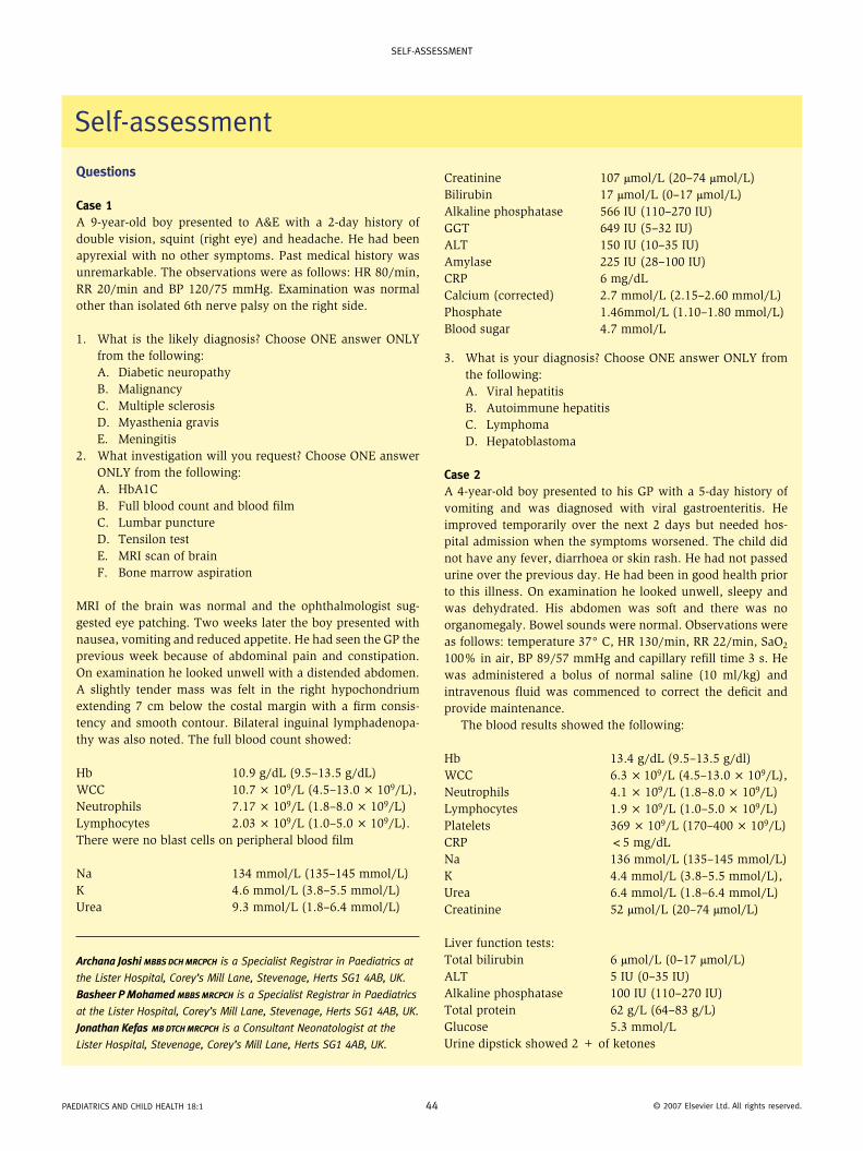
SELF-ASSESSMENT PAEDIATRICS AND CHILD HEALTH 18:1 44 © 2007 Elsevier Ltd. All rights reserved. Questions Case 1 A 9-year-old boy presented to A&E with a 2-day history of double vision, squint (right eye) and headache. He had been apyrexial with no other symptoms. Past medical history was unremarkable. The observations were as follows: HR 80/min, RR 20/min and BP 120/75 mmHg. Examination was normal other than isolated 6th nerve palsy on the right side. 1. What is the likely diagnosis? Choose ONE answer ONLY from the following: A. Diabetic neuropathy B. Malignancy C. Multiple sclerosis D. Myasthenia gravis E. Meningitis 2. What investigation will you request? Choose ONE answer ONLY from the following: A. HbA1C B. Full blood count and blood film C. Lumbar puncture D. Tensilon test E. MRI scan of brain F. Bone marrow aspiration MRI of the brain was normal and the ophthalmologist sug- gested eye patching. Two weeks later the boy presented with nausea, vomiting and reduced appetite. He had seen the GP the previous week because of abdominal pain and constipation. On examination he looked unwell with a distended abdomen. A slightly tender mass was felt in the right hypochondrium extending 7 cm below the costal margin with a firm consis- tency and smooth contour. Bilateral inguinal lymphadenopa- thy was also noted. The full blood count showed: Hb 10.9 g/dL (9.5–13.5 g/dL) WCC 10.7 × 10 9 /L (4.5–13.0 × 10 9 /L), Neutrophils 7.17 × 10 9 /L (1.8–8.0 × 10 9 /L) Lymphocytes 2.03 × 10 9 /L (1.0–5.0 × 10 9 /L). There were no blast cells on peripheral blood film Na 134 mmol/L (135–145 mmol/L) K 4.6 mmol/L (3.8–5.5 mmol/L) Urea 9.3 mmol/L (1.8–6.4 mmol/L) Archana Joshi MBBS DCH MRCPCH is a Specialist Registrar in Paediatrics at the Lister Hospital, Corey’s Mill Lane, Stevenage, Herts SG1 4AB, UK. Basheer P Mohamed MBBS MRCPCH is a Specialist Registrar in Paediatrics at the Lister Hospital, Corey’s Mill Lane, Stevenage, Herts SG1 4AB, UK. Jonathan Kefas MB DTCH MRCPCH is a Consultant Neonatologist at the Lister Hospital, Stevenage, Corey’s Mill Lane, Herts SG1 4AB, UK. Self-assessment Creatinine 107 μmol/L (20–74 μmol/L) Bilirubin 17 μmol/L (0–17 μmol/L) Alkaline phosphatase 566 IU (110–270 IU) GGT 649 IU (5–32 IU) ALT 150 IU (10–35 IU) Amylase 225 IU (28–100 IU) CRP 6 mg/dL Calcium (corrected) 2.7 mmol/L (2.15–2.60 mmol/L) Phosphate 1.46mmol/L (1.10–1.80 mmol/L) Blood sugar 4.7 mmol/L 3. What is your diagnosis? Choose ONE answer ONLY from the following: A. Viral hepatitis B. Autoimmune hepatitis C. Lymphoma D. Hepatoblastoma Case 2 A 4-year-old boy presented to his GP with a 5-day history of vomiting and was diagnosed with viral gastroenteritis. He improved temporarily over the next 2 days but needed hos- pital admission when the symptoms worsened. The child did not have any fever, diarrhoea or skin rash. He had not passed urine over the previous day. He had been in good health prior to this illness. On examination he looked unwell, sleepy and was dehydrated. His abdomen was soft and there was no organomegaly. Bowel sounds were normal. Observations were as follows: temperature 37° C, HR 130/min, RR 22/min, SaO 2 100% in air, BP 89/57 mmHg and capillary refill time 3 s. He was administered a bolus of normal saline (10 ml/kg) and intravenous fluid was commenced to correct the deficit and provide maintenance. The blood results showed the following: Hb 13.4 g/dL (9.5–13.5 g/dl) WCC 6.3 × 10 9 /L (4.5–13.0 × 10 9 /L), Neutrophils 4.1 × 10 9 /L (1.8–8.0 × 10 9 /L) Lymphocytes 1.9 × 10 9 /L (1.0–5.0 × 10 9 /L) Platelets 369 × 10 9 /L (170–400 × 10 9 /L) CRP <5 mg/dL Na 136 mmol/L (135–145 mmol/L) K 4.4 mmol/L (3.8–5.5 mmol/L), Urea 6.4 mmol/L (1.8–6.4 mmol/L) Creatinine 52 μmol/L (20–74 μmol/L) Liver function tests: Total bilirubin 6 μmol/L (0–17 μmol/L) ALT 5 IU (0–35 IU) Alkaline phosphatase 100 IU (110–270 IU) Total protein 62 g/L (64–83 g/L) Glucose 5.3 mmol/L Urine dipstick showed 2 + of ketones
-
Upload
archana-joshi -
Category
Documents
-
view
214 -
download
1
Transcript of Self-assessment
self-assessment
Questions
Case 1A 9-year-old boy presented to A&E with a 2-day history of double vision, squint (right eye) and headache. He had been apyrexial with no other symptoms. Past medical history was unremarkable. The observations were as follows: HR 80/min, RR 20/min and BP 120/75 mmHg. Examination was normal other than isolated 6th nerve palsy on the right side.
1. What is the likely diagnosis? Choose ONE answer ONLY from the following:A. Diabetic neuropathyB. MalignancyC. Multiple sclerosisD. Myasthenia gravisE. Meningitis
2. What investigation will you request? Choose ONE answer ONLY from the following:A. HbA1CB. Full blood count and blood filmC. Lumbar punctureD. Tensilon testE. MRI scan of brainF. Bone marrow aspiration
MRI of the brain was normal and the ophthalmologist sug-gested eye patching. Two weeks later the boy presented with nausea, vomiting and reduced appetite. He had seen the GP the previous week because of abdominal pain and constipation. On examination he looked unwell with a distended abdomen. A slightly tender mass was felt in the right hypochondrium extending 7 cm below the costal margin with a firm consis-tency and smooth contour. Bilateral inguinal lymphadenopa-thy was also noted. The full blood count showed:
Hb 10.9 g/dL (9.5–13.5 g/dL)WCC 10.7 × 10⁹/L (4.5–13.0 × 10⁹/L),Neutrophils 7.17 × 10⁹/L (1.8–8.0 × 10⁹/L)Lymphocytes 2.03 × 10⁹/L (1.0–5.0 × 10⁹/L).There were no blast cells on peripheral blood film
Na 134 mmol/L (135–145 mmol/L)K 4.6 mmol/L (3.8–5.5 mmol/L)Urea 9.3 mmol/L (1.8–6.4 mmol/L)
Archana Joshi MBBS DCH MRCPCH is a Specialist Registrar in Paediatrics at
the Lister Hospital, Corey’s Mill Lane, Stevenage, Herts SG1 4AB, UK.
Basheer P Mohamed MBBS MRCPCH is a Specialist Registrar in Paediatrics
at the Lister Hospital, Corey’s Mill Lane, Stevenage, Herts SG1 4AB, UK.
Jonathan Kefas MB DTCH MRCPCH is a Consultant Neonatologist at the
Lister Hospital, Stevenage, Corey’s Mill Lane, Herts SG1 4AB, UK.
self-assessment
Creatinine 107 μmol/L (20–74 μmol/L)Bilirubin 17 μmol/L (0–17 μmol/L)Alkaline phosphatase 566 IU (110–270 IU)GGT 649 IU (5–32 IU)ALT 150 IU (10–35 IU)Amylase 225 IU (28–100 IU)CRP 6 mg/dLCalcium (corrected) 2.7 mmol/L (2.15–2.60 mmol/L)Phosphate 1.46mmol/L (1.10–1.80 mmol/L)Blood sugar 4.7 mmol/L
3. What is your diagnosis? Choose ONE answer ONLY from the following:A. Viral hepatitisB. Autoimmune hepatitisC. LymphomaD. Hepatoblastoma
Case 2A 4-year-old boy presented to his GP with a 5-day history of vomiting and was diagnosed with viral gastroenteritis. He improved temporarily over the next 2 days but needed hos-pital admission when the symptoms worsened. The child did not have any fever, diarrhoea or skin rash. He had not passed urine over the previous day. He had been in good health prior to this illness. On examination he looked unwell, sleepy and was dehydrated. His abdomen was soft and there was no organomegaly. Bowel sounds were normal. Observations were as follows: temperature 37° C, HR 130/min, RR 22/min, SaO2 100% in air, BP 89/57 mmHg and capillary refill time 3 s. He was administered a bolus of normal saline (10 ml/kg) and intravenous fluid was commenced to correct the deficit and provide maintenance.
The blood results showed the following:
Hb 13.4 g/dL (9.5–13.5 g/dl)WCC 6.3 × 10⁹/L (4.5–13.0 × 10⁹/L),Neutrophils 4.1 × 10⁹/L (1.8–8.0 × 10⁹/L)Lymphocytes 1.9 × 10⁹/L (1.0–5.0 × 10⁹/L)Platelets 369 × 109/L (170–400 × 109/L)CRP <5 mg/dLNa 136 mmol/L (135–145 mmol/L)K 4.4 mmol/L (3.8–5.5 mmol/L),Urea 6.4 mmol/L (1.8–6.4 mmol/L)Creatinine 52 μmol/L (20–74 μmol/L)
Liver function tests:Total bilirubin 6 μmol/L (0–17 μmol/L)ALT 5 IU (0–35 IU)Alkaline phosphatase 100 IU (110–270 IU)Total protein 62 g/L (64–83 g/L)Glucose 5.3 mmol/LUrine dipstick showed 2 + of ketones
Paediatrics and child health 18:1 44 © 2007 elsevier ltd. all rights reserved.
self-assessment
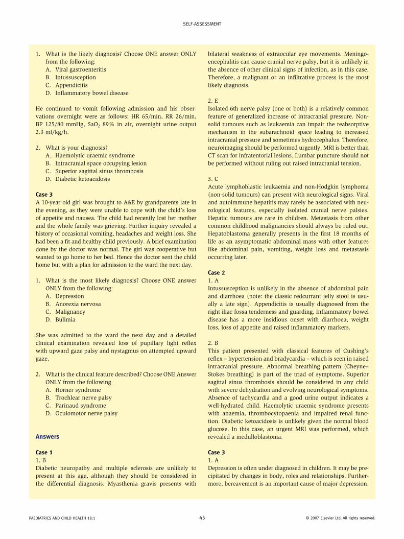
1. What is the likely diagnosis? Choose ONE answer ONLY from the following:A. Viral gastroenteritisB. IntussusceptionC. AppendicitisD. Inflammatory bowel disease
He continued to vomit following admission and his obser-vations overnight were as follows: HR 65/min, RR 26/min, BP 125/80 mmHg, SaO2 89% in air, overnight urine output 2.3 ml/kg/h.
2. What is your diagnosis?A. Haemolytic uraemic syndromeB. Intracranial space occupying lesionC. Superior sagittal sinus thrombosisD. Diabetic ketoacidosis
Case 3A 10-year old girl was brought to A&E by grandparents late in the evening, as they were unable to cope with the child’s loss of appetite and nausea. The child had recently lost her mother and the whole family was grieving. Further inquiry revealed a history of occasional vomiting, headaches and weight loss. She had been a fit and healthy child previously. A brief examination done by the doctor was normal. The girl was cooperative but wanted to go home to her bed. Hence the doctor sent the child home but with a plan for admission to the ward the next day.
1. What is the most likely diagnosis? Choose ONE answer ONLY from the following:A. DepressionB. Anorexia nervosaC. MalignancyD. Bulimia
She was admitted to the ward the next day and a detailed clinical examination revealed loss of pupillary light reflex with upward gaze palsy and nystagmus on attempted upward gaze.
2. What is the clinical feature described? Choose ONE Answer ONLY from the followingA. Horner syndromeB. Trochlear nerve palsyC. Parinaud syndromeD. Oculomotor nerve palsy
Answers
Case 11. BDiabetic neuropathy and multiple sclerosis are unlikely to present at this age, although they should be considered in the differential diagnosis. Myasthenia gravis presents with
bilateral weakness of extraocular eye movements. Meningo-encephalitis can cause cranial nerve palsy, but it is unlikely in the absence of other clinical signs of infection, as in this case. Therefore, a malignant or an infiltrative process is the most likely diagnosis.
2. EIsolated 6th nerve palsy (one or both) is a relatively common feature of generalized increase of intracranial pressure. Non-solid tumours such as leukaemia can impair the reabsorptive mechanism in the subarachnoid space leading to increased intracranial pressure and sometimes hydrocephalus. Therefore, neuroimaging should be performed urgently. MRI is better than CT scan for infratentorial lesions. Lumbar puncture should not be performed without ruling out raised intracranial tension.
3. CAcute lymphoblastic leukaemia and non-Hodgkin lymphoma (non-solid tumours) can present with neurological signs. Viral and autoimmune hepatitis may rarely be associated with neu-rological features, especially isolated cranial nerve palsies. Hepatic tumours are rare in children. Metastasis from other common childhood malignancies should always be ruled out. Hepatoblastoma generally presents in the first 18 months of life as an asymptomatic abdominal mass with other features like abdominal pain, vomiting, weight loss and metastasis occurring later.
Case 21. AIntussusception is unlikely in the absence of abdominal pain and diarrhoea (note: the classic redcurrant jelly stool is usu-ally a late sign). Appendicitis is usually diagnosed from the right iliac fossa tenderness and guarding. Inflammatory bowel disease has a more insidious onset with diarrhoea, weight loss, loss of appetite and raised inflammatory markers.
2. BThis patient presented with classical features of Cushing’s reflex – hypertension and bradycardia – which is seen in raised intracranial pressure. Abnormal breathing pattern (Cheyne–Stokes breathing) is part of the triad of symptoms. Superior sagittal sinus thrombosis should be considered in any child with severe dehydration and evolving neurological symptoms. Absence of tachycardia and a good urine output indicates a well-hydrated child. Haemolytic uraemic syndrome presents with anaemia, thrombocytopaenia and impaired renal func-tion. Diabetic ketoacidosis is unlikely given the normal blood glucose. In this case, an urgent MRI was performed, which revealed a medulloblastoma.
Case 31. ADepression is often under diagnosed in children. It may be pre-cipitated by changes in body, roles and relationships. Further-more, bereavement is an important cause of major depression.
Paediatrics and child health 18:1 45 © 2007 elsevier ltd. all rights reserved.
self-assessment
Depressed mood, weight loss or gain, insomnia or hypersom-nia, fatigue, feeling of worthlessness or guilt are features of depression. When these symptoms occur within 3 months of an identifiable stressor, this is considered as adjustment dis-order with depressed mood. Anorexia nervosa is a psychiatric disorder characterized by intense fear of becoming obese, altered self perception of body image and refusal to main-tain body weight over a minimal normal weight for age and height. Bulimia is defined as a separate clinical entity by DSM IV criteria with the following features: recurrent episodes of binge eating and fear of not being able to stop eating during a binge, and regularly engaging in activities such as self-induced vomiting, abuse of laxatives, rigorous exercise which counter-acts the effects of binge eating. Eating disorders are commonly seen in the 16–18 year age group and more commonly involve girls than boys. The incidence is on the rise in all western countries. Malignancy should be considered in the differential diagnosis of weight loss in children and hence a full clinical examination is vital, before a diagnosis of non-organic cause is made.
2. CThe clinical features are consistent with a condition referred to as Parinaud syndrome, which is due to mid-brain dysfunc-tion resulting from pressure by pineal tumours. The clinical
features include supranuclear palsy of upward gaze with preservation of downward gaze, loss of pupillary light reflex and nystagmus on attempted upward gaze. This child was diagnosed with pinealoma. A pineal tumour compressing the anterior hypothalamus causes loss of vision, diabetes insipi-dus, precocious puberty, emaciation (diencephalic syndrome) and sometimes obesity from insatiable appetite.
Horner syndrome results from cervical sympathetic chain injury and comprises ptosis, miosis, anhydrosis and enoph-thalmos. Ptosis in Horner’s syndrome is often incomplete, whereas in 3rd nerve palsy there is complete ptosis along with mydriasis. There is inability to elevate and adduct the eye. Trochlear nerve palsy, on the other hand, results in paralysis of the superior oblique muscle and hence causes difficulty with looking down and in. Children typically present with a head tilt and face turned towards the unaffected side.
Further reAding
fenichel Gm. clinical Pediatric neurology: a sign and symptoms
approach, 5th edn. Philadelphia: WB saunders, 2005.
Kleigman rm, Behrman re, Jenson hB, stanton Bf eds. nelson’s
textbook of Pediatrics, 18th edn. Philadelphia: WB saunders,
2007.
Paediatrics and child health 18:1 46 © 2007 elsevier ltd. all rights reserved.