Thermosensitive polymer-grafted iron oxide nanoparticles ...

Feature Article
576
Self-Assembly Strategy for the Preparation ofPolymer-Based Nanoparticles for Drug andGene Delivery
Si Chen, Si-Xue Cheng,* Ren-Xi Zhuo
Nanoparticulate drug-delivery systems have attained much importance because of theirinjectable property, the possibility to achieve passive targeting and active targeting, andunique advantages to realize stimuli tailored delivery. Molecular self-assembly is a powerfulmethod for fabricating polymer-based nanoparticles,which involves various driving forces, such as hydro-phobic interactions, electrostatic interactions, stereo-complexation, host/guest interactions and hydrogenbonding. By fine tuning one or many types of theseinteractions, self-assemblies with a wide range of struc-tures and functions could be fabricated. In this article,recent developments in different self-assembly strat-egies for the preparation of polymer-based nanoparticu-late delivery systems are discussed.
Introduction
Compared with conventional therapeutic systems, drug
delivery systems with the aim of achieving controlled,
systematic or site specific drug release over an extended
period of time offer numerous advantages, including
prolonged duration time, reduced side effects, improved
drug bioactivity and enhanced therapeutic efficiency.[1–3]
Among various formulations, nanoparticulate drug
delivery systems have attained much importance and
been extensively explored because of their injectable
property. In particular, for nanoparticulate drug delivery
systems with a hydrophobic core/hydrophilic shell struc-
ture, the hydrophilic shells could prevent the nanoparticles
S. Chen, S.-X. Cheng, R.-X. ZhuoKey Laboratory of Biomedical Polymers of Ministry of Education,Department of Chemistry, Wuhan University, Wuhan 430072,ChinaFax: þ86 27 6875 4509;E-mail: [email protected], [email protected]
Macromol. Biosci. 2011, 11, 576–589
� 2011 WILEY-VCH Verlag GmbH & Co. KGaA, Weinheim wileyonline
from aggregating and being trapped in the reticuloen-
dothelial system (RES) to ensure a long circulation period in
the blood. Thus the particles could be accumulated inmany
tumors via the enhanced permeability and retention (EPR)
effectwhen their sizes are inparticular ranges.[4–7] This is of
extreme importance in cancer treatment. In addition,
nanoparticles are of special interest for oral drug delivery
because their small size and large surface area favor their
absorption compared to larger carriers.
Molecular self-assembly is a rapid and powerful method
for fabricating nano-sized materials with various supramo-
lecular architectures, and self-assemblyhas been extensively
used to prepare polymer nanoparticles for drug and gene
delivery.A largenumberofpolymerswithdifferent chemical
structures have been reported for preparing self-assembled
delivery systems. The common hydrophilic shell-forming
segments include synthetic polymers such as poly(ethylene
glycol) (PEG)/poly(ethylene oxide) (PEO), poly[N-(2-hydro-
xypropyl)methacrylamide] (PHPMA), polyvinylpyrrolidone
(PVP), ionized pH-sensitive polymers, such as poly(acrylic
acid) (PAA), at particular pH ranges, thermosensitive
library.com DOI: 10.1002/mabi.201000427
Si Chen received his BS degree from WuhanUniversity in 2006. He is currently a PhD studentin the Department of Chemistry at Wuhan Uni-versity. His research is focused on functionalpolymers for drug and gene delivery.
Si-Xue Cheng received her BS degree from SouthChina University of Technology in 1992, an MSdegree from the University of Science and Tech-nology of China in 1995, and a PhD from theNational University of Singapore in 2000. In2001, she joined the Institute of MaterialsResearch and Engineering in Singapore as aresearch associate. From 2001 to 2003, shewas an associate professor in the Departmentof Chemistry at Wuhan University. Since 2003,she has been a professor at Wuhan University.Currently her main research interests arefocused on polymeric biomaterials, polymersand I/O hybrid materials for drug and genedelivery, and polymer self-assembly.
Ren-Xi Zhuo received his BS from Fudan Univer-sity in 1953. After graduation, he joined WuhanUniversity. He has been a professor since 1982.He was elected as an Fellow of the ChineseAcademy of Sciences in 1997. His research isfocused on biomedical polymers.
Self-Assembly Strategy for the Preparation of Polymer-Based . . .
www.mbs-journal.de
polymers based on poly(N-isopropylacrylamide) (PNI-
PAAm/PNIPAM) and polyphosphoesters (PPEs) at tempera-
tures lower than their lower critical solution temperatures
(LCSTs), and natural polymers such as polysaccharides
and their derivatives. The common hydrophobic core-
forming segments include poly(propylene oxide) (PPO),
poly(butylene oxide) (PBO), polystyrene (PS/PSt), polylactide
(PLA) including poly(D,L-lactide) (PDLLA), poly(D-lactide)
(PDLA) and poly(L-lactide) (PLLA), poly(lactide-co-glycolide)
(PLGA), poly(e-caprolactone) (PCL), poly(b-benzyl-L-aspartate)(PBLA), poly(g-benzyl-L-glutamate) (PBLG), poly(undecenoic
acid), polycarbonates such as poly(trimethylene carbonate)
(PTMC), deionized pH-sensitive polymers, such as poly-
(aspartic acid) (PAsp), poly(L-glutamic acid) (PGlu), poly-
(L-histidine) (PHis), and poly[4-(2-vinylbenzyloxy-N-
picolylnicotinamide)] [P(2-VBOPNA)] at particular pH
ranges, and thermosensitive polymers based on
PNIPAAm and PPEs at temperatures higher than their
LCSTs.[8–20]
To enhance the bioavailability of therapeutics and to
deliver therapeutic agents to particular tissues and cells,
ideal drug carriers should have stimuli responsibility
combined with target ability. Due to the small size and
the relatively large surface area, which facilitates the
functionalization of nanoparticles, nanoparticulate drug
delivery systems have unique advantages to realize stimuli
tailored drug delivery and to achieve active targeting
comparedwith other drug formulations. To endow the self-
assemblies with stimuli responsibility and target ability,
stimuli-responsive segments or motifs and targeting
ligands need to be introduced to the polymer chains. The
extensively investigated stimuli responsibilities of nano-
particles include pH, temperature, redox potential, glucose
level, light andmagnetic sensitivities. To achieve enhanced
synergy effects, double/multiple stimuli-responsive sys-
tems, which could achieve more functionalities and could
bemodulated throughdifferent parameters, have attracted
more and more research interest in recent years.[21–25] To
endow thenanoparticleswith an active targeting property,
numerous studies have been carried out on the incorpora-
tion of various targeting ligands to self-assemblies, mainly
through covalent bonds and the biotin/avidin interaction.
Frequently reported targeting ligands include monoclonal
antibodies,[26] peptides,[27] carbohydrates such as galactose
for hepatocyte targeting[28,29] and vitamins such as
folate.[30]
During the preparation of polymer-based self-assembled
nanoparticles, variousdriving forces for self-assembly, such
as hydrophobic interactions, electrostatic interactions,
stereocomplexation, host/guest interactions and hydrogen
bondingmaybe involved. Byfine tuning oneormany types
of these interactions, self-assemblies with a wide range of
structures and functions can be produced (Table 1). In this
article, we focus our discussion on recent developments in
www.MaterialsViews.com
Macromol. Biosci. 201
� 2011 WILEY-VCH Verlag Gmb
different self-assembly strategies and methods for the
preparation of polymer-based nanoparticle delivery sys-
tems, with highlights of their properties in drug and gene
delivery.
Self-Assembly of Polymer Nanoparticlesthrough Hydrophobic Interactions
Hydrophobic interactions are the most important
noncovalent interactions leading to the self-assembly
of polymers to form nanoparticulate drug and gene
delivery systems. Many types of self-assembled drug
formulations have been developed based on polymer
amphiphiles possessing both hydrophilic and hydrophobic
parts. It should be noted that, in some polymer self-
assembled systems formed through hydrophobic interac-
tions, other noncovalent interactions such as hydrogen
bonding and electrostatic interactions may also exist. For
the examples discussed in this section, the hydrophobic
interactions are the dominant driving force for the
self-assembly.
Self-Assembly of Polymers with Different Topologies
Amphiphilic polymers for preparing drug-loaded self-
assemblies mainly fall into one of five categorized
1, 11, 576–589
H & Co. KGaA, Weinheim577

Table 1. Properties of polymer-based nanoparticle self-assemblies with various noncovalent forces for drug and gene delivery.
Noncovalent force
involved in self-assembly
Typical self-assembly formed Drug and gene delivery properties
hydrophobic interaction micelles and vesicles composed of one or
various types of amphiphilic copolymers
with different architectures (linear, graft,
star, and hyperbranched structures)
hydrophobic drugs could be loaded in the
hydrophobic cores of micelles and the
lamellar bilayers of vesicles
hydrophilic drugs, peptides, proteins and
genes could be entrapped in the water-filled
cores of vesicles
DNA and RNA could be complexed with
cationic segments
self-assemblies with hyperbranched
structures exhibit improved drug loading
capacity and enhanced thermodynamic
and kinetic stabilities
mixed micelles and vesicles could achieve
multifunctionality (targeting ability,
stimuli-responsibility, etc.) and modulated
drug loading and release properties con-
veniently
electrostatic interaction micelles and vesicles composed of
polymers with oppositely charged
segments, or charged polymers and
oppositely charged compounds and ions
self-assemblies exhibit stimuli (pH- and
ionic strength-) responsivity
nanoparticles composed of oppositely
charged polyelectrolytes
nanoparticles composed of oppositely
charged cores (vector/gene complexes,
drugs, polymers and other templates)
and coating layers
drug release rate could be controlled by
adjusting the assembling multilayers
introduction of oppositely charged com-
ponents to vector/gene complexes could
reduce toxicity and incorporate target
ligands conveniently
stereocomplexation micelles composed of copolymers with
chiral segments with opposite chirality
stereocomplexation could improve
drug loading capacity and enhance
thermodynamic and kinetic stabilities
host/guest interaction micelles formed by polyrotaxanes Self-assemblies exhibit stimuli
(temperature, pH, and shearing) responsivity
nanoparticles formed by inclusion of
various compounds in cyclodextrins
free cyclodextrin cavities could further be
used to entrap drugs
hydrogen bonding micelles and vesicles with hydrogen
bonded parts
self-assemblies exhibit stimuli (pH)
responsivity
578 Macromol. Biosci. 2011, 11, 576–589
� 2011 WILEY-VCH Verlag GmbH & Co. KGaA, Weinheim www.MaterialsViews.com
www.mbs-journal.de
S. Chen, S.-X. Cheng, R.-X. Zhuo
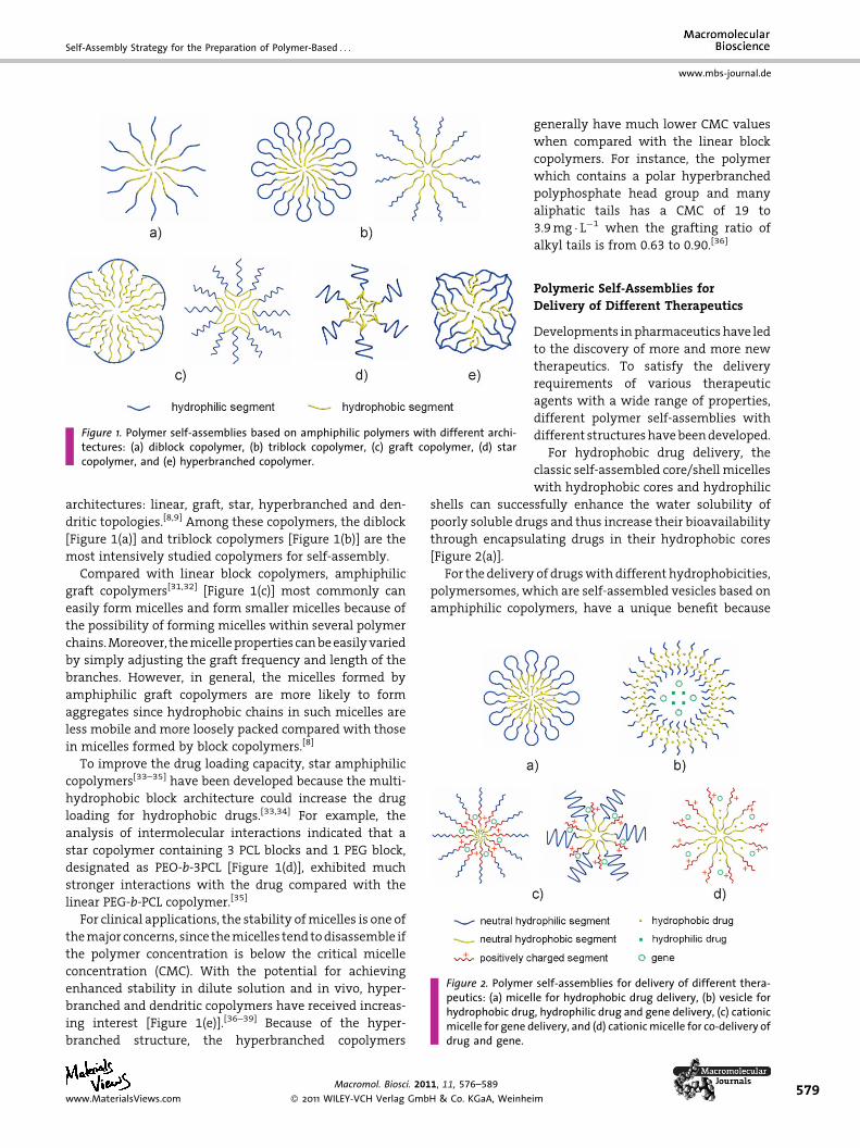
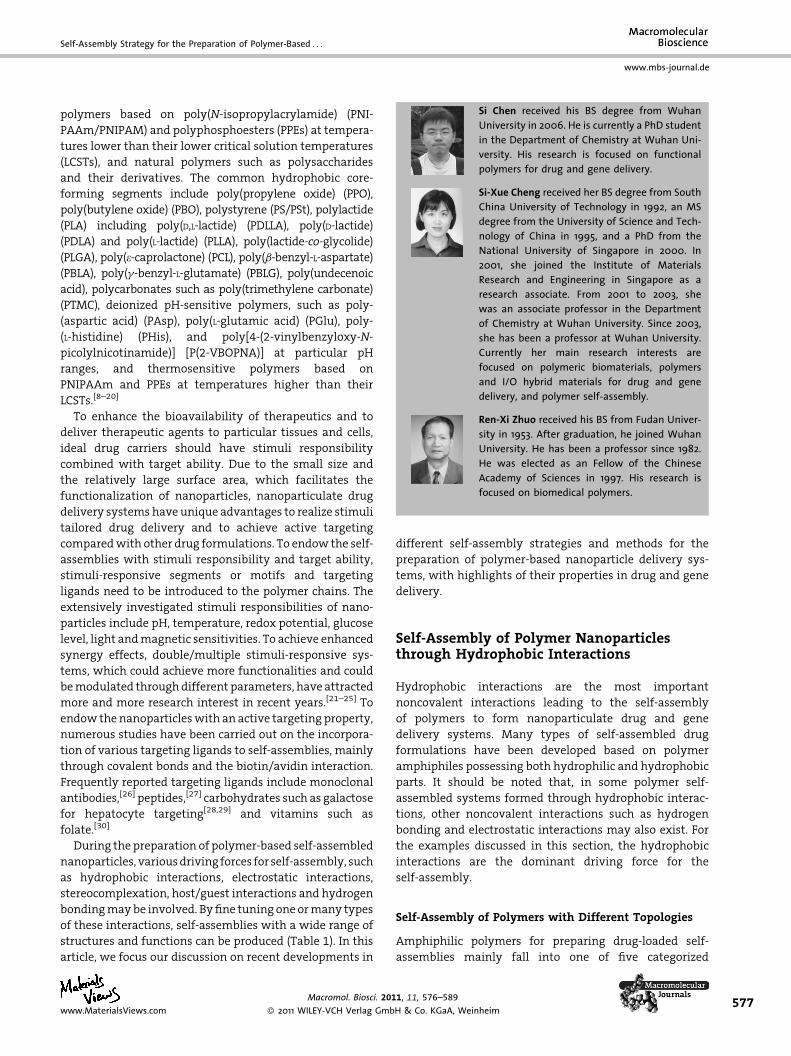
Figure 1. Polymer self-assemblies based on amphiphilic polymers with different archi-tectures: (a) diblock copolymer, (b) triblock copolymer, (c) graft copolymer, (d) starcopolymer, and (e) hyperbranched copolymer.
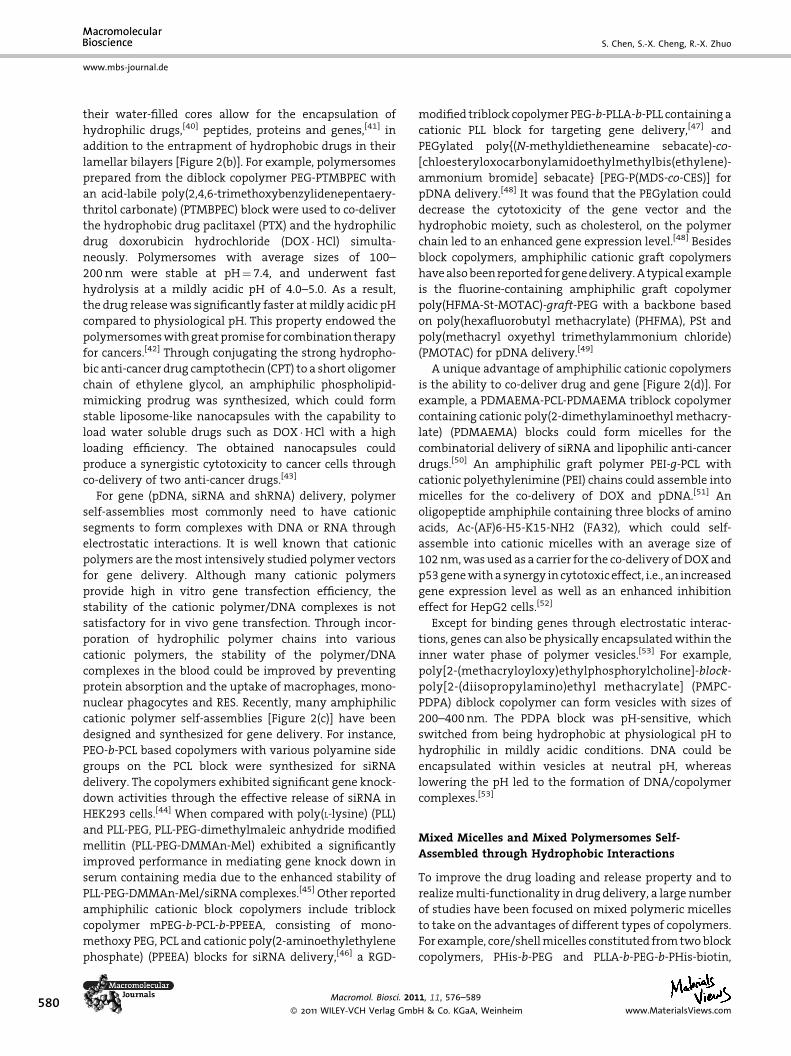
Figure 2. Polymer self-assemblies for delivery of different thera-peutics: (a) micelle for hydrophobic drug delivery, (b) vesicle forhydrophobic drug, hydrophilic drug and gene delivery, (c) cationicmicelle for gene delivery, and (d) cationicmicelle for co-delivery ofdrug and gene.
Self-Assembly Strategy for the Preparation of Polymer-Based . . .
www.mbs-journal.de
architectures: linear, graft, star, hyperbranched and den-
dritic topologies.[8,9] Among these copolymers, the diblock
[Figure 1(a)] and triblock copolymers [Figure 1(b)] are the
most intensively studied copolymers for self-assembly.
Compared with linear block copolymers, amphiphilic
graft copolymers[31,32] [Figure 1(c)] most commonly can
easily form micelles and form smaller micelles because of
the possibility of forming micelles within several polymer
chains.Moreover, themicelleproperties canbeeasilyvaried
by simply adjusting the graft frequency and length of the
branches. However, in general, the micelles formed by
amphiphilic graft copolymers are more likely to form
aggregates since hydrophobic chains in such micelles are
less mobile and more loosely packed compared with those
in micelles formed by block copolymers.[8]
To improve the drug loading capacity, star amphiphilic
copolymers[33–35] have been developed because the multi-
hydrophobic block architecture could increase the drug
loading for hydrophobic drugs.[33,34] For example, the
analysis of intermolecular interactions indicated that a
star copolymer containing 3 PCL blocks and 1 PEG block,
designated as PEO-b-3PCL [Figure 1(d)], exhibited much
stronger interactions with the drug compared with the
linear PEG-b-PCL copolymer.[35]
For clinical applications, the stability ofmicelles is one of
themajor concerns, since themicelles tend todisassemble if
the polymer concentration is below the critical micelle
concentration (CMC). With the potential for achieving
enhanced stability in dilute solution and in vivo, hyper-
branched and dendritic copolymers have received increas-
ing interest [Figure 1(e)].[36–39] Because of the hyper-
branched structure, the hyperbranched copolymers
www.MaterialsViews.com
Macromol. Biosci. 2011, 11, 576–589
� 2011 WILEY-VCH Verlag GmbH & Co. KGaA, Weinhe
generally have much lower CMC values
when compared with the linear block
copolymers. For instance, the polymer
which contains a polar hyperbranched
polyphosphate head group and many
aliphatic tails has a CMC of 19 to
3.9mg � L�1 when the grafting ratio of
alkyl tails is from 0.63 to 0.90.[36]
Polymeric Self-Assemblies forDelivery of Different Therapeutics
Developments inpharmaceuticshave led
to the discovery of more and more new
therapeutics. To satisfy the delivery
requirements of various therapeutic
agents with a wide range of properties,
different polymer self-assemblies with
different structureshavebeendeveloped.
For hydrophobic drug delivery, the
classic self-assembled core/shell micelles
with hydrophobic cores and hydrophilic
shells can successfully enhance the water solubility of
poorly soluble drugs and thus increase their bioavailability
through encapsulating drugs in their hydrophobic cores
[Figure 2(a)].
For the delivery of drugswith different hydrophobicities,
polymersomes, which are self-assembled vesicles based on
amphiphilic copolymers, have a unique benefit because
im579
580
www.mbs-journal.de
S. Chen, S.-X. Cheng, R.-X. Zhuo
their water-filled cores allow for the encapsulation of
hydrophilic drugs,[40] peptides, proteins and genes,[41] in
addition to the entrapment of hydrophobic drugs in their
lamellar bilayers [Figure 2(b)]. For example, polymersomes
prepared from the diblock copolymer PEG-PTMBPEC with
an acid-labile poly(2,4,6-trimethoxybenzylidenepentaery-
thritol carbonate) (PTMBPEC) block were used to co-deliver
the hydrophobic drug paclitaxel (PTX) and the hydrophilic
drug doxorubicin hydrochloride (DOX �HCl) simulta-
neously. Polymersomes with average sizes of 100–
200nm were stable at pH¼ 7.4, and underwent fast
hydrolysis at a mildly acidic pH of 4.0–5.0. As a result,
the drug releasewas significantly faster atmildly acidic pH
compared to physiological pH. This property endowed the
polymersomeswithgreatpromise for combination therapy
for cancers.[42] Through conjugating the strong hydropho-
bic anti-cancer drug camptothecin (CPT) to a short oligomer
chain of ethylene glycol, an amphiphilic phospholipid-
mimicking prodrug was synthesized, which could form
stable liposome-like nanocapsules with the capability to
load water soluble drugs such as DOX �HCl with a high
loading efficiency. The obtained nanocapsules could
produce a synergistic cytotoxicity to cancer cells through
co-delivery of two anti-cancer drugs.[43]
For gene (pDNA, siRNA and shRNA) delivery, polymer
self-assemblies most commonly need to have cationic
segments to form complexes with DNA or RNA through
electrostatic interactions. It is well known that cationic
polymers are themost intensively studied polymer vectors
for gene delivery. Although many cationic polymers
provide high in vitro gene transfection efficiency, the
stability of the cationic polymer/DNA complexes is not
satisfactory for in vivo gene transfection. Through incor-
poration of hydrophilic polymer chains into various
cationic polymers, the stability of the polymer/DNA
complexes in the blood could be improved by preventing
protein absorption and the uptake of macrophages, mono-
nuclear phagocytes and RES. Recently, many amphiphilic
cationic polymer self-assemblies [Figure 2(c)] have been
designed and synthesized for gene delivery. For instance,
PEO-b-PCL based copolymers with various polyamine side
groups on the PCL block were synthesized for siRNA
delivery. The copolymers exhibited significant gene knock-
down activities through the effective release of siRNA in
HEK293 cells.[44] When compared with poly(L-lysine) (PLL)
and PLL-PEG, PLL-PEG-dimethylmaleic anhydride modified
mellitin (PLL-PEG-DMMAn-Mel) exhibited a significantly
improved performance in mediating gene knock down in
serum containing media due to the enhanced stability of
PLL-PEG-DMMAn-Mel/siRNA complexes.[45] Other reported
amphiphilic cationic block copolymers include triblock
copolymer mPEG-b-PCL-b-PPEEA, consisting of mono-
methoxy PEG, PCL and cationic poly(2-aminoethylethylene
phosphate) (PPEEA) blocks for siRNA delivery,[46] a RGD-
Macromol. Biosci. 201
� 2011 WILEY-VCH Verlag Gmb
modified triblock copolymer PEG-b-PLLA-b-PLL containing a
cationic PLL block for targeting gene delivery,[47] and
PEGylated poly{(N-methyldietheneamine sebacate)-co-
[chloesteryloxocarbonylamidoethylmethylbis(ethylene)-
ammonium bromide] sebacate} [PEG-P(MDS-co-CES)] for
pDNA delivery.[48] It was found that the PEGylation could
decrease the cytotoxicity of the gene vector and the
hydrophobic moiety, such as cholesterol, on the polymer
chain led to an enhanced gene expression level.[48] Besides
block copolymers, amphiphilic cationic graft copolymers
havealsobeenreported forgenedelivery.A typical example
is the fluorine-containing amphiphilic graft copolymer
poly(HFMA-St-MOTAC)-graft-PEG with a backbone based
on poly(hexafluorobutyl methacrylate) (PHFMA), PSt and
poly(methacryl oxyethyl trimethylammonium chloride)
(PMOTAC) for pDNA delivery.[49]
A unique advantage of amphiphilic cationic copolymers
is the ability to co-deliver drug and gene [Figure 2(d)]. For
example, a PDMAEMA-PCL-PDMAEMA triblock copolymer
containing cationic poly(2-dimethylaminoethyl methacry-
late) (PDMAEMA) blocks could form micelles for the
combinatorial delivery of siRNA and lipophilic anti-cancer
drugs.[50] An amphiphilic graft polymer PEI-g-PCL with
cationic polyethylenimine (PEI) chains could assemble into
micelles for the co-delivery of DOX and pDNA.[51] An
oligopeptide amphiphile containing three blocks of amino
acids, Ac-(AF)6-H5-K15-NH2 (FA32), which could self-
assemble into cationic micelles with an average size of
102nm,wasused as a carrier for the co-delivery ofDOXand
p53genewithasynergy in cytotoxic effect, i.e., an increased
gene expression level as well as an enhanced inhibition
effect for HepG2 cells.[52]
Except for binding genes through electrostatic interac-
tions, genes can also be physically encapsulatedwithin the
inner water phase of polymer vesicles.[53] For example,
poly[2-(methacryloyloxy)ethylphosphorylcholine]-block-
poly[2-(diisopropylamino)ethyl methacrylate] (PMPC-
PDPA) diblock copolymer can form vesicles with sizes of
200–400nm. The PDPA block was pH-sensitive, which
switched from being hydrophobic at physiological pH to
hydrophilic in mildly acidic conditions. DNA could be
encapsulated within vesicles at neutral pH, whereas
lowering the pH led to the formation of DNA/copolymer
complexes.[53]
Mixed Micelles and Mixed Polymersomes Self-Assembled through Hydrophobic Interactions
To improve the drug loading and release property and to
realizemulti-functionality in drug delivery, a large number
of studies have been focused on mixed polymeric micelles
to take on the advantages of different types of copolymers.
For example, core/shellmicelles constituted fromtwoblock
copolymers, PHis-b-PEG and PLLA-b-PEG-b-PHis-biotin,
1, 11, 576–589
H & Co. KGaA, Weinheim www.MaterialsViews.com
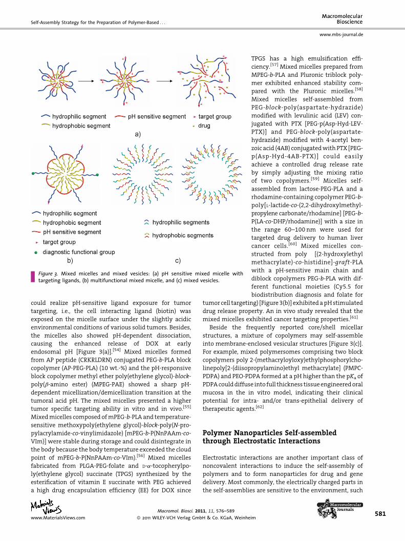
Figure 3. Mixed micelles and mixed vesicles: (a) pH sensitive mixed micelle withtargeting ligands, (b) multifunctional mixed micelle, and (c) mixed vesicles.
Self-Assembly Strategy for the Preparation of Polymer-Based . . .
www.mbs-journal.de
could realize pH-sensitive ligand exposure for tumor
targeting, i.e., the cell interacting ligand (biotin) was
exposed on the micelle surface under the slightly acidic
environmental conditions of various solid tumors. Besides,
the micelles also showed pH-dependent dissociation,
causing the enhanced release of DOX at early
endosomal pH [Figure 3(a)].[54] Mixed micelles formed
from AP peptide (CRKRLDRN) conjugated PEG-b-PLA block
copolymer (AP-PEG-PLA) (10 wt.-%) and the pH-responsive
block copolymer methyl ether poly(ethylene glycol)-block-
poly(b-amino ester) (MPEG-PAE) showed a sharp pH-
dependent micellization/demicellization transition at the
tumoral acid pH. The mixed micelles presented a higher
tumor specific targeting ability in vitro and in vivo.[55]
Mixedmicelles composed ofmPEG-b-PLAand temperature-
sensitive methoxypoly(ethylene glycol)-block-poly(N-pro-
pylacrylamide-co-vinylimidazole) [mPEG-b-P(NnPAAm-co-
VIm)] were stable during storage and could disintegrate in
the body because the body temperature exceeded the cloud
point of mPEG-b-P(NnPAAm-co-VIm).[56] Mixed micelles
fabricated from PLGA-PEG-folate and D-a-tocopherylpo-
ly(ethylene glycol) succinate (TPGS) synthesized by the
esterification of vitamin E succinate with PEG achieved
a high drug encapsulation efficiency (EE) for DOX since
www.MaterialsViews.com
Macromol. Biosci. 2011, 11, 576–589
� 2011 WILEY-VCH Verlag GmbH & Co. KGaA, Weinhe
TPGS has a high emulsification effi-
ciency.[57] Mixed micelles prepared from
MPEG-b-PLA and Pluronic triblock poly-
mer exhibited enhanced stability com-
pared with the Pluronic micelles.[58]
Mixed micelles self-assembled from
PEG-block-poly(aspartate-hydrazide)
modified with levulinic acid (LEV) con-
jugated with PTX [PEG-p(Asp-Hyd-LEV-
PTX)] and PEG-block-poly(aspartate-
hydrazide) modified with 4-acetyl ben-
zoic acid (4AB) conjugatedwithPTX [PEG-
p(Asp-Hyd-4AB-PTX)] could easily
achieve a controlled drug release rate
by simply adjusting the mixing ratio
of two copolymers.[59] Micelles self-
assembled from lactose-PEG-PLA and a
rhodamine-containing copolymer PEG-b-
poly[L-lactide-co-(2,2-dihydroxylmethyl-
propylene carbonate/rhodamine] [PEG-b-
P(LA-co-DHP/rhodamine)] with a size in
the range 60–100 nm were used for
targeted drug delivery to human liver
cancer cells.[60] Mixed micelles con-
structed from poly [(2-hydroxylethyl
methacrylate)-co-histidine]-graft-PLA
with a pH-sensitive main chain and
diblock copolymers PEG-b-PLA with dif-
ferent functional moieties (Cy5.5 for
biodistribution diagnosis and folate for
tumor cell targeting) [Figure3(b)] exhibitedapHstimulated
drug release property. An in vivo study revealed that the
mixed micelles exhibited cancer targeting properties.[61]
Beside the frequently reported core/shell micellar
structures, a mixture of copolymers may self-assemble
into membrane-enclosed vesicular structures [Figure 3(c)].
For example, mixed polymersomes comprising two block
copolymers poly 2-(methacryloyloxy)ethylphosphorylcho-
linepoly[2-(diisopropylamino)ethyl methacrylate] (PMPC-
PDPA) and PEO-PDPA formed at a pH higher than the pKa of
PDPAcoulddiffuse into full thickness tissueengineeredoral
mucosa in the in vitro model, indicating their clinical
potential for intra- and/or trans-epithelial delivery of
therapeutic agents.[62]
Polymer Nanoparticles Self-assembledthrough Electrostatic Interactions
Electrostatic interactions are another important class of
noncovalent interactions to induce the self-assembly of
polymers and to form nanoparticles for drug and gene
delivery. Most commonly, the electrically charged parts in
the self-assemblies are sensitive to the environment, such
im581
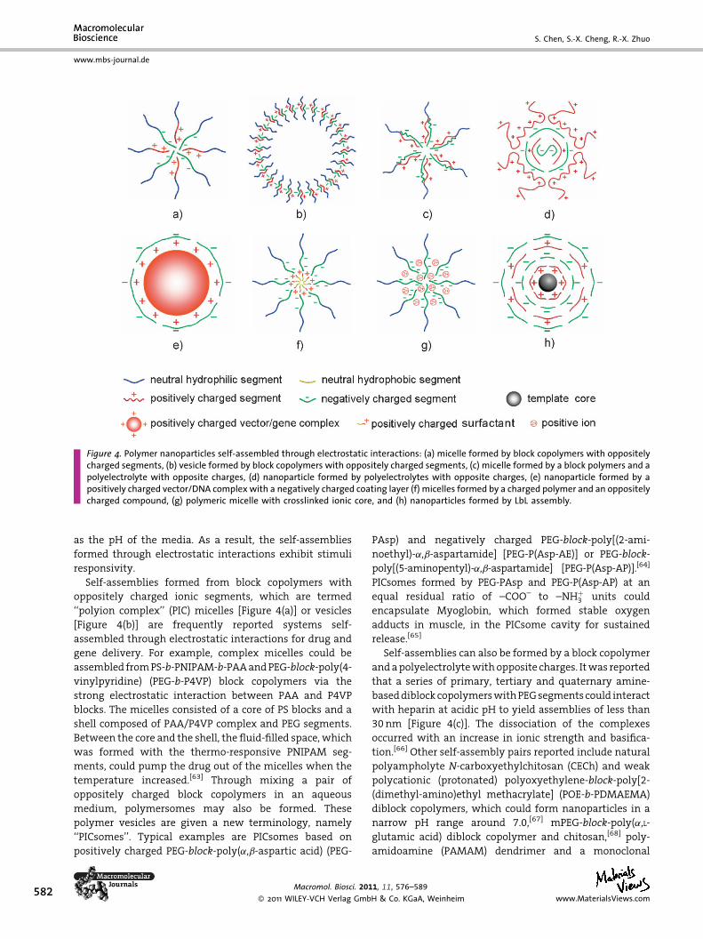
Figure 4. Polymer nanoparticles self-assembled through electrostatic interactions: (a) micelle formed by block copolymers with oppositelycharged segments, (b) vesicle formed by block copolymers with oppositely charged segments, (c) micelle formed by a block polymers and apolyelectrolyte with opposite charges, (d) nanoparticle formed by polyelectrolytes with opposite charges, (e) nanoparticle formed by apositively charged vector/DNA complex with a negatively charged coating layer (f) micelles formed by a charged polymer and an oppositelycharged compound, (g) polymeric micelle with crosslinked ionic core, and (h) nanoparticles formed by LbL assembly.
582
www.mbs-journal.de
S. Chen, S.-X. Cheng, R.-X. Zhuo
as the pH of the media. As a result, the self-assemblies
formed through electrostatic interactions exhibit stimuli
responsivity.
Self-assemblies formed from block copolymers with
oppositely charged ionic segments, which are termed
‘‘polyion complex’’ (PIC) micelles [Figure 4(a)] or vesicles
[Figure 4(b)] are frequently reported systems self-
assembled through electrostatic interactions for drug and
gene delivery. For example, complex micelles could be
assembled fromPS-b-PNIPAM-b-PAAandPEG-block-poly(4-
vinylpyridine) (PEG-b-P4VP) block copolymers via the
strong electrostatic interaction between PAA and P4VP
blocks. The micelles consisted of a core of PS blocks and a
shell composed of PAA/P4VP complex and PEG segments.
Between the core and the shell, the fluid-filled space, which
was formed with the thermo-responsive PNIPAM seg-
ments, could pump the drug out of the micelles when the
temperature increased.[63] Through mixing a pair of
oppositely charged block copolymers in an aqueous
medium, polymersomes may also be formed. These
polymer vesicles are given a new terminology, namely
‘‘PICsomes’’. Typical examples are PICsomes based on
positively charged PEG-block-poly(a,b-aspartic acid) (PEG-
Macromol. Biosci. 201
� 2011 WILEY-VCH Verlag Gmb
PAsp) and negatively charged PEG-block-poly[(2-ami-
noethyl)-a,b-aspartamide] [PEG-P(Asp-AE)] or PEG-block-
poly[(5-aminopentyl)-a,b-aspartamide] [PEG-P(Asp-AP)].[64]
PICsomes formed by PEG-PAsp and PEG-P(Asp-AP) at an
equal residual ratio of –COO– to –NHþ3 units could
encapsulate Myoglobin, which formed stable oxygen
adducts in muscle, in the PICsome cavity for sustained
release.[65]
Self-assemblies can also be formed by a block copolymer
andapolyelectrolytewithopposite charges. Itwas reported
that a series of primary, tertiary and quaternary amine-
baseddiblockcopolymerswithPEGsegmentscould interact
with heparin at acidic pH to yield assemblies of less than
30nm [Figure 4(c)]. The dissociation of the complexes
occurred with an increase in ionic strength and basifica-
tion.[66] Other self-assembly pairs reported include natural
polyampholyte N-carboxyethylchitosan (CECh) and weak
polycationic (protonated) polyoxyethylene-block-poly[2-
(dimethyl-amino)ethyl methacrylate] (POE-b-PDMAEMA)
diblock copolymers, which could form nanoparticles in a
narrow pH range around 7.0,[67] mPEG-block-poly(a,L-
glutamic acid) diblock copolymer and chitosan,[68] poly-
amidoamine (PAMAM) dendrimer and a monoclonal
1, 11, 576–589
H & Co. KGaA, Weinheim www.MaterialsViews.com

Self-Assembly Strategy for the Preparation of Polymer-Based . . .
www.mbs-journal.de
antibody fragment incorporated PEG-block-poly[(propyl
methacrylate)-co-(methacrylic acid)] [PEG-b-P(PrMA-co-
MAA)] for siRNA delivery.[69]
PICs are sensitive to the environment, such as ionic
strength or pH. Although this property is useful for stimuli-
responsive delivery, the instability is unfavorable for some
particular applications. By introducing hydrophobic inter-
actions, PICs could be stabilized. A representative work
involved using anionic amphiphilic copolymer g-PGA-g-L-
Phe(g-PGA-Phe), which was obtained by the modification
of poly(g-glutamic acid) by hydrophobic L-phenylalanine
(L-Phe), as a component to form PICs with cationic
poly(e-lysine). The resultant PICs exhibited significantly
improved stability under physiological conditions.[70]
Two polyelectrolytes with opposite charges can form
self-assembled nanoparticles [Figure 4(d)]. For instance,
the complexation of dextran sulfate or alginate with
chitosan resulted in nanoparticles with a diameter
ranging from 423–850nm, which could be used for
insulin delivery.[71]
Introducing oppositely charged polymers to polycation
gene vectors through electrostatic interactions is an
important strategy to reduce the toxicity and to improve
the in vivo stability of gene delivery systems. For example,
negatively chargedpeptides containingRGD ligands coated
onto positively charged poly(b-amino ester)/DNA com-
plexes could reduce potential toxicity and facilitate cell and
tissue-specific gene delivery [Figure 4(e)].[72,73] In siRNA
delivery, using the complexes formed by electrostatic
complexation of negatively charged hyaluronic acid (HA)
and cationic poly(L-arginine) (PLR) to condense siRNA could
significantly reduce the cytotoxicity.[74] In addition to
improved biocompatibility, electrostatic complexation can
also be utilized to introduce target ligands to gene delivery
systems. For instance, heparin-biotin couldbe coatedon the
surface of polyamidoamine dendrimer/DNA complexes via
electrostatic interactions to form PAMAM/DNA/heparin-
biotin terplexes, which exhibited much higher cellular
uptake into HeLa cells due to the specific interactions
between biotin and biotin receptors on the HeLa cells.[75]
In addition to a pair of oppositely charged polymers,
nanoparticles can also be formed by a charged polymer and
an oppositely charged compound. For instance, the cationic
block copolymer PEG-poly{N-[N’-(2-aminoethyl)-2-ami-
noethyl]aspartamide} [PEG-pAsp(DET)] could form stable
micelles with anionic modified proteins. After the PIC
micellesenteredcells, themodifiedproteinswereconverted
to the original proteins.[76] Nanosized complex micelles
could be spontaneously produced from ionic interactions
between folate incorporated PEG-conjugated oligonucleo-
tide and a cationic lipid, lipofectamine,which could beused
for target gene delivery.[77] The block copolymer PEO-block-
poly(tert-butyl methacrylate) (PEO-b-PMA) and an oppo-
sitely charged surfactant (hexadecyltrimethylammonium
www.MaterialsViews.com
Macromol. Biosci. 201
� 2011 WILEY-VCH Verlag Gmb
bromide) formed nanoparticles with sizes in the 60–90nm
range [Figure 4(f)]. Decreasing the ionization degree of the
PMA block upon decreasing the pH caused elevation of the
particle size at pH< 5.5, followed by formation of large
aggregates at pH< 4.[78] Two types of negatively charged
polymers (one with an S–S bond) and a positively charged
surfactant could formmicelle-typeassemblies,whichcould
bedisassembled throughtailoringtheredoxcharacteristics,
ionic strength and pH of the medium.[79] Due to the
electrostatic interaction between the polymers with
negatively charged segments and cationic ions, polymeric
micelles with crosslinked ionic cores could be formed. For
example, PEO-block-poly(methacrylic acid) and Ca2þ could
formmicelles displaying pH- and ionic strength-responsive
properties [Figure 4(g)].[80,81]
The layer-by-layer (LbL) assembly technique based on
electrostatic interactions is commonly used to fabricate
films and microsized particles, yet LbL assembly can also
been utilized to prepared nanosized particles. For LbL
assembly, inorganic substrates, such as CaCO3, MnCO3 and
CdCO3, which can be conveniently removed by dissolution
with an ethylenediamine tetraacetate (EDTA) solution, are
now used more frequently.[82] For the purpose of drug
delivery, drug nanoparticles can be used as the cores for LbL
assembly [Figure 4(h)]. As reported, nanoparticles could be
formed by the LbL assembly of poly(allylamine hydro-
chloride) and poly(styrene sulfonate) (PSS) on the surface of
drug nanoparticles (dexamethasone, a steroid used in
conjugation with chemotherapy) followed by further
modification through covalently attaching PEG to the outer
surface of the nanoparticles.[83] Through ultrasonic treat-
ment of microsized poorly soluble drug particles (the anti-
cancer drugs tamoxifen and PTX), the size of drug particles
could be decreased to the nano level (between 100 and
200nm). Through LbL coating of the polycation poly(di-
methyldiallylamideammonium chloride) (PDDA) and
negatively charged PSS, a stable coated nanocolloidal drug
dispersion was formed with a high drug loading content.
The drug release rate from such nanocolloidal particles
could be easily controlled by adjusting the density and
thickness of the assembling multilayers. After further
introducingmonoclonalnucleosome-specific2C5antibody,
the nanoparticles showed promising application for cancer
targetingdrugdelivery.[84] The LbLassembly techniquewas
also applied for the surface modification of biodegradable
PLGA nanoparticles, using PAA and PEI as building blocks.
After five layers of PAA and PEI were deposited, the
multilayers were crosslinked with amino terminated PEG
or amino terminated folate decorated PEG (PEG-FA). These
nanoparticles couldpotentiallybeusedas specific targeting
carriers for anti-cancer drugs.[85] Other reported cores for
LbL assembly include sulfate modified fluorescent poly-
styrene nanobeads with a diameter of 200nm for LbL
deposition of PLL, chitosan and heparin sulfate,[86] and gold
1, 11, 576–589
H & Co. KGaA, Weinheim583

Figure 5. Polymer nanoparticles self-assembled through stereo-complexation: (a) diblock stereo copolymer, (b) triblock stereocopolymer, (d) Y-shaped stereo copolymer, and (d) graft stereocopolymer.
584
www.mbs-journal.de
S. Chen, S.-X. Cheng, R.-X. Zhuo
nanoparticles with a size of �10nm for LbL coating with
poly(allylamine) (PAH) and PSS.[87]
Polymer Nanoparticles Self-Assembledthrough Stereocomplexation
In comparison to low-molecular-weight surfactant
micelles, polymeric micelles feature unique attributes,
including higher thermodynamic and kinetic stabilities.
However, for in vivo applications, the stability of polymeric
micelles still remains a major concern. It is reported that
micelles prepared from PEG-b-PDLLA dissociated after
intravenous injection and were rapidly excreted in urine.
Chemical crosslinking of either core or shell segments is the
common strategy to retain the integrity of polymer
micelles.However, the crosslinkingmayunfavorably affect
the bioactivity of encapsulated drugs and the biodegrad-
ability of the system. Stabilizing the core through stereo-
complex formation is an alternative strategy, which could
overcome the disadvantages caused by the chemical
crosslinking.[88]
The most commonly studied polymers for stereocom-
plexation are PDLA and PLLA.[89] Other polymers that could
form stereocomplexes, investigated for biomedical pur-
poses, include enantiopure homopolymers of N-acryloyl-
D,L-leucine methyl ester.[90]
In previous studies, the PDLA/PLLA segment contained
copolymerswith various architectureswere synthesized to
form stereocomplex self-assemblies. The investigated
stereocomplex pairs include block copolymers PEG-b-
PDLA/PEG-b-PLLA [Figure 5(a)],[88,91,92] PDLA-b-PEG-b-
PDLA/PLLA-b-PEG-b-PLLA [Figure 5(b)],[93] Y-shaped copo-
lymers [Figure 5(c)] PEG-PDLA-PDLA/PEG-PLLA-PLLA, PEG-
PLLA-PDLA/PEG-PDLA-PLLA[94] and the graft copolymers
dextran-g-PLLA/dextran-g-PDLA [Figure 5(d)].[95]
Due to the stereocomplexation, stereocomplex self-
assemblies exhibit strong thermodynamic stability as well
as kinetic stability, and stereocomplex micelles are less
prone to aggregation during the lyophilization pro-
cess.[88,95] In addition, both drug loading level and
encapsulation efficiency could be improved through
stereocomplexation,[92] and the drug release is slower from
stereocomplex micelles than it is from corresponding
conventional micelles without stereocomplexation.[92,94]
Besides, the polydispersity of stereocomplex micelles is
lower compared with conventional micelles.[94]
Polymer Nanoparticulate Self-Assemblieswith Host/Guest Interactions
Host/guest interactions have always been considered to
be an important class of noncovalent interactions for
inducing self-assembly. For biomedical applications, the
Macromol. Biosci. 201
� 2011 WILEY-VCH Verlag Gmb
most commonly investigated host/guest interactions are
based on the cyclodextrins (CDs). CDs are a series of cyclic
oligosaccharides with a hydrophobic cone-shaped central
cavity, which can act as hosts for many macromolecular
guests to form polyrotaxanes (PRs) by threading CD
molecules onto polymer chains with the inclusion driven
by the geometric compatibility and hydrophobic interac-
tions between the CDs and the polymer segments.[96] The
interactions in PRs can be cleaved by temperature changes
and pH changes to induce ionization of the polymer
segments and shearing. As a result, CDmolecules canmove
along the polymer chain and even de-thread, resulting in
thedisassociationof thePRaggregates. Thisunique stimuli-
response character is favorable in controlled drug release.
Generally, to form PR-based self-assemblies with host/
guest interactions, other noncovalent interactions, such
as hydrogen bonding among CDs and hydrophobic inter-
1, 11, 576–589
H & Co. KGaA, Weinheim www.MaterialsViews.com

Figure 6. Polymer nanoparticles self-assembled through host/guest interactions:(a) micelle formed by PEO-b-PAA with PEO segment threaded in CDs, (b) micellesformed by a polymer with CDs in side groups through hosting hydrophobic smallmolecules and a polymer with hydrophobic groups, (c) nanoparticles formed by a graftcopolymer with hydrophobic side chains and a cyclodextrin polymer, and (d) nanopar-ticle formed through CD/Ad recognition.
Self-Assembly Strategy for the Preparation of Polymer-Based . . .
www.mbs-journal.de
actions, are still involved in the self-assembly. For example,
PEG-b-PLL with PEG segments threaded in a-CDs could
form pH switchable nanoparticles. At pH> 10, PLL seg-
ments became less soluble and aggregated in aqueous
solution, leading to gel formation.[97] PEO-b-PAA with PEO
threaded in a-CDs could self-organize to formmicelleswith
a spherical shape and a size of 100nm in alkaline media
[Figure 6(a)]. Decreasing the pH led to the deionization of
free PAA segments and thus made assemblies fuse to form
microspheres.[98]
In addition to forming assemblies through threading CD
molecules onto polymer chains, CD-based self-assemblies
can be formed in other patterns. For instance, diblock
copolymer PEG-b-PCDwith a PEG block and a block bearing
b-CD side groups could be dissolved in water, and could
assemble into core/shell nanocarriers if the b-CDs in the
side groups served as the hosts and formed inclusion
complexeswithvariouscompounds includinghydrophobic
small molecules such as pyrene and particular polymers
www.MaterialsViews.com
Macromol. Biosci. 2011, 11, 576–589
� 2011 WILEY-VCH Verlag GmbH & Co. KGaA, Weinhe
such as poly(b-benzyl-L-aspartate) (PBLA)
[Figure 6(b)]. The assemblies prepared by
this procedure exhibited chemical sensi-
tivity, which was of importance for
responsive delivery and bio- or chemical
sensing.[99] The association of two hydro-
soluble polymers, dextranbearinghydro-
phobic lauryl side chains and a cyclodex-
trin polymer (b-CD crosslinked with
epichlorohydrin), in aqueous media led
to the formation of spherical-shaped
nanogels of 100–200nm due to the
inclusion of lauryl chains in b-CD moi-
eties [Figure 6(c)]. The remaining
unthreaded b-CD cavities were available
for the inclusion of hydrophobic mole-
cules. [100] Using three building blocks,
adamantine-grafted polyamidoamine
dendrimer (n-Ad-PAMAM), b-CD-grafted
branched polyethylenimine (CD-PEI) and
Ad-functionalized PEG (Ad-PEG), supra-
molecular nanoparticles through CD/Ad
recognition could be prepared
[Figure 6(d)]. By tuning the mixing ratios
of the three building blocks, the sizes of
the nanoparticles could be controlled
from 30–150nm.[101]
To prepare self-assemblies based on
CDs, a great challenge that exists is the
insolubility of CD-based self-assemblies
in most solvents because of the strong
intermolecular hydrogen bonds between
CDs, leading to the unfavorable precipi-
tation of CD-contained amphiphiles.
Using modified CD derivates, such as
maleic anhydride modified a-CD, could effectively over-
come this problem through weakening the intermolecular
hydrogen bonding.[102]
Polymer Nanoparticulate Self-Assemblieswith Hydrogen Bonding
Hydrogen bonding interactions exist in many self-
assembled systems, with association of other noncovalent
interactions such as hydrophobic interactions and electro-
static forces. Yet, in some self-assembly systems, the
hydrogen bonding interactions are of critical importance
in the self-assembly process. In this section, only systems
with hydrogen bonding that plays a critical role in self-
assembly and the properties of the self-assemblies are
discussed.
It has been reported that, in addition to hydrophobic
interactions, hydrogen bonding between PVPhol segments
im585

Figure 7. Polymer nanoparticle self-assemblies with hydrogenbonding: (a) micelle formed by two block copolymers with hydro-gen bonded PVP and PVPhol segments as a core (yellow area), (b)micelle formed by two block copolymers with hydrogen bondedP4VP and PAA segments as amiddle layer (yellow area), (c) micelleformed by a star terpolymer with hydrogen bonded PEG andPMMA segments as a core (yellow area), and (d) vesicle withhydrogen bonding between stilbazole group and gallic acidgroup.
586
www.mbs-journal.de
S. Chen, S.-X. Cheng, R.-X. Zhuo
and PVP segments played an important role in the
formation of mixed micelles composed of two copolymers,
tris(dibenzoylmethanato)europium(III)-coordinated
poly{N-isopropylacrylamide-co-4-(1-ethyl-1H-imidazo[4,5-
f][1,10]phenanthrolin-2-yl)phenyl methacrylate}-block-
polyvinylphenol [P(NIPAAm-co-EIPPMMA)-b-PVPhol] and
FITC-conjugated poly[N-isopropylacrylamide-co-(hydroxy-
lethyl methacrylate]-block-polyvinylpyridine [P(NIPAAm-
co-HEMA)-b-PVP] [Figure 7(a)]. The fluorescence properties
of the micelles could be controlled by temperature and pH
changes due to the phase transition of temperature
sensitive PNIPAAm contained segments and the deproto-
nation/protonation of PVP segments. An in vitro study
showed that the micelles incorporated by A549 cells also
exhibited temperature and pH-sensitive fluorescence.[103]
Among hydrogen-bonded self-assemblies, an important
class is the stimuli-responsive micelles, where their pH
sensitive segments deionize at particular pH ranges and
thus the hydrogen bonding becomes the dominant driving
force to induce the self-assembly. A good example is multi-
Macromol. Biosci. 201
� 2011 WILEY-VCH Verlag Gmb
layered micelles prepared using three copolymers, PS-b-
PAA, PNIPAM-b-P4VP and PEG-b-P4VP. The micelles were
composed of a core of PS segments, a middle layer
constructed through the electrostatic interactions and
hydrogen bonding between the PAA block and the P4VP
block, and a corona consisting of PNIPAM and PEG chains
[Figure 7(b)]. At temperatures higher than the LCST, the
PNIPAMchains collapsedonto thePAA/P4VP complex layer
while the PEG chains still stretched into the solution
through the collapsed PNIPAM layer,which could act as the
hydrophilic channels for drug release. The drug release rate
could be controlled by changing the ratio of PEG chains to
PNIPAM chains in the corona.[104] Another typical example
is self-assemblies based on the ABC miktoarm star
terpolymer PEG(-b-PMAA)-b-PDEA consisting of PEG, poly-
(methacrylic acid (PMAA)) and poly[2-(diethylamino)ethyl
methacrylate (PDEA)] arms. PEG(-b-PMAA)-b-PDEA could
self-assemble into three types of aggregates at different
pHs, namely, at pH< 4, hydrogen bonding interactions
between fully protonated PMAA and PEG led to the
formation of micellar aggregates stabilized by protonated
PDEA coronas [Figure 7(c)]; at pH¼ 5–7, micelles possessed
polyion complex cores formed by charge compensation
between partially ionized PMAA and partially protonated
PDEA sequences; above pH¼ 8,micelleswith deprotonated
PDEA cores and PEG/ionized PMAA coronas were
formed.[105]
As well as hydrogen bonded micelles, hydrogen bonded
vesicles have also been reported. For example, vesicleswith
a bilayer structure could be formed by Py-EO12 (a PEO chain
with a stilbazole head group) and TCB-COOH (a dendron of
gallic acid with three alkyl chains) through hydrogen
bonding between the stilbazole group and the gallic acid
moiety [Figure 7(d)].[106]
Other Self-assembled Polymer Nanoparticles
In addition to the dominant noncovalent interactions
inducing theself-assemblydiscussedabove, somepolymer-
based self-assembled systems may involve multi/inter-
molecular interactions. For example, in some natural
polymer-based self-assembled systems, the self-assembly
is realized by precisely adjusting the hydrophilic/hydro-
phobic balance of the polymer chains by the addition of
inorganic ions. The natural polymers, such as alginate and
pectin with carboxyl groups, have particular pKa values. In
basic and neutral solutions, the polymer chains exist in the
form of a stretching conformation, due to the repulsion
betweenthedeprotonatedcarboxylgroups, andhaveahigh
hydrophilicity. While in an acidic solution, the polymer
chains tend to aggregate because of the protonation of
carboxyl groups, which leads to the decreased hydrophi-
licity. Precisely controlling the hydrophilic/hydrophobic
1, 11, 576–589
H & Co. KGaA, Weinheim www.MaterialsViews.com
Self-Assembly Strategy for the Preparation of Polymer-Based . . .
www.mbs-journal.de
balance of the polymer chains is critical to inducing self-
assembly of natural polymers. As ionic polysaccharides,
alginateandpectinhavetheability tobinddivalent cations,
and the divalent cations can induce interchain association,
leading to the formation of junction zones. As reported,
through adjusting the concentrations of Ca2þ ions, which
coordinate with alginate[107] and pectin[108] chains, and
carbonic ions (CO2�3 ), which stabilize the systems due to the
electrostatic interaction with Ca2þ ions, the hydrophilic/
hydrophobicbalanceof thenaturalpolymerchainscouldbe
fine tuned, i.e., the polymer chains in theCa2þ-rich domains
became more condensed, whereas the polymer chains in
the Ca2þ deficient domains had the stronger electrostatic
repulsion between the –COO– groups and thus had a higher
affinity with water molecules. The hydrophilicity differ-
ence between the Ca2þ rich domains and the Ca2þ deficient
domains led to the formation of aggregates with different
morphologies includingnanospheresandvesicles.With the
presence of Ca2þ and CO2�3 in the natural polymer-based
nanospheres/vesicles, thepermeabilityof theencapsulated
drug could be reduced, resulting in effectively sustained
drug release. The release rates from nanospheres and
vesicles were strongly affected by the pH value of the
release medium. Another example of inducing the self-
assembly of natural polymers is to use K2S2O8 as amediate
proton donor agent to prepare assemblies of alginate by
partial protonation of carboxyl groups in alginate chains in
water. The decreased number of dissociated carboxylic
groups in alginate chains made alginate lose its hydro-
philicity to some extent, leading to the formation of
alginate assemblies. The size of the alginate assemblies
decreased with decreasing pH because of the increased
hydrophobic segments in the alginate chains.[109]
Summary and Perspectives
Despite the numerous efforts rendered on versatile nano-
sized delivery systems based on polymers with different
chemical structures and properties for the delivery of
different therapeutics (chemotherapeutic agents, proteins,
DNAs and RNAs), in vivo experimental data to comprehen-
sively understand the influence of polymer structures and
long time polymer degradation on the fate of drugs is still
very limited. Since toxicological and immunological issues
for new polymers and carriers are of high concern, features
that are often ignored during the research phase, more
extensive work combining expertise in chemistry, phar-
maceutics and biology is urgently required.
In the years to come, it is expected that the emergence of
nanotechnology platforms and progress in pharmaceutics,
biology and polymer science will catalyze the further
development of multifunctional nanoparticulate delivery
systems to meet the ultimate goal for controlled drug
www.MaterialsViews.com
Macromol. Biosci. 201
� 2011 WILEY-VCH Verlag Gmb
release, which is to maximize therapeutic activity while
minimizing the negative side effects of the drug.
Acknowledgements: Financial support from National NaturalScience Foundation of China (21074099) is gratefully acknowl-edged.
Received: October 21, 2010; Published online: December 27, 2010;DOI: 10.1002/mabi.201000427
Keywords: drug delivery systems; micelles; nanoparticles; self-assembly; vesicles
[1] J. Shi, A. R. Votruba, O. C. Farokhzad, R. Langer, Nano Lett.2010, 10, 3223.
[2] K. E. Uhrich, S. M. Cannizzaro, R. S. Langer, K. M. Shakesheff,Chem. Rev. 1999, 99, 3181.
[3] D. F. Williams, Biomaterials 2009, 30, 5897.[4] S. Kim, J. H. Kim, O. Jeon, I. C. Kwon, K. Park, Eur. J. Pharm.
Biopharm. 2009, 71, 420.[5] Y. Kakizawa, K. Kataoka, Adv. Drug Deliv. Rev. 2002, 54, 203.[6] M. Talelli, C. J. F. Rijcken, C. F. van Nostrum, G. Storm, W. E.
Hennink, Adv. Drug Deliv. Rev. 2010, 62, 231.[7] H. Maeda, Bioconjugate Chem. 2010, 21, 797.[8] V. P. Torchilin, J. Controlled Release 2001, 73, 137.[9] L. Y. Qiu, Y. H. Bae, Pharm. Res. 2006, 23, 1.[10] D. Sutton, N. Nasongkla, E. Blanco, J. Gao, Pharm. Res. 2007,
24, 1029.[11] M. C. Branco, J. P. Schneider, Acta Biomater. 2009, 5, 817.[12] A. Blanazs, S. P. Armes, A. J. Ryan,Macromol. Rapid Commun.
2009, 30, 267.[13] N. Wiradharma, Y. Zhang, S. Venkataraman, J. L. Hedrick,
Y. Y. Yang, Nano Today 2009, 4, 302.[14] S. F. M. van Dongen, H. P. M. de Hoog, R. J. R. W. Peters,
M. Nallani, R. J. M. Nolte, J. C. M. van Hest, Chem. Rev. 2009,109, 6212.
[15] H. J. Yoon, W. D. Jang, J. Mater. Chem. 2010, 20, 211.[16] J. Jagur-Grodzinski, Polym. Adv. Technol. 2009, 20, 595.[17] A. D. Baldwin, K. L. Kiick, Pept. Sci. 2010, 94, 128.[18] Y. C. Wang, Y. Y. Yuan, J. Z. Du, X. Z. Yang, J. Wang,Macromol.
Biosci. 2009, 9, 1154.[19] Y. Zhang, R. X. Zhuo, Biomaterials 2005, 26, 2089.[20] K. M. Huh, H. S. Min, S. C. Lee, H. J. Lee, S. Kim, K. Park,
J. Controlled Release 2008, 126, 122.[21] F. Meng, Z. Zhong, J. Feijen, Biomacromolecules 2009, 10, 198.[22] V. Bulmus, M. Woodward, L. Lin, N. Murthy, P. Stayton,
A. Hoffman, J. Controlled Release 2003, 93, 105.[23] Y. Y. Li, H. Q. Dong, K. Wang, D. L. Shi, X. Z. Zhang, R. X. Zhuo,
Sci. China 2010, 53, 447.[24] H. Wei, S. X. Cheng, X. Z. Zhang, R. X. Zhuo, Prog. Polym. Sci.
2009, 34, 893.[25] A. Napoli, M. J. Boerakker, N. Tirelli, R. J. M. Nolte, N. A. J. M.
Sommerdijk, J. A. Hubbell, Langmuir 2004, 20, 3487.[26] Z. Pang, W. Lu, H. Gao, K. Hu, J. Chen, C. Zhang, X. Gao,
X. Jiang, C. Zhu, J. Controlled Release 2008, 128, 120.
1, 11, 576–589
H & Co. KGaA, Weinheim587

588
www.mbs-journal.de
S. Chen, S.-X. Cheng, R.-X. Zhuo
[27] C. Y. Quan, C. Chang, H. Wei, C. S. Chen, X. D. Xu, S. X. Cheng,X. Z. Zhang, R. X. Zhuo, Nanotechnology 2009, 20, 335101.
[28] F. Suriano, R. Pratt, J. P. K. Tan, N. Wiradharma, A. Nelson,Y. Y. Yang, P. Dubois, J. L. Hedrick, Biomaterials 2010, 31,2637.
[29] T. Hiratsuka, M. Goto, Y. Kondo, C. S. Cho, T. Akaike, Macro-mol. Biosci. 2008, 8, 231.
[30] T. Lu, J. Sun, X. Chen, P. Zhang, X. Jing,Macromol. Biosci. 2009,9, 1059.
[31] Z. M. Miao, S. X. Cheng, X. Z. Zhang, R. X. Zhuo, Biomacro-molecules 2006, 7, 2020.
[32] J. Babinot, E. Renard, V. Langlois,Macromol. Rapid Commun.2010, 31, 619.
[33] Y. Y. Li, X. Z. Zhang, G. C. Kim, H. Cheng, S. X. Cheng, R. X.Zhuo, Small 2006, 2, 917.
[34] K. Xiao, J. Luo, W. L. Fowler, Y. Li, J. S. Lee, L. Xing, R. H. Cheng,L. Wang, K. S. Lam, Biomaterials 2009, 30, 6006.
[35] S. K. Patel, A. Lavasanifar, P. Choi, Biomaterials 2010, 31,1780.
[36] J. Liu, Y. Pang,W. Huang, X. Zhu, Y. Zhou, D. Yan, Biomaterials2010, 31, 1334.
[37] J. Liu, W. Huang, Y. Pang, X. Zhu, Y. Zhou, D. Yan, Langmuir2010, 26, 10585.
[38] S. Chen, X. Z. Zhang, S. X. Cheng, R. X. Zhuo, Z. W. Gu,Biomacromolecules 2008, 9, 2578.
[39] F. Wang, T. K. Bronich, A. V. Kabanov, R. D. Rauh, J. Roovers,Bioconjugate Chem. 2005, 16, 397.
[40] C. Zheng, L. Qiu, K. Zhu, Polymer 2009, 50, 1173.[41] D. A. Christian, S. Cai, D. M. Bowen, Y. Kim, J. D. Pajerowski,
D. E. Discher, Eur. J. Pharm. Biopharm. 2009, 71, 463.[42] W. Chen, F. Meng, R. C. , Z. Zhong, J. Controlled Release 2010,
142, 40.[43] Y. Shen, E. Jin, B. Zhang, C. J. Murphy, M. Sui, J. Zhao, J. Wang,
J. Tang, M. Fan, E. V. Kirk, W. J. Murdoch, J. Am. Chem. Soc.2010, 132, 4259.
[44] X. B. Xiong, H. Uludag, A. Lavasanifar, Biomaterials 2009, 30,242.
[45] K. Buyens,M.Meyer, E. Wagner, J. Demeester, S. C. De Smedt,N. N. Sanders, J. Controlled Release 2010, 141, 38.
[46] T. M. Sun, J. Z. Du, L. F. Yan, H. Q. Mao, J. Wang, Biomaterials2008, 29, 4348.
[47] C. Deng, X. Chen, H. Yu, J. Sun, T. Lu, X. Jing, Polymer 2007, 48,139.
[48] Y. Wang, C. Y. Ke, C. W. Beh, S. Q. Liu, S. H. Goh, Y. Y. Yang,Biomaterials 2007, 28, 5358.
[49] S. D. Xiong, L. Li, J. Jiang, L. P. Tong, S. Wu, Z. S. Xu, P. K. Chu,Biomaterials 2010, 31, 2673.
[50] C. Zhu, S. Jung, S. Luo, F. Meng, X. Zhu, T. G. Park, Z. Zhong,Biomaterials 2010, 31, 2408.
[51] L. Y. Qiu, Y. H. Bae, Biomaterials 2007, 28, 4132.[52] N.Wiradharma, Y.W. Tong, Y. Y. Yang, Biomaterials 2009, 30,
3100.[53] H. Lomas, I. Canton, S. MacNeil, J. Du, S. P. Armes, A. J. Ryan,
A. L. Lewis, G. Battaglia, Adv. Mater. 2007, 19, 4238.[54] E. S. Lee, K. Na, Y. H. Bae, Nano Lett. 2005, 5, 325.[55] X. L. Wu, J. H. Kim, H. Koo, S. M. Bae, H. Shin, M. S. Kim, B. H.
Lee, R. W. Park, I. S. Kim, K. Choi, I. C. Kwon, K. Kim, D. S. Lee,Bioconjugate Chem. 2010, 21, 208.
[56] C. L. Lo, S. J. Lin, H. C. Tsai, W. H. Chan, C. H. Tsai, C. H. D.Cheng, G. H. Hsiue, Biomaterials 2009, 30, 3961.
[57] H. Zhao, L. Y. L. Yung, J. Biomed. Mater. Res. 2009, 91A, 505.[58] C. F. Mu, P. Balakrishnan, F. D. Cui, Y. M. Yin, Y. B. Lee, H. G.
Choi, C. S. Yong, S. J. Chung, C. K. Shim, D. D. Kim, Biomater-ials 2010, 31, 2371.
Macromol. Biosci. 201
� 2011 WILEY-VCH Verlag Gmb
[59] A. W. G. Alani, Y. Bae, D. A. Rao, G. S. Kwon, Biomaterials2010, 31, 1765.
[60] P. Ma, S. Liu, Y. Huang, X. Chen, L. Zhang, X. Jing, Biomaterials2010, 31, 2646.
[61] H. C. Tsai, W. H. Chang, C. L. Lo, C. H. Tsai, C. H. Chang,T. W. Ou, T. C. Yen, G. H. Hsiue, Biomaterials 2010, 31,2293.
[62] V. Hearnden, H. Lomas, S. MacNeil, M. Thornhill, C. Murdoch,A. Lewis, J. Madsen, A. Blanazs, S. Armes, G. Battaglia, Pharm.Res. 2009, 26, 1718.
[63] H. Wang, Y. An, N. Huang, R. Ma, J. Li, L. Shi,Macromol. RapidCommun. 2008, 29, 1410.
[64] A. Koide, A. Kishimura, K. Osada, W. D. Jang, Y. Yamasaki,K. Kataoka, J. Am. Chem. Soc. 2006, 128, 5988.
[65] A. Kishimura, A. Koide, K. Osada, Y. Yamasaki, K. Kataoka,Angew. Chem. Int. Ed. 2007, 46, 6085.
[66] M. Dufresne,1 J. C. Leroux, Pharm. Res. 2004, 21, 160.[67] R. Mincheva, F. Bougard, D. Paneva, C. A. Vachaudez,
M. Fustin, J. F. Gohy, N. Manolova, I. Rashkov, P. Dubois,J. Polym. Sci., Part A: Polym. Chem. 2009, 47, 2105.
[68] K. Luo, J. Yin, Z. Song, L. Cui, B. Cao, X. Chen, Biomacromo-lecules 2008, 9, 2653.
[69] M. Elsabahy, N. Wazen, N. Bayo-Puxan, G. Deleavey,M. Servant, M. J. Damha, J. C. Leroux, Adv. Funct. Mater.2009, 19, 3862.
[70] T. Akagi, K.Watanabe, H. Kim,M. Akashi, Langmuir 2010, 26,2406.
[71] B. Sarmento, S. Martins, A. Ribeiro, F. Veiga, R. Neufeld,D. Ferreira, Inter. J. Peptide Res. Therap. 2006, 12, 131.
[72] J. J. Green, E. Chiu, E. S. Leshchiner, J. Shi, R. Langer, D. G.Anderson, Nano Lett. 2007, 7, 874.
[73] T. J. Harris, J. J. Green, P. W. Fung, R. Langer, D. G. Anderson,S. N. Bhatia, Biomaterials 2010, 31, 998.
[74] E. J. Kim, G. Shim, K. Kim, I. C. Kwon, Y. K. O, C. K. Shim,J. Gene Med. 2009, 11, 791.
[75] Q. Zhang, S. Chen, R. X. Zhuo, X. Z. Zhang, S. X. Cheng,Bioconjugate Chem. in press, DOI: 10.1021/bc100309e
[76] Y. Lee, T. Ishii, H. J. Kim, N. Nishiyama, Y. Hayakawa,K. Itaka, K. Kataoka, Angew. Chem. Int. Ed. 2010, 49,2552.
[77] S. H. Kim, J. H. Jeong, H. Mok, S. H. Lee, S. W. Kim, T. G. Park,Biotechnol. Prog. 2007, 23, 232.
[78] S. V. Solomatin, T. K. Bronich, T. W. Bargar, A. Eisenberg, V. A.Kabanov, A. V. Kabanov, Langmuir 2003, 19, 8069.
[79] S. Ghosh, V. Yesilyurt, E. N. Savariar, K. Irvin,S. Thayumanavan, J. Polym. Sci., Part A: Polym. Chem.2009, 47, 1052.
[80] T. K. Bronich, P. A. Keifer, L. S. Shlyakhtenko, A. V. Kabanov,J. Am. Chem. Soc. 2005, 127, 8236.
[81] J. O. Kim, G. Sahay, A. V. Kabanov, T. K. Bronich, Biomacro-molecules 2010, 11, 919.
[82] D. Lensen, D. M. Vriezema, J. C. M. van Hest, Macromol.Biosci. 2008, 8, 991.
[83] A. S. Zahr, M. de Villiers, M. V. Pishko, Langmuir 2005, 21,403.
[84] A. Agarwal, Y. Lvov, R. Sawant, V. Torchilin, J. ControlledRelease 2008, 128, 255.
[85] J. Zhou, G. Romero, E. Rojas, S. Moya, L. Ma, C. Gao,Macromol.Chem. Phys. 2010, 211, 404.
[86] A. S. Zahr, C. A. Davis, M. V. Pishko, Langmuir 2006, 22, 8178.[87] G. F. Schneider, V. Subr, K. Ulbrich, G. Decher, Nano Lett.
2009, 9, 636.[88] N. Kang, M. E. Perron, R. E. Prud’homme, Y. Zhang,
G. Gaucher, J. C. Leroux, Nano Lett. 2005, 5, 315.
1, 11, 576–589
H & Co. KGaA, Weinheim www.MaterialsViews.com

Self-Assembly Strategy for the Preparation of Polymer-Based . . .
www.mbs-journal.de
[89] F. K. Wolf, A. M. Hofmann, H. Frey,Macromolecules 2010, 43,3314.
[90] J. Skey, C. F. Hansell, R. K. O’Reilly,Macromolecules 2010, 43,1309.
[91] J. P. K. Tan, S. H. Kim, F. Nederberg, E. A. Appel, R. M. Way-mouth, Y. Zhang, J. L. Hedrick, Y. Y. Yang, Small 2009, 5,1504.
[92] L. Chen, Z. Xie, J. Hu, X. Chen, X. Jing, J. Nanoparticle Res.2007, 9, 777.
[93] D. W. Lim, T. G. Park, J. Appl. Polym. Sci. 2000, 75, 1615.[94] F. Nederberg, E. Appel, P. K. Tan, S. H. Kim, K. Fukushima,
J. Sly, R. D. Miller, R. M. Waymouth, Y. Y. Yang, J. L. Hedrick,Biomacromolecules 2009, 10, 1460.
[95] K. Nagahama, Y. Mori, Y. Ohya, T. Ouchi, Biomacromolecules2007, 8, 2135.
[96] J. Li, X. J. Loh, Adv. Drug Delivery Rev. 2008, 60, 1000.[97] R. Yuan, X. Shuai, J. Polym. Sci., Part B: Polym. Phys. 2008, 46,
782.[98] J. Huang, L. Ren, Y. Chen, Polym. Int. 2008, 57, 714.[99] J. Zhang, X. Ma, Angew. Chem. Int. Ed. 2009, 48, 964.
www.MaterialsViews.com
Macromol. Biosci. 201
� 2011 WILEY-VCH Verlag Gmb
[100] S. Daoud-Mahammed, P. Couvreur, K. Bouchemal,M. Cheron, G. Lebas, C. Amiel, R. Gref, Biomacromolecules2009, 10, 547.
[101] H. Wang, S. Wang, H. Su, K. J. Chen, A. L. Armijo, W. Y. Lin,Y. Wang, J. Sun, K. Kamei, J. Czernin, C. G. Radu, H. R. Tseng,Angew. Chem. Int. Ed. 2009, 48, 4344.
[102] H. Dong, Y. Li, S. Cai, R. Zhuo, X. Zhang, L. Liu, Angew. Chem.Int. Ed. 2008, 47, 5573.
[103] Y. Y. Li, H. Cheng, J. L. Zhu, L. Yuan, Y. Dai, S. X. Cheng, X. Z.Zhang, R. X. Zhuo, Adv. Mater. 2009, 21, 2402.
[104] D. Xiong, Y. An, Z. Li, R. Ma, Y. Liu, C. Wu, L. Zou, L. Shi,J. Zhang, Macromol. Rapid Commun. 2008, 29, 1895.
[105] H. Liu, C. Li, H. Liu, S. Liu, Langmuir 2009, 25, 4724.[106] Q. Liu, Y. Wang, W. Li, L. Wu, Polymer 2008, 49, 4159.[107] C. Y. Yu, L. H. Jia, B. C. Yin, X. Z. Zhang, S. X. Cheng, R. X. Zhuo,
J. Phys. Chem. C 2008, 112, 16774.[108] C. Y. Yu, H. Cao, X. C. Zhang, F. Z. Zhou, S. X. Cheng, X. Z.
Zhang, R. X. Zhuo, Langmuir 2009, 25, 11720.[109] Y. Cao, X. Shen, Y. Chen, J. Guo, Q. Chen, X. Jiang, Bioma-
cromolecules 2005, 6, 2189.
1, 11, 576–589
H & Co. KGaA, Weinheim589