Selective JAK3 Inhibitors with a Covalent Reversible ... · Selective JAK3 Inhibitors with a...
7
Brief Communication Selective JAK3 Inhibitors with a Covalent Reversible Binding Mode Targeting a New Induced Fit Binding Pocket Graphical Abstract Highlights d Identification and characterization of novel covalent reversible JAK3 inhibitors d Picomolar affinities along with both high isoform and kinome selectivity is achieved d Covalent-reversible interaction and a new induced binding pocket confirmed by X-ray structures d High potency and selectivity are successfully proven in cellular models Authors Michael Forster, Apirat Chaikuad, Silke M. Bauer, ..., Kamran Ghoreschi, Stefan Knapp, Stefan A. Laufer Correspondence [email protected] (S.K.), [email protected] (S.A.L.) In Brief JAK1/3 signaling plays a major role in immunological and inflammatory processes, but the reciprocal dependency of this receptor-sharing kinase pair still remains unclear. Forster et al. report highly selective covalent reversible JAK3 inhibitors as promising tools to elucidate this issue. Accession Numbers 5LWM 5LWN Forster et al., 2016, Cell Chemical Biology 23, 1335–1340 November 17, 2016 ª 2016 The Authors. Published by Elsevier Ltd. http://dx.doi.org/10.1016/j.chembiol.2016.10.008
Transcript of Selective JAK3 Inhibitors with a Covalent Reversible ... · Selective JAK3 Inhibitors with a...

Brief Communication
Selective JAK3 Inhibitors with a Covalent Reversible
Binding Mode Targeting a New Induced Fit BindingPocketGraphical Abstract
Highlights
d Identification and characterization of novel covalent
reversible JAK3 inhibitors
d Picomolar affinities along with both high isoform and kinome
selectivity is achieved
d Covalent-reversible interaction and a new induced binding
pocket confirmed by X-ray structures
d High potency and selectivity are successfully proven in
cellular models
Forster et al., 2016, Cell Chemical Biology 23, 1335–1340November 17, 2016 ª 2016 The Authors. Published by Elsevier Lhttp://dx.doi.org/10.1016/j.chembiol.2016.10.008
Authors
Michael Forster, Apirat Chaikuad,
Silke M. Bauer, ..., Kamran Ghoreschi,
Stefan Knapp, Stefan A. Laufer
[email protected](S.K.),[email protected] (S.A.L.)
In Brief
JAK1/3 signaling plays a major role in
immunological and inflammatory
processes, but the reciprocal
dependency of this receptor-sharing
kinase pair still remains unclear. Forster
et al. report highly selective covalent
reversible JAK3 inhibitors as promising
tools to elucidate this issue.
Accession Numbers
5LWM
5LWN
td.

Please cite this article as: Forster et al., Selective JAK3 Inhibitors with a Covalent Reversible Binding Mode Targeting a New Induced Fit BindingPocket, Cell Chemical Biology (2016), http://dx.doi.org/10.1016/j.chembiol.2016.10.008
Cell Chemical Biology
Brief Communication
Selective JAK3 Inhibitors with a CovalentReversible Binding Mode Targetinga New Induced Fit Binding PocketMichael Forster,1 Apirat Chaikuad,2 Silke M. Bauer,1 Julia Holstein,3 Matthew B. Robers,4 Cesear R. Corona,4
Matthias Gehringer,1,5 Ellen Pfaffenrot,1 Kamran Ghoreschi,3 Stefan Knapp,2,6,* and Stefan A. Laufer1,7,*1Department of Pharmaceutical/Medicinal Chemistry, Eberhard-Karls-University Tuebingen, Auf der Morgenstelle 8, 72076 Tuebingen,
Germany2Nuffield Department of Clinical Medicine, Structural Genomics Consortium and Target Discovery Institute, University of Oxford, Old Road
Campus Research Building, Roosevelt Drive, Oxford OX3 7DQ, UK3Department of Dermatology, University Medical Center, Eberhard-Karls-University Tuebingen, Liebermeisterstraße 25, 72076 Tuebingen,
Germany4Promega Corporation, 2800 Woods Hollow Road, Madison, WI 53711, USA5Present address: Department of Chemistry and Applied Biosciences, Institute of Pharmaceutical Sciences, Swiss Federal Institute of
Technology (ETH) Zurich, Vladimir-Prelog-Weg 1–5/10, 8093 Zurich, Switzerland6Present address: Institute for Pharmaceutical Chemistry, Johann Wolfgang Goethe-University and Buchmann Institute for Molecular LifeSciences, Max-von-Laue-Straße 9, 60438 Frankfurt am Main, Germany7Lead Contact
*Correspondence: [email protected] (S.K.), [email protected] (S.A.L.)
http://dx.doi.org/10.1016/j.chembiol.2016.10.008
SUMMARY
Janus kinases (JAKs) are a family of cytoplasmatictyrosine kinases that are attractive targets for thedevelopment of anti-inflammatory drugs given theirroles in cytokine signaling. One question regardingJAKs and their inhibitors that remains under inten-sive debate is whether JAK inhibitors should be iso-form selective. Since JAK3 functions are restricted toimmune cells, an isoform-selective inhibitor for JAK3could be especially valuable to achieve clinicallymore useful and precise effects. However, the highdegree of structural conservation makes isoform-se-lective targeting a challenging task. Here, we presentpicomolar inhibitors with unprecedented kinome-wide selectivity for JAK3. Selectivity was achievedby concurrent covalent reversible targeting of aJAK3-specific cysteine residue and a ligand-inducedbinding pocket. We confirmed that in vitro activityand selectivity translate well into the cellular environ-ment and suggest that our inhibitors are powerfultools to elucidate JAK3-specific functions.
INTRODUCTION
While the other isoforms of the Janus kinase family (JAK1, JAK2,
and TYK2) have a broad spectrum of functions in different
tissues, JAK3 plays a specific role in the development of im-
mune-competent cells (Ghoreschi et al., 2009). The key function
of JAK3 in the immune system is further supported by the fact
that loss-of-function mutations of JAK3 cause severe combined
immunodeficiency syndrome (SCID). Therefore, JAK3-selective
Cell Chemical Biology 23, 1335–1340, NovemThis is an open access article und
inhibitors are considered as promising candidates for immuno-
suppressive and anti-inflammatory therapies (Pesu et al.,
2005). However, the sufficiency of specific JAK3 inhibition for
efficient immunosuppression is heavily debated. The cause of
the controversy rests on the invariable co-localization of JAK3
and JAK1 on common g chain (gc) cytokine receptor dimers,
suggesting that dual JAK1 and JAK3 inhibition is required for
efficient suppression of cytokine signaling (Haan et al., 2011;
Thorarensen et al., 2014). To resolve this enigma, highly JAK3-
selective chemical probes are required. To date, only a few
compounds with appropriate isoform selectivity for JAK3 have
been reported. The current gold standard for investigating
JAK-dependent signaling is the selective pan-JAK inhibitor tofa-
citinib (1, Figure 1) (Knapp et al., 2013). Although initially claimed
as JAK3 specific (Flanagan et al., 2010), further studies demon-
strated the poor selectivity of 1 within the JAK family (Thoma
et al., 2014). Due to the immense structural conservation among
the JAKs, isoform selectivity remains a challenging task. Thoma
et al. (2011) developed reversible maleinimide-derived inhibitors
with reasonable JAK3 selectivity (>120 fold), which have been
utilized with limited success to probe the JAK1/3 dependency
issue (Haan et al., 2011). Very recently, Goedken et al. (2015)
and Tan et al. (2015) reported irreversible JAK3 inhibitors
showing good isoform selectivity but also potent off-target activ-
ity in the remaining kinome. Merck patented several irreversible
acrylamide-based inhibitors (Ahearn et al., 2013), and recently
Smith et al. (2016) utilized an inhibitor from this structural class
for comprehensive investigation of the time dependency of
JAK1/3 signaling in T cells. Although high JAK3 isoform selec-
tivity was demonstrated, kinome-wide selectivity was not par-
ticularly investigated. Furthermore, the collective consequences
of JAK3 inhibition in an exclusively irreversible covalent manner
are still unclarified and may not be favorable for all purposes.
An alternative to classical Michael acceptors are covalent
reversible cyano-acrylamide-based inhibitors. This principle
ber 17, 2016 ª 2016 The Authors. Published by Elsevier Ltd. 1335er the CC BY license (http://creativecommons.org/licenses/by/4.0/).

Figure 1. Structures of Tofacitinib 1, Lead Compound 2, and Novel
JAK3 Inhibitors 3–8See also Tables S1, S2, S3, and S6.
Please cite this article as: Forster et al., Selective JAK3 Inhibitors with a Covalent Reversible Binding Mode Targeting a New Induced Fit BindingPocket, Cell Chemical Biology (2016), http://dx.doi.org/10.1016/j.chembiol.2016.10.008
was successfully applied by London et al. (2014) to obtain JAK3
inhibitors with reasonable in vitro activity and isoform selectivity,
but without a proven cellular activity and kinome-wide selec-
tivity. The present study reports a novel class of covalent revers-
ible JAK3 inhibitors providing both high isoform and kinome
selectivity as well as potent cellular activity and selectivity.
RESULTS AND DISCUSSION
Identification of Covalent Reversible JAK3 Inhibitors4 and 5In our quest for a highly selective JAK3 probe, we aimed to
exploit a non-catalytic cysteine (C909), which is not present in
the other JAK family members. C909 is situated in the solvent-
exposed front part of the ATP binding site, where the other
isoforms possess a serine residue (Chrencik et al., 2010). The
nucleophilic nature of the cysteine thiol group can be utilized to
covalently trap inhibitors bearing an electrophilic group (Singh
et al., 2010). Besides JAK3, only ten human kinases feature a
cysteine at an equivalent position (Liu et al., 2013).
Based on modeling studies, we identified compound 2 (Fig-
ure 1) with a half maximal inhibitory concentration (IC50) of
63 nM as a reasonable starting point for the development of
covalent JAK3 inhibitors targeting C909. We substituted the
imidazole C2 atom with suitable linker moieties bearing an elec-
1336 Cell Chemical Biology 23, 1335–1340, November 17, 2016
trophilic warhead. Initial efforts focused on para- and meta-
substituted phenyl linkers and simple acrylamides as Michael
acceptors. This strategy provided limited success since all
compounds (6–8, Figure 1) in this series exhibited decreased
inhibitory activity compared with 2 (Table S1). Switching the
linker from phenyl to a 2,5-disubstituted furyl moiety furnished
more promising results. Compound 3 (Figure 1) bearing a clas-
sical acrylamide Michael acceptor demonstrated a slightly
higher inhibitory activity (IC50 = 51 nM) than template 2. However,
the increase in activity was not substantial enough to assume
covalent binding. Accordingly, the initial design strategy was
revised in two ways. First, the reactivity of the Michael acceptor
was tuned by introducing an additional nitrile group at the aC
atom. Cyano-acrylamides were postulated as covalent revers-
ible Michael acceptors, since the covalent bond formation can
be reversed under physiological conditions (Serafimova et al.,
2012). As a second feature, an additional methyl groupwas intro-
duced at position 2 of the cyclohexyl moiety to mimic the chiral
side chain of 1 more appropriately. Both modifications were
well tolerated and yielded highly potent compounds 4 and 5
(Figure 1) with IC50 values of 9 nM and 17 nM, respectively, which
are close to the lower detection limit of our ELISA.
Compounds 4 and 5 Demonstrate High JAK Isoformand Kinome SelectivityThis 4- to 7-fold increase in JAK3 inhibitory activity compared
with 2 prompted us to determine the JAK isoform selectivity for
compounds 1, 3, 4, and 5 in a commercial assay (Kinase
HotSpot, Reaction Biology Corp.). As previously observed
(Gehringer et al., 2014), this assay format is more sensitive and
therefore provides comparatively lower IC50 values than the pre-
viously applied ELISA (Table S1). The selectivity profiles of 1 and
the classical amide-derived Michael acceptor 3 are significantly
different from cyano-acrylamides 4 and 5. While 3 and 1 were
reasonably potent (IC50 = 22 nM and 292 pM, respectively)
but unselective within the JAK family, both 4 and 5 exhibited
JAK3 IC50 values in the picomolar range (127 pM and 154 pM,
respectively), even lower than the reference compound 1, and
demonstrated 400-, 2,700- and 3,600-fold or 400-, 1,700-, and
5,800-fold selectivity over JAK1, JAK2, and TYK2, respectively
(Table S1). We screened 4 and 5 against a panel of 410 kinases
(Kinase 410-Profiler, ProQinase) at concentrations of 100 nM
and 500 nM. Both compounds had no relevant effect on the ac-
tivity of any tested kinases except JAK3 at a concentration of
100 nM. At 500 nM, compound 4 moderately inhibited 11 other
kinases besides JAK3 with residual activities below 50%, while
compound 5 revealed only one off-target (Table S2). The some-
what better selectivity profile of 5 can be attributed to the addi-
tional exocyclic methyl group, which is supposed to be also
the key driver for the exceptional kinome selectivity of 1 (Chren-
cik et al., 2010). It is noteworthy that both compounds show no
significant activity against the other ten kinases carrying a
cysteine at the equivalent position. To verify these results in a
cellular setting, we used a bioluminescence resonance energy
transfer (BRET) assays in HeLa cells expressing the NanoLuc
fused tyrosine kinases BTK, BLK, and TEC, which all harbor a
cysteine at the same position. We detected no measurable inter-
action of 4 with TEC or BTK, and only BLK interacted weakly at
micromolar concentrations (Figures S1D–S1F). Thus, 4 and 5
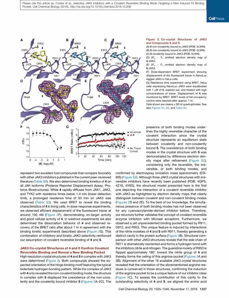
Figure 2. Co-crystal Structures of JAK3
and Compounds 4 and 5(A) 4 non-covalently bound to JAK3 (PDB: 5LWM).
(B) 5 non-covalently bound to JAK3 (PDB: 5LWN).
(C) 5 covalently bound to JAK3 (PDB: 5LWN).
(D) 2Fo � Fc omitted electron density map of
4-JAK3.
(E) 2Fo � Fc omitted electron density map of
5-JAK3.
(F) Dose-dependent BRET experiment showing
displacement of the fluorescent tracer in NanoLuc
tagged JAK3 in HeLa cells.
(G) Residence time experiment using BRET. HeLa
cells expressing NanoLuc JAK3 were equilibrated
with 1 mM of 4, washed out, and treated with high
concentrations of tracer. Displacement of 4 was
monitored by BRET. BRET levels of full occupancy
control were reached after approx. 1 hr.
Data shown are means ± SD of quadruplicates. See
also Figures S1, S2, and Table S4.
Please cite this article as: Forster et al., Selective JAK3 Inhibitors with a Covalent Reversible Binding Mode Targeting a New Induced Fit BindingPocket, Cell Chemical Biology (2016), http://dx.doi.org/10.1016/j.chembiol.2016.10.008
represent two excellent tool compounds that compare favorably
with other JAK3 inhibitors published in the current peer-reviewed
literature (Table S6). We also determined binding kinetics of 4 on
all JAK isoforms (Proteros Reporter Displacement Assay, Pro-
teros Biostructures). While 4 rapidly diffuses from JAK1, JAK2,
and TYK2 with residence times below 1.4 min (lower detection
limit), a prolonged residence time of 50 min on JAK3 was
observed (Table S3). We used BRET to reveal the binding
characteristics of 4 in living cells. In dose-response experiments,
we observed efficient displacement of the fluorescent tracer at
around 100 nM (Figure 2F), demonstrating on-target activity
and good cellular activity of 4. In washout experiments we also
determined the dissociation behavior of 4 and observed re-
covery of the BRET ratio after about 1 hr in agreement with the
binding kinetic experiments described above (Figure 2G). This
combination of inhibitory and kinetic JAK3 selectivity reinforced
our assumption of covalent reversible binding of 4 and 5.
JAK3 Co-crystal Structures of 4 and 5 Confirm CovalentReversible Binding and Reveal a New Binding PocketHigh resolution crystal structures of 4 and 5 in complexwith JAK3
were determined (Figure 2). Both compounds showed the ex-
pected orientation of the hinge binding motif featuring the typical
bidentate hydrogen bonding pattern. While the complex of JAK3
with4only revealed the non-covalent bindingmode, the structure
in complex with 5 displayed the coexistence of the non-cova-
lently and the covalently bound inhibitor 5 (Figures 2A–2C). The
Cell Chemical Biolog
presence of both binding modes under-
lines the highly reversible character of the
covalent interaction since the crystal
structure represents an equilibrium state
between covalently and non-covalently
bound 5. The coexistence of both binding
modes in the crystal structure with 5 was
demonstrated by difference electron den-
sity maps after refinement (Figure S2),
considering only the reversible, the irre-
versible, or both binding modes, and
confirmed by electrospray ionization mass spectrometry (ESI-
MS) (Figure S2). Although three JAK3 crystal structures with irre-
versible inhibitors have recently been published (PDB: 4QPS,
4Z16, 4V0G), the structural model presented here is the first
one depicting the interaction of a covalent reversible inhibitor
with JAK3 as highlighted by electron density maps that clearly
distinguish between covalent and non-covalent binding modes
(Figures 2D and 2E). To the best of our knowledge, the simulta-
neous presence of both binding modes has not been observed
for any cyanoacrylamide-derived inhibitor before. Therefore,
our structure further validates the concept of covalent reversible
enzyme inhibition with Michael acceptors. Furthermore, we
observed a yet unprecedented binding pocket formed by R911,
D912, and R953. This unique feature is induced by interactions
of the nitrile moieties of 4 and 5 with R911, thereby generating a
distinct cavity in the protein surface (Figure 3B). Structural com-
parison with other JAK3 structures reveals that the side chain of
R911 is dramatically reoriented and forms a hydrogen bond with
the inhibitors nitrile and nitrogen. The guanidinemoiety of R953 is
flipped approximately 180� toward the nitrile group of 4 and
thereby forms the ceiling of this arginine pocket (Figures 3A and
3B). Alignment of the other 16 available JAK3 crystal structures
revealed that the orientation of the aforementioned arginine res-
idues is conserved in those structures, confirming the induction
of the arginine pocket to be a unique feature of our inhibitor class
(Figure 3C). To assess the contribution of this cavity to the
outstanding selectivity of 4 and 5, we aligned the amino acid
y 23, 1335–1340, November 17, 2016 1337
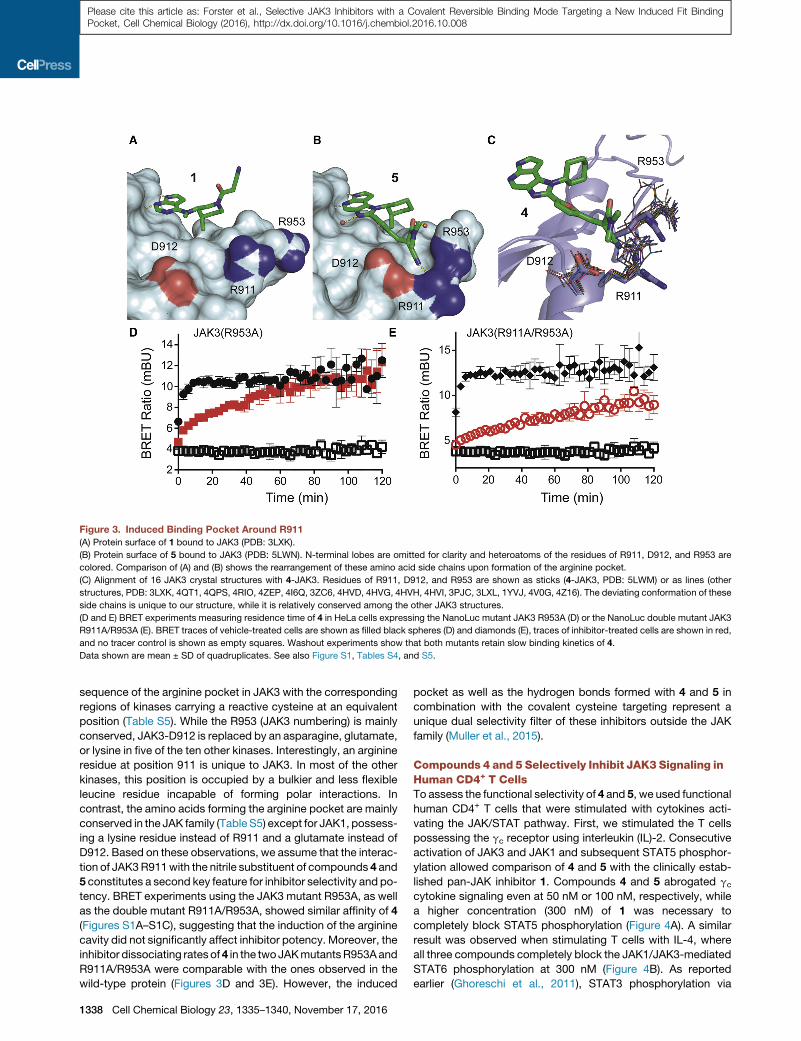
Figure 3. Induced Binding Pocket Around R911
(A) Protein surface of 1 bound to JAK3 (PDB: 3LXK).
(B) Protein surface of 5 bound to JAK3 (PDB: 5LWN). N-terminal lobes are omitted for clarity and heteroatoms of the residues of R911, D912, and R953 are
colored. Comparison of (A) and (B) shows the rearrangement of these amino acid side chains upon formation of the arginine pocket.
(C) Alignment of 16 JAK3 crystal structures with 4-JAK3. Residues of R911, D912, and R953 are shown as sticks (4-JAK3, PDB: 5LWM) or as lines (other
structures, PDB: 3LXK, 4QT1, 4QPS, 4RIO, 4ZEP, 4I6Q, 3ZC6, 4HVD, 4HVG, 4HVH, 4HVI, 3PJC, 3LXL, 1YVJ, 4V0G, 4Z16). The deviating conformation of these
side chains is unique to our structure, while it is relatively conserved among the other JAK3 structures.
(D and E) BRET experiments measuring residence time of 4 in HeLa cells expressing the NanoLuc mutant JAK3 R953A (D) or the NanoLuc double mutant JAK3
R911A/R953A (E). BRET traces of vehicle-treated cells are shown as filled black spheres (D) and diamonds (E), traces of inhibitor-treated cells are shown in red,
and no tracer control is shown as empty squares. Washout experiments show that both mutants retain slow binding kinetics of 4.
Data shown are mean ± SD of quadruplicates. See also Figure S1, Tables S4, and S5.
Please cite this article as: Forster et al., Selective JAK3 Inhibitors with a Covalent Reversible Binding Mode Targeting a New Induced Fit BindingPocket, Cell Chemical Biology (2016), http://dx.doi.org/10.1016/j.chembiol.2016.10.008
sequence of the arginine pocket in JAK3 with the corresponding
regions of kinases carrying a reactive cysteine at an equivalent
position (Table S5). While the R953 (JAK3 numbering) is mainly
conserved, JAK3-D912 is replaced by an asparagine, glutamate,
or lysine in five of the ten other kinases. Interestingly, an arginine
residue at position 911 is unique to JAK3. In most of the other
kinases, this position is occupied by a bulkier and less flexible
leucine residue incapable of forming polar interactions. In
contrast, the amino acids forming the arginine pocket are mainly
conserved in the JAK family (Table S5) except for JAK1, possess-
ing a lysine residue instead of R911 and a glutamate instead of
D912. Based on these observations, we assume that the interac-
tion of JAK3R911with the nitrile substituent of compounds 4 and
5 constitutes a second key feature for inhibitor selectivity and po-
tency. BRET experiments using the JAK3 mutant R953A, as well
as the double mutant R911A/R953A, showed similar affinity of 4
(Figures S1A–S1C), suggesting that the induction of the arginine
cavity did not significantly affect inhibitor potency. Moreover, the
inhibitor dissociating ratesof4 in the twoJAKmutantsR953Aand
R911A/R953A were comparable with the ones observed in the
wild-type protein (Figures 3D and 3E). However, the induced
1338 Cell Chemical Biology 23, 1335–1340, November 17, 2016
pocket as well as the hydrogen bonds formed with 4 and 5 in
combination with the covalent cysteine targeting represent a
unique dual selectivity filter of these inhibitors outside the JAK
family (Muller et al., 2015).
Compounds 4 and 5 Selectively Inhibit JAK3 Signaling inHuman CD4+ T CellsTo assess the functional selectivity of 4 and 5, we used functional
human CD4+ T cells that were stimulated with cytokines acti-
vating the JAK/STAT pathway. First, we stimulated the T cells
possessing the gc receptor using interleukin (IL)-2. Consecutive
activation of JAK3 and JAK1 and subsequent STAT5 phosphor-
ylation allowed comparison of 4 and 5 with the clinically estab-
lished pan-JAK inhibitor 1. Compounds 4 and 5 abrogated gccytokine signaling even at 50 nM or 100 nM, respectively, while
a higher concentration (300 nM) of 1 was necessary to
completely block STAT5 phosphorylation (Figure 4A). A similar
result was observed when stimulating T cells with IL-4, where
all three compounds completely block the JAK1/JAK3-mediated
STAT6 phosphorylation at 300 nM (Figure 4B). As reported
earlier (Ghoreschi et al., 2011), STAT3 phosphorylation via

Figure 4. Selective Inhibition of JAK3-Medi-
ated Cytokine Signaling by Compounds
4 and 5Human CD4+ T cells were pre-incubated for 1 hr with
the indicated concentrations of the JAK inhibitors
(JAKi) 1, 4 or 5 and stimulated for 30 min with IL-2
(activates JAK3/JAK1) (A), IL-4 (activates JAK3/
JAK1) (B), IL-6 (activates JAK2/JAK1/TYK2) (C) or
IFN-a (activates JAK1/TYK2) (D). Phosphorylation of
STAT5 (A), STAT6 (B), STAT3 (C), or STAT1 (D) was
determined by phospho-specific Abs and immuno-
blotting (ns, no cytokine stimulation). Levels of actin
were determined to show equal loading. See also
Table S1.
Please cite this article as: Forster et al., Selective JAK3 Inhibitors with a Covalent Reversible Binding Mode Targeting a New Induced Fit BindingPocket, Cell Chemical Biology (2016), http://dx.doi.org/10.1016/j.chembiol.2016.10.008
JAK1, JAK2, and TYK2, triggered by stimulation with IL-6, is
inhibited by 1 in a dose-dependent manner at concentrations
R300 nM. In sharp contrast, 4 and 5 did not affect STAT3 acti-
vation at doses up to 1,000 nM (Figure 4C), confirming the selec-
tivity toward JAK3 in functional cells. The selectivity difference
between our compounds and 1 was even more pronounced
when T cells were stimulated with interferon (IFN)-a. While 1
clearly inhibits IFN-a-mediated JAK1/TYK2 signaling at concen-
trations >100 nM, compounds 4 and 5 did not influence pSTAT1
levels at concentrations up to 1,000 nM (Figure 4D). These
results demonstrate that the JAK3 selectivity of 4 and 5 observed
in enzyme assays are maintained in a cellular context.
SIGNIFICANCE
In this study, a new class of covalent reversible JAK3 inhib-
itors was developed. Crystallographic data confirmed a
reversible covalent binding mode as well as a previously
unseen binding cavity induced by a nitrile-arginine interac-
tion. The combination of both structural features paved the
way for a new generation of JAK3-selective inhibitors. With
compounds 4 and 5, we provide JAK3 inhibitors with pico-
molar affinities and outstanding selectivity within the JAK
family and against the whole kinome. Moreover, it was
demonstrated that activity and selectivity translate well in
a cellular environment. These compounds are thus suitable
to serve as chemical probes to elucidate the effect of selec-
tive JAK3 inhibition. They might be used for studying the
Cell Chemical Biolo
JAK1/3 interplay with particular regard
to their respective roles in cytokine
signaling and the resultant clinically
relevant immunosuppressive effects.
EXPERIMENTAL PROCEDURES
Chemical Synthesis
Synthetic schemes and detailed synthetic proced-
ures are described in the Supplemental Experi-
mental Procedures.
JAK3 ELISA
For initial JAK3 IC50 determination, different
concentrations of inhibitors were incubated in
substrate-coated 96-well plates with ATP and
recombinant JAK3 kinase domain (amino acids
781–1124). The degree of phosphorylation was
determined by detection via monoclonal anti-pTyr-HRP-conjugated anti-
bodies, followed by a color reaction. See the Supplemental Experimental Pro-
cedures for details and Bauer et al. (2014).
Protein Expression, Purification, Crystallization, and Structure
Determination
Recombinant JAK3 kinase domain with a tobacco etch virus (TEV)-cleavable
His tag was expressed in Sf9 cells. The cells were lysed and protein was
initially purified by Ni-affinity chromatography. Protein was incubated over-
night with the inhibitors and TEV protease. The cleaved protein was further
purified by reverse Ni-affinity and size exclusion chromatography. JAK3-inhib-
itor complexes were crystallized using sitting-drop vapor diffusion. The
obtained crystals were cryoprotected and diffraction data were collected at
Diamond Light Source. More details on the procedures and crystallographic
data refinement are described in the Supplemental Experimental Procedures.
BRET Experiments
Dose-response experiments were conducted in HeLa cells expressing
NanoLuc JAK3 or the studied mutants using Promega tracer 5. Binding
kinetic experiments in living cells after compound washout were conducted
using the same constructs and tracers as described in Robers et al. (2015).
A detailed description is provided in the Supplemental Experimental
Procedures.
CD4+ T Cell Cytokine Stimulation Assays
T cells were purified from peripheral blood mononuclear cells from human do-
nors. Equal numbers of cells were incubated for 1 hr with JAK inhibitors or
DMSO control and stimulated with cytokines for 30 min. The cells were lysed,
and the proteins were separated via PAGE and transferred to a polyvinylidene
fluoride membrane. The proteins of interest were blotted with specific anti-
bodies and visualized with an infrared imaging system. A detailed description
can be found in the Supplemental Experimental Procedures.
gy 23, 1335–1340, November 17, 2016 1339

Please cite this article as: Forster et al., Selective JAK3 Inhibitors with a Covalent Reversible Binding Mode Targeting a New Induced Fit BindingPocket, Cell Chemical Biology (2016), http://dx.doi.org/10.1016/j.chembiol.2016.10.008
ACCESSION NUMBERS
The accession number for compound 4 co-crystallized with JAK3 is PDB:
5LWM. The accession number for compound 5 co-crystallized with JAK3 is
PDB: 5LWN.
SUPPLEMENTAL INFORMATION
Supplemental Information includes Supplemental Experimental Procedures,
two figures, and six tables and can be found with this article online at http://
dx.doi.org/10.1016/j.chembiol.2016.10.008.
AUTHOR CONTRIBUTIONS
S.A.L. and S.K. initiated and supervised this study. M.F. and M.G. conceived
the chemical experiments and M.F. carried them out. A.C. conceived the pro-
tein X-ray experiments and carried them out. S.M.B. conceived the experi-
ments for the initial biological evaluation and carried them out. M.B.R. and
C.R.C. conceived and carried out the BRET experiments. J.H. and K.G.
conceived the CD4+ T cell experiments and J.H. carried them out. M.F.,
A.C., M.G., S.M.B., and J.H. carried out the data analysis. M.F., M.G.,
S.M.B., E.P., A.C., K.G., and S.K. wrote the paper.
ACKNOWLEDGMENTS
K.G. would like to thank the DFG for funding (Transregio SFB TR156). S.K. is
grateful for support by the SGC, a registered charity (number 1097737)
receiving funds from AbbVie, Bayer Pharma AG, Boehringer Ingelheim, Can-
ada Foundation for Innovation, Eshelman Institute for Innovation, Genome
Canada through Ontario Genomics Institute, Innovative Medicines Initiative
(EU/EFPIA) (ULTRA-DD grant no. 115766), Janssen, gs9:Merck, Novartis
Pharma AG, Ontario Ministry of Economic Development and Innovation,
Pfizer, Sao Paulo Research Foundation-FAPESP, Takeda, and the Wellcome
Trust (grant no. 092809/Z/10/Z) as well as the Innovative Medicines Initiative
(EU/EFPIA) (grant K4DD).
Received: April 6, 2016
Revised: July 21, 2016
Accepted: October 13, 2016
Published: November 10, 2016
REFERENCES
Ahearn, S.P., Christopher, M., Jung, J., Pu, Q., Rivkin, A., Scott, M.E., Witter,
D.J., Woo, H.C., Cash, B., Dinsmore, C., et al. (2013). Pyrrolopyrimidines as
Janus Kinase Inhibitors. WO Patent 2013/085802. https://www.google.com/
patents/WO2013085802A1?cl=enIt.
Bauer, S.M., Gehringer, M., and Laufer, S.A. (2014). A direct enzyme-linked
immunosorbent assay (ELISA) for the quantitative evaluation of Janus
Kinase 3 (JAK3) inhibitors. Anal. Methods 6, 8817–8822.
Chrencik, J.E., Patny, A., Leung, I.K., Korniski, B., Emmons, T.L., Hall, T.,
Weinberg, R.A., Gormley, J.A., Williams, J.M., Day, J.E., et al. (2010).
Structural and thermodynamic characterization of the TYK2 and JAK3 kinase
domains in complex with CP-690550 and CMP-6. J. Mol. Biol. 400, 413–433.
Flanagan, M.E., Blumenkopf, T.A., Brissette, W.H., Brown, M.F., Casavant,
J.M., Poa, C.S., Doty, J.L., Elliott, E.A., Fisher, M.B., Hines, M., et al. (2010).
Discovery of CP-690’550: a potent and selective Janus Kinase (JAK) inhibitor
for the treatment of autoimmune diseases and organ transplant rejection.
J. Med. Chem. 53, 8468–8484.
Gehringer, M., Pfaffenrot, E., Bauer, S., and Laufer, S.A. (2014). Design and
synthesis of tricyclic JAK3 inhibitors with picomolar affinities as novel molec-
ular probes. ChemMedChem 9, 277–281.
1340 Cell Chemical Biology 23, 1335–1340, November 17, 2016
Ghoreschi, K., Laurence, A., and O’Shea, J.J. (2009). Selectivity and therapeu-
tic inhibition of kinases: to be or not to be? Nat. Immunol. 10, 356–360.
Ghoreschi, K., Jesson, M.I., Li, X., Lee, J.L., Ghosh, S., Alsup, J.W., Warner,
J.D., Tanaka, M., Steward-Tharp, S.M., Gadina, M., et al. (2011). Modulation
of innate and adaptive immune responses by tofacitinib (CP-690,550).
J. Immunol. 186, 4234–4243.
Goedken, E.R., Argiriadi, M.A., Banach, D.L., Fiamengo, B.A., Foley, S.E.,
Frank, K.E., George, J.S., Harris, C.M., Hobson, A.D., Ihle, D.C., et al.
(2015). Tricyclic covalent inhibitors selectively target Jak3 through an active
site thiol. J. Biol. Chem. 290, 4573–4589.
Haan, C., Rolvering, C., Raulf, F., Kapp, M., Dr€uckes, P., Thoma, G.,
Behrmann, I., and Zerwes, H.-G. (2011). Jak1 has a dominant role over jak3
in signal transduction through gamma-c-containing cytokine receptors.
Chem. Biol. 18, 314–323.
Knapp, S., Arruda, P., Blagg, J., Burley, S., Drewry, D.H., Edwards, A., Fabbro,
D., Gillespie, P., Gray, N.S., Kuster, B., et al. (2013). A public-private partner-
ship to unlock the untargeted kinome. Nat. Chem. Biol. 9, 3–6.
Liu, Q., Sabnis, Y., Zhao, Z., Zhang, T., Buhrlage, S.J., Jones, L.H., and Gray,
N.S. (2013). Developing irreversible inhibitors of the protein kinase cysteinome.
Chem. Biol. 20, 146–159.
London, N., Miller, R.M., Krishnan, S., Uchida, K., Irwin, J.J., Eidam, O.,
Gibold, L., Cimerman�ci�c, P., Bonnet, R., Shoichet, B.K., et al. (2014).
Covalent docking of large libraries for the discovery of chemical probes. Nat.
Chem. Biol. 10, 1066–1072.
Muller, S., Chaikuad, A., Gray, N.S., and Knapp, S. (2015). The ins and outs of
selective kinase inhibitor development. Nat. Chem. Biol. 11, 818–821.
Pesu, M., Candotti, F., Husa, M., Hofmann, S.R., Notarangelo, L.D., and
O’Shea, J.J. (2005). Jak3, severe combined immunodeficiency, and a new
class of immunosuppressive drugs. Immunol. Rev. 203, 127–142.
Robers, M.B., Dart, M.L., Woodroofe, C.C., Zimprich, C.A., Kirkland, T.A.,
Machleidt, T., Kupcho, K.R., Levin, S., Hartnett, J.R., Zimmerman, K., et al.
(2015). Target engagement and drug residence time can be observed in living
cells with BRET. Nat. Commun. 6, 10091.
Serafimova, I.M., Pufall, M.A., Krishnan, S., Duda, K., Cohen, M.S., Maglathlin,
R.L., McFarland, J.M., Miller, R.M., Frodin, M., and Taunton, J. (2012).
Reversible targeting of noncatalytic cysteines with chemically tuned electro-
philes. Nat. Chem. Biol. 8, 471–476.
Singh, J., Petter, R.C., and Kluge, A.F. (2010). Targeted covalent drugs of the
kinase family. Curr. Opin. Chem. Biol. 14, 475–480.
Smith, G.A., Uchida, K., Weiss, A., and Taunton, J. (2016). Essential biphasic
role for JAK3 catalytic activity in IL-2 receptor signaling. Nat. Chem. Biol. 12,
373–379.
Tan, L., Akahane, K., McNally, R., Reyskens, K.M.S.E., Ficarro, S.B., Liu, S.,
Herter-Sprie, G.S., Koyama, S., Pattison, M.J., Labella, K., et al. (2015).
Development of selective covalent Janus Kinase 3 inhibitors. J. Med. Chem.
58, 6589–6606.
Thoma, G., Nuninger, F., Falchetto, R., Hermes, E., Tavares, G.A.,
Vangrevelinghe, E., and Zerwes, H.-G. (2011). Identification of a potent
Janus kinase 3 inhibitor with high selectivity within the Janus kinase family.
J. Med. Chem. 54, 284–288.
Thoma, G., Dr€uckes, P., and Zerwes, H.-G. (2014). Selective inhibitors of the
Janus kinase Jak3—Are they effective? Bioorg. Med. Chem. Lett. 24, 4617–
4621.
Thorarensen, A., Banker, M.E., Fensome, A., Telliez, J.-B., Juba, B., Vincent,
F., Czerwinski, R.M., and Casimiro-Garcia, A. (2014). ATP-mediated kinome
selectivity: the missing link in understanding the contribution of individual jak
kinase isoforms to cellular signaling. ACS Chem. Biol. 9, 1552–1558.