Cerebellar anatomy as applied to cerebellar microsurgical resections

REVIEW ARTICLEpublished: 10 January 2013
doi: 10.3389/fncir.2012.00116
Seeking a unified framework for cerebellar function anddysfunction: from circuit operations to cognitionEgidio D’Angelo1,2* and Stefano Casali1*
1 Department of Brain and Behavioral Sciences, Pavia, Italy2 IRCCS C. Mondino, Brain Connectivity Center, Pavia, Italy
Edited by:
Chris I. De Zeeuw, Erasmus MedicalCenter, Netherlands
Reviewed by:
Yosef Yarom, Hebrew University,IsraelChristian Hansel, Erasmus MedicalCenter, Netherlands
*Correspondence:
Egidio D’Angelo and Stefano Casali,Department of Brain and BehavioralSciences, Via Forlanini 6,I-27100 Pavia, Italy.e-mail: [email protected];[email protected]
Following the fundamental recognition of its involvement in sensory-motor coordinationand learning, the cerebellum is now also believed to take part in the processingof cognition and emotion. This hypothesis is recurrent in numerous papers reportinganatomical and functional observations, and it requires an explanation. We argue that asimilar circuit structure in all cerebellar areas may carry out various operations using acommon computational scheme. On the basis of a broad review of anatomical data, it isconceivable that the different roles of the cerebellum lie in the specific connectivity ofthe cerebellar modules, with motor, cognitive, and emotional functions (at least partially)segregated into different cerebro-cerebellar loops. We here develop a conceptual andoperational framework based on multiple interconnected levels (a meta-levels hypothesis):from cellular/molecular to network mechanisms leading to generation of computationalprimitives, thence to high-level cognitive/emotional processing, and finally to the sphereof mental function and dysfunction. The main concept explored is that of intimateinterplay between timing and learning (reminiscent of the “timing and learning machine”capabilities long attributed to the cerebellum), which reverberates from cellular to circuitmechanisms. Subsequently, integration within large-scale brain loops could generate thedisparate cognitive/emotional and mental functions in which the cerebellum has beenimplicated. We propose, therefore, that the cerebellum operates as a general-purposeco-processor, whose effects depend on the specific brain centers to which individualmodules are connected. Abnormal functioning in these loops could eventually contributeto the pathogenesis of major brain pathologies including not just ataxia but also dyslexia,autism, schizophrenia, and depression.
Keywords: cerebellum, cognition, motor control, timing, prediction, autism, schizophrenia, dyslexia
INTRODUCTIONThe cerebellum is classically thought to control movement coor-dination (Flourens, 1824; Luciani, 1891) and motor learning(Marr, 1969; Albus, 1972) but recent experimental evidence sug-gests that it may also play a key role in cognition and emo-tion (Schmahmann, 2004; Schmahmann and Caplan, 2006; Ito,2008) 1. This clearly raises broader questions: how can the samecircuit cope with so many different tasks? Is signal processingin the cerebellar circuits always based on the same computa-tional scheme? Is it conceivable that what underlies the differentroles of the cerebellum is the specific connectivity of cerebellarmodules, rather than specific microcircuit properties? In order to
1We need to clarify the terminology used. We here treat brain functions ona “neurological” level, where cognition and emotion are kept distinct. Thisdiffers from the definition used in neuroinformatics, in which “cognitiveprocessing” refers to computational primitives—like pattern recognition, gen-eralization, and abstraction by artificial neural networks. These definitions,furthermore, should not be confused with the tenet of classic cognitive psy-chology, which maintained that emotion is a category of cognition (Spitzer,1998).
answer these questions, here we propose a conceptual and opera-tional framework, or meta-levels hypothesis, based on four levels:(1) cellular/molecular, (2) network, primitives of circuit process-ing, (3) high-level cognitive/emotional processing, and (4) mentalprocessing. We first review neuroanatomical, neuropsychological,neuropsychiatric, and neuroimaging studies in order to elucidatehow the cerebellum might take part in cognitive and emotionalfunctions, and how cerebellar damage could determine neuro-logical and neuropsychiatric disorders. We then argue that thecerebellum carries out basic computational functions, timing,and learning, applicable in different cases. The cerebellum hasbeen reported to assist brain operations by providing accuratetiming of multiple series of signals coming from the cerebral cor-tex and the sensory systems [reviewed in Bower (1997, 2002);Jacobson et al. (2008); D’Angelo and De Zeeuw (2009); D’Angeloet al. (2009, 2010); D’Angelo (2010a,b, 2011); De Zeeuw et al.(2011)]. This could underlie the implementation of processeslike sensory prediction, novelty detection, error detection, timematching, and sequence ordering (Ivry and Baldo, 1992; Ivryet al., 2002; Ghajar and Ivry, 2009). This multi-dimensional com-putation would allow the same circuit to contribute to functions
Frontiers in Neural Circuits www.frontiersin.org January 2013 | Volume 6 | Article 116 | 1
NEURAL CIRCUITS

D’Angelo and Casali Unified cerebellar function
as diverse as voluntary movement (a cognitive process, after all)and thought, provided that appropriate connections with dif-ferent cortical and subcortical centers were established and thatcommunication between these centers occurred over the appro-priate frequency bands and using compatible codes (Ito, 1993,2008; D’Angelo, 2011). We propose, therefore, that the cerebel-lum operates as a general co-processor, whose effect depends onthe centers to which different modules are connected, affectingcognitive functions as well as sensory-motor processing.
BRAIN PROCESSING AND THE CEREBELLUMThe cerebellum has long been linked to the concept of motor con-trol but now several observations indicate that it is also involvedin cognitive/emotional processing. This extension of its role raisesa key question: is there formal similarity between these two typesof processing? A critical observation, in this regard, is that cog-nitive and sensory-motor processing should not, in principle, bevery different. This prevents from a serious computational para-dox: if the two processes were different, they may use differentcoding strategies. But then, how could the basic neuronal circuitof the cerebellum, which appears to be invariant across differ-ent areas, be able to process different signal codes? This wouldviolate the idea that the cerebellum develops a single general algo-rithm. Indeed, different sections of the cerebral cortex (sensory,motor, and associative in nature) communicate with each otheras well as with various cerebellar areas and so the neural codesare likely to be homogeneous. As a corollary of this, it is well-documented that motor planning means predicting the sensoryconsequences of a motor act: a motor plan is coded in terms of ananticipated sensory state (Blakemore et al., 1998a,b). This is akinto the general hypothesis of the “prediction imperative” that needsto be satisfied in order to allow brain processing (Llinás and Roy,2009). Prediction processes are normally performed by “forwardcontrollers,” which use internal memory to represent the sys-tem state (Diedrichsen et al., 2010; Shadmehr and Mussa-Ivaldi,2012).
IS THE CEREBELLUM A GENERALIZED FORWARD CONTROLLER?On the basis of studies of the vestibulo-ocular reflex (VOR),eye-blink conditioning, and saccadic eye movements, and thefundamental theoretical concepts of motor learning (Marr, 1969),the cerebellum has been suggested to provide forward models ofthe motor system. These forward models can predict the pos-ture or motion of body parts following a motor command and,by a further transformation, predict the sensory consequencesof actions (Miall and Reckess, 2002). More precisely, a copy ofmotor commands generated by the motor cortex (efference copy)is sent to the cerebellum, which uses its internal forward modelto predict their sensory consequences (corollary discharge). Thesensory predictions are then compared to actual sensory feedback(Wolpert et al., 1998): in the presence of errors (or novelty, i.e.,deviations from prediction), the cerebellum emits corrective sig-nals. A fully characterized example of generation of predictionsby cerebellar circuits is provided by electro-perception in weaklyelectric fishes, in which a cerebellar-like structure compares theexpected electric field generated by the fish with the actual elec-tric field sensed by the electroreceptors, thus gaining information
on the structure of the environment through the changes that thislatter has caused in the field itself (Bell et al., 2008).
In the presence of persistent deviations from prediction thecerebellum learns to modify the forward model itself. Learningappears to occur through two distinct processes, one faster andmore labile, involving the cerebellar forward controller, the other,which may at least partly reside outside the cerebellum, slowerand consolidated (Shadmehr and Mussa-Ivaldi, 2012). In fact, thecerebellar cortex is thought to process the faster component ofmemory, while the deep cerebellar nuclei may elaborate its slowercomponent (Medina and Mauk, 2000). The cerebellum is thoughtto share its “predictor function” with the parietal lobes, in such away that these two structures might work in parallel (Blakemoreand Sirigu, 2003): the cerebellum as a whole is likely to gener-ate faster but unconscious predictions, while the parietal lobesprobably generate slower but conscious ones.
Given the anatomical connections of the cerebellum with asso-ciative areas (see below) and the similarity of motor planning andcognitive processing, it seems logical to generalize the forwardcontroller role of the cerebellum to cognition. Indeed, Ito (2005)hypothesized that the cerebellum could operate as a generalizedforward controller regulating cognition as well as sensory-motorcontrol2.
THE FORWARD CONTROLLER AND MENTAL ACTIVITYThe fundamental postulate about brain/mind functioning is thatthe brain generates a virtual reality (Churchland and Sejinowski,1992; Churchland, 1998; Llinas and Paré, 1998), probably con-ferring an evolutionary advantage by predicting possible envi-ronmental configurations and allowing symbolic representationand communication. Several observations show that perceptionis not a copy of the external or internal energy patterns, butrather a mental elaboration endowed with quality and deformedby imperfect receptor sampling, adaptation, memories, and emo-tions. This makes conscious perception unique and subjective. Atthis point, one may speculate on how the cerebellum, being deeplyinterconnected with the cerebral cortex, might be involved in pro-cessing conscious percepts. We propose a somewhat provocativereflection.
A first issue is that whereas reality is perceived as instanta-neous, computation in neurons and synapses actually takes timeand the cerebral cortex needs hundreds of milliseconds to gen-erate a conscious percept. This delay, in addition to violating the
2A conceptual obstacle is that sensory-motor processing is thought to bemostly automatic (and unconscious) while cognition and emotion are boundto consciousness. Akin with this, the thalamo-cortical circuit is usuallythought of as the site of conscious processing and the cerebro-cerebellar cir-cuit as the site of automatic control. Indeed, the cerebellum probably does nottake part in conscious representations directly, given that abnormalities of thecerebellum do not seem to impair consciousness. In their hypothesis, Tononiand Edelman suggest that the ability of circuits to generate consciousness liesin the strength of their internal connectivity, which is quite high in the cor-tex but comparatively much poorer in the cerebellum (Tononi and Edelman,1998). This would explain why the cortex is capable of developing consciouspercepts, while the cerebellum is not. However, the cerebellum is deeply inter-connected in closed loops with the cerebral cortex over multiple independentlines involving associative as well as sensory-motor areas (see below), so thatthe activity of the two structures is strongly integrated.
Frontiers in Neural Circuits www.frontiersin.org January 2013 | Volume 6 | Article 116 | 2

D’Angelo and Casali Unified cerebellar function
idea of the instantaneity of subjective perception, is far too longto allow movement and thought to be controlled in a purposeful,dynamic, and interactive manner. Therefore, the virtual realitygenerated by the brain has to be “anticipatory” and to occursomehow in advance of the elaboration of objective reality basedon cortical processing of sensory signals. This anticipatory processmay be based on the use of previous information and memory onvarious time scales, as would occur in a forward controller, whichis exactly what the cerebellum is thought to be. A second issueis that reality is perceived as continuous, even though computa-tional cycles during cerebro-cortical cognitive processing actuallylast about 25 ms (a γ-band cycle) and longer cycles about 100 ms(a θ-band cycle) (Buzsaki, 2006). The cerebellum, by exerting mil-lisecond control of its output spikes, may help to maintain the fastcontinuity required for spatiotemporal integration of consciouspercepts.
Thus, the fact that the cerebellum does not, clinically, appear tobe needed to generate consciousness (Tononi and Edelman, 1998)does not mean that it is extraneous to the mechanisms controllingthe relationship between objective reality and internal represen-tation. Indeed, functional activation of the cerebellum has beenrevealed in relation to the conscious representation of time intasks using internal memories (Addis et al., 2009; Nyberg et al.,2010; Szpunar, 2010, 2011). It should be noted at this point thatone main theory on the working of the cerebellum is that it actsas a “comparator of intentionality with execution,” which is pre-cisely what the whole brain continuously does in order to relateneuronal activity to the world. On this basis, we conclude thatit can hardly be considered surprising that the cerebellum takespart in cognition and emotion, that it can influence attention andintelligence (Cotterill, 2001), and that its dysfunction can affect“internal coherence” in dissociative diseases.
THE EXTENDED CEREBRO-CEREBELLAR LOOPSThe cerebellar cortex has, from the earliest studies, always beenreported to have a similar structure in all its sections, and its cir-cuit to show a regular “lattice”-like organization (Eccles et al.,1967) (Figure 1). The cerebellar circuit can be schematicallydescribed as follows: mossy fibers activate granule and Golgi cellsin the granular layer. Granule cells emit parallel fibers and activateall the other neurons in the cerebellar cortex. Golgi cells are dou-bly activated by mossy and parallel fibers providing feedforwardand feedback inhibition to granule cells. The granular layer alsocontains other interneurons, namely, Lugaro cells and unipolarbrush cells (only in the flocculo-nodular lobe). In the molecu-lar layer, parallel fibers activate Purkinje cells and also stellateand basket cells, which in turn inhibit Purkinje cells. Purkinjecells are also activated by climbing fibers generated by the inferiorolive. Purkinje cells in turn project to the deep-cerebellar nuclei.In this context, the modules and the cerebello-thalamo-cerebro-cortical circuits (CTCCs) can be considered the main structuralelements.
THE CEREBELLAR MODULAR ORGANIZATIONMacroscopically, the cerebellum consists of a tightly folded layerof cortex with white matter beneath in which deep nuclei areembedded. At microscopic level, each part of the cortex consists
FIGURE 1 | Schematic representation of the cerebellar circuit. Thecerebellar circuit consists of cortical and subcortical sections. At subcorticallevel, the afferent fibers activate DCN cells (DCN-C) and IO cells (IO-C).The DCN emits the output and at the same time inhibits the IO. In thecerebellar cortex, there are different types of neurons including granulecells (GrC), Golgi cells (GoC), Purkinje cells (PC), stellate and basket cells(SC, BC), Lugaro cells, and unipolar brush cells (not shown). The two maininputs are represented by mossy fibers (mf) originating in various brainstem and spinal cord nuclei, and by climbing fibers (cf) originating from theIO. Signals conveyed through the mossy fibers diverge to DCN and activatethe granular layer (containing GrC and GoC). The ascending axon of the GrCbifurcates in the molecular layer (containing PC, SC, and BC) formingthe parallel fibers (pf). The cerebellar cortical circuit is organized as afeedforward excitatory chain assisted by inhibitory loops: mfs excite GrCs,which activate all the other cortical elements. In the granular layer, inhibitionis provided by GoC, in the molecular layer by SC and BC. Finally, PC inhibitDCN. The IO, which is also activated by brain stem and spinal cord nuclei,controls PC activity though a single powerful synapse. Thus, the wholesystem can be seen as a complex mechanism controlling the DCN output.
of the same small set of neuronal elements, laid out accordingto a highly stereotyped geometry. At an intermediate level, thecerebellum and its auxiliary structures can be broken down intoseveral hundred or thousand microzones or microcompartments,
Frontiers in Neural Circuits www.frontiersin.org January 2013 | Volume 6 | Article 116 | 3

D’Angelo and Casali Unified cerebellar function
which are thought to represent effective cerebellar functionalunits (Figure 2). These can be further differentiated into stripes,zones, and multizonal microcomplexes, which are effective func-tional modules (Andersson and Oscarsson, 1978; Apps andGarwicz, 2005; Apps and Hawkes, 2009)3.
A module is a conglomerate of several, non-adjacent parasagit-tal bands of Purkinje cells projecting to specific areas of deepcerebellar nuclei and gating segregated projections from the infe-rior olive (Cerminara, 2010; Oberdick and Sillitoe, 2011; Ruigrok,2010). Likewise, the mossy fibers projecting to a certain groupof Purkinje cells through the granular layer also project to thesame deep cerebellar nucleus neuron receiving input from thosePurkinje cells (Ito, 1984; Pijpers et al., 2006; Voogd, 2010). Themodules have almost segregated inputs, since climbing fibersbifurcate on the parasagittal plane to as many as 10 not necessar-ily adjacent Purkinje cells (mossy fiber bifurcations spread acrossboth planes). Thus, the majority of connections between neuronsand interneurons in the cerebellar cortex occur within individualmodules. The connections between modules occur almost exclu-sively via parallel fibers, which contact Purkinje cells and the otherinhibitory interneurons (Lainé and Axelrad, 1998; Dieudonnéand Dumoulin, 2000; Dean et al., 2004).
The modules have a very similar if not identical structure anddo not show major differences in their neuronal properties, eventhough some variants have been reported. One of these concernsthe vestibulocerebellum, which contains an additional cell type,the unipolar brush cell (Mugnaini et al., 2011), and may exhibitmore sustained discharges to Purkinje cells (Kim et al., 2011).Another peculiar aspect is glycine feedback from the lateral cere-bellar nuclei, which is sent only to the hemispheres and not to thevermis (Uusisaari and Knopfel, 2012). Finally, evident organiza-tion of genetic markers along the sagittal plane leads to a further“biochemical” compartmentalization3. These local properties donot undermine the general concept of a unified cerebellar com-putational algorithm, but they may bias certain modules towardspecific functional states, as is thought to occur in other braincircuits in relation to neuromodulators and neuropeptides (e.g.,LeBeau et al., 2005).
The cerebellar circuit appears to be organized in a feed-forward manner, with information passing through the cortexwithout recurrent loops and with limited intermodular con-nectivity. This is in apparent contrast with the cerebral cortex,
3The cerebellum is made up of large compartments generally known as zones,which can be broken down into smaller compartments known as microzones.The demonstration, by microneurography, of cortical zones shows that eachbody part maps to specific points in the cerebellum, even though there arenumerous repetitions of the basic map forming an arrangement that hasbeen called “fractured somatotopy.” A different indication of compartmen-talization is obtained by immune staining for certain types of protein (e.g.,zebrin, NOS etc.). This reveals stripes oriented perpendicular to the cerebellarfolds. Different markers generate different sets of stripes, and the widths andlengths vary as a function of location, but they all have the same general shape.Oscarsson in the 1970s proposed that these cortical zones can be partitionedinto smaller units called microzones. In 2005, Apps and Garwicz showed thatseveral microzones can be organized into a multizonal microcomplex. Allmicrozones in the microcomplex are connected to the same group of deepcerebellar nuclei and inferior olivary neurons and correspond to the cerebellarmodules defined by Voogd.
which shows zonal differences in thickness, in the proportion ofgranular and pyramidal neurons, in intracortical connectivity, inneuronal subtypes and spine distribution (Elston and DeFelipe,2002; Douglas and Martin, 2004; Lubke and Feldmeyer, 2007).Moreover, while there is poor intermodular connectivity in thecerebellum, the cerebral cortex shows strong intercolumnar con-nectivity [the relevance of which has been commented above(Tononi and Edelman, 1998)]. Clearly, the different anatomo-functional organization of the two cortices implies differentcomputational strategies. However, since the two cortices aredeeply interconnected through serial parallel loops, the productof cerebro-cortical elaborations is continuously relayed to specificmodules of the cerebellar cortex. Thus, in addition to the need tounderstand how cerebral and cerebellar cortical modules operate,it is essential to look in more detail at this interconnection of thetwo structures.
CEREBELLO-THALAMO-CEREBRO-CORTICAL CIRCUITS (CTCCs)There is growing evidence that the CTCCs include several afferentand efferent cerebral cortical areas of a motor, sensory, or associa-tive nature (Figure 3) (Strick et al., 2009). Most cerebro-cerebellarafferent projections pass through the basal (anterior or ven-tral) pontine nuclei and intermediate cerebellar peduncle, whilemost cerebello-cerebral efferent projections pass through dentateand ventrolateral (VL) thalamic nuclei. Some of these loops arehere considered in more detail in relation to sensory-motor andcognitive-emotional functions: the motor and somatosensory loops(including those involved in oculomotor control), the parietalloops, the prefrontal loops, the oculomotor loops, and the loopsformed with the basal ganglia and the limbic system. Cerebello-cerebral loops are highly segregated (Habas et al., 2009; Krienenand Buckner, 2009) and form complex interconnections also withthe basal ganglia and subcortical areas. Interestingly, during phy-logenesis, the cerebellar hemispheres evolve in parallel with theassociative rather than the motor or sensory areas, which sup-ports the progressive involvement of the cerebellum in cognitiveprocessing.
Motor and somatosensory loopsThe cerebellum projects both to motor and somatosensory areas.The output to the primary motor area (M1) is conveyed throughthe VL thalamic nuclei projecting to layers IV and V, while out-puts to the primary somatosensory area (S1) pass through theintralaminar nuclei projecting to intragranular and superficiallayers (Molinari et al., 2002). Through these projections to MI,the cerebellum can modulate motor cortex excitability in relationto the incoming sensory input (Luft et al., 2005). The cerebel-lum is also interconnected with premotor (Dum and Strick, 2003)and supplementary motor areas (Rouiller et al., 1994) involved inmovement planning. Interestingly, transcranial magnetic stimu-lation (TMS) of the lateral cerebellum can strongly affect the con-tralateral cerebral motor cortex (Oliveri et al., 2005). CerebellarTMS regulates the functional connectivity between Purkinje cellsand deep cerebellar nuclei, modifying the excitability of inter-connected motor areas, as shown by changes in motor-evokedpotential amplitude and in short and long intracortical inhibition(Koch et al., 2009a).
Frontiers in Neural Circuits www.frontiersin.org January 2013 | Volume 6 | Article 116 | 4

D’Angelo and Casali Unified cerebellar function
FIGURE 2 | The modular organization of the cerebellum. The pictureshows a flattened view of the cerebellum. Four ideal zones are shown incolor, each one containing microzones forming a multizonal microcomplex.The microzones have the basic structure reported in the expansion on theright (same symbols as in Figure 1, inhibitory interneurons are overlaid inblue). A microzone is defined as a group of the order of 1000 Purkinje cells allhaving the same somatotopic receptive field. These Purkinje cells arearranged in a long, narrow strip, oriented perpendicular to the cortical folds,so that Purkinje cell dendrites are flattened in the same direction as themicrozones extend and are crossed by parallel fibers at right angles. The
climbing fibers branches (about 10) usually innervate Purkinje cells belongingto the same microzone and the olivary neurons generating such climbingfibers tend to be coupled by gap junctions. This helps synchronizing Purkinjecells within a microzone on a millisecond time scale. The Purkinje cellsbelonging to a microzone all send their axons to the same small cluster ofoutput cells within the deep cerebellar nuclei. Finally, the axons of basketcells are much longer in the longitudinal direction than in the mediolateraldirection (not shown), causing them to be confined largely to a singlemicrozone. Thus, cellular interactions within a microzone are much strongerthan those between different microzones.
Parietal loopsThe cerebellum is closely connected with the parietal lobes. Thecerebellum sends input to area 7b of the inferior parietal lobe,in particular to the anterior intraparietal (AIP) area, throughVL thalamic nuclei (Clower et al., 2001). AIP neurons are acti-vated in response to the sight of an object, as well as to the actof grasping it, in reach-to-grasp arm movements (Tunik et al.,2005), and in the creation of crossmodal sensorial representa-tions of objects (Grefkes et al., 2002). The cerebellar input to theAIP passes through a specific “output channel” of the dentatenucleus. The cerebellar-VL thalamic inputs to motor and pre-motor areas send secondary afferents to the AIP (Clower et al.,2005). The cerebellum also targets other parietal regions, namelythe ventral lateral intraparietal area (vLIP) and medial intrapari-etal area (MIP) (Prevosto et al., 2010). Importantly, vLIP neurons
can represent salient visual stimuli and are important for visualattentional control (Kusunoki et al., 2000), while the MIP is cru-cial for visual-motor coordinate transformation (Grefkes et al.,2004). In humans, the AIP is also connected to the ventral pre-motor cortex, while theMIP shows relatively strong projectionsto parahippocampal regions (Rushworth et al., 2006) formingcomplex loops involving multiple cortical areas, the thalamus, thecerebellum, and the basal ganglia.
Prefrontal loopsThe cerebellum is reciprocally connected, through the thala-mus (Middleton and Strick, 2001), with the medial prefrontalcortex (MPFC) (Watson et al., 2009), the dorsolateral pre-frontal cortex (DLPFC) (Kelly and Strick, 2003), and the anteriorprefrontal cortex (APFC) (Krienen and Buckner, 2009). The
Frontiers in Neural Circuits www.frontiersin.org January 2013 | Volume 6 | Article 116 | 5

D’Angelo and Casali Unified cerebellar function
FIGURE 3 | The cerebello-thalamo-cerebro-cortical circuits (CTCCs). Thefigure represents schematically the bidirectional connectivity between thecerebellum and the telencephalon, in particular with the cereberal cortex.Telencephalic projections from the cortex and basal ganglia (through thesubthalamic nucleus, STN) and limbic areas are relayed to the cerebellumthrough the anterior pontine nuclei (APN). The cerebellum in turn sends its
output through the deep cerebellar nuclei (DCN), red nucleus (RN), andanterior thalamic nucleus (ATN) to various telencephalic areas including themotor cortex (MC), the prefrontal cortex (PFC), the parietal cortex (PC),and the temporal cortex (TC). These connections, which are supportedby anatomical and functional data, forming several bidirectionalcerebello-thalamo-cerebro-cortical circuits (CTCCs).
MPFC is important in saccadic movements and cognitive control(Ridderinkhof et al., 2004) and is strongly involved in determin-ing behavior on the basis of expectations (Amodio and Frith,2006). Moreover, this cortical area plays a key role in fear extinc-tion processes (Morgan et al., 1993; Milad and Quirk, 2002).The DLPFC is particularly important in working memory con-trol (Petrides, 2000), mental preparation of imminent actions(Pochon et al., 2001), and procedural learning (Pascual-Leoneet al., 1996) and its functional alteration is involved in major psy-choses (Weinberger et al., 1986, 1988; Dolan et al., 1993). TheAPFC is less understood (Ramnani and Owen, 2004) but its mainfunction could be that of integrating multiple distinct cognitiveprocesses during goal-directed complex behaviors.
Temporal loopsThe exact nature of the connections between temporalareas—including the hippocampus and amygdala—and thecerebellum is still unclear. However, some studies have shownthat the temporal cortex makes a “negligible” contribution tothe corticopontine fiber tract (both in humans and in macaquemonkeys) (Ramnani et al., 2006). This probably means thatthe cerebellum is unlikely to receive strong direct afferents fromtemporal areas. On the other hand, cerebellar fastigial nucleiseem to project to several temporal areas, like the hippocampusand amygdala (at least in monkeys and cats) (Heath and Harper,1974). Accordingly, a recent human fMRI resting-state studyfound significant functional connectivity between the bilateralanterior inferior cerebellum and bilateral hippocampus andtemporal lobes (He and Zang, 2004). Furthermore, dynamic
causal modeling proved that, during a rhyming judgment task,the cerebellum and the lateral anterior temporal lobe are stronglyand bidirectionally interconnected (Booth et al., 2007). Moreextensive studies are clearly required in order to elucidate thepattern of connectivity between the cerebellum and temporalareas; however, it seems reasonable to speculate that there existssome kind of functional interplay between these two structures.
Oculomotor loopsThe cerebellum is also deeply involved in oculomotor regulation,which involves several cortical and subcortical areas participat-ing in automatic and cognitive control processes. Besides theVOR, to which the cerebellar flocculo-nodular lobe is specifi-cally devoted, the cerebellum is involved in the control of sac-cadic and smooth pursuit eye movements (Alahyane et al., 2008;Colnaghi et al., 2010; Panouillères et al., 2011). Both the lat-eral and posterior cerebellum, mainly the vermis, are involved inthe control of ocular saccades (Robinson et al., 1993; Hashimotoand Ohtsuka, 1995; Goffart et al., 2003). The lateral cerebel-lum and the vermisare also involved in controlling the precisionand velocity of smooth pursuit movements (Takagi et al., 1999).Saccades and pursuit, used in order to execute different cognitive-perceptual tasks (basically, saccades are required when searchingfor a static target, while pursuit is needed to track moving targets),are thought to be different outcomes of a single sensory-motorprocess aimed at orienting the visual axis (Xivry and Lefèvre,2007). The cerebellum and the fastigial oculomotor region havebeen shown to play a major role both in controlling the execu-tion of saccades and in elaborating the visuospatial information
Frontiers in Neural Circuits www.frontiersin.org January 2013 | Volume 6 | Article 116 | 6

D’Angelo and Casali Unified cerebellar function
concerning the target (Tilikete et al., 2006; Guerrasio et al., 2009).The oculomotor system comprises different areas. The retinaprojects to the superior colliculus (Lefèvre et al., 1998), which, inturn, sends afferents to the cerebellum and the lateral intraparietalarea (LIP). The LIP is connected with the frontal eye field (FEF)and the basal ganglia and superior colliculus gate input from theFEF to the LIP (Straube and Buttner, 2007). A recent DiffusionTensor Imaging (DTI) study (Doron et al., 2010) showed the cere-bellum to be strongly connected with the precentral gyrus and thesuperior frontal gyrus, which take part in motor and oculomo-tor processes as well as the processing of spatial working memory(Boisgueheneuc et al., 2006). The cerebellum has thus been shownto be deeply integrated in processes controlling both the motorand cognitive components of eye movements.
Loops formed with the basal ganglia and limbic systemThe cerebellum has recently been shown to form bidirectionalconnections with the basal ganglia. The cerebellum-basal gan-glia pathway starts from the dentate nucleus, goes throughthe thalamus and reaches the striatum; the basal ganglia-cerebellum pathway starts from the subthalamic nucleus andends in the cerebellar cortex, passing through the pontine nuclei(Bostan and Strick, 2010; Bartolo et al., 2011). The cerebel-lum is also thought to be connected with the limbic system,although few anatomical studies are available. Low-frequencystimulation of the cerebellar fastigial nucleus has an anti-epileptogenic effect when seizures are induced by amygdaloidkindling (Wang et al., 2008) and there exists evidence suggest-ing that the cerebellum may be connected with the amygdala,hippocampus, and septal nuclei (Snider and Maiti, 1976). Thecerebellum is also connected with the hypothalamus (Haineset al., 1990) and, as indicated above, with limbic cortices likethe DLPFC.
FUNCTIONAL ACTIVATION OF CEREBRO-CEREBELLAR LOOPSOne of the greatest recent achievements of neurophysiologyhas been to open a window on the mechanisms governingcognitive and emotional functions. Techniques like fMRI andMagnetoencefalography (MEG) have proved fundamental in thisrespect, since they provide information on the localization andcorrelation of active areas during controlled behavioral tasks.Moreover, the use of TMS has made it possible to intervene selec-tively on the CTCCs (by directly exciting or inhibiting or bymodifying synaptic plasticity). In this way, neuroanatomy can beturned into functional connectivity, linking circuit organizationwith system functions and behavior, so that mental activity andmajor mental disorders can be explored on a physiological basis.In parallel with these developments, understanding of cerebellarfunctions is also improving greatly.
The close relationship between the cerebellum and cerebralcortex was first revealed by crossed cerebellar diaschisis, a reduc-tion in metabolism and blood flow in the cerebellar hemispherecontralateral to a cerebral lesion (Beldarrain et al., 1997). Detailedinvestigations have since provided structural and functionalevidence (see also below) of multiple cerebro-cerebellar loopsprocessing, in concert, sensory-motor and emotional/cognitivetasks. In fMRI studies, cognitive and motor functions in human
CTCCs appear segregated (Salmi et al., 2010). A non-verbal audi-tory working memory task was found to be associated withenhanced brain activity in the parietal, dorsal premotor, andlateral prefrontal cortices and in lobules VII–VIII of the pos-terior cerebellum. A sensory-motor control task activated themotor/somatosensory, medial prefrontal, and posterior cingu-late cortices, and lobules V/VI of the anterior cerebellum. Apurely cognitive task activated fronto-parietal cerebro-corticalareas and crus I/II in the lateral cerebellum. The tracts betweenthe cerebral and the cerebellar areas exhibiting cognitive andsensory-motor activity are mainly projected via segregated pon-tine (input) and thalamic (output) nuclei. For example, crus I/IIin the lateral cerebellum is linked with the DLPFC and is acti-vated during cognitive tasks, whereas the anterior cerebellar lobeis not.
Functional imaging studies have helped to confirm the rela-tionship between the specific activation of the latero-posteriorlobe and cognitive processes during cerebellar damage, oftenassociated with a frontal-like syndrome (see below) with mem-ory deficits and aphasia, thought dysmetria, and incoordinationbetween mental processing and motor execution (Arriada-Mendicoa et al., 1999). Moreover, malformations of or damage tothe cerebellar vermis are commonly linked to affective alterations(Schmahmann and Sherman, 1998; Tavano and Borgatti, 2010).These observations support the view that cognitive/emotionaland motor functions are at least partially segregated in the cere-bellum, with cognitive functions localized in the lateral-posteriorcerebellum.
FROM MOTOR CONTROL TO COGNITION AND EMOTIONNeurology classically considers the cerebellum in relation toataxia, i.e., the motor consequences of cerebellar damage. Ataxia(from the Greekα–ταξισ, meaning “lack of order”) is a neu-ropathological state consisting of gross lack of coordination ofmuscle movements. It is caused by dysfunction of those parts ofthe nervous system that coordinate movement and it includesforms of cerebellar, sensory, and vestibular origin. Cerebellarataxia is expressed through a variety of elementary neurologi-cal deficits, such as antagonist hypotonia, asynergy, dysmetria,dyschronometria, and dysdiadochokinesia. How and where theseabnormalities manifest themselves depends on which cerebellarstructures have been damaged and whether the lesion is bilateralor unilateral. In very general terms, we can observe three maingroups of symptoms4:
• impairment of body balance (Romberg test) and of eye move-ment control (saccade alterations, nystagmus) due to specificdysfunction of the vestibulocerebellum;
4According to a comparative anatomical, functional, and phylogenetic sub-division of the cerebellum, the vestibulocerebellum (archicerebellum) canbe identified with the flocculo-nodular lobe, the spinocerebellum (paleo-cerebellum) with the rest of the vermis and para-vermal areas, and thecerebro-cerebellum (neocerebellum) with the cerebellar hemispheres. Hence,evolutionarily, there is a progressive increase in cerebro-cerebellar connec-tions, which reach their maximum development in primates and humans, inparallel with the increased extension of the associative cortices.
Frontiers in Neural Circuits www.frontiersin.org January 2013 | Volume 6 | Article 116 | 7

D’Angelo and Casali Unified cerebellar function
• impairment of gait (wide-based, “drunken sailor” gait,characterized by uncertain starting and stopping, lateral devi-ations, and uneven steps) due to dysfunction of the spinocere-bellum;
• difficulty executing voluntary, planned movements due toimpairment of the cerebro-cerebellum. Disturbances includeintention tremor (coarse trembling, accentuated on the execu-tion of voluntary movements, possibly involving the head andeyes as well as the limbs and torso), peculiar writing abnor-malities (large, uneven letters, irregular underlining), and apeculiar pattern of dysarthria (slurred speech, sometimes char-acterized by explosive variations in voice intensity despite aregular rhythm).
Quite apart from their undisputed clinical importance, theseobservations lend support to the idea that different motor func-tions are localized in specific cerebro-cerebellar loops and that thelateral cerebellum is involved, through cerebro-cerebellar loops,in the cognitive components of movement planning. In addition,on careful analysis, patients with focal cerebellar lesions have alsobeen found to show cognitive-affective alterations (Schmahmannand Sherman, 1998) constituting a picture that might be calleddysmetria of thought. The concept of “dysmetria of thought”or “cognitive dysmetria” has been proposed as a unitary neu-rocognitive framework of reference for schizophrenia symptoms[(Andreasen et al., 1998), see below] and involves a neuralnetwork with the main nodes in the prefrontal cortex (PFC),thalamus, and cerebellum. Cognitive dysmetria comprises:
• impairment of executive functions, such as planning, set-shifting, abstract reasoning, working memory, and verbal flu-ency;
• difficulties with spatial cognition, both in visuospatial organi-zation and visuospatial working memory;
• personality change, with blunting of affect and/or disinhibitedand inappropriate behavior;
• language deficits including agrammatism, dysprosodia, andmild anomia.
This constellation of symptoms, which is reminiscent of a pre-frontal syndrome (Schmahmann, 2004; Schweizer et al., 2007), iscalled cerebellar cognitive affective syndrome. Clearly these symp-toms are not exclusive to cerebellar damage; indeed, the afore-mentioned cognitive and affective alterations can also be foundin patients with disorders of the cortical associative areas (espe-cially prefrontal) and paralimbic areas, or with disorders of thesubcortical areas to which the former are connected. It wouldbe safe to say that these symptoms involve the whole CTCCloop. Anatomically, lesions of the posterior lobe are associated, inparticular, with cognitive symptoms, while lesions of the vermisare consistently observed in patients with pronounced affectivealterations. The anterior lobe seems to be less involved in thegeneration of these cognitive and behavioral deficits, while ante-rior lobe lesions are well-known to cause motor ataxia (Dienerand Dichgans, 1992) (Figure 3). Functional neuroimaging studieshave consistently shown: (1) activation in the anterior lobe dur-ing motor learning and classical conditioning, (2) activation of
the posterior lobe during several kinds of purely cognitive testsof executive functions (cognitive planning, set-shifting, work-ing memory), language (verbal memory tasks, verb for nounsubstitution, synonym generation), mental imagery, and sen-sory discrimination, (3) activation of the vermal region duringtests evaluating emotional modulation. Finally, (4) abnormalactivation of the cerebellar vermis and posterior lobe has beenobserved in several primary psychiatric disorders, most notablyschizophrenia, autism, and dyslexia, further discussed below.
THE EXTENDED COORDINATING AND PREDICTING ACTION OF THECEREBELLUMThe cerebellum is assumed to contribute to sensory-motor pro-cessing in an automatic manner. After having received, analyzed,and recognized a sensory or a motor pattern (as a prediction of afuture sensory state), the cerebellum produces gain and phase cor-rections that make it possible to regulate the force and activationof large sets of muscles 5. The predicted and actual patterns arethen compared; this is followed by the provision of appropriatecorrection sand thus the generation of movement coordination.As an extension of this, patterns coming from various cerebro-cortical areas can be processed, allowing the “coordination” ofhigher cognitive functions. Once activated, the CTCC loops couldbe used not just for automatic but also for controlled functions.These can be set in the more general framework of cognitivecontrol and executive function6.
The cerebellum may take part in cognitive control by regu-lating executive functions, which it could do by manipulatingdifferent “objects.” These can be considered parts of a set ofvirtual representations, given that they may be purely symbolic(e.g., thoughts) or applied to symbolic expression (e.g., speech)or voluntary movement (which, after all, is based on a virtual
5Motor controlis preprogrammed in a feed forward manner and is based onthe coordination of elementary movements consisting of straight and curvedsegments. These “jerks” can be modulated in delay, duration, and strength.The analysis of a simple preprogrammed ballistic “jerk” movement, the sac-cadic movement needed to direct the eye toward a desired target, has shownthat the cerebellum can indeed learn to control the beginning, the end, andthe velocity of this elementary segment of movement.6Cognitive control is the ability to direct mental processing toward complextargets. To achieve it, it is necessary to divert attention from actual sen-sory inputs, prioritize, and coordinate sequences of actions, and prolong thiscontrol until the target is achieved beyond immediate environmental interfer-ences. Cognitive control allows regulation of executive functions, which can beautomatic or controlled. Automatic executive functions are driven bottom-upthrough stimuli that activate internal behavioral modules, while the controlledexecutive functions operate in a top-down manner and imply choices basedon general principles and experience. In general, once a controlled process islearned, it can be transformed into an automatic process, thereby accelerat-ing its execution. The main properties of executive functions are that they are(1) goal-directed, (2) learned rather than innate, (3) multimodal; in addition,they (4) require generalization, (5) require working memory, (6) have limitedcapacity (only a few processes can be controlled at a time), and (7) requirechoices and selections to focus attention on a selected target. Executive con-trol can associate, coordinate, and select multiple inputs and outputs, learningthe procedure and generating behavioral flexibility and complexity. It shouldbe noted that these aspects of cognitive control do not formally differ muchfrom those of voluntary motor control.
Frontiers in Neural Circuits www.frontiersin.org January 2013 | Volume 6 | Article 116 | 8

D’Angelo and Casali Unified cerebellar function
representation of its sensory consequences—see above). The cere-bellum then integrates these multiple internal representations(of a motor, sensory, or cognitive/emotional nature) with exter-nal stimuli and with voluntary (or self-generated) responses.Indeed, cognitive dysmetria, which is the loss of these functions,is characterized by difficulty in prioritizing, processing, and coor-dinating responses to incoming information (Andreasen et al.,1996; Crespo-Facorro et al., 1999). Importantly, the involvementof the cerebellum in executive functions becomes more prominentas the complexity of these functions increases (Gottwald et al.,2004). Deficits in semantic and phonemic fluency and poor per-formances reported in some memory tasks can be traced backto a deficit in executive functions. Moreover, performance on“basic” attentional tasks (e.g., Go/NoGo) is substantially nor-mal, but performance on “high level” attentional tasks (e.g., the“divided attention” paradigm, where subjects have to respondsimultaneously to multiple cognitive tasks) is impaired (Baddeleyet al., 1984; Craik et al., 1996). Finally, patients with right-sidedlesions are more impaired than those with left-sided lesions. Thissupports the idea of lateralization of cerebellar functions, withverbal deficits mostly occurring in the presence of right cerebellarlesions and visuospatial deficits tending to occur in left cerebellarlesions. Clearly, this lateralization replicates the division of cog-nitive competences between the two cerebral hemispheres, withwhich the cerebellum is cross-connected via the pontine nucleiand thalamus.
A similar role of the cerebellum in prioritizing, processing, andcoordinating responses to incoming information could underliecerebellar control of emotional experience7. Lesions of the cere-bellum interfere with affective expectations from a given behav-ioral context. This is evident in fear conditioning paradigms, inwhich the relationship between a conditioning stimulus and afrightening unconditioned stimulus can be precisely controlled(Sacchetti et al., 2005). Vermal lesions can decrease reactivityto frightening stimuli, probably by controlling the output tothe hypothalamus, amygdala, hippocampus, septal nuclei, andnucleus accumbens. Likewise, neuroimaging studies show thatthe cerebellum and the anterior cingulate cortex (ACC) arestrongly activated when a painful stimulus is expected after agiven cue (Ploghaus et al., 2003). While the cerebellum buildsup the expectation of pain, the ACC, which is strongly con-nected with the cerebellum, plays an important role in severalneurocognitive mechanisms capable of modulating pain percep-tion, mainly attention, expectation, and reappraisal (Wiech et al.,2008). Moreover, the cerebellum, together with the ACC andthe insula, is strongly activated when perceiving pain in others(Jackson et al., 2005), and these same structures (together withthe primary and secondary somatosensory cortices, putamen, andthalamus) have been found to show activation that is related to the
7Emotional control, like cognitive control, has both a bottom-up and a top-down component. In the first case, a somato-visceral reaction (emotionalresponse) is generated by detection of a significant pattern and the subjectlearns about it and subsequently elaborates an emotion (the James-Langemodel). In the second case, the subject first elaborates an emotion andthis then activates appropriate somato-visceral responses (the Cannon-Bardmodel).
intensity of pain (Coghill et al., 1999). Finally, the cerebellum mayalso regulate the quality of emotional experience (Turner et al.,2007). Patients with cerebellar stroke report reduced pleasant feel-ings in response to happiness-evoking stimuli (while unpleasantexperience to frightening stimuli was substantially similar to thatrecorded in controls).
The prefrontal cerebral cortex has classically been consideredto be the main station exerting cognitive control and the limbicsystem cortices to be the ones primarily involved, together withamygdala and hippocampus, in affective control. Infact, signalsprocessed in the cerebral cortex are continuously sent to subcorti-cal structures, including the cerebellum, which then sends back tothe cortex signals able to refine and control cerebro-cortical pro-cessing. This process resembles the control of movement planningoccurring in the sensory-motor CTCC loops (Figure 4).
META-LEVELS OF SIGNAL PROCESSING IN CTCC LOOPSSo far we have considered observations suggesting that the cere-bellum, in addition to taking part in sensory-motor control,is also involved in cognitive/emotional functions. These obser-vations are based on evidence of cerebellar activation duringspecific cognitive/emotional tasks and on the existence of connec-tions between the cerebellum and relevant cerebro-cortical areas.Moreover, we have tried to make sense of all this by setting cere-bellar activity within the general framework of brain functioningand cognitive control. But the question, now, is how can the cere-bellum support these multiple operations? The basic hypothesis isthat the cerebellum uses, throughout, the same circuit structure,and that different outcomes depend on the specific connections todifferent brain areas. This also implies that the same code is usedfor all the operations involving the cerebellum and that motorcontrol and cognition/emotion have an equivalent structure at thelevel of spike coding.
On an operational level, in order to connect basic circuit func-tions with cognitive/emotional and mental processing, a seriesof meta-levels needs to be considered. Ideally, it should be pos-sible, first, to demonstrate the connection between neighboringmeta-levels, and thereafter to link the cellular/molecular mecha-nisms with cognitive/emotional processing and then with mentalfunction and dysfunction.
1. Cellular/molecular to circuit. As regards the relationshipbetween the cellular-molecular level and the circuit level ofcerebellar operations, several specific hypotheses have beenadvanced, which are currently under investigation and havebeen discussed elsewhere (D’Angelo, 2011). The idea, basically,is that the cerebellum is able to exploit spike timing, neuronaldynamics and long-term synaptic plasticity in order to processincoming signals in the spatial, temporal, frequency, and phasedomains. At circuit level, timing and plasticity in neurons andsynapses can implement adaptable signal processing capabil-ities, which appear to be the prerequisites for the emergenceof cerebellar processing (Hansel et al., 2001; D’Angelo and DeZeeuw, 2009). The outcome of circuit operations on cerebellarfunctions are themselves bound to signal timing and learning,in line with the original main theories of the cerebellum as atiming and learning device (Albus, 1972; Ivry and Keele, 1989).
Frontiers in Neural Circuits www.frontiersin.org January 2013 | Volume 6 | Article 116 | 9
D’Angelo and Casali Unified cerebellar function
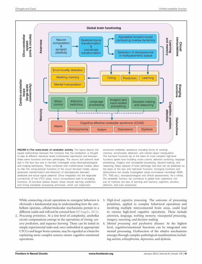
FIGURE 4 | The meta-levels of cerebellar activity. The figure depicts thecausal relationships between the functions that the cerebellum is thoughtto play at different operative levels (meta-levels hypothesis) and betweenthese same functions and brain pathologies. The neuron and network levellays in the blue box and is normally investigate using electrophysiologicaland imaging techniques. These combined with mathematical models, allowto infer the computational functions of the circuit (forward model, variousparameter transformation and detection of discrepancies betweenpredicted and actual signal patterns). Once integrated into the large-saleconnectivity of the CTCC loops, circuit computations lead to emergingfunctions. At low-level (yellow boxes), these include learning, prediction,and timing (cerebellar processing primitives), which can implement
structured cerebellar operations including forms of workingmemory, error/novelty detection, and mental object manipulation.The low-level functions lay at the basis of more complex high-levelfunctions (green box) including motor control, attention switching, languageprocessing, imagery and visuospatial processing, decision-making, andreasoning. Major aspects of brain pathology (red box) can be predicted onthe basis of the low- and high-level functions. Emerging functions anddysfunctions are usually investigated using non-invasive recordings (fMRI,DTI, TMS etc.), neuropsychological and clinical assessments. As a whole,the cerebellar function can contribute to global brain operations notjust of motricity but also of learning and memory, cognition, emotion,attention, and even awareness.
While connecting circuit operations to emergent behaviors isobviously a fundamental step in understanding how the cere-bellum operates, cellular/molecular mechanisms pertain to adifferent realm and will not be covered here (D’Angelo, 2011).
2. Processing primitives. At a low-level of complexity, cerebellarcircuit computations emerge in the operations of timing, sen-sory prediction, and sequence learning. These can be tested insimple experimental tasks and, once embedded in appropriateCTCCs and larger brain systems, may be regarded as a basis forexplaining more complex sensory-motor cognitive-emotionaloperations.
3. High-level cognitive processing. The outcome of processingprimitives, applied to complex behavioral operations andinvolving multiple interconnected brain areas, could leadto various high-level cognitive operations. These includeattention, language, working memory, visuospatial processing,imagery, reasoning, and decision-making.
4. Mental processing and psychiatric diseases. At the highestlevel, cognitive/emotional functions can be integrated intomental processing. Dysfunction of the relative mechanismsemerges through complex pathological manifestations includ-ing autism, schizophrenia, depression, and dyslexia.
Frontiers in Neural Circuits www.frontiersin.org January 2013 | Volume 6 | Article 116 | 10

D’Angelo and Casali Unified cerebellar function
THE CEREBELLAR PROCESSING PRIMITIVESUnderstanding how the cerebellum contributes to so many appar-ently disparate functions would be an enormous step forward asit would mean understanding the common processing primitivesof the cerebellar circuit. The most plausible hypothesis is that thecerebellum has a predictive function, i.e., the ability to anticipateincoming information and thus to ensure that actions correctlyanticipate changes in the environment (Moberget et al., 2008).This hypothesis has two parts. The timing hypothesis postulatesthat the cerebellum is critical for representing the temporal rela-tionship between task-relevant events, while the sensory predictionhypothesis postulates that it is critical in generating expectan-cies regarding incoming information (Ivry et al., 2002). The twohypotheses are not mutually exclusive; rather, they seem to be setat two different hierarchical levels, with timing being more ele-mentary than prediction. Indeed, whereas timing is merely theestablishment of an ordered relationship between two elements,the ability to predict future patterns (as in a forward controller)requires, in addition, the ability to compare different incomingpatterns and predict their consequences on the basis of inter-nally stored memory. Computationally, timing requires only oneprocessing line while sensory prediction requires several. In thePellionisz and Llinas hypothesis (Pellionisz and Llinàs, 1982), thissensory prediction corresponds to a tensorial transformation inthe spatiotemporal hyperspace of the cerebellar circuit. Finally,the cerebellum is likely to use internal memories to adapt itscomputational schemes. The meaning of cerebellar learning hasbeen hotly debated, with controversy often arising over the roleof long-term synaptic plasticity in motor learning. Nonetheless,compelling evidence suggests that learning helps to automate tim-ing and sensory prediction with respect to specific motor andcognitive sequences.
TIMINGMotor coordination, which fails in cerebellar patients, is essen-tially a precise spatiotemporal sequence of movements of oneor more body segments, which must show appropriate posi-tion, velocity, and acceleration. As Ivry underlines (Ivry, 2000),the cerebellum probably operates as an internal timing systemproviding a precise temporal representation for motor and non-motor tasks. Experiments of “irregularity detection,” measuringcortical mismatch-negativity, have indicated that the cerebel-lum selectively contributes to processing the temporal propertiesof stimuli (Ivry, 2000). With regard to time estimation abil-ity (timing), a recent review (Koch et al., 2009b) showed thatthe cerebellum is crucial when normal subjects are required toestimate the passage of brief time intervals and when time is com-puted in relation to given salient events. In turn, circuits involvingthe striatum and substantia nigra, which project to the PFC, aremainly implicated in processing supra-second time intervals inrelationship with various cognitive functions.
One critical issue in physics and biology is velocity estimation,a process that could occur in different locations in the brain, suchas the thalamo-cortical circuit (Ahissar et al., 2000; Szwed et al.,2003). As the cerebellum is a dedicated space-time processor, itis to be expected that it is also involved in velocity estimation.Indeed, a recent fMRI study (O’Reilly et al., 2008) identified a
region in the posterior cerebellum (lobule VII crus 1) that isselectively activated during velocity judgment tasks (prospectivespatiotemporal model). Conversely, when perceptual judgmentsare based only on the spatial (direction) characteristics of anobject, this specific area is not significantly activated. Moreover,the functional connectivity between the posterior cerebellum andthe anterior putamen (bilaterally), which is involved in timing(Matell and Meck, 2004), is enhanced during the velocity judg-ment task, which is essentially perceptual, with an only minimalmotor component.
PREDICTIONAs we have pointed out, the cerebellum has been considered to actas a forward controller (Miall and Reckess, 2002; Wolpert et al.,1998) implementing the contravariant transformations that arenecessary in order to convert predictive sensory plans into motorrepresentations. The involvement of the cerebellum is shown bythe ability to predict the sensory consequences of one’s ownmotor actions. Typically, in the absence of visual feedback, cere-bellar patients have great difficulty in estimating the directionof pointing (Synofzik et al., 2008). The cerebellum signals dis-crepancies between predicted and actual sensory consequences ofmovements, triggering appropriate corrections. In a recent study,subjects were required to use their right hand to move a roboticarm; the motion of this arm determined the position of a sec-ond robotic arm, which made contact with subject’s left palm.Computer-controlled delays were introduced between the move-ment of the right hand and the tactile stimulation on the leftpalm. Activity in the right lateral cerebellar cortex, measured withPET, showed a positive correlation with delay, i.e., with the timeprediction error (Blakemore et al., 2001). This suggests that thecerebellum is less activated by a movement that generates a tactilestimulation than by a movement that does not (which signifies anerror due to lack of sensorial feedback from the target). A sim-ilar phenomenon is seen with tickling, whose sensory effect issuppressed during self-stimulation (which signifies perfect can-cellation of error) (Blakemore et al., 1998a). Accordingly, thesomatosensory cortex is significantly more activated by an exter-nally generated tactile stimulus than by a self-generated one, andthe cerebellum has been shown to provide the signal needed toattenuate the sensory responses to self-generated tactile stimuli(Blakemore et al., 1998a, 1999; Blakemore and Sirigu, 2003).
LEARNINGAnother basic function of the cerebellum is sequence learning. Ina scenario simulating the absence of coordination in ataxia, Shinand Ivry (2003) showed that patients with cerebellar lesions werenot able to learn simultaneously presented spatial and temporalsequences (conversely, patients with Parkinson’s disease were ableto learn these sequences, but not the relationship between them).
Along the same lines, cognitive sequencing functions canbe selectively damaged in patients with cerebellar lesions; forexample, patients with left-side cerebellar lesions perform poorlyon script sequences based on pictorial material and patientswith right-side cerebellar lesions on script sequences requir-ing verbal elaboration (Leggio et al., 2008). These deficitswere not correlated with general intelligence, or with general
Frontiers in Neural Circuits www.frontiersin.org January 2013 | Volume 6 | Article 116 | 11
D’Angelo and Casali Unified cerebellar function
neuropsychological impairment. Furthermore, they were foundboth in patients with focal lesions and in subjects with degen-erative cerebellar pathologies. It is noteworthy that when thesepatients were asked to order a set of cards representing sev-eral behavioral sequences, they were unable to work out thecorrect order, even though they could correctly describe andunderstand the meaning of the single cards. Interestingly, whilecerebellar patients are not necessarily impaired in learning simplevisual or spatial sequences, their ability to discriminate differ-ent durations of auditory stimuli is generally impaired. Indeed,learning sequences of auditory tones with different durations hasbeen found to be rather difficult for cerebellar patients, eventhough the same patients can normally learn visual sequencesand sequences of tones with different frequencies but not differ-ent durations (Frings et al., 2006; Ivry and Keele, 1989). Thesedata are obviously consistent with the “timing hypothesis”; how-ever, the impairment in script sequences could be related to moreabstract cognitive processes and possibly to a lack of executivefunctions.
The role played by cerebellar structures in sequence learningdepends on experience-related factors; in motor sequence learn-ing tasks, the cerebellum shows prominent activation during earlyphases of learning; instead, after extended practice, the activationis located mainly at the level of the basal ganglia (Doyon et al.,2002). Notably, within the early phase of learning, the activationhas been found to shift gradually from the cerebellar cortex tothe deep cerebellar nuclei (Medina and Mauk, 2000; Shadmehrand Mussa-Ivaldi, 2012). Moreover, some researchers hypothesizethat the cerebro-cerebellar loop is primarily involved in motoradaptation processes (e.g., adapting to environmental changes orperturbations), rather than in effective motor learning processes(e.g., learning new sequences of movements), which could be pro-cessed by cerebro-striatal circuits (Doyon et al., 2003; Debas et al.,2010). The cerebellum, coupled with the PFC, is particularlyimportant in learning new visuomotor procedures by imitation(Petrosini, 2007) in the manner of mirror neuron effects. Finally,cerebellar damage can lead to severe impairment of non-motorassociative learning independently of motor alteration (Drepperet al., 1999).
THE CEREBELLUM AND HIGH-LEVEL COGNITIVEPROCESSINGThe timing, predictive, and learning properties of the cerebellum,once integrated within the circuits formed with the cerebral cor-tex, basal ganglia, and limbic system, can lead to control of morecomplex cognitive/emotional functions, including attention, lan-guage, memory, imagery, and reasoning.
ATTENTIONThe cerebellar contribution to attentive functions has beenrevealed in several physiological and pathological conditions.Both autistic and cerebellar patients show a selective impair-ment in attention shifting from visual to auditory stimuli,although attention focusing is normal (Courchesne et al., 1994a).Moreover, the cerebellum, controlling the precision of saccades,probably plays an important role in orienting attention to a visualcue (especially in covert attention tasks). This role seems to be
linked to procedural spatial learning functions, which are stronglyrelated to the ability of the cerebellum to learn goal-directed tra-jectories, as recently supported by experimental results (Burguièreet al., 2005) and computational modeling (Passot et al., 2009).
Indeed, patients with cerebellar lesions are able to correctlyorient visual attention but their reaction times are rather slow(800 and 1200 ms) compared with those of normal control sub-jects (100 ms on average) (Townsend et al., 1999). Attentionswitching is reinforced when subjects have to reassign motorresponses to different stimuli. In agreement with this “attentivehypothesis,” some cerebellar areas show significant activation,measured with fMRI, during early phases of skill learning (bothfor motor and non-motor skills) and during pure visual attentiontasks (Allen et al., 1997).
One theory is that the primary role of attention is to gen-erate time-based expectancies of sensory information (Ghajarand Ivry, 2009). Essentially the suggestion is that, the higherthe level of attention, the lower the performance variability,because the subject is less likely to be distracted by irrele-vant information. The authors observe that the cerebellum isconstantly activated after an attentional cue, independently ofactual execution of movements, and even if the preparationof a potential motor response may be required. Accordingly,the cerebellum is bilaterally activated when a cue precedes thebeginning of a motor task, whilst the primary motor cor-tex is activated only—and mainly contralaterally—during theexecution of the task itself (Cui et al., 2000). Furthermore,it has been shown that PFC-projecting zones of the cerebel-lum process the symbolic content of sensory cues (Balsters andRamnani, 2008). Ghajar and Ivry argue that the cerebellummay be actively involved in an attentional network comprisingmainly the PFC, the inferior parietal lobule, and the cerebellumitself. The specialized role of the cerebellum might be to helpencode the precise timing of sensory predictions. Cerebellar pre-dictive activity probably works in a time frame of 2.5 s, so thatevents that fall within this window can be considered temporallybound.
Thus, according to Ghajar and Ivry’s hypothesis, the predictivefunction of the cerebellum may be seen as a defining trait of atten-tion. However, we can speculate that, in many tasks, attention isnot necessarily closely bound up with sensory anticipation. Theexecution of visual search and feature match tasks, for example,may not rely on anticipatory mechanisms and may not involve thecerebellum directly. Nevertheless, cerebellar patients can fail intasks of this kind, too, because impaired ocular movement controlmay lead to incomplete exploration of stimuli.
LANGUAGE PROCESSING AND VERBAL WORKING MEMORYThe cerebellum is deeply implicated in language, involving bothmotor and cognitive processing organized in the “phonolog-ical loop.” Cerebellar pathology impairs acquisition of motorskills and primary articulatory abilities and the resulting reducedarticulation speed impairs working memory for verbal material,reducing sensitivity to the onset, rime, and phonemic structureof language. This impairment of the phonological loop, in turn,leads to difficulty in language acquisition and dyslexia (Nicolsonet al., 2001b) (see below).
Frontiers in Neural Circuits www.frontiersin.org January 2013 | Volume 6 | Article 116 | 12

D’Angelo and Casali Unified cerebellar function
Cerebellar damage can result in impairment of verbal workingmemory (Justus et al., 2005). Cerebellar patients demonstrate areduction of the “phonological similarity effect” (normal sub-jects show more difficulties in memorizing phonologically sim-ilar words than phonologically dissimilar ones). Desmond et al.(1997) attempted to clarify the difference between the cerebel-lar contribution to phonological “rehearsal” mechanisms and toproper verbal working memory processes. During simple letterrepetition tasks under fMRI, specific areas of the posterior ver-mis (lobules VI and VIIA) and of the cerebellar hemispheres (leftsuperior HVIIA, right HVI) were activated. The same areas wereactivated together with an additional part of the right cerebellarhemisphere (HVIIB) during a sequential verbal working mem-ory task. It was hypothesized that (1) HVIIA and HVI activationsrepresent input from the frontal lobes (which are connected withthe articulatory control processes of verbal working memory) andthat (2) HVIIB reflects input from temporal and parietal areas(which, in turn, are probably the key areas of the phonologi-cal store), and that the function of the cerebellum during verbalworking memory tasks could be to compare the output of sub-vocal articulation with the content of the phonological store. Theverbal working memory deficit in cerebellar subjects is specificand is, both “forward and backward,” independent of dysarthricsymptoms, which suggests that the cerebellum is involved inthe initial phonological encoding and, possibly, in strengtheningmemory traces (Ravizza et al., 2006). In normal subjects, single-pulse TMS delivered to the cerebellum during the encoding phaseof a verbal working memory test does not affect the accuracyof the performance but lengthens the reaction times (Desmondet al., 2005). Clearly, the involvement of the cerebellum in linguis-tic processing reflects the role of this structure in timing, learning,prediction, and attention.
Cerebellar patients show poor performances on phonologi-cal verbal fluency tasks, but not on semantic verbal fluency tasks[(Leggio et al., 2000); but see Smet et al. (2007)], and there-fore show a dissociation between their processing of phonologicaland semantic material. Patients with aright posterolateral cere-bellar lesion are selectively impaired in verb-noun associations(Gebhart et al., 2002). This impairment is not observed when thetask is to associate verbs with visual stimuli (pictures of objects)(Richter et al., 2004). It should be noted that cerebellar patients,unlike patients with Parkinson’s disease, are normally able toperform category learning tasks (Maddox et al., 2005). Whenlistening to disyllabic stimuli, subjects with bilateral cerebellarpathology do not show the phoneme-boundary effect generallyshown by neurologically normal subjects. This may be due to theirimpaired ability to discriminate between intervals of differentduration (Ackermann et al., 1997). Clinical studies also suggestthat cerebellar pathology can play a causal role in prefrontal apha-sic symptoms (Marien et al., 1996). Moreover, cerebellar activityswitches hemispheres (from right to left) according to recruit-ment of right PFC, during linguistic tasks, in aphasia followinga stroke of left cerebral hemisphere (Connor et al., 2006).
The (right) cerebellum is strongly activated during seman-tic disambiguation tasks (Bedny et al., 2008) and, bilaterally,during lexical decision tasks with semantic priming (Rissmanet al., 2003). The cerebellum is activated during different kinds
of verb-noun association tasks (Seger et al., 2000). Also, thecerebellum is strongly activated by semantic discrimination tasksand the intensity of the activation correlates positively with thedifficulty of the task (Xiang et al., 2003). Finally, it should benoted that cerebellar theta-burst stimulation with TMS has beenshown to selectively enhance associative priming, while semanticpriming was unaffected (Argyropoulos, 2011).
IMAGERY AND VISUOSPATIAL PROCESSINGThe cerebellum is involved in pure imagery processes, both motor(Ryding et al., 1993; Naito et al., 2002) and visual (Ishai et al.,2000; Mellet et al., 2000). Indeed, patients affected by unilat-eral cerebellar stroke show slowed or impaired motor imagery(González et al., 2005; Battaglia et al., 2006). Moreover, cerebellarpatients are impaired in tests of mental rotation of objects (a typ-ical example of a visual imagery process) while, at the same time,failing to show significant deficits in tasks evaluating basic per-ceptual functioning or sensory discrimination (Molinari et al.,2004). Some cerebellar patients show purer perceptual alterations,such as hemispatial neglect (Silveri et al., 2001), and there is evi-dence that the cerebellum could be involved in metric judgmentprocesses, as tested in the line bisection task (Fink et al., 2000).
The neural networks involved in imagery processes show astrong inter-individual and inter-trial variability; for example,Gerardin et al. (2000) found the cerebellum to be constantlyactivated during actual execution of motor actions, whilst thereemerged strong inter-individual differences in its degree of activa-tion during the execution of motor imagery tasks. Along the samelines, Grealy and Lee recently described a cerebellar patient foundto be more impaired in monitoring imagined simple actions thanin controlling the actual execution of the same actions (Grealyand Lee, 2011). Conversely, a different study reported cerebel-lar activation only during actual execution of motor acts andnot while imaging the same acts (Nair et al., 2003) and a fur-ther one reported reduced cerebellar activity during imaginedmovements compared with actual execution of the same move-ments (Lotze et al., 1999). These heterogeneous results may beexplained by individual differences, differences in the nature ofthe cerebellar lesions, and in the complexity or novelty of the tasksinvolved. However, another possible reason for the aforemen-tioned differences could be that the cerebellum is actively engagedin manipulating and monitoring mental images rather than ingenerating them. In the last two studies (Lotze et al., 1999; Nairet al., 2003), the subjects were asked to imagine themselves exe-cuting relatively simple finger-tapping movements. Conversely,in the other study (Grealy and Lee, 2011) the patient was askedto imagine himself doing a pointing movement toward a spe-cific location in space and to guess the amount of time requiredto execute the complete movement. Thus, in this case, the sub-ject (who showed no difficulties of any kind in generating mentalimages) needed to actively monitor his motor imagery processand to estimate specific spatiotemporal information. Similarly,in the other reported studies linking the cerebellum with motorimagery, subjects were required to extrapolate some specific infor-mation from their imagery processes and/or to mentally imaginerather complex activities, such as playing tennis. In the same way,visual imagery tasks often require subjects to infer some kind
Frontiers in Neural Circuits www.frontiersin.org January 2013 | Volume 6 | Article 116 | 13

D’Angelo and Casali Unified cerebellar function
of information from the mentally generated images and/or toactively manipulate these mental images (e.g., mental rotations).It is thus possible that the cerebellum is primarily engaged inmanipulating mental images and in estimating spatiotemporalinformation related to dynamic motor imagery processes, whilstthe pure generation of mental images probably does not relyprimarily on cerebellar computations.
Furthermore, studies on hemicerebelloctomized rats, not dis-playing pure (declarative) spatial memory alteration, suggestthat the cerebellum can play a major role in spatial navigation(Petrosini et al., 1998; Foti et al., 2010) and could be involved indeveloping procedural spatial search strategies.
DECISION-MAKING AND REASONINGThe cerebellum is involved in decision-making under uncertainty(Blackwood et al., 2004) (probabilistic reasoning), which suggeststhat it can construct probabilistic models of external events. Ina two-alternative forced-choice task condition, brain processingadvances in four stages: processing of sensory information, optionevaluation, intention formation, and, finally, action execution. Ina recent MEG study (Guggisberg et al., 2007), the cerebellumand the inferior parietal cortex showed high frequency activity(gamma-band) during the intention formation and action exe-cution stages (and, in some conditions, also during the optionevaluation stage, mainly when all the options had the same value).
The cerebellum is also likely to be involved in reasoningprocesses of different types. For example, cerebellar activationhas been observed during probabilistic and deductive reasoning(Osherson et al., 1998). Interestingly, deductive reasoning prefer-entially activates the left cerebellar hemisphere, while inductivereasoning activates the right cerebellar hemisphere (Goel andDolan, 2004). Cerebellar activity in deductive reasoning seemsto be independent of the presence/absence of semantic content(Goel et al., 2000), and also of its nature, concrete, or abstract(Goel and Dolan, 2001).
Although the meaning, if any, of cerebellar activation in rea-soning is not fully understood, the cerebellum is thought to takepart in creating and controlling adaptive working models of theenvironment, in cooperation with cortical structures, mainly thePFC (Vandervert, 2003; Vandervert et al., 2007). Indeed, thereis interesting evidence that logical reasoning could be based onspecific mental models and that, in turn, the internal struc-ture of these models could directly influence the reasoning pro-cess (Johnson-Laird, 1980; Schaeken et al., 1996; Johnson-Laird,2001). Therefore, the cerebellum could play an important role inmanipulating the mental models required for logical reasoning.
MENTAL PROCESSING: CEREBELLAR INVOLVEMENTIN NEUROPSYCHIATRIC DISORDERSAbnormal cerebellar processing can lead to alterations in mentalfunctions. Cerebellar patients often show mood disorders, per-sonality change, cognitive disorders, and dementia which maybe integrated into the pathological frameworks of schizophrenia,depression, autism, and dyslexia. The rate of psychiatric mor-bidity associated with cerebellar degenerative diseases is aboutdouble that found in normal subjects (Leroi et al., 2002): 77% ofpatients with cerebellar degenerative diseases are affected by
psychiatric disorders, compared with only 41% of neurologi-cally healthy control subjects. Interestingly, the components ofcognitive processing related to cerebellar activity also appear tobe related to the pathogenesis of these diseases.
SCHIZOPHRENIASchizophrenia is a mental disorder characterized by a dissociationbetween internal representations and external reality. It is knownthat “cognitive dysmetria,” typical of psychoses like schizophre-nia, is also observed in cerebellar patients. A role of the cerebellumin early onset schizophrenia was recently reported in a DTI studywhich revealed reduced fractional anisotropy in the white matterof the parietal association cortex and in the left cerebellar pedun-cle (Kyriakopoulos et al., 2008). Moreover, neurological soft signsin schizophrenic patients are inversely correlated with volumeof the right cerebellar hemisphere (Bottmer et al., 2005). Thecerebellar dysfunction may impair the ability of schizophrenicsubjects to recognize an action on the basis of a subject’s inten-tion. Indeed, schizophrenic patients are not able to correctlyestimate the sensory consequences of their own actions (Synofziket al., 2010), a deficit usually observed in cerebellar patients. Inother words, this means that the consequences of their actionsare not in agreement with the expected sensory results of theseactions and with the subject’s intentions. This is hardly surprisinggiven the predictive function of the cerebellum.
Neuroanatomically, there is evidence showing that damageto a CTCC could be the primary pathophysiological alterationin schizophrenic patients (Konarski et al., 2005). Several imag-ing studies (CT, MRI) have reported abnormal volume of thecerebellar vermis (either hypoplasia orhyperplasia), while othershave reported global cerebellar atrophy. Cerebellar hypoactiva-tion (or even non-activation) has been measured with fMRI incognitive tasks involving the prefrontal-cerebellar loop, tasks suchas the (1) Wisconsin Card-Sorting Test, (2) working memory[n–back] task, and (3) periodic sequence-learning tasks. An onto-genetic substrate can be traced back to abnormalities in infantmotor development (IMD) and executive function development(Ridler et al., 2006). IMD and executive function development arenormally associated with increased gray matter density (GMD)in the premotor cortex, striatum, and cerebellum, reinforcing thefronto-cerebellar network. Schizophrenic patients have delayedIMD and deficits in executive functions correlating with disrup-tion of the fronto-cerebellar network. In postmortem studies,reduction in the size of the anterior vermis in schizophrenicpatients is associated with reductions in the density and sizeof Purkinje cells. Moreover, the synaptogenesis process couldbe impaired, both for excitatory and inhibitory neurons, and acore alteration may concern the NMDA receptors and synapticplasticity (Stephan et al., 2009).
Overall, in view of the reported impairment of the cerebellumand Purkinje cells, it can be hypothesized that the neural basisof schizophrenia might partially overlap that of autism (Bosoet al., 2010). Considering the general function of the cerebel-lum, it is possible that schizophrenic patients are impaired inswitching from an egocentric frame of reference to an allocentricone (Yakusheva et al., 2007). In agreement with this hypothe-sis, when asked to imagine an object from another perspective,
Frontiers in Neural Circuits www.frontiersin.org January 2013 | Volume 6 | Article 116 | 14
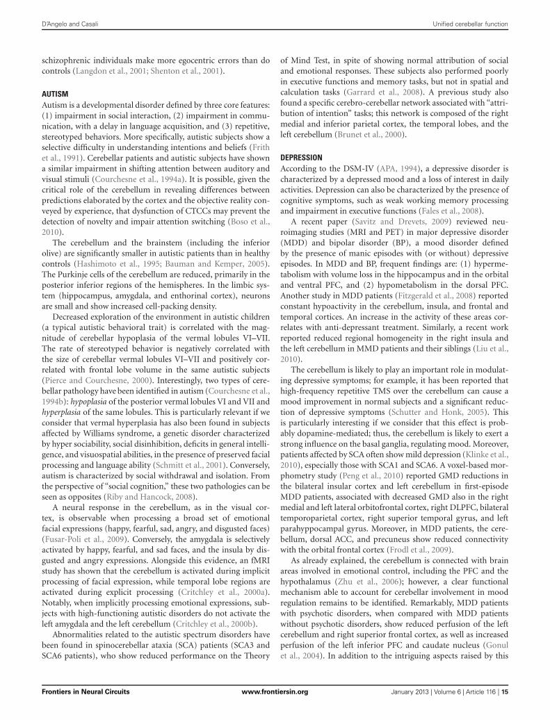
D’Angelo and Casali Unified cerebellar function
schizophrenic individuals make more egocentric errors than docontrols (Langdon et al., 2001; Shenton et al., 2001).
AUTISMAutism is a developmental disorder defined by three core features:(1) impairment in social interaction, (2) impairment in commu-nication, with a delay in language acquisition, and (3) repetitive,stereotyped behaviors. More specifically, autistic subjects show aselective difficulty in understanding intentions and beliefs (Frithet al., 1991). Cerebellar patients and autistic subjects have showna similar impairment in shifting attention between auditory andvisual stimuli (Courchesne et al., 1994a). It is possible, given thecritical role of the cerebellum in revealing differences betweenpredictions elaborated by the cortex and the objective reality con-veyed by experience, that dysfunction of CTCCs may prevent thedetection of novelty and impair attention switching (Boso et al.,2010).
The cerebellum and the brainstem (including the inferiorolive) are significantly smaller in autistic patients than in healthycontrols (Hashimoto et al., 1995; Bauman and Kemper, 2005).The Purkinje cells of the cerebellum are reduced, primarily in theposterior inferior regions of the hemispheres. In the limbic sys-tem (hippocampus, amygdala, and enthorinal cortex), neuronsare small and show increased cell-packing density.
Decreased exploration of the environment in autistic children(a typical autistic behavioral trait) is correlated with the mag-nitude of cerebellar hypoplasia of the vermal lobules VI–VII.The rate of stereotyped behavior is negatively correlated withthe size of cerebellar vermal lobules VI–VII and positively cor-related with frontal lobe volume in the same autistic subjects(Pierce and Courchesne, 2000). Interestingly, two types of cere-bellar pathology have been identified in autism (Courchesne et al.,1994b): hypoplasia of the posterior vermal lobules VI and VII andhyperplasia of the same lobules. This is particularly relevant if weconsider that vermal hyperplasia has also been found in subjectsaffected by Williams syndrome, a genetic disorder characterizedby hyper sociability, social disinhibition, deficits in general intelli-gence, and visuospatial abilities, in the presence of preserved facialprocessing and language ability (Schmitt et al., 2001). Conversely,autism is characterized by social withdrawal and isolation. Fromthe perspective of “social cognition,” these two pathologies can beseen as opposites (Riby and Hancock, 2008).
A neural response in the cerebellum, as in the visual cor-tex, is observable when processing a broad set of emotionalfacial expressions (happy, fearful, sad, angry, and disgusted faces)(Fusar-Poli et al., 2009). Conversely, the amygdala is selectivelyactivated by happy, fearful, and sad faces, and the insula by dis-gusted and angry expressions. Alongside this evidence, an fMRIstudy has shown that the cerebellum is activated during implicitprocessing of facial expression, while temporal lobe regions areactivated during explicit processing (Critchley et al., 2000a).Notably, when implicitly processing emotional expressions, sub-jects with high-functioning autistic disorders do not activate theleft amygdala and the left cerebellum (Critchley et al., 2000b).
Abnormalities related to the autistic spectrum disorders havebeen found in spinocerebellar ataxia (SCA) patients (SCA3 andSCA6 patients), who show reduced performance on the Theory
of Mind Test, in spite of showing normal attribution of socialand emotional responses. These subjects also performed poorlyin executive functions and memory tasks, but not in spatial andcalculation tasks (Garrard et al., 2008). A previous study alsofound a specific cerebro-cerebellar network associated with “attri-bution of intention” tasks; this network is composed of the rightmedial and inferior parietal cortex, the temporal lobes, and theleft cerebellum (Brunet et al., 2000).
DEPRESSIONAccording to the DSM-IV (APA, 1994), a depressive disorder ischaracterized by a depressed mood and a loss of interest in dailyactivities. Depression can also be characterized by the presence ofcognitive symptoms, such as weak working memory processingand impairment in executive functions (Fales et al., 2008).
A recent paper (Savitz and Drevets, 2009) reviewed neu-roimaging studies (MRI and PET) in major depressive disorder(MDD) and bipolar disorder (BP), a mood disorder definedby the presence of manic episodes with (or without) depressiveepisodes. In MDD and BP, frequent findings are: (1) hyperme-tabolism with volume loss in the hippocampus and in the orbitaland ventral PFC, and (2) hypometabolism in the dorsal PFC.Another study in MDD patients (Fitzgerald et al., 2008) reportedconstant hypoactivity in the cerebellum, insula, and frontal andtemporal cortices. An increase in the activity of these areas cor-relates with anti-depressant treatment. Similarly, a recent workreported reduced regional homogeneity in the right insula andthe left cerebellum in MMD patients and their siblings (Liu et al.,2010).
The cerebellum is likely to play an important role in modulat-ing depressive symptoms; for example, it has been reported thathigh-frequency repetitive TMS over the cerebellum can cause amood improvement in normal subjects and a significant reduc-tion of depressive symptoms (Schutter and Honk, 2005). Thisis particularly interesting if we consider that this effect is prob-ably dopamine-mediated; thus, the cerebellum is likely to exert astrong influence on the basal ganglia, regulating mood. Moreover,patients affected by SCA often show mild depression (Klinke et al.,2010), especially those with SCA1 and SCA6. A voxel-based mor-phometry study (Peng et al., 2010) reported GMD reductions inthe bilateral insular cortex and left cerebellum in first-episodeMDD patients, associated with decreased GMD also in the rightmedial and left lateral orbitofrontal cortex, right DLPFC, bilateraltemporoparietal cortex, right superior temporal gyrus, and leftparahyppocampal gyrus. Moreover, in MDD patients, the cere-bellum, dorsal ACC, and precuneus show reduced connectivitywith the orbital frontal cortex (Frodl et al., 2009).
As already explained, the cerebellum is connected with brainareas involved in emotional control, including the PFC and thehypothalamus (Zhu et al., 2006); however, a clear functionalmechanism able to account for cerebellar involvement in moodregulation remains to be identified. Remarkably, MDD patientswith psychotic disorders, when compared with MDD patientswithout psychotic disorders, show reduced perfusion of the leftcerebellum and right superior frontal cortex, as well as increasedperfusion of the left inferior PFC and caudate nucleus (Gonulet al., 2004). In addition to the intriguing aspects raised by this
Frontiers in Neural Circuits www.frontiersin.org January 2013 | Volume 6 | Article 116 | 15

D’Angelo and Casali Unified cerebellar function
lateralization, these observations suggest that, in MDD, cerebellaractivity correlates more closely with psychotic traits (e.g., delu-sions of control) than with “pure” depressive symptoms. Thecerebellum is also involved in BP, with bipolar patients found toshow poor emotional homeostasis, mania, and cognitive dysfunc-tions (Strakowski et al., 2005) and, on MRI scanning, a volumereduction in region V2 and V3 of the cerebellar vermis (Millset al., 2005; Monkul et al., 2008).
DYSLEXIADevelopmental dyslexia (DD) is characterized by a selective diffi-culty in acquiring reading skills, in spite of normal general intelli-gence (Habib, 2000). There are three main hypotheses regardingthe core deficit responsible for DD symptoms: the magnosys-tem deficit hypothesis (MDH) (Stein, 2001), the phonologicaldeficit hypothesis (PDH) (Ramus, 2003), and the cerebellar deficithypothesis (CDH). The MDH takes its name from the “magno-cellular neurons” of the lateral geniculate nucleus, which mostlyfeed the visual “dorsal stream” dedicated to the analysis of move-ment (the “where” pathway). Normal magnocellular function isnecessary for high motion sensitivity and stable binocular per-ception which, in turn, are essential for proper development oforthographic skills. Many dyslexics show poor visual localiza-tion and their motion sensitivity is impaired, which suggeststhat abnormal development of the magnocellular system couldplay a pathogenetic role in DD. The PDH postulates that DD iscaused by an impaired ability to represent and process speechsounds. The CDH, on the other hand, is based on the observa-tion that dyslexic children show deficits in motor coordination,motor skills, and automatic processing, which suggests that acerebellar dysfunction constitutes the neurological basis of thedisease (Fawcett and Nicolson, 2004). Indeed, in a PET study,dyslexic subjects learning a motor sequence showed abnormal-ities in cerebellar activation during both automatic processingand new learning. An early cerebellar deficit has been hypoth-esized to impair the development of articulatory and writingskills (Nicolson et al., 2001b), and non-fluent articulation wouldengage more attentional resources, thereby impairing sensoryfeedback processing. It can thus be hypothesized that a cerebellarimpairment causes a marked phonological deficit which, coupledwith an automation deficit, results in DD. Cerebellar dysfunctioncan also explain the specific impairment in time estimation tasksshown by dyslexic subjects (Nicolson et al., 2001a). On the whole,the PDH can be seen as a part of the CDH. Conversely, the CDHdoes not completely explain the MDH, even though there is a sub-type of DD characterized by magnocellular impairment (Tallalet al., 2006).
Dyslexics show small right cerebellar anterior lobes on MRI(Eckert et al., 2003), suggesting that a fronto-cerebellar circuitimpairment could indeed cause the symptoms of DD. The rightcerebellum is the brain area that best discriminates dyslexicsfrom healthy control subjects (Pernet et al., 2009). Moreover,dyslexic subjects have symmetric cerebellar (as well as parietaland temporal cortex) gray matter, while healthy controls show agreater asymmetry (Hier et al., 1978; Rae et al., 2002). Therefore,there is substantial evidence that the cerebellum takes part in thepathogenesis of DD.
DISCUSSIONRecently, an impressive amount of new data has revealed a dis-concerting heterogeneity of functional roles attributed to thecerebellum. Here we propose a unified framework that might pro-vide a logical explanation of the numerous operations in whichthe cerebellum is involved.
DIFFERENT INTERPRETATIONS OR DISPARATE OPERATIONALPROCESSES?Ito, highlighting that the cerebellum can acquire forward modelsof a controlled object (e.g., the arm) through practice, proposesthat the cerebellum uses internal models in order to adapt motoracts and mental activities to contextual information (Ito, 1993,2008). By virtue of these models, the cerebellum is able to exertfast, precise, and automated control of well-learned movements.If we also think of thoughts and cognitive processes as controlledobjects, the logical conclusion is that these same internal modelscan be applied to cognitive processing as well. This is hardly sur-prising, as mental products are virtual objects and there may notbe much difference between a “thought” and a “thought to move.”
Along the same lines, Vandervert conjectures that the cerebel-lum is engaged in a dynamic interplay with working memory;his main idea is that repetitive working memory processes areactively adapted by the cerebellum and that this must result inbetter and faster attentional control and, consequently, in moreprecise and better timed cognitive processes (Vandervert, 2003,2007; Vandervert et al., 2007).
Again, a comparable theoretical framework was advancedsome years ago by Courchesne and Allen (1997). They hypothe-sized that the main function of the cerebellum is to predict theinternal conditions required for different cognitive and motoractivities and to rapidly and precisely set those conditions.Arguably, this “predict and prepare” function of the cerebellummust be implemented mainly unconsciously and automatically.Accordingly, the cerebellum is thought to be involved in implicitlearning and, on the contrary, not to play a relevant role in declar-ative, explicit learning (Doyon et al., 1997). The cerebellum islikely to adopt a “trial-and-error” learning rule, unlike the cere-bral cortex and basal ganglia, which seem to learn, respectively,through frequency-based and reward-based rules (Doya, 2000).
Finally, Ivry suggested that the cerebellum is involved in everytask requiring precise timing processes (Ivry et al., 2002), includ-ing the production of skilled movements, eye-blink conditioning,duration discrimination of perceptual events, fast and preciseregulation of attention and working memory, and some specificlinguistic skills.
Other proposals are that the cerebellum is directly involved incognitive/emotional processes, insofar as these are linked to somekind of motor or oculomotor activity (Doron et al., 2010), andthat the main function of the cerebellum is to regulate the acquisi-tion of sensory data across several sensory modalities, and thus tosupport various sensory, motor, and cognitive functions (Petacchiet al., 2005).
Clearly, although these frameworks are valid for interpretingdata sets relative to specific experiments or methodologies, thelink between these observations has remained largely specula-tive or unaddressed. Moreover, the involvement of the cerebellum
Frontiers in Neural Circuits www.frontiersin.org January 2013 | Volume 6 | Article 116 | 16

D’Angelo and Casali Unified cerebellar function
in some cognitive functions, like attention, language, mentalimagery, and reasoning, as well as in neuropsychiatric disordersand emotion, has remained obscure.
A UNIFIED INTERPRETATION THROUGH THE META-LEVELSHYPOTHESISWhat we suggest in this paper is that, as a result of modularconnectivity with the cerebral cortex, causal relationships existbetween the low- and high-level cognitive functions that the cere-bellum is thought to play. Finally, we conjecture that failure of thissystem can lead to mental pathologies. The identified low-levelfunctions, timing, prediction, and learning, directly implementstructured cerebellar operations including forms of working mem-ory, error/novelty detection, and mental object manipulation. Onthe one hand, timing, prediction, and learning have been relatedto cellular and network operations [e.g., see Medina and Mauk(2000); D’Angelo (2011); D’Angelo et al. (2011)], even thoughthere is, as yet, no precise understanding of the mechanismsinvolved. On the other hand, these same functions may lie atthe basis of more complex functions including motor control,attention switching, language processing, imagery and visuospatialprocessing, decision-making, and reasoning. Finally, major aspectsof brain pathology can be predicted on the basis of these samelow- and high-level functions. In some cases, direct evidence ofthese relationships has been demonstrated, while in others theseinter-dependencies are still implicit, providing scope for testing ofthe hypothesis of cerebellar functioning based on its organizationinto meta-levels (Figure 4).
Timing seems the most fundamental of all the low-level func-tions. Timing may be directly explained on the basis of cir-cuit mechanisms, whose cellular and synaptic components havebeen partly revealed [e.g., see D’Angelo and De Zeeuw (2009);D’Angelo et al. (2009)]. Timing is reflected in the ordering ofcomplex sequences ordinarily processed by the cerebellum and itis deeply integrated with prediction and learning.
Sensory prediction, or, more generally, prediction, has beenshown to predominate in motor control, attention switch-ing, working memory, and language processing (Shadmehr andMussa-Ivaldi, 2012), but it is also thought to intervene duringimagery and visuospatial processing (which are also componentsof motor planning) and during decision-making and reasoning.So-called fluid intelligence, a form of cognitive control involvedin executive functions (e.g., in the Tower of Hanoi test), rests ona dynamic sequence of selective attention, planning, storage inthe working memory, hypothesis updating, attention redirecting,and so on. It is thought that the cerebellum is involved, at leastwhen the problem is unusual and unexpected, according to itserror/novelty-detection function. The circuits underlying predic-tion are unclear. It can be supposed that different inputs collide inproper time frames generating patterns that can, at some level, berecognized by pattern detectors. A model based on timing controlin the granular layer and pattern detection in the molecular layerhas been proposed (D’Angelo, 2011).
Learning in the cerebellum, and the original concepts relatedto this, probably need to be revised and extended in the light ofnew cellular and system physiology data. On the one hand, thecerebellar cortex and nuclei are clearly endowed with numerous
mechanisms of long-term synaptic plasticity and therefore prob-ably undergo changes during activity not just at the parallelfiber-Purkinje cell synapse, but also at other synaptic sites (Hanselet al., 2001; Jorntell and Hansel, 2006; Ohtsuki et al., 2009; Gaoet al., 2012). These distributed changes could have very differ-ent meanings, for example that of controlling the spatiotemporaldynamics of signal processing in the granular layer and percep-tron operations in Purkinje cells. On the other hand, functionalimaging shows that the cerebellum is primarily involved whenunknown problems or circumstances are encountered. This sug-gests that the cerebellum can incorporate specialized forms ofprocedural memory that can be reconfigured and transmittedto other brain areas. This learning can modify cerebellar spa-tiotemporal processing in terms of timing, pattern recognition,and coincidence detection, ultimately affecting the internal for-ward model and the consequent predictive properties. Cerebellarlearning could contribute to working memory.
A puzzling implication of all this is that cerebellar processingmight, ultimately, take part in generating conscious and coherentrepresentations of the world, a function typically ascribed to thethalmo-cortical loops. Indeed, rapid continuous cerebellar pro-cessing in the cerebellar circuit through feedforward independentmodules could enhance the immediate and continuous represen-tation of events, which is one of the key aspects of consciousness(Addis et al., 2009; Nyberg et al., 2010; Szpunar, 2010, 2011).Also worth noting is the specific involvement of the cerebellum inelaborating problems of a statistical nature. In this case, it can beimagined that, by imposing an internal dynamic model, the cere-bellum helps to automatically elaborate the trend in a complexdata distribution on the basis of its previous acquisition of themost probable data sets. Interestingly, many biologically relevantproblems have a statistical nature and the role of the cerebellumin this sense should be further explored.
A dramatic translation of low-level into high-level functionsis observed in mental processing and even more clearly inthe related dysfunction occurring in certain brain diseases. Inschizophrenia, there is major failure of prediction-based com-parison between internal and external representations, and ofcoordinate transformation and therefore manipulation of mentalmodels. Similarly, autism involves a failure in redirecting atten-tion, which can either be locked into internal contents or behyperfocused on selective objects. In depression, psychotic symp-toms may be regarded as a loss of internal coherence betweeninternally and externally generated signals, with consequent dys-regulation of mood homeostasis. Finally, in dyslexia, a combi-nation of failures in the phonological loop involving workingmemory and recognition and manipulation of mental objectscould be involved.
CONCLUSIONSThe meta-levels hypothesis provides a key through which toorder the multitude of manifestations of cerebellar physiologyand pathology and reconcile the basic cerebellar functional mech-anisms with the emerging properties of the network. The meta-levels hypothesis leads to testable predictions and opens the waysfor new experimental designs. These can be broadly divided intothose addressing (1) how the olivo-cerebellar system generates
Frontiers in Neural Circuits www.frontiersin.org January 2013 | Volume 6 | Article 116 | 17

D’Angelo and Casali Unified cerebellar function
its internal representations and operations, (2) how the emerg-ing functions derive from connections of the cerebellum withother brain structures, and (3) how dysfunction of the systemcould lead to pathology. In general, tools like genetic engineeringin experimental animals, large-scale mathematical modeling andnon-invasive stimulation/recording technology (like TMS andfMRI) in humans could provide valuable information. For exam-ple, genetic mutations impairing LTP (Long-Term Potentiation)or LTD (Long-Term Depression) could be used to investigate thepotential role of the various forms of long-term synaptic plastic-ity expressed by cerebellar synapses not just with respect to motorlearning but also with respect to cognitive processing extendingthrough the CTCCs. Moreover, the role of cellular properties oncircuit and system computations could be analyzed using detailedmathematical models, the CTCCs could be investigated by DTI,
and their functional activation during specific tasks identified byfMRI. These same techniques could improve brain disease anal-ysis and therapy. For example, cerebellar TMS has an impact ondifferent pathologies, including Parkinson’s disease (Koch et al.,2009a), epilepsy (Brighina et al., 2006), and stroke (Webster et al.,2006). Ultimately, this analysis could contribute to the devel-opment of a theory on global brain functioning in which thecerebellum is considered an integral part and not just a structurepurely devoted to motor control.
ACKNOWLEDGMENTSThis work was supported by the European Union (CEREBNETFP7-ITN238686 and REALNET FP7-ICT270434) and by theItalian Ministry of Health (RF-2008-1143418 and RF-2009-1475845) to Egidio D’Angelo.
REFERENCESAckermann, H., Gräber, S., Hertrich,
I., and Daum, I. (1997). Categoricalspeech perception in cerebellar dis-orders. Brain Lang. 60, 323–331.
Addis, D. R., Pan, L., Vu, M. A.,Laiser, N., and Schacter, D. L.(2009). Constructive episodic sim-ulation of the future and the past:distinct subsystems of a core brainnetwork mediate imagining andremembering. Neuropsychologia 47,2222–2238.
Ahissar, E., Sosnik, R., and Haidarliu,S. (2000). Transformation fromtemporal to rate coding in asomatosensory thalamocorticalpathway. Nature 406, 302–306.
Alahyane, N., Fonteille, V., Urquizar,C., Salemme, R., Nighoghossian, N.,Pelisson, D., et al. (2008). Separateneural substrates in the human cere-bellum for sensory-motor adapta-tion of reactive and of scanningvoluntary saccades. Cerebellum 7,595–601.
Albus, J. S. (1972). A theory of cerebel-lar function. Math. Biosci. 10, 25–61.
Allen, G., Buxton, R. B., Wong, E.C., and Courchesne, E. (1997).Attentional activation of thecerebellum independent ofmotor involvement. Science 275,1940–1943.
Amodio, D. M., and Frith, C. D. (2006).Meeting of minds: the medialfrontal cortex and social cognition.Nature 7, 268–277.
Andersson, G., and Oscarsson, O.(1978). Climbing fiber microzonesin cerebellar vermis and theirprojection to different groupsof cells in the lateral vestibu-lar nucleus. Exp. Brain Res. 32,565–579.
Andreasen, N. C., O’Leary, D. S.,Cizadlo, T., Arndt, S., Rezai,K., Ponto, L. L., et al. (1996).Schizophrenia and cognitive
dysmetria: a positron-emissiontomography study of dysfunctio-nal prefrontal-thalamic-cerebellarcircuitry. Proc. Natl. Acad. Sci.U.S.A. 93, 9985–9990.
Andreasen, N. C., Paradiso, S., andO’Leary, D. S. (1998). “Cognitivedysmetria” as an integrative theoryof schizophrenia: a dysfunctionin cortical-subcortical-cerebellarcircuitry? Schizophr. Bull. 24,203–218.
APA. (1994). Diagnostic and StatisticalManual of Mental Disorders 4.Washington, DC: AmericanPsychiatric Association.
Apps, R., and Garwicz, M. (2005).Anatomical and physiological foun-dations of cerebellar informationprocessing. Nat. Rev. Neurosci. 6,297–311.
Apps, R., and Hawkes, R. (2009).Cerebellar cortical organization:a one-map hypothesis. Nat. Rev.Neurosci. 10, 670–681.
Argyropoulos, G. P. (2011). Cerebellartheta-burst stimulation selectivelyenhances lexical associative prim-ing. Cerebellum 10, 540–550.
Arriada-Mendicoa, N., Otero-Siliceo,E., and Corona-Vazquez, T. (1999).[Current concepts regarding thecerebellum and cognition]. Rev.Neurol. 29, 1075–1082.
Baddeley, A., Lewis, V., Eldridge, M.,and Thomson, N. (1984). Attentionand retrieval from long-term mem-ory. J. Exp. Psychol. 113, 518–540.
Balsters, J. H., and Ramnani, N.(2008). Symbolic representationsof action in the human cerebellum.Neuroimage 43, 388–398.
Bartolo, P. D., Gelfo, F., Burello, L.,Giorgio, A. D., Petrosini, L., andGranato, A. (2011). Plastic changesin striatal fast-spiking interneuronsfollowing hemicerebellectomyand environmental enrichment.Cerebellum 10, 624–632.
Battaglia, F., Quartarone, A., Ghilardi,M. F., Dattola, R., Bagnato, S.,Rizzo, V., et al. (2006). Unilateralcerebellar stroke disrupts movementpreparation and motor imagery.Clin. Neurophysiol. 117, 1009–1016.
Bauman, M. L., and Kemper, T. L.(2005). Neuroanatomic observa-tions of the brain in autism: areview and future directions. Int.J. Dev. Neurosci. 23, 183–187.
Bedny, M., McGill, M., and Thompson-Schill, L. S. (2008). Semantic adap-tation and competition during wordcomprehension. Cereb. Cortex 18,2574–2585.
Beldarrain, M. G., García-Moncó,J., Quintana, J., Llorens, V., andRodeño, E. (1997). Diaschisis andneuropsychological performanceafter cerebellar stroke. Eur. Neurol.37, 82–89.
Bell, C. C., Han, V., and Sawtell, N. B.(2008). Cerebellum-like structuresand their implications for cerebellarfunction. Annu. Rev. Neurosci. 31,1–24.
Blackwood, N., Ffytche, D., Simmons,A., Bentallc, R., Murray, R., andHoward, R. (2004). The cerebellumand decision making under uncer-tainty. Brain Res. Cogn. Brain Res.20, 46–53.
Blakemore, S.-J., Frith, C. D., andWolpert, D. M. (2001). The cere-bellum is involved in predictingthe sensory consequences of action.Neuroreport 12, 1879–1884.
Blakemore, S.-J., and Sirigu, A. (2003).Action prediction in the cerebellumand in the parietal lobe. Exp. BrainRes. 153, 239–245.
Blakemore, S.-J., Wolpert, D. M., andFrith, C. D. (1998a). Central cancel-lation of self-produced tickle sensa-tion. Nature 1, 635–640.
Blakemore, S. J., Goodbody, S. J., andWolpert, D. M. (1998b). Predictingthe consequences of our own
actions: the role of sensorimotorcontext estimation. J. Neurosci. 18,7511–7518.
Blakemore, S.-J., Wolpert, D. M., andFrith, C. D. (1999). The cerebel-lum contributes to somatosensorycortical activity during self-produced tactile stimulation.Neuroimage 10, 448–459.
Booth, J. R., Wood, L., Lu, D., Houk,J. C., and Bitan, T. (2007). The roleof the basal ganglia and cerebellumin language processing. Brain Res.1133, 136–144.
Boso, M., Emanuele, E., Prestori, F.,Politi, P., Barale, F., and D’Angelo,E. (2010). Autism and genius: isthere a link? The involvement ofcentral brain loops and hypothesesfor functional testing. Funct. Neurol.25, 15–20.
Bostan, A. C., and Strick, P. L. (2010).The cerebellum and basal gangliaare interconnected. Neuropsychol.Rev. 20, 261–270.
Bottmer, C., Bachmann, S., Pantel, J.,Essig, M., Amann, M., Schad, L.R., et al. (2005). Reduced cerebel-lar volume and neurological softsigns in first-episode schizophrenia.Psychiatry Res. 140, 239–250.
Bower, J. M. (1997). Is the cerebellumsensory for motor’s sake, or motorfor sensory’s sake: the view from thewhiskers of a rat? Prog. Brain Res.114, 463–496.
Bower, J. M. (2002). The organizationof cerebellar cortical circuitry revis-ited: implications for function. Ann.N.Y. Acad. Sci. 978, 135–155.
Brighina, F., Daniele, O., Piazza, A.,Giglia, G., and Fierro, B. (2006).Hemispheric cerebellar rTMSto treat drug-resistant epilepsy:case reports. Neurosci. Lett. 397,229–233.
Brunet, E., Sarfati, Y., Hardy-Bayle,M.-C., and Decety, J. (2000). A PETinvestigation of the attribution of
Frontiers in Neural Circuits www.frontiersin.org January 2013 | Volume 6 | Article 116 | 18
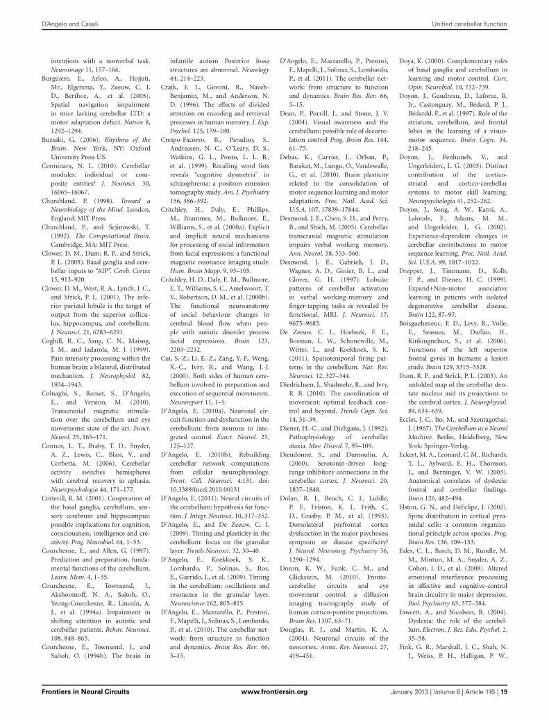
D’Angelo and Casali Unified cerebellar function
intentions with a nonverbal task.Neuroimage 11, 157–166.
Burguière, E., Arleo, A., Hojjati,Mr., Elgersma, Y., Zeeuw, C. I.D., Berthoz, A., et al. (2005).Spatial navigation impairmentin mice lacking cerebellar LTD: amotor adaptation deficit. Nature 8,1292–1294.
Buzsaki, G. (2006). Rhythms of theBrain. New York, NY: OxfordUniversity Press US.
Cerminara, N. L. (2010). Cerebellarmodules: individual or com-posite entities? J. Neurosci. 30,16065–16067.
Churchland, P. (1998). Toward aNeurobiology of the Mind. London,England: MIT Press.
Churchland, P., and Sejinowski, T.(1992). The Computational Brain.Cambridge, MA: MIT Press.
Clower, D. M., Dum, R. P., and Strick,P. L. (2005). Basal ganglia and cere-bellar inputs to “AIP”. Cereb. Cortex15, 913–920.
Clower, D. M., West, R. A., Lynch, J. C.,and Strick, P. L. (2001). The infe-rior parietal lobule is the target ofoutput from the superior collicu-lus, hippocampus, and cerebellum.J. Neurosci. 21, 6283–6291.
Coghill, R. C., Sang, C. N., Maisog,J. M., and Iadarola, M. J. (1999).Pain intensity processing within thehuman brain: a bilateral, distributedmechanism. J. Neurophysiol. 82,1934–1943.
Colnaghi, S., Ramat, S., D’Angelo,E., and Versino, M. (2010).Transcranial magnetic stimula-tion over the cerebellum and eyemovements: state of the art. Funct.Neurol. 25, 165–171.
Connor, L. T., Braby, T. D., Snyder,A. Z., Lewis, C., Blasi, V., andCorbetta, M. (2006). Cerebellaractivity switches hemisphereswith cerebral recovery in aphasia.Neuropsychologia 44, 171–177.
Cotterill, R. M. (2001). Cooperation ofthe basal ganglia, cerebellum, sen-sory cerebrum and hippocampus:possible implications for cognition,consciousness, intelligence and cre-ativity. Prog. Neurobiol. 64, 1–33.
Courchesne, E., and Allen, G. (1997).Prediction and preparation, funda-mental functions of the cerebellum.Learn. Mem. 4, 1–35.
Courchesne, E., Townsend, J.,Akshoomoff, N. A., Saitoh, O.,Yeung-Courchesne, R., Lincoln, A.J., et al. (1994a). Impairment inshifting attention in autistic andcerebellar patients. Behav. Neurosci.108, 848–865.
Courchesne, E., Townsend, J., andSaitoh, O. (1994b). The brain in
infantile autism Posterior fossastructures are abnormal. Neurology44, 214–223.
Craik, F. I., Govoni, R., Naveh-Benjamin, M., and Anderson, N.D. (1996). The effects of dividedattention on encoding and retrievalprocesses in human memory. J. Exp.Psychol. 125, 159–180.
Crespo-Facorro, B., Paradiso, S.,Andreasen, N. C., O’Leary, D. S.,Watkins, G. L., Ponto, L. L. B.,et al. (1999). Recalling word listsreveals “cognitive dysmetria” inschizophrenia: a positron emissiontomography study. Am. J. Psychiatry156, 386–392.
Critchley, H., Daly, E., Phillips,M., Brammer, M., Bullmore, E.,Williams, S., et al. (2000a). Explicitand implicit neural mechanismsfor processing of social informationfrom facial expressions: a functionalmagnetic resonance imaging study.Hum. Brain Mapp. 9, 93–105.
Critchley, H. D., Daly, E. M., Bullmore,E. T., Williams, S. C., Amelsvoort, T.V., Robertson, D. M., et al. (2000b).The functional neuroanatomyof social behaviour changes incerebral blood flow when peo-ple with autistic disorder processfacial expressions. Brain 123,2203–2212.
Cui, S.-Z., Li, E.-Z., Zang, Y.-F., Weng,X.-C., Ivry, R., and Wang, J.-J.(2000). Both sides of human cere-bellum involved in preparation andexecution of sequential movements.Neuroreport 11, 1–5.
D’Angelo, E. (2010a). Neuronal cir-cuit function and dysfunction in thecerebellum: from neurons to inte-grated control. Funct. Neurol. 25,125–127.
D’Angelo, E. (2010b). Rebuildingcerebellar network computationsfrom cellular neurophysiology.Front. Cell. Neurosci. 4:131. doi:10.3389/fncel.2010.00131
D’Angelo, E. (2011). Neural circuits ofthe cerebellum: hypothesis for func-tion. J. Integr. Neurosci. 10, 317–352.
D’Angelo, E., and De Zeeuw, C. I.(2009). Timing and plasticity in thecerebellum: focus on the granularlayer. Trends Neurosci. 32, 30–40.
D’Angelo, E., Koekkoek, S. K.,Lombardo, P., Solinas, S., Ros,E., Garrido, J., et al. (2009). Timingin the cerebellum: oscillations andresonance in the granular layer.Neuroscience 162, 805–815.
D’Angelo, E., Mazzarello, P., Prestori,F., Mapelli, J., Solinas, S., Lombardo,P., et al. (2010). The cerebellar net-work: from structure to functionand dynamics. Brain Res. Rev. 66,5–15.
D’Angelo, E., Mazzarello, P., Prestori,F., Mapelli, J., Solinas, S., Lombardo,P., et al. (2011). The cerebellar net-work: from structure to functionand dynamics. Brain Res. Rev. 66,5–15.
Dean, P., Porrill, J., and Stone, J. V.(2004). Visual awareness and thecerebellum: possible role of decorre-lation control Prog. Brain Res. 144,61–75.
Debas, K., Carrier, J., Orban, P.,Barakat, M., Lungu, O., Vandewalle,G., et al. (2010). Brain plasticityrelated to the consolidation ofmotor sequence learning and motoradaptation. Proc. Natl. Acad. Sci.U.S.A. 107, 17839–17844.
Desmond, J. E., Chen, S. H., and Perry,B., and Shieh, M. (2005). Cerebellartranscranial magnetic stimulationimpairs verbal working memory.Ann. Neurol. 58, 553–560.
Desmond, J. E., Gabrieli, J. D.,Wagner, A. D., Ginier, B. L., andGlover, G. H. (1997). Lobularpatterns of cerebellar activationin verbal working-memory andfinger-tapping tasks as revealed byfunctional, MRI. J. Neurosci. 17,9675–9685.
De Zeeuw, C. I., Hoebeek, F. E.,Bosman, L. W., Schonewille, M.,Witter, L., and Koekkoek, S. K.(2011). Spatiotemporal firing pat-terns in the cerebellum. Nat. Rev.Neurosci. 12, 327–344.
Diedrichsen, J., Shadmehr, R., and Ivry,R. B. (2010). The coordination ofmovement: optimal feedback con-trol and beyond. Trends Cogn. Sci.14, 31–39.
Diener, H.-C., and Dichgans, J. (1992).Pathophysiology of cerebellarataxia. Mov. Disord. 7, 95–109.
Dieudonné, S., and Dumoulin, A.(2000). Serotonin-driven long-range inhibitory connections in thecerebellar cortex. J. Neurosci. 20,1837–1848.
Dolan, R. J., Bench, C. J., Liddle,P. F., Friston, K. J., Frith, C.D., Grasby, P. M., et al. (1993).Dorsolateral prefrontal cortexdysfunction in the major psychoses;symptom or disease specificity?J. Neurol. Neurosurg. Psychiatry 56,1290–1294.
Doron, K. W., Funk, C. M., andGlickstein, M. (2010). Fronto-cerebellar circuits and eyemovement control: a diffusionimaging tractography study ofhuman cortico-pontine projections.Brain Res. 1307, 63–71.
Douglas, R. J., and Martin, K. A.(2004). Neuronal circuits of theneocortex. Annu. Rev. Neurosci. 27,419–451.
Doya, K. (2000). Complementary rolesof basal ganglia and cerebellum inlearning and motor control. Curr.Opin. Neurobiol. 10, 732–739.
Doyon, J., Gaudreau, D., Laforce, R.Jr., Castonguay, M., Bédard, P. J.,Bédardd, F., et al. (1997). Role of thestriatum, cerebellum, and frontallobes in the learning of a visuo-motor sequence. Brain Cogn. 34,218–245.
Doyon, J., Penhuneb, V., andUngerleiderc, L. G. (2003). Distinctcontribution of the cortico-striatal and cortico-cerebellarsystems to motor skill learning.Neuropsychologia 41, 252–262.
Doyon, J., Song, A. W., Karni, A.,Lalonde, F., Adams, M. M.,and Ungerleider, L. G. (2002).Experience-dependent changes incerebellar contributions to motorsequence learning. Proc. Natl. Acad.Sci. U.S.A. 99, 1017–1022.
Drepper, J., Timmann, D., Kolb,F. P., and Diener, H. C. (1999).Expand+Non-motor associativelearning in patients with isolateddegenerative cerebellar disease.Brain 122, 87–97.
Boisgueheneuc, F. D., Levy, R., Volle,E., Seassau, M., Duffau, H.,Kinkingnehun, S., et al. (2006).Functions of the left superiorfrontal gyrus in humans: a lesionstudy. Brain 129, 3315–3328.
Dum, R. P., and Strick, P. L. (2003). Anunfolded map of the cerebellar den-tate nucleus and its projections tothe cerebral cortex. J. Neurophysiol.89, 634–639.
Eccles, J. C., Ito, M., and Szentagothai,J. (1967). The Cerebellum as a NeuralMachine. Berlin, Heidelberg, NewYork: Springer-Verlag.
Eckert, M. A., Leonard, C. M., Richards,T. L., Aylward, E. H., Thomson,J., and Berninger, V. W. (2003).Anatomical correlates of dyslexia:frontal and cerebellar findings.Brain 126, 482–494.
Elston, G. N., and DeFelipe, J. (2002).Spine distribution in cortical pyra-midal cells: a common organiza-tional principle across species. Prog.Brain Res. 136, 109–133.
Fales, C. L., Barch, D. M., Rundle, M.M., Mintun, M. A., Snyder, A. Z.,Cohen, J. D., et al. (2008). Alteredemotional interference processingin affective and cognitive-controlbrain circuitry in major depression.Biol. Psychiatry 63, 377–384.
Fawcett, A., and Nicolson, R. (2004).Dyslexia: the role of the cerebel-lum. Electron. J. Res. Edu. Psychol. 2,35–58.
Fink, G. R., Marshall, J. C., Shah, N.J., Weiss, P. H., Halligan, P. W.,
Frontiers in Neural Circuits www.frontiersin.org January 2013 | Volume 6 | Article 116 | 19
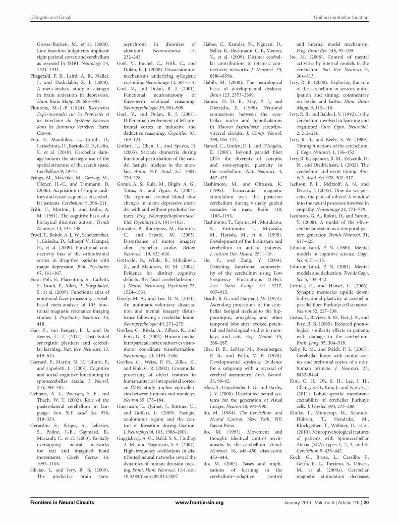
D’Angelo and Casali Unified cerebellar function
Grosse-Ruyken, M., et al. (2000).Line bisection judgments implicateright parietal cortex and cerebellumas assessed by fMRI. Neurology 54,1324–1331.
Fitzgerald, P. B., Laird, A. R., Maller,J., and Daskalakis, Z. J. (2008).A meta-analytic study of changesin brain activation in depression.Hum. Brain Mapp. 29, 683–695.
Flourens, M.-J.-P. (1824). RecherchesExpèrimentales sur les Propriétés etles Fonctions du Système Nerveuxdans les Animaux Vertébres. Paris:Crevot.
Foti, F., Mandolesi, L., Cutuli, D.,Laricchiuta, D., Bartolo, P. D., Gelfo,F., et al. (2010). Cerebellar dam-age loosens the strategic use of thespatial structure of the search space.Cerebellum 9, 29–41.
Frings, M., Maschke, M., Gerwig, M.,Diener, H.-C., and Timmann, D.(2006). Acquisition of simple audi-tory and visual sequences in cerebel-lar patients. Cerebellum 5, 206–211.
Frith, U., Morton, J., and Leslie, A.M. (1991). The cognitive basis of abiological disorder: autism. TrendsNeurosci. 14, 433–438.
Frodl, T., Bokde, A. L. W., Scheuerecker,J., Lisiecka, D., Schoepf, V., Hampel,H., et al. (2009). Functional con-nectivity bias of the orbitofrontalcortex in drug-free patients withmajor depression. Biol. Psychiatry67, 161–167.
Fusar-Poli, P., Placentino, A., Carletti,F., Landi, P., Allen, P., Surguladze,S., et al. (2009). Functional atlas ofemotional faces processing: a voxel-based meta-analysis of 105 func-tional magnetic resonance imagingstudies. J. Psychiatry Neurosci. 34,418.
Gao, Z., van Beugen, B. J., and DeZeeuw, C. I. (2012). Distributedsynergistic plasticity and cerebel-lar learning. Nat. Rev. Neurosci. 13,619–635.
Garrard, P., Martin, N. H., Giunti, P.,and Cipolotti, L. (2008). Cognitiveand social cognitive functioning inspinocerebellar ataxia. J. Neurol.255, 398–405.
Gebhart, A. L., Petersen, S. E., andThach, W. T. (2002). Role of theposterolateral cerebellum in lan-guage. Ann. N.Y. Acad. Sci. 978,318–333.
Gerardin, E., Sirigu, A., Lehéricy,S., Poline, J.-B., Gaymard, B.,Marsault, C., et al. (2000). Partiallyoverlapping neural networksfor real and imagined handmovements. Cereb. Cortex 10,1093–1104.
Ghajar, J., and Ivry, R. B. (2009).The predictive brain state:
asynchrony in disorders ofattention? Neuroscientist 15,232–243.
Goel, V., Buchel, C., Frith, C., andDolan, R. J. (2000). Dissociation ofmechanisms underlying syllogisticreasoning. Neuroimage 12, 504–514.
Goel, V., and Dolan, R. J. (2001).Functional neuroanatomy ofthree-term relational reasoning.Neuropsychologia 39, 901–909.
Goel, V., and Dolan, R. J. (2004).Differential involvement of left pre-frontal cortex in inductive anddeductive reasoning. Cognition 93,109–121.
Goffart, L., Chen, L., and Sparks, D.(2003). Saccade dysmetria duringfunctional perturbation of the cau-dal fastigial nucleus in the mon-key. Annu. N.Y. Acad. Sci. 1004,220–228.
Gonul, A. S., Kula, M., Bilgin, A. G.,Tutus, A., and Oguz, A. (2004).The regional cerebral blood flowchanges in major depressive disor-der with and without psychotic fea-tures. Prog. Neuropsychopharmacol.Biol. Psychiatry 28, 1015–1021.
González, B., Rodríguez, M., Ramirez,C., and Sabaté, M. (2005).Disturbance of motor imageryafter cerebellar stroke. Behav.Neurosci. 119, 622–626.
Gottwald, B., Wilde, B., Mihajlovic,Z., and Mehdorn, H. M. (2004).Evidence for distinct cognitivedeficits after focal cerebellarlesions.J. Neurol. Neurosurg. Psychiatry 75,1524–1531.
Grealy, M. A., and Lee, D. N. (2011).An automatic-voluntary dissocia-tion and mental imagery distur-bance following a cerebellar lesion.Neuropsychologia 49, 271–275.
Grefkes, C., Ritzla, A., Zillesa, K., andFink, G. R. (2004). Human medialintraparietal cortex subserves visuo-motor coordinate transformation.Neuroimage 23, 1494–1506.
Grefkes, C., Weiss, P. H., Zilles, K.,and Fink, G. R. (2002). Crossmodalprocessing of object features inhuman anterior intraparietal cortex:an fMRI study implies equivalen-cies between humans and monkeys.Neuron 35, 173–184.
Guerrasio, L., Quinet, J., Büttner, U.,and Goffart, L. (2009). Fastigialoculomotor region and the con-trol of foveation during fixation.J. Neurophysiol. 103, 1988–2001.
Guggisberg, A. G., Dalal, S. S., Findlay,A. M., and Nagarajan, S. S. (2007).High-frequency oscillations in dis-tributed neural networks reveal thedynamics of human decision mak-ing. Front. Hum. Neurosci. 1:14. doi:10.3389/neuro.09.014.2007
Habas, C., Kamdar, N., Nguyen, D.,Keller, K., Beckmann, C. F., Menon,V., et al. (2009). Distinct cerebel-lar contributions to intrinsic con-nectivity networks. J. Neurosci. 29,8586–8594.
Habib, M. (2000). The neurologicalbasis of developmental dyslexia.Brain 123, 2373–2399.
Haines, D. D. E., May, P. J., andDietrichs, E. (1990). Neuronalconnections between the cere-bellar nuclei and hypothalamusin Macaca fascicularis: cerebello-visceral circuits. J. Comp. Neurol.299, 106–122.
Hansel, C., Linden, D. J., and D’Angelo,E. (2001). Beyond parallel fiberLTD: the diversity of synapticand non-synaptic plasticity inthe cerebellum. Nat. Neurosci. 4,467–475.
Hashimoto, M., and Ohtsuka, K.(1995). Transcranial magneticstimulation over the posteriorcerebellum during visually guidedsaccades in man. Brain 118,1185–1193.
Hashimoto, T., Tayama, M., Murakawa,K., Yoshimoto, T., Miyazaki,M., Harada, M., et al. (1995).Development of the brainstem andcerebellum in autistic patients.J. Autism Dev. Disord. 25, 1–18.
He, Y., and Zang, Y. (2004).Detecting functional connectiv-ity of the cerebellum using LowFrequency Fluctuations (LFFs).Lect. Notes Comp. Sci. 3217,907–915.
Heath, R. G., and Harper, J. W. (1974).Ascending projections of the cere-bellar fastigial nucleus to the hip-pocampus, amygdala, and othertemporal lobe sites: evoked poten-tial and histological studies in mon-keys and cats. Exp. Neurol. 45,268–287.
Hier, D. B., LeMay, M., Rosenberger,P. B., and Perlo, V. P. (1978).Developmental dyslexia. Evidencefor a subgroup with a reversal ofcerebral asymmetry. Arch. Neurol.35, 90–92.
Ishai, A., Ungerleider, L. G., and Haxby,J. V. (2000). Distributed neural sys-tems for the generation of visualimages. Neuron 28, 979–990.
Ito, M. (1984). The Cerebellum andNeural Control. New York, NY:Raven Press.
Ito, M. (1993). Movement andthought: identical control mech-anisms by the cerebellum. TrendsNeurosci. 16, 448–450. discussion:453–444.
Ito, M. (2005). Bases and impli-cations of learning in thecerebellum—adaptive control
and internal model mechanism.Prog. Brain Res. 148, 95–109.
Ito, M. (2008). Control of mentalactivities by internal models in thecerebellum. Nat. Rev. Neurosci. 9,304–313.
Ivry, R. B. (2000). Exploring the roleof the cerebellum in sensory antic-ipation and timing: commentaryon tesche and karhu. Hum. BrainMapp. 9, 115–118.
Ivry, R. B., and Baldo, J. V. (1992). Is thecerebellum involved in learning andcognition? Curr. Opin. Neurobiol.2, 212–216.
Ivry, R. B., and Keele, S. W. (1989).Timing functions of the cerebellum.J. Cogn. Neurosci. 1, 136–152.
Ivry, R. B., Spencer, R. M., Zelaznik, H.N., and Diedrichsen, J. (2002). Thecerebellum and event timing. Ann.N.Y. Acad. Sci. 978, 302–317.
Jackson, P. L., Meltzoff, A. N., andDecety, J. (2005). How do we per-ceive the pain of others? A windowinto the neural processes involved inempathy. Neuroimage 24, 771–779.
Jacobson, G. A., Rokni, D., and Yarom,Y. (2008). A model of the olivo-cerebellar system as a temporal pat-tern generator. Trends Neurosci. 31,617–625.
Johnson-Laird, P. N. (1980). Mentalmodels in cognitive science. Cogn.Sci. 4, 71–115.
Johnson-Laird, P. N. (2001). Mentalmodels and deductionSci. 5, 434–442.
Jorntell, H., and Hansel, C. (2006).Synaptic memories upside down:bidirectional plasticity at cerebellarparallel fiber-Purkinje cell synapses.Neuron 52, 227–238.
Justus, T., Ravizza, S. M., Fiez, J. A., andIvry, R. B. (2005). Reduced phono-logical similarity effects in patientswith damage to the cerebellum.Brain Lang. 95, 304–318.
Kelly, R. M., and Strick, P. L. (2003).Cerebellar loops with motor cor-tex and prefrontal cortex of a non-human primate. J. Neurosci. 23,8432–8444.
Kim, C. H., Oh, S. H., Lee, J. H.,Chang, S. O., Kim, J., and Kim, S. J.(2011). Lobule-specific membraneexcitability of cerebellar Purkinjecells. J. Physiol. 590, 273–288.
Klinke, I., Minnerop, M., Schmitz-Hübsch, T., Hendriks, M.,Klockgether, T., Wüllner, U., et al.(2010). Neuropsychological featuresof patients with SpinocerebellarAtaxia (SCA) types 1, 2, 3, and 6.Cerebellum 9, 433–442.
Koch, G., Brusa, L., Carrillo, F.,Gerfo, E. L., Torriero, S., Oliveri,M., et al. (2009a). Cerebellarmagnetic stimulation decreases
Frontiers in Neural Circuits www.frontiersin.org January 2013 | Volume 6 | Article 116 | 20
. Trends Cogn.

D’Angelo and Casali Unified cerebellar function
levodopa-induced dyskinesias inParkinson disease. Neurology 73,113–119.
Koch, G., Oliveri, M., and Caltagirone,C. (2009b). Neural networksengaged in milliseconds and sec-onds time processing: evidencefrom transcranial magnetic stim-ulation and patients with corticalor subcortical dysfunction. Philos.Trans. R. Soc. Lond. B Biol. Sci. 364,1907–1918.
Konarski, J. Z., McIntyre, R. S., Grupp,L. A., and Kennedy, S. H. (2005).Is the cerebellum relevant in thecircuitry of neuropsychiatric dis-orders? J. Psychiatry Neurosci. 30,178–186.
Krienen, F. M., and Buckner, R. L.(2009). Segregated fronto-cerebellarcircuits revealed by intrinsic func-tional connectivity. Cereb. Cortex19, 2485–2497.
Kusunoki, M., Gottlieb, J., andGoldberg, M. E. (2000). Thelateral intraparietal area as asalience map: the representationof abrupt onset, stimulus motion,and task relevance. Vis. Res. 40,1459–1468.
Kyriakopoulos, M., Vyasa, N. S.,Barkerb, G. J., Chitnisb, X. A., andFrangoua, S. (2008). A diffusiontensor imaging study of whitematter in early-onset schizophrenia.Biol. Psychiatry 63, 519–523.
Lainé, J., and Axelrad, H. (1998).Lugaro cells target basket and stel-late cells in the cerebellar cortex.Neuroreport 9, 2399–2403.
Langdon, R., Coltheart, M., Ward,P. B., and Catts, S. V. (2001).Visual and cognitive perspective-taking impairments in schizophre-nia: a failure of allocentric sim-ulation? Cogn. Neuropsychiatry 6,241–269.
LeBeau, F. E., El Manira, A., and Griller,S. (2005). Tuning the network:modulation of neuronal micro-circuits in the spinal cord andhippocampus. Trends Neurosci. 28,552–561.
Lefèvre, P., Quaia, C., and Opticana,L. M. (1998). Distributed modelof control of saccades by superiorcolliculus and cerebellum. NeuralNetw. 11, 1175–1190.
Leggio, M. G., Silveri, M. C., Petrosini,L., and Molinari, M. (2000).Phonological grouping is specifi-cally affected in cerebellar patients:a verbal fluency study. J. Neurol.Neurosurg. Psychiatry 69, 102–106.
Leggio, M. G., Tedesco, A. M.,Chiricozzi, F. R., Clausi, S., Orsini,A., and Molinari, M. (2008).Cognitive sequencing impairmentin patients with focal or atrophic
cerebellar damage. Brain 131,1332–1343.
Leroi, I., O’Hearn, E., Marsh, L.,Lyketsos, C. G., Rosenblatt,A., Ross, C. A., et al. (2002).Psychopathology in patients withdegenerative cerebellar diseases:a comparison to Huntington’sdisease. Am. J. Psichiatry 159,1306–1314.
Liu, Z., Xu, C., Xu, Y., Wang, Y., Zhao,B., Lv, Y., et al. (2010). Decreasedregional homogeneity in insula andcerebellum: a resting-state fMRIstudy in patients with major depres-sion and subjects at high risk formajor depression. Psychiatry Res.182, 211–215.
Llinas, R., and Paré, D. (1998). TheBrain as a Closed System Modulatedby the Senses. London, England:MIT Press.
Llinás, R. R., and Roy, S. (2009). The“prediction imperative” as the basisfor self-awareness. Philos. Trans.R. Soc. Lond. B Biol. Sci. 364,1301–1307.
Lotze, M., Montoya, P., Erb, M.,Hülsmann, E., Flor, H., Klose, U.,et al. (1999). Activation of corti-cal and cerebellar motor areas dur-ing executed and imagined handmovements: an fMRI study. J. Cogn.Neurosci. 11, 491–501.
Lubke, J., and Feldmeyer, D. (2007).Excitatory signal flow and connec-tivity in a cortical column: focus onbarrel cortex. Brain Struct. Funct.212, 3–17.
Luciani, L. (1891). Il Cervelletto.Nuovi Studi di Fisiologia Normale ePatologica. Firenze: Le Monnier.
Luft, A. R., Manto, M.-U., and Taib,N. O. B. (2005). Modulation ofmotor cortex excitability by sus-tained peripheral stimulation: theinteraction between the motor cor-tex and the cerebellum. Cerebellum4, 90–96.
Maddox, W. T., Aparicio, P., Marchant,N. L., and Ivry, R. B. (2005). Rule-based category learning is impairedin patients with Parkinson’s diseasebut not in patients with cerebel-lar disorders. J. Cogn. Neurosci. 17,707–723.
Marien, P., Saerens, J., Nanhoe, R.,Moens, E., Nagels, G., Pickut, B.A., et al. (1996). Cerebellar inducedaphasia: case report of cerebel-lar induced prefrontal aphasic lan-guage phenomena supported bySPECT findings. J. Neurol. Sci. 144,34–43.
Marr, D. (1969). A theory of cerebellarcortex. J. Physiol. 202, 437–470.
Matell, M. S., and Meck, W. H. (2004).Cortico-striatal circuits and intervaltiming: coincidence detection of
oscillatory processes. Cogn. BrainRes. 21, 139–170.
Medina, J. F., and Mauk, M. D. (2000).Computer simulation of cerebel-lar information processing. Nat.Neurosci. 3(Suppl.), 1205–1211.
Mellet, E., Tzourio-Mazoyer, N.,Bricogne, S., Mazoyer, B., Kosslyn,S. M., and Denis, M. (2000).Functional anatomy of high-resolution visual mental imagery.J. Cogn. Neurosci. 12, 98–109.
Miall, R. C., and Reckess, G. Z. (2002).The cerebellum and the timing ofcoordinated eye and hand tracking.Brain Cogn. 48, 212–226.
Middleton, F. A., and Strick, P. L.(2001). Cerebellar projections to theprefrontal cortex of the primate.J. Neurosci. 21, 700–712.
Milad, M. R., and Quirk, G. J. (2002).Neurons in medial prefrontal cortexsignal memory for fear extinction.Nature 420, 70–74.
Mills, N. P., DelBello, M. P., Adler, C.M., and Strakowski, S. M. (2005).MRI analysis of cerebellar vermalabnormalities in bipolar disorder.Am. J. Psichiatry 162, 1530–1532.
Moberget, T., Karns, C. M., Deouell,L. Y., Lindgren, M., Knight, R. T.,and Ivry, R. B. (2008). Detectingviolations of sensory expectanciesfollowing cerebellar degenera-tion: a mismatch negativity study.Neuropsychologia 46, 2569–2579.
Molinari, M., Filippini, V., and Leggio,M. G. (2002). Neuronal plastic-ity of interrelated cerebellar andcortical networks. Neuroscience 3,863–870.
Molinari, M., Petrosini, L., Misciagna,S., and Leggio, M. G. (2004).Visuospatial abilities in cerebel-lar disorders. J. Neurol. Neurosurg.Psychiatry 75, 235–240.
Monkul, E. S., Hatch, J. P., Sassi,R. B., Axelson, D., Brambilla, P.,Nicoletti, M. A., et al. (2008).MRI study of the cerebellumin young bipolar patients. Prog.Neuropsychopharmacol. Biol.Psychiatry 32, 613–619.
Morgan, M. A., Romanskib, L. M., andLeDoux, J. E. (1993). Extinction ofemotional learning: contribution ofmedial prefrontal cortex. Neurosci.Lett. 163, 109–111.
Mugnaini, E., Sekerková, G., andMartina, M. (2011). The unipolarbrush cell: a remarkable neuronfinally receiving deserved attention.Brain Res. Rev. 66, 220–245.
Nair, D. G., Purcott, K. L., Fuchs,A., Steinberg, F., and Kelso, J. A.S. (2003). Cortical and cerebel-lar activity of the human brainduring imagined and executedunimanual and bimanual action
sequences: a functional MRI study.Brain Res. Cogn. Brain Res. 15,250–260.
Naito, E., Kochiyama, T., Kitada,R., Nakamura, S., Matsumura,M., Yonekura, Y., et al. (2002).Internally simulated movementsensations during motor imageryactivate cortical motor areas andthe cerebellum. J. Neurosci. 22,3683–3691.
Nicolson, R. I., Fawcett, A. J., and Dean,P. (2001a). Developmental dyslexia:the cerebellar deficit hypothesis.Trends Neurosci. 24, 508–511.
Nicolson, R. I., Fawcett, A. J., Dean, P.,Zeffiro, T., and Eden, G. (2001b).A TINS debate – Hindbrain ver-sus the forebrain: a case for cere-bellar deficit dyslexia: the cerebellardeficit hypothesis. Trends Neurosci.24, 508–511.
Nyberg, L., Kim, A. S., Habib, R.,Levine, B., and Tulving, E. (2010).Consciousness of subjective timein the brain. Proc. Natl. Acad. Sci.U.S.A. 107, 22356–22359.
Oberdick, J., and Sillitoe, R. V. (2011).Cerebellar zones: history, develop-ment, and function. Cerebellum 10,301–306.
Ohtsuki, G., Piochon, C., and Hansel,C. (2009). Climbing fiber signal-ing and cerebellar gain control.Front. Cell. Neurosci. 3:4. doi:10.3389/neuro.03.004.2009
Oliveri, M., Koch, G., Torriero, S., andCaltagirone, C. (2005). Increasedfacilitation of the primary motorcortex following 1 Hz repetitivetranscranial magnetic stimulationof the contralateral cerebellum innormal humans. Neurosci. Lett. 376,188–193.
Osherson, D., Perani, D., Cappa, S.,Schnur, T., Grassi, F., and Fazio,F. (1998). Distinct brain loci indeductive versus probabilisticreasoning. Neuropsychologia 36,369–376.
O’Reilly, J. X., Mesulam, M. M., andNobre, A. C. (2008). The cerebellumpredicts the timing of perceptualevents. J. Neurosci. 28, 2252–2260.
Panouillères, M., Neggers, S., Gutteling,T., Salemme, R., Stigchel, S., Geest,J., et al. (2011). Transcranial mag-netic stimulation and motor plas-ticity in human lateral cerebellum:dual effect on saccadic adaptation.Hum. Brain Mapp. 33, 1512–1525.
Pascual-Leone, A., Wassermann, E. M.,Grafman, J., and Hallett, M. (1996).The role of the dorsolateral pre-frontal cortex in implicit procedu-ral learning 107,479–485.
Passot, J.-B., Rondi-Reig, L., andArleo, A. (2009). Cerebellum and
Frontiers in Neural Circuits www.frontiersin.org January 2013 | Volume 6 | Article 116 | 21
. Exp. Brain Res.

D’Angelo and Casali Unified cerebellar function
spatial cognition: a connectionistapproach. Adv. Comput. Intell.Learn. 287–292.
Pellionisz, A. J., and Llinàs, R. (1982).Space-time representation in thebrain the cerebellum as a pre-dictive space-time metric tensor.Neuroscience 7, 2949–2970.
Peng, J., Liu, J., Nie, B., Li, Y., Shan,B., Wang, G., et al. (2010). Cerebraland cerebellar gray matter reductionin first-episode patients with majordepressive disorder: a voxel-basedmorphometry study. Eur. J. Radiol.80, 395–399.
Pernet, C. R., Poline, J. B., Demonet,J. F., and Rousselet, G. A. (2009).Brain classification reveals the rightcerebellum as the best biomarker ofdyslexia. BMC Neurosci. 10:67. doi:10.1186/1471-2202-10-67
Petacchi, A., Laird, A. R., Fox, P. T., andBower, J. M. (2005). Cerebellum andauditory function: an ALE meta-analysis of functional neuroimag-ing studies. Hum. Brain Mapp. 25,118–128.
Petrides, M. (2000). The role of themid-dorsolateral prefrontal cortexin working memory. Exp. Brain Res.133, 44–54.
Petrosini, L. (2007). “Do What IDo” and “Do How I Do”: differ-ent components of imitative learn-ing are mediated by different neu-ral structures. Neuroscientist 13,335–348.
Petrosini, L., Leggio, M. G., andMolinari, M. (1998). The cerebel-lum in the spatial problem solving:a co-star or a guest star? Prog.Neurobiol. 56, 191–210.
Pierce, K., and Courchesne, E. (2000).Evidence for a cerebellar role inreduced exploration and stereo-typed behavior in autism. Biol.Psychiatry 49, 655–664.
Pijpers, A., Apps, R., Pardoe, J., Voogd,J., and Ruigrok, T. J. (2006). Precisespatial relationships between mossyfibers and climbing fibers in ratcerebellar cortical zones. J. Neurosci.26, 12067–12080.
Ploghaus, A., Becerra, L., Borras, C.,and Borsook, D. (2003). Neural cir-cuitry underlying pain modulation:expectation, hypnosis, placebo.Trends Cogn. Sci. 7, 197–200.
Pochon, J.-B., Levy, R., Poline, J.-B.,Crozier, S., Lehéricy, S., Pillon, B.,et al. (2001). The role of dor-solateral prefrontal cortex in thepreparation of forthcoming actions:an fMRI study. Cereb. Cortex 11,260–266.
Prevosto, V., Graf, W., and Ugolini,G. (2010). Cerebellar inputs tointraparietal cortex areas LIP andMIP: functional frameworks for
adaptive control of eye movements,reaching, and Arm/Eye/Head move-ment coordination. Cereb. Cortex20, 214–228.
Rae, C., Harasty, J. A., Dzendrowskyj,T. E., Talcott, J. B., Simpson, J.M., Blamire, A. M., et al. (2002).Cerebellar morphology in develop-mental dyslexia. Neuropsychologia40, 1285–1292.
Ramnani, N., Behrens, T. E., Johansen-Berg, H., Richter, M. C., Pinsk, M.A., Andersson, J. L., et al. (2006).The evolution of prefrontal inputsto the cortico-pontine system:diffusion imaging evidence fromMacaque monkeys and humans.Cereb. Cortex 16, 811–818.
Ramnani, N., and Owen, A. M. (2004).Anterior prefrontal cortex: insightsinto function from anathomy andneuroimaging. Nature 5, 184–194.
Ramus, F. (2003). Developmentaldyslexia: specific phonologicaldeficit or general sensorimotordysfunction? Curr. Opin. Neurobiol.13, 212–218.
Ravizza, S. M., McCormick, C. A.,Schlerf, J. E., Justus, T., Ivry, R. B.,and Fiez, J. A. (2006). Cerebellardamage produces selective deficitsin verbal working memory. Brain129, 306–320.
Riby, D. M., and Hancock, P. J.B. (2008). Viewing it differ-ently: social scene perception inWilliams syndrome and Autism.Neuropsychologia 46, 2855–2860.
Richter, S., Kaiser, O., Hein-Kropp, C.,Dimitrova, A., Gizewski, E., Beckc,A., et al. (2004). Preserved verbgeneration in patients with cere-bellar atrophy. Neuropsychologia 42,1235–1246.
Ridderinkhof, K. R., Ullsperger, M.,Crone, E. A., and Nieuwenhuis,S. (2004). The role of the medialfrontal cortex in cognitive control.Science 306, 443–447.
Ridler, K., Veijola, J. M., Tanskanen, P.,Miettunen, J., Chitnis, X., Suckling,J., et al. (2006). Fronto-cerebellarsystems are associated with infantmotor and adult executive func-tions in healthy adults but not inschizophrenia. Proc. Natl. Acad. Sci.U.S.A. 103, 15651–15656.
Rissman, J., Eliassen, J. C., andBlumstein, S. E. (2003). An event-related fMRI investigation ofimplicit semantic priming. J. Cogn.Neurosci. 15, 1160–1175.
Robinson, F. R., Straube, A., and Fuchs,A. F. (1993). Role of the caudal fasti-gial nucleus in saccade generation.II. Effects of muscimol inactivation.J. Neurophysiol. 70, 1741–1758.
Rouiller, E. M., Liang, F., Babalian, A.,Moret, V., and Wiesendanger, M.
(1994). Cerebellothalamocorticaland pallidothalamocortical pro-jections to the primary andsupplementary motor cortical areas:a multiple tracing study in macaquemonkeys. J. Comp. Neurol. 345,185–213.
Ruigrok, T. J. H. (2010). Ins and outs ofcerebellar modules. Cerebellum 10,464–474.
Rushworth, M. F. S., Behrens, T. E.J., and Johansen-Berg, H. (2006).Connection patterns distinguish 3regions of human parietal cortex.Cereb. Cortex 16, 1418–1430.
Ryding, E., Decety, J., Sjöholm, H.,Stenberg, G., and Ingvar, D. H.(1993). Motor imagery activatesthe cerebellum regionally. A SPECTrCBF study with 99mTc-HMPAO.Brain Res. Cogn. Brain Res. 1,94–99.
Sacchetti, B., Scelfo, B., and Strata, P.(2005). The cerebellum: synapticchanges and fear conditioning.Neuroscientist 11, 217–227.
Salmi, J., Pallesen, K. J., Neuvonen, T.,Brattico, E., Korvenoja, A., Salonen,O., et al. (2010). Cognitive andmotor loops of the human cerebro-cerebellar system. J. Cogn. Neurosci.22, 2663–2676.
Savitz, J., and Drevets, W. C. (2009).Bipolar and major depres-sive disorder: neuroimagingthe developmental-degenerativedivide. Neurosci. Biobehav. Rev. 33,699–771.
Schaeken, W., Johnson-Laird, P. N.,and D’Ydewalle, G. (1996). Mentalmodels and temporal reasoning.Cognition 60, 205–234.
Schmahmann, J. D. (2004). Disordersof the cerebellum: ataxia, dysme-tria of thought, and the cerebel-lar cognitive sffective syndrome.J. Neuropsychiatry Clin. Neurosci.16, 367–378.
Schmahmann, J. D., and Caplan, D.(2006). Cognition, emotion and thecerebellum. Brain 129, 288–292.
Schmahmann, J. D., and Sherman, J.C. (1998). The cerebellar cogni-tive affective syndrome. Brain 121,561–579.
Schmitt, J. E., Eliez, S., Warsofsky, I.S., Bellugi, U., and Reissa, A. L.(2001). Enlarged cerebellar vermisin Williams syndrome. J. Psychiatr.Res. 35, 225–229.
Schutter, D. J., and Honk, J. (2005).A framework for targeting alter-native brain regions with repeti-tive transcranial magnetic stimula-tion in the treatment of depres-sion. J. Psychiatry Neurosci. 30,91–97.
Schweizer, T. A., Levine, B., Rewilak,D., O’Connor, C., Turner, G.,
Alexander, M. P., et al. (2007).Rehabilitation of executivefunctioning after focal damageto the cerebellum. Neurorehabil.Neural Repair 22, 72–77.
Seger, C. A., Desmond, J. E., Glover,G. H., and Gabrieli, J. D. E.(2000). Functional magneticresonance imaging evidence forright-hemisphere involvementin processing unusual semanticrelationships. Neuropsychology 14,361–369.
Shadmehr, R., and Mussa-Ivaldi, S.(2012). Biological Learning andControl. Cambridge: MIT Press.
Shenton, M. E., Dickey, C. C., Frumin,M., and McCarley, R. W. (2001).A review of MRI findings inschizophrenia. Schizophr. Res. 49,1–52.
Shin, J. C., and Ivry, R. B. (2003).Spatial and temporal sequencelearning in patients withParkinson’s disease or cerebel-lar lesions. J. Cogn. Neurosci. 15,1232–1243.
Silveri, M. C., Misciagna, S., andTerrezza, G. (2001). Right sideneglect in right cerebellar lesion.J. Neurol. Neurosurg. Psychiatry 71,114–117.
Smet, H. J. D., Baillieux, H., Deyn,P. P. D., Mariën, P., and Paquier,P. (2007). The cerebellum andlanguage: the story so far. FoliaPhoniatr. Logop. 59, 165–170.
Snider, R. S., and Maiti, A. (1976).Cerebellar contributions to thepapez circuit. J. Neurosci. Res. 2,133–146.
Spitzer, M. (1998). The Mind Withinthe Net: Model of Learning, Thinkingand Acting. Cambridge: MIT Press.
Stein, J. (2001). The magnocellulartheory of developmental dyslexia.Dyslexia 7, 12–36.
Stephan, K. E., Friston, K. J., andFrith, C. D. (2009). Dysconnectionin schizophrenia: from abnormalsynaptic plasticity to failures ofself-monitoring. Schizophr. Bull.35, 509–527.
Strakowski, S. M., Adler, C. M.,Holland, S. K., Mills, N. P., DelBello,M. P., and Eliassen, J. C. (2005).Abnormal fMRI brain activation ineuthymic bipolar disorder patientsduring a counting stroop interfer-ence task. Am. J. Psichiatry 162,1697–1705.
Straube, A., and Buttner, U. (2007).Neuro-Ophthalmology. NeuronalControl of Eye Movements.Magdeburg: Karger.
Strick, P. L., Dum, R. P., and Fiez, J. A.(2009). Cerebellum and nonmotorfunction. Annu. Rev. Neurosci. 32,413–434.
Frontiers in Neural Circuits www.frontiersin.org January 2013 | Volume 6 | Article 116 | 22

D’Angelo and Casali Unified cerebellar function
Synofzik, M., Lindner, A., and Thier,P. (2008). The cerebellum updatespredictions about the visual con-sequences of one’s behavior. Curr.Biol. 18, 814–818.
Synofzik, M., Thier, P., Leube, D. T.,Schlotterbeck, P., and Lindner, A.(2010). Misattributions of agency inschizophrenia are based on impre-cise predictions about the sensoryconsequences of one’s actions. Brain133, 262–271.
Szpunar, K. K. (2010). Evidence foran implicit influence of memory onfuture thinking. Mem. Cognit. 38,531–540.
Szpunar, K. K. (2011). On subjectivetime. Cortex 47, 409–411.
Szwed, M., Bagdasarian, K., andAhissar, E. (2003). Encoding ofvibrissal active touch. Neuron 40,621–630.
Takagi, M., Zee, D. S., and Tamargo,R. J. (1999). Effects of lesions ofthe oculomotor cerebellar vermison eye movements in primate:smooth pursuit. J. Neurophysiol. 83,2047–2062.
Tallal, P., Miller, S., and Holly, F.R. (2006). Neurobiological basis ofspeech: a case for the preeminenceof temporal processing. Ann. N.Y.Acad. Sci. 682, 27–47.
Tavano, A., and Borgatti, R. (2010).Evidence for a link among cogni-tion, language and emotion in cere-bellar malformations. Cortex 46,907–918.
Tilikete, C., Koene, A., Nighoghossian,N., Vighetto, A., and Pélisson,D. (2006). Saccadic lateropulsionin Wallenberg syndrome: a win-dow to access cerebellar controlof saccades? Exp. Brain Res. 174,555–565.
Tononi, G., and Edelman, G. M.(1998). Consciousness and com-plexity. Science 282, 1846–1851.
Townsend, J., Courchesne, E.,Covington, J., Westerfield, M.,Harris, N. S., Lyden, P., et al. (1999).Spatial attention deficits in patientswith acquired or developmentalcerebellar abnormality. J. Neurosci.19, 5632–5643.
Tunik, E., Frey, S. H., and Grafton,S. T. (2005). Virtual lesions ofthe anterior intraparietal areadisrupt goal-dependent on-lineadjustments of grasp. Nature 8,505–511.
Turner, B. M., Paradiso, S., Marvel,C. L., Pierson, R., BolesPonto, L.L., Hichwa, R. D., et al. (2007).The cerebellum and emotionalexperience. Neuropsychologia 45,1331–1341.
Uusisaari, M. Y., and Knopfel, T.(2012). Diversity of neuronal ele-ments and circuitry in the cerebellarnuclei. Cerebellum 11, 420–421.
Vandervert, L. R. (2003). How work-ing memory and cognitive mod-eling functions of the cerebellumcontribute to discoveries in math-ematics. New Ideas Psychol. 21,15–29.
Vandervert, L. R. (2007). Cognitivefunctions of the cerebellum explainhow Ericsson’s deliberate practiceproduces giftedness. High Abil. Stud.18, 89–92.
Vandervert, L. R., Schimpf, P. H., andLiu, H. (2007). How working mem-ory and the cerebellum collaborateto produce creativity and innova-tion. Creat. Res. J. 19, 1–18.
Voogd, J. (2010). Cerebellar zones:a personal history. Cerebellum 10,334–350.
Wang, S., Wu, D.-C., Ding, M.-P., Li,Q., Zhuge, Z.-B., Zhang, S.-H., et al.(2008). Low-frequency stimula-tion of cerebellar fastigial nucleusinhibits amygdaloid kindling acqui-sition in Sprague–Dawley rats.Neurobiol. Dis. 29, 52–58.
Watson, T. C., Jones, M.W., and Apps, R. (2009).Electrophysiological mapping ofnovel prefrontal–cerebellar path-ways. Front. Integr. Neurosci. 3:18.doi: 10.3389/neuro.07.018.2009
Webster, B. R., Celnik, P. A., and Cohen,L. G. (2006). Noninvasive brainstimulation in stroke rehabilitation.NeuroRX 3, 474–481.
Weinberger, D. R., Berman, K. F.,and Illowsky, B. P. (1988).Physiological dysfunction ofdorsolateral prefrontal cortex inschizophrenia, I. I. I. A New Cohortand Evidence for a MonoaminergicMechanism. Arch. Gen. Psychiatry45, 609–615.
Weinberger, D. R., Berman, K. F., andZec, R. F. (1986). Physiologic dys-function of dorsolateral prefrontalcortex in schizophrenia, I. Regionalcerebral blood flow evidence. Arch.Gen. Psychiatry 43, 114–124.
Wiech, K., Ploner, M., and Tracey, I.(2008). Neurocognitive aspects ofpain perception. Trends Cogn. Sci.12, 306–313.
Wolpert, D. M., Miall, R. C., andKawato, M. (1998). Internal modelsin the cerebellum. Trends Cogn. Sci.2, 338–347.
Xiang, H., Lin, C., Ma, X., Zhang,Z., Bower, J. M., Weng, X., et al.(2003). Involvement of the cerebel-lum in semantic discrimination: anfMRI study. Hum. Brain Mapp. 18,208–214.
Xivry, J.-J., and Lefèvre, P. (2007).Saccades and pursuit: two outcomesof a single sensorimotor process.J. Physiol. 584, 11–23.
Yakusheva, T. A., Shaikh, A. G., Green,A. M., Blazquez, P. M., Dickman,J. D., and Angelaki, D. E. (2007).Purkinje cells in posterior cerebel-lar vermis encode motion in aninertial reference frame. Neuron 54,973–985.
Zhu, J.-N., Yung, W.-H., Chow,B. K.-C., Chand, Y.-S., and Wanga,J.-J. (2006). The cerebellar-hypothalamic circuits: Potentialpathways underlying cerebellarinvolvement in somatic-visceralintegration. Brain Res. Rev. 52,93–106.
Conflict of Interest Statement: Theauthors declare that the researchwas conducted in the absence of anycommercial or financial relationshipsthat could be construed as a potentialconflict of interest.
Received: 18 August 2012; accepted: 17December 2012; published online: 10January 2013.Citation: D’Angelo E and Casali S (2013)Seeking a unified framework for cere-bellar function and dysfunction: fromcircuit operations to cognition. Front.Neural Circuits 6:116. doi: 10.3389/fncir.2012.00116Copyright © 2013 D’Angelo and Casali.This is an open-access article dis-tributed under the terms of the CreativeCommons Attribution License, whichpermits use, distribution and reproduc-tion in other forums, provided the origi-nal authors and source are credited andsubject to any copyright notices concern-ing any third-party graphics etc.
Frontiers in Neural Circuits www.frontiersin.org January 2013 | Volume 6 | Article 116 | 23