Section 6 Electroencephalogram (EEG), Wakefulness and Sleep.
35
Section 6 Electroencephalogram (EEG), Wakefulness and Sleep
-
Upload
virgil-gibbs -
Category
Documents
-
view
226 -
download
2
Transcript of Section 6 Electroencephalogram (EEG), Wakefulness and Sleep.
Section 6
Electroencephalogram (EEG), Wakefulness and Sleep
I.I. Electroencephalogram Electroencephalogram (EEG)(EEG)
1. Brain Waves
Brain Waves: State of the BrainBrain Waves: State of the Brain Normal brain function involves continuous electrical activity Patterns of neuronal electrical activity recorded are called brain
waves Brain waves change with age, sensory stimuli, brain disease, and the
chemical state of the body An electroencephalogram (EEG) records this activity EEGs can be used to diagnose and localize brain lesions, tumors,
infarcts, infections, abscesses, and epileptic lesions A flat EEG (no electrical activity) is clinical evidence of death
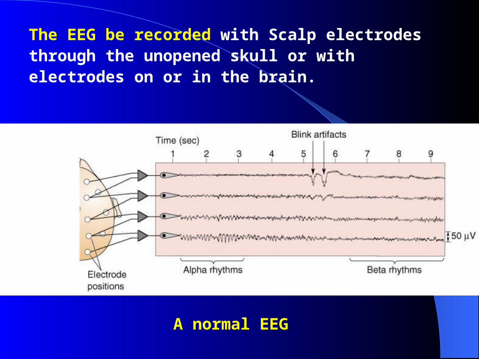
The EEG be recorded with Scalp electrodes through the unopened skull or with electrodes on or in the brain.
A normal EEG
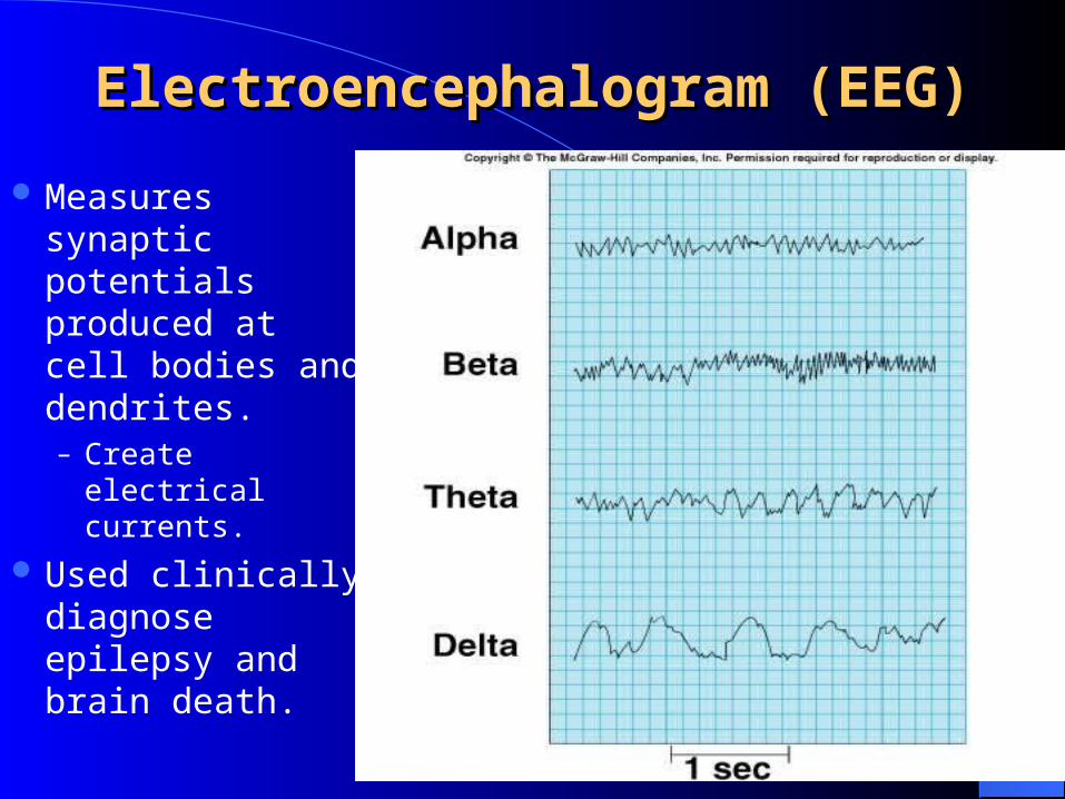
Electroencephalogram (EEG)Electroencephalogram (EEG)
Measures synaptic potentials produced at cell bodies and dendrites.– Create electrical
currents.
Used clinically diagnose epilepsy and brain death.
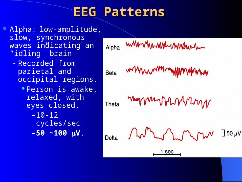
EEG PatternsEEG Patterns Alpha: low-amplitude,
slow, synchronous waves indicating an “idling” brain– Recorded from
parietal and occipital regions.
Person is awake, relaxed, with eyes closed.
– 10-12 cycles/sec– 50 ~100 V.
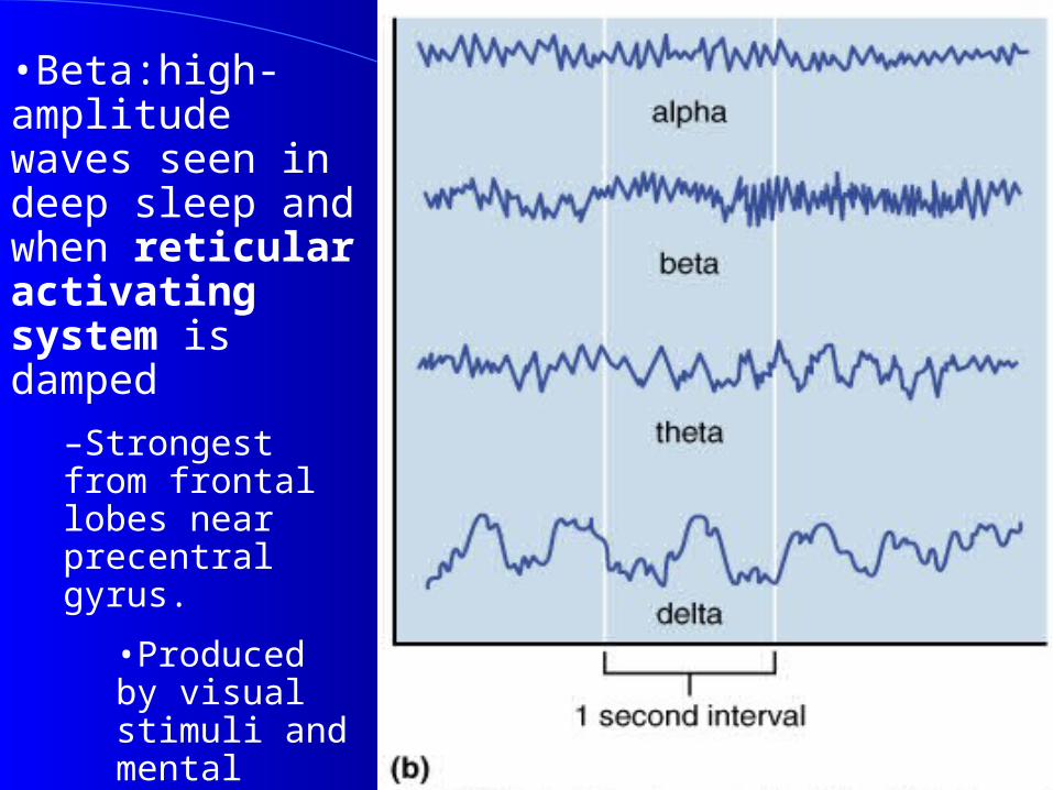
•Beta:high-amplitude waves seen in deep sleep and when reticular activating system is damped
–Strongest from frontal lobes near precentral gyrus.
•Produced by visual stimuli and mental activity.
•Evoked activity.
–13-25 cycles/sec.
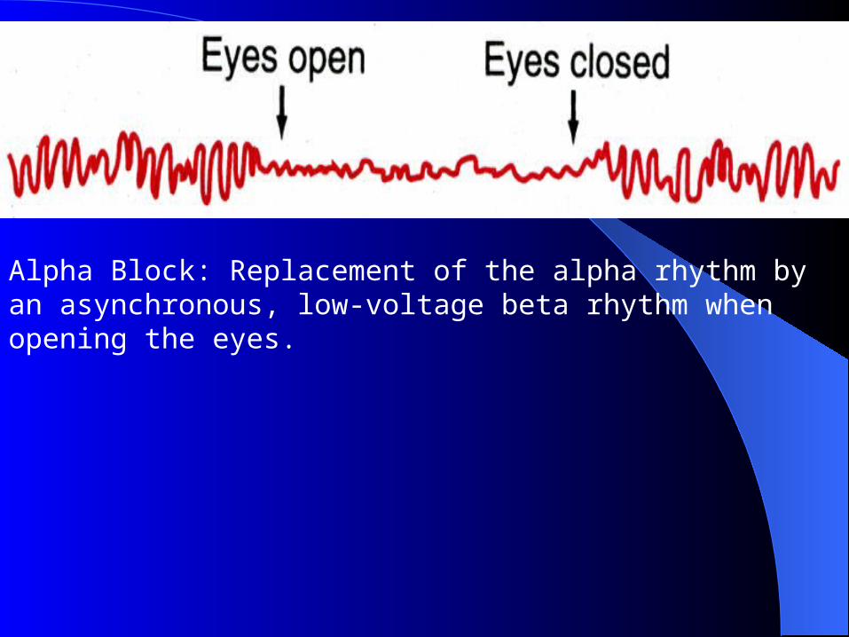
Alpha Block: Replacement of the alpha rhythm by an asynchronous, low-voltage beta rhythm when opening the eyes.
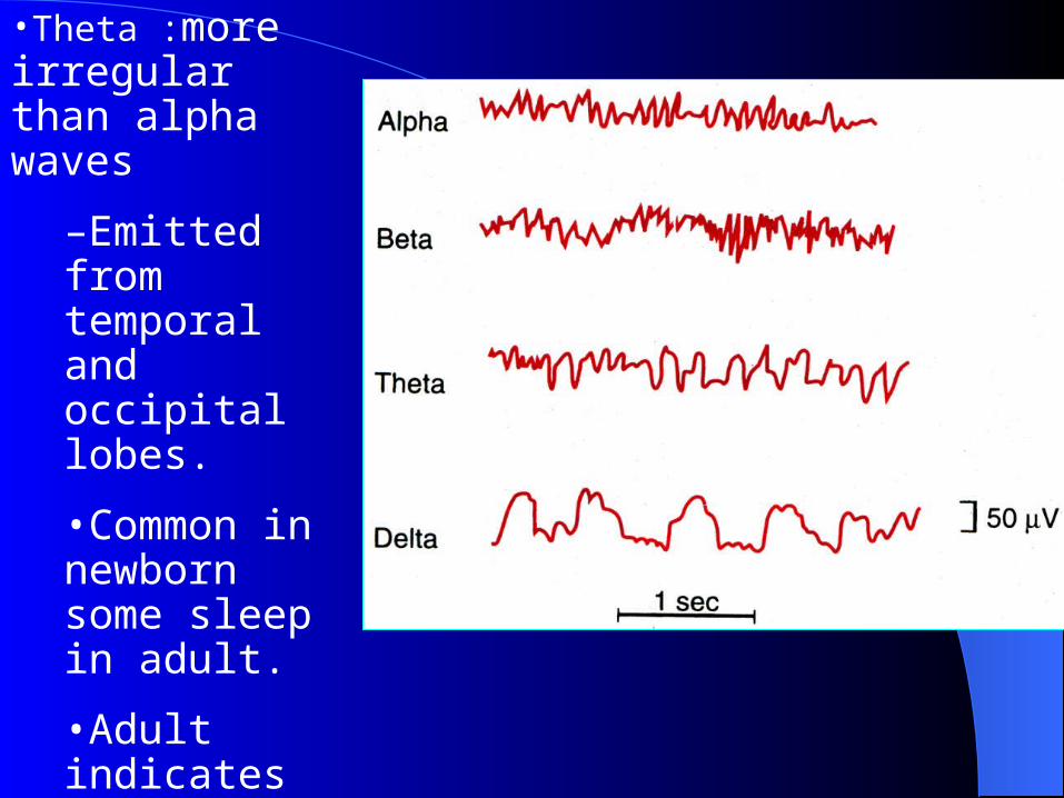
•Theta :more irregular than alpha waves
–Emitted from temporal and occipital lobes.
•Common in newborn some sleep in adult.
•Adult indicates severe emotional stress.
–5-8 cycles/sec.
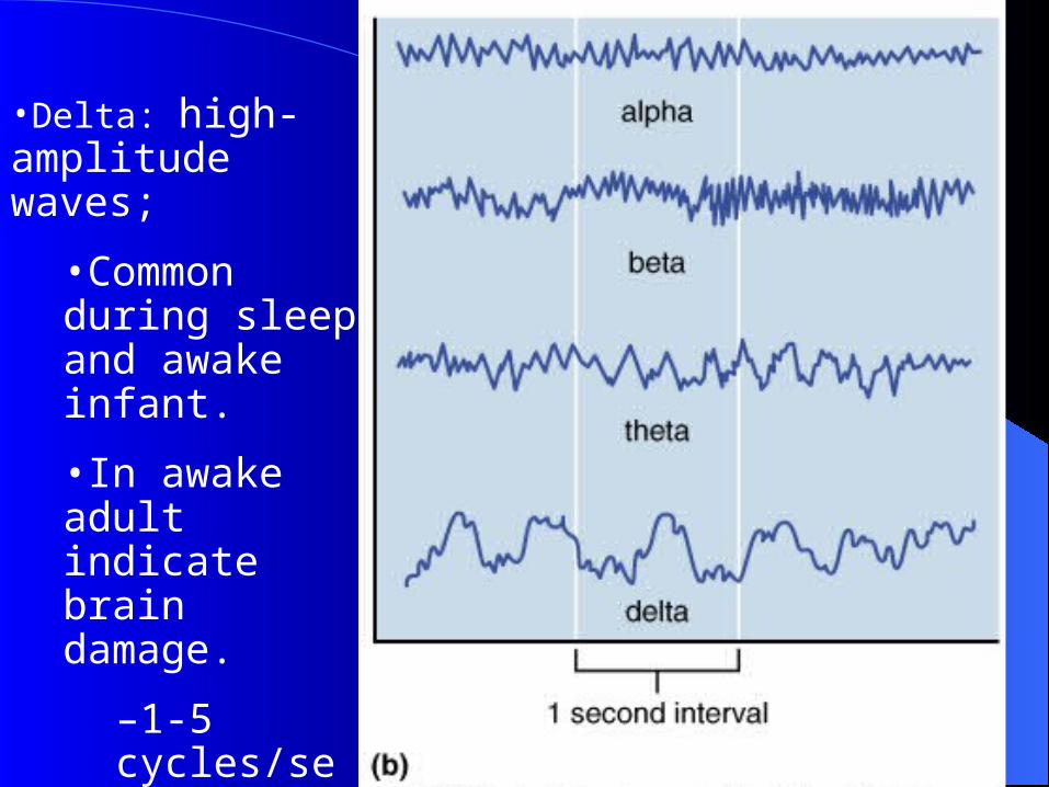
•Delta: high-amplitude waves;
•Common during sleep and awake infant.
•In awake adult indicate brain damage.
–1-5 cycles/sec.
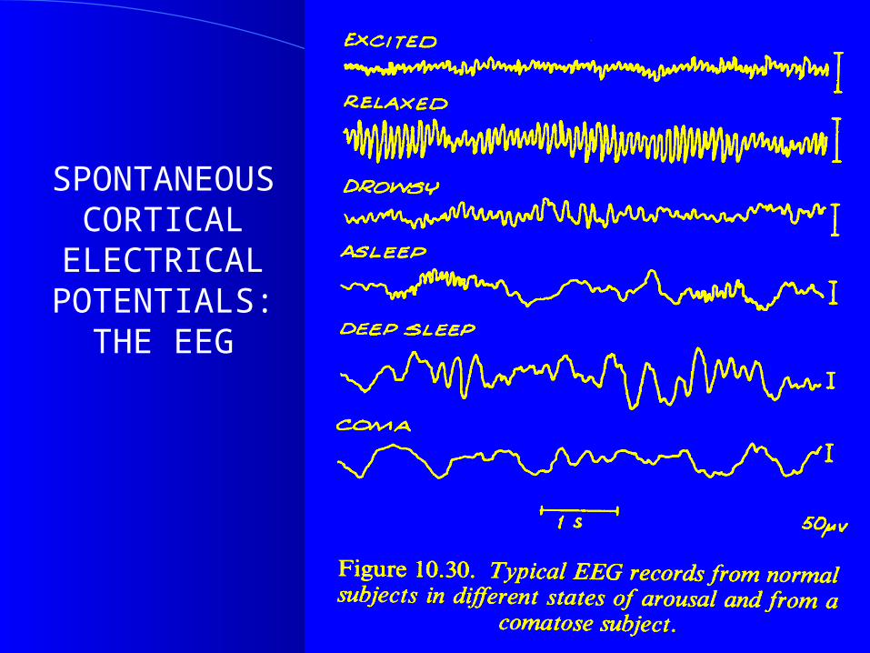
SPONTANEOUS CORTICAL
ELECTRICAL POTENTIALS:
THE EEG
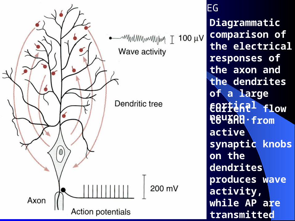
Diagrammatic comparison of the electrical responses of the axon and the dendrites of a large cortical neuron.
2. Mechanism of EEG
Current flow to and from active synaptic knobs on the dendrites produces wave activity, while AP are transmitted along the axon.

Mechanism of EEGMechanism of EEG
Continuous graph of changing voltage fields at scalp surface resulting from ongoing synaptic activity in underlying cortex
Inputs from subcortical structures– Thalamus– Brainstem reticular formation
•EEG signals generated by cortex•Currents in extracellular space generated by summation of EPSPs and IPSPs
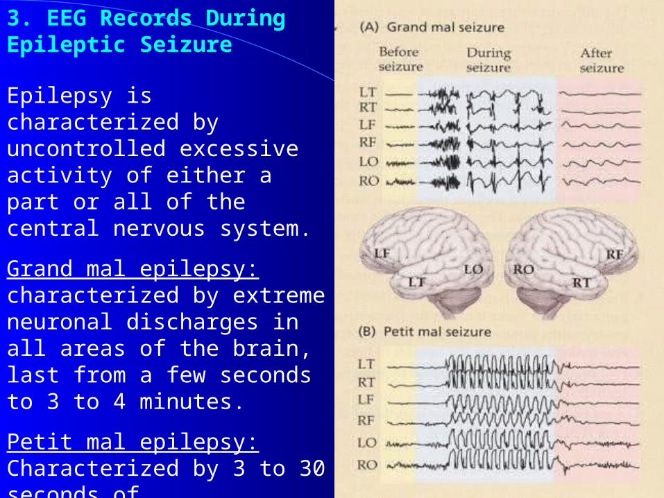
3. EEG Records During Epileptic Seizure
Epilepsy is characterized by uncontrolled excessive activity of either a part or all of the central nervous system.
Grand mal epilepsy: characterized by extreme neuronal discharges in all areas of the brain, last from a few seconds to 3 to 4 minutes.
Petit mal epilepsy: Characterized by 3 to 30 seconds of unconsciousness or diminished consciousness during which the person has several twitch-like contractions of the muscle.
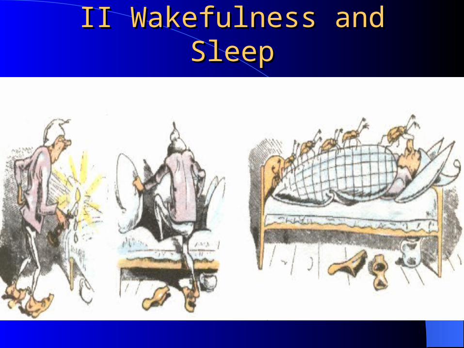
II Wakefulness and SleepII Wakefulness and Sleep
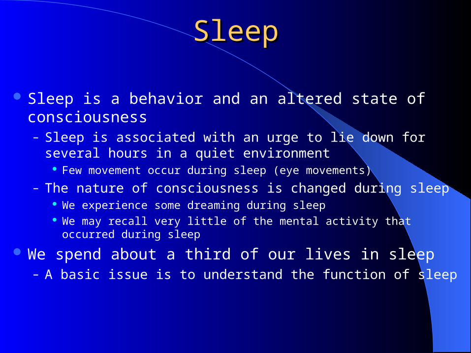
SleepSleep
Sleep is a behavior and an altered state of consciousness– Sleep is associated with an urge to lie down for several hours in
a quiet environment Few movement occur during sleep (eye movements)
– The nature of consciousness is changed during sleep We experience some dreaming during sleep We may recall very little of the mental activity that occurred during sleep
We spend about a third of our lives in sleep– A basic issue is to understand the function of sleep
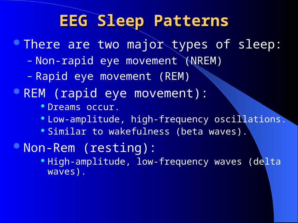
EEG Sleep PatternsEEG Sleep PatternsThere are two major types of sleep:
– Non-rapid eye movement (NREM)– Rapid eye movement (REM)
REM (rapid eye movement): Dreams occur. Low-amplitude, high-frequency oscillations. Similar to wakefulness (beta waves).
Non-Rem (resting): High-amplitude, low-frequency waves (delta waves).
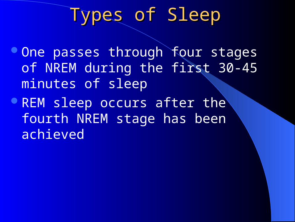
Types of SleepTypes of Sleep
One passes through four stages of NREM during the first 30-45 minutes of sleep
REM sleep occurs after the fourth NREM stage has been achieved
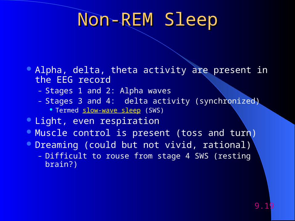
Non-REM SleepNon-REM Sleep
Alpha, delta, theta activity are present in the EEG record– Stages 1 and 2: Alpha waves– Stages 3 and 4: delta activity (synchronized)
Termed slow-wave sleep (SWS)
Light, even respiration Muscle control is present (toss and turn) Dreaming (could but not vivid, rational)
– Difficult to rouse from stage 4 SWS (resting brain?)
9.19
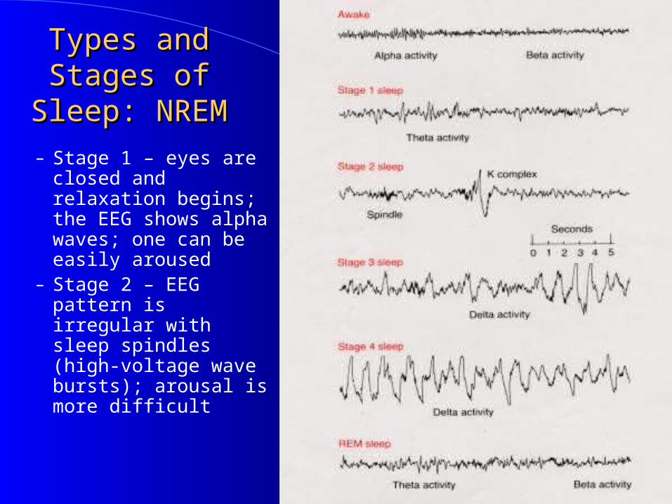
Types and Stages Types and Stages of Sleep: NREMof Sleep: NREM
– Stage 1 – eyes are closed and relaxation begins; the EEG shows alpha waves; one can be easily aroused
– Stage 2 – EEG pattern is irregular with sleep spindles (high-voltage wave bursts); arousal is more difficult
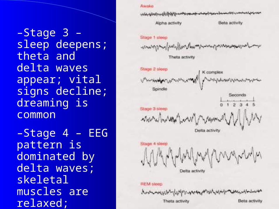
–Stage 3 – sleep deepens; theta and delta waves appear; vital signs decline; dreaming is common
–Stage 4 – EEG pattern is dominated by delta waves; skeletal muscles are relaxed; arousal is difficult
REM SleepREM Sleep Presence of beta activity (desynchronized EEG
pattern) Physiological arousal threshold increases
Heart-rate quickens Breathing more irregular and rapid Brainwave activity resembles wakefulness Genital arousal
Pontine-Geniculate-Occipital (PGO) waves? Loss of muscle tone (paralysis) Vivid, emotional dreams May be involved in memory consolidation
9.22
Pontine-geniculate-occipital (PGO) wave –
A synchronized burst of electrical activity that originates in the pons and like a wave it activates the lateral geniculate nucleus (first relay of visual information)
and then the occipital lobe, specifically in the visual cortex (which receives and puts together the visual information that comes from the lat. geniculate nucleus).
PGO waves appear seconds before and during REM sleep.
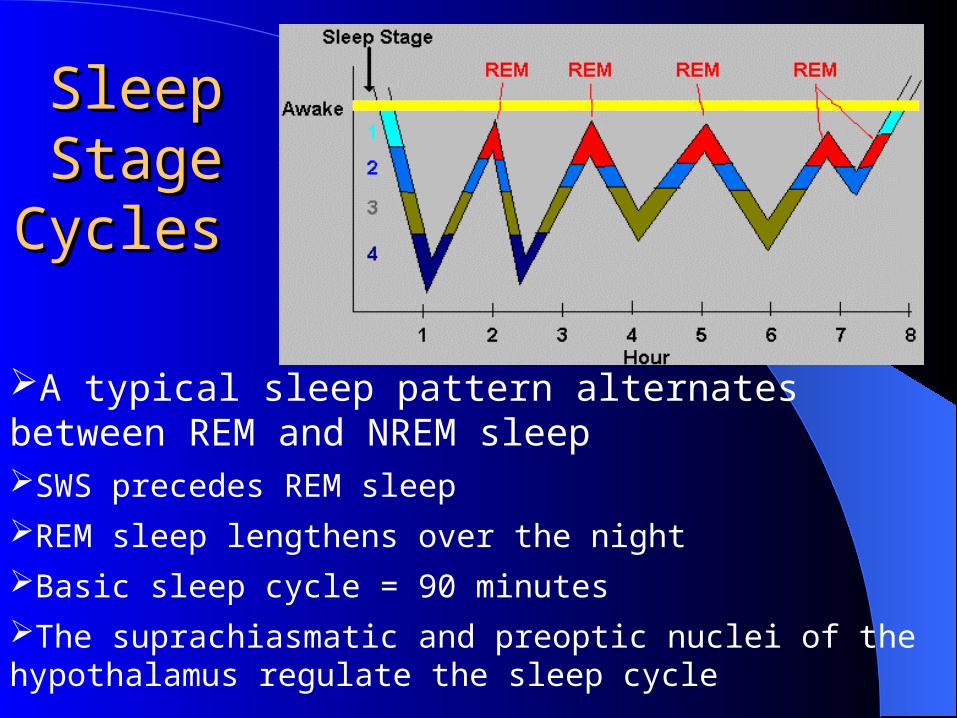
Sleep Sleep Stage Stage
Cycles Cycles
A typical sleep pattern alternates between REM and NREM sleepSWS precedes REM sleep
REM sleep lengthens over the night
Basic sleep cycle = 90 minutes
The suprachiasmatic and preoptic nuclei of the hypothalamus regulate the sleep cycle
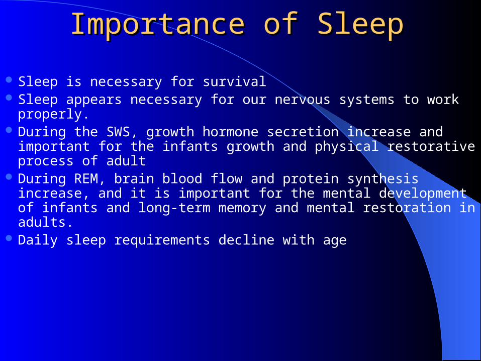
Importance of SleepImportance of Sleep
Sleep is necessary for survival Sleep appears necessary for our nervous systems to work properly. During the SWS, growth hormone secretion increase and important
for the infants growth and physical restorative process of adult During REM, brain blood flow and protein synthesis increase, and
it is important for the mental development of infants and long-term memory and mental restoration in adults.
Daily sleep requirements decline with age
What Happens if We are What Happens if We are Deprived of Sleep?Deprived of Sleep?
Lack of alertness Fatigue Memory problems Irritability Depression Lack of motivation Accidents Fibro Myalgia
Tips for Getting a Good NightTips for Getting a Good Night’’s Sleeps Sleep
Avoid caffeine and alcohol after dinnerKeep a routineDon’t nap during the dayDon’t go to bed hungry or right after eatingExerciseStop smoking
Rules for Optimal SleepRules for Optimal Sleep
Get an adequate amount of sleep every night
Establish a regular sleep scheduleGet continuous sleepMake up for lost sleep
Chemical Control of Sleep/WakingChemical Control of Sleep/Waking
Sleep is regulated: loss of SWS or REM sleep is made up somewhat on following nights– Does the body produce a sleep-promoting chemical during
wakefulness or a wakefulness-promoting chemical during sleep?
Unlikely that sleep is controlled by blood-borne chemicals in the general circulation given:– Siamese twins share the same circulatory system, but sleep
independently– Bottle-nose dolphins: the two hemispheres sleep
independently
9.29
Neural Regulation of ArousalNeural Regulation of Arousal
Electrical stimulation of the brain stem induces arousal– Dorsal path: RF--> to medial thalamus --> cortex– Ventral path: RF --> to lateral hypothalamus, basal ganglia, and the forebrain
Neurotransmitters involved in arousal:– NE neurons in rat locus coeruleus (LC) show high activity during
wakefulness, low activity during sleep (zero during REM sleep) LC neurons may play a role in vigilance
– Activation of ACh neurons produces behavioral activation and cortical desynchrony
ACh agonists increase arousal, ACh antagonists decrease arousal– 5-HT: stimulation of the raphe nuclei induces arousal whereas 5-HT
antagonists reduce cortical arousal
9.30
Neural Control of SWSNeural Control of SWS
The ventrolateral preoptic area (VLPA) is important for the control of sleep– Lesions of the preoptic area produce total insomnia,
leading to death– Electrical stimulation of the preoptic area induces signs
of drowsiness in cats– VLPA neurons promote sleep
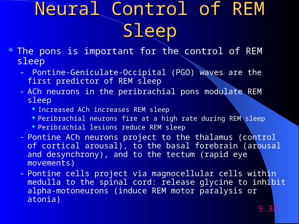
Neural Control of REM SleepNeural Control of REM Sleep
The pons is important for the control of REM sleep– Pontine-Geniculate-Occipital (PGO) waves are the first
predictor of REM sleep– ACh neurons in the peribrachial pons modulate REM sleep
Increased ACh increases REM sleep Peribrachial neurons fire at a high rate during REM sleep Peribrachial lesions reduce REM sleep
– Pontine ACh neurons project to the thalamus (control of cortical arousal), to the basal forebrain (arousal and desynchrony), and to the tectum (rapid eye movements)
– Pontine cells project via magnocellular cells within medulla to the spinal cord: release glycine to inhibit alpha-motoneurons (induce REM motor paralysis or atonia)
9.32
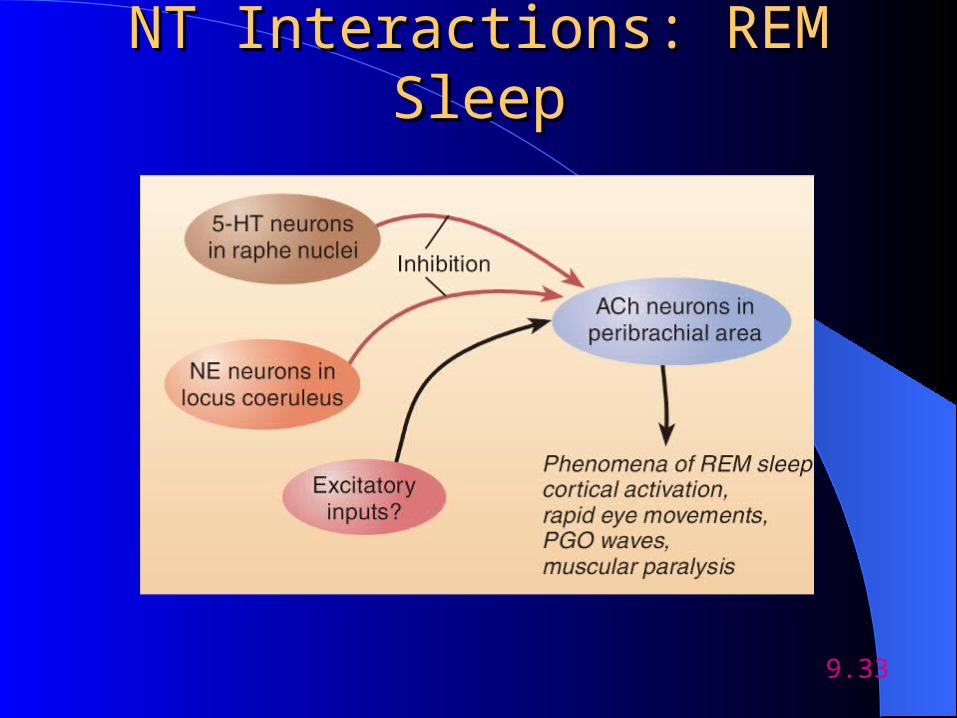
NT Interactions: REM SleepNT Interactions: REM Sleep
9.33
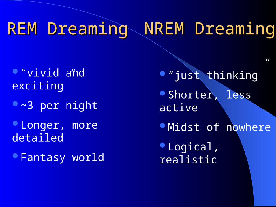
REM DreamingREM Dreaming NREM DreamingNREM Dreaming
“vivid and exciting”
~3 per night
Longer, more detailed
Fantasy world
“just thinking”
Shorter, less active
Midst of nowhere
Logical, realistic
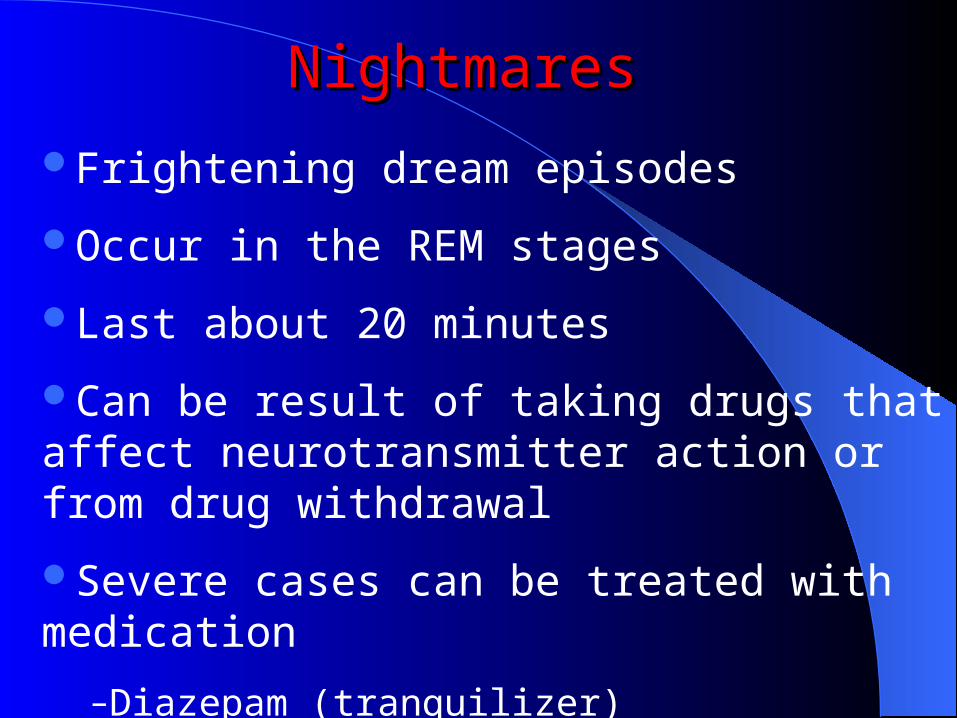
Frightening dream episodes
Occur in the REM stages
Last about 20 minutes
Can be result of taking drugs that affect neurotransmitter action or from drug withdrawal
Severe cases can be treated with medication
–Diazepam (tranquilizer)
NightmaresNightmares