Seasonal Infestation of Small Ruminant by Nasal Bots in ......using forceps and pen knife to expose...
23
Seasonal Infestation of Small Ruminant by Nasal Bots in Kaduna State, Northwestern Nigeria. Owolabi, Y.H 1 ., George, B.D.J. and A.J. Natala Department of Veterinary Parasitology and Entomology, Ahmadu Bello University, Zaria-Nigeria
Transcript of Seasonal Infestation of Small Ruminant by Nasal Bots in ......using forceps and pen knife to expose...

Seasonal Infestation of Small Ruminant by Nasal Bots in Kaduna State,
Northwestern Nigeria.
Owolabi, Y.H1., George, B.D.J. and A.J. Natala
Department of Veterinary Parasitology and Entomology, Ahmadu Bello University,
Zaria-Nigeria

INTRODUCTION
• Livestock is an important component of Nigeria's agricultural economy, especially the small ruminant production in meeting animal protein requirements and providing vital raw materials for the agro-based industries.
• It also provides employment for many Nigerians who engage in the production and marketing of livestock and it's by-products. In addition, it plays an important cultural role which cannot be measured in monetary values (Gatenby, 1991).
• Oestrus ovis infestation limits productivity of sheep and goats throughout the humid and sub humid zones (Gatenby, 1982).
• The most severe effect of the disease and parasite in adult sheep and goats arise from production losses including reduced fertility (Devendra, 1987).

INTRODUCTION/2
• Information on the prevalence of diseases in each distinct climatic and sociological situation is a prerequisite for the rational design and introduction of economical and effective preventive and control programme.
• This is the background from which a prevalence study of oestrosis in sheep and goats was conducted during a 24month period covering two consecutive dry and wet seasons

Life Cycle of Oestrus ovis
Adapted from www.icb.usp.br/-marcelcp/oestrus.htm
sheep
1st Instar
2nd Instar 3rd Instar
Pupa
Adult Emerging
from Pupa
Adult Fly

METHODOLOGY
Study Area
• The study was carried out in five Local Government Areas (LGAs) of Kaduna State located in the Northern Guinea Savannah vegetational zone of Nigeria.
• These include Sabon gari, Zaria, Giwa, Kudan and Makarfi local government areas (LGAs).
• The five LGAs were purposely selected for nearness to Zaria( university base, easy access to the slaughter floors and abattoirs and availability of sheep and goat heads after slaughter.
Study Period
• The study was conducted over a period of two years from November 2005 to October 2007. This period covered two consecutive dry and wet seasons.

Sample Size
• The sample size was obtained using the formula outlined by FAO, 1990
n = Z2pq q= 1 – p p = anticipated prevalence
d2 d = desired precision = 0.05
z = value for std normal deviate = 1.96
Sampling
• The heads of these slaughtered ruminants were purchased from butchers in market places and abattoirs on weekly basis from the selected local government areas.
• They were then transported in polythene bags to the Entomology Laboratory of the Dept. of Veterinary Parasitology and Entomology, Faculty of Vet. Med., ABU Zaria where they were further processed.
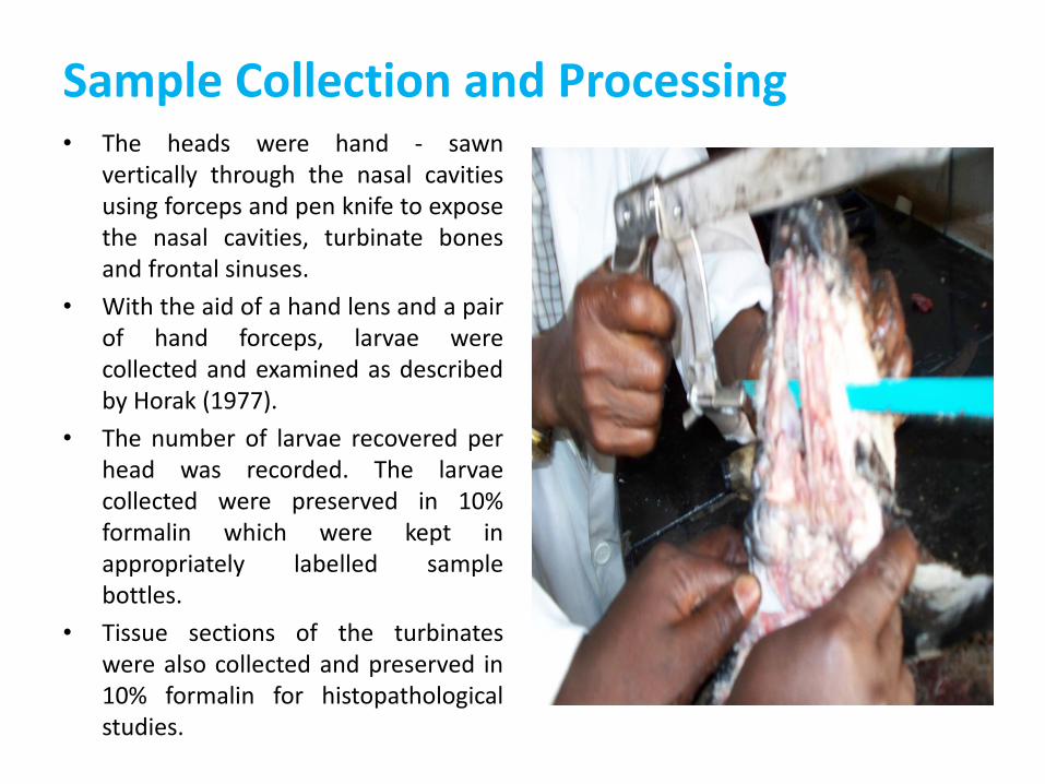
Sample Collection and Processing • The heads were hand - sawn
vertically through the nasal cavities using forceps and pen knife to expose the nasal cavities, turbinate bones and frontal sinuses.
• With the aid of a hand lens and a pair of hand forceps, larvae were collected and examined as described by Horak (1977).
• The number of larvae recovered per head was recorded. The larvae collected were preserved in 10% formalin which were kept in appropriately labelled sample bottles.
• Tissue sections of the turbinates were also collected and preserved in 10% formalin for histopathological studies.


METHODOLOGY CONT’D
Histopathological Studies
• Sections of the nasal cavity and turbinate bones were processed using Technicon tissue processor.
• The tissues were sectioned and stained using Heamatoxylin and Eosin (H & E) stain and mounted on glass slides and later examined under oil immersion objective (x100) lens for histopathological lesions using the methods described by Kiernan, (1990)
Statistical Analysis
• The prevalence data collected were analysed using one way analysis of variance (ANOVA) to test differences between the LGAs where values of P<0.05 were considered significant. Chi square test was also used to test the level of significance between wet and dry season.

RESULTS

TABLE 1: PREVALENCE OF OESTRUS OVIS IN SHEEP AND GOATS HEADS EXAMINED IN THE FIVE LGAS OF KADUNA STATE
LGA SHEEP GOATS
# Samples
Collected
No Positive
/ (%)
# Samples
Collected
No Positive
/ (%)
S/Gari 63 22 (34.9)a 218 50 (22.9)a
Zaria 56 18 (32.1)a 192 38 (19.8)a
Giwa 50 9 (18.0)b 192 27 (14.1)c
Kudan 46 7 (15.2)b 153 26 (17.0)b
Makarfi 43 7 (16.2)b 138 26 (18.8)b
TOTAL 258 63 (24.4) 893 167 (18.7)
Values with different superscript down the column differ significantly at P<0.05

TABLE 2: PREVALENCE OF OESTRUS OVIS IN SHEEP AND GOATS DURING THE DRY AND WET SEASONS
Season SHEEP GOATS
# of
Samples
# Positive /
(%)
# of
Samples
# Positive / (%)
Dry
(Nov- April) 115 28 (24.3) 448 66 (14.7)
Wet
(May- Oct) 143 35 (24.5) 445 101 (22.7)
Total 258 63 (24.4) 893 167 (18.7)
Chi square= 0.001
df= 1 p=0.981
Chi square=9.314
df=1 p=0.002

FIG. 1: PREVALENCE OF SHEEP OESTRUS OVIS IN THE FIVE LGAS OF KADUNA STATE DURING THE DRY SEASONS
0.0
5.0
10.0
15.0
20.0
25.0
30.0
35.0
40.0
SABON GARI ZARIA GIWA KUDAN MAKARFI MEAN
PR
EVA
LEN
CE
(%)
1ST YEAR DRY SEASON 2ND YEAR DRY SEASON MEAN FOR COMBINED DRY SEASON

FIG. 2: PREVALENCE OF SHEEP OESTRUS OVIS IN THE FIVE LGAS OF KADUNA STATE DURING THE WET SEASONS
0.0
5.0
10.0
15.0
20.0
25.0
30.0
35.0
40.0
SABON GARI ZARIA GIWA KUDAN MAKARFI MEAN
PR
EVA
LEN
CE
(%)
1ST YEAR WET SEASON 2ND YEAR WET SEASON MEAN FOR COMBINED WET SEASON

FIG.3: PREVALENCE OF GOAT OESTRUS OVIS IN THE FIVE LGAS OF KADUNA STATE DURING THE DRY SEASONS

FIG.4: PREVALENCE OF GOAT OESTRUS OVIS IN FIVE LGAS OF KADUNA STATE DURING THE WET SEASONS

Fig. 5 Mean Monthly Prevalence Rate of Infestation of Sheep Oestrus ovis in the five LGAs during the study period

Fig. 6 Mean Monthly Prevalence Rate of Infestation of Sheep Oestrus ovis in the five LGAs during the study period

PLATE 1: PHOTOMICROGRAPH OF THE TURBINATE TISSUE FROM SHEEP NON INFESTED WITH OESTRUS OVIS. NOTE TURBINATE BONE (ARROW) AND
ABSENCE OF HISTOPATHOLOGICAL LESIONS

PLATE 2: PHOTOMICROGRAPH OF THE TURBINATE TISSUE FROM SHEEP INFESTED WITH OESTRUS OVIS. NOTE CONGESTION OF VENOUS SINUSES IN
SUB – MUCOSA (V). THE TURBINATE BONE IS SHOWN (ARROW)
V
V
V

PLATE 3: PHOTOMICROGRAPH OF THE TURBINATE TISSUE FROM SHEEP INFESTED WITH OESTRUS OVIS. NOTE CONGESTION OF VENOUS SINUSES IN
SUB – MUCOSA (V). THE TURBINATE BONE IS SHOWN (ARROW)
V
V

CONCLUSION • Prevalence of Oestrus ovis in the small ruminant heads studied is
20%. It is significantly higher (P<0.05) in sheep (24.4%) than in goats (19.0%)
• There is significant difference (P<0.05) in the prevalence of Oestrus ovis in sheep and goats in all the five LGAs studied even though the LGAs are located in the same vegetation zones
• Although in humid tropical countries adult fly activity and larvae development occur all year round, Oestrus ovis infestation was observed to be significantly higher (P<0.05) during the wet (22.7%) than dry season (14.7%) in goats, but in sheep, there was no significant difference (P>0.05) in rate of infestation in the dry season (24.3%) compared to the wet season (24.5%).
• Histopathological studies of the infected sheep and goats showed congestion of the venous sinuses in the sub mucosa, whereas the uninfected sheep and goat heads showed no histopathological lesions.

THANK
YOU
ALL