Searching for Noninvasive Predictors of the Diagnosis and ...
14
biomolecules Article Searching for Noninvasive Predictors of the Diagnosis and Monitoring of Eosinophilic Esophagitis—The Importance of Biomarkers of the Inflammatory Reaction Involving Eosinophils Joanna Sarbinowska 1 , Benita Wiatrak 2, * and Dorota Wa´ sko-Czopnik 1 Citation: Sarbinowska, J.; Wiatrak, B.; Wa´ sko-Czopnik, D. Searching for Noninvasive Predictors of the Diagnosis and Monitoring of Eosinophilic Esophagitis—The Importance of Biomarkers of the Inflammatory Reaction Involving Eosinophils. Biomolecules 2021, 11, 890. https://doi.org/10.3390/ biom11060890 Academic Editor: Cecilia M. P. Rodrigues Received: 28 April 2021 Accepted: 8 June 2021 Published: 15 June 2021 Publisher’s Note: MDPI stays neutral with regard to jurisdictional claims in published maps and institutional affil- iations. Copyright: © 2021 by the authors. Licensee MDPI, Basel, Switzerland. This article is an open access article distributed under the terms and conditions of the Creative Commons Attribution (CC BY) license (https:// creativecommons.org/licenses/by/ 4.0/). 1 Department of Gastroenterology and Hepatology, Wroclaw Medical University, Borowska 213, 50-556 Wroclaw, Poland; [email protected] (J.S.); [email protected] (D.W.-C.) 2 Department of Pharmacology, Wroclaw Medical University, Mikulicza-Radeckiego 2, 50-345 Wroclaw, Poland * Correspondence: [email protected]; Tel.: +48-717-841-438 Abstract: Background: Invasive and costly endoscopic diagnosis is obligatory for the diagnosis and monitoring of eosinophilic esophagitis (EoE). This study aims to evaluate the usefulness of serum biomarkers involved in eosinophil-mediated inflammation in the management of EoE. Methods: A prospective cohort study was conducted in 58 patients with dysphagia. Each participant com- pleted a health questionnaire, underwent esophagogastroduodenoscopy with esophageal biopsy for histopathological examination and assessment of total, inflammatory and fibrostenotic Eosinophilic Esophagitis Reference Score (EREFS). Serum levels of interleukin 5 (IL-5), interleukin 13 (IL-13), transforming growth factor β1 (TGF-β1), major basic protein (MBP), and eotaxin 3 were determined by enzyme immunoassays. Total of 16 patients meeting the histological criteria for EoE were treated with proton pump inhibitors for 8 weeks, and then the same diagnostics was performed again. Results: Statistically significantly higher concentrations of MBP and TGF-β1 were demonstrated in the group of patients with EoE, while MBP and eotaxin 3 correlated with the peak eosinophil count (PEC). Baseline MBP levels and eotaxin 3 after treatment significantly positively correlated with EREFS. There was a negative correlation between IL-13 and fibrostenotic EREFS. Additionally, after treatment, a negative correlation TGF-β1 was noted with the inflammatory EREFS and a positive correlation with the fibrostenotic EREFS. Conclusions: The potential role of MBP in predicting the diagnosis of EoE, eotaxin 3 in predicting the advancement and correlation of IL-13 and TGF-β1 in differentiating the inflammatory and fibrotic course of the disease may facilitate the management and individualization of EoE therapy. Keywords: eosinophilic esophagitis; eotaxin 3; interleukin 5; interleukin 13; major basic protein; transforming growth factor beta 1 1. Introduction In recent decades, in response to global trends aimed at minimizing the invasiveness and the cost of treatments, we observe a significant development of innovative diagnostic methods and technologies to detect new biomarkers based on the study of pathomecha- nisms of diseases, especially those with a chronic nature and increasing morbidity. Eosinophilic esophagitis (EoE) is a disease that has undoubtedly been a clinical chal- lenge in the last two decades, not only due to the almost 30-fold increase in the incidence, more than 20-fold increase in the frequency of performing diagnostic tests [1,2], but also due to chronic course and recurrent nature of the disease, often leading to complications [2]. The dynamic development of research on EoE, which was initially considered a purely pe- diatric disease [3], is reflected in multiple changes to diagnostic and therapeutic guidelines that have been updated six times since the disease’s first reports [4–9]. According to the Biomolecules 2021, 11, 890. https://doi.org/10.3390/biom11060890 https://www.mdpi.com/journal/biomolecules
Transcript of Searching for Noninvasive Predictors of the Diagnosis and ...

biomolecules
Article
Searching for Noninvasive Predictors of the Diagnosis andMonitoring of Eosinophilic Esophagitis—The Importance ofBiomarkers of the Inflammatory ReactionInvolving Eosinophils
Joanna Sarbinowska 1 , Benita Wiatrak 2,* and Dorota Wasko-Czopnik 1
�����������������
Citation: Sarbinowska, J.; Wiatrak,
B.; Wasko-Czopnik, D. Searching for
Noninvasive Predictors of the
Diagnosis and Monitoring of
Eosinophilic Esophagitis—The
Importance of Biomarkers of the
Inflammatory Reaction Involving
Eosinophils. Biomolecules 2021, 11, 890.
https://doi.org/10.3390/
biom11060890
Academic Editor: Cecilia M. P.
Rodrigues
Received: 28 April 2021
Accepted: 8 June 2021
Published: 15 June 2021
Publisher’s Note: MDPI stays neutral
with regard to jurisdictional claims in
published maps and institutional affil-
iations.
Copyright: © 2021 by the authors.
Licensee MDPI, Basel, Switzerland.
This article is an open access article
distributed under the terms and
conditions of the Creative Commons
Attribution (CC BY) license (https://
creativecommons.org/licenses/by/
4.0/).
1 Department of Gastroenterology and Hepatology, Wroclaw Medical University, Borowska 213,50-556 Wroclaw, Poland; [email protected] (J.S.);[email protected] (D.W.-C.)
2 Department of Pharmacology, Wroclaw Medical University, Mikulicza-Radeckiego 2, 50-345 Wroclaw, Poland* Correspondence: [email protected]; Tel.: +48-717-841-438
Abstract: Background: Invasive and costly endoscopic diagnosis is obligatory for the diagnosis andmonitoring of eosinophilic esophagitis (EoE). This study aims to evaluate the usefulness of serumbiomarkers involved in eosinophil-mediated inflammation in the management of EoE. Methods:A prospective cohort study was conducted in 58 patients with dysphagia. Each participant com-pleted a health questionnaire, underwent esophagogastroduodenoscopy with esophageal biopsy forhistopathological examination and assessment of total, inflammatory and fibrostenotic EosinophilicEsophagitis Reference Score (EREFS). Serum levels of interleukin 5 (IL-5), interleukin 13 (IL-13),transforming growth factor β1 (TGF-β1), major basic protein (MBP), and eotaxin 3 were determinedby enzyme immunoassays. Total of 16 patients meeting the histological criteria for EoE were treatedwith proton pump inhibitors for 8 weeks, and then the same diagnostics was performed again.Results: Statistically significantly higher concentrations of MBP and TGF-β1 were demonstrated inthe group of patients with EoE, while MBP and eotaxin 3 correlated with the peak eosinophil count(PEC). Baseline MBP levels and eotaxin 3 after treatment significantly positively correlated withEREFS. There was a negative correlation between IL-13 and fibrostenotic EREFS. Additionally, aftertreatment, a negative correlation TGF-β1 was noted with the inflammatory EREFS and a positivecorrelation with the fibrostenotic EREFS. Conclusions: The potential role of MBP in predicting thediagnosis of EoE, eotaxin 3 in predicting the advancement and correlation of IL-13 and TGF-β1 indifferentiating the inflammatory and fibrotic course of the disease may facilitate the managementand individualization of EoE therapy.
Keywords: eosinophilic esophagitis; eotaxin 3; interleukin 5; interleukin 13; major basic protein;transforming growth factor beta 1
1. Introduction
In recent decades, in response to global trends aimed at minimizing the invasivenessand the cost of treatments, we observe a significant development of innovative diagnosticmethods and technologies to detect new biomarkers based on the study of pathomecha-nisms of diseases, especially those with a chronic nature and increasing morbidity.
Eosinophilic esophagitis (EoE) is a disease that has undoubtedly been a clinical chal-lenge in the last two decades, not only due to the almost 30-fold increase in the incidence,more than 20-fold increase in the frequency of performing diagnostic tests [1,2], but alsodue to chronic course and recurrent nature of the disease, often leading to complications [2].The dynamic development of research on EoE, which was initially considered a purely pe-diatric disease [3], is reflected in multiple changes to diagnostic and therapeutic guidelinesthat have been updated six times since the disease’s first reports [4–9]. According to the
Biomolecules 2021, 11, 890. https://doi.org/10.3390/biom11060890 https://www.mdpi.com/journal/biomolecules
Biomolecules 2021, 11, 890 2 of 14
definition published in 2017 and maintained in 2020 in the latest recommendations of theAmerican Gastroenterological Association and the Joint Task Force on Allergy-ImmunologyPractice Parameters, EoE is the primary immune-mediated esophageal disease manifestedby esophageal dysfunction in the form of dysphagia and food impaction [9]. Histologi-cally are observed chronic inflammatory infiltrates with a predominance of intraepithelialeosinophils [9]. Detection of ≥15 eosinophils/HPF (per high power field) in a biopsy ofthe esophageal mucosa, the coexistence of clinical symptoms and the exclusion of otherconditions with systemic or local eosinophilic infiltration are therefore mandatory for thediagnosis of the disease, but also for monitoring the advancement and effectiveness oftherapy [7–9]. Periodic panendoscopy with the collection of numerous specimens fromthe esophageal mucosa for histopathological evaluation, although considered the only“gold standard”, is not a perfect solution. Apart from being cost-intensive and invasive,which, together with persistent or recurrent symptoms, significantly deteriorate patients’quality of life [10], they also carry a high risk of underdiagnosis and therapeutic delay. Atoo small number of samples taken (less than 6), as well as their incorrect location, limitedto only one-half of the esophagus or only lesions (while a normal endoscopic image doesnot rule out the disease) [7,11,12] significantly reduce the chances of a correct diagnosis.Although precisely specified, the histological criterion is based on the pathologist’s sub-jective assessment, which, as the research shows, in as many as 22% of cases, may lead tounderestimation and erroneous exclusion of the diagnosis [13].
In response to the current needs, numerous studies are being developed to maximizethe individualization of EoE management by assessing already recognized methods andcompletely newly developed and implemented technologies [14]. Among them, a min-imally invasive targeting marker would be of strategic importance. Even in the case ofnon-specific clinical symptoms, it should be able to specifically suggest endoscopic andhistopathological diagnostics (which are the only methods that ultimately differentiatefrom other causes of dysphagia, including neoplasm) and be sensitive enough to replace itin control and monitoring the effectiveness of the therapy [15]. All these criteria would bemet by a biochemical marker detectable in the blood of patients. However, no substancewith sufficient sensitivity and specificity to be included in the guidelines has yet beenidentified [7,9].
In this study, an attempt was made to assess the concentrations of serum biomarkersinvolved in the Th2-dependent immune response, and thus influencing the formation andadvancement of EoE. These were the cytokines associated with stimulating intra-tissuemigration and degranulation of eosinophils—interleukin 5 (IL-5), interleukin 13 (IL-13), andeotaxin 3, as well as biomarkers involved in increasing muscle reactivity, development offibrosis, and remodeling—eosinophil major basic protein (MBP) and transforming growthfactor β1 (TGF-β1) [16–18].
The aim of the study was, therefore, to evaluate the use of serum biomarkers (IL-5, IL-13, eotaxin 3, MBP, and TGF-β1) in the diagnosis and monitoring of EoE by assessing theircorrelation with the occurrence, as well as endoscopic and histopathological advancementof EoE in patients diagnosed with dysphagia.
This study is not the first attempt to assess the diagnostic and prognostic significanceof serum markers with a recognized pathophysiological role in EoE. However, the designof this study was adjusted to consider the main allegations raised in the evaluation ofprevious studies analyzing the concentration of biomarkers in EoE—the prospective natureof the study was adopted, and the time between taking serum samples and performingendoscopic examinations with biopsies was shortened as much as possible [14].
2. Materials and Methods2.1. Study Design and Population
This prospective cohort study was conducted at the Department of Gastroenterol-ogy and Hepatology and the Department of Otolaryngology, Head and Neck Surgeryat Wroclaw Medical University in Poland. From 1 November 2017 to 30 April 2020, the

Biomolecules 2021, 11, 890 3 of 14
58 adult patients were recruited to the project for endoscopic diagnosis of dysphagia. Thecriterion of exclusion from participation in the study were already diagnosed chronicdiseases with possible eosinophilic infiltration of the gastrointestinal tract (eosinophilicesophagitis, eosinophilic gastroenteritis, Crohn’s disease, celiac disease), rheumatological,dermatological, infectious and genetic disorders with possible peripheral eosinophilia, aswell as dysphagia caused by a diagnosed neoplastic infiltration of the esophagus. None ofthe project participants was a transplant recipient, and no one reported heartburn as anaccompanying symptom of dysphagia. Before enrollment in the project, high-resolutionesophageal manometry (HRM) was performed to rule out patients with achalasia as apotential cause of esophageal eosinophilia. HRM always precedes panendoscopy to avoidthe therapeutic effect of the endoscope and possible effects on the manometric parametersand esophageal motility assessment.
Each project participant completed a questionnaire on health and existing diseases,with particular emphasis on atopy. Esophagogastroduodenoscopy was performed, andserum levels of cytokines IL-5 and IL-13, TGF-β1, and eotaxin 3, as well as the productof eosinophil degranulation—MBP, were determined. Diagnostic panendoscopies wereperformed by one endoscopist—gastroenterology specialist using an Olympus GIF-Q180device (Olympus, Tokyo, Japan). During the medical examination, the presence of endo-scopic features of esophagitis, hiatal hernia, and Schatzki ring were analyzed in detail.Retrospectively, based on the obtained photographic documentation and results descrip-tion, the presence and advancement of features included in the EoE Endoscopic ReferenceScore (EREFS) were assessed, including edema, rings, exudates, furrows and strictures, andalso crepe paper esophagus, i.e., mucosal fragility or laceration upon passage of diagnosticendoscope [19]. EREFS was assessed similarly to the study by Dellon et al. [20], takinginto account the current modified EREFS classification system [19]. Total EREFS (ratedon a scale from 0 to 9) was the sum of the points obtained in assessing all the EREFSclassification features. The inflammatory subscore was the sum of the points given for thepresence of exudate, edema and furrows (from 0 to 4), and fibrostenotic subscore for thediagnosis of rings and strictures (from 0 to 4). Regardless of the presence of the describedmacroscopic changes, from each participant during the study, six esophageal mucosabiopsy specimens were collected (two each for distal, middle, and proximal esophagus).The obtained material was sent for histopathological examination to assess peak eosinophilcount (PEC) at each biopsy, interpreted as the maximum number of eosinophils per HPF(standard size ~0.3 mm2). Each biopsy sample was re-verified by a second independentspecialist—a pathologist. A venous blood sample was also collected from each participantwithin a maximum of 7 days after endoscopy, centrifuged, and the collected serum wasstored at −70 ◦C. Quantification of IL-5, IL-13, and TGF-β1 levels was performed usingDiaclone enzyme immunoassays (Diaclone SAS, Besancon, France), and eotaxin 3 and MBPusing Cloud-Clone enzyme immunoassays (Cloud-Clone Corp., Houston, TX, USA). Bothtest protocols and reference values of biomarkers for the general population were adoptedin accordance with the recommendations of the assay manufacturers (for IL-5 from 0 to18.49 pg/mL, for IL-13 from 0 to 7.28 pg/mL, for TGF-β1 from 5222 to 13,731 pg/mL, foreotaxin 3 from 18.6 to 51.2 pg/mL, and for MBP from 372.1 to 685.4 ng/mL).
After completing medical examinations, the project participants were divided accord-ing to the histopathological criterion’s fulfillment for the diagnosis of EoE. Patients with≥15 eosinophils/HPF in the biopsy samples constituted the group of patients with EoE,while the remaining patients—the non-EoE group. EoE patients were then treated for8 weeks with proton pump inhibitor (PPI)—omeprazole in the dose of 20 mg twice daily(following current therapeutic UEG, EAACI ESPGHAN, and EUREOS guidelines from 2017with later amendments) [7–9]. After 8 weeks, each patient in the EoE group completed thehealth and symptom questionnaire again, had a second panendoscopy with distal, middle,and proximal esophagus biopsies for histopathological examination. Venous blood wascollected again to determine eosinophil-mediated inflammatory biomarkers (the protocolswere identical to those used for qualifying patients to the project).

Biomolecules 2021, 11, 890 4 of 14
2.2. Statistical Analysis
The sample size was calculated using the general linear model (α = 0.05; power = 0.90;effect size = 0.25). The required number of patients was calculated as 39. We assumed adropout rate of 30%, and a sample size of 58 patients was selected.
The data distribution was analyzed with the Shapiro–Wilk test, and it turned outthat there was no normal distribution. Data are presented as median and interquartileranges (IQR). The comparison of demographic data between the group of patients withEoE and the control group was performed using the chi-squared test. Quantitative valuesobtained in pre-treatment and post-treatment groups of patients were compared using theWilcoxon test.
One-dimensional logistic models were used to assess the relationships and predictionpotential of the studied biomarkers. The dependent variable was the variable representingthe diagnosis of EoE, and the independent variable was the biomarker under study. Sig-nificant statistical models are marked in red. Sensitivity and specificity were calculatedand presented for biomarkers for the EoE pre-diagnosis score. Spearman’s rank correla-tion coefficients were used to investigate correlations between biomarker levels, PEC inesophageal biopsies and diagnosis of EoE.
Statistical analyses were performed using Statistica 13.0 software (Dell Software Inc.,Round Rock, TX, USA). In the data analysis, p < 0.05 was used as the level of significance.
2.3. Ethical Considerations
The Bioethics Committee at the Wroclaw Medical University approved the projecton 17 August 2017 (KB no. 544/2017), with a subsequent extension on 6 December 2018(KB no. 730/2018). All project participants gave informed written consent to participate inthe study.
3. Results3.1. Study Population
During the 30 months of the study, taking into account the assumed exclusion criteria,58 patients were recruited for endoscopic diagnosis due to dysphagia. Based on the firsthistopathological evaluation of the specimens from the esophageal mucosa collected duringesophagogastroduodenoscopy, initially, 15 patients met the histopathological criteria forthe diagnosis of EoE. However, in 16 patients (27.6%), EoE features were confirmed bymicroscopic examination after re-evaluating the specimens. The remaining 42 persons(72.4%) belonged to the non-EoE group, in which 6 patients were diagnosed with hiatalhernia, and 4 persons with erosive esophagitis and Schatzki ring as a possible cause ofdysphagia reported upon admission. In 28 participants, the cause of the symptoms wasnot identified in the endoscopic and histopathological examination.
Despite the disproportion in both groups’ size, no statistically significant differenceswere observed in terms of age, the burden of atopic diseases or clinical symptoms relatedto esophageal dysfunction (Table 1). The demographic feature differentiating the studiedpopulations was gender—in the EoE group, a statistically significant majority of patientswere men (68.75% vs. 40.48%, p = 0.05).
The described groups significantly differed in terms of histopathological, endoscopic,and biochemical features (Table 1, Figure 1). As predicted, EoE patients had significantlyhigher median PEC values than the non-EoE group (p = 0.0001). However, endoscopicfeatures of esophagitis (p = 0.33), hiatal hernia (p = 0.86), and Schatzki’s ring (p = 0.12) arenot characteristic of patients with dysphagia in the course of EoE in the studied population.Among the six key features included in the EREFS, the presence of edema (p = 0.026),as well as all endoscopic features of fibrostenosis, i.e., esophageal rings (p = 0.046) andstrictures (p = 0.02), was significantly more often observed in the group of patients withEoE. The results for both EREFS subscores—inflammatory (p = 0.003) and fibrostenotic(p = 0.02), and also total EREFS (p = 0.0015) turned out to be significantly higher in the EoEgroup compared to the control group.

Biomolecules 2021, 11, 890 5 of 14
Table 1. Characteristics of the study participants divided into the group with eosinophilic esophagitis(EoE) and without EoE (EREFS—Eosinophilic Esophagitis Endoscopic Reference Score, PEC—peakeosinophil count).
Parameters EoE Without EoE p
Patients [n (%)] 16 (27.6) 42 (72.4) -
Age median (range) 28.5 (20–50) 36.5 (24–68) 0.47
Male [n (%)] 11 (68.75) 17 (40.48) 0.05
Atopy [n (%)] 8 (50.00) 20 (47.62) 0.87
Atopy
inhalation allergies [n (%)] 4 (25.00) 10 (23.81) 0.92food allergies [n (%)] 4 (25.00) 5 (11.90) 0.21bronchial asthma [n (%)] 0 (0.00) 5 (11.90) 0.14atopic dermatitis [n (%)] 2 (12.50) 3 (7.14) 0.52allergic sinusitis [n (%)] 1 (6.25) 3 (7.14) 0.90
Clinicalsymptoms
choking [n (%)] 9 (56.25) 18 (42.86) 0.36food impaction [n (%)] 9 (56.25) 21 (50.00) 0.67odynophagia [n (%)] 7 (43.75) 18 (42.86) 0.95
Endoscopicfeatures
inflammation [n (%)] 3 (18.75) 4 (9.52) 0.33endoscopic features of a hiatalhernia [n (%)] 2 (12.50) 6 (14.29) 0.86
Schatzki ring [n (%)] 4 (25.00) 4 (9.52) 0.12edema [n (%)] 6 (37.50) 5 (11.90) 0.026rings [n (%)] 8 (50.00) 10 (23.38) 0.046exudates [n (%)] 3 (18.75) 5 (11.90) 0.49furrows [n (%)] 3 (18.75) 3 (7.14) 0.19strictures [n (%)] 1 (12.50) 0 0.02crepe paper esophagus[n (%)] 0 0 -
EREFSinflammatory [n (%)] 9 (56.25) 8 (19.04) 0.003fibrostenotic [n (%)] 9 (56.25) 10 (23.81) 0.02total [n (%)] 12 (75.00) 12 (28.57) 0.0015
PEC median (range) 45 (15–100) 0 (0–5) 0.0001
Biomolecules 2021, 11, x FOR PEER REVIEW 6 of 14
Figure 1. Comparison of serum biomarker concentrations between the group of patients with EoE and the non-EoE group:
(A) interleukin 5—IL-5, (B) interleukin 13—IL-13, (C) transforming growth factor β1—TGF-β1, (D) eotaxin 3, (E) major
basic protein—MBP. Statistical significance was evaluated with a Mann–Whitney U test.
The concentration of the studied biomarkers was compared between the group of
patients with EoE and the control group. Statistically significantly higher concentrations
of MBP (p = 0.002) and TGF-β1 (p = 0.04) were demonstrated in the EoE patients (Figure 1). A
higher level was also observed in the case of eotaxin 3, where the difference was close to
statistical significance (p = 0.07).
Similar results were obtained from the analysis in terms of exceeding the reference
values of individual biomarkers. Obtained serum levels of TGF-β1 (p = 0.04) and MBP (p
= 0.0001) exceed the upper limit of the general population’s reference values. The de-
scribed dependence was also observed for eotaxin 3, but without statistical significance (p
= 0.31).
Relationships between biomarkers, diagnosis of EoE and PEC were evaluated using
Spearman’s rank correlation coefficients (Table 2). Levels of IL-5 and IL-13 showed a pos-
itive, statistically significant correlation between them. Simultaneously, there were weak
negative correlations between these cytokines and PEC, diagnosis of EoE and concentra-
tions of other biomarkers (for eotaxin 3 and in the case of IL-13 vs. TGF-β1 statistically
significant). The concentration of TGF-β1 significantly correlated with the diagnosis of
EoE and showed a weak positive correlation with the PEC. The opposite situation was
observed for eotaxin 3, which significantly correlated with the PEC, without a significant
positive correlation with the EoE diagnosis. The strongest statistically significant correla-
tion was obtained for MBP, both with the PEC and diagnosis, which indicates the potential
importance of this biomarker in diagnosing EoE.
Table 2. Spearman’s rank correlation coefficients between biomarkers, diagnosis and PEC, before and after treatment.
Parameters
Before Treatment (n = 16) After Treatment (n = 7)
PEC PEC Diagnosis of
EoE IL-13 IL-5 TGF- β1 Eotaxin 3
IL-13 −0.12 −0.19 0.02
IL-5 −0.04 0.10 0.42 0.71
TGF-β1 0.10 0.27 −0.33 −0.12 0.53
Eotaxin 3 0.33 0.24 −0.46 −0.15 0.08 0.66
MBP 0.53 0.41 0.03 −0.13 0.06 −0.01 0.43
In addition to assessing the possible role of biomarkers in predicting the diagnosis of
EoE, this study also attempts to assess their importance in predicting histological
Figure 1. Comparison of serum biomarker concentrations between the group of patients with EoE and the non-EoE group:(A) interleukin 5—IL-5, (B) interleukin 13—IL-13, (C) transforming growth factor β1—TGF-β1, (D) eotaxin 3, (E) majorbasic protein—MBP. Statistical significance was evaluated with a Mann–Whitney U test.

Biomolecules 2021, 11, 890 6 of 14
3.2. Biomarkers in the Prediction of Diagnosis and Histopathological Advancement
The assessment of the concentration of biomarkers of the eosinophil-mediated in-flammatory reaction in the blood serum revealed markers that could predict the disease’sdiagnosis. The median (IQR) concentrations determined during biomarker diagnostics areas follows: IL-5—4.25 (range 1.30–23.40) pg/mL, IL-13—3.00 (range 0.79–33.00) pg/mL,eotaxin 3—50.85 (range 1.98–233.10) pg/mL, MBP—682.5 (range 299.0–1096.0) ng/mL andTGF-β1—7995 (range 3150–17,604) pg/mL.
The concentration of the studied biomarkers was compared between the group ofpatients with EoE and the control group. Statistically significantly higher concentrations ofMBP (p = 0.002) and TGF-β1 (p = 0.04) were demonstrated in the EoE patients (Figure 1). Ahigher level was also observed in the case of eotaxin 3, where the difference was close tostatistical significance (p = 0.07).
Similar results were obtained from the analysis in terms of exceeding the referencevalues of individual biomarkers. Obtained serum levels of TGF-β1 (p = 0.04) and MBP(p = 0.0001) exceed the upper limit of the general population’s reference values. Thedescribed dependence was also observed for eotaxin 3, but without statistical significance(p = 0.31).
Relationships between biomarkers, diagnosis of EoE and PEC were evaluated usingSpearman’s rank correlation coefficients (Table 2). Levels of IL-5 and IL-13 showed apositive, statistically significant correlation between them. Simultaneously, there wereweak negative correlations between these cytokines and PEC, diagnosis of EoE and concen-trations of other biomarkers (for eotaxin 3 and in the case of IL-13 vs. TGF-β1 statisticallysignificant). The concentration of TGF-β1 significantly correlated with the diagnosis ofEoE and showed a weak positive correlation with the PEC. The opposite situation wasobserved for eotaxin 3, which significantly correlated with the PEC, without a significantpositive correlation with the EoE diagnosis. The strongest statistically significant correla-tion was obtained for MBP, both with the PEC and diagnosis, which indicates the potentialimportance of this biomarker in diagnosing EoE.
Table 2. Spearman’s rank correlation coefficients between biomarkers, diagnosis and PEC, before and after treatment.
ParametersBefore Treatment (n = 16)
After Treatment (n = 7)PECPEC Diagnosis
of EoE IL-13 IL-5 TGF- β1 Eotaxin 3
IL-13 −0.12 −0.19 0.02IL-5 −0.04 0.10 0.42 0.71
TGF-β1 0.10 0.27 −0.33 −0.12 0.53Eotaxin 3 0.33 0.24 −0.46 −0.15 0.08 0.66
MBP 0.53 0.41 0.03 −0.13 0.06 −0.01 0.43
In addition to assessing the possible role of biomarkers in predicting the diagnosis ofEoE, this study also attempts to assess their importance in predicting histological remis-sion. For this purpose, the concentrations of biomarkers obtained from patients with EoEafter 8 weeks of PPIs therapy were correlated with the PEC in samples collected from theesophageal mucosa during the control esophagogastroduodenoscopy. Due to the invasive-ness and nuisance of the follow-up examination and limited endoscopic control duringthe COVID-19 pandemic, only 7 patients (i.e., 43.75% of project participants diagnosedwith EoE) participated in the re-evaluation after two months of treatment. Among them,5 patients (71.43%) achieved histopathological remission, defined as a reduction in the num-ber of eosinophils found in esophageal mucosa biopsies below 15/HPF (median 10, range0–70 eosinophils/HPF). The median (IQR) concentrations of the biomarkers after treatmentare as follows: IL-5—5.8 (range 3.2–8.8) pg/mL, IL-13—4.1 (range 1.4–26.2) pg/mL, eotaxin3—61.3 (range 34.9–120.7) pg/mL, MBP—577 (range 349–637) ng/mL, and TGF-β1—6690(range 5670–15,024) pg/mL. Analysis of these values showed a strong positive but not

Biomolecules 2021, 11, 890 7 of 14
statistically significant correlation of TGF-β1, eotaxin 3, and MBP with PEC value (Table 2).The correlation of these markers with diagnosis and histopathological advancement wasthus confirmed both at the diagnosis of EoE and after the first 8 weeks of treatment, butstatistically significant values were obtained only in the first examination (Table 2).
3.3. Biomarkers in the Assessment of Endoscopic Advancement and Prognosis of Inflammatory orFibrostenotic Course
In addition to the possible diagnostic potential in predicting histopathological advance-ment of EoE, the usefulness of the studied biomarkers in correlation with an endoscopicassessment of total, inflammatory and fibrostenotic EREFS was also checked (Table 3).
Table 3. Spearman’s rank correlation coefficients between biomarkers and EREFS subscores, beforeand after treatment.
Parameter IL-13 IL-5 TGF-β1 Eotaxin 3 MBP
BeforeTreatment
(n = 16)
InflammatoryEREFS 0.021 −0.037 0.145 −0.014 0.526
FibrostenoticEREFS −0.134 −0.142 0.226 0.106 0.264
EFERS −0.063 −0.084 0.224 0.094 0.447
AfterTreatment
(n = 7)
InflammatoryEREFS 0.144 0.000 −0.144 0.577 0.874
FibrostenoticEREFS −0.722 0.289 0.289 0.289 −0.291
EFERS −0.535 0.267 0.134 0.802 0.539
MBP was a marker most strongly (statistically significantly) correlated with eosinophilicinfiltration and endoscopic advancement in all EREFS subscores. Contrary to the resultsobtained before treatment, the correlation with fibrostenotic EREFS after treatment wasweak negative (but statistically significant). IL-13 significantly correlated only with post-treatment fibrostenotic EREFS, and this correlation was strong negative. The moderatenegative correlation between IL-13 and total EREFS after treatment is also noteworthy.A relationship pattern opposite to MBP after treatment was observed for TGF-β1 aftertreatment—there were significant weak correlations, negative with inflammatory EREFSand positive with fibrostenotic EREFS. After treatment, positive statistically significantcorrelations were obtained for eotaxin 3—moderate for inflammatory EREFS, weak in thefibrostenosis, and strong for a total score. In the case of IL-5, only weak and statisticallyinsignificant correlations with EREFS were observed, which does not allow includingthis interleukin among the markers of prognostic importance in assessing endoscopicadvancement.
3.4. Diagnostic Potential of the Studied Biomarkers
Biomarker concentrations before diagnosis and after 8 weeks of therapy in the groupof patients with EoE are presented in Figure 2. After treatment with PPI, a statisticallysignificant decrease in MBP concentration was observed (p = 0.05). The treatment alsocaused an increase in the IL-13 level (p = 0.03).
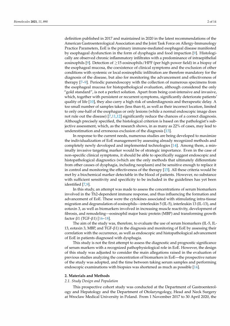
A graphical representation of the effectiveness of studied biomarkers in predictingEoE diagnosis is presented in Figure 3 as ROC curves. The calculated AUC values (areaunder the ROC curve) for all markers oscillated in the range of 0.593–0.742. The highestAUC value was obtained for the MPB, simultaneously with the lowest AUC error value.

Biomolecules 2021, 11, 890 8 of 14Biomolecules 2021, 11, x FOR PEER REVIEW 8 of 14
Figure 2. Biomarker levels in EoE patients before and after treatment: (A) IL-5, (B) IL-13, (C) TGF-β1, (D) eotaxin 3, (E)
MBP. Statistical significance was evaluated with a Wilcoxon test.
A graphical representation of the effectiveness of studied biomarkers in predicting
EoE diagnosis is presented in Figure 3 as ROC curves. The calculated AUC values (area
under the ROC curve) for all markers oscillated in the range of 0.593–0.742. The highest
AUC value was obtained for the MPB, simultaneously with the lowest AUC error value.
Figure 3. ROC curves for IL-5 (A), IL-13 (B), TGF-β1 (C), eotaxin 3 (D), and MBP (E), showing their effectiveness as markers
predicting the diagnosis of EoE. Optimal cut-off points were determined using Youden index analysis (AUC—area under
the curve, SE—standard error).
4. Discussion
So far, many studies have attempted to identify a tissue marker correlating with di-
agnosis [21], progression [22–25], and response to EoE treatment [22,26,27], allowing for
the differentiation of esophageal diseases with accompanying dysphagia [28–30] and
Figure 2. Biomarker levels in EoE patients before and after treatment: (A) IL-5, (B) IL-13, (C) TGF-β1, (D) eotaxin 3, (E) MBP.Statistical significance was evaluated with a Wilcoxon test.
Biomolecules 2021, 11, x FOR PEER REVIEW 8 of 14
Figure 2. Biomarker levels in EoE patients before and after treatment: (A) IL-5, (B) IL-13, (C) TGF-β1, (D) eotaxin 3, (E)
MBP. Statistical significance was evaluated with a Wilcoxon test.
A graphical representation of the effectiveness of studied biomarkers in predicting
EoE diagnosis is presented in Figure 3 as ROC curves. The calculated AUC values (area
under the ROC curve) for all markers oscillated in the range of 0.593–0.742. The highest
AUC value was obtained for the MPB, simultaneously with the lowest AUC error value.
Figure 3. ROC curves for IL-5 (A), IL-13 (B), TGF-β1 (C), eotaxin 3 (D), and MBP (E), showing their effectiveness as markers
predicting the diagnosis of EoE. Optimal cut-off points were determined using Youden index analysis (AUC—area under
the curve, SE—standard error).
4. Discussion
So far, many studies have attempted to identify a tissue marker correlating with di-
agnosis [21], progression [22–25], and response to EoE treatment [22,26,27], allowing for
the differentiation of esophageal diseases with accompanying dysphagia [28–30] and
Figure 3. ROC curves for IL-5 (A), IL-13 (B), TGF-β1 (C), eotaxin 3 (D), and MBP (E), showing their effectiveness as markerspredicting the diagnosis of EoE. Optimal cut-off points were determined using Youden index analysis (AUC—area underthe curve, SE—standard error).
4. Discussion
So far, many studies have attempted to identify a tissue marker correlating withdiagnosis [21], progression [22–25], and response to EoE treatment [22,26,27], allowingfor the differentiation of esophageal diseases with accompanying dysphagia [28–30] and

Biomolecules 2021, 11, 890 9 of 14
being a trigger marker in disease development, and thus an effective target of biologicaltherapies [31]. Due to the predicted low specificity of markers involved simultaneouslyin the pathomechanisms of numerous allergic diseases [32] and the ambiguous results ofresearch on tissue markers in EoE, little attention was paid to assessing the significance ofthese markers’ serum levels.
In this study, we looked for a minimally invasive marker, determined in venousblood serum, having a potential predictive value for the diagnosis, histopathological andendoscopic advancement of EoE, and correlated with the response to PPI treatment.
Based on the results of our study, it can be concluded that MBP was a serum markermost strongly (statistically significantly) correlated with both the diagnosis of EoE, aswell as the peak number of eosinophils/HPF and endoscopic advancement (assessed atdiagnosis by inflammatory, fibrostenotic, and total EREFS). The highest sensitivity andspecificity also characterized this marker. Although the correlation between the level ofMBP in blood serum [33] or saliva of patients [34] and the diagnosis or stage of EoE has notbeen proven so far, this marker’s importance in the esophageal string test has been repeat-edly emphasized [35,36], and above all in tissue tests. Positive correlations were found inpredicting the diagnosis of EoE [28,29] and in assessing the response to treatment [26]. Theadvantage of MBP1 over the peak eosinophil count (PEC) in diagnosing the disease wasalso proven in two research studies based on the assessment of tissue markers [23,37]. Thiswas justified pathophysiologically by the degranulation of eosinophils, which by releasinggranular proteins, including MBP, into the tissues, lose their cellular morphological pheno-type and therefore are not included in the histopathological examination result [23,37]. Thecorrelation between MBP and the diagnosis of EoE found in our study, although strongand statistically significant, is weaker than the correlation between the diagnosis and PEC.This is probably the price of less invasive serological determinations, but in the face of alimited number of studies on this group of potential predictors of EoE diagnosis, it doesnot undermine the sense of the study.
A serum marker that was also strongly correlated with PEC in our study was eotaxin 3,while the level of TGF-β1 correlated with the diagnosis of EoE. For these three markers, i.e.,MBP, eotaxin 3, and TGF-β1, there were also strong positive, but not statistically significant,correlations with the PEC after 8-week PPI therapy. Due to the small group of patients withEoE recruited to the project, low attendance (43.75%) in control studies after 8-week PPItherapy, as well as the lack of statistical significance of the correlations between markersand PEC after treatment, it is difficult in this study to select a serum marker predicting thehistological remission of the disease.
The levels of MBP, TGF-β1, and eotaxin 3 were positively correlated with each other.In turn, a negative correlation occurred between these markers and IL-5 and IL-13 cytokines(with a positive correlation between them). It can be interpreted as a synergy of theseproteins’ actions at subsequent stages of developing the inflammatory reaction involvingeosinophils. The pathophysiology of the disease confirms this. After the significant partici-pation of IL-5 and IL 13 in the stimulation of the influx of eosinophils to the esophagealmucosa, with the development of inflammation, their importance and tissue concentrationdecrease, and secondarily also their concentration in the blood serum. They give way toinduced eosinophil-activating chemokines, such as eotaxin 3, and products of eosinophildegranulation, including MBP and TGF-β [38].
Considering the small number of studies on blood serum markers to date, an attemptto predict the course and advancement of inflammatory and fibrostenotic EoE based on thecorrelation with the endoscopic assessment of EREFS seems innovative. Apart from thealready discussed correlation with MBP, we also observed strong and moderate statisticallysignificant correlations of eotaxin 3 with remission scores in each of the post-treatmentEREFS subscores in our study. The inhibitory effect of treatment with conventional dosesof PPIs on the expression of eotaxin 3, and secondarily on the development of the dis-ease [39–41], would therefore be reflected in the results of this study and would settle the

Biomolecules 2021, 11, 890 10 of 14
hitherto ambiguous observations confirming [42] or denying [33,43,44] the importance ofeotaxin 3 concentration in monitoring the course of EoE.
Based on the interpretation of the IL-13 and TGF-β1 concentrations, it seems possibleto differentiate the course of the inflammatory and fibrostenotic EoE in the studied group ofpatients. The increase in the concentration of TGF-β1, with the simultaneous decrease in theconcentration of IL-13 in the serum, may correspond to the development of fibrostenosisin the course of EoE. Conversely, a low concentration of TGF-β1 in the serum, with asimultaneous increase in the concentration of IL-13, may indicate less advanced diseaseand the predominance of inflammatory processes over fibrostenotic processes. Thesecorrelations were observed despite the apparent individual low specificity of both TGF-β1and IL-13 in the diagnosis and monitoring of EoE [33]. TGF-β1 is considered the “mainmediator of fibrosis” responsible for the activation of fibroblasts and the induction ofepithelial-mesenchymal transformation in many fibrostenotic processes [45]. In turn, IL-13 is well-known for its role in many atopic diseases, where it contributes to eosinophilchemotaxis, goblet cell hyperplasia, collagen deposition and an increase in smooth musclecontractility [17].
Another investigated serum marker with a confirmed role in the pathomechanismof EoE is IL-5. Previous studies assessing the importance of this cytokine in diagnos-ing and monitoring EoE have not confirmed the correlation of its concentration withthe diagnosis and course of the disease in the group of adult patients [34] and the pedi-atric population [46]. In another prospective study evaluating serum biomarker levelsin EoE after PPIs therapy, a statistically significant negative correlation was found be-tween IL-5 and esophageal eosinophilia and no prediction of the post-treatment tissueeosinophilia [44]. Similar conclusions can be drawn from this study, but the negativecorrelation with esophageal eosinophilia was not statistically significant. In the cited study,the described relationship was justified by the high accuracy of the ELISA test used [44],which may also be reflected in our study (at the detection threshold of 5 pg/mL, the medianIL-5 concentration in the group of patients with EoE was 4.07 pg/mL and 4.30 pg/mL inthe control group). A negative correlation with tissue eosinophilia can also be observedin the case of IL-13, the concentration of which, similarly to IL-5, significantly increasesin the serum after treatment. The described observation is not entirely clear but may bedue to the mediation of these interleukins in the remodeling process, leading to esophagealmotility disorders, which may persist regardless of the active eosinophilic inflammation,even after its complete resolution [47–49].
This study’s undoubted advantage is an attempt to minimize the invasiveness ofdiagnosis and monitoring of EoE by evaluating the so far rarely assessed or not assessedmarkers in blood serum with a confirmed pathophysiological relationship with EoE. Im-portant aspects are also: the prospective nature of the study, the shortest possible timeinterval between taking serum samples and performing endoscopic examinations withbiopsies, re-verification of all histopathological examinations of the specimens collectedduring the project, as well as the correct selection of the study population—homogeneousin terms of age and symptoms, allergic burden, and heterogeneous only in terms of gender.Male gender is a significant risk factor for EoE resulting from the suggested sex-dependentassociation between single nucleotide polymorphisms in the thymic stromal lymphopoietingene and its receptor and the protective effect of estrogen hormone signaling in women [7].The weakness of this study is the relatively small study group, the population limitedto adults only, and the lack of a pH-metric assessment that would allow for objectiveclassification of patients with possible gastroesophageal reflux, often coexisting with EoEor being an independent cause of dysphagia in the group of patients without EoE diagnosis.The limitations of this project suggest the need to continue research on noninvasive bloodserum biomarkers and confirm the obtained results in the validation cohort, taking intoaccount the possible effect of co-occurrence and overlapping of EoE and GERD, as well asdepending on the pharmacotherapy used: PPI, local steroid therapy or elimination diet.

Biomolecules 2021, 11, 890 11 of 14
5. Conclusions
Based on the results of this study and the available literature data, it is not possible toselect one serum biomarker with pleiotropic predictive and prognostic functions in EoE.
The observed trend, suggesting the importance of MBP in predicting the diagnosisand eotaxin 3 in predicting disease advancement, emphasizes the potential for improvingthe management and increasing the individualization of treatment. However, the necessarycondition is to determine the markers several times, and not one parameter should be con-sidered, but the whole group of them together, taking into account the pathophysiologicalrole and interdependencies.
It can be predicted that this project, as well as the existing high-quality prospectivestudies correlating the concentration of individual markers in the blood serum with thediagnosis and progression of EoE, has developed a material for the creation of an automatedalgorithm that would provide intelligent analysis of the obtained data and could improvethe precision of EoE diagnostics and therapy in the future.
Author Contributions: Study Design: D.W.-C., B.W., J.S.; data collection: D.W.-C., J.S.; statisticalanalysis: B.W.; data interpretation: J.S., B.W., D.W.-C.; manuscript preparation: J.S., B.W., D.W.-C.;literature search: J.S.; funds collection: J.S., D.W.-C. All authors have read and agreed to the publishedversion of the manuscript.
Funding: This study was funded by the Wroclaw Medical University Research Program for YoungScientists (Project: STM.C130.17.045).
Institutional Review Board Statement: The study was conducted according to the guidelines ofthe Declaration of Helsinki, and approved by the Bioethics Committee of the Medical University ofWroclaw, Poland (KB no. 544/2017, 17 August 2017) with a subsequent extension (KB no. 730/2018,6 December 2018).
Informed Consent Statement: Informed consent was obtained from all subjects involved in the study.
Data Availability Statement: The data generated and analyzed during the current study are availablefrom the corresponding author upon reasonable request.
Acknowledgments: The authors would like to thank Maria Jasinska and Beata Marczak-Karpinafrom the Department of Gastroenterology’s Scientific Laboratory (Wroclaw Medical University) fortheir lab assistance with this work.
Conflicts of Interest: The authors declare no conflict of interest.
Trial Registration
This trial was registered with ClinicalTrials.gov (no. NCT04803162). Registered 17March 2021—Retrospectively registered, https://clinicaltrials.gov/ct2/show/NCT04803162 (accessed on 13 June 2021).
Abbreviations
AUC area under the curveELISA enzyme-linked immunosorbent assayEoE eosinophilic esophagitisEREFS Eosinophilic Esophagitis Endoscopic Reference ScoreHPF high power fieldIL-5 interleukin 5IL-13 interleukin 13IQR interquartile rangesMBP major basic proteinPEC peak eosinophil countPPI proton pump inhibitorROC receiver operating characteristicTGF-β1 transforming growth factor β1

Biomolecules 2021, 11, 890 12 of 14
References1. Dellon, E.S. Epidemiology of Eosinophilic Esophagitis. Gastroenterol. Clin. N. Am. 2014, 43, 201–218. [CrossRef] [PubMed]2. Arias, Á.; Lucendo, A.J. Epidemiology and risk factors for eosinophilic esophagitis: Lessons for clinicians. Expert Rev. Gastroenterol.
Hepatol. 2020, 14, 1069–1082. [CrossRef] [PubMed]3. Gonsalves, N. Eosinophilic Esophagitis: History, Nomenclature, and Diagnostic Guidelines. Gastrointest. Endosc. Clin. N. Am.
2008, 18, 1–9. [CrossRef] [PubMed]4. Furuta, G.T.; Liacouras, C.A.; Collins, M.H.; Gupta, S.K.; Justinich, C.; Putnam, P.E.; Bonis, P.; Hassal, E.; Straumann, A.;
Rothenberg, M.E. Eosinophilic Esophagitis in Children and Adults: A Systematic Review and Consensus Recommendations forDiagnosis and Treatment. Sponsored by the American Gastroenterological Association (AGA) Institute and North AmericanSociety of Pediatric Gastroenterol. Gastroenterology 2007, 133, 1342–1363. [CrossRef] [PubMed]
5. Liacouras, C.A.; Furuta, G.T.; Hirano, I.; Atkins, D.; Attwood, S.E.; Bonis, P.A.; Burks, A.W.; Chehade, M.; Collins, M.H.; Dellon,E.S.; et al. Eosinophilic esophagitis: Updated consensus recommendations for children and adults. J. Allergy Clin. Immunol. 2011,128, 3–20.e6. [CrossRef] [PubMed]
6. Dellon, E.S.; Gonsalves, N.; Hirano, I.; Furuta, G.T.; Liacouras, C.A.; Katzka, D.A. ACG Clinical Guideline: Evidenced BasedApproach to the Diagnosis and Management of Esophageal Eosinophilia and Eosinophilic Esophagitis (EoE). Am. J. Gastroenterol.2013, 108, 679–692. [CrossRef] [PubMed]
7. Lucendo, A.J.; Molina-Infante, J.; Arias, Á.; von Arnim, U.; Bredenoord, A.J.; Bussmann, C.; Dias, J.A.; Gonzalez-Cervera, J.;Larsson, H.; Miehlke, S.; et al. Guidelines on eosinophilic esophagitis: Evidence-based statements and recommendations fordiagnosis and management in children and adults. United Eur. Gastroenterol. J. 2017, 5, 335–358. [CrossRef] [PubMed]
8. Dellon, E.S.; Liacouras, C.A.; Molina-Infante, J.; Furuta, G.T.; Spergel, J.M.; Zevit, N.; Spechler, S.J.; Attwood, S.E.; Straumann, A.;Aceves, S.S.; et al. Updated international consensus diagnostic criteria for eosinophilic esophagitis: Proceedings of the AGREEconference. Gastroenterology 2018, 155, 1022.e10–1033.e10. [CrossRef]
9. Hirano, I.; Chan, E.S.; Rank, M.A.; Sharaf, R.N.; Stollman, N.H.; Stukus, D.R.; Wang, K.; Greenhawt, M.; Falck-Ytter, Y.T.;Chachu, K.A.; et al. AGA institute and the joint task force on allergy-immunology practice parameters clinical guidelines for themanagement of eosinophilic esophagitis. Ann. Allergy Asthma Immunol. 2020, 124, 416–423. [CrossRef]
10. Schoepfer, A.; Safroneeva, E.; Straumann, A. How to measure disease activity in eosinophilic esophagitis. Dis. Esophagus 2015, 29,959–966. [CrossRef]
11. Nielsen, J.A.; Lager, D.J.; Lewin, M.; Rendon, G.; Roberts, C.A. The optimal number of biopsy fragments to establish a morphologicdiagnosis of eosinophilic esophagitis. Am. J. Gastroenterol. 2014, 109, 515–520. [CrossRef] [PubMed]
12. Kim, H.P.; Vance, R.B.; Shaheen, N.J.; Dellon, E.S. The Prevalence and Diagnostic Utility of Endoscopic Features of EosinophilicEsophagitis: A Meta-analysis. Clin. Gastroenterol. Hepatol. 2012, 10, 988–996.e5. [CrossRef] [PubMed]
13. Stucke, E.M.; Clarridge, K.E.; Collins, M.H.; Henderson, C.J.; Martin, L.J.; Rothenberg, M.E. Value of an Additional Review forEosinophil Quantification in Esophageal Bi-opsies. J. Pediatr. Gastroenterol. Nutr. 2015, 61, 65–68. [CrossRef] [PubMed]
14. Godwin, B.; Wilkins, B.; Muir, A.B. EoE disease monitoring: Where we are and where we are going. Ann Allergy. Asthma Immunol.2020, 124, 240–247. [CrossRef] [PubMed]
15. Fujiwara, Y. Symptom-based diagnostic approach for eosinophilic esophagitis. J. Gastroenterol. 2020, 55, 833–845. [CrossRef][PubMed]
16. Muir, A.B.; Wang, J.X.; Nakagawa, H. Epithelial-stromal crosstalk and fibrosis in eosinophilic esophagitis. J. Gastroenterol. 2019,54, 10–18. [CrossRef] [PubMed]
17. O’Shea, K.M.; Aceves, S.S.; Dellon, E.S.; Gupta, S.K.; Spergel, J.M.; Furuta, G.T.; Rothenberg, M.E. Pathophysiology of EosinophilicEsophagitis. Gastroenterology 2018, 154, 333–345. [CrossRef]
18. D’Alessandro, A. Eosinophilic esophagitis: From pathophysiology to treatment. World J. Gastrointest. Pathophysiol. 2015, 6,150–158. [CrossRef]
19. Hirano, I.; Moy, N.; Heckman, M.G.; Thomas, C.S.; Gonsalves, N.; Achem, S.R. Endoscopic assessment of the oesophageal featuresof eosinophilic oesophagitis: Validation of a novel classification and grading system. Gut 2013, 62, 489–495. [CrossRef]
20. Dellon, E.S.; Cotton, C.C.; Gebhart, J.H.; Higgins, L.L.; Beitia, R.; Woosley, J.T.; Shaheen, N.J. Accuracy of the EosinophilicEsophagitis Endoscopic Reference Score in Diagnosis and Determining Response to Treatment. Clin. Gastroenterol. Hepatol. 2016,14, 31–39. [CrossRef]
21. Wright, B.L.; Doyle, A.D.; Shim, K.P.; Pai, R.K.; Barshow, S.M.; Horsley-Silva, J.L.; Luo, H.; Rank, M.A.; Jacobsen, E.A.; Katzka,D.A.; et al. Image Analysis of Eosinophil Peroxidase Immunohistochemistry for Diagnosis of Eosinophilic Esophagitis. Dig. Dis.Sci. 2021, 66, 775–783. [CrossRef]
22. Kim, G.H.; Park, Y.S.; Jung, K.W.; Kim, M.; Na, H.K.; Ahn, J.Y.; Lee, J.H.; Kim, D.H.; Choi, K.D.; Song, H.J.; et al. An IncreasingTrend of Eosinophilic Esophagitis in Korea and the Clinical Implication of the Biomarkers to Determine Disease Activity andTreatment Response in Eosinophilic Esophagitis. J. Neurogastroenterol. Motil. 2019, 25, 525–533. [CrossRef]
23. Peterson, K.A.; Gleich, G.J.; Limaye, N.S.; Crispin, H.; Robson, J.; Fang, J.; Saffari, H.; Clayton, F.; Leiferman, K.M. Eosinophilgranule major basic protein 1 deposition in eosinophilic esophagitis correlates with symptoms independent of eosinophil counts.Dis. Esophagus 2019, 32, doz055. [CrossRef]

Biomolecules 2021, 11, 890 13 of 14
24. Johansson, M.W.; McKernan, E.M.; Fichtinger, P.S.; Angulo, E.L.; Bagley, J.L.; Lee, K.E.; Evans, M.D.; Lomeli, L.D.; Mosher,D.F.; Cook, S.M.; et al. αIIb-Integrin (CD41) associated with blood eosinophils is a potential biomarker for disease activity ineosinophilic esophagitis. J. Allergy Clin. Immunol. 2020, 145, 1699–1701. [CrossRef]
25. Bartig, K.A.; Lee, K.E.; Mosher, D.F.; Mathur, S.K.; Johansson, M.W. Platelet association with leukocytes in active eosinophilicesophagitis. PLoS ONE 2021, 16, e0250521. [CrossRef] [PubMed]
26. Dellon, E.S.; Woosley, J.T.; McGee, S.J.; Moist, S.E.; Shaheen, N.J. Utility of major basic protein, eotaxin-3, and mast cell tryptasestaining for prediction of response to topical steroid treatment in eosinophilic esophagitis: Analysis of a randomized, double-blind,double dummy clinical trial. Dis. Esophagus 2020, 33. [CrossRef] [PubMed]
27. Lingblom, C.; Albinsson, S.; Johansson, L.; Larsson, H.; Wennerås, C. Patient-Reported Outcomes and Blood-Based ParametersIdentify Response to Treatment in Eosinophilic Esophagitis. Dig. Dis. Sci. 2021, 66, 1556–1564. [CrossRef] [PubMed]
28. Dellon, E.S.; Chen, X.; Miller, R.C.; Woosley, J.T.; Shaheen, N.J. Diagnostic Utility of Major Basic Protein, Eotaxin-3, and LeukotrieneEnzyme Staining in Eosinophilic Esophagitis. Am. J. Gastroenterol. 2012, 107, 1503–1511. [CrossRef] [PubMed]
29. Dellon, E.S.; Speck, O.; Woodward, K.; Covey, S.; Rusin, S.; Gebhart, J.H.; Chen, X.; Woosley, J.T.; Shaheen, N.J. Markersof Eosinophilic Inflammation for Diagnosis of Eosinophilic Esophagitis and Proton Pump Inhibitor–Responsive EsophagealEosinophilia: A Prospective Study. Clin. Gastroenterol. Hepatol. 2014, 12, 2015–2022. [CrossRef]
30. Clayton, S.; Cauble, E.; Kumar, A.; Patil, N.; Ledford, D.; Kolliputi, N.; Lopes-Virella, M.F.; Castell, D.; Richter, J. Plasma levels ofTNF-α, IL-6, IFN-γ, IL-12, IL-17, IL-22, and IL-23 in achalasia, eosinophilic esophagitis (EoE), and gastroesophageal reflux disease(GERD). BMC Gastroenterol. 2019, 19, 28. [CrossRef]
31. Lucendo, A.J.; López-Sánchez, P. Targeted Therapies for Eosinophilic Gastrointestinal Disorders. BioDrugs 2020, 34, 477–493.[CrossRef]
32. Hines, B.T.; Rank, M.A.; Wright, B.L.; Marks, L.A.; Hagan, J.B.; Straumann, A.; Greenhawt, M.; Dellon, E.S. Minimally invasivebiomarker studies in eosinophilic esophagitis: A systematic review. Ann. Allergy Asthma Immunol. 2018, 121, 218–228. [CrossRef]
33. Dellon, E.S.; Rusin, S.; Gebhart, J.H.; Covey, S.; Higgins, L.L.; Beitia, R.; Speck, O.; Woodward, K.; Woosley, J.T.; Shaheen, N.J.Utility of a noninvasive serum biomarker panel for diagnosis and monitoring of eosinophilic esophagitis: A prospective study.Am. J. Gastroenterol. 2015, 110, 821–827. [CrossRef]
34. Avinashi, V.; Chan, J.M.; Bush, J.W.; Vallance, B.A.; Yang, H.; Portales-Casamar, E.; Soller, L.; Mill, C.; Chan, E.S. Poor Correlationof Oral Swabs with Esophageal Eosinophil Counts. Dysphagia 2019, 35, 773–779. [CrossRef] [PubMed]
35. Furuta, G.T.; Kagalwalla, A.F.; Lee, J.J.; Alumkal, P.; Maybruck, B.T.; Fillon, S.; Masterson, J.C.; Ochkur, S.; Protheroe, C.; Moore,W.; et al. The oesophageal string test: A novel, minimally invasive method measures mucosal inflammation in eosinophilicoesophagitis. Gut 2012, 62, 1395–1405. [CrossRef] [PubMed]
36. Ackerman, S.J.; Kagalwalla, A.F.; Hirano, I.; Gonsalves, N.; Katcher, P.M.; Gupta, S.; Wechsler, J.B.; Grozdanovic, M.; Pan, Z.;Masterson, J.C.; et al. One-Hour Esophageal String Test: A Nonendoscopic Minimally Invasive Test That Accurately DetectsDisease Activity in Eosinophilic Esophagitis. Am. J. Gastroenterol. 2019, 114, 1614–1625. [CrossRef] [PubMed]
37. Peterson, K.A.; Cobell, W.J.; Clayton, F.C.; Krishnamurthy, C.; Ying, J.; Pease, L.F.; Saffari, H.; Georgelas, A.; Fang, J.; Gleich, G.J.;et al. Extracellular Eosinophil Granule Protein Deposition in Ringed Esophagus with Sparse Eosinophils. Dig. Dis. Sci. 2015, 60,2646–2653. [CrossRef] [PubMed]
38. Travers, J.; Rothenberg, M.E. Eosinophils in mucosal immune responses. Mucosal Immunol. 2015, 8, 464–475. [CrossRef] [PubMed]39. Cheng, E.; Zhang, X.; Huo, X.; Yu, C.; Zhang, Q.; Wang, D.H.; Spechler, S.J.; Souza, R.F. Omeprazole blocks eotaxin-3 expression
by oesophageal squamous cells from patients with eosinophilic oesophagitis and GORD. Gut 2013, 62, 824–832. [CrossRef]40. Zhang, X.; Cheng, E.; Huo, X.; Yu, C.; Zhang, Q.; Pham, T.H.; Wang, D.H.; Spechler, S.J.; Souza, R.F. Omeprazole Blocks STAT6
Binding to the Eotaxin-3 Promoter in Eosinophilic Esophagitis Cells. PLoS ONE 2012, 7, e50037. [CrossRef]41. Molina-Infante, J.; Rivas, M.D.; Hernandez-Alonso, M.; Vinagre-Rodríguez, G.; Mateos-Rodríguez, J.M.; Dueñas-Sadornil,
C.; Perez-Gallardo, B.; Ferrando-Lamana, L.; Fernandez-Gonzalez, N.; Bañares, R.; et al. Proton pump inhibitor-responsiveoesophageal eosinophilia correlates with downregulation of eotaxin-3 and Th2 cytokines overexpression. Aliment. Pharmacol.Ther. 2014, 40, 955–965. [CrossRef]
42. Konikoff, M.R.; Blanchard, C.; Kirby, C.; Buckmeier, B.K.; Cohen, M.B.; Heubi, J.E.; Putnam, P.E.; Rothenberg, M.E. Potential ofBlood Eosinophils, Eosinophil-Derived Neurotoxin, and Eotaxin-3 as Biomarkers of Eosinophilic Esophagitis. Clin. Gastroenterol.Hepatol. 2006, 4, 1328–1336. [CrossRef] [PubMed]
43. Ishihara, S.; Shoda, T.; Ishimura, N.; Ohta, S.; Ono, J.; Azuma, Y.; Okimoto, E.; Izuhara, K.; Nomura, I.; Matsumoto, K.; et al.Serum biomarkers for the diagnosis of eosinophilic esophagitis and eosinophilic gastroenteritis. Intern. Med. 2017, 56, 2819–2825.[CrossRef]
44. Min, S.B.; Nylund, C.M.; Baker, T.P.; Ally, M.; Reinhardt, B.; Chen, Y.-J.; Nazareno, L.; Moawad, F.J. Longitudinal Evaluation ofNoninvasive Biomarkers for Eosinophilic Esophagitis. J. Clin. Gastroenterol. 2017, 51, 127–135. [CrossRef]
45. Armbruster-Lee, J.; Cavender, C.P.; Lieberman, J.A.; Samarasinghe, A.E. Understanding fibrosis in eosinophilic esophagitis: Arewe there yet? J. Leukoc. Biol. 2018, 104, 31–40. [CrossRef] [PubMed]
46. Subbarao, G.; Rosenman, M.B.; Ohnuki, L.; Georgelas, A.; Davis, M.; Fitzgerald, J.F.; Molleston, J.P.; Croffie, J.M.; Pfefferkorn,M.D.; Corkins, M.R.; et al. Exploring Potential Noninvasive Biomarkers in Eosinophilic Esophagitis in Children. J. Pediatr.Gastroenterol. Nutr. 2011, 53, 651–658. [CrossRef] [PubMed]

Biomolecules 2021, 11, 890 14 of 14
47. Nhu, Q.M.; Aceves, S.S. Tissue Remodeling in Chronic Eosinophilic Esophageal Inflammation: Parallels in Asthma and Thera-peutic Perspectives. Front. Med. 2017, 4, 128. [CrossRef] [PubMed]
48. Mavi, P.; Rajavelu, P.; Rayapudi, M.; Paul, R.J.; Mishra, A. Esophageal functional impairments in experimental eosinophilicesophagitis. Am. J. Physiol. Liver Physiol. 2012, 302, G1347–G1355. [CrossRef]
49. Zuo, L.; Fulkerson, P.C.; Finkelman, F.D.; Mingler, M.; Fischetti, C.A.; Blanchard, C.; Rothenberg, M.E. IL-13 Induces EsophagealRemodeling and Gene Expression by an Eosinophil-Independent, IL-13Rα2–Inhibited Pathway. J. Immunol. 2010, 185, 660–669.[CrossRef]