ScopeMed-fulltext (2)
3
Case Report Chromoblastomy cosis NJIRM 2011; Vol. 2(1).Jan-March eISSN: 0975-9840 pISSN: 2230 - 9969 P a g e 5 7 Chromoblastomycosis Dr. Harshada Shah*, Dr. K.Verma**, Dr. P.L.Dhand***, Dr. Rajesh Soni****, Ms. Pragya Shakya **** *Prof. & Head, Dept. of Microbiology, **Prof. & Head, Dept. of Skin & Venereal Disease, ***Prof. & Head, Dept. of Pathology, ***Assistant Professor, ****P.G. Student, Dept. of Microbiology, RDGMC, Ujjain Abstract: Chromoblastomycosis is a chronic fungal infection primarily of skin and subcutaneous tissue caused by a variety of dematiaceous fungal species belonging to different genera. We report a case which remains undiagnosed for 15 years although it presented with the most common manifestation. It was diagnosed and treated with excellent clinico-microbiological and histo-pathological correlation. Key words: Chromoblastomycosis, Cladosporium carionii, Copper penny bodies. Corresponding Author: Dr. Harshada Shah, Professor & Head, Dept of Microbiology, R.D.Gardi Medical College, Surasa, Agar Road, Ujjain (MP) 456006, E –mail: [email protected] INTRODUCTION: Chromoblastomycosis is a chronic, relatively uncommon, localized mycotic infection of the skin and subcutaneous tissue. Normally, it follows a traumatic implantation of dematiaceous fungi. The lesions range from verrucoid, ulcerated, crustate or cauliflower like growth to raised or flat plaques. Satellite lesions develop following auto- inoculation and by lymphatic spread to adjacent areas. The disease has been described worldwide, but the incidence is greater in subtropical regions. Large numbers of cases have been reported from Madagascar and Brazil. Farming is the most common occupation associated with this lesion 1 . In India, most of the cases have been reported in belts from North to South sparing the Eastern and Western regions 2 . We present a case of Chromoblastomycosis from Central India. CASE REPORT: A 50 year old male agriculturist from Ujjain district in Madhya Pradesh visited the Skin OPD of C.R Gardi hospital in Jan, 2008. He presented with a 15 years long history and a cauliflower like lesion associated with itching and depigmentation of right lower limb. History revealed that it began with a small papular lesion on the external surface of right foot with itching. He went pole to post to seek medical attention until the lesion increased in size over a period of last 5 years. He also tried several home remedies. He was diagnosed as a case of tuberculosis at other center and received anti-tubercular treatment without getting any clinical response. The lesion continued to progress and reached up to his knee (fig.1). Fig 1 Initial KOH mount showed a fair number of round, non-septate structures resembling fungal spores. The skin biopsy done at our center revealed a pseudoepitheliomatous hyperplasia with micro abscesses and granulomatous infiltrate (fig.2). Fig 2
-
Upload
ernel-tosoc -
Category
Documents
-
view
221 -
download
0
Transcript of ScopeMed-fulltext (2)
8/4/2019 ScopeMed-fulltext (2)
http://slidepdf.com/reader/full/scopemed-fulltext-2 1/3
Case Report Chromoblastomycosis
NJIRM 2011; Vol. 2(1).Jan-March eISSN: 0975-9840 pISSN: 2230 - 9969
5 7
Chromoblastomycosis
Dr. Harshada Shah*, Dr. K.Verma**, Dr. P.L.Dhand***, Dr. Rajesh Soni****, Ms. Pragya Shakya ****
*Prof. & Head, Dept. of Microbiology, **Prof. & Head, Dept. of Skin & Venereal Disease, ***Prof. & Head, Dept. of Pathology,
***Assistant Professor, ****P.G. Student, Dept. of Microbiology, RDGMC, Ujjain
Abstract: Chromoblastomycosis is a chronic fungal infection primarily of skin and subcutaneous tissue caused by
a variety of dematiaceous fungal species belonging to different genera. We report a case which remains
undiagnosed for 15 years although it presented with the most common manifestation. It was diagnosed and
treated with excellent clinico-microbiological and histo-pathological correlation.
Key words: Chromoblastomycosis, Cladosporium carionii, Copper penny bodies.
Corresponding Author: Dr. Harshada Shah, Professor & Head, Dept of Microbiology, R.D.Gardi Medical College,
Surasa, Agar Road, Ujjain (MP) 456006, E –mail: [email protected]
INTRODUCTION: Chromoblastomycosis is a chronic,
relatively uncommon, localized mycotic infection of
the skin and subcutaneous tissue. Normally, it
follows a traumatic implantation of dematiaceous
fungi. The lesions range from verrucoid, ulcerated,
crustate or cauliflower like growth to raised or flat
plaques. Satellite lesions develop following auto-
inoculation and by lymphatic spread to adjacent
areas. The disease has been described worldwide,
but the incidence is greater in subtropical regions.
Large numbers of cases have been reported from
Madagascar and Brazil. Farming is the mostcommon occupation associated with this lesion1
. In
India, most of the cases have been reported in belts
from North to South sparing the Eastern and
Western regions2. We present a case of
Chromoblastomycosis from Central India.
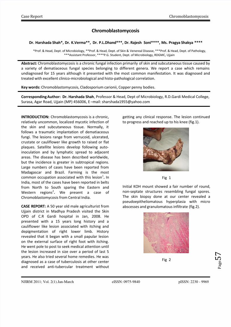
CASE REPORT: A 50 year old male agriculturist from
Ujjain district in Madhya Pradesh visited the Skin
OPD of C.R Gardi hospital in Jan, 2008. He
presented with a 15 years long history and a
cauliflower like lesion associated with itching and
depigmentation of right lower limb. History
revealed that it began with a small papular lesion
on the external surface of right foot with itching.
He went pole to post to seek medical attention until
the lesion increased in size over a period of last 5
years. He also tried several home remedies. He was
diagnosed as a case of tuberculosis at other center
and received anti-tubercular treatment without
getting any clinical response. The lesion continued
to progress and reached up to his knee (fig.1).
Fig 1
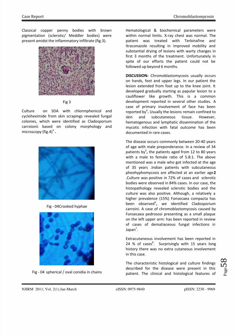
Initial KOH mount showed a fair number of round,
non-septate structures resembling fungal spores.
The skin biopsy done at our center revealed a
pseudoepitheliomatous hyperplasia with micro
abscesses and granulomatous infiltrate (fig.2).
Fig 2
8/4/2019 ScopeMed-fulltext (2)
http://slidepdf.com/reader/full/scopemed-fulltext-2 2/3
Case Report Chromoblastomycosis
NJIRM 2011; Vol. 2(1).Jan-March eISSN: 0975-9840 pISSN: 2230 - 9969
5 8
Classical copper penny bodies with brown
pigmentation (sclerotic/ Meddler bodies) were
present amidst the inflammatory infiltrate (fig.3).
Fig 3
Culture on SDA with chlormphenicol and
cycloheximide from skin scrapings revealed fungalcolonies, which were identified as Cladosporium
carroionii based on colony morphology and
microscopy (fig.4)3
.
Fig - 04Crooked hyphae
Fig - 04 spherical / oval conidia in chains
Hematological & biochemical parameters were
within normal limits. X-ray chest was normal. The
patient was treated with Terbinafine and
Itraconazole resulting in improved mobility and
substantial drying of lesions with warty changes in
first 3 months of the treatment. Unfortunately in
spite of our efforts the patient could not be
followed up beyond 6 months.
DISCUSSION: Chromoblastomycosis usually occurs
on hands, feet and upper legs. In our patient the
lesion extended from foot up to the knee joint. It
developed gradually starting as papular lesion to a
cauliflower like growth. This is a common
development reported in several other studies. A
case of primary involvement of face has been
reported by4. Usually the lesions remain confined to
skin and subcutaneous tissue. However,hematogenous and lymphatic dissemination of the
mycotic infection with fatal outcome has been
documented in rare cases.
The disease occurs commonly between 20-40 years
of age with male preponderance. In a review of 34
patients by5, the patients aged from 12 to 80 years
with a male to female ratio of 5.8:1. The above
mentioned was a male who got infected at the age
of 35 years .Indian patients with subcutaneous
pheohyphomycosis are affected at an earlier age2
.Culture was positive in 72% of cases and scleroticbodies were observed in 84% cases. In our case, the
histopathology revealed sclerotic bodies and the
culture was also positive. Although, a relatively a
higher prevalence (15%) Fonsecaea compacta has
been observed6, we identified Cladosporium
carroini. A case of chromoblastomycosis caused by
Fonsecaea pedrososi presenting as a small plaque
on the left upper arm: has been reported in review
of cases of dematiaceous fungal infections in
Japan7.
Extracutaneous involvement has been reported in
24 % of cases8. Surprisingly with 15 years long
history there was no extra cutaneous involvement
in this case.
The characteristic histological and culture findings
described for the disease were present in this
patient. The clinical and histological features of

8/4/2019 ScopeMed-fulltext (2)
http://slidepdf.com/reader/full/scopemed-fulltext-2 3/3
Case Report Chromoblastomycosis
NJIRM 2011; Vol. 2(1).Jan-March eISSN: 0975-9840 pISSN: 2230 - 9969
5 9
chromoblastomycosis are often misdiagnosed as
squamous cell carcinoma or cutaneous tuberculosis,
if a high level of suspicion and early diagnosis is not
made. Similarly our patient was treated for
cutaneous tuberculosis for two years.
Numerous studies reported from India describe thedistribution of the disease in the country in a belt
from north to south, more so in sub-Himalayan
belt6, 9,10
. There are sporadic reports from eastern
and western regions. Since, there are sparse reports
from our region, we report this case.
Intra-lesional or systemic administration of
amphotericin-B, flucytosine, calciferol with or
without potassium iodide and INH has produced
better results than ketoconazole, thiabendazole and
topical heat and X-ray therapy. Variable results withketoconazole alone have been reported. In the
above referred case, terbinafine and itraconazole
halted the progression of the fungus.
In our opinion, a high clinical suspicion by the
dermatologist and collaborative efforts done by the
clinician with the pathology and microbiology
laboratory has resulted into an excellent diagnosis
which was unfortunately not revealed during last 15
years in spite of approaching several clinicians, from
various places by the patient. Secondly, lack of
microbiology facilities at most of the places maylead to under or wrong diagnosis leading to delay in
proper treatment in these patients.
REFERENCES:
1. Seema V. Pradhan et al; Indian Journal of
Dermatology, Venerology and Leprology 2007,
Vol 73, Page no 176
2. Sharma NL, Mahajan V, Sharma RC, Sharma A.,
2002., Subcutaneous pheohypomycosis in India-
a case report and review. Int. J. Dermatol. Vol
41(1), p-16-20.3. Jagdish Chander – Textbook of medical
mycology – 3rd edition – 2009.P -181
4. Rakesh Bharti, S K Malhotra, Singh Manjit Bal, –
Indian journal of dermatology, venereology and
leprology, 1995, vol – 61(1), p – 54,55
5. Sharma NL, Sharma RC, Grover PS, Gupta ML,
Sharma AK, Mahajan VK(1999).,
Chromoblastomycosis in India. Int. J .Dermatol
38(11)., 846- 51.
6. Rajendran C, Ramesh V, Misra RS, Kandhari S, U.
preti HB, Datta K K(1997) .
Chromoblastomycosis in India. Int. J. Dermatol
36(1), p-29-33.
7. Kikuchi Y, Kondo M, Yaguchi H, Hiruma M, Ikeda
S. 2007. A case of chromoblastomycosis caused
by Fonsecaea pedrososi presenting as a small
plaque on the left upper arm: a review of
reported cases of dematiaceous fungal
infection in Japan. Nippon Ishinkin Gakkai
Zasshi. 48(2) 85-9.
8. 8.. Chromoblastomycosis in India – Nandlal
Sharma, Ramesh Chander Sharma, Prem singh
Grover, ML Gupta, AK Sharma, VK Mahajan –
International journal of dermatology – 2001, v –
38(Nov), p –
846 -851.9. Vyas MC, Joshi YR, Bhargava N, Joshi KR, Tanwar
RK. 2000. Cerebral chromoblastomicosis – a
rare case report of cerebral abcess and brief
review of literature – a case report.Indian .J.
Pathol. Microbiol.43 (1), p 81- 10.
10. Garg N, Devi IB, Vajramani GV, Nagarathna S,
Sampath S, Chandramouli BA, Chandramukhi A,
Shankar SK.Central nervous system
cladosporiosis: an account of ten culture-
proven cases. Neurol India, 55(3): 282-8.