Scleromyxedema: a case treated with oral prednisone ... · é difícil, não existindo na...
4
Received on August 01, 2004. Approved by the Consultive Council and accepted for publication on December 07, 2005. * Work done at Dermatology Service of the Hospital Universitário Onofre Lopes/Universidade Federal do Rio Grande do Norte – UFRN - (RN), Brazil Conflict of interests: None 1 Associate Professor of Dermatology at the Universidade Federal do Rio Grande do Norte – UFRN – Rio Grande do Norte (RN), Brazil. Ph.D. in Dermatology from the Universidade Federal do Rio de Janeiro – UFRJ – Rio de Janeiro (RJ), Brazil. 2 Pathologist at the Hospital Universitário Onofre Lopes Universidade Federal do Rio Grande do Norte – UFRN – Rio Grande do Norte (RN), Brazil. 3 Resident Physician at Dermatology Service of the Hospital Universitário Onofre Lopes - Universidade Federal do Rio Grande do Norte – UFRN – Rio Grande do Norte (RN), Brazil. 4 Resident Physician at Dermatology Service of the Hospital Universitário Onofre Lopes - Universidade Federal do Rio Grande do Norte – UFRN – Rio Grande do Norte (RN), Brazil. ©2006 by Anais Brasileiros de Dermatologia Scleromyxedema: a case treated with oral prednisone * Escleromixedema: um caso tratado com prednisona oral * Pedro Bezerra da Trindade Neto 1 Alexandre de Oliveira Sales 2 Aldavânea Cabral de Oliveira e Silva 3 Juliana Cristina Soares Nunes 4 Abstract: Scleromyxedema is an idiopathic cutaneous mucinosis characterized by a papular eruption, skin induration and paraproteinemia. Histologically, fibrolast proliferation can be observed in the upper dermis associated with a mucine deposition. Treatment is difficult and at present there is no totally effective therapeutic modality to control the disease. The present report is on a 68-year-old patient with scleromyxedema without systemic manifestation, who responded to oral steroid therapy. Keywords: Adrenal cortex hormones; Hyaluronic acid; Hypergammaglobulinemia; Mucinoses Resumo: O escleromixedema é uma mucinose cutânea idiopática caracterizada por erupção papulosa, induração da pele e paraproteinemia. Histologicamente, se observa pro- liferação de fibroblastos na derme superior associada a depósito de mucina. O tratamento é difícil, não existindo na atualidade modalidade terapêutica totalmente eficaz para con- trolar a enfermidade. Relata-se o caso de um paciente de 68 anos com escleromixedema, sem manifestação sistêmica, que respondeu à terapia oral com corticosteróide. Palavras-chave: Ácido hialurônico; Corticosteróides; Hipergamaglobulinemia; Mucinoses An Bras Dermatol. 2006;81(1):55-8. Case Report 55 INTRODUCTION Scleromyxedema is a primary cutaneous muci- nosis of unknown origin, characterized by infiltrating lesions in the skin, a mucine deposit in the upper der- misis and monoclonal paraproteinemia. 1,2 The myxede- matous lichen spectre (papular mucinosis) was original- ly described by Dubreuilh and Reitman, in 1906. 3 In 1953, Montgomery and Underwood 4 classi- fied the myxedematous lichen into four clinical forms: (1) generalized lichenous papular eruption; (2) non- confluent papular form; (3) localized or generalized lichenous plaques; and (4) urticaria-like plaques and nodular lichenous eruptions. In 1954, Gottron and Arndt introduced the term scleromyxedema to refer to a variant of papular muci- nosis which corresponds to a generalized diffuse lichenous eruption with skin sclerosis and normal thy- roid function. 1,5 As a response to the indiscriminate use of the terms papular mucinosis, myxedematous lichen, and scleromyxedema, in 2001, Rongioletti et al., 6 based on anatomic-clinical criteria, adopted a new classification for this group of diseases: (1) generalized myxedematous lichen (scleromyxedema); (2) localized myxedematous lichen; (3) atypical forms of myxedematous lichen. Even though there have been reports of diverse therapeutic regimens, the benefits for this condition are variable. 7 The objective of this paper is to describe a case of this rare entity, stressing the favorable clinical response to the systemic use of a corticosteroid. CASE REPORT 68-year-old male, anos, do sexo masculino, mar- ried, agricultural worker, born in and coming from
Transcript of Scleromyxedema: a case treated with oral prednisone ... · é difícil, não existindo na...

Received on August 01, 2004.Approved by the Consultive Council and accepted for publication on December 07, 2005.* Work done at Dermatology Service of the Hospital Universitário Onofre Lopes/Universidade Federal do Rio Grande do Norte – UFRN - (RN), Brazil Conflict of interests: None
1 Associate Professor of Dermatology at the Universidade Federal do Rio Grande do Norte – UFRN – Rio Grande do Norte (RN), Brazil. Ph.D. in Dermatology from the Universidade Federal do Rio de Janeiro – UFRJ – Rio de Janeiro (RJ), Brazil.
2 Pathologist at the Hospital Universitário Onofre Lopes Universidade Federal do Rio Grande do Norte – UFRN – Rio Grande do Norte (RN), Brazil.3 Resident Physician at Dermatology Service of the Hospital Universitário Onofre Lopes - Universidade Federal do Rio Grande do Norte – UFRN – Rio
Grande do Norte (RN), Brazil.4 Resident Physician at Dermatology Service of the Hospital Universitário Onofre Lopes - Universidade Federal do Rio Grande do Norte – UFRN – Rio
Grande do Norte (RN), Brazil.
©2006 by Anais Brasileiros de Dermatologia
Scleromyxedema: a case treated with oral prednisone*
Escleromixedema: um caso tratado com prednisona oral*
Pedro Bezerra da Trindade Neto1 Alexandre de Oliveira Sales2
Aldavânea Cabral de Oliveira e Silva3 Juliana Cristina Soares Nunes4
Abstract: Scleromyxedema is an idiopathic cutaneous mucinosis characterized by a papulareruption, skin induration and paraproteinemia. Histologically, fibrolast proliferation can beobserved in the upper dermis associated with a mucine deposition. Treatment is difficult andat present there is no totally effective therapeutic modality to control the disease. The presentreport is on a 68-year-old patient with scleromyxedema without systemic manifestation, whoresponded to oral steroid therapy.Keywords: Adrenal cortex hormones; Hyaluronic acid; Hypergammaglobulinemia; Mucinoses
Resumo: O escleromixedema é uma mucinose cutânea idiopática caracterizada porerupção papulosa, induração da pele e paraproteinemia. Histologicamente, se observa pro-liferação de fibroblastos na derme superior associada a depósito de mucina. O tratamentoé difícil, não existindo na atualidade modalidade terapêutica totalmente eficaz para con-trolar a enfermidade. Relata-se o caso de um paciente de 68 anos com escleromixedema,sem manifestação sistêmica, que respondeu à terapia oral com corticosteróide.Palavras-chave: Ácido hialurônico; Corticosteróides; Hipergamaglobulinemia; Mucinoses
An Bras Dermatol. 2006;81(1):55-8.
Case Report55
INTRODUCTIONScleromyxedema is a primary cutaneous muci-
nosis of unknown origin, characterized by infiltratinglesions in the skin, a mucine deposit in the upper der-misis and monoclonal paraproteinemia.1,2 The myxede-matous lichen spectre (papular mucinosis) was original-ly described by Dubreuilh and Reitman, in 1906.3
In 1953, Montgomery and Underwood4 classi-fied the myxedematous lichen into four clinical forms:(1) generalized lichenous papular eruption; (2) non-confluent papular form; (3) localized or generalizedlichenous plaques; and (4) urticaria-like plaques andnodular lichenous eruptions.
In 1954, Gottron and Arndt introduced the termscleromyxedema to refer to a variant of papular muci-nosis which corresponds to a generalized diffuselichenous eruption with skin sclerosis and normal thy-roid function.1,5
As a response to the indiscriminate use of theterms papular mucinosis, myxedematous lichen, andscleromyxedema, in 2001, Rongioletti et al.,6 based onanatomic-clinical criteria, adopted a new classification forthis group of diseases: (1) generalized myxedematouslichen (scleromyxedema); (2) localized myxedematouslichen; (3) atypical forms of myxedematous lichen.
Even though there have been reports of diversetherapeutic regimens, the benefits for this condition arevariable.7
The objective of this paper is to describe a caseof this rare entity, stressing the favorable clinicalresponse to the systemic use of a corticosteroid.
CASE REPORT 68-year-old male, anos, do sexo masculino, mar-
ried, agricultural worker, born in and coming from

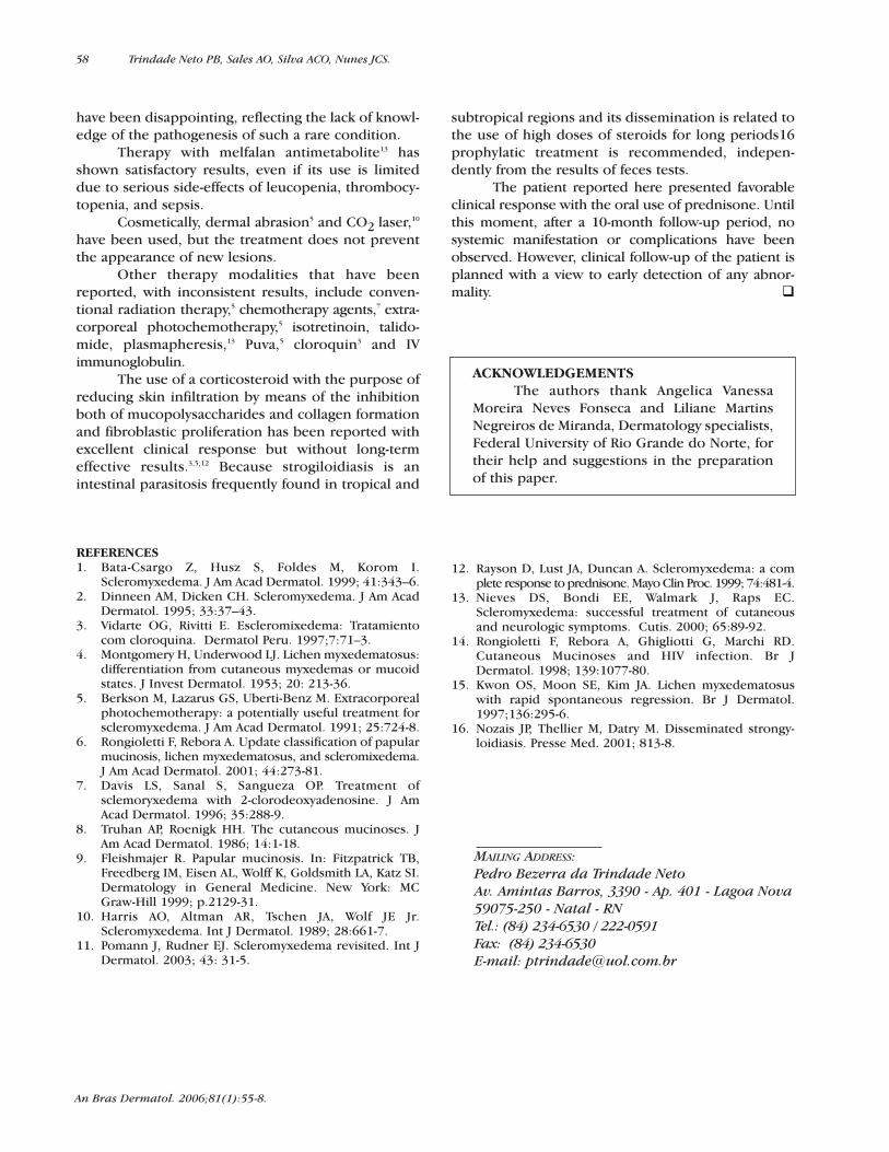
Espirito Santo, state of Rio Grande do Norte. For oneyear the patient has presented confluent erythemaaccompanied by slight pruritus, inicially on the faceand upper portion of the thorax, extending to the restof the body, with the further appearance of progres-sive skin hardening. The dermatological examrevealed diffuse skin infiltration associated to multiplepapules, sparse or confluent, with diameters rangingfrom 2 to 4 mm, located symmetrically on the extend-ing surface of the arms, retroauricular areas (Figure 1)thorax and face, with intensification in the foreheadfolds and glabellar area (Figure 2). A scarcity of hairwas also observed in the eyebrows and axillae.
Hemogram and liver, kidney and thyreoid func-tions were normal. Electrophoresis of serum proteinsrevealed hypergammaglobulinemia. Quantitatively,there was an increase of IgG immunoglobulin:2.110mg/dl (normal: 564-1765mg/dl), but with normalvalues for IgE, IgM, and IgA. Serum immunoelec-trophoresis by immunofixation showed monoclonalparaprotein of the IgG type with a predominance of thelambda chain.
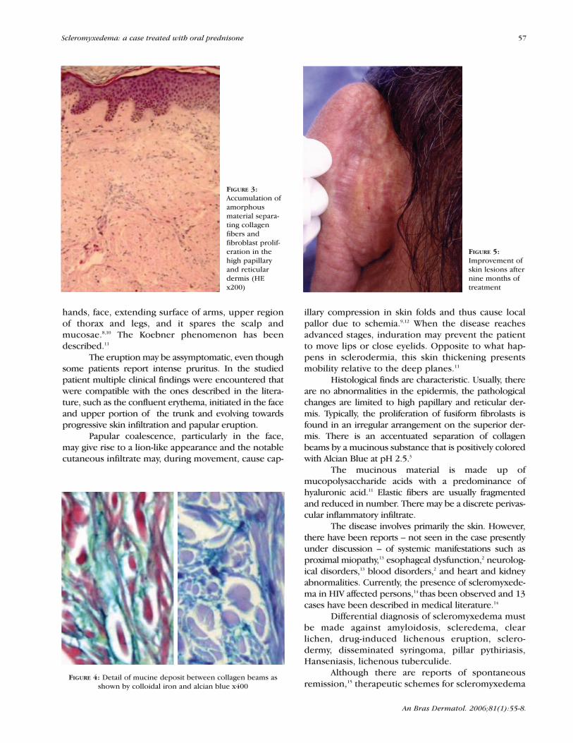
The histopathological examination of the skinshowed a separation of collagen beams by a slightlybasophilous amorphous material in the high papillarand reticular dermis, with a proliferation of enlargedfusiform fibroblasts (Figure 3). By means of alcian blueand colloidal iron dyeing the mucine deposit was madeevident. (Figure 4).
The diagnostic criteria proposed for scle-romyxedema included the presence of skin infiltration,the deposition of mucinous material on the dermis,exclusion of clinical and lab-tested thyroid dysfunction,as well as demonstration of paraproteinemia.
The patient began treatment with prednisone,60mg/day, having had previous prophylaxis for dissem-inated strongiloydiasis with cambendazol (5 mg/kg) at asingle dose. The second month of follow-up showed areduction of erythema and pruritus. After six monthsthere was a regression of papular eruption, skin infil-tration, and IgG paraprotein was reduced to 1.11 ng/dl.By the ninth month of prednisone therapy and the clin-ical improvement of the lesions (Figure 5), steroidreduction was begun.
DISCUSSIONScleromyxedema is an idiopathic cutaneous
mucinosis with a chronic, progressive course, charac-terized by general papular eruption with skin indura-tion, frequently associated with a peculiar monoclon-al paraproteinemia (83.2% of cases).6
The role of paraproteinemia in the pathogene-sis of this disease remains unclarified. Habitually, it isa lambda chain IgG, although some patients may pre-sent kappa chain or IgA.5
The paraproteinemia differs from normal IgGbecause it is extremely basic globulin of a small size(molecular mass: 110Kda; normal: 160Kda)8 andbecause of the absence of the significant antigen portionof the Fd fragment.5,9 In the reported case, the lambdaIgG type monoclonal paraproteinemia was observed.
The disease affects adults between 30 and 70years of age without gender preference. The cuta-neous feature is the erythema associated with papulareruption, as well as the diffuse sclerodermiform thick-ening. The 1-4 mm papules are normally disposed ina symmetrical manner, either isolated or confluent,and they are located preferentially on the back of
56 Trindade Neto PB, Sales AO, Silva ACO, Nunes JCS.
An Bras Dermatol. 2006;81(1):55-8.
FIGURE 1: Smalllichenouspapules behindthe ear
FIGURE 2: Diffuseface infiltration
An Bras Dermatol. 2006;81(1):55-8.
hands, face, extending surface of arms, upper regionof thorax and legs, and it spares the scalp andmucosae.8,10 The Koebner phenomenon has beendescribed.11
The eruption may be assymptomatic, even thoughsome patients report intense pruritus. In the studiedpatient multiple clinical findings were encountered thatwere compatible with the ones described in the litera-ture, such as the confluent erythema, initiated in the faceand upper portion of the trunk and evolving towardsprogressive skin infiltration and papular eruption.
Papular coalescence, particularly in the face,may give rise to a lion-like appearance and the notablecutaneous infiltrate may, during movement, cause cap-
illary compression in skin folds and thus cause localpallor due to schemia.9,12 When the disease reachesadvanced stages, induration may prevent the patientto move lips or close eyelids. Opposite to what hap-pens in sclerodermia, this skin thickening presentsmobility relative to the deep planes.11
Histological finds are characteristic. Usually, thereare no abnormalities in the epidermis, the pathologicalchanges are limited to high papillary and reticular der-mis. Typically, the proliferation of fusiform fibrolasts isfound in an irregular arrangement on the superior der-mis. There is an accentuated separation of collagenbeams by a mucinous substance that is positively coloredwith Alcian Blue at pH 2.5.3
The mucinous material is made up ofmucopolysaccharide acids with a predominance ofhyaluronic acid.11 Elastic fibers are usually fragmentedand reduced in number. There may be a discrete perivas-cular inflammatory infiltrate.
The disease involves primarily the skin. However,there have been reports – not seen in the case presentlyunder discussion – of systemic manifestations such asproximal miopathy,13 esophageal dysfunction,2 neurolog-ical disorders,13 blood disorders,2 and heart and kidneyabnormalities. Currently, the presence of scleromyxede-ma in HIV affected persons,14 thas been observed and 13cases have been described in medical literature.14
Differential diagnosis of scleromyxedema mustbe made against amyloidosis, scleredema, clearlichen, drug-induced lichenous eruption, sclero-dermy, disseminated syringoma, pillar pythiriasis,Hanseniasis, lichenous tuberculide.
Although there are reports of spontaneousremission,15 therapeutic schemes for scleromyxedema
FIGURE 5:Improvement ofskin lesions afternine months oftreatment
FIGURE 3:Accumulation ofamorphousmaterial separa-ting collagenfibers andfibroblast prolif-eration in thehigh papillaryand reticulardermis (HEx200)
FIGURE 4: Detail of mucine deposit between collagen beams asshown by colloidal iron and alcian blue x400
Scleromyxedema: a case treated with oral prednisone 57
REFERENCES1. Bata-Csargo Z, Husz S, Foldes M, Korom I.
Scleromyxedema. J Am Acad Dermatol. 1999; 41:343–6.2. Dinneen AM, Dicken CH. Scleromyxedema. J Am Acad
Dermatol. 1995; 33:37–43.3. Vidarte OG, Rivitti E. Escleromixedema: Tratamiento
com cloroquina. Dermatol Peru. 1997;7:71–3.4. Montgomery H, Underwood LJ. Lichen myxedematosus:
differentiation from cutaneous myxedemas or mucoidstates. J Invest Dermatol. 1953; 20: 213-36.
5. Berkson M, Lazarus GS, Uberti-Benz M. Extracorporeal photochemotherapy: a potentially useful treatment forscleromyxedema. J Am Acad Dermatol. 1991; 25:724-8.
6. Rongioletti F, Rebora A. Update classification of papular mucinosis, lichen myxedematosus, and scleromixedema.J Am Acad Dermatol. 2001; 44:273-81.
7. Davis LS, Sanal S, Sangueza OP. Treatment of sclemoryxedema with 2-clorodeoxyadenosine. J AmAcad Dermatol. 1996; 35:288-9.
8. Truhan AP, Roenigk HH. The cutaneous mucinoses. J Am Acad Dermatol. 1986; 14:1-18.
9. Fleishmajer R. Papular mucinosis. In: Fitzpatrick TB, Freedberg IM, Eisen AL, Wolff K, Goldsmith LA, Katz SI. Dermatology in General Medicine. New York: MC Graw-Hill 1999; p.2129-31.
10. Harris AO, Altman AR, Tschen JA, Wolf JE Jr.Scleromyxedema. Int J Dermatol. 1989; 28:661-7.
11. Pomann J, Rudner EJ. Scleromyxedema revisited. Int JDermatol. 2003; 43: 31-5.
12. Rayson D, Lust JA, Duncan A. Scleromyxedema: a complete response to prednisone. Mayo Clin Proc. 1999; 74:481-4.
13. Nieves DS, Bondi EE, Walmark J, Raps EC. Scleromyxedema: successful treatment of cutaneous and neurologic symptoms. Cutis. 2000; 65:89-92.
14. Rongioletti F, Rebora A, Ghigliotti G, Marchi RD. Cutaneous Mucinoses and HIV infection. Br JDermatol. 1998; 139:1077-80.
15. Kwon OS, Moon SE, Kim JA. Lichen myxedematosuswith rapid spontaneous regression. Br J Dermatol.1997;136:295-6.
16. Nozais JP, Thellier M, Datry M. Disseminated strongy-loidiasis. Presse Med. 2001; 813-8.
An Bras Dermatol. 2006;81(1):55-8.
MAILING ADDRESS:Pedro Bezerra da Trindade Neto Av. Amintas Barros, 3390 - Ap. 401 - Lagoa Nova59075-250 - Natal - RNTel.: (84) 234-6530 / 222-0591Fax: (84) 234-6530E-mail: [email protected]
58 Trindade Neto PB, Sales AO, Silva ACO, Nunes JCS.
have been disappointing, reflecting the lack of knowl-edge of the pathogenesis of such a rare condition.
Therapy with melfalan antimetabolite13 hasshown satisfactory results, even if its use is limiteddue to serious side-effects of leucopenia, thrombocy-topenia, and sepsis.
Cosmetically, dermal abrasion5 and CO2 laser,10
have been used, but the treatment does not preventthe appearance of new lesions.
Other therapy modalities that have beenreported, with inconsistent results, include conven-tional radiation therapy,5 chemotherapy agents,7 extra-corporeal photochemotherapy,5 isotretinoin, talido-mide, plasmapheresis,13 Puva,5 cloroquin3 and IVimmunoglobulin.
The use of a corticosteroid with the purpose ofreducing skin infiltration by means of the inhibitionboth of mucopolysaccharides and collagen formationand fibroblastic proliferation has been reported withexcellent clinical response but without long-termeffective results.3,5,12 Because strogiloidiasis is anintestinal parasitosis frequently found in tropical and
subtropical regions and its dissemination is related tothe use of high doses of steroids for long periods16prophylatic treatment is recommended, indepen-dently from the results of feces tests.
The patient reported here presented favorableclinical response with the oral use of prednisone. Untilthis moment, after a 10-month follow-up period, nosystemic manifestation or complications have beenobserved. However, clinical follow-up of the patient isplanned with a view to early detection of any abnor-mality. �
ACKNOWLEDGEMENTSThe authors thank Angelica Vanessa
Moreira Neves Fonseca and Liliane MartinsNegreiros de Miranda, Dermatology specialists,Federal University of Rio Grande do Norte, fortheir help and suggestions in the preparationof this paper.


![Surto de Pasteurelose por Pasteurella multocida tipo A em ... · na cunicultura [11]. Normalmente a enfermidade se desenvolve após quadros de estresse, transporte, mu-danças bruscas](https://static.fdocuments.in/doc/165x107/5e60b81e584f4b374c1a4199/surto-de-pasteurelose-por-pasteurella-multocida-tipo-a-em-na-cunicultura-11.jpg)