SCIENTIFIC TECHNICAL PROFILE MTA-Fillapex
36
MTA-Fillapex Bioceramic endodontic sealer SCIENTIFIC TECHNICAL PROFILE FDA AUTHORIZED
Transcript of SCIENTIFIC TECHNICAL PROFILE MTA-Fillapex
MTA-FillapexBioceramic endodontic sealer
SCIENTIFIC TECHNICAL PROFILE
FDA AUTHORIZED

Summary
03-2016
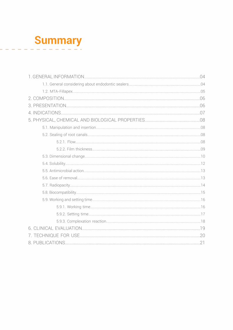
Summary
1. GENERAL INFORMATION.......................................................................................................04 1.1. General considering about endodontic sealers.............................................................................04
1.2. MTA-Fillapex.........................................................................................................................................05
2. COMPOSITION.........................................................................................................................06 3. PRESENTATION.......................................................................................................................06 4. INDICATIONS.............................................................................................................................07 5. PHYSICAL, CHEMICAL AND BIOLOGICAL PROPERTIES.................................................08 5.1. Manipulation and insertion................................................................................................................08
5.2. Sealing of root canals.........................................................................................................................08
5.2.1. Flow......................................................................................................................................08
5.2.2. Film thickness....................................................................................................................09
5.3. Dimensional change............................................................................................................................10
5.4. Solubility.................................................................................................................................................12
5.5. Antimicrobial action.............................................................................................................................13
5.6. Ease of removal....................................................................................................................................13
5.7. Radiopacity............................................................................................................................................14
5.8. Biocompatibility..............................................................................................................................................15
5.9. Working and setting time....................................................................................................................16
5.9.1. Working time......................................................................................................................16
5.9.2. Setting time........................................................................................................................17
5.9.3. Complexation reaction.....................................................................................................18
6. CLINICAL EVALUATION.........................................................................................................19 7. TECHNIQUE FOR USE...........................................................................................................20 8. PUBLICATIONS........................................................................................................................21
SCIENTIFIC TECHNICAL PROFILE - MTA-Fillapex
4
1. GENERAL INFORMATION
1.1. General considering about endodontic sealers
Since ancient times there is a concern of dental professionals in relation to root canal filling.
The science of Endodontics has been continuously seeking to improve the knowledge and performance
of endodontic sealers, as well as other materials and instruments used in this specialty field. The aim
of filling a root canal is to keep the periapical tissues healthy. McElroy in 1955 had already described
many substances that were used for filling root canals. With the evolution of research, new materials
became available in the market for root canal filling.
According to GROSSMAN (1974), root canal filling materials must have the following
properties:
A. It should be easily introduce inside the root canal.
B. It should seal the whole root canal system, including lateral and accessory canals.
C. Once inserted, it should not shrink.
D. It should be impervious to moisture.
E. It should be antimicrobial or, at least, unsuitable for microbial growth.
F. It should be radiopaque.
G. It should not stain the tooth structure.
H. It should be sterile or capable of being easily and quickly sterilized.
I. It should not irritate the periapical tissues.
J. If necessary, it should be easy to remove.
Being established the ideal profile of a filling material, it is possible to point the ideal
parameters for research and development of new products, as well as the evaluation of those already
on the market.
SCIENTIFIC TECHNICAL PROFILE - MTA-Fillapex
5
1.2. MTA-Fillapex
MTA-Fillapex is an bioceramic endodontic sealer based on MTA, developed by Angelus
(Londrina/Parana/Brazil) and launched commercially in 2010. It is a new product that combines the
proven advantages of MTA with a superior canal obturation product. Its formulation in the paste/paste
system allows a complete filling of the entire root canal, including accessory and lateral canals.
MTA, present in the composition of MTA-Fillapex, is more stable than calcium hydroxide,
providing constant release of calcium ions for the tissues and maintaining a pH which elicits antibacterial
effects. The tissue recovery and the lack of inflammatory response are optimized by the use of MTA
and disalicylate resin. The product is eugenol free and will not interfere with adhesive procedures inside
the root canal. Also, it does not cause discoloration of the tooth structure.
MAIN FEATURES AND ADVANTAGES
A. Presence of MTA in the formula: allows the formation of new tissue, including root
cementum;
B. Biocompatibility: rapid recovery of tissues without causing inflammatory reaction;
C. High Radiopacity: perfect radiographic visualization;
D. Does not stain tooth structure as it is free of eugenol and bismuth oxide;
E. Excellent Flow: the flowable consistency of MTA-Fillapex is engineered to penetrate
and also to fill lateral canals;
F. Setting expansion: provides excellent sealing of the root canal, avoiding the penetration
of tissue fluids and/or bacterial recontamination;
G. Calcium ion release: induces rapid tissue regeneration in sites with bone lesion and
microbial activity;
H. System paste x paste: easy handling and insertion
I. Working time: allows adequate working time to be used by specialists and/or general
practitioners;
J. Easy removal: allows easy removal for retreatment, particularly when used with GP
points.

SCIENTIFIC TECHNICAL PROFILE - MTA-Fillapex
6
2. COMPOSITION
3. PRESENTATION
The product is presented in dual syringes with automix tips or in tubes.
Syringe with automix tip - 4gBase paste tube - 18g
Catalyst paste tube - 12g
PASTE A
PASTE B
CHEMICAL NAME
Salicylate resin
Calcium Tungstate
Fumed Silica
Fumed Silica Titanium Dioxide
Mineral TrioxideAggregate
Base resin
Fumed SilicaTitanium Dioxide
Tricalcium silicateDicalcium SilicateCalcium OxideTricalcium Aluminate
Pentaerythritol Rosinate P - Toluenesolfonamide
FillerPigment
Active ingredient andresponsible for ionicpolymer formation
Plasticity
Plasticity
Methyl SalicylateButylene Glycol Colophony
Calcium Tungstate
Fumed Silica
Ionic polymer formation
Radiopacity
Filler
COMPONENT NAME FUNCTION
SCIENTIFIC TECHNICAL PROFILE - MTA-Fillapex
7
4. INDICATIONS
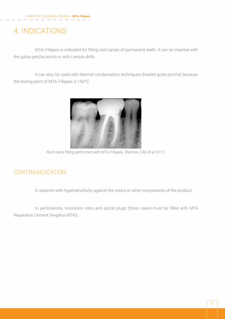
MTA-Fillapex is indicated for filling root canals of permanent teeth. It can be inserted with
the gutta-percha points or with Lentulo drills.
It can also be used with thermal condensation techniques (heated gutta percha) because
the boiling point of MTA-Fillapex is 150°C.
CONTRAINDICATION
In patients with hypersensitivity against the resins or other components of the product.
In perforations, resorption sites and apical plugs (these cases must be filled with MTA
Reparative Cement (Angelus MTA)).
Root canal filling performed with MTA-Fillapex. (Ramos, CAS et al 2011)
SCIENTIFIC TECHNICAL PROFILE - MTA-Fillapex
8
5. PHYSICAL, CHEMICAL AND BIOLOGICAL PROPERTIES
5.1. Manipulation and insertion
The MTA-Fillapex is paste/paste material presented in automix double syringes or tubes
which provide an adequate consistency for the cement insertion in the root canal. The presence of
nanoparticles enables a homogeneous mixture and better flow of the product.
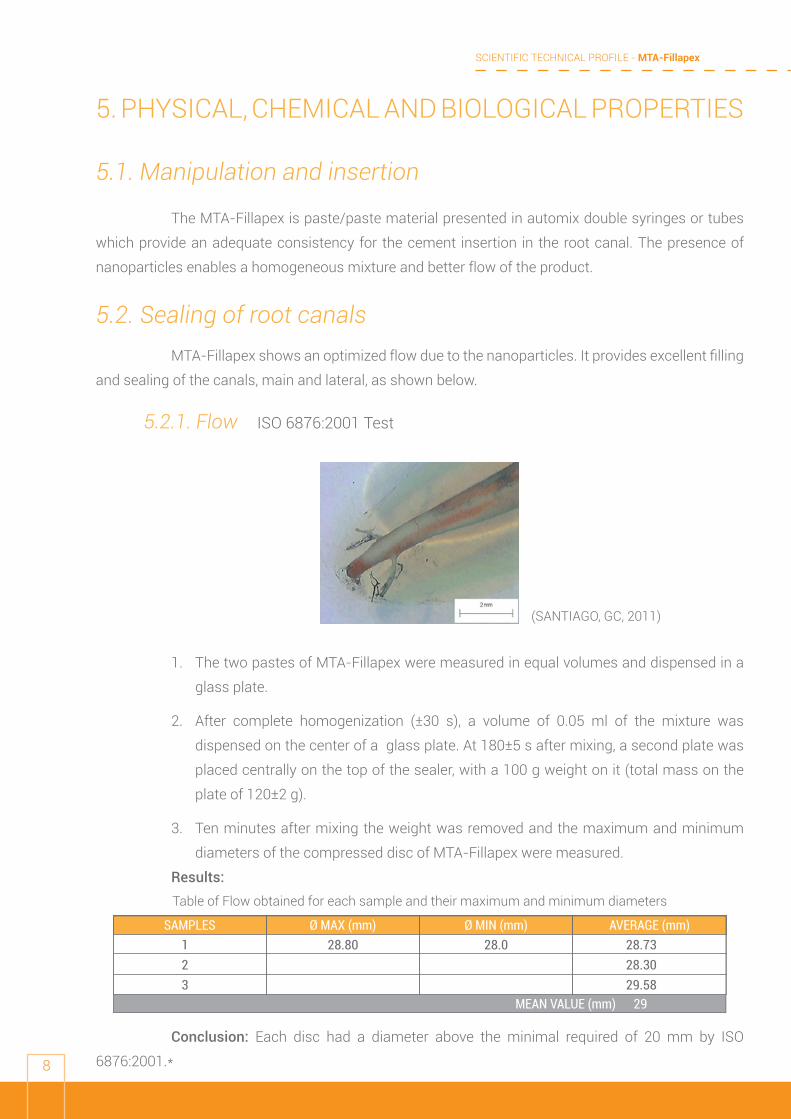
5.2. Sealing of root canalsMTA-Fillapex shows an optimized flow due to the nanoparticles. It provides excellent filling
and sealing of the canals, main and lateral, as shown below.
5.2.1. Flow ISO 6876:2001 Test
1. The two pastes of MTA-Fillapex were measured in equal volumes and dispensed in a
glass plate.
2. After complete homogenization (±30 s), a volume of 0.05 ml of the mixture was
dispensed on the center of a glass plate. At 180±5 s after mixing, a second plate was
placed centrally on the top of the sealer, with a 100 g weight on it (total mass on the
plate of 120±2 g).
3. Ten minutes after mixing the weight was removed and the maximum and minimum
diameters of the compressed disc of MTA-Fillapex were measured.
Results:
Conclusion: Each disc had a diameter above the minimal required of 20 mm by ISO
6876:2001.*
Ø MAX (mm) Ø MIN (mm) AVERAGE (mm)123
28.80 28.0 28.7328.3029.58
SAMPLES
MEAN VALUE (mm) 29
Table of Flow obtained for each sample and their maximum and minimum diameters
(SANTIAGO, GC, 2011)
SCIENTIFIC TECHNICAL PROFILE - MTA-Fillapex
9
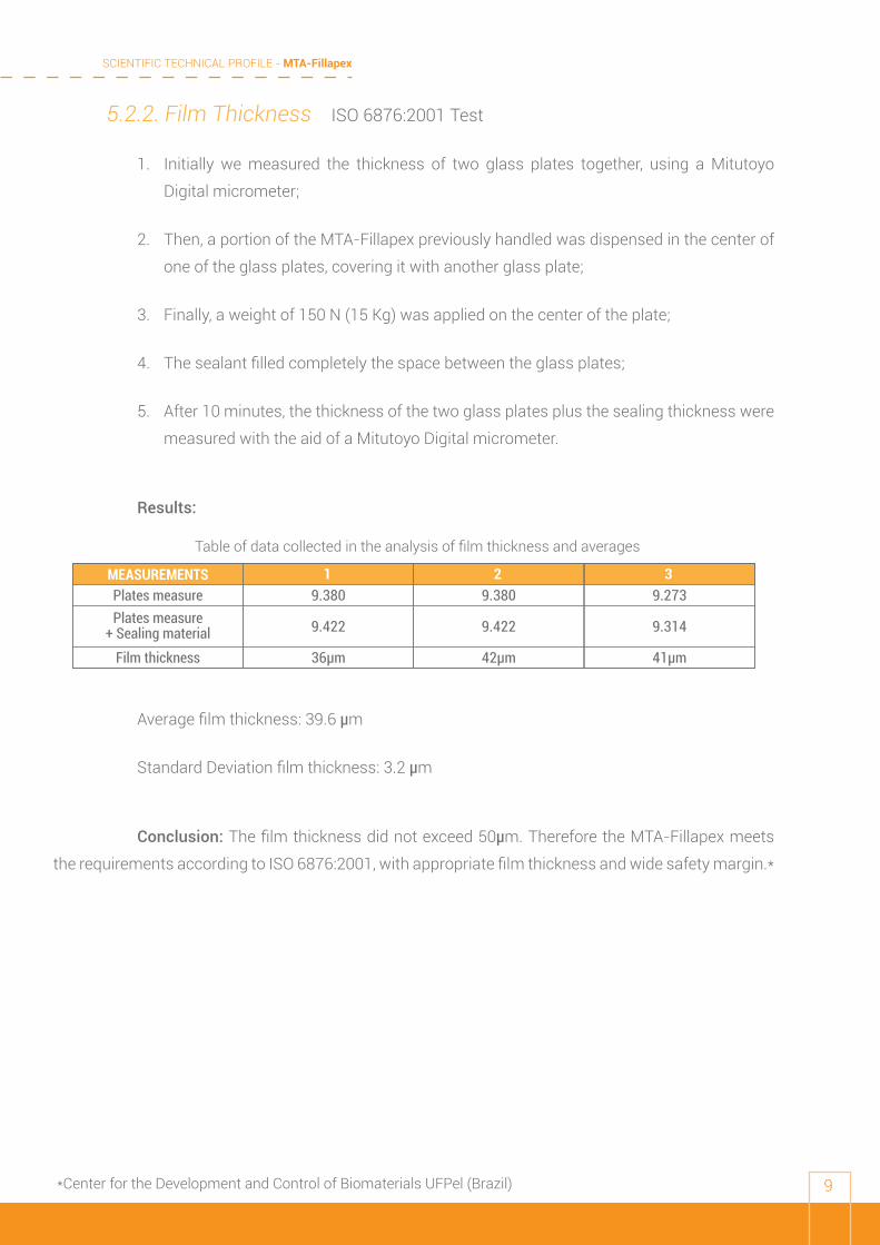
5.2.2. Film Thickness ISO 6876:2001 Test
1. Initially we measured the thickness of two glass plates together, using a Mitutoyo
Digital micrometer;
2. Then, a portion of the MTA-Fillapex previously handled was dispensed in the center of
one of the glass plates, covering it with another glass plate;
3. Finally, a weight of 150 N (15 Kg) was applied on the center of the plate;
4. The sealant filled completely the space between the glass plates;
5. After 10 minutes, the thickness of the two glass plates plus the sealing thickness were
measured with the aid of a Mitutoyo Digital micrometer.
Results:
Average film thickness: 39.6 µm
Standard Deviation film thickness: 3.2 µm
Conclusion: The film thickness did not exceed 50μm. Therefore the MTA-Fillapex meets
the requirements according to ISO 6876:2001, with appropriate film thickness and wide safety margin.*
1 2 39.380
9.422
36µm
9.380
9.422
42µm
9.273
9.314
41µm
Plates measure
Film thickness
Plates measure+ Sealing material
MEASUREMENTS
Table of data collected in the analysis of film thickness and averages
*Center for the Development and Control of Biomaterials UFPel (Brazil)
SCIENTIFIC TECHNICAL PROFILE - MTA-Fillapex
10
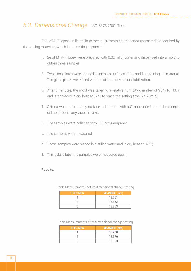
5.3. Dimensional Change ISO 6876:2001 Test
The MTA-Fillapex, unlike resin cements, presents an important characteristic required by
the sealing materials, which is the setting expansion.
1. 2g of MTA-Fillapex were prepared with 0.02 ml of water and dispensed into a mold to
obtain three samples;
2. Two glass plates were pressed up on both surfaces of the mold containing the material.
The glass plates were fixed with the aid of a device for stabilization;
3. After 5 minutes, the mold was taken to a relative humidity chamber of 95 % to 100%
and later placed in dry heat at 37°C to reach the setting time (2h 20min);
4. Setting was confirmed by surface indentation with a Gilmore needle until the sample
did not present any visible marks;
5. The samples were polished with 600 grit sandpaper;
6. The samples were measured;
7. These samples were placed in distilled water and in dry heat at 37°C;
8. Thirty days later, the samples were measured again.
Results:
MEASURE (mm)13.26113.38213.363
1
32
SPECIMEN
Table Measurements before dimensional change testing
MEASURE (mm)13.28013.37913.363
1
32
SPECIMEN
Table Measurements after dimensional change testing
SCIENTIFIC TECHNICAL PROFILE - MTA-Fillapex
11
The values obtained before and after dimensional changes testing were calculated in
percentage to obtain the value of dimensional change for each sample.
Sample 1 - showed 0.1% expansion
Sample 2 - showed 0.022% shrinkage
Sample 3 - showed 0.022% expansion
Average overall dimensional change (from 3 specimens ) = 0.088%
Conclusion: ISO sets that the average dimensional change of the material should not exceed
1.0% shrinkage or 0.1% expansion. Thus it is concluded that the material fulfilled the requirements
standardized by ISO, considering each sample individually, as well as the average change in the material
adding all samples tested.*
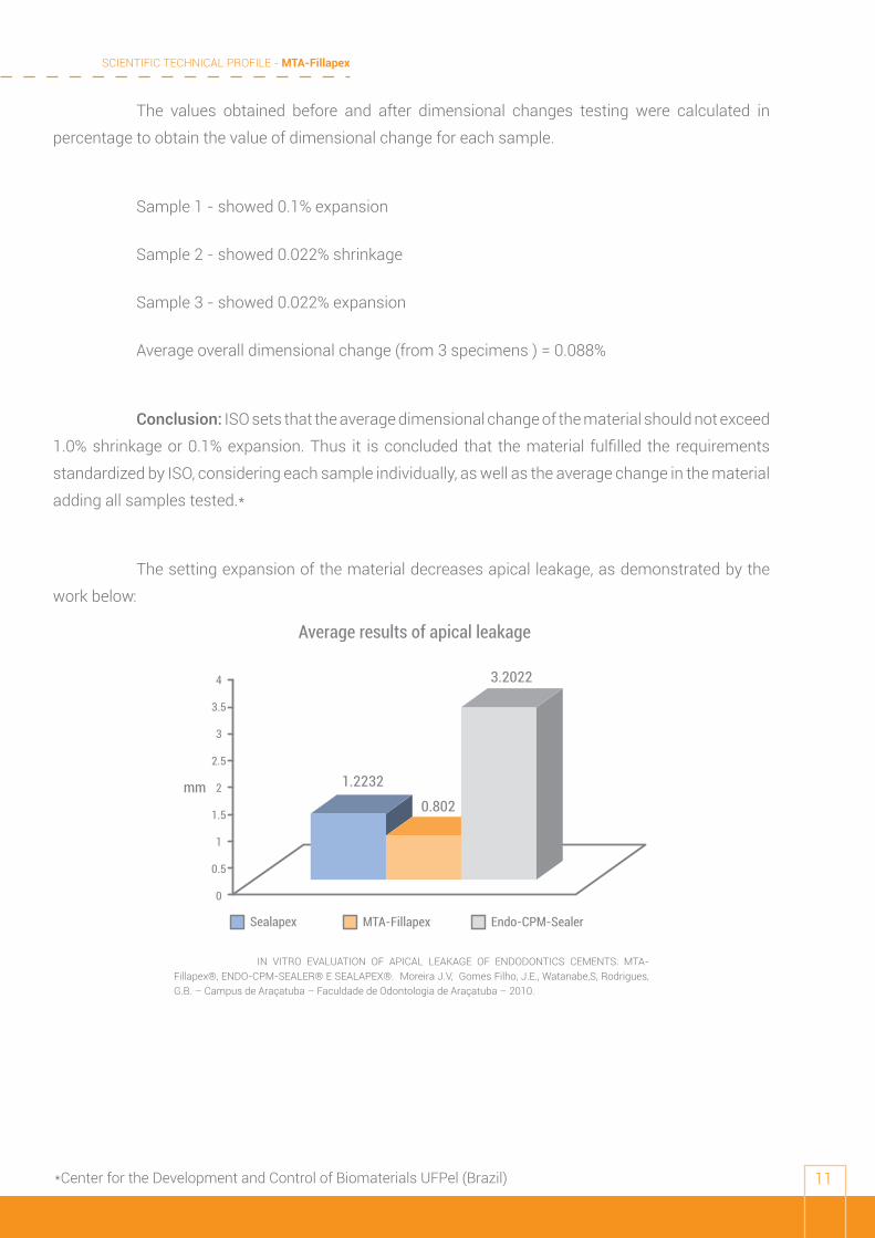
The setting expansion of the material decreases apical leakage, as demonstrated by the
work below:
Average results of apical leakage
Sealapex MTA-Fillapex Endo-CPM-Sealer
4
3.5
3
2.5
2
1.5
1
0.5
0
1.2232
0.802
3.2022
mm
IN VITRO EVALUATION OF APICAL LEAKAGE OF ENDODONTICS CEMENTS: MTA-Fillapex®, ENDO-CPM-SEALER® E SEALAPEX®. Moreira J.V, Gomes Filho, J.E., Watanabe,S, Rodrigues, G.B. – Campus de Araçatuba – Faculdade de Odontologia de Araçatuba – 2010.
*Center for the Development and Control of Biomaterials UFPel (Brazil)
SCIENTIFIC TECHNICAL PROFILE - MTA-Fillapex
12
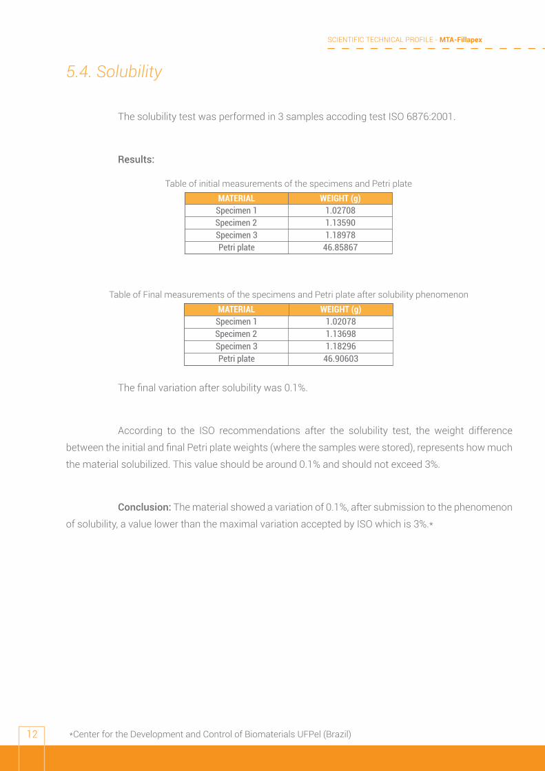
5.4. Solubility
The solubility test was performed in 3 samples accoding test ISO 6876:2001.
Results:
The final variation after solubility was 0.1%.
According to the ISO recommendations after the solubility test, the weight difference
between the initial and final Petri plate weights (where the samples were stored), represents how much
the material solubilized. This value should be around 0.1% and should not exceed 3%.
Conclusion: The material showed a variation of 0.1%, after submission to the phenomenon
of solubility, a value lower than the maximal variation accepted by ISO which is 3%.*
WEIGHT (g)1.027081.135901.18978
Specimen 1
Specimen 3Specimen 2
MATERIAL
46.85867Petri plate
Table of initial measurements of the specimens and Petri plate
WEIGHT (g)1.020781.136981.18296
Specimen 1
Specimen 3Specimen 2
MATERIAL
46.90603Petri plate
Table of Final measurements of the specimens and Petri plate after solubility phenomenon
*Center for the Development and Control of Biomaterials UFPel (Brazil)
SCIENTIFIC TECHNICAL PROFILE - MTA-Fillapex
13
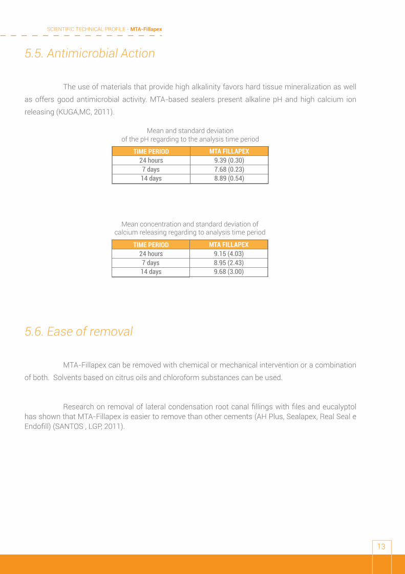
5.5. Antimicrobial Action
The use of materials that provide high alkalinity favors hard tissue mineralization as well
as offers good antimicrobial activity. MTA-based sealers present alkaline pH and high calcium ion
releasing (KUGA,MC, 2011).
5.6. Ease of removal
MTA-Fillapex can be removed with chemical or mechanical intervention or a combination
of both. Solvents based on citrus oils and chloroform substances can be used.
Research on removal of lateral condensation root canal fillings with files and eucalyptol has shown that MTA-Fillapex is easier to remove than other cements (AH Plus, Sealapex, Real Seal e Endofill) (SANTOS , LGP, 2011).
MTA FILLAPEX9.39 (0.30)7.68 (0.23)8.89 (0.54)
24 hours
14 days7 days
TIME PERIOD
Mean and standard deviationof the pH regarding to the analysis time period
MTA FILLAPEX9.15 (4.03)8.95 (2.43)9.68 (3.00)
24 hours
14 days7 days
TIME PERIOD
Mean concentration and standard deviation ofcalcium releasing regarding to analysis time period
SCIENTIFIC TECHNICAL PROFILE - MTA-Fillapex
14
5.7. Radiopacity ISO 6876:2001 Test
1. MTA-Fillapex was mixed according to the manufacturer’s instructions and placed in
the mold.
2. The covers on the top and bottom were pressed to make a 1mm thick sample.
3. The sample was placed in the center of an X-ray film adjacent to the step wedge.
4. The system was irradiated in accordance with ISO 6876:2001.
5. After developing, fixing and drying the exposed film, the densities of the image of the
sample and the step wedge were compared using Image J.
6. The results were expressed in millimeters of aluminum.
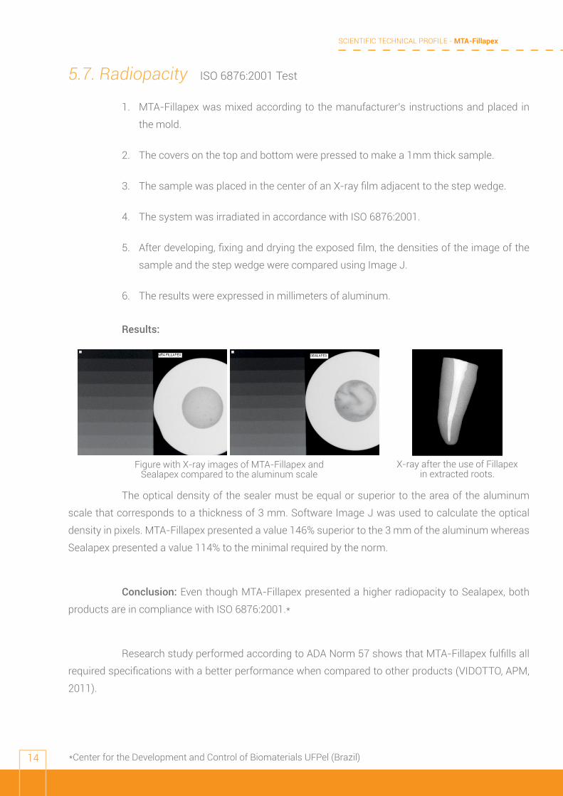
Results:
The optical density of the sealer must be equal or superior to the area of the aluminum
scale that corresponds to a thickness of 3 mm. Software Image J was used to calculate the optical
density in pixels. MTA-Fillapex presented a value 146% superior to the 3 mm of the aluminum whereas
Sealapex presented a value 114% to the minimal required by the norm.
Conclusion: Even though MTA-Fillapex presented a higher radiopacity to Sealapex, both
products are in compliance with ISO 6876:2001.*
Research study performed according to ADA Norm 57 shows that MTA-Fillapex fulfills all
required specifications with a better performance when compared to other products (VIDOTTO, APM,
2011).
Figure with X-ray images of MTA-Fillapex and Sealapex compared to the aluminum scale
X-ray after the use of Fillapex in extracted roots.
*Center for the Development and Control of Biomaterials UFPel (Brazil)
SCIENTIFIC TECHNICAL PROFILE - MTA-Fillapex
15
5.8. Biocompatibility
The biological properties inherent to conventional MTA used for treatment of root
perforations replicates in MTA-Fillapex. When in contact with water, CaO can be converted into Ca(OH)2
and dissociated into Ca+2 and OH. The diffusion of hydroxyl ions from the root canal increases the pH at
the surface of the root adjacent to the periodontal tissues, possibly interfering with osteoclastic activity
and promoting alkalinization in the adjacent tissues, which favors healing. Calcium ions participate in
the activation of calcium-dependent adenosine triphosphatase and react with carbonic gas to form
calcium carbonate crystals (birefringent to polarized light), which serve as a nucleus for calcification
and favor mineralization. A rich extracellular network of fibronectin in close contact with these crystals
strongly supports the role of calcite crystals and fibronectin as an initiating step in the formation of a
hard tissue. Calcium is also needed for cell migration and differentiation. Because MTA-Fillapex and
MTA have similar chemical composition and elicit similar tissue reactions, it is expected that MTA-
Fillapex will act similarly to MTA when used in clinical situations, but be easier to handle because of its
paste/paste presentation.
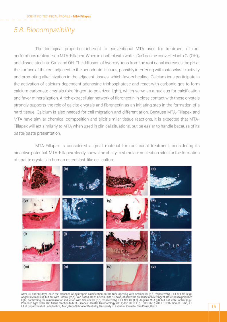
MTA-Fillapex is considered a great material for root canal treatment, considering its
bioactive potential. MTA-Fillapex clearly shows the ability to stimulate nucleation sites for the formation
of apatite crystals in human osteoblast-like cell culture.
After 30 and 90 days, note the presence of dystrophic calcification on the tube opening with Sealapex® (a,c, respectively), FILLAPEX® (e,g), Angelus MTA® (i,k), but not with Control (m,o). Von Kossa 100x. After 30 and 90 days, observe the presence of birefringent structures to polarized light, confirming the mineralization induction with Sealapex® (b,d, respectively), FILLAPEX® (f,h), Angelus MTA (j,l), but not with Control (n,p). Polarized light 100x. Rat tissue reaction to MTA-Fillapex - Dental Traumatology 2011; doi: 10.1111/j.1600-9657.2011.01096. Gomes-Filho, J.E ET al Department of Endodontics, Arac¸atuba School of Dentistry, University of Estadual Paulista, São Paulo, Brazil
SCIENTIFIC TECHNICAL PROFILE - MTA-Fillapex
16
5.9 Working Time and Setting Time
5.9.1 Working time Test ISO 6876:2001
1. The two pastes of MTA-Fillapex were measured in equal volumes and dispensed in a
glass plate.
2. After complete homogenization (±30 s), a volume of 0.05 ml of the mixture was
dispensed on the center of a glass plate using a graduated syringe.
3. At increasing intervals after mixing and the setting time of MTA-Fillapex, a second plate
was placed centrally on the top of the sealer, with a 100 g weight (total mass on the
plate: 120±2 g).
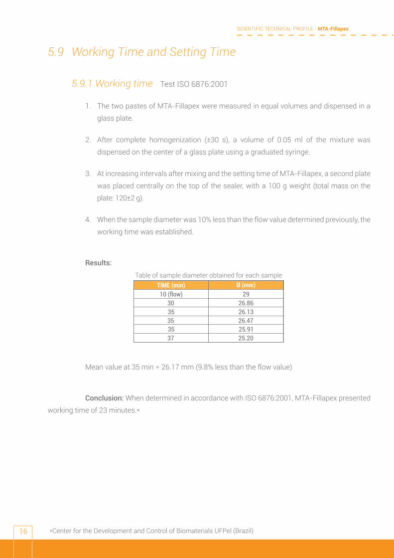
4. When the sample diameter was 10% less than the flow value determined previously, the
working time was established.
Results:
Mean value at 35 min = 26.17 mm (9.8% less than the flow value)
Conclusion: When determined in accordance with ISO 6876:2001, MTA-Fillapex presented
working time of 23 minutes.*
Ø (mm)29
26.8626.13
10 (flow)
3530
3535
3725.9126.47
25.20
TIME (min)Table of sample diameter obtained for each sample
*Center for the Development and Control of Biomaterials UFPel (Brazil)
SCIENTIFIC TECHNICAL PROFILE - MTA-Fillapex
17
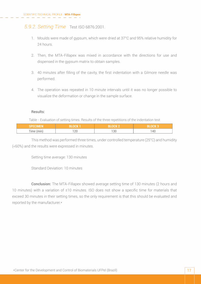
5.9.2. Setting Time Test ISO 6876:2001.
1. Moulds were made of gypsum, which were dried at 37°C and 95% relative humidity for
24 hours.
2. Then, the MTA-Fillapex was mixed in accordance with the directions for use and
dispensed in the gypsum matrix to obtain samples.
3. 40 minutes after filling of the cavity, the first indentation with a Gilmore needle was
performed.
4. The operation was repeated in 10 minute intervals until it was no longer possible to
visualize the deformation or change in the sample surface.
Results:
This method was performed three times, under controlled temperature (25°C) and humidity
(<60%) and the results were expressed in minutes.
Setting time average: 130 minutes
Standard Deviation: 10 minutes
Conclusion: The MTA-Fillapex showed average setting time of 130 minutes (2 hours and
10 minutes) with a variation of ±10 minutes. ISO does not show a specific time for materials that
exceed 30 minutes in their setting times, so the only requirement is that this should be evaluated and
reported by the manufacturer.*
BLOCK 1 BLOCK 2 BLOCK 3120 130 140Time (min)
SPECIMEN
Table - Evaluation of setting times. Results of the three repetitions of the indentation test
*Center for the Development and Control of Biomaterials UFPel (Brazil)
SCIENTIFIC TECHNICAL PROFILE - MTA-Fillapex
18
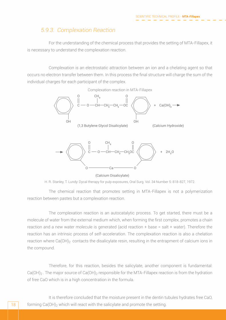
5.9.3. Complexation Reaction
For the understanding of the chemical process that provides the setting of MTA-Fillapex, it
is necessary to understand the complexation reaction.
Complexation is an electrostatic attraction between an ion and a chelating agent so that
occurs no electron transfer between them. In this process the final structure will charge the sum of the
individual charges for each participant of the complex.
The chemical reaction that promotes setting in MTA-Fillapex is not a polymerization
reaction between pastes but a complexation reaction.
The complexation reaction is an autocatalytic process. To get started, there must be a
molecule of water from the external medium which, when forming the first complex, promotes a chain
reaction and a new water molecule is generated (acid reaction + base = salt + water). Therefore the
reaction has an intrinsic process of self-acceleration. The complexation reaction is also a chelation
reaction where Ca(OH)2 contacts the disalicylate resin, resulting in the entrapment of calcium ions in
the compound.
Therefore, for this reaction, besides the salicylate, another component is fundamental:
Ca(OH)2 . The major source of Ca(OH)2 responsible for the MTA-Fillapex reaction is from the hydration
of free CaO which is in a high concentration in the formula.
It is therefore concluded that the moisture present in the dentin tubules hydrates free CaO,
forming Ca(OH)2 which will react with the salicylate and promote the setting.
Complexation reaction in MTA-Fillapex
H. R. Stanley, T. Lundy: Dycal therapy for pulp exposures, Oral Surg. Vol. 34 Number 5: 818-827, 1972.
OH
O O
C O CH
CH³
CH²
CH²
OC
OH
+ Ca(OH)²
(1,3 Butylene Glycol Disalicylate) (Calcium Hydroxide)
O Ca
O O
C O CH
CH³
CH²
CH²OC
O
+ 2H²O
(Calcium Disalicylate)
SCIENTIFIC TECHNICAL PROFILE - MTA-Fillapex
19
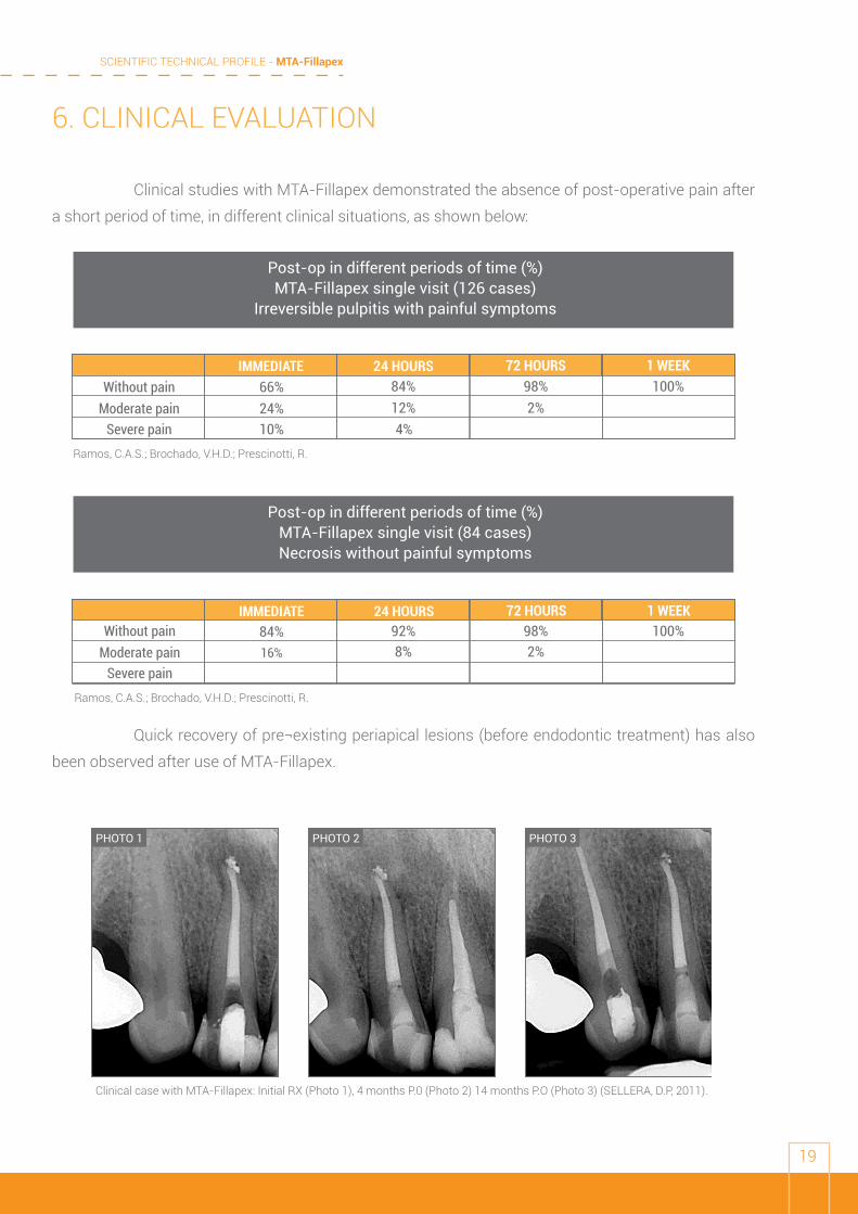
6. CLINICAL EVALUATION
Clinical studies with MTA-Fillapex demonstrated the absence of post-operative pain after
a short period of time, in different clinical situations, as shown below:
Quick recovery of pre¬existing periapical lesions (before endodontic treatment) has also
been observed after use of MTA-Fillapex.
Post-op in different periods of time (%)MTA-Fillapex single visit (126 cases)
Irreversible pulpitis with painful symptoms
Post-op in different periods of time (%)MTA-Fillapex single visit (84 cases)Necrosis without painful symptoms
Ramos, C.A.S.; Brochado, V.H.D.; Prescinotti, R.
Ramos, C.A.S.; Brochado, V.H.D.; Prescinotti, R.
24 HOURS 72 HOURS 1 WEEK66%24%10%
84%12%4%
98%2%
100%Without pain
Severe painModerate pain
IMMEDIATE
84%16%
92%8%
98%2%
100%24 HOURS 72 HOURS 1 WEEK
Without pain
Severe painModerate pain
IMMEDIATE
PHOTO 1 PHOTO 2 PHOTO 3
Clinical case with MTA-Fillapex: Initial RX (Photo 1), 4 months P.0 (Photo 2) 14 months P.O (Photo 3) (SELLERA, D.P, 2011).
SCIENTIFIC TECHNICAL PROFILE - MTA-Fillapex
20
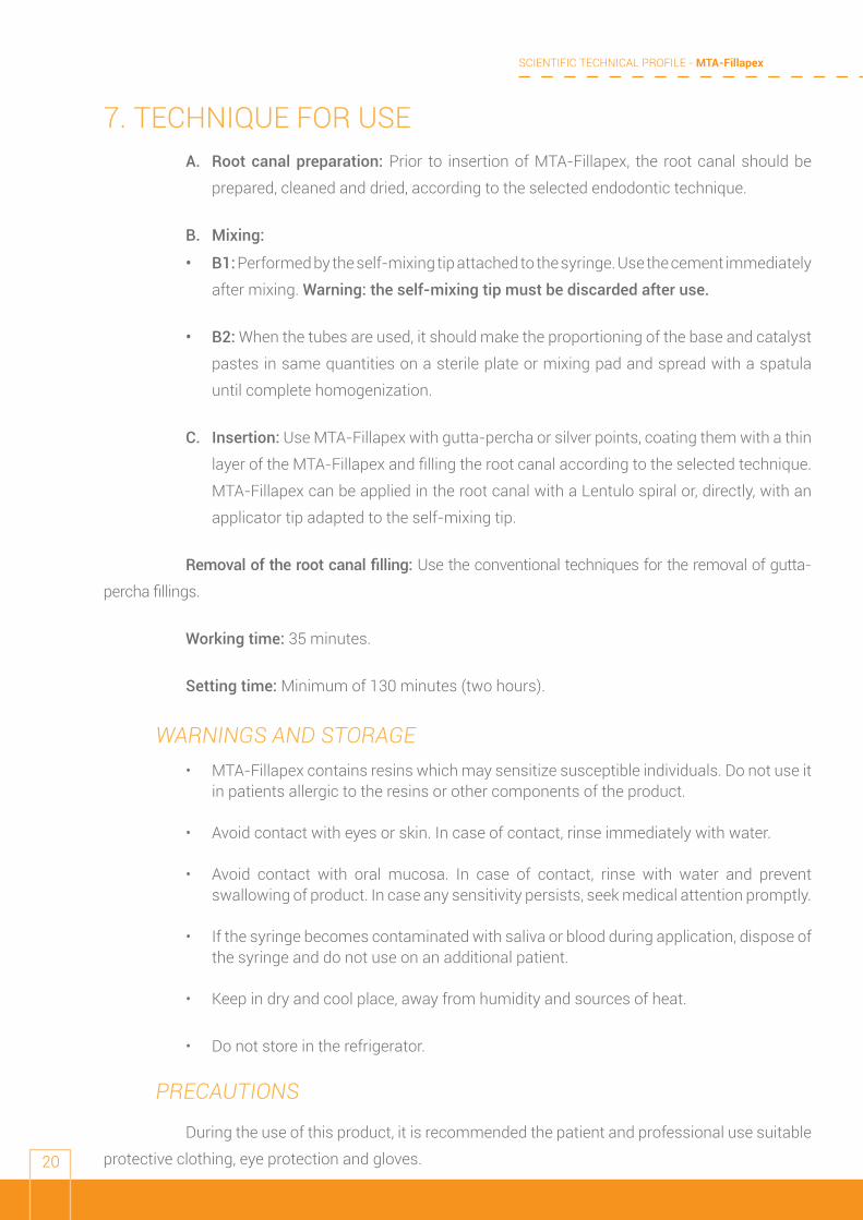
7. TECHNIQUE FOR USEA. Root canal preparation: Prior to insertion of MTA-Fillapex, the root canal should be
prepared, cleaned and dried, according to the selected endodontic technique.
B. Mixing:
• B1: Performed by the self-mixing tip attached to the syringe. Use the cement immediately
after mixing. Warning: the self-mixing tip must be discarded after use.
• B2: When the tubes are used, it should make the proportioning of the base and catalyst
pastes in same quantities on a sterile plate or mixing pad and spread with a spatula
until complete homogenization.
C. Insertion: Use MTA-Fillapex with gutta-percha or silver points, coating them with a thin
layer of the MTA-Fillapex and filling the root canal according to the selected technique.
MTA-Fillapex can be applied in the root canal with a Lentulo spiral or, directly, with an
applicator tip adapted to the self-mixing tip.
Removal of the root canal filling: Use the conventional techniques for the removal of gutta-
percha fillings.
Working time: 35 minutes.
Setting time: Minimum of 130 minutes (two hours).
WARNINGS AND STORAGE• MTA-Fillapex contains resins which may sensitize susceptible individuals. Do not use it
in patients allergic to the resins or other components of the product.
• Avoid contact with eyes or skin. In case of contact, rinse immediately with water.
• Avoid contact with oral mucosa. In case of contact, rinse with water and prevent swallowing of product. In case any sensitivity persists, seek medical attention promptly.
• If the syringe becomes contaminated with saliva or blood during application, dispose of the syringe and do not use on an additional patient.
• Keep in dry and cool place, away from humidity and sources of heat.
• Do not store in the refrigerator.
PRECAUTIONS
During the use of this product, it is recommended the patient and professional use suitable
protective clothing, eye protection and gloves.
SCIENTIFIC TECHNICAL PROFILE - MTA-Fillapex
21
8. PUBLICATIONS1. ASSMAN, E. Avaliação da resistência de união à dentina dos cimentos à base de MTA
e à base de resina epóxica através do teste de micropush out; Trabalho de Conclusão de Curso de Especialização de Endodontia da Universidade Federal do Rio Grande do Sul; 2010
2. ASSMAN, E.; ET AL. Dentin Bond Strength of Two Mineral Trioxide Aggregate–based and One Epoxy Resin–based Sealers; JOE — Volume 38, Number 2, February 2012
3. AZNAR, F.D.C.T. Tratamento de reabsorção interna empregando cimento endodôntico à base de MTA: Relato de Caso Clínico. Angelus. 2015
4. BALDISSERA, R.S. Avaliação da penetrabilidade dentinária apical dos cimentos AH Plus e MTA Fillapex por meio da microscopia confocal. Trabalho de conclusão de especialização. Universidade Federal do Rio Grande do Sul. Faculdade de Odontologia. Curso de Especialização em Endodontia <http://hdl.handle.net/10183/79931. 2015>
5. BIN C V; VALERA M C, CAMARGO S E A, RABELO S B, SILVA G O, BALDUCCI I, CAMARGO C H R, Cytotoxicity and Genotoxicity of Root Canal Sealers Based on Mineral Trioxide Aggregate Endod 2012;38:495–500
6. BORGES, A.H.; ET AL. Analysis of Chemical Elements and Heavy Metals in MTA Fillapex and AH Plus. OHDM - Vol. 13 - No. 4 - December, 2014
7. BORGES, A.H.; et al. Arsenic Release of Portland and MTA-Based Cements. IADR Thursday, March 21, 2013
8. BORGES, R.P., Avaliação da solubilidade de cimentos obturadores dos canais radiculares à base de silicato de cálcio – Tese de Doutorado da Universidade de Ribeirão Preto (UNAERP), área de concentração Endodontia, Ribeirão Rreto, 2011
9. BRAIT, A H, Cirurgia paraendodôntica com retroinstrumentação ultrassônica e retrobturação com Fillapex/MTA http://4.bp.blogspot.com/_LAB40W-pjXk/TBy8kPflw6I/AAAAAAAAAXM/aAUNlx2GC5s/s1600/Slide1.JPG
10. BRAUN, A.; ET AL. Germ reduction during endodontic treatment of a geminated tooth with a 970 nm laser. International magazine of laser dentistry. august 2013
11. BROOM, N.J.; ET AL. Reparacion de periodontitis apical crónica con cemento sellador MTA Fillapex en una sesion. Endoodncia Actual. Noviembre 2012, vol VII. N.3
12. BROOM, N.J.; ET AL. Reparación expectante de lesiones periapicales de origen endodóntico con cemento MTA-Fill-Apex en una sola cita. Endodoncia • Volumen 31 • Número 2 • Abril-Junio 2013
13. CAMILLERI, J. ET AL. Sealers and Warm Gutta-percha Obturation Techniques. JOE — Volume 41, Number 1, January 2015
14. CARVALHO, V.H.M.; ET AL. Comparação de métodos radiográficos para determinação da radiopacidade de cimentos endodônticos. Braz Oral Res 2015
SCIENTIFIC TECHNICAL PROFILE - MTA-Fillapex
22
15. CASTRO, V.L.D. Análise comparativa entre dois sistemas dosadores do cimento MTA Fillapex®. Monografia apresentada ao Curso de Especialização da Faculdade São Leopoldo Mandic, Belo Horizonte MG, para obtenção do título de Especialista em Endodontia. BELO HORIZONTE, 2012
16. CECHELLA, B.C. Influência Da Exposição Do Biodentine Ao Tampão Fosfato-Salino Sobre O Selamento Apical E A Resistência De União À Dentina (Push-Out). Dissertação submetida ao Programa de Pós-Graduação em Odontologia da Universidade Federal de Santa Catarina para a obtenção do Grau de Mestre em Odontologia. Área de concentração: Endodontia Florianópolis 2014
17. COLLARES KF, CAMARGO-JUNIOR AS, KNABACH CB, OLIVEIRA LP, JARDIM PS, JACINTO RC Influência de cimento endodôntico a base de MTA na resistência de união de pinos de fibra de vidro Brasilian Oral Rearch, Pie015, Volume 25 Supplement 1 September 2011
18. CORNÉLIO ALG, SALLES LP, ROSSA-JUNIOR C, GUERREIRO-TANOMARU JM, TANOMARU-FILHO M Biocompatibilidade e bioatividade do MTA-Fillapex em cultura de células ósseas humanas, Brasilian Oral Rearch,PNf 016, Volume 25 Supplement 1 September 2011
19. COSTA CCR, ROCHA VGN, HABITANTE SM, RALDI DP, LAGE-MARQUES JL. Análise da infiltração apical de um novo cimento endodôntico a base de MTA. Cienc Odontol Bras 2009;12:35-40
20. CRISTOFAMO, A.S. ET AL. Ação Antimicrobiana De Um Cimento Endodôntico À Base De Silicato De Cálcio Sobre Dois Tipos Bacterianos. Anais Da 21a. Jornada Acadêmica De Odontologia Da Universidade Do Oeste Paulista – Unoeste 03 A 07 De Novembro Presidente Prudente – SP_ 2014
21. CRUZ, ATG. Influência do hidróxido de cálcio na penetração dos cimentos AH Plus E MTA Fillapex nos túbulos dentinários. Braz Oral Res 2014;28 (Suppl. 1)
22. CUNHA RAG, ROMAGNOLI C, BERGER SB, GUIRALDO RD, MOURA SK, CARVALHO RV, COSTA JM, LOPES MB Propriedades físicas de cimento a base de MTA. Brasilian Oral Rearch,PIc071 Volume 25 Supplement 1 September 2011
23. DU, T.; ET AL. Combined Antibacterial Effect of Sodium Hypochlorite and Root Canal Sealers against Enterococcus faecalis Biofilms in Dentin Canals. JOE — Volume 41, Number 8, August 2015
24. EHSANI, M.; ET AL. Antimicrobial activity of three different endodontic sealers on the enterococcus faecalis and lactobacillus (in vitro). Caspian J Dent Res -September 2013, 2(2): 8-14
25. EHSANI, M.; ET AL. Evaluation of Apical Micro-leakage of Different Endodontic Sealers in the Presence and Absence of Moisture. J Dent Res Dent Clin Dent Prospects. 2014 Summer; 8(3): 125–129.
PUBL
ICAT
ION
S -
ABST
RACT
S
SCIENTIFIC TECHNICAL PROFILE - MTA-Fillapex
23
26. ELBAZ, A.A.; ET AL. Longitudinal Assessment of Biocompatibility and Healing Response of MTA Fillapex International Dental Journal 2013; 63 (Suppl. 2): 1--98. FDI World Dental Federation
27. FARAONI, G. ET AL. Avaliação comparativa do escoamento e tempo de presa do cimento MTA Fillapex® Comparative assessment of flow and setting time of the MTA
28. FARIA-JÚNIOR NB, TANOMARU-FILHO M, BERBERT FLCV, GUERREIRO-TANOMARU JM Atividade antimicrobiana de cimentos obturadores endodônticos sobre biofilme de Enterococcus faecalis Brasilian Oral Rearch, PNe036Volume 25 , Supplement 1 September 2011
29. FELIPPE, G.S. Capacidade De Promover Biomineralização De Diferentes Bioagregados: Análise In Vivo. Dissertação apresentada ao Programa de Pós-Graduação em Odontologia, da Universidade Federal de Santa Catarina, como parte dos requisitos para obtenção do título de Mestre em Odontologia. Área de concentração: Endodontia. 2012
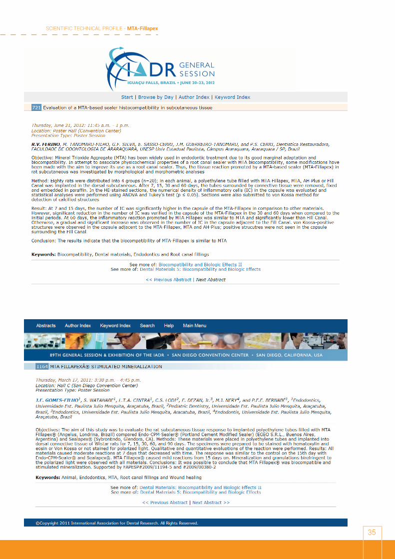
30. FERINO, R.V; TANOMARU-FILHO,M.; SILVA,G.F; SASSO-CERRI,E. GUERREIRO-TANOMARU, J.M;CERRI, P.S. Evaluation of a MTA-based sealer histocompatibility in subcutaneous tissue 721 Thursday, June 21, 2012: 11:45 a.m. - 1 p.m IADR 2012
31. FERREIRA, M.M. ET AL. Comparison of the apical seal on filled root canals with Topseal® vs MTA Fillapex® sealers: A quantitative scintigraphic analysis. Open Journal of Stomatology, 2013, 3, 128-132
32. Fillapex™ sealer. RFO, Passo Fundo, v. 18, n. 2, p. 180-184, maio/ago. 2013
33. FINGER, M.S.; et al. Comparative evaluation of pH and solubility of MTA Fillapex® endodontic sealer. RSBO. 2014 Jan-Mar;11(1):41-6
34. FREITAS, C.G.; ET AL. Radiopacity of 4 cements obturators through the tomographic analysis; Rev. APCD ,2012. 66(1) 36-40
35. GLASSMANN, G. Bioactive endododontic obturation: Combining the new with the tried and true MTA Fillapex and Continuous Wave of Condensation; ROOTS, 3; 2013
36. GOMES FIILHO, J E; Rat tissue reaction to MTA-Fillapex Dental Traumatology 2011; doi: 10.1111/j.1600-9657.2011.
37. GOMES FILHO, J. E.; et al. Effect of MTA-based sealer on the healing of periapical lesions. J Appl Oral Sci.2013;21(3):235-42
38. GOMES-FILHO J.E, MOREIRA J.V, WATANABE S., LODI C.S., CINTRA L.T.S, DEZAN JUNIOR, E., BERNABÉ P.F.E, NERY M.J, OTOBONI FILHO, J.A Sealability of MTA and calcium hydroxide containing Sealers. J Appl Oral Sci. 2012;20(3):347-51
39. GOMES-FILHO JE, WATANABE S, BERNABÉ PFE, COSTA MTM. A mineral trioxide aggregate sealer stimulated mineralization. Journal of Endodontics 35:256- 60 (2009)
PUBL
ICAT
ION
S -
ABST
RACT
S

SCIENTIFIC TECHNICAL PROFILE - MTA-Fillapex
24
40. GÜMRÜ, B.; ET AL. Evaluation of The Radiopacity of a MTA-Based Root-Canal Filling Material using Digital Radiography. Journal of Marmara University Institute of Health Sciences Volume: 3, Number: 1, 2013
41. IOANNIDIS, K.; ET AL. Spectrophotometric analysis of crown discoloration induced by MTA- and ZnOE-based sealers. J. Appl. Oral Sci. vol.21 no.2 Bauru Mar./Apr. 2013
42. KESKIN, S.; ET AL. Efficacy of Different Activation Regimes of Chitosan. International Dental Journal © 2013 FDI World Dental Federation
43. KUGA , M .C.; ET AL. Effects of calcium hydroxide addition on the physical and chemical properties of a calcium silicate-based sealer. J Appl Oral Sci. 2014 May-Jun; 22(3): 180–184
44. KUGA, M. C.; ET AL. Evaluation of the pH, calcium release and antibacterial activity of MTA Fillapex. Rev Odontol UNESP. 2013 Sept-Oct; 42(5): 330-335
45. KUGA,M.C. et al. Hydrogen ion and calcium releasing of MTA-Fillapex® and MTA-based formulations. RSBO. 2011 Jul-Sep;8(3):271-6
46. MACHADO, G.L.N. Estudo Comparativo Da Infiltração Apical Entre Um Cimento A Base De MTA E Um Cimento Resinoso. Monografia apresentada ao Programa de Especialização em Endodontia do ICS – FUN Ilhéus-BA, como parte dos requisitos para obtenção do título de Especialista. Ilhéus – BA, 2013
47. MADANI, Z .S.; ET AL. Comparative evaluation of antimicrobial activity of two root canal sealers: MTA Fillapex and AH26. Minerva Stomatol, 2014. 63; 1-2
48. MALKA, V.B. Radiopacidade de cimentos endodonticos: comparação entre dois métodos in vitro.Trabalho de conclusão de curso ( graduação). Universidade Federal do Rio Grande do Sul. Porto Alegre 2012
49. MARCIANO, M.A. Physical And Chemical Properties Of Experimental Portland-based Sealers Saturday, March 23, 2013: 2 p.m. - 3:15 p.m. Location: Hall 4 (Washington State Convention Center)IADR, MARCH, 2013
50. MARCIANO, M.A.; ET AL. Alteração de cor de quatro cimentos obturadores de canais radiculares: análise por meio de espectrofotometria Color alteration of four root canal sealers: a spectrophotometer analysis. Full Dent. Sci. 2013; 5(17):221-225.
51. MARQUES, N.C.T.; ET AL. Rat subcutaneous tissue response to MTA Fillapex® and Portland cement. Braz. Dent. J. vol.24 no.1 Ribeirão Preto 2013
52. MEIRELLES VIDOTTO, A.p.; SANCHES CUNHA, R; GREGATTO ZEFERINO, E; GUIMARÃES P.R.D; SIGRIST DE MARTIN, A; DA SILVEIRA BUENO,C.E. Comparison of MTA-Fillapex radiopacity with five root canal sealers RSBO Revista Sul-Brasileira de Odontologia, vol. 8, núm. 4, octubre-diciembre, 2011, pp. 404-409
53. MELO, T.A.F.; ET AL;. Filling analysis of artificial lateral canals after main canal obturation through three different endodontic sealers. RSBO. 2014 Oct-Dec;11(4):369-74
SCIENTIFIC TECHNICAL PROFILE - MTA-Fillapex
25
54. MELO, W.O.S.; ET AL. Influência da adição de quitosana na radiopacidade do cimento endodôntico MTA FILLAPEX . Brazilan O. Research, 2015
55. MOREIRA, J.V; GOMES-FIHO, J.E.; WATANABE, S. RODRIGUES, G.B. Avaliação in vitro da infiltração apical dos cimentos endodônticos: MTA-Fillapex®, Endo-CPM-Sealer® e Sealapex®. – Campus de Araçatuba – Faculdade de Odontologia de Araçatuba – Odontologia –2010
56. MORENO, M.B.P. Avaliação de propriedades físico-químicas de cimentos endodônticos experimentais à base de MTA e salicilato modificados por cálcio. Braz Oral Res 2013
57. MORGENTAL RD, VIER-PELISSER FV, OLIVEIRA SD, ANTUNES FC, COGO DM, KOPPER PM Avaliação da resistência de união à dentina dos cimentos à base de MTA e à base de resina epóxica, através do teste de micro push-out ,.; Antibacterial activity of two MTA-based root canal sealers. Int Endod J. 2011 Dec;44(12):1128-33
58. MORGENTAL, R. D; et al. Antibacterial activity of two MTA-based root canal sealers. International Endodontic Journal, 44, 1128–1133, 2011
59. MOTTA JUNIOR, A.G. Caracterização química de fases cristalográficase físicas dos cimentos MTA-Fillapex, AH Plus, Sealer 26 e Endofill. Tese apresentada como requisito parcial para obtenção do título de Doutor, ao Programa de Pós-Graduação em Odontologia, da Universidade do estado do Rio de Janeiro. Área de concentração Endodontia. 2011.
60. NAKAZOME, P.A.G.; ET AL. Propriedades físico-químicas e antibiofilme do MTA Fillapex associado à nanopartículas de óxido de cálcio ou de hidróxido de cálcio. Brazilan O. Research, 2015
61. NASCIMENTO, C.A; GUERREIRO TANOMARU,J.M; FARIA-JUNIOR,N.B; TANOMARU-FILHO,M. pH, Solubility and Antibacterial Activity Against Biofilm of Endodontic Sealers 164291 Friday, June 22, 2012: 11:45 a.m. - 1 p.m. IADR 2012
62. NES, F.C.; ET AL. Avaliação do pH e da atividade antibacteriana de cimentos à base de MTA. XII Salão de Iniciação Científica – PUCRS, 03 a 07 de outubro de 2011
63. NIKHIL,V.; ET AL. Effect of technique of sealer agitation on percentage and depth of MTA Fillapex sealer penetration: A comparative in-vitro study. J Conserv Dent. 2015 Mar-Apr; 18(2): 119–123
64. OZEROGLU. E.; ET AL. Comparison of Various Smear Layer Removal Techniques on push-out Bond Strength of MTA Fillapex. International Dental Journal 2013; 63 (Suppl. 1): 1--98
65. PEREIRA, L. Uma abordagem endodôntica contemporânea com o uso de cimento biocerâmico. SITE ANGELUS, 2015
66. PEREIRA. L.A.P.;BERTI, L.S.A. Abordaje y resolución exitosa de perforaciones radiculares: 3 experiencias clínicas. Canal Abierto Revista de la Sociedad de Endodoncia de Chile, N 32. Octubre 2015
SCIENTIFIC TECHNICAL PROFILE - MTA-Fillapex
26
67. PEREZ , J.A.T., BROOM, N.A.; ET AL. Respuesta inflamatoria de los cementos selladores: MTAFillapex, Adseal y Roth en tejido subcutáneo de ratas. Revista Endodoncia Actual / Febrero- Mayo 2015/Vol . X. No.1. PP. 34-39
68. PORFIRIO R, MINOTTI PG, NUNES E, SILVEIRA FF, SOARES JA, MORAES IG, ORDINOLA-ZAPATA R, DUARTE MAH. Análise da solubilidade, pH e liberação de cálcio de quatro cimentos endodônticos. Brasilian Oral Rearch, PIb 010 Volume 25 , Supplement 1 September 2011
69. QUEIROZ FF, BRUM CBB, BRUM JR, SENA NT Estudo in vitro da atividade antimicrobiana de cimentos endodônticos frente ao Enterococcus faecalis Brasilian Oral Rearch, AO 012 - Volume 25 • Supplement 1 September 2011
70. RAWTIYA, M.; ET AL. MTA root canal sealers. JOFR, 2013 (3) 1, 16-21.
71. RESENDE, M. A. M. Estudo in vitro das propriedades físico-químicas, escoamento e tempo de presa do MTA-Fillapex em comparação com dois cimentos endodônticas: 2012. 58 f. Monografia (Graduação em Odontologia) - Centro Universitário de Lavras, Lavras, 2012
72. REVIEJO, M.; ET AL. Confocal Laser Scanning Microscope Study Of The Penetration Of Mta Fillapex And Ah-¬‐Plus Into Root Canal Dentinal Tubules. Conseuro, Pairs, 2013
73. REYHANI, M.F.; ETA L; Apical microleakage of AH plus and MTA Fillapex® sealers in association with immediate and delayed post space preparation: a bacterial leakage study. MINERVA STOMATOL 2015;64:1-2
74. RODRIGUES, R.L. Avaliação do efeito antimicrobiano de cimentos endodônticos associados ao óxido de zinco nanoparticulado. XXVI Congresso de Iniciação Científica. Faculdade de Odontologia de Araraquara – UNESP 2015
75. SADEGUI, S.; AKIBARI, A. Ex-vivo Apical Seal Comparison of Root Canal Obturation Using MTA Fillapex Versus MTA Orthograde. Journal of Dentomaxillofacial Radiology, Pathology and Surgery Vol 2, No 2, Summer 2013
76. SAĞSEN,B.; ÜSTÜN,Y.; PALA, K; DEMİRBUĞA, S. Resistance to fracture of roots fi lled with different sealers. Dental Materials Journal 2012; 31(4): 528–532SALLES LP, GOMES-CORNÉLIO AL, GUIMARÃES FC, HERRERA BS, BAO SN, ROSSA-JUNIOR C, GUERREIRO-TANOMARU JM, TANOMARU-FILHO M. Mineral trioxide aggregate-based endodontic sealer stimulates hydroxyapatite nucleation in human osteoblast-like cell culture. J Endod. 2012 Jul;38(7):971-6. Epub 2012 Apr 6.
77. SALLES,L.P;CORNÉLIO,A.L; GUERREIRO-TANOMARU J.M., TANOMARU-FILHO, M. Bioactivity of MTA-Fillapex in Human Osteoblast-Like Cells 882 Thursday, June 21, 2012: 11:45 a.m. - 1 p.m IADR 2012
78. SANTIAGO, G.C. Estudo comparativo “in vitro” de selamento apical utilizando as técnicas de Condensação Lateral e Híbrida de Tagger com os cimentos Pulp Canal Sealer e MTA-Fillapex - Monografia apresentada ao Curso de Especialização da Faculdade de Pós graduação FAISA/CIODONTO, Sete LagoasMG, como requisito parcial para obtenção do título de Especialista - Faculdade de Sete Lagoas, 2010.
SCIENTIFIC TECHNICAL PROFILE - MTA-Fillapex
27
79. SANTOS LGP, TEDESCO M, FELIPPE WT, FREITAS SFT, TEIXEIRA CS, BORTOLUZZI EA, FELIPPE MCS Retratamento endodôntico: Avaliação ex vivo da presença de remanescentes de diferentes materiais obturadores Brasilian Oral Rearch, PNe014, Volume 25 Supplement 1 September 2011
80. SANTOS, L.G.P. Difusão de íons hidroxila através da dentina radicular nos casos de retratamento endodôntico [dissertação] : estudo ex vivo. Dissertação apresentada ao Programa de Pós-Graduação em Odontologia da Universidade Federal de Santa Catarina como parte dos requisitos para a obtenção do título de Mestre em Odontologia. Área de concentração: Endodontia. Florianópolis, 2012
81. SCELZA, M.C, LINHARES A.B, DA SILVA L.E., J. GRANJEIRO, M. ALVES, G.G. A multiparametric assay to compare the cytotoxicity of endodontic sealers with primary human osteoblasts. International Endodontic Journal, 45, 12–18, 2012
82. SCHMITT, G. U. ; ET AL . Influências de cimentos endodônticos na resistência de união De pinos de fibra de vidro. XX Congresso de iniciação científica UFPEL - 2011
83. SCHMITT, G.U.; ET AL. Influencia De Cimentos Endodonticos Na Resistência De União De Pinos De Fibra De Vidro. XX congresso de iniciação cientifica UFPEL, 2011
84. SILVA, E.J.N.L.; ET AL.. Evaluation of Cytotoxicity and Physicochemical Properties of Calcium Silicate-based Endodontic Sealer MTA Fillapex. JOE — Volume 39, Number 2, February 2013
85. SILVA, P.A.M. Avaliação on vitro da capacidade seladora e da qualidade de obturação de quatro cimentos endodonticos utilizando a técnica de cone único. Dissertação apresentada para obtenção do título de Mestre em Ciências. Área de concentração de Endodontia. Bauru, 2013.
86. SILVA, R. S. F. et al. Análise histológica tecidual frente a um novo cimento endodôntico à base de MTA. São Paulo: Braz. Oral. Res. , 2006. p. 220 In: SBPQO 2006, Atibaia.
87. SPIRONELLI RAMOS, C.A. ET al. Tratamento endodôntico utilizando-se o MTA-Fillapex® como cimento obturador: relato de um caso clínico .Publicado em 14 de Março de 2011 Odontocase in: <<http://www.odontocases.com.br/Cases/5/endodontia/61/tratamento-endodontico-utilizando-se-o-mta-fillapex®-como-cimento-obturador>>
88. SPOSITO, O.S. Avaliação da dor pós-operatória em dentes com canais radiculares obturados com MTA Fillapex ou Endofill. Braz Oral Res 2013
89. SPOSITO, O.S.; et al. Avaliação da influência do cimento obturador na dor após o tratamento do canal radicular. XV ENPos. UFPEL.2011
90. TANOMARU-FILHO, M.; ET AL. Radiopacity and flow of different endodontic sealers. Acta odontol. latinoam. vol.26 no.2 Buenos Aires oct. 2013
91. TEIXEIRA, J.F.R.N. Revisão sobre os cimentos de obturação utilizados em Endodontia. Trabalho apresentado à Universidade Fernando Pessoa como parte dos requisitos para a obtenção do grau de Mestre em Medicina Dentária Universidade Fernando Pessoa, Porto,2014
SCIENTIFIC TECHNICAL PROFILE - MTA-Fillapex
28
92. TONINI, R.; et Al . La Microinfiltrazione Del Sigillo Apicale: Valutazioni Al Icroscopio Ottico Nel Confronto Tra Due Sistemi Di Otturazione Con Guttaperca Veicolata Da Carrier Associati A Tre Diversi Cementi Endodontici. Universita’ Degli Studi Di Brescia Facolta’ Di Medicina E Chirurgia Corso Di Laurea Specialistica In Odontoiatria E Protesi Dentaria ( 2013)
93. TYAGI, S.; ET AL. Evolution of root canal sealers: An insight story. European Journal of General Dentistry | Vol 2 | Issue 3 | September-December 2013 |
94. USTUN, Y.; ET AL. In vitro antimicrobial efficiency of different root canal sealers against Enterecoccus faecalis. European Journal of General Dentistry | Vol 2 | Issue 2 | May-August 2013 |
95. UZUNOGLU,E.; ET AL. Retreatability of Root Canals Obturated Using Gutta-Percha with Bioceramic, MTA and Resin-Based Sealers. Iran Endod J. 2015; 10(2): 93–98. Published online 2015 Mar
96. VEDOVELLO, G.; KAMACHI,J. Caso de Retratamento com obturação do Cimento MTA-Fillapex - http://eecampinas.blogspot.com.br/ 2011
97. VENÇÃO, A.C. Efeitos sobre as propriedades físico-químicas e atividade antimicrobiana de modificações na composição de um cimento contendo silicato de cálcio, Dissertação (Mestrado) – Universidade Estadual Paulista, Faculdade de Odontologia. Araraquara: [s.n.], 2014.
98. VIAPIANA R, BOSSO R, REIS JMSN, GUERREIRO-TANOMARU JM, TANOMARU-FILHO M Radiopacidade e escoamento dos cimentos endodônticos AH Plus, Endo CPM Sealer, Fillapex, Sealapex, Epiphany e Epiphany SE. Brasilian Oral Rearch,PNf026, Volume 25 Supplement 1 September 2011
99. VIDOTTO, A.P.M ET AL. Comparison of MTA Fillapex radiopacity with five root canal sealers : RSBO. 2011 Oct-Dec;8(4):404-9
100. VITTI, R.P. Avaliação do pH e liberação de cálcio de cimentos endodônticos experimentais à base de MTA e salicilato modificados por cálcio. Braz Oral Res 2013
101. VITTI, R.P. Estudo de cimentos comerciais e experimentais à base de MTA para obturação de canais radiculares. Tese (Doutorado) - Universidade Estadual de Campinas, Faculdade de Odontologia de Piracicaba. Piracicaba, SP : [s.n.], 2013.
102. VITTI, R.P.; ET AL. Physical Properties of MTA Fillapex Sealer. JOE — Volume 39, Number 7, July 2013
103. VIVAN, R.R.; ET AL. Avaliação da radiopacidade de diferentes materiais utilizados como retrobturadores. SALUSVITA, Bauru, v. 31, n. 2, p. 105-117, 2012.
104. VOGT, B.F. Efeito do Agregado Trióxido Mineral intracanal em um modelo de reimplante tardio em dentes de ratos – análise histológica e imunoistoquímica. Tese de doutorado, Área de Concentração em Cirurgia e Traumatologia Bucomaxilofacial. Faculdade de Odontologia da Pontifícia Universidade Católica do Rio Grande do Sul.2011
SCIENTIFIC TECHNICAL PROFILE - MTA-Fillapex
29
105. VOGT,B.F. Efeito do agregado trióxido mineral intracanal em um modelo de reimplante tardio em dentes de ratos - análise histológica e imunoistoquímica. Tese apresentada como parte dos requisitos para a obtenção do título de Doutor em Odontologia, Área de Concentração em Cirurgia e Traumatologia Bucomaxilofacial, pelo Programa de Pós-Graduação da Faculdade de Odontologia da Pontifícia Universidade Católica do Rio Grande do Sul 2011
106. WATANABE S, GOMES-FILHO JE, CINTRA LTA, BERNABÉ PFE, LODI CS, DEZAN-JUNIOR E, NERY MJ, OTOBONI-FILHO JÁ Reação do tecido conjuntivo de rato ao novo cimento a base de MTA Brasilian Oral Rearch, PNd025, Volume 25 Supplement 1 September 2011
107. ZAGO B.A.A., TRUIZ CF, ARAÚJO MC, HIRATA BS, RAMOS CAS; Cimento obturador MTA-Fillapex- Caso clínico - UEL 2011
108. ZAGO, B.A.A.; et al. Cimento obturador MTA Fillapex- Caso clínico - UEL 2011
109. ZEMENER, O.; LALIS, R.M.; PAMEIJER, C.; CHAVES, C.; KOKUBU,G. Intraosseous biocompatibility of an MTA-based and a zinc oxide and eugenol root canal sealer Endodontic Practice, august 2013 XX
110. ZEMENER, O.; LALIS, R.M.; PAMEIJER,C.; CHAVES,C.; KOKUBU,G.. Intraosseous Biocompatibility of an MTA-based and a zinc oxide and eugenol root canal sealer Endodontic Practice, august 2013 XX
111. ZHOU, H.M; SHEN, Y.; ZHENG, W.; HAAPSALAO, M. Physical Properties of 5 Root Canal
Sealers. JOE — Volume 39, Number 10, October 2013
SCIENTIFIC TECHNICAL PROFILE - MTA-Fillapex
30
PUBL
ICAT
ION
S -
ABST
RACT
S
J Appl Oral Sci. 347
ABSTRACT
www.scielo.br/jaos
Sealability of MTA and calcium hydroxide-containing sealers
João Eduardo GOMES-FILHO2, Jaqueline Viana MOREIRA5, Simone WATANABE4, Carolina Simonetti LODI4, Luciano Tavares Angelo CINTRA3, Eloi DEZAN JUNIOR2, Pedro Felício Estrada BERNABÉ1, Mauro Juvenal NERY2, José Arlindo OTOBONI FILHO2
1- DDS, MSc, PhD, Full Professor, Department of Endodontics, Araçatuba School of Dentistry, UNESP - Univ. Estadual Paulista, Araçatuba, SP, Brazil.2- DDS, MSc, PhD, Associate Professor, Department of Endodontics, Araçatuba School of Dentistry, UNESP - Univ. Estadual Paulista, Araçatuba, SP, Brazil.3- DDS, MSc, PhD, Assistant Professor, Department of Endodontics, Araçatuba School of Dentistry, UNESP - Univ. Estadual Paulista, Araçatuba, SP, Brazil.4- DDS, MSc, Graduate Program in Pediatrics, Araçatuba School of Dentistry, UNESP - Univ. Estadual Paulista, Araçatuba, SP, Brazil.5- Undergraduate student, Araçatuba School of Dentistry, UNESP - Univ. Estadual Paulista, Araçatuba, SP, Brazil.
Corresponding address: Dr. João Eduardo Gomes-Filho - Faculdade de Odontologia de Araçatuba, Universidade Estadual Paulista - R. José Bonifácio, 1193 - Araçatuba - SP - Brasil - Phone (0055) 18 36363252 - Fax: (0055) 18 36363279 - e-mail: [email protected]
Received: October 2, 2010 - Modification: August 16, 2011 - Accepted: September 1, 2011
Objectives: The aim of this study was to evaluate the apical sealability of Fillapex®, endo-CPM-Sealer® and Sealapex®. Material and Methods: Ninety-four freshly extracted
single-rooted teeth were selected and decoronated. All teeth were radiographed to confirm the existence of a single and straight root canal, which was prepared using Protaper Universal and 2.5% sodium hypochlorite. The teeth were randomly divided in groups of 10 specimens each according to the sealer, and the canals were filled using the single cone technique and one of the sealers. Four additional teeth were used as controls. The teeth were submitted to dye leakage with Rhodamine B for 24 h but using vacuum on the initial 15 min. Thereafter, they were cut longitudinally and the leakage was measured in a linear fashion from apex to crown. Data were analyzed by ANOVA and Tukey’s tests at 5% significance level. Results: Fillapex® and Sealapex® showed significantly less dye leakage than endo-CPM-Sealer® (p<0.05). Conclusions: It was concluded that Fillapex® and Sealapex® were able to prevent apical dye leakage differently from endo-CPM-Sealer®.
Key words: Root canal filling material. Leakage. Fillapex.
INTRODUCTION
One of the of root canal obturation goals is to obtain hermetic sealing of the root canal system favoring the process of apical and periapical repair after endodontic therapy17. Inadequate filling can result in fluid movements into the filling defects favoring a periapical chronic inflammatory reaction and compromising the treatment success29.
Root canal ramifications, such as lateral, secondary and accessory canals can establish connection between the main root canal and periodontal ligament, as well as the apical foramen3,9.
Several authors described that localized periodontal problems might be associated with necrotic and infected root canal ramifications highlighting the importance of the capacity of the endodontic sealer to flow into these irregularities3,4. Despite the significance of this physical property, the relationship
between flow and its ability to penetrate narrow root canal ramifications has not been investigated3,30.
Root canal sealers used clinically have several bases including zinc oxide-eugenol, epoxy resin, glass ionomer, and calcium hydroxide. Sealapex®
(Sybron endo Glendora, CA, USA) is an endodontic sealer that contains calcium oxide, which, in contact with water, forms calcium hydroxide and it was used in the present article as a reference.
A new formulation of MTA-labeled endo-CPM-Sealer® (eGeO S.R.L., Buenos Aires, Buenos Aires, Argentina) was created to be used as root canal sealer. The composition of CPM Sealer is MTA with the addition of calcium carbonate to reduce the pH from 12.5 to 10.0 after set. This way, the surface necrosis in contact with the material is restricted, which allows the action of the alkaline phosphatase14.
Besides CPM Sealer, Fillapex® (Angelus Indústria
2012;20(3):347-51
SCIENTIFIC TECHNICAL PROFILE - MTA-Fillapex
31
ISSN: Printed version: 1806-7727Electronic version: 1984-5685RSBO. 2011 Jul-Sep;8(3):271-6
Original Research Article
Hydrogen ion and calcium releasing of mTA fillapex® and mTA-based formulations
Milton Carlos Kuga1
Edson Alves de Campos1
Paloma Hernandez Viscardi2
Paula Zapparoli Carrilho2
Fernanda Castilho Xaviér2
Nayara Pereira Silvestre2
Corresponding author:Milton Carlos KugaAvenida Saul Silveira, n. 5-01.CEP 17018-260 – Bauru – SP – BrasilE-mail: [email protected]
1 School of Dentistry of Araraquara, Sao Paulo State University – Araraquara – SP – Brazil. 2 School of Dentistry, University Center of Rio Preto – Sao Jose do Rio Preto – SP – Brazil.
Received for publication: January 12, 2011. Accepted for publication: February 23, 2011.
Abstract
Introduction: MTA is composed of various metal oxides, calcium oxide and bismuth. It has good biological properties and is indicated in cases of endodontic complications. Several commercial formulations are available and further studies are necessary to evaluate these materials. Objective: To evaluate pH and calcium releasing of MTA Fillapex® compared with gray and white MTA. Material and methods: Gray and white MTA (Angelus) and MTA Fillapex® (Angelus) were manipulated and placed into polyethylene tubes and immersed in distilled water. The pH of these solutions was measured at 24 hours, 7 days and 14 days. Simultaneously, at these same aforementioned periods, these materials’ calcium releasing was quantified, through atomic absorption spectrophotometry. The results were submitted to ANOVA, with level of significance at 5%. Results: Concerning to pH, the materials present similar behaviors among each other at 24 hours (p > 0.05). At 7 and 14 days, MTA Fillapex® provided significantly lower pH values than the other materials (p < 0.05). Regarding to calcium releasing, at 24 hours and 7 days, MTA Fillapex® provided lower releasing than the other materials (p < 0.05). After 14 days, differences were found between MTA Fillapex® and gray MTA (p < 0.05). Conclusion: All materials showed alkaline pH and calcium releasing, with significantly lower values for MTA Fillapex® sealer.
Keywords: MTA; pH; calcium; sealer.
PUBL
ICAT
ION
S -
ABST
RACT
S
SCIENTIFIC TECHNICAL PROFILE - MTA-Fillapex
32
PUBL
ICAT
ION
S -
ABST
RACT
S
Original Research Article
Comparison of MTA Fillapex radiopacity with five root canal sealers
Ana Paula Meirelles Vidotto¹ Rodrigo Sanches Cunha² Eduardo Gregatto Zeferino¹Daniel Guimarães Pedro Rocha¹ Alexandre Sigrist de Martin¹Carlos Eduardo da Silveira Bueno¹
Corresponding author:Ana Paula Meirelles VidottoAvenida Santa Isabel, n.º 241CEP 13084-755 – Campinas – SP – BrasilE-mail: [email protected]
1 São Leopoldo Mandic Research Center – Campinas – SP – Brazil.2 University of Manitoba – Winnipeg – MB – Canada.
Received for publication: February 4, 2011. Accepted for publication: March 30, 2011.
Abstract
Introduction: The endodontic sealer is a filling material whose physicochemical properties are mandatory for the achievement of endodontic therapy final goal. An ideal endodontic sealer should have some properties, including radiopacity. Objective: This study compared MTA Fillapex™ radiopacity with the radiopacity of five others endodontic sealers: Endométhasone-N™, AH Plus™, Acroseal™, Epiphany SE™ and RoekoSeal™. Material and methods: Five cylindrical samples of each sealer were used, constructed with the aid of a matrix. On an occlusal film, a sample of each sealer was placed along with an aluminum stepwedge and five radiographic shots were taken. The radiographic images were digitized and each sample’s gray scales were compared with each shade of the aluminum stepwedge, by using software. Results: The results, in decreasing order of radiopacity, were: AH Plus™ was statistically the most radiopaque sealer (9.4 mm Al), followed by Epiphany SE™ (7.8 mm Al), MTA Fillapex™ (6.5 mm Al), RoekoSeal™ (5.8 mm Al), Endométhasone-N™ (4.5 mm Al), and Acroseal™, the least statistically radiopaque (3.5 mm Al). Conclusion: It can be concluded that MTA Fillapex™ was the third most radiopaque sealer among all tested sealers. Also, MTA Fillapex™ has the radiopacity degree in agreement with ADA specification No. 57 (1983).
Keywords: obturation; root canal sealer; radiopacity.
ISSN: Printed version: 1806-7727Electronic version: 1984-5685RSBO. 2011 Oct-Dec;8(4):404-9
SCIENTIFIC TECHNICAL PROFILE - MTA-Fillapex
33
Rat tissue reaction to MTA FILLAPEX�
Generally the main goal of root canal therapy is theproper cleaning and shaping of the root canal systemfollowed by filling of the canal with gutta-percha andsealer. Ideally, sealers should have favorable physicaland chemical properties (1). In addition, it is highlydesirable for sealers to be biocompatible because theycan come in direct contact with the periodontal tissuesthrough the apical foramen and accessory communica-tions. Because they could delay wound healing, it isimportant to study the reaction of tissues to these sealersbefore their clinical use (2).
The presence and release of substances from sealersmay generate different reactions when in contact withtissues. The reaction varies according to the substance, theamount released, and the resorption speed. Sealapex�
(SybronEndo, Glendora, CA, USA) is a sealer thatcontains calcium oxide (CaO), which in contact withwater forms calcium hydroxide (Ca(OH)2) (3, 4). Seal-apex� has been shown to induce only amild inflammatoryreaction when it contacts the periapical tissues (2, 3, 5).
Mineral trioxide aggregate (MTA) has been exten-sively studied. It was designed to be used in pathologic oriatrogenic root perforations and in root-end cavities (6,7). Studies have shown that MTA promotes favorabletissue reactions characterized by the absence of severeinflammatory reactions, the presence of a fibrous cap-sule, and the induction of mineralized repair tissue (8, 9).
However, despite its favorable characteristics, MTA doesnot exhibit the physical properties needed to be used as asealer, owing to its working time, setting time, anddifficult handling (1, 10).
An MTA-based sealer (Angelus�; Londrina, Parana,Brazil) was recently introduced to the market. It is apaste–paste sealer whose composition is a trade secret.However, it is known that synthetic Portland Cementclinkers, which are dark gray nodular materials made byheating ground limestone and clay at a temperature ofabout 1400–1500�C, and disalicylate are the basiccomponents and form an ionic polymer. According tothe manufacturer, it has the following physical proper-ties: working time, 35 min; flow capacity, 27.66 mm;setting time, 130 min; optical density, 77%; and solubil-ity, 0.1%. Moreover, it is easily manipulated. However,no study has evaluated its biological characteristics.Thus, the aim of this study was to compare the tissuereactions of MTA FILLAPEX, Sealapex and AngelusMTA in the subcutaneous connective tissues of the rat,including their ability to stimulate mineralization.
Material and methods
Thirty male 4- to 6-month-old Wistar Albino rats,weighing 250–280 g, were used in the study. The animalswere housed in temperature-controlled rooms and
Dental Traumatology 2011; doi: 10.1111/j.1600-9657.2011.01096.x
� 2011 John Wiley & Sons A/S 1
Joao Eduardo Gomes-Filho,
Simone Watanabe, Carolina
Simonetti Lodi, Luciano Tavares
Angelo Cintra, Mauro Juvenal
Nery, Jose Arlindo Otoboni Filho,
Eloi Dezan Jr, Pedro Felıcio
Estrada Bernabe
Department of Endodontics, Aracatuba School
of Dentistry, University of Estadual Paulista, Sao
Paulo, Brazil
Correspondence to: Dr Joao EduardoGomes-Filho, Department of Endodontics,Aracatuba School of Dentistry, University ofEstadual Paulista, R. Jose Bonifacio, 1193,
Aracatuba, Sao Paulo, BrazilTel.: +0055 18 36363252Fax: +0055 18 36363279e-mail: [email protected]
Accepted 6 November, 2011
Abstract – The aim of this study was to evaluate the rat subcutaneous tissuereaction to implanted polyethylene tubes filled with mineral trioxide aggregate(MTA) FILLAPEX� compared to the reaction to tubes filled with Sealapex� orAngelus MTA�. These materials were placed in polyethylene tubes andimplanted into the dorsal connective tissue of Wistar rats for 7, 15, 30, 60, and90 days. The specimens were stained with hematoxylin and eosin or Von Kossaor left unstained for examination under polarized light. Qualitative andquantitative evaluations of the reaction were performed. All materials causedmoderate reactions after 7 days, which decreased with time. The reactions weremoderate and similar to that evoked by the control and Sealapex� on the 15thday. MTA FILLAPEX� and Angelus MTA caused mild reactions beginningafter 15 days. Mineralization and granulation birefringent to polarized lightwere observed with all materials. It was concluded that MTA FILLAPEX� wasbiocompatible and stimulated mineralization.
PUBL
ICAT
ION
S -
ABST
RACT
S
SCIENTIFIC TECHNICAL PROFILE - MTA-Fillapex
34
PUBL
ICAT
ION
S -
ABST
RACT
S
Mineral Trioxide Aggregate–based Endodontic SealerStimulates Hydroxyapatite Nucleation in HumanOsteoblast-like Cell CultureLoise Pedrosa Salles, MSc,*† Ana L�ıvia Gomes-Corn�elio, MSc,* Felipe Coutinho Guimar~aes, MSc,†
Bruno Schneider Herrera, PhD,‡ Sonia Nair Bao, PhD,† Carlos Rossa-Junior, PhD,‡
Juliane Maria Guerreiro-Tanomaru, PhD,* and Mario Tanomaru-Filho, PhD*
AbstractIntroduction: The main purpose of this study was toevaluate the biocompatibility and bioactivity of a newmineral trioxide aggregate (MTA)-based endodonticsealer, MTA Fillapex (MTA-F; Angelus, Londrina, Brazil),in human cell culture. Methods: Human osteoblast-likecells (Saos-2) were exposed for 1, 2, 3, and 7 days toMTA-F, Epiphany SE (EP-SE; SybronEndo, Orange, CA),and zinc oxide–eugenol sealer (ZOE). Unexposedcultures were the control group (CT). The viability ofthe cells was assessed by MTT assay and themorphology by scanning electron microscopy (SEM).The bioactivity of MTA-F was evaluated by alkalinephosphatase activity (ALP) and the detection of calciumdeposits in the culture with alizarin red stain (ARS).Energy-dispersive X-ray spectroscopy (EDS) was usedto chemically characterize the hydroxyapatite crystallites(HAP). Saos-2 cells were cultured for 21 days for ARSand SEM/EDS. ARS results were expressed as thenumber of stained nodules per area. Statistical analysiswas performed with analysis of variance and Bonferronitests (P < .01). Results:MTA-F exposure for 1, 2, and3 days resulted in increased cytotoxicity. In contrast,viability increased after 7 days of exposure to MTA-F.Exposure to EP-SE and ZOE was cytotoxic at all timepoints. At day 7, ALP activity increase was significantin the MTA-F group. MTA-F presented the highestpercentage of ARS-stained nodules (MTA-F > CT >EP-SE > ZOE). SEM/EDS analysis showed hydroxyap-atite crystals only in the MTA-F and CT groups. In theMTA-F group, crystallite morphology and chemicalcomposition were different from CT. Conclusions:After setting, the cytotoxicity of MTA-F decreasesand the sealer presents suitable bioactivity to stimulateHAP crystal nucleation. (J Endod 2012;-:1–6)
Key WordsBioactivity, biocompatibility, hydroxyapatite, mineral trioxide aggregate sealer
Mineral trioxide aggregate (MTA) emerged as the material of choice for root perfo-ration repairs and root-end fillings in the 90s, a revolutionary period marked by
many advances in endodontics (1). MTA was developed at Loma Linda University andreceived approval from the Food and Drug Administration for human use in 1998 (2,3). Since then, MTA has shown excellent biological properties in several in vivo andin vitro studies (4–9). In cell culture systems, for example, MTA has been shown toenhance proliferation of periodontal ligament fibroblasts (6), to induce differentiationof osteoblasts (7, 8), and to stimulate mineralization of dental pulp cells (9). Thisbiocompatibility and bioactive potential raised the interest of scientists worldwide toimprove the handling characteristics and some physicochemical properties of MTAwith the intention of expanding its applicability in endodontics. Consequently, newMTA-based root-end filling cements and root canal sealers have been proposed(10–12), such as MTA Fillapex (MTA-F; Angelus, Londrina, Brazil).
The new MTA-based sealers reflect a current requirement to have materials forendodontic therapy that are able to stimulate the healing process of periapical tissues,instead of merely biocompatible or inert materials. As a result, MTA-F represents theeffort in combining a material of excellent biological properties as MTA with resinsand other components to improve diverse required properties of an endodontic sealerincluding adhesiveness, dimensional stability, working time, radiopacity, flow, and anti-bacterial effects. According to the manufacturer’s information, MTA-F is composed ofsalicylate resin, resin diluent, natural resin, bismuth oxide as radiopacifying agent, silicananoparticles, MTA, and pigments. The MTA itself consists of fine hydrophilic particlesof tricalcium silicate, tricalcium aluminum oxide, tricalcium oxide, gypsum (calciumsulfate dihydrate), and other mineral oxides (3). Gypsum is an important determinantof setting time. MTA cements generally contain less gypsum to allowmore handling time.Unfortunately, MTA-F data sheet lacks details about the natural resin, pigments, anddiluents composition.
It is important to investigate if the combination of these resins and other constit-uents influence the bioactive potential of MTA in the new endodontic sealer. Therefore,the main purpose of this study was to evaluate the biocompatibility and the bioactivity ofMTA-F in stimulating mineralization in Saos-2 cell culture compared with Epiphany SE
From the *Department of Restorative Dentistry, Dental School of S~ao Paulo State University, Araraquara, S~ao Paulo, Brazil; †Cellular Biology Department, Institute ofBiological Sciences, University of Bras�ılia, Distrito Federal, Brazil; and ‡Department of Diagnosis and Surgery, Araraquara Dental School, S~ao Paulo State University,Araraquara, S~ao Paulo, Brazil.
To CNPQ (Brazil) and CAPES (Brazil) for the fellowship grants (to L.P.S. and A.L.G.-C.).To CNPq, FAPDF (Brazil), FINEP (Brazil), and FAPESP (Brazil, grant 2010/10769-1) for supporting this study.Address requests for reprints to Dr M�ario Tanomaru-Filho, Rua Humait�a, 1680, Caixa Postal 331, Centro, 14801-903 Araraquara, SP, Brazil. E-mail address:
[email protected]/$ - see front matter
Copyright ª 2012 American Association of Endodontists.doi:10.1016/j.joen.2012.02.018
Basic Research—Biology
JOE — Volume -, Number -, - 2012 MTA-based Endodontic Sealer Stimulates Hydroxyapatite Nucleation 1
SCIENTIFIC TECHNICAL PROFILE - MTA-Fillapex
35
PUBL
ICAT
ION
S -
ABST
RACT
S

www.angelus.ind.br
Simplified!