
Scientific & Clinical Compendium - … · 2 AmnioFix® Scientific & Clinical Compendium MiMedx...
12
Scientific & Clinical Compendium March 2013
Transcript of Scientific & Clinical Compendium - … · 2 AmnioFix® Scientific & Clinical Compendium MiMedx...

Scientific & Clinical Compendium March 2013

AmnioFix® PRODUCT DESCRIPTION AmnioFix® Amniotic Membrane Allografts are comprised of a dehydrated human amniotic membrane tissue (dHAM) that has undergone a patented PURION® Process. The bilayered amnion/chorion membrane graft has non-viable cells, active growth factors, specialized cytokines and structural extracellular matrix (ECM) proteins.
AmnioFix® CONFIGURATIONS AmnioFix® Amniotic Membrane Allograft: This amnion/chorion configuration is delivered as a dry sheet embossed with the monogram “SB”, which enables the user to identify the correct orientation so the more adherent of the 2 sides makes contact with the desired surface. The allograft may be used dry or hydrated prior to use with normal saline solution. AmnioFix® is supplied in multiple sizes.
AmnioFix® Micronized dehydrated Amniotic Membrane Allograft: This amnion/chorion configuration presents as a powder that can be mixed with sterile saline to create a liquid (micro-grafts are suspended in the saline) for injection into or adjacent to the targeted damaged soft tissue. AmnioFix® Injectable is for single use (single or multiple injection sites) in a single patient.
AmnioFix® Amniotic Membrane Wrap Allograft: This amnion/chorion configuration is delivered in a dry sheet embossed with the monogram “SB”. This configuration differs from the traditional AmnioFix® Membrane because some of the layers of the membrane have been reversed to provide two less adherent sides. The goal when configuring this graft was to provide two slick surfaces where gliding may happen on both sides. When the graft is placed surgically around a nerve or tendon and stitched to itself, the graft provides both protection and the configuration facilitates gliding of the repaired area.
PACKAGING All PURION® Processed amniotic membrane allografts are dehydrated and packaged aseptically into an inner peel pouch and sealed with an outer peel pouch system within a clean room environment. The outer peel pouch is NOT considered sterile. The inner pouch, which contains the graft, is considered sterile unless damaged or compromised.
SOURCE OF AMNIOTIC TISSUE Eligible amnion donors are living mothers that have delivered a live birth through Caesarean section. All tissues are recovered under full informed consent of the donor. Each donor must then answer a series of questions to ensure the donor has not engaged in behaviors to place her at an increased risk for the transmission of infectious diseases and to ensure the donor has not shown signs or symptoms of illnesses. Donor procurement and screening processes were developed to prevent the transmission of infectious diseases from donors to recipients of the material. These processes follow the FDA regulations and American Association of Tissue Banks (AATB) standards.
2 AmnioFix® Scientific & Clinical Compendium MiMedx Group, Inc.
- Basement membrane: Collagen III, IV, V, laminin, fibronectin - Compact Layer: Collagen I, III, V, VI, fibronectin
- Fibroblast Layer: Collagen I, III, VI, laminin, fibronectin
- Basement Membrane: Collagen IV, fibronectin, laminin- Fibroblast Layer; Collagen I, III, IV, V, VI, proteoglycans
AMNION
CHORION
Histology cross section of AmnioFix® amniotic membrane allograft at 40xFIGURE 1

THE PURION® PROCESS The PURION® Process is a patented process that safely and gently cleans the tissue. The membrane layers are then dehydrated in a way that preserves the key elements associated with healing. The sterilized tissue is packaged and stored at room temperature and has a 5 year shelf life. The tissue may be delivered in a dried sheet configuration using an onlay surgical or clinical technique. Dehydrated human amnion/chorion membrane allografts can also be micronized to create a powder configuration which can be administered as a topical powder or can be mixed with a saline to create an injectable solution. For more information on the PURION® Process ask your account executive for a copy of the “PURION® Processed Dehydrated Human Amnion/Chorion Membrane Allograft” Monograph (SB123.006).
IMMUNE RESPONSE The amnion and chorion layers of human amniotic membrane serve as the principle tissue separation between mother and child. The layers do not have vascularity nor do they present antigens that the mother or child would react with during the pregnancy.1 This lack of antigens enables tissue transplants to take place without the need to match blood types or other indicators that would cause a rejection. Amniotic tissues have shown little to no HLA-A, B, C antigens and β2 microglobulin.2 This is clinically supported by the fact the tissue delivers multiple immunosuppressive cytokines (IL-10 and TGF-b)3 and over 85,000 implants have taken place to date. The company has not received any adverse event reports attributed to the allograft. An extensive literary search was conducted and no peer review papers note amnion or chorion producing an immune response.
FDA REGULATION OF HUMAN AMNIOTIC TISSUE PURION® Processed dehydrated human amniotic tissue is regulated under Section 361 of the Public Health Service Act by the United States Food and Drug Administration (FDA). PURION® Processed dehydrated allografts are minimally manipulated and intended for homologous use.
AmnioFix® Scientific & Clinical Compendium MiMedx Group, Inc. 3
FDA Regulatory Classifications for Tissue and Cell Based ProductsTABLE 1
361 HCT/Ps (Human Cell, Tissue and Cellular and Tissue based Products)
510(k) Clearance
Premarket Approval (PMA)
Biologic License Application (BLA)
New Drug Application (NDA)
Human Tissue (Allograft)
Medical Device (Example: decellularized human dermis, xenografts, collagen dressings, bone void filler)
Medical Device (Example: human living skin substitutes, bone substitute)
Biologic product (Example: Cell products, such as those containing hematopoietic progenitor cells, vaccines, and blood components)
Drug product (Example: living stem cells non-autologous, second degree relative, or autologous stem cells that are expanded in the laboratory)
Minimally manipulated, intended for homologous use. No clearance or pre-market approval required. Requires FDA Good Tissue Practices (GTP)
Requires FDA Substantial Equivalence, shorter submission and less required verses PMA. Based on predicate device. Requires FDA Current Good Manufacturing Practice (cGMP)
Requires extensive FDA Pre-Market approval process, including comprehensive clinical trials. Requires FDA Current Good Manufacturing Practice (cGMP)
Requires extensive FDA Pre-Market approval process, including comprehensive pre-clinical and clinical trials. Requires compliance to FDA Current Good Manufacturing Practice (cGMP)
Requires extensive FDA Pre-Market approval process, including comprehensive clinical trials. Requires FDA Current Good Manufacturing Practice (cGMP)

SCIENTIFIC EVIDENCEGROWTH FACTORS FOUND IN AmnioFix® The following growth factors are known to be present within AmnioFix®. This list is a sample of growth factors MiMedx® has determined to be present within AmnioFix®: epidermal growth factor (EGF), transforming growth factors alpha and beta (TGF-α&β), basic fibroblast growth factor (bFGF), platelet derived growth factors (PDGF AA & BB), and vascular endothelial growth fact (VEGF).4
SPECIALIZED CYTOKINES FOUND IN AmnioFix® Some of the known specialized cytokines and proteins found in dHAM include: Interleukin I receptor antagonist (IL-1ra), Interleukin 4 (IL-4) and Interleukin 10 (IL-10), which contribute to the immuno-privileged properties of tissue.8
UNIQUE ENZYME INHIBITORS Matrix Metalloproteases (MMPs) are one of the contributing factors to non-healing wounds. MMPs are produced by cells to degrade ECM components to assist with cell migration and debris clean-up after tissue has been damaged. In chronic wounds, MMPs can go unchecked, due to a patient’s inability to produce enough tissue inhibitors of metalloproteases (TIMPs) which are responsible for inhibiting these MMPs. Excessive MMPs can cause major degradation of newly formed granulations tissue which, in turn, causes a delay in healing. Some of the TIMPs in AmnioFix® are TIMP-1, 2 and 4.
FACTOR PRIMARY ACTIVITY5
TABLE 2
EGF
TGF-α
TGF-β
bFGF
PDGF
VEGF
The best documented activity of EGF is its ability to promote proliferation and differentiation of mesenchymal and epithelial cells. In vitro, EGF is a proliferation factor for fibroblasts, epithelial and endothelial cells, and promotes colony formation of epidermal cells in culture. In vivo, EGF induces epithelial development and promotes angiogenesis (new blood vessel growth).Important for normal wound healing, TGF-alpha shows a similar potency to EGF as a mitogen (stimulated cell division) for fibroblasts and as an inducer of epithelial development in vivo. TGF-alpha is reportedly more potent than EGF as an angiogenic factor in vivo and as a stimulator for keratinocyte migration.TGF-beta is proposed to act as cellular switches that regulate processes such as immune function, proliferation, epithelial-mesenchymal transition, and wound healing.Promotes proliferation of many cells; The Fibroblast Growth Factors (FGFs) constitute a large family of proteins involved in many aspects of development including cell proliferation, growth, and differentiation. They act on several cell types to regulate diverse physiologic functions including angiogenesis, cell growth, pattern formation, embryonic development, metabolic regulation, cell migration, neurotrophic effects, and tissue repair. Platelet-derived growth factor (PDGF) is a major mitogen (promotes cell division) for connective tissue cells and certain other cell types. In-vitro, activation of PDGF receptors leads to stimulation of cell growth, but also to changes in cell shape and motility; PDGF induces reorganization of the actin filament system and stimulates chemotaxis, i.e., a directed cell movement toward a gradient of PDGF. In vivo, PDGF has important roles during the embryonic development as well as during wound healing.6
In vitro, VEGF is a potent endothelial cell mitogen (promotes cell division). VEGF has been shown to stimulate von Willebrand factor release from endothelial cells and induce expression of tissue factor activity in endothelial cells as well as in monocytes. VEGF has also been shown to be chemotactic. In vivo, VEGF can induce angiogenesis as well as increase micro-vascular permeability. The modified extracellular matrix subsequently promotes the migration of macrophages, fibroblasts and endothelial cells. Based on its in vitro and in vivo properties, VEGF is expected to play important roles during normal and pathological angiogenesis, a process that is associated with wound healing and embryonic development.5
4 AmnioFix® Scientific & Clinical Compendium MiMedx Group, Inc.

ECM FOUND IN AmnioFix® Major structural proteins found in amnion/chorion and its anchoring basement membranes include: collagen types (I7, III7, IV7, V7, and VII8), fibronectin7, laminins7, and proteoglycans9.
GROWTH FACTORS RELEASE PROFILE The growth factor release profiles have been tested in AmnioFix®. This list is a sample of growth factors MiMedx® has determined to be present within AmnioFix®: platelet derived growth factor (PDGF AA & BB), basic fibroblast growth factor (bFGF), transforming growth factor beta (TGF-β), epidermal growth factor (EGF), and vascular endothelial growth factor (VEGF).
PURION® Processed dehydrated human amnion/chorion was micronized into a powder-like material. Neutral saline was used to rehydrate the micronized amniotic tissue and then centrifuged for ten minutes to create a supernatant liquid and solid material pellet. The supernatant liquid was tested for PDGF-AA, PDGF-BB, bFGF, TGF-β, and bFGF using ELISA protocols. The results showed that less than 50% of the TGF- β, bFGF, PDGF-AA, and PDGF-BB were found to be released into solution, except for EGF. EGF released 63% in to solution (Table 3).
As the remaining solid is absorbed over time, it has been proposed that the growth factors still bound into the extracellular matrix are released into the surrounding tissue, providing a continual release of growth factors during the tissue regeneration process10 (Figure 2).
THE ROLE OF THE CHORION
The amniotic layer is the inner lining of the amniotic sac and is derived from the embryo. The adjacent layer, the chorion, surrounds the amniotic layer and is also derived from the embryo. The proprietary PURION® Processed AmnioFix® grafts include both membranes. The chorion is included because of the substantial amount of ECM, growth factors and cytokines within the layer4, and it does not contain antigens1.
While some tissue processors have decided to exclude the chorion layer, MiMedx® has chosen to include it. This is made possible by the proprietary processing technology. No published data supports presence of an immunologic effect on a patient from the chorion. Single layered and amnion only membrane grafts are typically thinner, less durable, and faster resorbing.
Native Growth Relative Amount Release Profile (n=5) Factors Present Determined by ELISA 24 hr incubation period at 4˚C Assay (n=5) Remaining in Tissue
PDGF-AA +++++
PDGF-BB +
bFGF ++
TGF-b1 ++
EGF +
*Growth factors were measured by ELISA in the incubation fluid and in the tissue.
13%
4%
44%
31%
63%
87%
96%
56%
69%
37%
AmnioFix® Scientific & Clinical Compendium MiMedx Group, Inc. 5
TABLE 3

FIGURE 2
FIGURE 3
GROWTH FACTOR ACTIVITY In vitro experiments were performed to determine whether dHAM affects human cell proliferation and migration. The experiments confirm that growth factors and cytokines found in dHAM stimulate cell migration and proliferation. These results were consistent with the results produced from outside laboratory testing.
The effects of dehydrated human amniotic membrane on cell proliferation were assessed in vitro using human dermal fibroblasts (HDF).10 Test media was generated through the incubation (37°C for 16 hours) of 2 mL serum free culture media with varying amounts of dehydrated human amniotic membrane (1 and ½ of a 16 mm diameter membrane disk). In addition, serum free and 10% serum culture media were also incubated to provide negative and positive controls respectively. The HDF cells were incubated (37°C, 5% CO2) with test media, negative and positive controls for 72 hours. Cell proliferation was then measured using XTT cell proliferation assay.
Research Report: Time Release of Growth Factors & Cytokines into Saline from AmnioFix® Injectable and EpiFix®. Rev.00 2012
Samples of dHAM were incubated in normal saline at 37°C for 1, 8, 24, 48, or 96 hours. At each time point, the incubation fluid and tissue were collected and the amount of the indicated growth factors was measured by ELISA assay. For one set of samples, after 96 hours, the tissue was digested with bacterial collagenase and the amount of growth factors in the digestion was measured by ELISA. The trend line illustrates the means for five replicate samples at each time point.
The time release properties of bFGF, PDGF-AA, and TGF-B1.
Cell proliferation amounts based on membrane size (n=3)
ABSO
RBAN
CE
6 AmnioFix® Scientific & Clinical Compendium MiMedx Group, Inc.
CULTUREMiMedx® Internal Report “The effects of AmnioFix® on cell proliferation In-Vitro” Research conducted by third party.
In all cases, the PURION® Processed dehydrated human amniotic membrane material showed statistically significant increase of cellular proliferation when compared to serum free control (Figure 3). The effects of dehydrated human amniotic membrane on human cell migration were evaluated in vitro in a trans-well culture apparatus according to established methods (Figure 4). Human cells were cultured in the upper chamber and PURION® Processed dehydrated human amniotic membrane was placed in the lower chamber.11
** = P <0.01 and * = P <0.05 in relation to serum free media treatment.
Serum free 10% Serum 1 disk 1/2 disk
1.4
1.2
1
0.8
0.6
0.4
0.2
0

*
FIGURE 4 Trans-well culture apparatus
AmnioFix® Scientific & Clinical Compendium MiMedx Group, Inc. 7
The number of human cells migrating through the porous membrane was counted after 24 hours of culture. Cell counts of migrating cells per micrograph relative to the FBS positive control are depicted in Figure 5.
Statistically higher cell counts were observed in High (12x13mm or 156mm2) and Medium-sized (4mm diameter disk or 12.6mm2) samples relative to the Low (1.5mm diameter disk or 1.77mm2) counterpart and the No Serum negative control.
FIGURE 5 Percent of cell migration relative to positive control (n=6)
% m
igra
tion
MiMedx® Internal Report - SBR -100005.00 “Amnion-influenced cell migration” Research conducted by third party.
* indicates significantly higher (p ≤ 0.05) migration rate than both No Serum and Low group
*
High156mm2
100
90
80
70
60
50
40
30
20
10
0Low
1.77mm2Medium12.6mm2
No Serum
Figure 5 demonstrates that increased cell migration correlates with higher amounts of PURION® Processed dehydrated human amniotic membrane.
Dehydrated Human Amniotic Membrane Dehydrated Human Amniotic Membrane
CLINICAL DATAAMNIOTIC MEMBRANE CLINICAL BENEFITAmniotic membrane allografts have been used in surgical applications for over 100 years. Clinical uses range from wound coverage for burn victims11 to healing of the conjuctiva after pterygium repair. The amniotic membrane natural properties provide the following benefits for surgical usage:
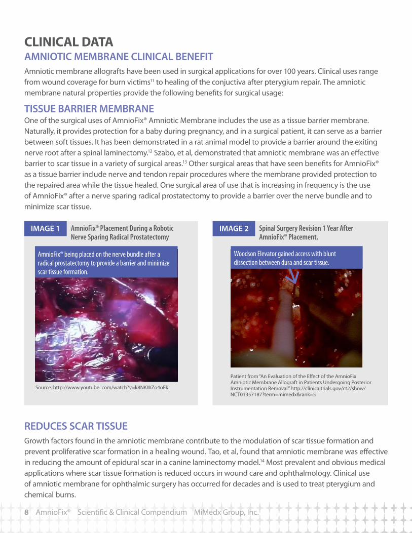
TISSUE BARRIER MEMBRANE One of the surgical uses of AmnioFix® Amniotic Membrane includes the use as a tissue barrier membrane. Naturally, it provides protection for a baby during pregnancy, and in a surgical patient, it can serve as a barrier between soft tissues. It has been demonstrated in a rat animal model to provide a barrier around the exiting nerve root after a spinal laminectomy.12 Szabo, et al, demonstrated that amniotic membrane was an effective barrier to scar tissue in a variety of surgical areas.13 Other surgical areas that have seen benefits for AmnioFix® as a tissue barrier include nerve and tendon repair procedures where the membrane provided protection to the repaired area while the tissue healed. One surgical area of use that is increasing in frequency is the use of AmnioFix® after a nerve sparing radical prostatectomy to provide a barrier over the nerve bundle and to minimize scar tissue.
Spinal Surgery Revision 1 Year After AmnioFix® Placement.
AmnioFix® Placement During a Robotic Nerve Sparing Radical Prostatectomy
REDUCES SCAR TISSUE Growth factors found in the amniotic membrane contribute to the modulation of scar tissue formation and prevent proliferative scar formation in a healing wound. Tao, et al, found that amniotic membrane was effective in reducing the amount of epidural scar in a canine laminectomy model.14 Most prevalent and obvious medical applications where scar tissue formation is reduced occurs in wound care and ophthalmology. Clinical use of amniotic membrane for ophthalmic surgery has occurred for decades and is used to treat pterygium and chemical burns.
8 AmnioFix® Scientific & Clinical Compendium MiMedx Group, Inc.
Patient from “An Evaluation of the Effect of the AmnioFix Amniotic Membrane Allograft in Patients Undergoing Posterior Instrumentation Removal.” http://clinicaltrials.gov/ct2/show/NCT01357187?term=mimedx&rank=5
Source: http://www.youtube..com/watch?v=k8NKWZo4oEk
IMAGE 1 IMAGE 2
Woodson Elevator gained access with blunt dissection between dura and scar tissue.
AmnioFix® being placed on the nerve bundle after a radical prostatectomy to provide a barrier and minimize scar tissue formation.

REDUCE INFLAMMATIONMany of the cytokines found in the amniotic membrane are critical to the healing process (Figure 6). To date MiMedx® has identified several interleukins and TIMPS in the PURION® Processed tissue, such as IL-1ra, that counteract pro-inflammatory interleukins including IL-1. For chronic and surgical wounds, restoring the wound to efficient wound healing is a desired outcome. A wound that is in a prolonged inflammatory state of wound healing may generate more fibroblasts in the area and thus more scar. The normal healing process can be confounded by comorbidities including obesity, diabetes, age, and smoking.
Source: Fetterolf, D. Preliminary Retrospective Analysis of Case Reports of AmnioFix® Injectable Human Amniotic Membrane Allograft in Soft Tissue Injuries and Inflammatory Conditions. 2012.
AmnioFix® Scientific & Clinical Compendium MiMedx Group, Inc. 9
NORMAL HEALING PROCESS (IN DAYS) (from point of injury)
INFLAMMATION PROLIFERATION REMODELING
0.1 1 10 30 100 0.1 1 10 30 ?
NON-HEALING WOUNDS (IN DAYS) (from point of injury)
PROLONGED INFLAMMATORY PHASE
OVERALL EFFECTIVENESS
Symptom Resolution Number of Cases Percent
Complete resolution (greater than or equal 11/26 patients 42%to 90% resolution of symptoms)
Partial resolution of symptoms 5/26 19%No resolution of symptoms 7/26 27% (less than or equal to 10%)
Information regarding effectiveness still pending 3/26 12%
Total 26/26 100%
Healing Time (Weeks) Cases PercentOne 1/26 4%Two 2/26 8%Three 3/26 12%Four 9/26 35%Five 0/26 0%Six 1/26 4%Greater than Six 2/26 8%No Resolution 6/26 23%Pending final analysis 3/26 12%
FIGURE 6
AmnioFix® Injectable Used for Soft Tissue Injuries
A total of 26 patient treatments from 5 different physicians were accumulated, representing a number of different conditions. All patients received only a single injection of the micronized amniotic membrane allograft. The data collected represents an informal discussion performed with the implanting physician who has used the injectable product.
Injuries or conditions identified by the physicians using the material include (number of cases):
• Plantar fasciitis (12)
• Epicondylitis (medial and lateral) (8)
• Toe joint related issues such as hallux limitus (2)
• Achilles tendonitis
• Biceps tendonitis
• Triceps tendonitis
• Acromioclavicular joint area inflammation
Source: Websites, Instructions For Use, and Promotional Literature
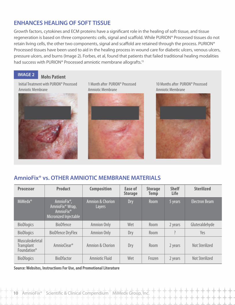
ENHANCES HEALING OF SOFT TISSUEGrowth factors, cytokines and ECM proteins have a significant role in the healing of soft tissue, and tissue regeneration is based on three components: cells, signal and scaffold. While PURION® Processed tissues do not retain living cells, the other two components, signal and scaffold are retained through the process. PURION® Processed tissues have been used to aid in the healing process in wound care for diabetic ulcers, venous ulcers, pressure ulcers, and burns (Image 2). Forbes, et al, found that patients that failed traditional healing modalities had success with PURION® Processed amniotic membrane allografts.15
AmnioFix® vs. OTHER AMNIOTIC MEMBRANE MATERIALS
10 AmnioFix® Scientific & Clinical Compendium MiMedx Group, Inc.
Mohs Patient Initial Treatment with PURION® Processed Amniotic Membrane
1 Month after PURION® Processed Amniotic Membrane
10 Months after PURION® Processed Amniotic Membrane
IMAGE 2
Processor Product Composition Ease of Storage Shelf Sterilized Storage Temp Life
MiMedx® AmnioFix®, Amnion & Chorion Dry Room 5 years Electron Beam AmnioFix® Wrap, Layers AmnioFix® Micronized Injectable
BioDlogics BioDfence Amnion Only Wet Room 2 years Gluteraldehyde
BioDlogics BioDfence DryFlex Amnion Only Dry Room ? Yes
Musculoskeletal Transplant AmnioClear® Amnion & Chorion Dry Room 2 years Not Sterilized Foundation®
BioDlogics BioDfactor Amniotic Fluid Wet Frozen 2 years Not Sterilized

AmnioFix® vs. OTHER AMNIOTIC MEMBRANE MATERIALS
AmnioFix® Scientific & Clinical Compendium MiMedx Group, Inc. 11
Source: MiMedx® Internal Report - SBR -100003.00 “Purion-Processed Dehydrated Human Amniotic Membrane Competitive Tissue Analysis ” Research conducted by MiMedx®.
Source: MiMedx® Internal Report - SBR -100003.00 “Purion-Processed Dehydrated Human Amniotic Membrane Competitive Tissue Analysis ” Research conducted by MiMedx®.
FIGURE 7
120%
100%
80%
60%
40%
20%
0%PDGF-AA PDGF-BB EGF bFGF TGF-β1
PDGF-AA PDGF-BB bFGF TGF-β1 EGF VEGF
AmnioFix® BioDfence BioDfence DryFlex BioDfactor
AmnioFix® InjectableBioDFactor
SUMMARYThe use of human amniotic membrane over the past 100 years has produced a significant amount of data in multiple areas of medicine. Based upon relevant immuno-histochemisty, ELISA, cell migration and proliferation studies conducted by MiMedx® and independent laboratories on PURION® Processed dehydrated human amniotic membrane, it is clear the patent protected PURION® Process provides a minimally manipulated and carefully preserved amniotic tissue that contains essential growth factors and extracellular matrix within the membrane. The sterilized dehydrated human amniotic membrane is processed to provide an easy to use, safe option for multiple surgical applications while providing a 5 year shelf life at room temperature. Many of the key components present in natural amniotic membrane are preserved during the gentle preservation of the PURION® Process, which accounts for the advantageous clinical properties observed when the allografts are used in the clinical applications described. The use of AmnioFix® as a tissue barrier that reduces scar tissue formation, reduces inflammation, and enhances healing in surgical applications can be advantageous for many medical specialties.
~
FIGURE 8
2018161410
86420
~
28x 112x
Relative Growth Factor Content (n=2)
The use of the PURION® Process to prepare the AmnioFix® membrane outperforms other processing methods by preserving the natural growth factors present in amniotic tissue. AmnioFix® and three other membranes were tested for relative quantities of growth factors. In the study, AmnioFix® outperformed all 3 competitive products with a higher quantity and variety of growth factors preserved in the tissue (Figure7).
AmnioFix® MICRONIZED INJECTABLE VS. OTHER INJECTABLEAmnioFix® Injectable is PURION® Processed graft that is micronized to create a powder configuration. A comparative test was conducted using standard protocols to determine the relative amounts of natural growth factors found in the amniotic tissue products (Figure 8). AmnioFix® Injectable contains higher levels of growth factors than BioDFactor.

60 Chastain Center Blvd., Suite 60, Kennesaw, GA 30144866.477.4219 • www.mimedx.com
Patents and patents pending see: www.mimedx.com/patentsAmnioFix®, PURION®, and MiMedx® are registered trademarks of MiMedx Group, Inc. AmnioFix® is processed by Surgical Biologics, a MiMedx Group Company.©2013 MiMedx Group, Inc. All rights reserved.
AM141.002
®
REFERENCES:1. Sherwood, Lauralee. Human Physiology, From Cells to Systems, 7th Ed. Belmont , CA: Cengage Learning, 2010.2. Baradaran-Rafii A, Aghayan H, Arjmand B, and Javadi M. “Amniotic Membrane Transplantation.” 3. Morisaki T, et al. Immunosuppressive cytokines (IL-10, TGF-beta) genes expression in human gastric carcinoma tissues. J Surg Oncol. 1996 Dec:63(4):234-9.4. Kay H; Nelson D; Wang Y. “The Placenta: From Development to Disease.” Wiley-Blackwell. 2011.5. http://www.rndsystems.com 6. Heldin CH, Westermark B. Mechanism of action and in vivo role of platelet-derived growth factor. Physiol Rev. 1999 Oct;79(4):1283-316.7. Fukuda K, Chikama T, Nakamura M, Nishida T. Differential distribution of subchains of the basement membrane components type IV collagen and laminin among the amniotic membrane, cornea, and conjunctiva. Cornea. 1999 Jan;18(1):73-9.8. Hassan Niknejad, Habibollah Peirovi, Masoumeh Jorjani, Abolhassan Ahmadiani, Jalal Ghanavi, Alexander, M. Seifalian. Properties of Amniotic Membrane for Potential Use in Tissue Engineering. European Cells and Materials Vol. 15 2008 (pages 88-99).9. Dietrich-Ntoukas T, Hofmann-Rummelt C, Kruse FE, Schlötzer-Schrehardt U. Comparative analysis of the basement membrane composition of the human limbus epithelium and amniotic membrane epithelium. Cornea. 2012 May;31(5):564-9.10. MiMedx® Internal Report - SBR-100001.00.11. Andonovska et al. “The advantages of the application of amnion membrane in the treatment of burns.” Prilozi. 2008.12. Allen. “Preclinical Study of Human Allograft Amniotic Membrane as a Barrier to Epidural Fibrosis in the Early Wound of a Postlaminectomy Rat Model.” SAS Poster presentation in April 2011. 13. Szabo et al. “Evaluation of seprafilm and amniotic membrane as adhesion prophylaxis in mesh repair of abdominal wall hernia in rats.” Eur Surg Res. 2000. 14. Tao, H and Fen, H “Implantation of amniotic membrane to reduce postlaminectomy epidural adhesions.” Eur Spine J. (18)8. 01-AUG-2009. pp 1202-12.15. Forbes, J and Fetterolf, D E “Dehydrated amniotic membrane allografts for the treatment of chronic wounds: a case series.” J Wound Care. (21)6. 06 2012.