Scientific Abstracts Hair India 2012 Scientific Abstracts ...
7
Jul-Sep 2012 / Vol 4 / Issue 3 ISSN 0974-7753 Scientific Abstracts Hair India 2012 nd 2 International Congress of Trichology On September 7-9, 2012, Mamallapuram, India
Transcript of Scientific Abstracts Hair India 2012 Scientific Abstracts ...
Jul-Sep 2012 / Vol 4 / Issue 3
ISSN 0974-7753
Inte
rna
tion
al Jo
urn
al o
f Tric
ho
log
y • V
olu
me 4
• Issue 3
• Ju
ly-S
ep
tem
ber 2
012 • P
age
s 15
3-2
32
Scientific Abstracts
Hair India 2012nd
2 International Congress of Trichology
On September 7-9, 2012, Mamallapuram, India
Scientific Abstracts
Hair India 2012th
16 Annual Meeting of theEuropean Hair Research Society
on June 21-23, 2012, Barcelona, Spain
158 International Journal of Trichology / Jul-Sep 2012 / Vol-4 / Issue-3
INTRODUCTION
Trichothiodistrophy (TTD) is a rare autosomal recessive condition that was first described by Price et al. in 1980.[1]
TTD is characterized by a distinctive congenital hair shaft dysplasia caused by deficiency of high-sulfur matrix proteins during the late stage of keratinocyte differentiation. It may be confirmed by a cromatography aminoacid determination or by XrMa of the hair shaft showing a deficit in the
Address for correspondence: Dr. José M. Mir-Bonafé,
Department of Dermatology, University Hospital of
Salamanca, C/ San Silvestre, 10, Portal 1, 1º A, Salamanca,
Spain. E-mail: [email protected]
Original Article Further Insights in Trichothiodistrophy: A Clinical, Microscopic, and Ultrastructural Study of 20 Cases and Literature ReviewJuan Ferrando, José M. Mir-Bonafé1, Rodrigo Cepeda-Valdés2, Anna Domínguez3, Jorge Ocampo-Candiani4, Javier García-Veigas3, Minerva Gómez-Flores4, Julio C. Salas-Alanis2,4
Department of Dermatology, Hospital Clinic, Barcelona, 1Department of Dermatology, University Hospital of Salamanca, Spain, 2Department of Dermatology, DEBRA (Dystrophic Epidermolysis Bullosa Research Association), Monterrey, México, 3Department of Scientific-Technical Services, University of Barcelona, Spain, 4Department of Dermatology, Hospital Universitario “Jose E. González” Universidad Autónoma de Nuevo León, México
ABSTRACT
Background: Trichothiodistrophy (TTD) is a rare autosomal recessive condition that is characterizedbyaspecificcongenitalhairshaftdysplasiacausedbydeficiencyofsulfurassociated with a wide spectrum of multisystem abnormalities. In this article, we study clinical,microscopic,andultrastructuralfindingsof20patientswithTTDwiththeaimtoadd further insights regarding to this rare condition. Additionally, analyses of our results are compared with those extracted from the literature in order to enhance its comprehensibility. Materials and Methods: Twenty cases of TTD were included: 7 from Mexico and 14 from Spain. Clinical, microscopic, scanning electron microscopy (SEM) studies and X-ray microanalysis (XrMa) were carried out in all of them. Genetic studies were performed in all seven Mexican cases. Patients with xeroderma pigmentosum and xeroderma pigmentosum/TTD-complex were excluded. Results: Cuticular changes and longitudinal crests of the hair shaft were demonstrated. These crests were irregular, disorganized, followingthehairlongestaxis.Hairshaftsulfurdeficiencywasdisposeddiscontinuouslyand intermittently rather than uniformly. This severe decrease of sulfur contents was located close to the trichoschisis areas.Only five patients did not show related disturbances.Micro-dolichocephalywasobservedinfivecasesandrepresentedthemostfrequentfacialdysmorphism found. It is also remarkable that all patients with urologic malformations also combined diverse neurologic disorders. Moreover, three Mexican sisters demonstrated the coexistence of scarce pubic vellus hair, developmental delay, onychodystrophy, and maxillar/mandibullar hypoplasia. Conclusions: TTD phenotype has greatly varied from very subtle forms to severe alterations such as neurologic abnormalities, blindness, lamellar ichthyosis and gonadal malformations. Herein, a multisystem study should be performed mandatorily in patients diagnosed with TTD.
Key words:Ichthyosis,neurologicabnormality,onychodystrophy,sulfurdeficiency,trichoschisis, trichothiodistrophy
Access this article onlineWebsite:
www.ijtrichology.com
DOI:
10.4103/0974-7753.100075
Quick Response Code:
cystine or sulfur content, respectively, fewer than half of normal (10–50%).[2] TTD may be associated with a wide spectrum of abnormalities affecting organs derived from ectoderm and neuroectoderm. The most commonly related disturbances include photosensitivity, ichthyosis, brittle hair, intellectual impairment, decreased fertility, and short stature (acronym: PIBIDS).[3,4] However, phenotype greatly varies from very subtle forms to extremely severe alterations. The hair of TTD patients may inconstantly be easily broken,
Ferrando, et al.: Further insights in trichothiodistrophy
International Journal of Trichology / Jul-Sep 2012 / Vol-4 / Issue-3 159
sparse, dry, and brittle. Pilotraction is positive. The light microscopy test reveals transverse fractures through the hair shafts (trichoschisis). Polarizing light microscopic examination exhibits an alternating light and dark (so-called tiger tail) banding pattern with trichoschisis. Moreover, absent or defective cuticle may be visualized by scanning electron microscopy (SEM).[5]
TTD is currently divided into photo and non-photosensitive types.[6] The former represents about 50% of the TTD patients and has been related to mutations in several different DNA repair genes. Specifically, these genes encodes transcription-DNA factor IIH (TFIIH) subunits (XPD, XPB, p8/TTDA)[3,6-9] and nucleotide excision repair (NER).[2] Mutations on the two alleles of the XPD gene are the most common. In addition to the photosensitive form of TTD, NER pathway defects are responsible for photosensivity in two further distinct autosomal recessive syndromes: xeroderma pigmentosum and Cockayne syndrome.[10] Contrarily to patients with xeroderma pigmentosum, there is a lack of predisposition to cancer in TTD.[3,9] Although there is little understanding in this particular distinction, it has been proposed that the altered basal transcription in TTD may lead to a decreased transcription of specific genes and therefore would block tumor progression.[2,10] On the other hand, transcription defects might lead to the rest of TTD-related neuroectodermal abnormalities.[10] Recently, TTDN1 mutations (chromosome 7p14) have been found in up to 10% of non-photosensitive cases.[7,11] Some investigators have suggested a third genetic group that comprises some non-photosensitive patients in which this mutation was excluded. However, their exact molecular bases are not still known.
In this article, we study clinical, microscopic and ultrastructural findings of twenty patients with TTD with the aim to add further insights regarding to this rare condition. Additionally, analyses of our results are compared with those extracted from the literature review in order to enhance its comprehensibility.
MATERIALS AND METHODS
Diagnosis of TTD is currently based on the following clinical or laboratory abnormalities: presence of brittle hair and/or hair shaft abnormalities, tiger tail banding with polarized microscopy, and decreased sulfur content of hair. Following these diagnostic criteria, 20 cases of TTD were included. Seven patients from the North East of Mexico (Sabinas: 4 cases, Nuevo Laredo: 2 cases, Saltillo: 1 case) and the rest from the Department of Dermatology of
Hospital Clinic (Barcelona, Spain) are described. Clinical, microscopical, SEM studies, and X-ray microanalysis (XrMa) were completed in all of them. Genetic studies were additionally performed in all seven Mexican cases. Patients with xeroderma pigmentosum and xeroderma pigmentosum/TTD-complex were excluded.
RESULTS
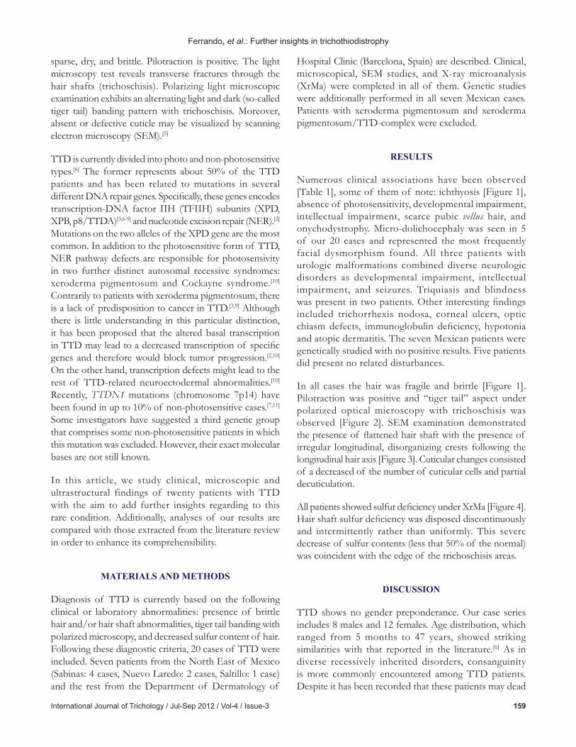
Numerous clinical associations have been observed [Table 1], some of them of note: ichthyosis [Figure 1], absence of photosensitivity, developmental impairment, intellectual impairment, scarce pubic vellus hair, and onychodystrophy. Micro-dolichocephaly was seen in 5 of our 20 cases and represented the most frequently facial dysmorphism found. All three patients with urologic malformations combined diverse neurologic disorders as developmental impairment, intellectual impairment, and seizures. Triquiasis and blindness was present in two patients. Other interesting findings included trichorrhexis nodosa, corneal ulcers, optic chiasm defects, immunoglobulin deficiency, hypotonia and atopic dermatitis. The seven Mexican patients were genetically studied with no positive results. Five patients did present no related disturbances.
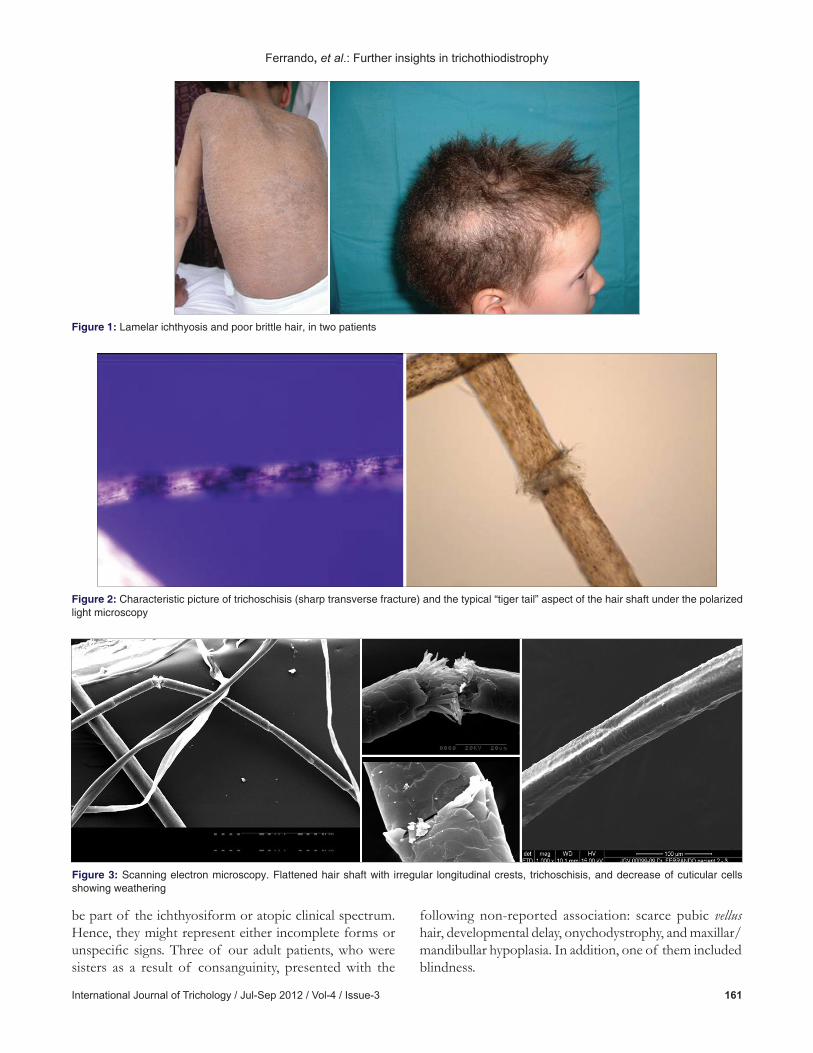
In all cases the hair was fragile and brittle [Figure 1]. Pilotraction was positive and “tiger tail” aspect under polarized optical microscopy with trichoschisis was observed [Figure 2]. SEM examination demonstrated the presence of flattened hair shaft with the presence of irregular longitudinal, disorganizing crests following the longitudinal hair axis [Figure 3]. Cuticular changes consisted of a decreased of the number of cuticular cells and partial decuticulation.
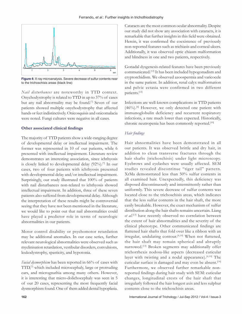
All patients showed sulfur deficiency under XrMa [Figure 4]. Hair shaft sulfur deficiency was disposed discontinuously and intermittently rather than uniformly. This severe decrease of sulfur contents (less that 50% of the normal) was coincident with the edge of the trichoschisis areas.
DISCUSSION
TTD shows no gender preponderance. Our case series includes 8 males and 12 females. Age distribution, which ranged from 5 months to 47 years, showed striking similarities with that reported in the literature.[6] As in diverse recessively inherited disorders, consanguinity is more commonly encountered among TTD patients. Despite it has been recorded that these patients may dead

Ferrando, et al.: Further insights in trichothiodistrophy
160 International Journal of Trichology / Jul-Sep 2012 / Vol-4 / Issue-3
in up to 20-fold higher compared with the United States general population at young age,[6] we did documented no deaths. However, it should be mentioned that a long-term follow up was limited since this was a transversal cohort.
Skin findings
Among TTD-associated abnormalities, ichthyosis is the most frequently documented skin abnormality. Similarly to our study, lamellar type is the most common. However, while data from literature shows a high incidence of ichthyosis among TTD patients (65%),[6] our study demonstrated a significant decreasing of this condition (20%). Unexpectedly, a recent study conducted by Morice-Picard et al.[7] showed a higher incidence of cases with clinical and microscopic aspects of ichthyosis vulgaris. In addition, their case series showed that congenital ichthyosis and collodion baby were more frequent in patients with clinical photosensitivity and abnormal DNA repair than those non-photosensitive. In contrast, only one of our five patients with ichthyosis demonstrated photosensitivity. Regarding to this, it is
interesting a patient that we previously published[12] who presented with the coexistence of congenital ichthyosis, developmental delay, intellectual impairment, renal calyx malformations, microcephaly, urethral duplicity, and hypercalciuria. Moreover, patient number 10 showed myelinisation retardation, nistagmus, neuronal transmission impairment, optic chiasms malformations and secondary blindness. Interestingly, a 1-year-old male black race patient showed congenital lamellar ichthyosis associated with TTD.
Photosensitivity is the second most reported skin feature in the literature (42%).[7] Unexpectedly, we only found it in two patients (10%). It is interesting that despite none of the seven patients genetically studied for TTDN1 gene was positive, only two of them had photosensitivity. This leads to hypothesise that patients with no TTDN1 gene mutation may be the majority in the non-photosensitivity group.
Other uncommon cutaneous features include erythroderma, ezcema, telangiectasias, follicular queratosis, cheilitis, or pruritus.[7] However, we suggest that these findings may
Table 1: Clinical associations of 20 patients with trichothiodistrophyPatient number
Age-sex
Skinfindings
Neurologicfindings
Relatedmalformations
Other features
1. 20 y-M Lamellar ichthyosis DI, II Renal calyx, urethral duplicity, microcephaly
Consanguinity, hypercalciuria
2. 5 y-M – – – –
3. 4 y-F Onicosquisis, onicomalacia DI – –
4. 8 y-M Onicosquisis DI, II, leukodystrophy, spasticity – –
5. 3 y-F Photosensitivity, atopic dermatitis
– Facial dysmorphy –
6. 8 y-F – – – –
7. 1 y-M Lamellar ichthyosis – – Black race
8. 5 y-M – – – –
9. 5 m-M Trichorrhexis nodosa Seizures, hypotonia Pelvic ectasia Systolic heart murmur
10. 8 m-F Lamellar ichthyosis DI, II, myelinisation retardation, nistagmus
Optic chiasm Neuronal transmission impairment, blindness
11. 11 y-M Lamellar ichthyosis – – –
12. 5 y-M – – – –
13. 2 y-F Ichthyosis, photosensitivity – – –
14. 2 y-F Onychodystrophy DI, II – Twin sister with patient 15
15. 2 y-F Onychodystrophy DI, II – Twin sister with patient 14
16. 3 y-F – – – –
17. 45 y-F Scarce pubic vellus hair, onychodystrophy
DI Mandibular hypoplasia Consanguinity, sister with patients 18 and 19
18. 47 y-F Scarce pubic vellus hair, onychodystrophy
DI Mandibular hypoplasia Consanguinity, sister with patients 17 and 19
19. 44 y-F Scarce pubic vellus hair, onychodystrophy
DI Maxillar hypoplasia Consanguinity, sister with patients 18 and 19, triquiasis, corneal ulcers, blindness
20. 27 y-M Keratosis pilaris DI Varicocele, azoospermia
Consanguinity, triquiasis, immunoglobulin deficiency
DI – Developmental impairment; II – Intellectual impairment; y – Years; m – Months; M – Male; F – Female
Ferrando, et al.: Further insights in trichothiodistrophy
International Journal of Trichology / Jul-Sep 2012 / Vol-4 / Issue-3 161
Figure 3: Scanning electron microscopy. Flattened hair shaft with irregular longitudinal crests, trichoschisis, and decrease of cuticular cells showing weathering
Figure 1: Lamelar ichthyosis and poor brittle hair, in two patients
Figure 2: Characteristic picture of trichoschisis (sharp transverse fracture) and the typical “tiger tail” aspect of the hair shaft under the polarized light microscopy
be part of the ichthyosiform or atopic clinical spectrum. Hence, they might represent either incomplete forms or unspecific signs. Three of our adult patients, who were sisters as a result of consanguinity, presented with the
following non-reported association: scarce pubic vellus hair, developmental delay, onychodystrophy, and maxillar/mandibullar hypoplasia. In addition, one of them included blindness.
Ferrando, et al.: Further insights in trichothiodistrophy
162 International Journal of Trichology / Jul-Sep 2012 / Vol-4 / Issue-3
Nail disturbances are noteworthy in TTD context. Onychodystrophy is related to TTD in up to 37% of cases but any nail abnormality may be found.[7] Seven of our patients showed multiple onychodystrophy that affected hands or feet indistinctively. Onicosquisis and onicomalacia were noted. Fungi cultures were negative in all cases.
Other associated clinical findings
The majority of TTD patients show a wide-ranging degree of developmental delay or intellectual impairment. The former was represented in 10 of our patients, while 6 presented with intellectual impairment. Literature review demonstrates an interesting association, since ichthyosis is closely linked to developmental delay (92%).[7] In our cases, two of four patients with ichthyosis presented with developmental delay and/or intellectual impairment. Surprisingly, our study illustrated that 100% of patients with nail disturbances non-related to ichthyosis showed intellectual impairment. In addition, three of these seven patients also suffered from developmental delay. Although the interpretation of these results might be controversial seeing that they have not been mentioned in the literature, we would like to point out that nail abnormalities could have played a predictor role in terms of neurologic abnormalities in our patients.
Motor control disability or psychomotor retardation may be additional anomalies. In our case series, further relevant neurological abnormalities were observed such as myelinisation retardation, vestibular disorders, convulsions, leukodystrophy, spasticity, and hypotonia.
Facial dysmorphism has been reported in 66% of cases with TTD,[7] which included microcephaly, large or protruding ears, and micrognathia among many others. However, it is interesting that micro-dolichocephaly was seen in 5 of our 20 cases, representing the most frequently facial dysmorphism found. One of them added dental hypoplasia.
Cataracts are the most common ocular abnormality. Despite our study did not show any association with cataracts, it is remarkable that further insights in this field were obtained. Herein, it was confirmed the coexistence of previously non-reported features such as trichiasis and corneal ulcers. Additionally, it was observed optic chiasm malformation and blindness in one and two patients, respectively.
Gonadal dysgenesis-related features have been previously communicated.[12] It has been included hypogonadism and cryptorchidism. We observed azoospermia and varicocele in the same patient. In addition, renal calyx malformation and pelvic ectasia were confirmed in two different patients.[12]
Infections are well-known complications in TTD patients (46%).[6] However, we only detected one patient with immunoglobulin deficiency and recurrent respiratory infections, a rate much lower than expected. Historically, chronic neutropenia has been commonly reported.[10]
Hair findings
Hair abnormalities have been demonstrated in all our patients. It was observed brittle and dry hair, in addition to clean transverse fractures through the hair shafts (trichoschisis) under light microscopy. Eyebrows and eyelashes were usually affected. SEM studies revealed discontinue “tiger tail” pattern. XrMa demonstrated less than 50% sulfur contents in all examined hair. Unexpectedly, this deficiency was disposed discontinuously and intermittently rather than uniformly. This severe decrease of sulfur contents was located close to the trichoschisis areas, which indicates that the less sulfur contents in the hair shaft, the more easily breakable. However, the exact mechanism of sulfur distribution along the hair shafts remains uncertain. Liang et al.[13] have recently observed no correlation between the extent of hair abnormalities and the severity of the clinical phenotype. Other communicated findings are flattened hair shafts that fold over like a ribbon with an irregular, undulating contour.[4,10] When not flattened, the hair shaft may remain spherical and abruptly narrowed.[13] Broken segments may additionally offer trichorrhexis nodosa-like aspects (decreased cuticular layer with twisting and a nodal appearance).[4,10] The cuticular surface is damaged and may even be absent.[14] Furthermore, we observed further remarkable non-reported findings during hair study with SEM: cuticular changes, longitudinal crests of the hair shaft that irregularly followed the hair longest axis and less sulphur contents close to the trichoschisis areas.
Figure 4: X-ray microanalysis. Severe decrease of sulfur contents near to the trichoschisis areas (black line)
Ferrando, et al.: Further insights in trichothiodistrophy
International Journal of Trichology / Jul-Sep 2012 / Vol-4 / Issue-3 163
In summary, our study revealed that TTD phenotype has greatly varied from very subtle forms to extremely severe alterations such as neurologic abnormalities, blindness, lamellar ichthyosis, and gonadal malformations. Hence, a patient diagnosed with TTD should mandatorily undergo a complete multisystem study.
ACKNOWLEDGMENTS
We would like to thank Dr. Angela Cristiano (Department of Genetics and Development, Columbia University, New York, United States) for the genetic study, as well as Drs. Montserrat Castro, Marta Cucurell, Eva Fernández Vilariño, Isabel Febrer, Valentín García Mellado, Dan López, Ana Martín Santiago, Alex Llambrich, José Carlos Moreno, Jorge Moreno, Anna Tuneu, Angel Vera and Ernesto Bonifazi for referring their patients to us for the study.
REFERENCES
1. Price VH, Odom RB, Ward WH, Jones FT. Trichothiodystrophy: Sulfur-deficient brittle hair as a marker for a neuroectodermal symptom complex. Arch Dermatol 1980;116:1375-84.
2. Bergmann E, Egly JM. Trichothiodystrophy, a transcription syndrome. Trends Genet 2001;17:279-86.
3. Lambert WC, Gagna CE, Lambert MW. Trichothiodystrophy: Photosensitive, TTD-P, TTD, Tay syndrome. Adv Exp Med Biol 2010;685:106-10.
4. Liang C, Kraemer KH, Morris A, Schiffmann R, Price VH, Menefee E, et al. Characterization of tiger-tail banding and hair shaft abnormalities in trichothiodystrophy. J Am Acad Dermatol 2005;52:224-32.
5. Price VH. Trichothiodystrophy: Update. Pediatr Dermatol 1992;9:369-70.6. Faghri S, Tamura D, Kraemer KH, Digiovanna JJ. Trichothiodystrophy: A
systematic review of 112 published cases characterises a wide spectrum of clinical manifestations. J Med Genet 2008;45:609-21.
7. Morice-Picard F, Cario-Andre M, Rezvani H, Lacombe D, Sarasin A, Taieb A. New clinico-genetic classification of trichothiodystrophy. Am J Med Genet A 2009;149A:2020-30.
8. Hashimoto S, Egly JM. Trichothiodystrophy view from the molecular basis of DNA repair/transcription factor TFIIH. Hum Mol Genet 2009;18: R224-30.
9. Stefanini M, Botta E, Lanzafame M, Orioli D. Trichothiodystrophy: From basic mechanisms to clinical implications. DNA Repair (Amst) 2010;9: 2-10.
10. Itin PH, Sarasin A, Pittelkow MR. Trichothiodystrophy: Update on the sulfur-deficient brittle hair syndromes. J Am Acad Dermatol 2001;44:891-920; quiz 1-4.
11. Nakabayashi K, Amann D, Ren Y, Saarialho-Kere U, Avidan N, Gentles S, et al. Identification of C7orf11 (TTDN1) gene mutations and genetic heterogeneity in nonphotosensitive trichothiodystrophy. Am J Hum Genet 2005;76:510-6.
12. Malvehy J, Ferrando J, Soler J, Tuneu A, Ballesta F, Estrach T. Trichothiodystrophy associated with urologic malformation and primary hypercalciuria. Pediatr Dermatol 1997;14:441-5.
13. Liang C, Morris A, Schlucker S, Imoto K, Price VH, Menefee E, et al. Structural and molecular hair abnormalities in trichothiodystrophy. J Invest Dermatol 2006;126:2210-6.
14. Grimalt R, Ferrando J. Shaft effects from cosmetics and styling. In: Camacho F, Randall VA, Price VH, editors. Hair and its Disorders. Biology, Pathology and Management. London: Martin Dunitz Ed., 2000. p. 325-38.
How to cite this article: Ferrando J, Mir-Bonafé JM, Cepeda-Valdés R, Domínguez A, Ocampo-Candiani J, García-Veigas J,
et al. Further insights in trichothiodistrophy: A clinical, microscopic, and ultrastructural study of 20 cases and literature review. Int J
Trichol 2012;4:158-63.
Source of Support: Nil, Conflict of Interest: None declared.