Title: Iron overload increases osteoclastogenesis and aggravates ...
Schistosome infection aggravates HCV-related liver disease and
induces changes in the regulatory T-cell phenotype
E. LOFFREDO-VERDE,1 I. ABDEL-AZIZ,2 J. ALBRECHT,1,3 N. EL-GUINDY,4 M. YACOB,4 A. SOLIEMAN,5 U. PROTZER,3,6
D. H. BUSCH,1,3 L. E. LAYLAND7 & C. U. PRAZERES DA COSTA1
1Institute for Medical Microbiology, Immunology and Hygiene, Technische Universität München, Munich, Germany, 2MedicalParasitology, Kasr Al-Eini University Clinic Cairo, Cairo, Egypt, 3Clinical Cooperation Group ‘Immune Monitoring’, Helmholtz ZentrumM€unchen and Technische Universität München, Munich,Germany, 4Clinical & Chemical Pathology, Kasr Al-Eini School of MedicineHospitals, Cairo Univeristy, Cairo,Egypt, 5Tropical Medicine Departments, Kasr Al-Eini School of Medicine Hospitals, Cairo Univeristy,Cairo, Egypt, 6Institute of Virology, Technische Universität and Helmholtz Zentrum München, Munich, Germany, 7Institute for MedicalMicrobiology, Immunology and Parasitology (IMMIP), University Clinic Bonn, Bonn, Germany
SUMMARY
Schistosome infections are renowned for their ability toinduce regulatory networks such as regulatory T cells(Treg) that control immune responses against homologousand heterologous antigens such as allergies. However, in thecase of co-infections with hepatitis C virus (HCV), schisto-somes accentuate disease progression and we hypothesizedthat expanding schistosome-induced Treg populations changetheir phenotype and could thereby suppress beneficial anti-HCV responses. We therefore analysed effector T cells andn/iTreg subsets applying the markers Granzyme B (GrzB)and Helios in Egyptian cohorts of HCV mono-infected(HCV), schistosome-co-infected (Sm/HCV) and infection-free individuals. Interestingly, viral load and liver transamin-ases were significantly elevated in Sm/HCV individuals whencompared to HCV patients. Moreover, overall Treg frequen-cies and HeliosposTreg were not elevated in Sm/HCV indi-viduals, but frequencies of GrzB+Treg were significantlyincreased. Simultaneously, GrzB+ CD8+ T cells were notsuppressed in co-infected individuals. This study demon-strates that in Sm/HCV co-infected cohorts, liver disease isaggravated with enhanced virus replication and Treg do notexpand but rather change their phenotype with GrzB possi-bly being a more reliable marker than Helios for iTreg.Therefore, curing concurrent schistosome disease could bean important prerequisite for successful HCV treatment asco-infected individuals respond poorly to interferon therapy.
Keywords Granzyme B, Helios, hepatitis C virus, liver dis-ease, regulatory T cells, Schistosoma mansoni
Abbreviations: GrzB, Granzyme B; HCV, hepatitis C virus;HD, healthy donors; S. mansoni, Schistosoma mansoni;SEA, soluble egg antigen; Sm/HCV, Schistosoma mansoni/HCV co-infection; Treg, Regulatory T cells
INTRODUCTION
Hepatitis C virus (HCV) is the second most common viralinfection worldwide and considered the most importantagent of liver disease and liver carcinoma. In general,approximately 70% of acute HCV cases develop chronichepatitis of which 15–20% will eventually evolve into cir-rhosis with a 10% risk of hepatocellular carcinoma (1).Currently, it is estimated that 180 million people areinfected with HCV, and, interestingly, Egypt has the high-est prevalence worldwide (31%) with 90% of those infectedindividuals being positive for the genotype 4 variant (2–5).HCV infections induce both CD4+ and CD8+ T-cellresponses, and during acute infection, several studies havedemonstrated that strong and sustained viral-specificCD4+ and CD8+ T-cell responses are required for sponta-neous and successful viral clearance. Individuals who failto mount or sustain such viral-specific responses usuallydevelop persistent viraemia and chronic infection (6).Although the underlying mechanisms of dysfunctionalvirus-specific immune responses remain poorly under-stood, research has shown that chronic HCV-infected indi-viduals present higher levels of peripheral CD4+CD25+
Foxp3+ regulatory T cells (Treg) which are able to sup-press virus-specific CD8+ T-cell responses (7, 8). The highprevalence of HCV infection in Egypt stems from an
Correspondence: Clarissa U. Prazeres da Costa, Institute for Med-ical Microbiology, Immunology and Hygiene, Technische Univer-sität München (TUM), Trogerstrasse 30, 81675 München,Germany (e-mail: [email protected]).Received: 29 July 2014Accepted for publication: 21 December 2014
© 2015 John Wiley & Sons Ltd 97
Parasite Immunology, 2015, 37, 97–104 DOI: 10.1111/pim.12171
iatrogenic epidemic that was introduced in the 1950s dueto mass treatment with antimony against schistosomiasis(9). Since then, concomitant human schistosomiasis andHCV infection are extremely common, especially in ruralareas of Egypt (10, 11) and co-infected individuals presentan accelerated onset of liver cirrhosis and hepatocellularcarcinoma (12). Indeed, whereas 30% of HCV mono-infected individuals recovered from acute infections, allthose with an additional Schistosoma mansoni-infectionprogressed to a chronic state (13). Moreover, compared toHCV mono-infected individuals, these patients exhibitedhigher HCV-positive RNA titres, higher necro-inflamma-tory and fibrotic scores in the liver and poor responses tointerferon therapy (10, 14).Schistosomiasis, caused by the helminth Schistosoma,
remains one of the most important parasitic diseasesworldwide, and current monitoring indicates that over 250million individuals are infected (15, 16). In endemic areas,these infections have a detrimental impact on both finan-cial and social sectors (17). S. mansoni and S. haematobi-um are endemic in many rural areas of Egypt, andcommunity prevalence often ranges between 15 and 45%(18). Immunologically, one of the most interesting aspectsof schistosome infection is the modulation of the immunesystem including a dynamic switch from an initial pro-inflammatory Th1 to a dominant helminth-beneficial Th2immune response once female worms become fecund. Asinfection enters the chronic phase, a more regulated equi-librium ensues with high levels of anti-inflammatory cyto-kines (IL-10 and TGF-b) and elevated frequencies ofregulatory T-cells (Treg) (19–22). In association, diseaseseverity is partially controlled by the balance of Th1 ver-sus Th2 type cytokines and/or the presence of Treg (19,20, 23). We and others have shown the essential role ofTreg using the murine model of S. mansoni and althoughTreg cannot prevent helminth infection per se, their neces-sity in controlling exaggerated immunopathology andeffector T-cell responses has been well documented (23–25). Indeed, those studies demonstrated that isolated Treg,from infected but not naive mice, could specifically sup-press schistosome-specific CD4+ T-cell responses in an IL-10 independent manner and moreover changed their phe-notype during infection (22, 24, 25). Specifically, schisto-some-induced Treg acquired cytotoxic T-cell activity asGranzyme B (GrzB) was strongly upregulated (25).Currently, no data are available whether there are char-
acteristic changes from nTreg (naturally occurring, thymusderived) to iTreg (peripherally induced) during humanschistosomiasis. Therefore, we investigated changes in thephenotype of Foxp3+ Treg, during human schistosomiasis,which could be responsible for the reported failure to raiseeffective antiviral CD4+ and CD8+ T-cell responses (7, 8,
26). In turn, this would lead to loss of viral replicationcontrol and eventually to more pronounced liver disease.Thus, we analysed liver function and viral load as well asTreg T effector cell (Teff) frequencies and specifically thenTreg/iTreg distribution using the novel markers Heliosand GrzB within patients with HCV mono-infection andthose with concomitant Schistosoma infection.
MATERIALS AND METHODS
Study subjects and collection of blood samples
In collaboration with the Hepatology Outpatient Clinic ofCairo University Hospital in Egypt in 2009, chronic hepa-titis C patients were recruited and further classified intoHCV mono-infected (n = 15–HCV) or co-infected withS. mansoni (n = 16–Sm/HCV). Schistosome infection wasdiagnosed on the presence of S. mansoni eggs within thestool with the Kato-Katz method and/or the presence ofhigh titres of schistosomal antibodies (>1/640) againstadult worms (Fumouze Diagnostics, Levallois-Perret,France). The study protocol was in accordance with theethical guidelines of the 1975 Declaration of Helsinki, andethical clearance was given by the Scientific Research Eth-ics Committee at the Faculty of Medicine, Cairo Univer-sity in Egypt and by the Ethics Commission of theFaculty of Medicine at the Technischen Universit€atM€unchen (TUM) in Germany. Patients exhibiting otherviral hepatic infections, hepatic cirrhosis, prolonged partialprothrombin time (PPT) and hepatocellular carcinomawere excluded, as well as other intestinal parasitic infec-tions by sodium-acetate-formalin (SAF) enrichmentmethod and microscopy. As the areas of patient recruit-ment in Egypt are considered malaria free, individualswere not tested for malaria. Peripheral blood mononuclearcells (PBMC) were isolated immediately after the collec-tion of fresh heparinized blood using Ficoll-Hypaque den-sity gradient centrifugation. Cell samples weresubsequently cryopreserved in 10% DMSO (dimethyl sul-phoxide) and 90% foetal calf serum (Sigma Chemicals,Hamburg, Germany; PAA Laboratories Inc., C€olbe, Ger-many) at �80°C and shipped to Germany on dry ice.HCV viral loads in plasma were quantified using real-timePCR (27). Further blood analysis of all study participantsincluded routine laboratory tests such as liver enzymesAST, ALT and AFP (Reflovet Plus Reader, Roche, Penz-berg, Germany), kidney function and coagulation tests,and a blood count. For comparison, further samples werecollected from age-matched, infection-free volunteers,which were mainly relatives of the patient cohort andresided therefore in the same endemic area (n = 13). Theyare denoted in this study as ‘healthy donors’ (HD).
98 © 2015 John Wiley & Sons Ltd, Parasite Immunology, 37, 97–104
E. Loffredo-Verde et al. Parasite Immunology
Flowcytometry cell staining
Phenotypic and functional characteristics of regulatoryand effector T-cell subsets were investigated using multi-colour surface and intracellular stainings based on flowcytometry technology. The following staining panel wasdesigned in order to characterize Treg and T effector cells:CD8 AmCyAn, CD127 PE, Granzyme B FITC (BectonDickinson, Heidelberg, Germany), CD3 APC-A750, CD25PE-Cy7 (Beckmann Coulter, Krefeld, Germany), CD103PE-Cy5, Foxp3 PB, Helios FITC (BioLegend, San Diego,CA, USA) and CD4 A700 (Affymetrix, Vienna, Austria).Viability was assessed by addition of ethidium monoazide([EMA], Molecular Probes/Invitrogen, Karlsruhe, Ger-many) and did not differ between patient groups (averagemean of viable cells in % � SD: HD 76�6% � 11�7%; Sm/HCV: 77�3% � 14�1%; HCV: 74�0% � 16�2%). In short,following surface staining, PBMCs were fixed and permea-bilized for 20 min at 4°C in 100 lL fixation/permeabiliza-tion working solution using the Foxp3 staining buffer setfrom eBioscience (Affymetrix). After washing, cells wereresuspended in permeabilization buffer containing anti-human antibodies specific to intracellular and intranuclearproteins for 30 min at 4°C. Cell acquisition was performedusing the LSRII flow cytometer (Becton Dickinson)equipped with a high-throughput system. Sample analysiswas performed using FlowJo version 9.5.3 (Tree Star, Ash-land, OR, USA).
Serum cytokine detection
Levels of IL-6 and IL-8 were measured in individualplasma samples using the human Th1/Th2/Th8/Th17/Th2213plex Kit FlowCytomix according to manufacturer’sinstructions (Affymetrix). Acquisition of the multipleximmunoassay was performed using the LSRII flow cytom-eter. Sample analysis was performed with FlowcytomixTM
Pro 3.0 Software (Affymetrix).
Statistical analysis
All statistical tests were performed with PRISM� 5.01
(GraphPad Software Inc., San Diego, CA, USA). D’Agos-tino and Pearson omnibus normality tests were performed,and parametrically distributed data were analysed withunpaired t-test (2 groups) and Mann–Whitney U-test wasused for nonparametric data. For more than two groups,1-way ANOVA tests was conducted and if data were non-parametric, a Kruskal–Wallis test with a confidence inter-val of 95% was employed. Results with a P value of <0�05were considered as significant and P values <0�01 ashighly significant.
RESULTS
Co-infected patients present higher viral load andstronger liver pathology
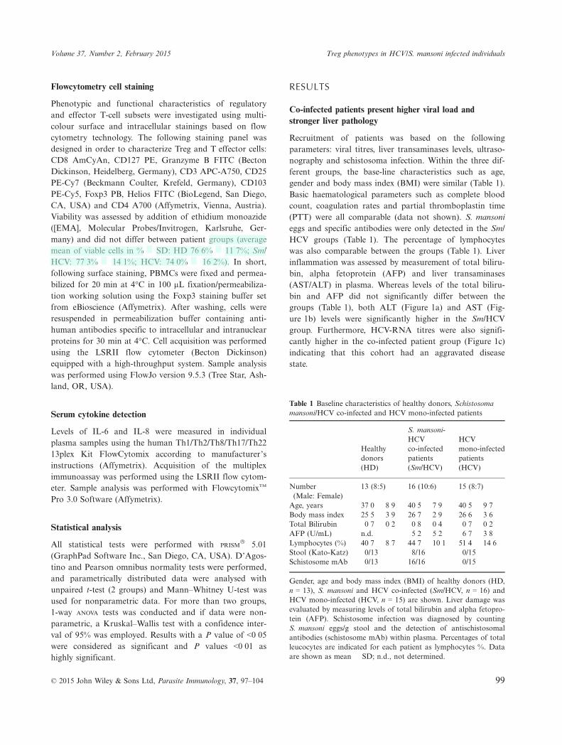
Recruitment of patients was based on the followingparameters: viral titres, liver transaminases levels, ultraso-nography and schistosoma infection. Within the three dif-ferent groups, the base-line characteristics such as age,gender and body mass index (BMI) were similar (Table 1).Basic haematological parameters such as complete bloodcount, coagulation rates and partial thromboplastin time(PTT) were all comparable (data not shown). S. mansonieggs and specific antibodies were only detected in the Sm/HCV groups (Table 1). The percentage of lymphocyteswas also comparable between the groups (Table 1). Liverinflammation was assessed by measurement of total biliru-bin, alpha fetoprotein (AFP) and liver transaminases(AST/ALT) in plasma. Whereas levels of the total biliru-bin and AFP did not significantly differ between thegroups (Table 1), both ALT (Figure 1a) and AST (Fig-ure 1b) levels were significantly higher in the Sm/HCVgroup. Furthermore, HCV-RNA titres were also signifi-cantly higher in the co-infected patient group (Figure 1c)indicating that this cohort had an aggravated diseasestate.
Table 1 Baseline characteristics of healthy donors, Schistosomamansoni/HCV co-infected and HCV mono-infected patients
Healthydonors(HD)
S. mansoni-HCVco-infectedpatients(Sm/HCV)
HCVmono-infectedpatients(HCV)
Number(Male: Female)
13 (8:5) 16 (10:6) 15 (8:7)
Age, years 37�0 � 8�9 40�5 � 7�9 40�5 � 9�7Body mass index 25�5 � 3�9 26�7 � 2�9 26�6 � 3�6Total Bilirubin 0�7 � 0�2 0�8 � 0�4 0�7 � 0�2AFP (U/mL) n.d. 5�2 � 5�2 6�7 � 3�8Lymphocytes (%) 40�7 � 8�7 44�7 � 10�1 51�4 � 14�6Stool (Kato-Katz) 0/13 8/16 0/15Schistosome mAb 0/13 16/16 0/15
Gender, age and body mass index (BMI) of healthy donors (HD,n = 13), S. mansoni and HCV co-infected (Sm/HCV, n = 16) andHCV mono-infected (HCV, n = 15) are shown. Liver damage wasevaluated by measuring levels of total bilirubin and alpha fetopro-tein (AFP). Schistosome infection was diagnosed by countingS. mansoni eggs/g stool and the detection of antischistosomalantibodies (schistosome mAb) within plasma. Percentages of totalleucocytes are indicated for each patient as lymphocytes %. Dataare shown as mean � SD; n.d., not determined.
© 2015 John Wiley & Sons Ltd, Parasite Immunology, 37, 97–104 99
Volume 37, Number 2, February 2015 Treg phenotypes in HCV/S. mansoni infected individuals
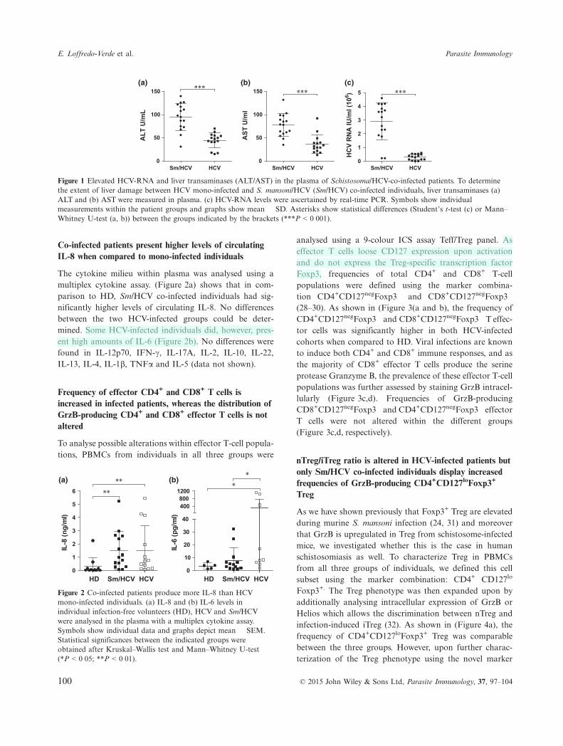
Co-infected patients present higher levels of circulatingIL-8 when compared to mono-infected individuals
The cytokine milieu within plasma was analysed using amultiplex cytokine assay. (Figure 2a) shows that in com-parison to HD, Sm/HCV co-infected individuals had sig-nificantly higher levels of circulating IL-8. No differencesbetween the two HCV-infected groups could be deter-mined. Some HCV-infected individuals did, however, pres-ent high amounts of IL-6 (Figure 2b). No differences werefound in IL-12p70, IFN-c, IL-17A, IL-2, IL-10, IL-22,IL-13, IL-4, IL-1b, TNFa and IL-5 (data not shown).
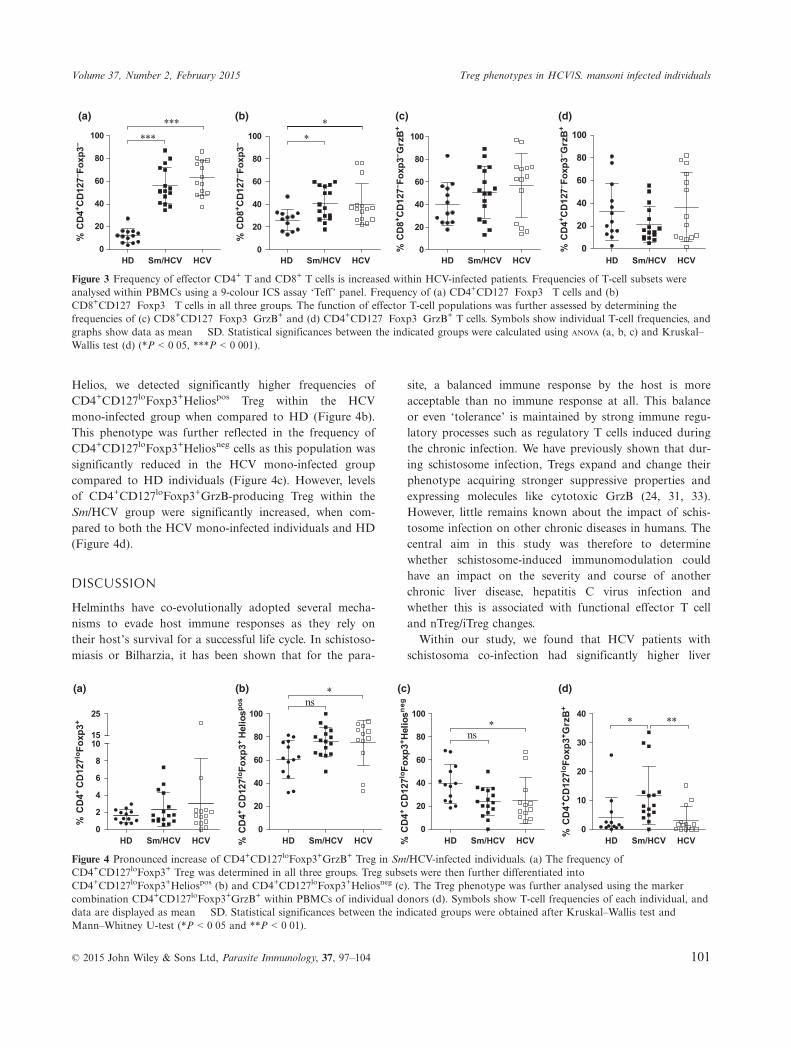
Frequency of effector CD4+ and CD8+ T cells isincreased in infected patients, whereas the distribution ofGrzB-producing CD4+ and CD8+ effector T cells is notaltered
To analyse possible alterations within effector T-cell popula-tions, PBMCs from individuals in all three groups were
analysed using a 9-colour ICS assay Teff/Treg panel. Aseffector T cells loose CD127 expression upon activationand do not express the Treg-specific transcription factorFoxp3, frequencies of total CD4+ and CD8+ T-cellpopulations were defined using the marker combina-tion CD4+CD127negFoxp3� and CD8+CD127negFoxp3�
(28–30). As shown in (Figure 3(a and b), the frequency ofCD4+CD127negFoxp3� and CD8+CD127negFoxp3� T effec-tor cells was significantly higher in both HCV-infectedcohorts when compared to HD. Viral infections are knownto induce both CD4+ and CD8+ immune responses, and asthe majority of CD8+ effector T cells produce the serineprotease Granzyme B, the prevalence of these effector T-cellpopulations was further assessed by staining GrzB intracel-lularly (Figure 3c,d). Frequencies of GrzB-producingCD8+CD127negFoxp3� and CD4+CD127negFoxp3� effectorT cells were not altered within the different groups(Figure 3c,d, respectively).
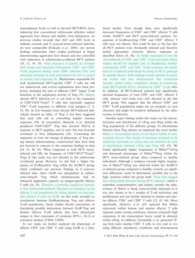
nTreg/iTreg ratio is altered in HCV-infected patients butonly Sm/HCV co-infected individuals display increasedfrequencies of GrzB-producing CD4+CD127loFoxp3+
Treg
As we have shown previously that Foxp3+ Treg are elevatedduring murine S. mansoni infection (24, 31) and moreoverthat GrzB is upregulated in Treg from schistosome-infectedmice, we investigated whether this is the case in humanschistosomiasis as well. To characterize Treg in PBMCsfrom all three groups of individuals, we defined this cellsubset using the marker combination: CD4+ CD127lo
Foxp3+. The Treg phenotype was then expanded upon byadditionally analysing intracellular expression of GrzB orHelios which allows the discrimination between nTreg andinfection-induced iTreg (32). As shown in (Figure 4a), thefrequency of CD4+CD127loFoxp3+ Treg was comparablebetween the three groups. However, upon further charac-terization of the Treg phenotype using the novel marker
Sm/HCV HCV0
50
100
150 ***(a)
ALT
U/m
L
Sm/HCV HCV0
50
100
150 ***(b)
AST
U/m
l
Sm/HCV HCV0
1
2
3
4
5 ***(c)
HC
V R
NA
IU/m
l (10
6 )
Figure 1 Elevated HCV-RNA and liver transaminases (ALT/AST) in the plasma of Schistosoma/HCV-co-infected patients. To determinethe extent of liver damage between HCV mono-infected and S. mansoni/HCV (Sm/HCV) co-infected individuals, liver transaminases (a)ALT and (b) AST were measured in plasma. (c) HCV-RNA levels were ascertained by real-time PCR. Symbols show individualmeasurements within the patient groups and graphs show mean � SD. Asterisks show statistical differences (Student’s t-test (c) or Mann–Whitney U-test (a, b)) between the groups indicated by the brackets (***P < 0�001).
HD Sm/HCV HCV0
1
2
3
4
5
6
(a)
****
IL-8
(ng/
ml)
HD Sm/HCV HCV0
10
20
30
40
400800
1200
(b)*
*
IL-6
(pg/
ml)
Figure 2 Co-infected patients produce more IL-8 than HCVmono-infected individuals. (a) IL-8 and (b) IL-6 levels inindividual infection-free volunteers (HD), HCV and Sm/HCVwere analysed in the plasma with a multiplex cytokine assay.Symbols show individual data and graphs depict mean � SEM.Statistical significances between the indicated groups wereobtained after Kruskal–Wallis test and Mann–Whitney U-test(*P < 0�05; **P < 0�01).
100 © 2015 John Wiley & Sons Ltd, Parasite Immunology, 37, 97–104
E. Loffredo-Verde et al. Parasite Immunology
Helios, we detected significantly higher frequencies ofCD4+CD127loFoxp3+Heliospos Treg within the HCVmono-infected group when compared to HD (Figure 4b).This phenotype was further reflected in the frequency ofCD4+CD127loFoxp3+Heliosneg cells as this population wassignificantly reduced in the HCV mono-infected groupcompared to HD individuals (Figure 4c). However, levelsof CD4+CD127loFoxp3+GrzB-producing Treg within theSm/HCV group were significantly increased, when com-pared to both the HCV mono-infected individuals and HD(Figure 4d).
DISCUSSION
Helminths have co-evolutionally adopted several mecha-nisms to evade host immune responses as they rely ontheir host’s survival for a successful life cycle. In schistoso-miasis or Bilharzia, it has been shown that for the para-
site, a balanced immune response by the host is moreacceptable than no immune response at all. This balanceor even ‘tolerance’ is maintained by strong immune regu-latory processes such as regulatory T cells induced duringthe chronic infection. We have previously shown that dur-ing schistosome infection, Tregs expand and change theirphenotype acquiring stronger suppressive properties andexpressing molecules like cytotoxic GrzB (24, 31, 33).However, little remains known about the impact of schis-tosome infection on other chronic diseases in humans. Thecentral aim in this study was therefore to determinewhether schistosome-induced immunomodulation couldhave an impact on the severity and course of anotherchronic liver disease, hepatitis C virus infection andwhether this is associated with functional effector T celland nTreg/iTreg changes.Within our study, we found that HCV patients with
schistosoma co-infection had significantly higher liver
HD Sm/HCV HCV0
20
40
60
80
100
(a)***
***
% C
D4+ C
D12
7– Fox
p3–
HD Sm/HCV HCV0
20
40
60
80
100
(b)*
*
HD Sm/HCV HCV0
20
40
60
80
100
(c)
% C
D8+ C
D12
7– Fox
p3– G
rzB
+
HD Sm/HCV HCV0
20
40
60
80
100
(d)
% C
D8+ C
D12
7– Fox
p3–
% C
D4+ C
D12
7– Fox
p3– G
rzB
+
Figure 3 Frequency of effector CD4+ T and CD8+ T cells is increased within HCV-infected patients. Frequencies of T-cell subsets wereanalysed within PBMCs using a 9-colour ICS assay ‘Teff’ panel. Frequency of (a) CD4+CD127�Foxp3� T cells and (b)CD8+CD127�Foxp3� T cells in all three groups. The function of effector T-cell populations was further assessed by determining thefrequencies of (c) CD8+CD127�Foxp3�GrzB+ and (d) CD4+CD127�Foxp3�GrzB+ T cells. Symbols show individual T-cell frequencies, andgraphs show data as mean � SD. Statistical significances between the indicated groups were calculated using ANOVA (a, b, c) and Kruskal–Wallis test (d) (*P < 0�05, ***P < 0�001).
HD Sm/HCV HCV0
2
4
6
8
1015
25
(a)
% C
D4+
CD
127lo
Foxp
3+
HD Sm/HCV HCV0
20
40
60
80
100
(b)ns
*
% C
D4+
CD
127lo
Foxp
3+ He
liospo
s
HD Sm/HCV HCV0
20
40
60
80
100
(c)
ns*
% C
D4+
CD
127lo
Foxp
3+ Hel
iosne
g
HD Sm/HCV HCV0
10
20
30
40
(d)
* **
% C
D4+ C
D12
7loFo
xp3+ G
rzB
+
Figure 4 Pronounced increase of CD4+CD127loFoxp3+GrzB+ Treg in Sm/HCV-infected individuals. (a) The frequency ofCD4+CD127loFoxp3+ Treg was determined in all three groups. Treg subsets were then further differentiated intoCD4+CD127loFoxp3+Heliospos (b) and CD4+CD127loFoxp3+Heliosneg (c). The Treg phenotype was further analysed using the markercombination CD4+CD127loFoxp3+GrzB+ within PBMCs of individual donors (d). Symbols show T-cell frequencies of each individual, anddata are displayed as mean � SD. Statistical significances between the indicated groups were obtained after Kruskal–Wallis test andMann–Whitney U-test (*P < 0�05 and **P < 0�01).
© 2015 John Wiley & Sons Ltd, Parasite Immunology, 37, 97–104 101
Volume 37, Number 2, February 2015 Treg phenotypes in HCV/S. mansoni infected individuals
transaminases levels as well as elevated HCV-RNA titres,indicating that concomitant schistosome infection indeedaggravates liver disease and inhibits virus elimination. Asprevious studies revealed that ALT and AST levels inendemic normals and S. mansoni-mono-infected individu-als were comparable (El-Kady et al., 2005), our currentfindings substantiate other studies performed in Egyptdemonstrating aggravated liver disease and less controlledviral replication in schistosome-co-infected HCV patients(10, 12, 34, 35). Viral clearance is known to correlatewith strong and sustained virus-specific CD4+ and CD8+
T-cell responses during acute HBV infection (36), andchronicity develops in such individuals who fail to mountor sustain such responses (6). Mechanisms responsible forsuch dysfunctional HCV-specific CD8+ T cells are stillnot understood, and several explanations have been pro-posed, including the lack of different CD4+ helper T-cellfunctions or the suppressive activity of Treg (37). Indeed,chronic HCV infection per se can lead to the expansionof CD4+CD25+Foxp3+ T cells that reportedly suppressCD8+ T-cell responses to different viral antigens (7, 8,36, 38). As liver biopsies from chronic HCV-infected indi-viduals showed an influx of Treg, it has been suggestedthat such cells aid in controlling hepatic immuneresponses (39). In association, Bolacchi et al. detectedperipheral CD4+CD25hi Treg that secreted TGF-b inresponse to HCV peptides, and in turn, this was inverselycorrelated to liver inflammation (26). Concerning theexpansion or even the change of phenotype of Treg dur-ing human schistosomiasis, controversial data have beenput forward in contrast to the consistent findings in mice(24, 33, 40, 41). When compared to both HCV mono-infected and HD, the frequency of CD4+CD127loFoxp3+
Tregs in this study was not elevated in the schistosomeco-infected group. However, we did find a higher fre-quency of GrzB-positive Treg within the Sm/HCV group,which confirmed our previous findings in S. mansoni-infected mice where GrzB was upregulated in schisto-some-induced Treg which simultaneously had anenhanced suppressive capacity on antigen-specific effectorT cells (24, 25). Therefore, a potential regulatory scenariois that schistosome-induced Treg have an influence on theeffector T-cell populations by acting directly on virus-spe-cific effector T cells via GrzB. As this study revealed nocorrelations between GrzB-producing Treg and effectorT-cell populations, future studies should concentrate onelucidating possible interactions between GrzB+Treg anddistinct effector T-cell subsets that have phenotypesunique in their expression of cytokines (IFN-c, IL-2) oractivation markers (CD44, CD69).In our study, we further analysed the phenotype of
effector CD4+ and CD8+ T cells using GrzB as a func-
tional marker. Even though there were significantlyincreased frequencies of CD4+ and CD8+ effector T cellswithin Sm/HCV and HCV mono-infected patients, fre-quencies of GrzB-secreting CD8+ and CD4+ effector Tcells were not altered. This could be due to the fact thatall HCV patients were chronically infected and thereforelacked appropriate cytotoxic effector responses asdescribed before (6, 36). As GrzB expression is not theonly hallmark of CD8+ and CD4+ T-cell activation, futurestudies should for example aim at deciphering whetherviral-specific CD8+ and CD4+ T-cell responses are modu-lated in co-infected individuals by using an HCV genotype4a peptide library. Such findings would expand on previ-ous studies that have demonstrated that co-infectedpatients exhibit either completely absent or transientlyweak HCV-specific IFN-c secretion by CD4+ T cells (42).In addition, all HCV-infected patients had significantlyelevated frequencies of total CD4+ and CD8+ effector Tcells, whereas viral loads were only elevated in the Sm/HCV group. This suggests that the effector CD4+ andCD8+ T-cell populations might not act properly on viralclearance and might be somehow influenced by the schis-tosome co-infection.Another major finding within this study was the charac-
terization and distribution of nTreg and iTreg within theTreg populations of the different groups. To discriminatebetween these Treg subsets, we employed the novel markerHelios, a transcription factor of the Ikaros family of tran-scription factors. This marker is currently proposed to beselectively expressed on nTreg and can therefore be usedto discriminate between nTreg and iTreg (32, 43). Wefound significantly higher frequencies of HeliosposnTregand decreased percentages of HeliosnegiTreg within theHCV mono-infected group, when compared to healthyindividuals. Although a tendency towards higher frequen-cies of HeliosposnTreg was observed within the Sm/HCVco-infected group compared to healthy controls, no signifi-cant differences could be determined, possibly due to thehigh variation within the group itself. These data suggestthat nTreg might increase during HCV infection, which issomewhat counterintuitive, and indeed, recently the selec-tiveness of Helios is being controversially discussed as itwas also shown to be a marker of T-cell activation andproliferation and was further detected in human and mur-ine effector CD4+ and CD8+ T cells (32, 43, 44). Morespecifically, Akimova et al. (43) reported that Heliosexpression within human and murine effector T cellsregresses under resting conditions, whereas constantly highexpression of the transcription factor could be detectedwithin nTreg. In addition, Gottschalk et al. generated iT-reg from na€ıve murine CD4+ T cells in vitro and in vivousing different stimulatory conditions and demonstrated
102 © 2015 John Wiley & Sons Ltd, Parasite Immunology, 37, 97–104
E. Loffredo-Verde et al. Parasite Immunology
that these iTreg only expressed Helios after being activatedby antigen-presenting cells (APCs) and downregulatedHelios expression under resting conditions. In contrast,nTreg constantly expressed the transcription factor Helios.iTreg generated without APCs but in the presence ofTGF-b and TCR stimulation showed no Helios expression(44). The study clearly demonstrates that Helios expressionwithin iTreg is derived from an APC-dependent stimulus.In this regard, our findings of higher levels of Heliospos
Treg might not only pertain to the nTreg compartment asin vivo many iTreg will have been generated upon APCcontact. We therefore believe that Helios might be a bettermarker to detect activated iTreg in humans rather than asa discrimination tool to dissect nTreg from iTreg. In thiscontext, it will be interesting to investigate the phenotypeof schistosome-induced ‘Heliospos’ iTreg’ and moreoverwhether such cells simultaneously upregulate GrzB as well.Previous studies have shown that HCV/S. mansoni-
infected individuals have enhanced pathology when com-pared to S. mansoni-infected patients. Moreover, thesestudies have associated this pathology with differences inimmune profiles of those groups including Treg numbers(10, 12, 13, 34, 35). Expanding on those findings, wefocused our study here on differences between HCV andHCV/S. mansoni-infected individuals and deciphered thatTreg have a distinct immune profile: elevated frequencies
of CD4+CD127loFoxp3+GrzB+ Treg. In conclusion, thisstudy provides additional knowledge on the broad influ-ence that chronic helminthic infections have on immuneresponses to unrelated antigens such as HCV. As withtheir ability to suppress autoimmune diseases, this phe-nomenon probably stems from the nature of their generalimmunosuppressive features that arise due to the expan-sion and/or induction of regulatory populations such asTregs (40, 45–48). As in this co-infection scenario there isa detrimental effect on the patient’s response to antiviralregimes, it will be important to study these immuneresponses after helminth treatment as this might improvethe outcome of HCV therapy.
ACKNOWLEDGEMENTS
The authors thank Christiane Fr€ommel for excellent tech-nical assistance and Dr. Kathrin Straubinger for criticallyreading the manuscript. This work was supported by theElse Kr€oner-Fresenius-Stiftung (EKFS) – grant numberA47/2010.
DISCLOSURES
None of the authors have a conflict of interest.
REFERENCES
1 Seeff LB. Natural history of hepatitis C. AmJ Med 1999; 107: 10S–15S.
2 Ray SC, Arthur RR, Carella A, Bukh J &Thomas DL. Genetic epidemiology of hepa-titis C virus throughout egypt. J Infect Dis2000; 182: 698–707.
3 Frank C, Mohamed MK, Strickland GT,et al. The role of parenteral antischistosomaltherapy in the spread of hepatitis C virus inEgypt. Lancet 2000; 355: 887–891.
4 Abdel-Aziz F, Habib M, Mohamed MK,et al. Hepatitis C virus (HCV) infection in acommunity in the Nile Delta: populationdescription and HCV prevalence. Hepatology2000; 32: 111–115.
5 Habib M, Mohamed MK, Abdel-Aziz F,et al. Hepatitis C virus infection in a com-munity in the Nile Delta: risk factors forseropositivity. Hepatology 2001; 33: 248–253.
6 Shoukry NH, Cawthon AG & Walker CM.Cell-mediated immunity and the outcome ofhepatitis C virus infection. Annu Rev Micro-biol 2004; 58: 391–424.
7 Billerbeck E, Bottler T & Thimme R. Regu-latory T cells in viral hepatitis. World J Gas-troenterol 2007; 13: 4858–4864.
8 Boettler T, Spangenberg HC, Neumann-Haefelin C, et al. T cells with aCD4+ CD25+ regulatory phenotype sup-
press in vitro proliferation of virus-specificCD8+ T cells during chronic hepatitis Cvirus infection. J Virol 2005; 79: 7860–7867.
9 Strickland GT. Liver disease in Egypt: hepa-titis C superseded schistosomiasis as a resultof iatrogenic and biological factors. Hepatol-ogy 2006; 43: 915–922.
10 Angelico M, Renganathan E, Gandin C,et al. Chronic liver disease in the Alexandriagovernorate, Egypt: contribution of schisto-somiasis and hepatitis virus infections. JHepatol 1997; 26: 236–243.
11 Strickland GT, Elhefni H, Salman T, et al.Role of hepatitis C infection in chronic liverdisease in Egypt. Am J Trop Med Hyg 2002;67: 436–442.
12 Kamal S, Madwar M, Bianchi L, et al. Clin-ical, virological and histopathological fea-tures: long-term follow-up in patients withchronic hepatitis C co-infected with S. man-soni. Liver 2000; 20: 281–289.
13 Kamal SM, Rasenack JW, Bianchi L, et al.Acute hepatitis C without and with schisto-somiasis: correlation with hepatitis C-specificCD4(+) T-cell and cytokine response. Gas-troenterology 2001; 121: 646–656.
14 Kamal SM, Madwar MA, Peters T, FawzyR & Rasenack J. Interferon therapy inpatients with chronic hepatitis C and schisto-somiasis. J Hepatol 2000; 32: 172–174.
15 Chitsulo L, Engels D, Montresor A & Sav-ioli L. The global status of schistosomiasisand its control. Acta Trop 2000; 77: 41–51.
16 Pearce EJ & MacDonald AS. The immuno-biology of schistosomiasis. Nat Rev Immunol2002; 2: 499–511.
17 Doenhoff MJ, Hagan P, Cioli D, et al. Pra-ziquantel: its use in control of schistosomia-sis in sub-Saharan Africa and currentresearch needs. Parasitology 2009; 136: 1825–1835.
18 El-Khoby T, Galal N, Fenwick A, et al. Theepidemiology of schistosomiasis in Egypt:summary findings in nine governorates. AmJ Trop Med Hyg 2000; 62: 88–99.
19 King CL, Medhat A, Malhotra I, et al.Cytokine control of parasite-specific anergyin human urinary schistosomiasis. IL-10modulates lymphocyte reactivity. J Immunol1996; 156: 4715–4721.
20 Malaquias LC, Falcao PL, Silveira AM,et al. Cytokine regulation of human immuneresponse to Schistosoma mansoni: analysis ofthe role of IL-4, IL-5 and IL-10 on periph-eral blood mononuclear cell responses. ScandJ Immunol 1997; 46: 393–398.
21 van der Kleij D, Latz E, Brouwers JF, et al.A novel host-parasite lipid cross-talk. Schis-tosomal lyso-phosphatidylserine activates
© 2015 John Wiley & Sons Ltd, Parasite Immunology, 37, 97–104 103
Volume 37, Number 2, February 2015 Treg phenotypes in HCV/S. mansoni infected individuals
toll-like receptor 2 and affects immunepolarization. J Biol Chem 2002; 277: 48122–48129.
22 Layland LE, Wagner H & da Costa CU.Lack of antigen-specific Th1 response altersgranuloma formation and composition inSchistosoma mansoni-infected MyD88-/-mice. Eur J Immunol 2005; 35: 3248–3257.
23 McKee AS & Pearce EJ. CD25+ CD4+ cellscontribute to Th2 polarization during hel-minth infection by suppressing Th1 responsedevelopment. J Immunol 2004; 173: 1224–1231.
24 Layland LE, Rad R, Wagner H & da CostaCU. Immunopathology in schistosomiasis iscontrolled by antigen-specific regulatory Tcells primed in the presence of TLR2. EurJ Immunol 2007; 37: 2174–2184.
25 Layland LE, Mages J, Loddenkemper C,et al. Pronounced phenotype in activatedregulatory T cells during a chronic helminthinfection. J Immunol 2010; 184: 713–724.
26 Bolacchi F, Sinistro A, Ciaprini C, et al.Increased hepatitis C virus (HCV)-specificCD4+ CD25+ regulatory T lymphocytesand reduced HCV-specific CD4+ T cellresponse in HCV-infected patients withnormal versus abnormal alanine aminotrans-ferase levels. Clin Exp Immunol 2006; 144:188–196.
27 Bruns T, Steinmetzer K, Ermantraut E &Stallmach A. Hepatitis C virus RNA quanti-tation in venous and capillary small-volumewhole-blood samples. J Clin Microbiol 2009;47: 3231–3240.
28 Golden-Mason L, Burton JR Jr, Castel-blanco N, et al. Loss of IL-7 receptor alpha-chain (CD127) expression in acute HCVinfection associated with viral persistence.Hepatology 2006; 44: 1098–1109.
29 Dunham RM, Cervasi B, Brenchley JM,et al. CD127 and CD25 expression definesCD4+ T cell subsets that are differentiallydepleted during HIV infection. J Immunol2008; 180: 5582–5592.
30 Paiardini M, Cervasi B, Albrecht H, et al.Loss of CD127 expression defines an expan-sion of effector CD8+ T cells in HIV-infected individuals. J Immunol 2005; 174:2900–2909.
31 Layland LE, Mages J, Loddenkemper C,et al. Pronounced phenotype in activatedregulatory T cells during a chronic helminthinfection. J Immunol 2010; 184: 713–724.
32 Thornton AM, Korty PE, Tran DQ, et al.Expression of Helios, an Ikaros transcriptionfactor family member, differentiates thymic-derived from peripherally induced Foxp3+T regulatory cells. J Immunol 2010; 184:3433–3441.
33 Hesse M, Piccirillo CA, Belkaid Y, et al.The pathogenesis of schistosomiasis is con-trolled by cooperating IL-10-producinginnate effector and regulatory T cells. JImmunol 2004; 172: 3157–3166.
34 Chieffi PP. Interrelationship between schisto-somiasis and concomitant diseases. Mem InstOswaldo Cruz 1992; 87(Suppl 4): 291–296.
35 Osada Y & Kanazawa T. Schistosome: Itsbenefit and harm in patients suffering fromconcomitant diseases. J Biomed Biotechnol2011; 2011: 10.
36 Post J, Ratnarajah S & Lloyd AR. Immuno-logical determinants of the outcomes fromprimary hepatitis C infection. Cell Mol LifeSci 2009; 66: 733–756.
37 Spengler U & Nattermann J. Immunopatho-genesis in hepatitis C virus cirrhosis. Clin Sci(Lond) 2007; 112: 141–155.
38 Langhans B, Braunschweiger I, Arndt S,et al. Core-specific adaptive regulatoryT-cells in different outcomes of hepatitis C.Clin Sci (Lond) 2010; 119: 97–109.
39 Ward SM, Fox BC, Brown PJ, et al. Quanti-fication and localisation of FOXP3+ T lym-phocytes and relation to hepaticinflammation during chronic HCV infection.J Hepatol 2007; 47: 316–324.
40 Wammes LJ, Hamid F, Wiria AE, et al. Reg-ulatory T cells in human geohelminth infec-
tion suppress immune responses to BCG andPlasmodium falciparum. Eur J Immunol2010; 40: 437–442.
41 Watanabe K, Mwinzi PN, Black CL, et al. Tregulatory cell levels decrease in peopleinfected with Schistosoma mansoni on effec-tive treatment. Am J Trop Med Hyg 2007;77: 676–682.
42 Teixeira-Carvalho A, Martins-Filho OA, Pe-ruhype-Magalhaes V, et al. Cytokines,chemokine receptors, CD4+ CD25HIGH+T-cells and clinical forms of human schisto-somiasis. Acta Trop 2008; 108: 139–149.
43 Akimova T, Beier UH, Wang L, Levine MH& Hancock WW. Helios expression is a mar-ker of T cell activation and proliferation.PLoS ONE 2011; 6: e24226.
44 Gottschalk RA, Corse E & Allison JP.Expression of Helios in peripherally inducedFoxp3+ regulatory T cells. J Immunol 2012;188: 976–980.
45 Specht S & Hoerauf A. Does helminth elimi-nation promote or prevent malaria? Lancet2007; 369: 446–447.
46 Borkow G & Bentwich Z. Chronic parasiteinfections cause immune changes that couldaffect successful vaccination. Trends Parasitol2008; 24: 243–245.
47 Cooper PJ, Chico M, Sandoval C, et al.Human infection with Ascaris lumbricoides isassociated with suppression of the interleu-kin-2 response to recombinant cholera toxinB subunit following vaccination with the liveoral cholera vaccine CVD 103-HgR. InfectImmun 2001; 69: 1574–1580.
48 Farid A, Al-Sherbiny M, Osman A, et al.Schistosoma infection inhibits cellularimmune responses to core HCV peptides.Parasite Immunol 2005; 27: 189–196.
104 © 2015 John Wiley & Sons Ltd, Parasite Immunology, 37, 97–104
E. Loffredo-Verde et al. Parasite Immunology