Scapular Stabilization in the Overhead Athlete WFATT Uhl2 HANDOUT... · Scapular Stabilization in...
18
6/10/2015 1 Scapular Stabilization in the Overhead Athlete Tim L. Uhl PhD ATC PT FNATA Division of Athletic Training Department of Rehabilitation Sciences College of Health Sciences University of Kentucky COI Disclosure Information Tim L. Uhl PhD PT ATC FNATA I have the following financial relationships to disclose. Leadership position/advisory role for: ASSET Past‐President Patents and royalties from: New Option Sports Inc. Honoraria(lecture fee) from: ASSET, Clemson Sports Medicine, Aspetera Hospital, Blount Memorial Hospital, Grant/Research funding from: Alignmed, NATA, NIH Other remuneration from: Lexington Clinic funds PhD Student Greetings Commonwealth of Kentucky Affiliations American Society of Shoulder Elbow Therapists (www.asset‐usa.org) American Shoulder and Elbow Surgeons (www.ases‐assm.org)
Transcript of Scapular Stabilization in the Overhead Athlete WFATT Uhl2 HANDOUT... · Scapular Stabilization in...

6/10/2015
1
Scapular Stabilization in the Overhead Athlete
Tim L. Uhl PhD ATC PT FNATADivision of Athletic Training
Department of Rehabilitation SciencesCollege of Health SciencesUniversity of Kentucky
COI Disclosure InformationTim L. Uhl PhD PT ATC FNATA
I have the following financial relationships to disclose.Leadership position/advisory role for: ASSET Past‐President
Patents and royalties from: New Option Sports Inc.
Honoraria(lecture fee) from: ASSET, Clemson Sports Medicine, Aspetera Hospital, Blount Memorial Hospital,
Grant/Research funding from: Alignmed, NATA, NIH
Other remuneration from: Lexington Clinic funds PhD Student
GreetingsCommonwealth of Kentucky Affiliations
American Society of ShoulderElbow Therapists (www.asset‐usa.org)
American Shoulder and Elbow Surgeons (www.ases‐assm.org)
6/10/2015
2
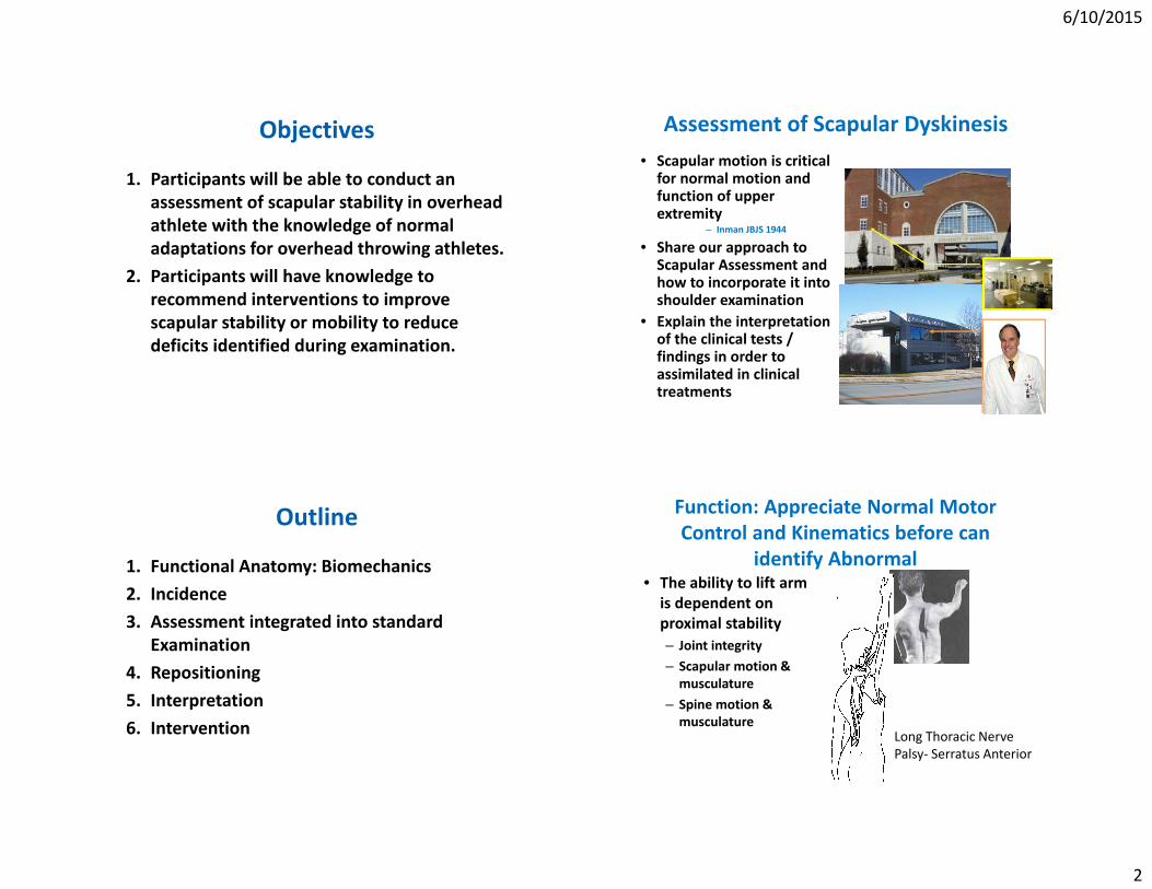
Objectives
1. Participants will be able to conduct an assessment of scapular stability in overhead athlete with the knowledge of normal adaptations for overhead throwing athletes.
2. Participants will have knowledge to recommend interventions to improve scapular stability or mobility to reduce deficits identified during examination.
Assessment of Scapular Dyskinesis• Scapular motion is critical for normal motion and function of upper extremity
– Inman JBJS 1944
• Share our approach to Scapular Assessment and how to incorporate it into shoulder examination
• Explain the interpretation of the clinical tests / findings in order to assimilated in clinical treatments
Outline
1. Functional Anatomy: Biomechanics2. Incidence3. Assessment integrated into standard
Examination4. Repositioning5. Interpretation6. Intervention
Function: Appreciate Normal Motor Control and Kinematics before can
identify Abnormal• The ability to lift arm is dependent on proximal stability– Joint integrity– Scapular motion & musculature
– Spine motion & musculature
Long Thoracic NervePalsy‐ Serratus Anterior
6/10/2015
3
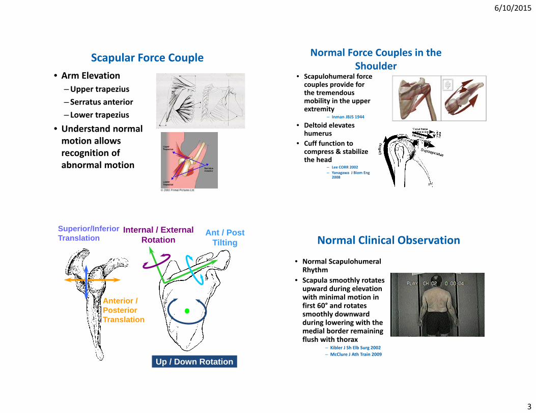
Scapular Force Couple• Arm Elevation
–Upper trapezius– Serratus anterior– Lower trapezius
• Understand normal motion allows recognition of abnormal motion
Normal Force Couples in the Shoulder
• Scapulohumeral force couples provide for the tremendous mobility in the upper extremity
– Inman JBJS 1944
• Deltoid elevates humerus
• Cuff function to compress & stabilize the head
– Lee CORR 2002– Yanagawa J Biom Eng
2008
Ant / PostTilting
Internal / ExternalRotation
Up / Down Rotation
Superior/Inferior Translation
Anterior /PosteriorTranslation
Normal Clinical Observation
• Normal Scapulohumeral Rhythm
• Scapula smoothly rotates upward during elevation with minimal motion in first 60° and rotates smoothly downward during lowering with the medial border remaining flush with thorax
– Kibler J Sh Elb Surg 2002– McClure J Ath Train 2009
6/10/2015
4
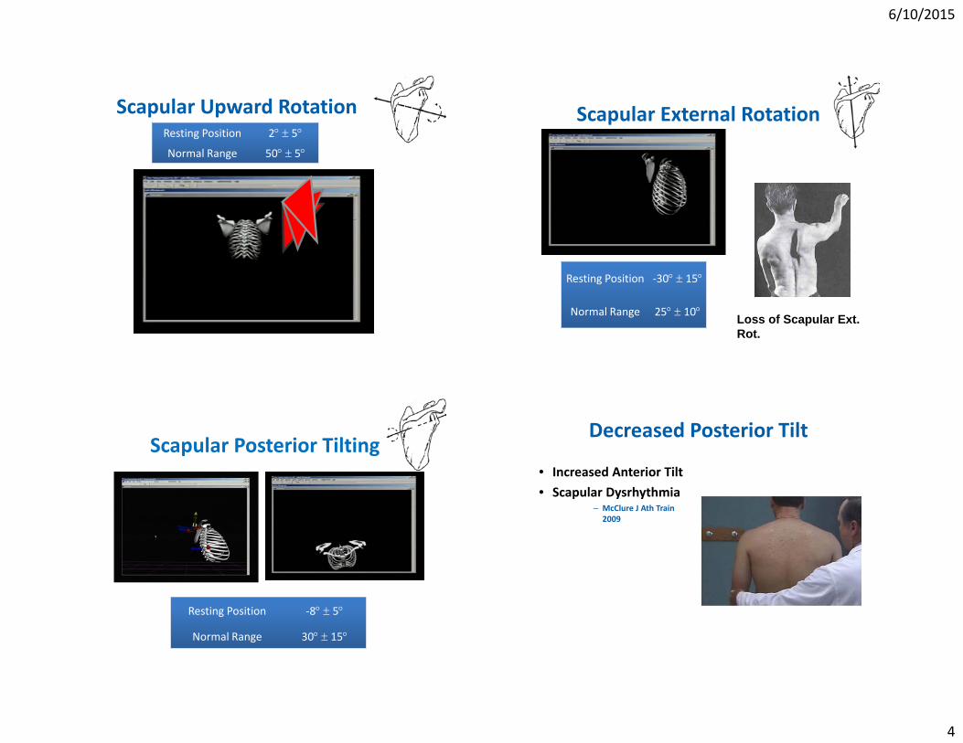
Scapular Upward RotationResting Position 2 5
Normal Range 50 5
Scapular External Rotation
Resting Position ‐30 15
Normal Range 25 10 Loss of Scapular Ext. Rot.
Scapular Posterior Tilting
Resting Position ‐8 5
Normal Range 30 15
Decreased Posterior Tilt
• Increased Anterior Tilt• Scapular Dysrhythmia
– McClure J Ath Train 2009
6/10/2015
5
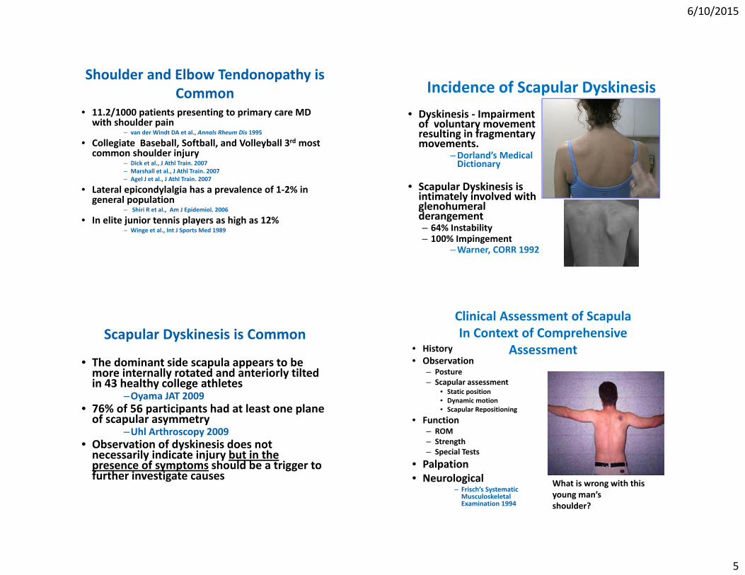
Shoulder and Elbow Tendonopathy is Common
• 11.2/1000 patients presenting to primary care MD with shoulder pain
– van der Windt DA et al., Annals Rheum Dis 1995
• Collegiate Baseball, Softball, and Volleyball 3rd most common shoulder injury
– Dick et al., J Athl Train. 2007– Marshall et al., J Athl Train. 2007– Agel J et al., J Athl Train. 2007
• Lateral epicondylalgia has a prevalence of 1‐2% in general population
– Shiri R et al., Am J Epidemiol. 2006
• In elite junior tennis players as high as 12%– Winge et al., Int J Sports Med 1989
Incidence of Scapular Dyskinesis• Dyskinesis ‐ Impairment of voluntary movement resulting in fragmentary movements.
–Dorland’s Medical Dictionary
• Scapular Dyskinesis is intimately involved with glenohumeral derangement– 64% Instability– 100% Impingement
–Warner, CORR 1992
Scapular Dyskinesis is Common
• The dominant side scapula appears to be more internally rotated and anteriorly tilted in 43 healthy college athletes
–Oyama JAT 2009• 76% of 56 participants had at least one plane of scapular asymmetry
–Uhl Arthroscopy 2009• Observation of dyskinesis does not necessarily indicate injury but in the presence of symptoms should be a trigger to further investigate causes
Clinical Assessment of Scapula In Context of Comprehensive
Assessment• History• Observation
– Posture– Scapular assessment
• Static position• Dynamic motion• Scapular Repositioning
• Function– ROM– Strength– Special Tests
• Palpation• Neurological
– Frisch’s Systematic Musculoskeletal Examination 1994
What is wrong with this young man’sshoulder?
6/10/2015
6
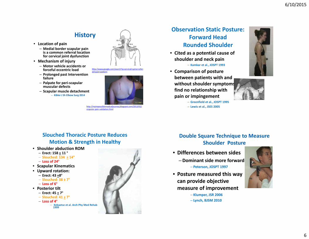
History• Location of pain
– Medial border scapular pain is a common referral location for cervical joint dysfunction
• Mechanism of injury– Motor vehicle accidents or forceful eccentric load
– Prolonged past Intervention failure
– Palpate for peri‐scapular muscular defects
– Scapular muscle detachment– Kibler J Sh Elbow Surg 2014
http://www.google.com/search?q=cervical+spine+referred+pain+pattern
http://myimpossiblemedicaljourney.blogspot.com/2013/02/scapular‐pain‐validation.html
Observation Static Posture:Forward Head
Rounded Shoulder• Cited as a potential cause of shoulder and neck pain
– Kamkar et al., JOSPT 1993
• Comparison of posture between patients with and without shoulder symptoms find no relationship with pain or impingement
– Greenfield et al., JOSPT 1995– Lewis et al., JSES 2005
Slouched Thoracic Posture Reduces Motion & Strength in Healthy
• Shoulder abduction ROM– Erect: 158 + 11 °– Slouched: 134 + 14°– Loss of 24
• Scapular Kinematics • Upward rotation:
– Erect: 43 +8– Slouched: 38 ± 7°– Loss of 6
• Posterior tilt – Erect: 45 + 7– Slouched: 41 + 7– Loss of 4
– Kebaetse et al. Arch Phy Med Rehab 1999
Double Square Technique to Measure Shoulder Posture
• Differences between sides–Dominant side more forward
– Peterson, JOSPT 1997
• Posture measured this way can provide objective measure of improvement
– Klumper, JSR 2006– Lynch, BJSM 2010
6/10/2015
7
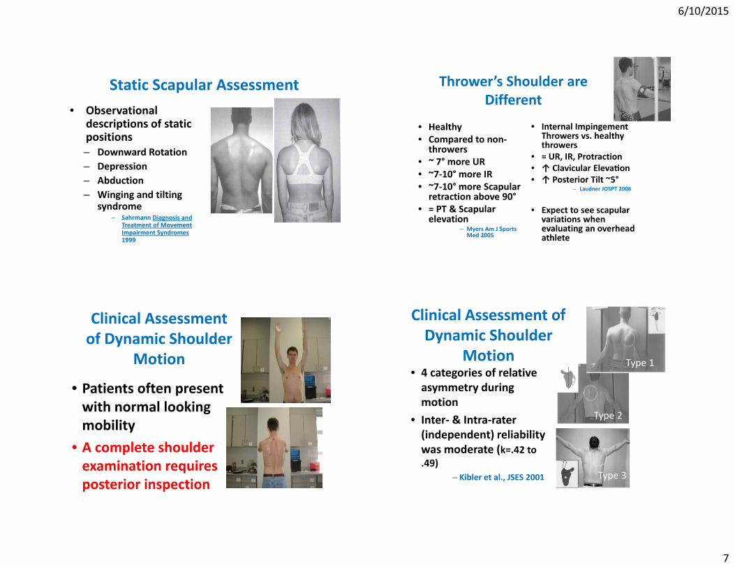
Static Scapular Assessment • Observational
descriptions of static positions – Downward Rotation– Depression– Abduction– Winging and tilting
syndrome– Sahrmann Diagnosis and
Treatment of Movement Impairment Syndromes1999
Thrower’s Shoulder are Different
• Healthy• Compared to non‐throwers
• ~ 7° more UR• ~7‐10° more IR• ~7‐10° more Scapular retraction above 90°
• = PT & Scapular elevation
– Myers Am J Sports Med 2005
• Internal Impingement Throwers vs. healthy throwers
• = UR, IR, Protraction• ↑ Clavicular Eleva on• ↑ Posterior Tilt ~5°
– Laudner JOSPT 2006
• Expect to see scapular variations when evaluating an overhead athlete
Clinical Assessment of Dynamic Shoulder
Motion
• Patients often present with normal looking mobility
• A complete shoulder examination requires posterior inspection
Clinical Assessment of Dynamic Shoulder
Motion• 4 categories of relative asymmetry during motion
• Inter‐ & Intra‐rater (independent) reliability was moderate (k=.42 to .49)
– Kibler et al., JSES 2001
Type 1
Type 2
Type 3
Type 2
Type 3

6/10/2015
8
Scapular Dyskinesis Test of Independent Scapular Motion
• 142 Athletes were examined
• Added load ( 3 or 5 lbs)
• Dyskinesis– Dysrhytmia – not smooth
– Winging – medial border displaced
• Each scapula was categorized – Normal– Subtle– Obvious
• 75‐82% percent agreement between raters– Kappa = .5 ‐ .6
– McClure et al., JAT 2009
Dynamic Scapular Assessment• Scapular Dykiniesis Test – 2 categories
– Obvious / Normal– McClure et al., J Ath. Train 2009
• Kiblers’ Scapular Assessment – 2 categories– Yes / No
– Uhl et al., Arthroscopy 2009
• Presence of Scapular Dyskinesis was not directly related to shoulder pain
• International Classification of Function– Body Functions
• Impairments (ROM, Strength, Motor Control, Scapular Dyskinesis)– Body Structures– Activities and Participation
Scapular Muscle Alterations in Patients with Shoulder Impingement
• ↓ac vity of the Serratus Anterior throughout the range of motion regardless of the load in patients with Shoulder impingement
• 26 manual labors with impingement were compared to 26 manual labors without impingement
• ↑Upper and Lower Trapezius between 60‐120with a load
– Ludewig & Cook PT 2000
Scapular Muscle Activation in Anterior Instability during Scaption
• RC Activation
• Ser. Ant. Activation– McMahon
JSES 1996
0
20
40
60
80
100
0‐30 30‐60 60‐90 90‐120
EMG Activity
(%MVIC)
Humeral Elevation Arcs
Normal Serratus Anterior
Anterior Instability Serratus Anterior
Normal Upper Trapezius
Anterior Instability Upper Trapezius
Normal Lower Trapezius
Anterior Instability Lower Trapezius

6/10/2015
9
HealthySwimmer with MDI
Scapular Dyskinesis Assessment
• Scapulohumeral Rhythm is necessary for normal function
• Scapular moves 3 dimensionally• Alterations can be observed
– Motor Control issues• Scapular muscular activation are altered
– Mobility Issues• Glenohumeral derangement
– Neurological Issues• Spinal Accessory• Long Thoracic
Repositioning the Scapula to Alter Symptoms
• Many impingement tests drive the humerus into the scapula to reproduce symptoms– Painful arc– Neer Impingement
• Would moving the scapula out of the way of the humerus alter symptoms– 1. Scapular Assistance Test– 2. Scapular Retraction Test
• Similar to distraction test for cervical radicular symptoms
6/10/2015
10
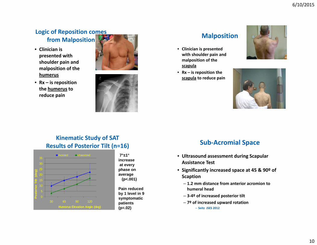
Logic of Reposition comes from Malposition
• Clinician is presented with shoulder pain and malposition of the humerus
• Rx – is reposition the humerus to reduce pain
Malposition
• Clinician is presented with shoulder pain and malposition of the scapula
• Rx – is reposition the scapula to reduce pain
Kinematic Study of SAT Results of Posterior Tilt (n=16)
0
5
10
15
20
25
30
35
30 60 90 120
Pos
teri
or T
ilt (
deg)
Humerus Elevation Angle (deg)
Assisted Unassisted 7°±1°increase at every phase on average
(p<.001)
Pain reduced by 1 level in 9 symptomatic patients (p=.02)
Sub‐Acromial Space
• Ultrasound assessment during Scapular Assistance Test
• Significantly increased space at 45 & 90º of Scaption– 1.2 mm distance from anterior acromion to humeral head
– 3‐4º of increased posterior tilt– 7º of increased upward rotation
– Seitz JSES 2012

6/10/2015
11
Scapular Retraction Test (SRT)• Perform provocative test to assess
arm elevation strength• Repeat test with patient actively
retracting the scapula and clinician manually stabilizing scapular medial border toward the thorax then retest strength
• (+) Test – Improved strength and /or pain reduction
• Interpretation ‐ to differentiate between true and apparent rotator cuff weakness in patients with shoulder pain
– Kibler & McMullen JAAOS 2003
Retraction Test• Increase in strength in retracted
position – 24% in injured– 13% in controls
• No change in pain• Apparent cuff weakness may be
arising from poor scapular control– Kibler et al., AJSM 2006
• Scapular Reposition Test – only manual support
• 98 overhead athletes with impingement signs– 46 had pain reduction of at least 1pt– <30% had strength increase with SRT
– Tate et al. JOSPT 2008
Results‐ Kinematics (Relative Change from Rest)
Not Stab. Stab Change p
UR 15.7±6.4 15±9 0 NS
IR 8.1±9.3 -.7±10 -8.9 .002
PT 5±5 16.6±7 11.6 .001
Prot. 2.6± -2.6± -5.3 .005
Elev. 8.9±11 -5.8±27 -14 .006
Kibler JOSPT 2009
Be careful of the Myopic View• Without proximal control
distal movement are compensating for a poor foundation
• Breakdown along the kinetic chain can– Increase demand on upper
extremity musculature – Change the biomechanical
stresses– Diminish performance
• 47% of 199 overhead throwing athletes with elbow pain had poor single leg squat control
– Kibler et al., AOSSM abstract 2009

6/10/2015
12
Functional Assessment of Core Stability
• Single leg balance– Trendlenberg
• SLS squat– Frontal plane control
• Hip• Knee
• Interpretation– Poor ‐ basic mat program that develops local muscle activation
– Good ‐ incorporate with distal extremity rehabilitation
Muscle Testing• Rotator Cuff
– Thumb up– Lift Off– External Rotation
– Kelly et al., Am J Sports Med 1996
• Scapular Muscle Test– Isolated Upper & Lower
Trapezius– Testing motions for
• Serratus• Rhomboids & Middle Trapezius
– Michener Phys Ther 2005
Shoulder Mobility• Isolate glenohumeral motion• Stabilize scapula improves
reliability– Awan, Arch Phy Med Rehab 2002
• Throwing athlete internal rotation deficit is common
– Wilk AJSM 2011– Kibler Clinics Sports Med 1988
• Loss of IR and TROM is predictive of injury
– Wilk AJSM 2011– Shanley AJSM 2011
• Horizontal Adduction deficit is present in pathological shoulder
– Myers, AJSM 2006
• Improves as symptoms improved during rehabilitation
– Tyler, AJSM 2010
Assessment to Intervention• History• Observation
– Static– Dynamic
• Function– Mobility – ROM – End Feel– Motor – Control –
Strength• Palpation• Neurological
– Spinal lesion– EMG/NCV if necessary
• Level of Irritability– Less is more
• Demand level– What is patient’s
expectation• Time of season• Impairments identified in
exam– Prioritize problems– Could I alter symptoms
during active motion• Scapula• Humeral

6/10/2015
13
Rehabilitation Interventions
• Goal is Patient Outcome• Is it a mobility problem• Is it a stability (motor control) issue
• Where is pain coming from?
Patient Outcome
Motor Control
Mobility
Pain Modulation
Stability
Scapular Rehabilitation Algorithm
Ellenbecker & Cools Br J Sports Med 2010
Scenario• Tennis player presents with acute tendonopathy of rotator cuff 2 due to increased activity level over last 5 days
• Limited ROM in ER and IR• Weak and painful ABD & ER• Scapular dyskinesis
Rehabilitation Plan
Patient Outcome
Motor Control
Mobility
Pain Modulation
Stability
6/10/2015
14
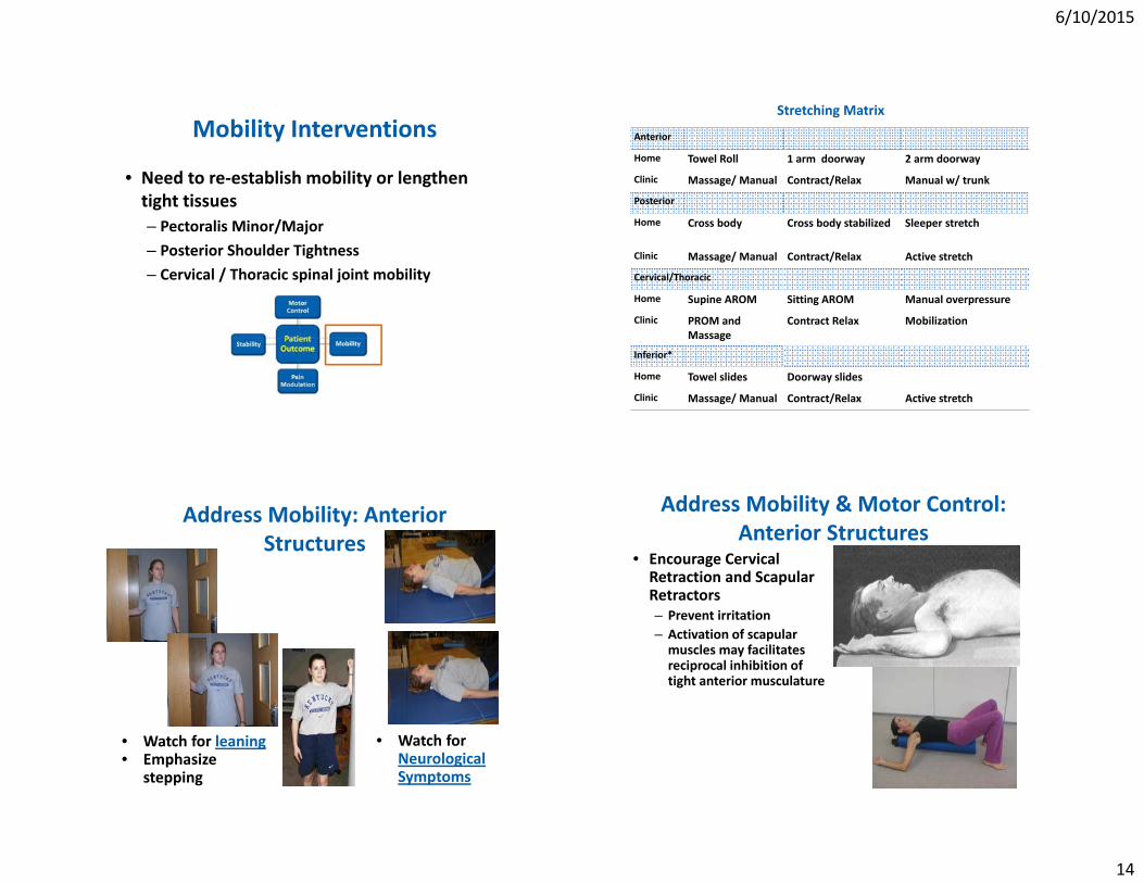
Mobility Interventions
• Need to re‐establish mobility or lengthen tight tissues– Pectoralis Minor/Major– Posterior Shoulder Tightness– Cervical / Thoracic spinal joint mobility
Stretching Matrix
Anterior
Home Towel Roll 1 arm doorway 2 arm doorway
Clinic Massage/ Manual Contract/Relax Manual w/ trunk
Posterior
Home Cross body Cross body stabilized Sleeper stretch
Clinic Massage/ Manual Contract/Relax Active stretch
Cervical/Thoracic
Home Supine AROM Sitting AROM Manual overpressure
Clinic PROM and Massage
Contract Relax Mobilization
Inferior*
Home Towel slides Doorway slides
Clinic Massage/ Manual Contract/Relax Active stretch
Address Mobility: Anterior Structures
• Watch for leaning• Emphasize
stepping
• Watch for Neurological Symptoms
Address Mobility & Motor Control: Anterior Structures
• Encourage Cervical Retraction and Scapular Retractors – Prevent irritation– Activation of scapular muscles may facilitates reciprocal inhibition of tight anterior musculature

6/10/2015
15
Address Mobility: Anterior Structures
• Other muscles attach to the coracoid– Short head of the biceps
– Coracobrachialis• Constantly check for scapular position & neurological Sx’s
Stretching the Posterior Tissues• Adaptive changes can occur
shortening posterior cuff/capsule
– Kibler et al., Clin Sports Med 1988
• Cross body adduction with scapula stable and humerus internally rotating
• Cross body adduction increases IR motion by 20o
– McClure et al., CSM 2005• Hold Stretch 30 seconds (No
Pain)– Bandy et al., Phy Ther 1994
• Considers variations in arm position and stabilizing scapula
Addressing Posterior Shoulder Tightness and Scapular Stability
• Stabilizing scapula with overpressure
• Integrate massage to teresminor/latissimus dorsi– Trigger point release
Spinal Mobilization Options
• Various techniques can be applied
• Consider patient level of irritability– Contraindications
• Great description & videos online through JOSPT
– Tate et al., JOSPT 2010
6/10/2015
16
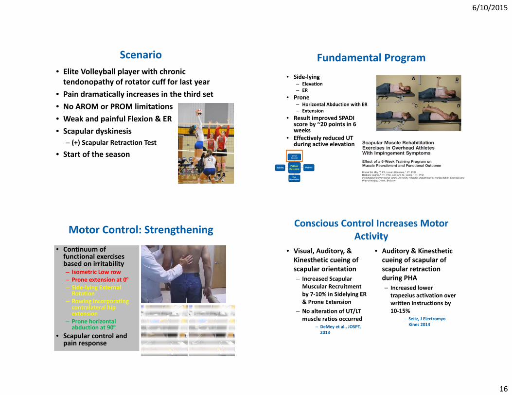
Scenario• Elite Volleyball player with chronic tendonopathy of rotator cuff for last year
• Pain dramatically increases in the third set• No AROM or PROM limitations• Weak and painful Flexion & ER• Scapular dyskinesis
– (+) Scapular Retraction Test
• Start of the season
Fundamental Program • Side‐lying
– Elevation– ER
• Prone – Horizontal Abduction with ER– Extension
• Result improved SPADI score by ~20 points in 6 weeks
• Effectively reduced UT during active elevation
Motor Control: Strengthening• Continuum of functional exercises based on irritability– Isometric Low row – Prone extension at 0– Side‐lying External Rotation
– Rowing incorporating contralateral hip extension
– Prone horizontal abduction at 90
• Scapular control and pain response
Conscious Control Increases Motor Activity
• Visual, Auditory, & Kinesthetic cueing of scapular orientation– Increased Scapular Muscular Recruitment by 7‐10% in Sidelying ER & Prone Extension
– No alteration of UT/LT muscle ratios occurred
– DeMey et al., JOSPT, 2013
• Auditory & Kinesthetic cueing of scapular of scapular retraction during PHA– Increased lower trapezius activation over written instructions by 10‐15%
– Seitz, J ElectromyoKines 2014
6/10/2015
17
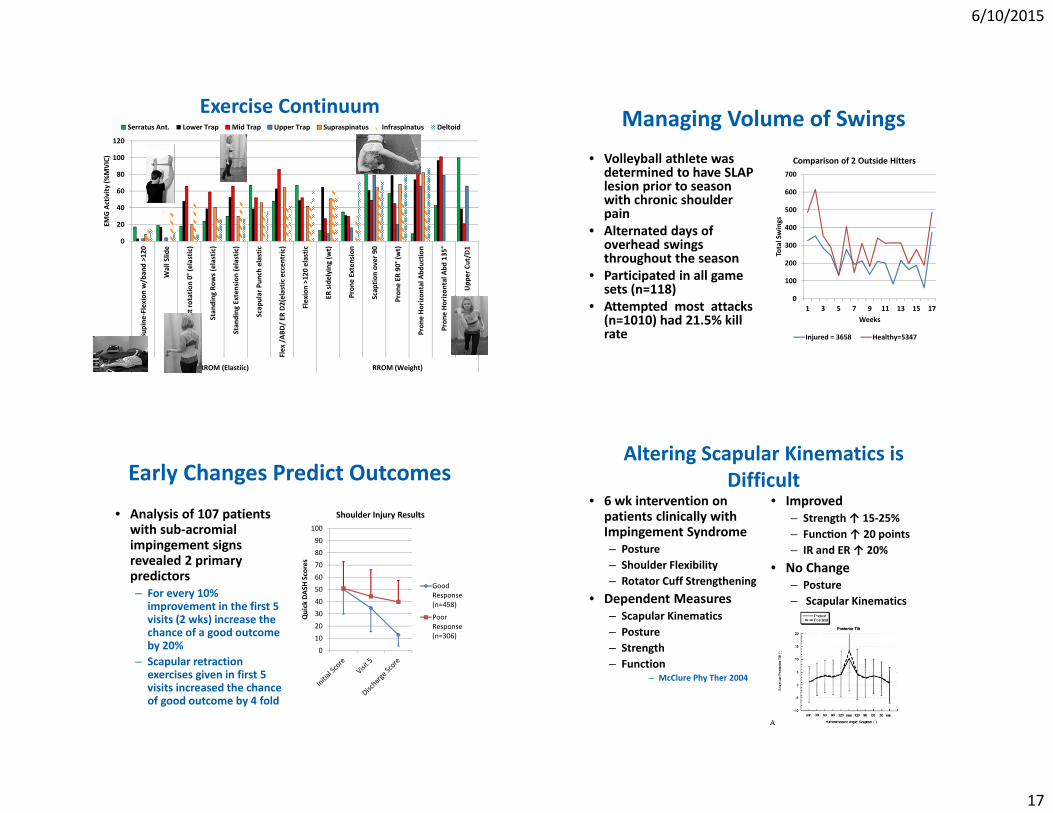
Exercise Continuum
0
20
40
60
80
100
120Supine
‐Flexion
w/ban
d >120
Wall Slid
e
Ext rotation 0° (e
lastic)
Stan
ding
Row
s (elastic)
Stan
ding
Exten
sion
(elastic)
Scap
ular Pun
ch elastic
Flex /AB
D/ ER
D2(elastic
eccen
tric)
Flexion >120
elastic
ER sidelying
(wt)
Pron
e Extension
Scap
tion over 90
Pron
e ER
90° (w
t)
Pron
e Horizon
tal A
bductio
n
Pron
e Horizon
tal A
bd 135°
Upp
er Cut/D
1
RROM (Elastiic) RROM (Weight)
EMG Activity
(%MVIC)
Serratus Ant. Lower Trap Mid Trap Upper Trap Supraspinatus Infraspinatus Deltoid Managing Volume of Swings• Volleyball athlete was
determined to have SLAP lesion prior to season with chronic shoulder pain
• Alternated days of overhead swings throughout the season
• Participated in all game sets (n=118)
• Attempted most attacks (n=1010) had 21.5% kill rate
0
100
200
300
400
500
600
700
1 3 5 7 9 11 13 15 17
Total Swings
Weeks
Comparison of 2 Outside Hitters
Injured = 3658 Healthy=5347
Early Changes Predict Outcomes
• Analysis of 107 patients with sub‐acromial impingement signs revealed 2 primary predictors– For every 10%
improvement in the first 5 visits (2 wks) increase the chance of a good outcome by 20%
– Scapular retraction exercises given in first 5 visits increased the chance of good outcome by 4 fold
0
10
20
30
40
50
60
70
80
90
100
Quick DAS
H Scores
Shoulder Injury Results
GoodResponse(n=458)
PoorResponse(n=306)
Altering Scapular Kinematics is Difficult
• 6 wk intervention on patients clinically with Impingement Syndrome– Posture– Shoulder Flexibility– Rotator Cuff Strengthening
• Dependent Measures– Scapular Kinematics– Posture– Strength– Function
– McClure Phy Ther 2004
• Improved– Strength ↑ 15‐25%– Func on ↑ 20 points– IR and ER ↑ 20%
• No Change– Posture– Scapular Kinematics

6/10/2015
18
Summary• Functional Anatomy: Biomechanics ‐ Know and understand
normal to recognize abnormal– Variations of normal among athletes
• Incidence ‐ Common • Assessment integrated into standard Examination – Easily
– Keep it simple • Repositioning– Reduction of pain indicates scapular
position/motion is a factor in shoulder pain• Interpretation – Impairment that may need to be
incorporated into treatment intervention• Intervention – Keep it patient centered
– Mobility issue– Motor Control issue– Modulate pain with exercise selected
Email: [email protected] Works
http://works.bepress.com/tim_uhl/
“The sun shines bright on my old Kentucky home…”
College of Health Sciences