scaffold Cells stimuli - unipi.it · The PAM2 system Robotic 3 axis micropositioner. PAM PAM2 Diode...
48
Tissue engineered product stimuli Cells scaffold Not required in all ATMP
Transcript of scaffold Cells stimuli - unipi.it · The PAM2 system Robotic 3 axis micropositioner. PAM PAM2 Diode...

Tissue engineered product
stimuli
Cells scaffold
Not required in all ATMP

What is a scaffold? A 3D structure which supports 3D tissue growth

What are the features of an ideal scaffold?
• 3D. Biocompatible, cell adhesive, bioerodable and bioactive
• Mechanical properties similar to those of natural tissue
• Optimal meso, micro- pores
• Well-defined, or quantifiable topology at meso- micro- and
nanoscales • 3D-matrix adhesions differ in content, structure, location, and function from
classically described in vitro adhesion, e.g., focal and fibrillar adhesions
• - cell adhesion in 3D-matrix more efficient (6-fold increase)
• - cell morphology is that of more in vivo-like (spindle shape)
• - cell migration speed increased by ~ 50%

Extracellular matrix features
• High degree of porosity
• Appropriate pore size
•High surface to volume ratio
• High degree of pore interconnectivity
•Biochemical factors & ECM features able to guide
cell function

Porosity and architecture Pore size, pore connectivity, porosity, pore distribution are all critical
•To fit cells •Fit at least a functional unit •Allow nutrient perfusion

Liver ECM

All polymers (materials) have to be porous in order to support 3 D tissue ingrowth.
Pores have to be interconnected (why, what is the difference between porosity and permeability?). Both porosity and permeability change when a material is degraded

Stimuli- the tripartite axis
Engineering Quasi-Vivo In Vitro Organ Models. Sbrana & Ahluwalia. Methods Adv Exp Med Biol. 2012;745:138-53.

Biochemical stimuli in scaffolds
• Synthetic biomaterials with ligands /proteins
• Natural biomaterials
• Decellularized Tissue

Mechano-structural stimulii

Forces are important
• Gravity
• Shear
• Pressure
• Tension
• Compression
Cyclic forces
Static forces (materials in constant tension)

Distraction osteogenesis: bone is pulled apart to encourage growth


Methods for generating (static) MS stimuli in scaffolds

Designer or Random?


Rapid prototyping: designer scaffolds

3D Printing/Digital Fabrication & RP

The PAM2 system
• 4 Position controlled brushless motors (resolution of 10 µm ± 1 µm)
• Working space 100×100×80 mm
• Working velocity 1-15 mm ∙s⁻¹ • Design of z-stage to locate
several modules
The PAM2 system
Robotic 3 axis micropositioner.
PAM PAM2 Diode laser Temperature control PAM2 software
Materials? Speed? Price? Fidelity? Tirella, De Maria, Vozzi, Ahluwalia Rapid Prot. J (2012);

Pressure Assisted Microsyringe (PAM)
Software
Syringe design
PAM system
Software
Vozzi et al,. Tissue Engineering, 8, 34, 2002. Vozzi et al , Biomaterials, 24, 2533, 2003, Vozzi et al, JBMRA, 71A, 326, 2004. Mariani et al., Tissue Eng. 12, 547, 2006. Bianchi et. Al. JBMR 81, 462, 2007.
Regulated air flow
Materials? Speed? Price? Fidelity?

Piston Assisted Microsyringe (PAM2)
Plunger driven
Materials? Speed? Price? Fidelity?

Smart-tunable modular scaffolds...
Resolution, fidelity, viscosity

Technique Material used RTM ratio (cm3/min)
Resolution (μm)
Cells used Limits
Membrane Lamination
Bioerodable polymers (PLA, PLGA, etc), bio-ceramics
Low (<1) 1000 Osteoblasts Structures not really porous, low resolution
Laser Sintering Calcium Phosphates, polymers (PLA, PLGA, etc)
Medium to high
< 400 Osteoblasts Presence of polymeric grains and of excess solvent
Photo-polymerisation
Photo-polymeric resins
0.5 (medium)
250 Osteoblasts Use of photo sensitive polymers and initiators which may be toxic
Fused Deposition Modelling
Bioerodable polymers (PLA, PLGA,etc)
7 (very high)
200 Various types Limited to non thermo labile materials. Layered structure very evident
3D Printing Bioerodable polymers, (PLA, PLGA, etc) and hydroxyapatite
Medium (about 1)
300 Various types, mainly skeletal
Presence of polymeric grains and of excess solvent
iRP Bioerodable polymers (PLA, PLGA, etc), collagen
0.1 (low) 300 Various types Complex to realise, build materials limited, low fidelity.
PAM2 Bioerodable polymers (PLA, PLGA, etc) and gels (alginate, gelatin)
1 (medium)
5-100 Neurons, endothelial cells, fibroblasts, hepatocytes, muscle
Highly water soluble materials cannot be used. Extrusion head very small.
InkJet Water, solvents, nanoparticle suspensions
Very low (<0.01)
10 Various Only low viscosity liquids.

Mattei. et al, Biomat. Acta, 2013
Uygun et al, Nature Med, 2010.

Price? Materials? Speed? Repeatibility ?
Organ Processing
Whole Organ Perfusion
• Detergents
• Intact microvasculature
• Slow and costly
Tissue Decellularization
• Detergents
• Rapid, less wasteful

Biomaterial Processing
• Freeze drying
• Phase separation
• Gas foaming
• Salt leaching
Price? Materials? Speed? Repeatibility ?
Freeze drying Phase separation Gas foaming Salt leaching

Electrospinning
Price? Materials? Speed? Repeatibility ?

Technique Material used RTM ratio (cm2/min)
Cells used Limits
Freeze drying Proteins, carbohydrates, polyesters, hydroxyapatite
High Variety Wide distribution of pore size
Phase Inversion Polyesters, PVA, polyurethanes, biogels (gelatin)
High Variety Low interconnectivity, difficult to control pore size
Salt leaching Polyesters, polyurethanes, hydroxyapatite
High Variety Salt residues, limited connectivity
Gas foaming Polyesters, PVA, polyurethanes, biogels (gelatin)
High Variety Quite expensive
Whole organ decell
Organs High Heart, liver, lung, etc
Whose organ? Detergents are aggressive Tissue decell Pieces of tissue High Many
Electrospinning Bioerodable polymers (PLA, PLGA, etc), proteins and gels (collagen, alginate, gelatin)
Very low (<1) Variety Gives rise to pseudo 3D “squashed” scaffolds
Stop here

Degradable Polymeric Biomaterials are materials which can be eliminated through hydrolytic degradation or enzyme attack. The synthetic ones are almost all polyesters (polycaprolactone, polyglycolide, polylactide)
• They do not give rise to a permanent and chronic “foreign body” response
• Some materials are capable of inducing tissue regeneration.
• They are used as temporary supports and scaffolds in tissue engineering. They cannot be used as permanent suppports but only for remodelling and repair.
Polyesters
R-C-O –R’
O

Requisites for bioerodabile materials 1) Provide an adequate mechanical support for a short period of time
without any problems after degradation. 2) Degradation rate match rate of new tissue generation 3) Provide an approriate biochemical environment for cell/cell and
cell/ECM interaction and supply nutrients and growth factors as necessary.
4) Guide tissue response as appropriate (enhance or suppress). 5) Not induce an inflammatory response. Low or negligible toxicity of
degradation products both locally and systemically. 6) Easy to produce and fabricate in large quantities 7) Compatible with drug delivery methods 8) Porous
Biodegradable biological polymers
Collagen: from animal sources. It is non immunogenic because it is a highly conserved protein.
Can be crosslinked to render it more stable, more resistant, increase degradation time, less hydrophilic, les soluble and increase tensile strength.
Very common in tissue engineered products, eg Alpigraf (collagen gel, fibroblasts+keratinocytes)
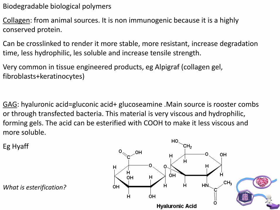
GAG: hyaluronic acid=gluconic acid+ glucoseamine .Main source is rooster combs or through transfected bacteria. This material is very viscous and hydrophilic, forming gels. The acid can be esterified with COOH to make it less viscous and more soluble.
Eg Hyaff
What is esterification?

Chitosan:polysaccharide
Chitosan has a high degree of biocompatibility but is not very resistant to loads or deformation. They are not reproducible (different sources are different) and may also carry infective agents.
From crabs, for example. Approved for cosmetic use in Japan

Synthetic biodegradables
The most widespread are those approved by the FDA. Polycaprolactone, polyglycolic acid and polylactic acid. All 3 are polyesters.
PGA : the simplest, crystalline (35-70%), insoluble (only in HFP), high mp (200C), used in sutures, Hydrophilic, degrades slowly
PLA: has an additional CH3 , (35% crytstalline) hydrophobic, degrades more slowly than PGA, more soluble on organic solvents. Chiral, so found in 3 forms: l, d and ld

PLLA: semi crystalline, hard, mp=180C, less crytalline than PGA (35%)
PDLLA: random chiarality. Amorphous. Degrades faster than PLLA (2-12 months)
O
O
CH3
CH3
CH3
HO
O
O n O
O
CH3
O
Opoly
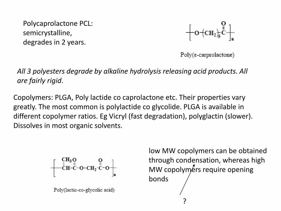
Polycaprolactone PCL: semicrystalline, degrades in 2 years.
All 3 polyesters degrade by alkaline hydrolysis releasing acid products. All are fairly rigid.
Copolymers: PLGA, Poly lactide co caprolactone etc. Their properties vary greatly. The most common is polylactide co glycolide. PLGA is available in different copolymer ratios. Eg Vicryl (fast degradation), polyglactin (slower). Dissolves in most organic solvents.
low MW copolymers can be obtained through condensation, whereas high MW copolymers require opening bonds
?

The degradation rate of PLGA depends on MW, hydrophilicity and the degree of crystallinity, pH and temperature
Copolymers are more amorphous

Problems with PGA e PLA e PCL: They are rigid. Do not possess functional groups to modify and bind proteins. Can generate too much local acidity. (degrade by hydrolysis). Question: write a reaction for hydrolysis of PGA (assume 3 monomers) On the other hand,compared with biological polymers they are more reproducible and less likely to carry infective agents (BSE). Moreover, biological polymers are not structurally strong. Please note that there is a whole world of polymers out there- but only a handful actually approved for in-vivo use.


Degradation rate of a polymer
Depends on
1)Polymer intrinsic properties:MW, crystallinity etc
2) environment: shear, acidity etc
3) Surface area
Problem: consider a unit cell of biodegradeable mateial with a pore in the center.
How does
1)Porosity
2) Maximum load
3) Mass of material
change with time

Where are we today?
Humans
• Skin
• Cartilage
• Trachea
• Bladder
• Pancreas
• In-vitro meat
Animals
• nude mouse

Live Scaffold Fabrication
cells
Direct Fabrication Composite materials
Cells Biomaterial Processing
Live Engineered
Scaffold
Live scaffold fabrication

Cell Printing
• Cell Printing (inkjet)
• Organ Printing (nozzle based)
• Living Inks, bioinks, bioprinter, bioplotter
Olivetti NanoBioJet

Cell dispensers and Bioprinters

• 2D...
• Small volumes in high spatial resolution patterns
• BioInk (i.e. protein based solutions)
• Particle based inks
• LivingInk (i.e cell suspensions)
80 pL
InkJet for Living Inks
Tirella et. al, Substrate stiffness influences high resolution printing of living cells with an ink-jet system. J Biosci Bioeng. 2011
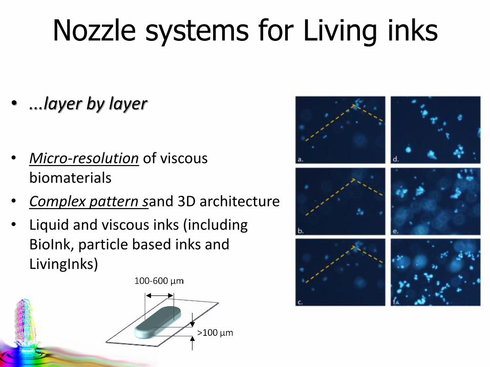
Nozzle systems for Living inks
• ...layer by layer
• Micro-resolution of viscous biomaterials
• Complex pattern sand 3D architecture
• Liquid and viscous inks (including BioInk, particle based inks and LivingInks)

V. Mironov et al. Biomaterials 30 (2009) 2164–2174
Organ Printing using cell suspensions as a material

Nano-in-micro (NIM) Live Scaffold Fabrication
Assembling:
• Living micro-spheres with
controlled mechanical and
properties and biomimetic
composition;
• Having:
• Cells
• Tissue matrix
• Release of known
moieties (e.g. ROS,
exogenous molecules)
• Scavenger properties
• Sensitive detectors[3]
[1] Tirella et al. JBMA 2013 ; [2] Mattei el all Biomat. Acta,; [3] Tirella , La Marca, Ahluwalia , Soft Matter submitted; [4] La Marca et al., ESB 2013.
Modular design to obtain a
fine spatially controlled and
tunable micro-environment
Living construct
Fluorescent signal [3]
Recreate an in vitro microsystem able to interact and
monitor living constructs in a non-invasive manner

Spherical Hydrogel Generator
Size controlled hydrogel micro-spheres as function of system working parameters and solution properties:
Solution viscosity (e.g. alginate w/v ratio, NPs concentration, cell concentration)
Nozzle diameter
Volumetric flow rate
External air flow
Sensitive/Functional domains can be easily fabricated
controlling sphere dimension, shape and composition
Shape is fixed via rapid physical gelation, e.g. for alginate microspheres form a gel in a beaker containing a 0.1 M CaCl2 solution in water.
Alginate
Alginate + NPs
Alginate + matrix
Alginate + matrix+cells
Alginate +matrix+ cellls + NPs