SAS-3 Serum Protein SB - Pera Medperamed.com/peramed/docs/300205_5060169852341_EN.pdf ·...
28
SAS-3 Serum Protein SB Instructions For Use REF 300205 SAS-3 Protéines Sériques SB Fiche technique SAS-3 Serumprotein SB Anleitung Sieroproteine SB SAS-3 Istruzioni per l’uso Proteínas Séricas SB SAS-3 Instrucciones de uso Contents English 1 Français 6 Deutsch 11 Italiano 16 Español 21
Transcript of SAS-3 Serum Protein SB - Pera Medperamed.com/peramed/docs/300205_5060169852341_EN.pdf ·...

SAS-3 Serum Protein SB
Instructions For UseREF 300205
SAS-3 Protéines Sériques SBFiche technique
SAS-3 Serumprotein SBAnleitung
Sieroproteine SB SAS-3 Istruzioni per l’uso
Proteínas Séricas SB SAS-3 Instrucciones de uso
Contents
English 1Français 6Deutsch 11Italiano 16Español 21


INTENDED PURPOSEThe SAS-3 Serum Protein SB kit is intended for the separation and quantitation of Serum Proteins byagarose gel electrophoresis.
Serum contains over 100 individual proteins, each with a specific set of functions which are subject tospecific variation in concentration under different pathological conditions
1.
Since the introduction of moving boundary electrophoresis by Tiselius2, and the subsequent use of zone
electrophoresis, Serum Proteins have been fractionated on the basis of their charge at a particular pH.The SAS-3 Serum Protein SB kit separates serum proteins into 6 main classes (albumin, alpha 1-globulin, alpha 2-globulin, beta1-globulin, beta 2-globulin, and gamma globulin) according to charge inan agarose gel. The proteins are then stained to allow visualisation and quantitative interpretation.Each of the classical electrophoretic zones, with the exception of albumin, normally contains 2 or morecomponents. The relative proportions of these fractions have proven to be useful aids in the diagnosisand prognosis of certain disease states
3-5.
WARNINGS AND PRECAUTIONSAll reagents are for in-vitro diagnostic use only. Do not ingest or pipette by mouth any kit component.Wear gloves when handling all kit components. Refer to the product safety data sheet for risk andsafety phrases and disposal information.
COMPOSITION1. SAS-3 Serum Protein SB Gel (x10)
Contains agarose in a Tris / Barbital buffer with thiomersal and sodium azide as preservative. The gel is ready for use as packaged.
2. Acid Blue Stain Concentrate (1x 75ml)Contains concentrated Acid Blue stain. Dilute the contents of the bottle to 700ml with purifiedwater. Stir overnight and filter before use. Store in a tightly stoppered bottle.
3. Destain Solution Concentrate (2x 100ml)Contains concentrated Destain Solution. Dilute the contents of the bottle to 5 litres with purifiedwater. Store in a tightly stoppered bottle.
4. Other Kit ComponentsEach kit contains Instructions For Use and sufficient Blotter C to complete 10 gels.
STORAGE AND SHELF-LIFE1. SAS-3 Serum Protein SB Gel
Gels should be stored at 15...30°C and are stable until the expiry date indicated on the package.DO NOT REFRIGERATE OR FREEZE. Deterioration of the gel may be indicated by 1) crystallineappearance indicating the gel has been frozen, 2) cracking and peeling indicating drying of the gelor 3) visible contamination of the agarose from bacterial or fungal sources.
2. Acid Blue StainThe stain concentrate should be stored at 15...30°C and is stable until the expiry date indicated onthe label. Diluted stain solution is stable for 6 months at 15...30°C. It is recommended to discardused stain immediately to prevent depletion of staining capability. Poor staining performance mayindicate deterioration of the stain solution.
1
SAS-3 SERUM PROTEIN SB
English

3. Destain SolutionThe destain concentrate should be stored at 15...30°C and is stable until the expiry date indicatedon the label. Diluted destain solution is stable for 6 months at 15...30°C. Cloudiness may indicatedeterioration of the destain solution.
ITEMS REQUIRED BUT NOT PROVIDEDCat. No. 310200 Sample Applicator Blades (1 x 10)Cat. No. 310300 Sample Applicator Blades (5 x 10)Cat. No. 210100 Disposable sample cups (100)Cat. No. 3100 REP Prep
SAMPLE COLLECTION AND PREPARATIONFreshly collected serum is the specimen of choice. Whilst samples can be stored at 15...30°C for upto 4 days, 2...6°C for up to 2 weeks or 6 months at -20°C for standard 5 band patterns
6, the beta 2-
globulin band (Complement C3) will degrade rapidly on storage. Urine and CSF can also be usedfollowing a suitable concentration step (50 - 100X). The use of plasma will result in a fibrinogen bandbetween the beta and gamma fractions.
Interfering Factors: 1) Haemolysis may cause false elevation in the alpha 2 and beta fractions. 2) Inaccurate results may be obtained on specimens left uncovered, due to evaporation.
STEP BY STEP PROCEDURE1. Program the following parameters into the instrument:
Step Time (mm:ss) Temperature (°C) Voltage OtherElectrophoresisLoad Sample 00:30 21 Speed 1Apply Sample 00:30 21 Speed 1 *Electrophoresis (60 sample) 13:00 18 480Electrophoresis (80 sample) 11:30 18 480Electrophoresis (100 sample) 09:45 18 480Dry 08:00 54* - Use Location 1 for SAS-3.
StainerStain 04:00 Recirculate ONDestain 02:00 Recirculate ONDestain 02:00 Recirculate ONDry 16:00 70
2

2. Place the correct number of applicator blade assemblies in position on the instrument:
3. Pipette 35µl of the sample into the appropriate disposable sample cups. Protect the samples fromevaporation.
4. Remove the gel from the packaging and discard the overlay.5. Dispense 2ml of REP Prep onto the left side of the chamber and carefully place the gel into the
chamber, aligning the holes in the gelbond with the pins in the chamber and avoiding bubbles underthe gel. Attach the electrodes onto the outside of the electrode posts so that they are in contactwith the gel blocks.
6. Blot the surface of the gel with a blotter C, discard the blotter.7. Select the Serum Protein test program and, following the prompts, apply the samples and perform
the electrophoresis.8. Following electrophoresis, remove the electrodes and remove both gel blocks using the Gel Block
Remover.9. Attach the gel to the staining chamber holder.10. Select the Serum Protein test program on the staining unit and, following the prompts, Stain,
Destain and Dry the gel.11. At the end of the staining cycle, remove the gel from the staining chamber. The gel is now ready
for examination.
QUALITY CONTROLKemtrol Serum Controls (Cat. No's. 7024 and 7025) can be used to verify all phases of the procedureand should be used on each plate run. Refer to the package insert provided for appropriate assayvalues.
INTERPRETATION OF RESULTS.It is recommended that any evaluation of the gels is performed against normal values produced for thismethod in each individual laboratory.
For a complete review of serum protein evaluation, see Ritzmann, S.E, 19825. Studies show that the
values are the same for both males and non-pregnant females. Some differences are seen in pregnantfemales at term and women on oral contraceptives.Age has some effect on normal levels. Cord blood has a decreased total protein, albumin, alpha 2 andbeta fractions; slightly increased alpha 1 and normal or increased gamma fraction (largely of maternalorigin). The gamma globulins drop rapidly until about 3 months of age, while other fractions havereached adult levels by this time. Adult levels of the gamma globulins are not reached until 10-16 yearsof age. The albumin decreases and beta globulin increases over the age of 40.1. Qualitative Evaluation:
The gels may be visually inspected for the presence or absence of particular bands of interest.2. Quantitative Evaluation:
Scan the gels, gel side down, at 595nm.
3
SAS-3 SERUM PROTEIN SB
English
Number of Samples Applicator Position
SAS-3
100 2, A, 9, 13, 16
80 3, 7, 12, 16
60 2, 8, 14

In either case, an elevation or decrease in particular serum components or the detection of unusualserum components require further investigation. The completed SAS-3 Serum Protein SB gel is stablefor an indefinite period of time.
LIMITATIONSSince all electrophoresis procedures are non-linear, it is important to follow these Instructions For Useclosely to ensure optimal resolution and reproducible results. Failure to follow these Instructions ForUse may affect the results obtained.
REFERENCE VALUESUsing 15 normal specimens from male and female donors with an age range of 20-59 years, thefollowing normal ranges were obtained (these are presented as a guideline only):
PERFORMANCE CHARACTERISTICSReproducibility
LinearityThe linearity of the method is a function of densitometer specification as well as gel performance. It is recommended that each customer determine the linearity of the method based upon thedensitometer in use in the laboratory.
4
Protein Fraction Range (%)
Albumin 52.3 - 63.5
Alpha1 2.0 - 4.9
Alpha2 5.6 - 11.8
Beta 1 9.2 - 12.5
Beta 2 2.9 - 4.5
Gamma 11.0 - 21.7
Within Gel (n=80) Between Gel (n=160)
Mean (%) CV (%) Mean (%) CV (%)
Albumin 60.8 3.9 61.0 3.5
Alpha 1 3.6 10.7 3.6 12.1
Alpha 2 8.8 5.6 8.8 5.2
Beta 1 8.8 4.6 8.7 4.2
Beta 2 6.2 6.9 6.2 8.1
Gamma 11.8 8.9 11.7 8.7

BIBLIOGRAPHY1. Alper, C.A. 'Plasma Protein Measurements as a Diagnostic Aid', N. Eng. J. Med., 1974; 291 : 287-290.2. Tiselius, A. 'A New Apparatus for Electrophoretic Analysis of Colloidal Mixtures', Trans. Faraday
Soc., 1937; 33 : 524.3. Ritzmann, S.E. and Daniels, J.C. 'Diagnostic Proteinology: Separation and Characterization of
Proteins, Qualitative and Quantitative Assays' in Laboratory Medicine, Harper and Row, Inc.,Hagerstown, 1979.
4. Tietz, N.W. (Ed.), Textbook of Clinical Chemistry, W.B. Saunders Co., Philadelphia, pages 579-582,1986.
5. Ritzmann, S.E. (Ed.), Protein Abnormalities Volume 1 : Physiology of Immunoglobulins -Diagnostic and Clinical Aspects’, Allen R. Liss, Inc., New York, 1982.
6. Tietz, N.W. (Ed.), Textbook of Clinical Chemistry, (3rd Edition), W.B. Saunders Co., Philadelphia,page 524, 1995.
5
SAS-3 SERUM PROTEIN SB
English

UTILISATIONLe kit SAS-3 Protéines Sériques SB est utilisé pour la séparation et la quantification des protéinessériques par électrophorèse en gel d'agarose.
Le sérum contient plus de 100 protéines qui ont chacune une fonction spécifique et qui peuvent subirdes variations quantitatives en fonction de diverses conditions pathologiques
1. Depuis l'introduction
par Tiselius2
de la mobilité électrophorètique, les protéines sériques sont fractionnées en fonction deleur charge à un pH déterminé. Le kit SAS-3 Protéines Sériques SB sépare les protéines sériques en 6fractions principales (albumine, alpha1, alpha 2, béta1, béta2 et gammaglobulines) selon leur charge engel d'agarose. Les protéines sont ensuite colorées pour permettre leur visualisation et l'interprétationsemi-quantitative. Chaque fraction, à l'exception de l'albumine, contient au moins 2 composants. Laproportion relative de ces différentes fractions peut aider à poser un diagnostic et à s'orienter verscertains stades de maladies
3-5.
PRECAUTIONSTous les réactifs sont à usage diagnostic in-vitro uniquement. Ne pas ingérer ou pipeter à la boucheaucun composant. Porter des gants pour la manipulation de tous les composants. Se reporter auxfiches de sécurité des composants du kit pour la manipulation et l'élimination.
COMPOSITION1. Plaque SAS-3 Protéines Sériques SB (x10)
Contient de l'agarose dans un tampon Tris / barbital additionné de thimérosal et d'azide de sodiumcomme conservateur. Le gel est prêt à l'emploi.
2. Colorant Acide Bleu (1x 75ml)Contient du colorant acide bleu concentré. Dissoudre le contenu du bouteille dans 700ml d'eaudistillée, laisser sous agitation toute une nuit. Filtrer avant utilisation. Conserver en bouteillehermétiquement fermée.
3. Solution décolorante (2x 100ml)Contient de solution décolorante concentrée. Diluer le contenu du bouteille dans 5 litres d'eaudistillée et conserver en récipient fermé.
4. Autres composants du kitChaque kit contient également 1 fiche technique et des buvard C pour 10 gels.
STOCKAGE ET CONSERVATION1. Plaque SAS-3 Protéines Sériques SB
Les gels doivent être conservés entre 15...30°C, ils sont stables jusqu'à la date d'expiration indiquéesur l'emballage. NE PAS REFRIGERER OU CONGELER. Les conditions suivantes indiquent unedétérioration du gel: 1) cristaux visibles indiquant que le gel a été congelé, 2) de craquelurestémoins d'une déshydratation du gel, 3) une contamination visible bactérienne ou fongique.
2. Colorant Acide BleuLe colorant concentré doit être conservé entre 15...30°C, il est stable jusqu'à la date de péremptionindiqué sur le flacon. Le colorant dilué est stable 6 mois entre 15...30°C. Il est recommandé derejeter le colorant utilisé afin de prévenir une diminution de la capacité de coloration. Une performance de coloration diminuée, indique une détérioration de la solution colorante.
6

3. DécolorantLe décolorant concentré doit être conservé entre 15...30°C, il est stable jusqu'à la date depéremption indiquée sur le flacon. Le décolorant dilué est stable 6 mois entre 15...30°C. Un aspect floconneux indique une détérioration de la solution décolorante.
MATERIELS NECESSAIRES NON FOURNISRéf. 310200 Applicateurs échantillons 1 x 10Réf. 310300 Applicateurs échantillons 5 x 10Réf. 210100 Cupules échantillons jetables 100Réf. 3100 REP Prep
PRELEVEMENTS DES ECHANTILLONSL'utilisation de sérums fraîchement prélevés est fortement recommandée. Les échantillons peuventêtre conservés entre 15...30°C pendant 4 jours, 2 semaines entre 2...6°C ou 6 mois à -20°C pourobtenir une séparation en 5 fractions
6, la bêta 2-globuline (Complément C3) est rapidement dégradée
durant la conservation. Urine et LCR peuvent être utilisés après une concentration préalable (50 à 100fois). L'utilisation de plasma laisse apparaître une bande entre les Béta et les gammaglobulines: le Fibrinogène.
Interférence: Un sérum hémolysé présente une bande entre les fractions alpha 2 et béta.Des résultats erronés peuvent être obtenus sur des échantillons ayant subi une concentration parévaporation.
METHODOLOGIE1. Entrer les paramètres suivants dans le instrument:
Etape Temps (mn:ss) Temp (°C) Voltage AutreModule d’électrophorèsePrevelement Ech 00:30 21 SPD 1Application Ech 00:30 21 SPD 1 *Electrophorese (60 Ech) 13:00 18 480Electrophorese (80 Ech) 11:30 18 480Electrophorese (100 Ech) 09:45 18 480Sechage 08:00 54* - Utiliser LOC 1 pour le SAS-3.
Module de colorationColore 04:00 REC OUIDecolore 02:00 REC OUIDecolore 02:00 REC OUISeche 16:00 70
7
SAS-3 PROTÉINES SÉRIQUES SB
Français

2. Placer le nombre correct d’applicateurs en position dans l’instrument:
3. Déposer 35µl d’échantillon dans les cuvettes jetables. Couvrir les échantillons afin d’éviterl’évaporation.
4. Sortir le gel de son emballage et retirer le film plastique.5. Déposer 2ml de REP Prep sur la partie gauche de la chambre de migration et délicatement placer
le gel dans la chambre. Aligner les trous de la plaque de gel avec les picots de fixation de lachambre en prenant soin de ne pas emprisonner de bulles sous le gel. Fixer les électrodes surl’extérieur des plots que sorte qu’elles soient en contact avec les ponts d’agarose.
6. Sécher la surface entière du gel à l’aide d’un buvard C, jeter ensuite le buvard.7. Sélectionner le programme PROTEINES SERIQUES et suivant les instructions, appliquer les
échantillons et lancer l’électrophorèse.8. A la fin de l’électrophorèse, retirer les électrodes et retirer les ponts d’agarose en utilisant la
raclette.9. Fixer le gel sur le portoir de coloration.10. Sélectionner le programme PROTEINES SERIQUES sur l’unité de coloration, decoloration et
suivre les instructions affichées.11. A la fin du cycle de coloration, sortir le gel de la chambre de coloration. Le gel est prêt pour la
lecture.
CONTROLE DE QUALITELes contrôles KEMTROL (Réf. 7024 ou 7025) doivent être utilisés pour vérifier toutes les étapes de lamanipulation sur chaque plaque. Se reporter à la fiche des contrôles pour la vérification des valeurs.
LECTURE DES PLAQUESIl est recommandé à chaque laboratoire d'établir ses propres normales selon cette méthode.
Pour une interprétation complète, se référer à Ritzmann, S.E, 19825. Différentes études montrent que
les valeurs normales sont les mêmes pour l'homme et la femme non-enceinte. Certaines modificationssont observées chez la femme à terme et chez la femme sous contraceptif oral.L'âge a quelques effets sur les valeurs normales. Le sang de cordon a des protéines totales diminuées,ainsi que l'Albumine, les Alpha 2 et les Béta. Les Alpha 1 sont légèrement augmentées et les Gammasont normales ou augmentées (en partie d'origine maternelle). Les gamma diminuent rapidement auxenvirons de 3 mois, pendant que les autres fractions atteignent progressivement les valeurs adultes.Les Gamma augmentent encore de 10 à 16 ans. Après 40 ans, l'albumine et les Bétaglobulinesaugmentent.1. Evaluation Qualitative:
Une lecture visuelle des plaques est nécessaire pour rechercher la présence éventuelle deprotéines anormales.
2. Evaluation Quantitative:Lire la plaque de gel, agarose vers le bas à 595 nm.
8
Nombre d’échantillons Position des applicateurs
SAS-3
100 2, A, 9, 13, 16
80 3, 7, 12, 16
60 2, 8, 14
Dans certains cas, une élévation ou diminution des composés particuliers du sérum ou la détection desprotéines inhabituelles doit déclencher d'autres investigations. Après lecture, la plaque de gel peut-être conservée indéfiniment.
LIMITESComme toutes les procédures d'électrophorèse ne sont pas linéaires, il est très important de respecterscrupuleusement la fiche technique afin d'obtenir une résolution optimale et des résultats reproductifs.Un non respect de la fiche peut affecter les résultats obtenus.
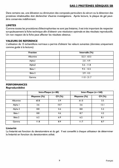
VALEURS DE REFERENCEL'utilisation de 15 échantillons normaux a permis d'obtenir les valeurs suivantes (données uniquementcomme guide à la lecture):
PERFORMANCESReproductibilité
LinéaritéLa linéarité est fonction du densitomètre et du gel. Il est conseillé à chaque utilisateur de déterminerla linéarité en fonction du densitomètre utilisé.
9
SAS-3 PROTÉINES SÉRIQUES SB
Français
Fraction Intervalle (%)
Albumine 52.3 - 63.5
Alpha1 2.0 - 4.9
Alpha2 5.6 - 11.8
Béta 1 9.2 - 12.5
Béta 2 2.9 - 4.5
Gamma 11.0 - 21.7
Intra-Plaque (n=80) Inter-Plaque (n=160)
Moyenne (%) CV (%) Moyenne (%) CV (%)
Albumine 60.8 3.9 61.0 3.5
Alpha 1 3.6 10.7 3.6 12.1
Alpha 2 8.8 5.6 8.8 5.2
Béta 1 8.8 4.6 8.7 4.2
Béta 2 6.2 6.9 6.2 8.1
Gamma 11.8 8.9 11.7 8.7

BIBLIOGRAPHIE1. Alper, C.A. 'Plasma Protein Measurements as a Diagnostic Aid', N. Eng. J. Med., 1974; 291 : 287-290.2. Tiselius, A. 'A New Apparatus for Electrophoretic Analysis of Colloidal Mixtures', Trans. Faraday
Soc., 1937; 33 : 524.3. Ritzmann, S.E. and Daniels, J.C. 'Diagnostic Proteinology: Separation and Characterization of
Proteins, Qualitative and Quantitative Assays' in Laboratory Medicine, Harper and Row, Inc.,Hagerstown, 1979.
4. Tietz, N.W. (Ed.), Textbook of Clinical Chemistry, W.B. Saunders Co., Philadelphia, pages 579-582,1986.
5. Ritzmann, S.E. (Ed.), Protein Abnormalities Volume 1 : Physiology of Immunoglobulins -Diagnostic and Clinical Aspects’, Allen R. Liss, Inc., New York, 1982.
6. Tietz, N.W. (Ed.), Textbook of Clinical Chemistry, (3rd Edition), W.B. Saunders Co., Philadelphia,page 524, 1995.
10

ANWENDUNGSBEREICHDer SAS-3 Serumprotein SB Kit dient zur Auftrennung und Auswertung der Serumprotein mittelsAgarosegel-Elektrophorese.
Serum enthält über 100 Einzelproteine, von denen jedes über eine Reihe von spezifischen Funktionenverfügt, die von der spezifischen Konzentrationsabweichung in unterschiedlichen pathologischenZuständen abhängen
1. Seit der Einführung der Moving-Boundary-Elektrophorese durch Tiselius
2und
der nachfolgenden Verwendung der Zonen-Elektrophorese, wurden Serumprotein sbe basierend aufihrer elektrischen Ladung bei einem bestimmten pH-Wert aufgetrennt. Mit dem SAS-3 SerumproteinSB Kit werden die Serumproteine entsprechend ihrer Ladung im Agarosegel in die 6 Hauptfraktionen(Albumin, Alpha-1-Globulin, Alpha-2-Globulin, Beta-1-Globulin, Beta-2-Globulin und Gamma-Globulin)aufgetrennt. Nach einer anschlieflenden Färbung können die Proteinbanden visuell oder quantitativausgewertet werden. Jede der klassischen Elektrophorese-Zonen, mit Ausnahme von Albumin, enthältüblicherweise 2 oder mehr Komponenten. Die relativen Ausmafle dieser Fraktionen haben sich alsnützliche Hilfsmittel in der Diagnose und Prognose von bestimmten Krankheitsstadien erwiesen
3-5.
WARNHINWEISE UND VORSICHTSMASSNAHMENAlle Reagenzien sind nur zur In-Vitro-Diagnostik bestimmt. Nicht einnehmen oder mit dem Mundpipettieren. Das Tragen von Handschuhen beim Umgang mit den Kit-Komponenten ist erforderlich.Bitte lesen Sie das Sicherheitsdatenblatt mit den Gefahrenhinweisen und Sicherheitsvorschlägen zu denKomponenten, sowie die Informationen zur Entsorgung.
INHALT1. SAS-3 Serumprotein SB-Gel (x10)
Enthält Agarose in einem Tris / Barbitalpuffer mit Thiomersal und Natriumazid alsKonservierungsmittel. Das Gel ist gebrauchsfertig verpackt.
2. Saures-Blau-Farbstoff (1x 75ml)Enthält eine konzentrierte Saures-Blau-Farbstoff-Lösung. Den Inhalt der Flasche mit 700ml dest.Wasser verdünnen. Über Nacht rühren und vor dem Gebrauch filtern. Lagerung des Farbstoffsin einer fest verschlossenen Flasche.
3. Entfärbelösung (2x 100ml)Enthält eine konzentrierte Entfärbelösung. Den Inhalt jeder Flasche mit 5 Liter dest. Wasserverdünnen und in einer fest verschlossenen Flasche lagern.
4. Weitere Kit-KomponentenJeder Kit enthält eine Methodenbeschreibung sowie ausreichend Blotter C für 10 Gele.
LAGERUNG UND STABILITÄT1. SAS-3 Serumprotein SB-Gel
Gele sollten bei 15...30°C gelagert werden und sind bis zum aufgedruckten Verfallsdatum stabil.NICHT IM KÜHLSCHRANK ODER TIEFKÜHLSCHRANK AUFBEWAHREN! Der Verfall desGels zeigt sich durch 1) Kristallisation, die auf ein Einfrieren des Gels hindeutet, 2) Brüchigkeit undAbblättern, die auf ein Austrocknen des Gels hindeuten, bzw. 3) sichtbare Kontaminierung derAgarose durch Bakterien oder Pilze.
11
SAS-3 SERUMPROTEIN SB
Deutsch

2. Saures-Blau-FarbstoffDas Farbstoff-Konzentrat sollte bei 15...30°C gelagert werden und ist bis zum aufgedrucktenVerfallsdatum stabil. Die Farbstofflösung ist für 6 Monate stabil bei einer Temperatur zwischen15...30°C. Es wird empfohlen, den benutzten Farbstoff unverzüglich zu entsorgen, um den Verlustder Färbungsfähigkeit zu verhindern. Eine schlechte Färbung weist auf den Verfall derFarbstofflösung hin.
3. EntfärbelösungDas Entfärbe-Konzentrat sollte bei 15...30°C gelagert werden und ist bis zum aufgedrucktenVerfallsdatum stabil. Die verdünnte Entfärbelösung ist für 6 Monate stabil bei einer Temperaturzwischen 15...30°C. Trübung kann auf den Verfall der Entfärbelösung hinweisen.
NICHT MITGELIEFERTES ABER BENÖTIGTES MATERIALKat. Nr. 310200 Probenapplikatormembranen (1 x 10)Kat. Nr. 310300 Probenapplikatormembranen (5 x 10)Kat. Nr. 210100 Einweg-Probenbecher (100)Kat. Nr. 3100 REP-Prep
PROBENAHME UND PROBENVORBEREITUNGFrisches Serum ist das Untersuchungsmaterial der Wahl. Proben können für das klassische 5-Bandenmuster
6, bei 15...30°C bis zu 4 Tagen, bei 2...6°C bis zu 2 Wochen und bei -20°C bis zu 6
Monate gelagert werden. Die Konzentration der Beta-2-Globulin-Fraktion, des C3-Komplements,nimmt bei Lagerung hingegen schnell ab. Urin und Liquores können nach einem angemessenenKonzentrierungsschritt (50-100-fach) ebenfalls verwendet werden. Die Verwendung von Plasmaresultiert in einer Fibrinogenbande im Bereich zwischen Beta- und Gamma-Fraktion.
Interferenzen: 1) Hämolytische Proben können falsche Alpha-2- und Beta-Werte zeigen. 2) LängereZeit unbedeckt stehende Proben können durch Verdunstungseffekte falsche Werte ergeben.
SCHRITT-FÜR-SCHRITT METHODE1. Geben Sie die folgenden Parameter in das Gerät ein.
Schritt Dauer (mm:ss) Temperatur (°C) Spannung SonstigesElektrophoreseProbe laden 00:30 21 Geschwindigkeit 1Probe auftragen 00:30 21 Geschwindigkeit 1*Elektrophorese (60 Probe) 13:00 18 480Elektrophorese (80 Probe) 11:30 18 480Elektrophorese (100 Probe) 09:45 18 480Trocknen 08:00 54* - Standort 1 für SAS-3 benutzen
FärbungFärben 04:00 Umlauf EINEntfärben 02:00 Umlauf EINEntfärben 02:00 Umlauf EINTrocknen 16:00 70
12

2. Die richtige Anzahl Applikatorkamm-Halterungen in Position auf dem Gerät anbringen:
3. Pipettieren Sie 35µl serum in der Einweg-Probenbecher. Schützen Sie die Proben vorVerdunstung.
4. Nehmen Sie das Gel aus der Schutzverpackung und verwerfen Sie den Überzug.5. Geben Sie 2ml REP Prep auf die linke Seite der Kammer und bringen Sie das Gel vorsichtig in die
Kammer ein. Richten Sie die Löcher im Gelbond auf die Stifte in der Kammer aus. Luftblasenunter dem Gel sind zu vermeiden. Die Elektroden an der Außenseite der Elektrodenhalternbefestigen, damit sie mit den Gelblöcken in Kontakt stehen.
6. Blotten Sie die Geloberfläche mit einem Blotter C und verwerfen Sie den Blotter anschlieflend.7. Wählen Sie das Serumprotein-Testprogramm an. Folgen Sie den Anweisungen und tragen Sie die
Proben auf. Führen Sie die Elektrophorese durch.8. Am Ende der Elektrophorese, entfernen Sie die Elektroden und entfernen beide Gelblöcke mit
dem speziellen Gelblock-Entferner.9. Befestigen Sie die Gel es am Färbekammerhalter.10. Wählen Sie das Serumprotein-Testprogramm auf der Färbeeinheit an. Färben, entfärben und
trocknen Sie das Gel entsprechend den Anweisungen.11. Am Ende des Färbevorgangs nehmen Sie das Gel aus der Färbekammer heraus. Es ist jetzt
prüfbereit.
QUALITÄTSKONTROLLEZur Überprüfung des gesamten Elektrophoreseablaufs sollten Sie bei jeder Trennung Kontrollenmitlaufen lassen, z.B. die Kemtrol-Kontrollseren: Kat. Nr. 7024 und 7025. Die entsprechendenVertrauenswerte finden sich in der Packungsbeilage.
AUSWERTUNGEs wird empfohlen, dass jegliche Auswertung der Gele im Vergleich mit Normalwerten durchgeführtwird, die in dem jeweiligen Labor für diese Methode ermittelt werden.
Für eine komplette Überprüfung der Serumprotein -Auswertung verweisen wir auf Ritzmann, S.E.1982
5. Studien zeigen, dass die Werte bei Männern und nicht schwangeren Frauen identisch sind.
Einige Abweichungen werden bei schwangeren Frauen am Geburtstermin und bei Frauen, die oraleVerhütungsmittel benutzen, festgestellt.Das Alter hat Auswirkungen auf die Normalwerte. Nabelschnurblut hat einen verringertenGesamtproteingehalt, Albumin, Alpha-2- und Beta-Fraktionen; leicht erhöhte Alpha-1 und normalebzw. erhöhte Gamma-Fraktion (weitgehend mütterlicher Herkunft). Die Gammaglobuline fallen raschbis zum Alter von 3 Monaten, während andere Fraktionen bis dahin bereits Erwachsenenwerteerreicht haben. Erwachsenenwerte der Gammaglobuline werden erst im Alter zwischen 10 und 16Jahren erreicht. Albumin verringert und Betaglobulin erhöht sich im Alter von 40+.
13
SAS-3 SERUMPROTEIN SB
Deutsch
Anzahl von Proben Applikatorposition
SAS-3
100 2, A, 9, 13, 16
80 3, 7, 12, 16
60 2, 8, 14

1. Qualitative Auswertung:Untersuchen Sie die Gele visuell auf das Vorhandensein bzw. Fehlen von Banden hin.
2. Quantitative Auswertung:Scannen Sie die Trennungen bei einer Wellenlänge von 595nm (Gelseite nach unten!).
In Fällen der Erhöhung oder Verringerung von bestimmten Serumkomponenten bzw. der Entdeckungvon ungewöhnlichen Serumkomponenten ist eine weitere Untersuchung erforderlich. Das fertige,trockene SAS-3 ist praktisch unbegrenzt haltbar.
EINSCHRÄNKUNGENDa alle Elektrophoreseverfahren nicht linear verlaufen, ist es wichtig, diese Anleitungen streng zubefolgen, um eine optimale Auflösung und reproduzierbare Ergebnisse zu erhalten. Die Nichteinhaltung dieser Anleitungen kann sich negativ auf die Ergebnisse auswirken.
REFERENZWERTEBei 15 normalen Proben von männlichen und weiblichen Spendern zwischen 20 und 59 Jahre wurdendie folgenden Normalwerte ermittelt (diese gelten nur als Richtlinie):
LEISTUNGSEIGENSCHAFTENReproduzierbarkeit
14
Protein-Fraktion Bereich (%)
Albumin 52.3 - 63.5
Alpha1 2.0 - 4.9
Alpha2 5.6 - 11.8
Beta 1 9.2 - 12.5
Beta 2 2.9 - 4.5
Gamma 11.0 - 21.7
Intra Assay (n=80) Inter Assay (n=160)
Mittel (%) CV (%) Mittel (%) CV (%)
Albumin 60.8 3.9 61.0 3.5
Alpha 1 3.6 10.7 3.6 12.1
Alpha 2 8.8 5.6 8.8 5.2
Beta 1 8.8 4.6 8.7 4.2
Beta 2 6.2 6.9 6.2 8.1
Gamma 11.8 8.9 11.7 8.7

LinearitätDie Linearität der Methode ist abhängig von der Densitometer-Spezifikation sowie der Leistung desGels. Es wird empfohlen, dass jeder Kunde die Linearität der Methode basierend auf dem im Laborverwendeten Densitometer selbst bestimmt.
LITERATUR1. Alper, C.A. ‘Plasma Protein Measurements as a Diagnostic Aid’, N. Eng. J. Med., 1974; 291 : 287-
290.2. Tiselius, A. ‘A New Approach for Electrophoretic Analysis of Colloidal Mixtures’, Trans. Faraday
Soc., 1937; 33 : 524.3. Ritzmann, S.E. and Daniels, J.C. ‘Diagnostic Proteinology: Separation and Characterization of
Proteins, Qualitative and Quantitative Assays’ in Laboratory Medicine, Harper and Row, Inc.,Hagerstown, 1979.
4. Tietz, N.W. (Ed.), Textbook of Clinical Chemistry, W.B. Saunders Co., Philadelphia, Seite 579-582, 1986.
5. Ritzmann, S.E. (Ed.), Protein Abnormalities Volume 1 : ‘Physiology of Immunoglobulins -Diagnostic and Clinical Aspects’, Allen R. Liss, Inc., New York, 1982.
6. Tietz, N.W. (Ed.), Textbook of Clinical Chemistry (3rd Edition), W.B. Saunders Co., Philadelphia,Seite 524, 1995.
15
SAS-3 SERUMPROTEIN SB
Deutsch
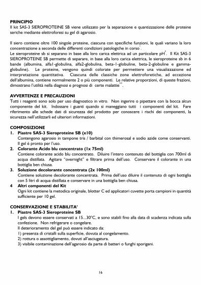
PRINCIPIOIl kit SAS-3 SIEROPROTEINE SB viene utilizzato per la separazione e quantizzazione delle proteineseriche mediante elettroforesi su gel di agarosio.
Il siero contiene oltre 100 singole proteine, ciascuna con specifiche funzioni, le quali variano la loroconcentrazione a seconda delle differenti condizioni patologiche in corso
1.
Le sieroproteine sb si separano in base alla loro carica elettrica ad un particolare pH2. Il Kit SAS-3
SIEROPROTEINE SB permette di separare, in base alla loro carica elettrica, le sieroproteine sb in 6bande (albumina, alfa1-globulina, alfa2-globulina, beta-1-globuline, beta-2-globuline e gamma-globuline). Le proteine, vengono quindi colorate per permettere una visualizzazione edinterpretazione quantitativa. Ciascuna delle classiche zone elettroforetiche, ad eccezionedell’albumina, contiene normalmente 2 o più componenti. Le relative proporzioni, di queste frazioni,dimostrano l’utilità nella diagnosi e prognosi di certe malattie
3-5.
AVVERTENZE E PRECAUZIONITutti i reagenti sono solo per uso diagnostico in vitro. Non ingerire o pipettare con la bocca alcuncomponente del kit. Indossare i guanti quando si maneggiano tutti i componenti del kit. Fareriferimento alle schede dati di sicurezza del prodotto per conoscere i rischi dei componenti, lasicurezza nell’utilizzarli ed ulteriori informazioni.
COMPOSIZIONE1. Piastre SAS-3 Sieroproteine SB (x10)
Contengono agarosio in tampone tris / barbital con thimerosal e sodio azide come conservanti. Il gel é pronto per l’uso.
2. Colorante Acido blu concentrato (1x 75ml)Contiene colorante acido blu concentrato. Diluire l’intero contenuto del bottiglia con 700ml diacqua distillata. Agitare “overnight” e filtrare prima dell’uso. Conservare il colorante in unabottiglia ben chiusa.
3. Soluzione decolorante concentrata (2x 100ml)Contiene soluzione decolorante concentrata. Prima dell’uso diluire il contenuto di ogni bottigliacon 5 litri di acqua distillata e conservare in una bottiglia ben chiusa.
4 Altri componenti del KitOgni kit contiene la metodica originale, blotter C ed applicatori cuvette porta campioni in quantitàsufficiente per 10 gel.
CONSERVAZIONE E STABILITA’1. Piastre SAS-3 Sieroproteine SB
I gels devono essere conservati a 15...30°C, e sono stabili fino alla data di scadenza indicata sullaconfezione. Non refrigerare o congelare.Il deterioramento del gel può essere indicato da:1) presenza di cristalli sulla superficie, dovuta al congelamento.2) rottura o assottigliamento, dovuti all’asciugatura.3) visibile contaminazione dell’agarosio da parte di batteri o funghi sporigeni.
16

2. Colorante Acido BluIl colorante concentrato deve essere conservato a 15...30°C, ed é stabile fino alla data riportatasull’etichetta. Il colorante ricostituito é stabile per 6 mesi, se conservato a 15...30°C. Scartareimmediatamente il colorante utilizzato. La scarsa colorazione può indicare un deterioramento delcolorante.
3. Soluzione decoloranteIl decolorante concentrato deve essere conservato a 15...30°C ed é stabile fino alla data discadenza riportata sull’etichetta. Il decolorante ricostituito é stabile per 6 mesi, se conservato a 15...30°C. La presenza di torbidità indica il deterioramento della soluzione decolorante.
RACCOLTA E PREPARAZIONE DEL CAMPIONESi consiglia di utilizzare siero fresco. Mentre i campioni possono essere conservati a 15...30°C fino a 4giorni, 2...6°C fino a 2 settimane oppure 6 mesi a -20°C per il tracciato standard a 5 bande (6), la bandebeta2-globulina (complemento C3) si degrada rapidamente con la conservazione.Si possono utilizzare campioni di urine e CSF previa opportuna concentrazione (50-100X).L’utilizzo del plasma può portare alla comparsa del fibrinogeno tra la frazione beta e la frazione gamma.
Fattori interferenti:- l’emolisi può causare falsi incrementi delle frazioni alfa2 e beta.- si possono ottenere risultati inattendibili se il campione viene conservato non correttamente
(ex. se non sigillata la provetta il campione si concentra a causa dell’evaporazione dello stesso).
PROCEDURA1. Programmare i seguenti parametri nella strumentazione.
Passaggio Tempo (mm:ss) Temp (°C) Voltaggio AltroElettroforesiCarico campione 00:30 21 Velocità 1Applicazione campione 00:30 21 Velocità 1 *Elettroforesi (60 campione) 13:00 18 480Elettroforesi (80 campione) 11:30 18 480Elettroforesi (100 campione) 09:45 18 480Asciugatura 08:00 54* - Usare posizione 1 per SAS-3
ColorazioneColorazione 04:00 Recirculate ONDecolorazione 02:00 Recirculate ONDecolorazione 02:00 Recirculate ONAsciugatura 16:00 70
17
SIEROPROTEINE SB SAS-3
Italiano

2. Collocare il numero esatto di lamelle di applicazione sullo strumento:
3. Pipettare 35µl di siero in nelle cuvette monouso. Proteggere i campioni dall’evaporazione.4. Rimuovere il gel dalla confezione ed eliminare il foglio protettivo.5. Dispensare 2ml di REP Prep nella camera elettroforetica e successivamente collocare il gel,
allineando i buchi del gel con i riferimenti nella camera, evitando la formazione di bolle sotto il gel.Fissare gli elettrodi all’esterno dei puntali, in modo tale che entrino a contatto con i blocchi di gel.
6. Asciugare la superficie del gel con un blotter C, poi scartarlo.7. Selezionare il programma SIEROPROTEINE, successivamente applicare i campioni e procedure
con l’elettroforesi.8. Al termine dell’elettroforesi, rimuova gli elettrodi e rimuovere entrambi I ponti di gel utilizzando
l’apposito “Rimuovi ponti di gel”.9. Collocarlo il gel nella camera di colorazione. Il gel deve essere rivolto verso l’operatore.10. Selezionare il programma SIEROPROTEINE, dopo la preparazione si procederà con la
colorazione, decolorazione ed asciugatura del gel.11. Al termine del ciclo di colorazione, rimuovere il gel dal coloratore. Il gel è ora pronto per essere
valutato.
INTERPRETAZIONE DEI RISULTATISi consiglia ad ogni singolo laboratorio di creare, con questo metodo, il proprio range di normalità.Per una completa valutazione delle sieroproteine, vedere Ritzmann, S.E., 1982
5.
Studi dimostrano che i valori proteici rimangono invariati per entrambi i sessi. Alcune differenze sipossono verificare nelle gestanti a termine e in donne che utilizzano contraccettivi orali.L’ età produce alcuni effetti sui valori normali.Legami di sangue portano:- una diminuizione delle proteine totali, albumina, alfa2 e beta.- lieve aumento alfa1.- rimane invariata oppure aumenta la frazione gamma (in gran misura da origini materne).
Le gamma globuline calano rapidamente sotto i 3 mesi di età ed i livelli restano sconosciuti fino a 10/16anni. Oltre i 40 anni, l’albumina diminuisce e le beta globuline aumentano.1. Valutazione qualitativa:
I gels possono essere visualizzati qualitativamente per la presenza o assenza di particolari bande.2. Valutazione quantitativa:
leggere i gels, a 595.
18
Numero di campioni Posizione applicatori
SAS-3
100 2, A, 9, 13, 16
80 3, 7, 12, 16
60 2, 8, 14

In altri casi, un aumento o diminuizione di particolari componenti del siero, oppure la presenza dicomponenti del siero non conosciute, richiede una nuova investigazione.La piastra completata di SIEROPROTEINE SB SAS-3, é stabile per un tempo indefinito.
CONTROLLO DI QUALITA'Per verificare la correttezza della procedura e controllare la lettura delle singole bande proteiche siconsiglia di usare il controllo Kemtrol - Normal cod. 7024 e Kemtrol - Abnormal cod. 7025. Per verificare i valori ottenuti, riferirsi alle istruzioni allegate nella confezione.
LIMITAZIONIDal momento che tutte le procedure elettroforetiche sono non-lineari, é importante seguireattentamente queste istruzioni per l’uso per ottenere un ottimale risoluzione e riproducibilità deirisultati.
VALORI DI RIFERIMENTOIl seguente range di normalità é stato ottenuto utilizzando 15 campioni normali.
CARATTERISTICHERiproducibilità
19
SIEROPROTEINE SB SAS-3
Italiano
Frazioni proteiche range (%)
Albumina 52.3 - 63.5
Alfa1 2.0 - 4.9
Alfa1 5.6 - 11.8
Beta 1 9.2 - 12.5
Beta 2 2.9 - 4.5
Gamma 11.0 - 21.7
entro la serie (n=80) tra la serie (n=160)
Media (%) CV (%) Media (%) CV (%)
Albumina 60.8 3.9 61.0 3.5
Alfa1 3.6 10.7 3.6 12.1
Alfa1 8.8 5.6 8.8 5.2
Beta 1 8.8 4.6 8.7 4.2
Beta 2 6.2 6.9 6.2 8.1
Gamma 11.8 8.9 11.7 8.7

LinearitàLa linearità del metodo é una funzione delle specifiche del densitometro. Si raccomanda che ognicliente determini la linearità del metodo in base al densitometro in uso nel laboratorio.
BIBLIOGRAFIA1. Alper, C.A. 'Plasma Protein Measurements as a Diagnostic Aid', N. Eng. J. Med., 1974; 291 : 287-290.2. Tiselius, A. 'A New Apparatus for Electrophoretic Analysis of Colloidal Mixtures', Trans. Faraday
Soc., 1937; 33 : 524.3. Ritzmann, S.E. and Daniels, J.C. 'Diagnostic Proteinology: Separation and Characterization of
Proteins, Qualitative and Quantitative Assays' in Laboratory Medicine, Harper and Row, Inc.,Hagerstown, 1979.
4. Tietz, N.W. (Ed.), Textbook of Clinical Chemistry, W.B. Saunders Co., Philadelphia, pages 579-582, 1986.
5. Ritzmann, S.E. (Ed.), Protein Abnormalities Volume 1 : Physiology of Immunoglobulins -Diagnostic and Clinical Aspects’, Allen R. Liss, Inc., New York, 1982.
6. Tietz, N.W. (Ed.), Textbook of Clinical Chemistry, (3rd Edition), W.B. Saunders Co., Philadelphia,page 524, 1995.
20

USO PREVISTOEl kit de proteínas séricas SB SAS-3 tiene como objeto la separación y cuantificación de proteínasséricas por electroforesis con gel de agarosa.
El suero contiene más de 100 proteínas individuales, cada una de ellas con una serie de funcionesespecíficas que están sujetas a variaciónes en su concentración bajo diferentes condiciones patológicas
1.
Desde la introducción de la electroforesis marginal móvil por Tiselius2
y el empleo subsiguiente de laelectroforesis de zona, las proteínas séricas SB se han fraccionado tomando como base su carga conun pH determinado. El kit de proteínas séricas SB SAS-3 separa, en un gel de agarosa las proteínas desuero en 6 clases principales (albúmina, alfa 1-globulina, alfa 2-globulina, beta1-globulina, beta2-globulina y gammaglobulina) según su carga. Luego las proteínas son coloreadas para permitir suvisualización e interpretación cuantitativa. Cada una de las zonas electroforéticas clásicas, con laexcepción de la albúmina, contiene normalmente 2 o más componentes. Las proporciones relativas deestas fracciones han demostrado su utilidad como ayuda en el diagnóstico y pronóstico de ciertosestados de enfermedad
3-5.
ADVERTENCIAS Y PRECAUCIONESTodos los reactivos son para utilizar únicamente en diagnóstico in vitro. No ingerir ni aspirar por laboca ningún componente del kit. Utilizar guantes para manipular los componentes del kit. Consultaren el prospecto de seguridad del producto las indicaciones sobre riesgos y seguridad así como lainformación acerca de su eliminación.
COMPOSICION1. Gel de proteínas séricas SB SAS-3. (x10)
Contiene agarosa en un tampón de Tris-Barbital, con Tiomersal y acída de sodio comoconservantes. El gel viene envasado listo para usar.
2. Colorante azul ácido concentrado. (1x 75ml)Contiene colorante azul ácido concentrado. Diluir el contenido del frasco en 700ml de aguadestilada. Dejar agitando durante toda la noche y filtrarlo antes del uso. Guardar el colorante enun frasco herméticamente cerrado.
3. Solución decolorante concentrada. (2x 100ml)Contiene solución decolorante concentrada. Diluir el contenido de cada frasco en 5 litros de aguadestilada y guardarlo en un frasco de tapón esmerilado herméticamente cerrado.
4. Otros componentes del kit.Cada kit contiene una hoja de instrucciones y secante C hasta completar 10 geles.
ALMACENAMIENTO Y VIDA UTIL DE ALMACENAJE1. Gel de proteínas séricas SB SAS-3.
Los geles deben guardarse a una temperatura entre 15...30°C y son estables hasta la fecha decaducidad indicada en el envase. NO REFRIGERAR NI CONGELAR. El deterioro del gel puedeser indicado por: 1) aspecto cristalino, indicio de que el gel se ha congelado, 2) agrietamiento yexfoliación, indicio de que el gel se ha secado, o 3) contaminación visible de la agarosa por fuentesbacterianas o micóticas.
21
PROTEÍNAS SÉRICAS SB SAS-3
Español

2. Colorante azul ácido.El colorante concentrado debe guardarse a una temperatura entre 15...30°C y es estable hasta lafecha de caducidad indicada en la etiqueta del frasco. La solución colorante preparada es establedurante 6 meses a una temperatura entre 15...30°C. Es aconsejable desechar inmediatamente elcolorante usado para prevenir el agotamiento de su capacidad de coloración. Unos malosresultados de coloración pueden ser indicio de deterioro de la solución colorante.
3. Solución decolorante.El decolorante concentrado debe guardarse a una temperatura entre 15...30°C y es estable hastala fecha de caducidad indicada en la etiqueta. La solución decolorante diluida es estable durante 6meses a una temperatura entre 15...30°C. La aparición de turbidez puede ser indicio de deteriorode la solución decolorante.
ACCESORIOS NECESARIOS, NO SUMINISTRADOSno de catàlogo 310200 Aplicadores de muestras 1 x 10no de catàlogo 310300 Aplicadores de muestras 5 x 10no de catàlogo 210100 Vasos de recogida de muestras desechables 100no de catàlogo 3100 REP Prep
RECOGIDA Y PREPARACION DE MUESTRASLa muestra consistirá en suero recién obtenido. Aunque las muestras pueden almacenarse 4 días a15...30°C, 2 semanas a 2...6°C, o 6 meses a -20°C para un patron standard de 5 bandas
6, la banda beta
2-globulina (Complemento C3) se degradará rapidamente al almacenarse. Orina y CSF también sepueden usar siguiendo una fase de concentración adecuada (50 - 100X). El empleo de plasma tendrácomo resultado la aparición de una banda de fibrinógeno entre las fracciones beta y gamma.
Factores de interferencia:1) La hemólisis puede causar una falsa elevación de las fracciones alfa-2 y beta.2) Se pueden obtener resultados imprecisos en muestras que se hayan dejado sin cubrir, debido a laevaporación.
PROCEDIMIENTO PASO A PASO1. Programar los siguientes paràmetros en el instrumento:
Paso Tiempo Temperatura Voltaje OtrosElectroforesis (mm:ss) (°C)Cargar muestra 00:30 21 Velocidad 1Aplicar muestra 00:30 21 Velocidad 1*Electroforesis (80 muestra) 13:00 18 480Electroforesis (80 muestra) 11:30 18 480Electroforesis (100 muestra) 09:45 18 480Secar 08:00 54* - Utilizar posición 1 para SAS-3
22

Paso Tiempo Temperatura Voltaje OtrosColoradorColorar 04:00 Recirculación ONDecolorar 02:00 Recirculación ONDecolorar 02:00 Recirculación ONSecar 16:00 70
2. Colocar en el instrumento el número correcto de unidades de aplicadores:
3. Introducir con una pipeta 35µl de la muestra en vasos de recogida de muestras desechables.Proteger las muestras de la evaporación.
4. Sacar el gel del envase y desechar la lámina sobrepuesta.5. Administrar 2ml de REP Prep al lado izquierdo de la cámara y colocar cuidadosamente el gel en la
cámara, alineando los orificios en la ligazón del gel con las espigas de la cámara y evitando laformación de burbujas debajo del gel. Conectar los electrodos a la parte externa de los bordes,de forma que estén en contacto con los bloques de gel.
6. Secar la superficie del gel con un secante C y luego desechar el secante.7. Seleccionar el programa de pruebas de seroproteínas y, siguiendo las indicaciones, aplicar las
muestras y realizar la electroforesis.8. Finalizada la electroforesis, sacar los electrodos y sacar ambos bloques de gel utilizando el
extractor de bloques de gel.9. Sacar el gel de la cámara y sujetarlo al soporte de la cámara de coloración.10. Seleccionar el programa de prueba de seroproteínas en la unidad de coloración y, siguiendo las
indicaciones, colorar, decolorar y secar el gel.11. Finalizado el ciclo de coloración, sacar el gel de la cámara de coloración. Ahora, el gel está listo
para ser examinado.
CONTROL DE CALIDADSe pueden aplicar los controles de suero Kemtrol (no de catàlogo 7024 y 7025) para verificar todas lasfases del procedimiento y también se pueden utilizar en cada placa. Consultar el prospecto del envase,en el que se indican los valores adecuados para los ensayos de laboratorio.
INTERPRETACION DE RESULTADOSEs aconsejable realizar cualquier evaluación de los geles contrastándola con valores normales obtenidospor este método en cada laboratorio en particular.Para obtener un análisis completo de la evaluación de proteínas séricas, leer a S. E. Ritzmann, 1982
5.
Los estudios muestran que los valores son los mismos tanto para hombres como para mujeres noembarazadas. Se han detectado algunas diferencias en mujeres al término de su embarazo y enmujeres que utilizan anticonceptivos orales. La edad tiene algún efecto sobre los niveles normales. La sangre del cordón umbilical tiene fracciones totales de proteínas, albúmina, alfa 2 y beta reducidas,la fracción alfa 1 ligeramente incrementada y gamma normal o incrementada (en gran parte de origen
23
PROTEÍNAS SÉRICAS SB SAS-3
Español
Número de muestras Posición del aplicador
SAS-3
100 2, A, 9, 13, 16
80 3, 7, 12, 16
60 2, 8, 14

materno). Las gammaglobulinas se reducen rápidamente cerca de los 3 meses de edad, mientras queotras fracciones ya han alcanzado niveles adultos. Los niveles adultos de las gammaglobulinas no sonalcanzados hasta los 10 - 16 años de edad. La albúmina decrece y la betaglobulina aumenta a partir delos 40 años de edad.1. Evaluación cuantitativa:
Los geles se pueden inspeccionar visualmente, para comprobar la existencia o ausencia dedeterminadas bandas de interés.
2. Evaluación cuantitativa: Analizar los geles, con el gel hacia abajo, a 595 nm.
En cualquier caso, una elevación o disminución de determinados componentes del suero o la detecciónde componentes inusuales en el suero requerirán una investigación posterior. El gel de proteínasséricas SB SAS-3 completado es estable durante un tiempo indefinido.
LIMITACIONESPuesto que los procedimientos de electroforesis son no lineales, es importante seguir atentamenteestas instrucciones de uso para asegurar unos resultados de resolución y reproductibilidad óptimos.Omitir el seguimiento de estas instrucciones de uso podría afectar a los resultados obtenidos.
VALORES DE REFERENCIALos siguientes márgenes nominales se obtuvieron utilizando 15 muestras normales obtenidas devarones y hembras donantes con edades comprendidas entre 20 y 59 años (estos valores, no obstante,se presentan sólo a modo de guía):
24
Fracción proteínica Margen (%)
Albúmina 52.3 - 63.5
Alfa1 2.0 - 4.9
Alfa1 5.6 - 11.8
Beta 1 9.2 - 12.5
Beta 2 2.9 - 4.5
Gamma 11.0 - 21.7

CARACTERISTICAS FUNCIONALESReproductibilidad
LinealidadLa linealidad del método es una especificación de funcionamiento del densitómetro así como de lascaracterísticas del gel. Es aconsejable que cada cliente determine la linealidad del método basándoseen el densitómetro utilizado en el laboratorio.
BIBLIOGRAFIA1. Alper, C.A. 'Plasma Protein Measurements as a Diagnostic Aid', N. Eng. J. Med., 1974; 291 : 287-
290.2. Tiselius, A. 'A New Apparatus for Electrophoretic Analysis of Colloidal Mixtures', Trans. Faraday
Soc., 1937; 33 : 524.3. Ritzmann, S.E. and Daniels, J.C. 'Diagnostic Proteinology: Separation and Characterization of
Proteins, Qualitative and Quantitative Assays' in Laboratory Medicine, Harper and Row, Inc.,Hagerstown, 1979.
4. Tietz, N.W. (Ed.), Textbook of Clinical Chemistry, W.B. Saunders Co., Philadelphia, pages 579-582, 1986.
5. Ritzmann, S.E. (Ed.), Protein Abnormalities Volume 1 : Physiology of Immunoglobulins -Diagnostic and Clinical Aspects’, Allen R. Liss, Inc., New York, 1982.
6. Tietz, N.W. (Ed.), Textbook of Clinical Chemistry, (3rd Edition), W.B. Saunders Co., Philadelphia,page 524, 1995.
25
PROTEÍNAS SÉRICAS SB SAS-3
Español
Dentro del gel (n=80) Entre el gel (n=160)
Media (%) CV (%) Media (%) CV (%)
Albúmina 60.8 3.9 61.0 3.5
Alfa1 3.6 10.7 3.6 12.1
Alfa1 8.8 5.6 8.8 5.2
Beta 1 8.8 4.6 8.7 4.2
Beta 2 6.2 6.9 6.2 8.1
Gamma 11.8 8.9 11.7 8.7

Helena Biosciences EuropeQueensway SouthTeam Valley Trading EstateGatesheadTyne and WearNE11 0SD
Tel: +44 (0) 191 482 8440Fax: +44 (0) 191 482 8442Email: [email protected]
HL-2-1432P 2008/09 (3)