
Sample A: Cover Page of Thesis, Project, or Dissertation ...
132
The use of histology, molecular techniques, and ex situ feeding experiments to investigate the feeding behavior of the coral reef predator Hermodice carunculata, the bearded fireworm A thesis submitted in partial fulfillment of the requirements for the degree of Master of Science at George Mason University By Staci A. Lewis Bachelor of Science Salem College, 2002 Director: Robert B. Jonas, Chair Department of Environmental Science and Policy Spring Semester 2009 George Mason University Fairfax, VA
Transcript of Sample A: Cover Page of Thesis, Project, or Dissertation ...
The use of histology, molecular techniques, and ex situ feeding experiments to investigate the feeding behavior of the coral reef predator Hermodice carunculata, the bearded
fireworm
A thesis submitted in partial fulfillment of the requirements for the degree of Master of Science at George Mason University
By
Staci A. Lewis Bachelor of Science Salem College, 2002
Director: Robert B. Jonas, Chair Department of Environmental Science and Policy
Spring Semester 2009 George Mason University
Fairfax, VA

Copyright 2009 Staci A. Lewis All Rights Reserved
ii
DEDICATION
To the late Dr. Joan Marsden, for her inspiration as a female scientist before her time To Dr. Suzanne Dorsey, for capturing my imagination and believing in my potential To the late Dr. Ronald Edwards, for your ever-present patience, encouragement, and advice To my Mom, Dad, brothers, and Michelle, for nurturing my love of marine biology and science
iii

ACKNOWLEDGEMENTS
I would like to thank everyone who helped me ponder, plan, execute, and analyze the many research projects which have culminated in this thesis. The Dauphin Island Sea Lab, Dr. Rich Aronson, and Dr. Ryan Moody reignited my passion for field research and provided amazing opportunities to pursue and expand my love for marine ecology. My research in Barbados was made possible by the Fulbright Program of the State Department. Thank you to the people working for the program in the U.S. and for the U.S. Embassy in Barbados for your support. I would also like to thank Geoff Cook for introducing me to George Mason University. We first talked about George Mason University many years ago during a coral research cruise. Thank you Geoff for your recruiting skills, companionship, and support since my journey began at GMU. Dr. Esther Peters was an integral part in my discovery of the art of histology. Thank you for your editing skills and patience. The Bellairs Research Institute was used as base camp for Chapter 3 and other projects I conducted while living in Barbados on and off from 2001-2005. I would like to recognize the fabulous crews of M/V Fling and Spree, the fellow researchers, and the NOAA employees on the many trips to the Flower Garden Banks and the Florida Keys National Marine Sanctuaries throughout my years as a project manager and a graduate student. You all made the trips unforgettable.
iv
TABLE OF CONTENTS
Page List of Tables…………………………………………………………………………....vi List of Figures………………………………………………………………………….vii Abstract…………………………………………………………………………………ix 1. Introduction .....…......................................................................................................…………… 1 2. A histological analysis of the invertebrate corallivore Hermodice carunculata ......12 3. The effect of a Millepora complanata feeding regime on the body weight of
Hermodice carunculata ............................................................................................52 4. The use of molecular techniques to investigate the feeding behavior of Hermodice
carunculata ...............................................................................................................85 5. Overall Conclusions ................................................................................................117
v
LIST OF TABLES Table Page 2.1 The total number of slides analyzed for each section .......................................……19 3.1 Statistically significant Pearson’s correlation tests ...........................................……72 3.2 Statistically significant Mann-Whitney U tests ................................................……72 4.1 Coral diseases associated with biota which has been proven through Koch’s postulates ..........................................................................................................……87 4.2 Coral diseases associated with abiotic stressors ...............................................……88 4.3 Swab Sample Summary Sheet ........................................................................……101 4.4 Core Sample Summary Sheet .........................................................................……102 4.5 Standard PCR Protocol with Taq Gold DNA Polymerase ..............................……103
vi

LIST OF FIGURES
Figure Page 2.1 A pictorial representation of the trimmed tissue sections .................................……19 2.2 Parasaggital section, dorsal view, of the epidermis from fireworm sample 07-53 III
...........................................................................................................................……22 2.3 Parasaggital section, ventral view, of fireworm sample 07-60 V with brown granular
pigment .............................................................................................................……23 2.4 Parasaggital section, dorsal view, of fireworm sample 07-53 III with green granular
pigment .............................................................................................................……24 2.5 Cross-section, ventral view, highlighting the outer cuticle with layer of cells from
fireworm sample 07-56 IVA .............................................................................……24 2.6 Side view of Hermodice carunculata ...............................................................……25 2.7 Cross-section of fireworm sample 07-58 IVP with toxic setae ........................……25 2.8 Parasaggital section, dorsal view, of the eye, brown-pigmented granules, dorsal cirri,
and the nuchal organ from fireworm sample 07-59 I .......................................……26 2.9 Parasaggital section, dorsal view, of the nuchal organ from fireworm sample 07-59 I
...........................................................................................................................……27 2.10 Dorsal view of H. carunculata........................................................................……27 2.11 Cross-section view of fireworm sample 07-58 IVP ............................................…28 2.12 Cross-section, ventral view, of the nerve cord from fireworm sample 07-59 IIA
.........................................................................................................................……29 2.13 Cross-section of pigmented granules associated with nerve cord penetrating the
cuticle from fireworm sample 07-59 IVP ........................................................……30 2.14 Parasaggital section of the cuticle-lined foregut from fireworm sample 07-56 I
.........................................................................................................................……31 2.15 Parasaggital section of proboscis with secretory cells from fireworm sample
07-57 I .............................................................................................................……32 2.16 Close-up image of Figure 2.15 ........................................................................……33 2.17 Parasaggital section of the specialized tissue in the foregut from fireworm sample
07-56 I .............................................................................................................……33 2.18 Cross-section of posterior digestive tract and foregut from fireworm sample 07-59
IIA ...................................................................................................................……34 2.19 Parasaggital section of secretory cells in digestive tract from fireworm sample 07-55
III .....................................................................................................................……35 2.20 Parasaggital section of posterior gut cavity with highly dense and long cilia from
fireworm sample 07-53 III ..............................................................................……36 2.21 Parasaggital section of digestive tract containing cuticles and coccoid cells from
fireworm sample 07-55 III ..............................................................................……37
vii
viii
2.22 Cross-section of the digestive tract containing various materials from fireworm sample 07-55 IIA .............................................................................................……39
2.23 Parasaggital section of the digestive tract containing coccoid cells from fireworm sample 07-53 III ..............................................................................................……39
3.1 Average weight change for the specimens in 1FCE .........................................……63 3.2 Average weight change for the specimens in 2FCE (all) ........................................65 3.3 Average weight change for the specimens in 2FCE ................................................67 3.4 Average weight change for the specimens in 2FCE Mann ......................................69 4.1 H. carunculata feeding on a Montastraea colony ...........................................……92 4.2 Suspect white plague killing a colony of Diploria strigosa .............................……93 4.3 Two H. carunculata specimens feeding on a Montastraea colony .................……94 4.4 Diver extracts 1.6-cm diameter cores .......................................................................98 4.5 PCR with Universal Protist Primer .........................................................................105 4.6 PCR with H. carunculata-specific primer Hc737RA .............................................106 4.7 PCR with H. carunculata-specific primer testing with coral DNA ........................107
ABSTRACT
THE USE OF HISTOLOGY, MOLECULAR TECHNIQUES, AND EX SITU FEEDING EXPERIMENTS TO INVESTIGATE THE FEEDING BEHAVIOR OF THE CORAL REEF PREDATOR HERMODICE CARUNCULATA, THE BEARDED FIREWORM Staci Lewis, MS George Mason University, 2009 Thesis Director: Dr. Robert Jonas Three studies on the invertebrate corallivore Hermodice carunculata, commonly known
as the bearded fireworm, were conducted to provide baseline information on the general
anatomical features, nutritional requirements, feeding behavior, and growth rates of this
poorly documented reef inhabitant. Through histological techniques, the species’ feeding
mechanisms, digestive system, and sensory structures were studied to understand its
ability to adapt to a changing reef habitat. Several previously unreported features were
observed including a simple layer of cells on the outer cuticle resembling, and the size of,
bacteria, a specialized tissue in the foregut, and pigment granules penetrating the ventral
cuticle at the nerve cord connection. This study also documented the appearance of gut
content in the digestive tract and the presence of secretory cells on the proboscis. These
observations provide insight into the feeding behavior of H. carunculata. Ex situ feeding
experiments were conducted in Barbados to monitor organisms’ weight change in
different coral reef feeding regimes as an indication of nutritional value of coral species.
During the experiments, H. carunculata specimens experienced a Millepora complanata
feeding regime and a short time period in a Montastraea annularis feeding regime. In
both experiments, the average weight change was not statistically significant, which
indicates the H. carunculata specimens did not gain weight in the presence of M.
complanata. These results suggest H. carunculata may supplement their diet with other
food sources to meet nutritional requirements. Finally, during an outbreak of an
unknown white syndrome on coral colonies in the Flower Garden Banks National Marine
Sanctuary, field samples, including H. carunculata specimens associated with the coral
tissue loss margins, were collected to test the use of the Polymerase Chain Reaction
(PCR) and DNA fingerprinting to identify H. carunculata associated with tissue loss
lesions. Specific H. carunculata primers were designed to amplify H. carunculata DNA
in the samples. The H. carunculata tissue samples (positive controls) were the only
samples amplified during the PCR tests. These results suggest H. carunculata organisms
do not leave detectable amounts of DNA at foraging sites. However, future molecular
tests should be conducted using other target sequences for amplification and take into
considerations the sensitivity of conditions including the number of PCR cycles.

1. Introduction
Coral reefs are among the most diverse ecosystems on the planet (Huston 1985,
Jackson 1991, Spalding et al. 2001, Folke et al. 2004, Wilkinson 2004, Ferner et al. 2009)
and are considered the rainforests of the sea (Huston 1985, Jackson 1991, Sebens 1994,
Knowlton 2001, Bellwood et al. 2004, Wilson et al. 2006, Jackson 2008, Yap 2009).
Although coral reefs only occupy around 0.09% of the ocean area (Wilson et al. 2006),
25% of all animals in the ocean rely on coral reefs for survival (Spalding et al. 2001,
Wilkinson 2004). These habitats are important to an enormous number of marine
organisms (Sebens 1994, Chapin et al. 2000, Knowlton 2001, Pandolfi and Jackson
2006) during at least one stage of their life for shelter (Menge and Lubchenco 1981, Sano
et al. 1984), food (Mumby et al. 2006, Rotjan and Lewis 2008), and breeding space
(Booth and Wellington 1998, Hughes and Tanner 2000, Syms and Jones 2000, Adams
and Ebersole 2002, Jones et al. 2004). The diversity of coral species offers an array of
physical structures, (Ferner et al. 2009, Yap 2009) including branches (Pandolfi and
Jackson 2006), plates (Huston 1985), and boulders (Edmunds and Elahi 2007), which are
vital to maintain the vast community of marine biota (Crowder and Cooper 1982, Jones
and Syms 1998, Syms and Jones 2000, Caley et al. 2001, Adams and Ebersole 2002,
Bellwood et al. 2003, Wilson et al. 2006, Ferner et al. 2009). Live coral tissue provides a
biological structure which promotes larval recruitment (Syms and Jones 2000), accretes
1
calcium carbonate skeleton (Hutson 1985), and provides food for corallivores (Bell and
Galzin 1984).
Coral reefs are being damaged worldwide at an alarming rate (Nystrom et al.
2000, Gardner et al. 2003, Bellwood et al. 2004, Hughes et al. 2007) from a variety of
threats including overfishing (Jackson et al. 2001, Pandolfi et al. 2003), storms (Hughes
1994, Aronson et al. 2004, Alvarez-Filip and Gil 2006, Cheal et al. 2008), pollution
(Pastorok and Bilyard 1985, Smith et al. 2008), and elevated sea surface temperatures
(Hoegh-Guldberg et al. 2007). Coral diseases can also lead to colony mortality (Bruckner
and Bruckner 1997, Hughes and Tanner 2000) which may exacerbate the worldwide
decline of coral reefs (Knowlton 2001, Pandolfi et al. 2003). Coral mortality has lead to
unprecedented phase shifts of dominant coral species (Hughes et al. 2003, Aronson et al.
2004, Green et al. 2008), changes in reef structural features (Edmunds and Elahi 2007,
Ferner et al. 2009), and increase in the number of algae-dominated reefs (Done 1992,
Knowlton 1992, Knowlton 2004, Pandolfi et al. 2005). Phase shifts and mass mortality of
corals can impact sessile and benthic marine organisms including the consumers of coral
tissue, corallivores (Rotjan and Lewis 2008).
The impacts of coral reef decline have been studied extensively for vertebrate
corallivores (Cheal et al. 2008) when compared with studies of invertebrate corallivores
(Rotjan and Lewis 2005). The following chapters examined a poorly understood, direct
consumer of coral tissue, the invertebrate facultative corallivore Hermodice carunculata,
one of 160 known corallivores. H. carunculata is in the Class Polychaeta, Family
Amphinomidae (Marsden 1962). It can be found on tropical reefs worldwide. Previous
2
field observations and microscopic analysis of gut content indicate that this species’ diet
includes coral tissue, sponges, anemones, and plant material (Marsden 1963a). Using its
muscular proboscis, H. carunculata everts its pharynx over its prey and moves tissue into
its foregut through use of its muscular pharynx (Marsden 1966). Disease events of
suspect white syndrome in Flower Gardens National Marine Sanctuary (FGBNMS) and
Navassa in the winters of 2004, 2005, 2006, and 2007 indicate there is a strong
correlation between H. carunculata and suspect white syndrome (Miller and Williams
2007, Jonas personal communication). These observations suggest the H. carunculata
populations were contributing to or the primary cause of coral tissue loss. However,
limited information on this species impairs our ability to address its ability to adapt in a
changing reef environment, its impact on a declining reef system, and its role in coral
tissue loss and etiology of diseases including white plague (WP). H. carunculata’s
feeding preferences, specific extraction mechanisms (i.e. the use of digestive enzymes to
aid in tissue removal), and capacity to transport microbes (e.g. vector of disease) need to
be well understood.
Periodic phenomena or long-term shifts in ocean conditions can affect coral reef
community ecology (May 1977, Knowlton 1992, Pandolfi et al. 2003) including the
abundance and biodiversity of coral species (Bythell et al. 1993, Hughes 1994, Aronson
and Precht 2001). Important reef-building coral species like Montastraea annularis have
been replaced by dominate and fast-growing species like Porites astreoides (Green et al.
2008). Similarly, the reef systems off of Barbados have seen a reduction in M. annularis
and Acropora cervicornis (Macintyre et al. 2007). In contrast to the decrease in
3

scleractinian corals, the hydrocoral Millepora complanata remains in high densities in the
shallow reefs (Lewis 2006). If the new dominant coral species are less nutritious per
tissue area, this may increase the area of coral tissue loss per feeding event (Baums et al.
2003b). Also, feeding behavior of corallivores (e.g., scrapers versus grazers) may not be
conducive to the newly dominant coral species morphology (Rotjan and Lewis 2008).
The overall nutritional value and availability of corals as a food resource could have a
detrimental effect on species (Baums et al. 2003b). If factors are not favorable for
consumption of the new coral species or if coral tissue becomes unavailable (i.e.,
complete coral mortality), facultative corallivores may utilize alternate food resources in
larger quantities, which could have compounding impacts on the reef community (Rotjan
and Lewis 2008). Through histopathological methods, Chapter 2 documents the tissues,
gut content, and anatomical features of eight specimens collected from the FGNMS in
2007 to develop baseline information on this species ability to detect and consume prey.
The experiments in Chapter 3 examined the body weight of H. carunculata, in the
presence of an abundant fire coral species, M. complanata from a system of declining
scleractinian coral cover in Barbados.
Studies investigating possible causes of coral disease have had inconsistent or
inconclusive findings (Ainsworth et al. 2007) including the uncertain role of corallivores
in the etiology of disease (Sussman et al. 2003, Miller and Williams 2007). Effective
management tools are needed to understand the correlation of microbial-induced tissue
loss to corallivory (Sussman et al. 2003) and to distinguish predation scars from disease
(Dalton and Godwin 2006, Miller and Williams 2007). In Chapter 4, samples of coral
4
tissue at the site of actively feeding organisms were tested for detectable H. carunculata
DNA to determine the association of corallivory feeding behavior of H. carunculata with
white syndrome tissue loss margins. Furthermore, the ability to trace H. carunculata
DNA on predation marks could aid in the development of accurate diagnosis criteria for
predation-induced versus microbial-induced tissue loss.
As coral reefs decline and distribution of coral species change, the impacts on reef
inhabitants which are dependent on coral reef complexity and live coral tissue need to be
understood (Sano et al. 1984, Chapin et al. 2000, Syms and Jones 2000, Booth and
Beretta 2002, Samways 2005, Rotjan and Lewis 2008). Basic information is not available
for some corallivores (Rotjan and Lewis 2008) including the invertebrate, facultative
corallivore, H. carunculata. The observations in the following chapters provide
fundamental information on the sensory structures, feeding behavior, and diagnosis
predation patterns of H. carunculata which are critical in determining the impact of a
deteriorating reef system on this coral reef dweller and its prey, including threatened
coral species.
5
Literature Cited Adams AJ, Ebersole JP (2002) Use of back-reef and lagoon habitats by coral reef fishes.
Mar Ecol-Prog Ser 228:213-226 Ainsworth TD, Kramasky-Winter E, Loya Y, Hoegh-Guldberg O, Fine M (2007) Coral
disease diagnostics: What's between a plague and a band? Appl Environ Microbiol 73:981-992
Alvarez-Filip L, Gil I (2006) Effects of Hurricanes Emily and Wilma on coral reefs in
Cozumel, Mexico. Coral Reefs 25:583-583 Aronson RB, MacIntyre IG, Wapnick CM, O'Neill MW (2004) Phase shifts, alternative
states, and the unprecedented convergence of two reef systems. Ecology 85:1876-1891
Aronson RB, Precht WF (2001) White-band disease and the changing face of Caribbean
coral reefs. Hydrobiologia 460:25-38 Baums IB, Miller MW, Szmant AM (2003b) Ecology of a corallivorous gastropod,
Coralliophila abbreviata, on two scleractinian hosts. II. Feeding, respiration and growth. Mar Biol 142:1093-1101
Bellwood DR, Hoey AS, Choat JH (2003) Limited functional redundancy in high
diversity systems: resilience and ecosystem function on coral reefs. Ecol Lett 6:281-285
Bellwood DR, Hughes TP, Folke C, Nystrom M (2004) Confronting the coral reef crisis.
Nature 429:827-833 Booth DJ, Beretta GA (2002) Changes in a fish assemblage after a coral bleaching event.
Mar Ecol-Prog Ser 245:205-212 Booth DJ, Wellington G (1998) Settlement preferences in coral-reef fishes: Effects on
patterns of adult and juvenile distributions, individual fitness and population structure. Blackwell Science, p 274-279
Bruckner AW, Bruckner RJ (1997) Outbreak of coral disease in Puerto Rico. Coral Reefs
16:260 Bythell JC, Gladfelter EH, Bythell M (1993) Chronic and catastrophic natural mortality
of 3 common Caribbean reef corals. . Coral Reefs 12:143-152
6

Caley MJ, Buckley KA, Jones GP (2001) Separating ecological effects of habitat fragmentation, degradation, and loss on coral commensals. Ecology 82:3435-3448
Chapin FS, Zavaleta ES, Eviner VT, Naylor RL, Vitousek PM, Reynolds HL, Hooper
DU, Lavorel S, Sala OE, Hobbie SE, Mack MC, Diaz S (2000) Consequences of changing biodiversity. Nature 405:234-242
Cheal A, Wilson SK, Emslie MJ, Dolman A, Sweatman H (2008) Responses of reef fish
sommunities to coral declines on the Great Barrier Reef. Mar Ecol-Prog Ser 372:211-223
Crowder LB, Cooper WE (1982) Habitat structural complexity and the interaction
between bluegills and their prey Ecology 63:1802-1813 Dalton SJ, Godwin S (2006) Progressive coral tissue mortality following predation by a
corallivorous nudibranch (Phestilla sp.). Coral Reefs 25:529-529 Done TJ (1992) Phase shifts in coral reef communities and their ecological significance
Hydrobiologia 247:121-132 Edmunds PJ, Elahi R (2007) The demographics of a 15-year decline in cover of the
Caribbean reef coral Montastraea annularis. Ecol Monogr 77:3-18 Emslie MJ, Cheal AJ, Sweatman H, Delean S (2008) Recovery from disturbance of coral
and reef fish communities on the Great Barrier Reef, Australia. Mar Ecol-Prog Ser 371:177-190
Ferner MC, Smee DL, Weissburg MJ (2009) Habitat complexity alters lethal and non-
lethal olfactory interactions between predators and prey. Mar Ecol-Prog Ser 374:13-22
Folke C, Carpenter S, Walker B, Scheffer M, Elmqvist T, Gunderson L, Holling CS
(2004) Regime shifts, resilience, and biodiversity in ecosystem management. Annu Rev Ecol Evol Syst 35:557-581
Gardner TA, Cote IM, Gill JA, Grant A, Watkinson AR (2003) Long-term region-wide
declines in Caribbean corals. Science 301:958-960 Green DH, Edmunds PJ, Carpenter RC (2008) Increasing relative abundance of Porites
astreoides on Caribbean reefs mediated by an overall decline in coral cover. Mar Ecol-Prog Ser 359:1-10
Hoegh-Guldberg O, Mumby PJ, Hooten AJ, Steneck RS, Greenfield P, Gomez E, Harvell
CD, Sale PF, Edwards AJ, Caldeira K, Knowlton N, Eakin CM, Iglesias-Prieto R,
7
Muthiga N, Bradbury RH, Dubi A, Hatziolos ME (2007) Coral reefs under rapid climate change and ocean acidification. Science 318:1737-1742
Hughes TP (1994) Catastrophes, phase shifts, and large scale degradation of a Caribbean
coral reef. Science 265:1547-1551 Hughes TP, Baird AH, Bellwood DR, Card M, Connolly SR, Folke C, Grosberg R,
Hoegh-Guldberg O, Jackson JBC, Kleypas J, Lough JM, Marshall P, Nystrom M, Palumbi SR, Pandolfi JM, Rosen B, Roughgarden J (2003) Climate change, human impacts, and the resilience of coral reefs. Science 301:929-933
Hughes TP, Rodrigues MJ, Bellwood DR, Ceccarelli D, Hoegh-Guldberg O, McCook L,
Moltschaniwskyj N, Pratchett MS, Steneck RS, Willis B (2007) Phase shifts, herbivory, and the resilience of coral reefs to climate change. Curr Biol 17:360-365
Hughes TP, Tanner JE (2000) Recruitment failure, life histories, and long-term decline of
Caribbean corals. Ecology 81:2250-2263 Huston MA (1985) Patterns of species diversity on coral reefs. Annu Rev Ecol Syst
16:149-177 Jackson JBC (1991) Adaptation and diversity of reef corals. Bioscience 41:475-482 Jackson JBC (2008) Ecological extinction and evolution in the brave new ocean. Proc
Natl Acad Sci U S A 105:11458-11465 Jackson JBC, Kirby MX, Berger WH, Bjorndal KA, Botsford LW, Bourque BJ, Bradbury
RH, Cooke R, Erlandson J, Estes JA, Hughes TP, Kidwell S, Lange CB, Lenihan HS, Pandolfi JM, Peterson CH, Steneck RS, Tegner MJ, Warner RR (2001) Historical overfishing and the recent collapse of coastal ecosystems. Science 293:629-638
Jones GP, McCormick MI, Srinivasan M, Eagle JV (2004) Coral decline threatens fish
biodiversity in marine reserves. Proc Natl Acad Sci U S A 101:8251-8253 Jones GP, Syms C (1998) Disturbance, habitat structure and the ecology of fishes on
coral reefs. Austral Science 23:287-297 Knowlton N (1992) Thresholds and multiple stable states in coral reef community
dynamics America Zoology 32:674-682 Knowlton N (2001) The future of coral reefs. Natl Acad Sciences, p 5419-5425
8

Knowlton N (2004) Multiple "stable" states and the conservation of marine ecosystems. Prog Oceanogr 60:387-396
Lewis JB (2006) Biology and ecology of the hydrocoral Millepora on coral reefs. In:
Advances in Marine Biology, Vol 50, Vol 50. Academic Press Ltd, London, p 1-55
Macintyre IG, Glynn PW, Toscano MA (2007) The demise of a major Acropora palmata
bank-barrier reef off the southeast coast of Barbados, West Indies. Coral Reefs 26:765-773
Marsden JR (1962) A coral-eating polyachaete. Nature 193:598 Marsden JR (1963a) A Preliminary Report on Digestive Enzymes of Hermodice
carunculata. Canadian Journal of Zoology/Revue Canadien de Zoologie 41:159-164
Marsden JR (1966) The coelomocytes of Hermodice carunculata (Polychaeta:
Amphimomidae) in relation to digestion and excretion. Canadian Journal of Zoology/Revue Canadien de Zoologie 44:377-389
May RM (1977) Thresholds and multiple stable states in coral reef community dynamics.
Nature 269:471-477 Menge BA, Lubchenco J (1981) Community organization in temperate and tropical rocky
intertidal habitats - prey refuges in relation to consumer pressure gradients Ecol Monogr 51:429-450
Miller MW, Williams DE (2007) Coral disease outbreak at Navassa, a remote Caribbean
island. Coral Reefs 26:97-101 Mumby PJ, Dahlgren CP, Harborne AR, Kappel CV, Micheli F, Brumbaugh DR, Holmes
KE, Mendes JM, Broad K, Sanchirico JN, Buch K, Box S, Stoffle RW, Gill AB (2006) Fishing, trophic cascades, and the process of grazing on coral reefs. Science 311:98-101
Nystrom M, Folke C, Moberg F (2000) Coral reef disturbance and resilience in a human-
dominated environment. Trends Ecol Evol 15:413-417 Pandolfi JM, Bradbury RH, Sala E, Hughes TP, Bjorndal KA, Cooke RG, McArdle D,
McClenachan L, Newman MJH, Paredes G, Warner RR, Jackson JBC (2003) Global trajectories of the long-term decline of coral reef ecosystems. Science 301:955-958
9
Pandolfi JM, Jackson JBC (2006) Ecological persistence interrupted in Caribbean coral reefs. Ecol Lett 9:818-826
Pandolfi JM, Jackson JBC, Baron N, Bradbury RH, Guzman HM, Hughes TP, Kappel
CV, Micheli F, Ogden JC, Possingham HP, Sala E (2005) Ecology - Are US coral reefs on the slippery slope to slime? Science 307:1725-1726
Pastorok RA, Bilyard GR (1985) Effects of sewage pollution on coral reef communities
Mar Ecol-Prog Ser 21:175-189 Rotjan RD, Lewis SM (2008) Impact of coral predators on tropical reefs. Mar Ecol-Prog
Ser 367:73-91 Samways MJ (2005) Breakdown of butterflyfish (Chaetodontidae) territories associated
with the onset of a mass coral bleaching event. Aquatic Conservation: Marine and Freshwater Ecosystems 15:S101-S107
Sano M, Shimizu M, Nose Y (1984) Changes in structure of coral-reef fish communities
by destruction of hermatypic corals - observational and experimental views. Pacific Science 38:51-79
Sebens KP (1994) Biodiversity of coral reefs - what are we losing and why. Amer Soc
Zoologists, p 115-133 Smith TB, Nemeth RS, Blondeau J, Calnan JM, Kadison E, Herzlieb S (2008) Assessing
coral reef health across onshore to offshore stress gradients in the US Virgin Islands. Mar Pollut Bull 56:1983-1991
Spalding M, Ravilious C, Green E (2001) World Atlas of Coral Reefs, Vol. University of
California Press, Berkeley Sussman M, Loya Y, Fine M, Rosenberg E (2003) The marine fireworm Hermodice
carunculata is a winter reservoir and spring-summer vector for the coral-bleaching pathogen Vibrio shiloi. Environmental Microbiology, 2003 Apr, 5(4):250-5
Syms C, Jones GP (2000) Disturbance, habitat structure, and the dynamics of a coral-reef
fish community. Ecology 81:2714-2729 Wilkinson C (ed) (2004) Status of coral reefs of the world, Vol 1. Global Coral Reef
Monitoring Network and Australian Institute of Marine Science, Townesville, Australia
10

Wilson SK, Graham NAJ, Pratchett MS, Jones GP, Polunin NVC (2006) Multiple disturbances and the global degradation of coral reefs: are reef fishes at risk or resilient? Glob Change Biol 12:2220-2234
Yap HT (2009) Local changes in community diversity after coral transplantation. Mar
Ecol-Prog Ser 374:33-41
11

2. A histological analysis of the invertebrate corallivore Hermodice carunculata
Abstract
The diversity of coral species provides habitat niches through their physical
structure and live tissue which are essential for many marine organisms. Given the
alarming rate of worldwide coral reef decline, many studies have questioned the
consequences for marine species which rely on the physical and biological structure of
coral reef diversity for shelter and food. The association of the reef community with coral
structure is significant, but may be different for dwellers like herbivores and omnivores
versus corallivores and coral-nesting fishes. The functional group corallivores,
specifically the invertebrate Hermodice carunculata, is of particular interest in this study.
Of the known 160 corallivores, H. carcunculata is one of the 5 corallivorous invertebrate
phyla. H. carunculata is poorly understood despite its documented vector capabilities and
its association with coral tissue loss margins. Through histopathological techniques, this
study provides an overview of H. carunculata’s key anatomical features including the
sensory structures, digestive system, and feeding apparatus. Previously unreported
structures were also observed including a thin layer of coccoid cells on the outside of its
cuticle which may have ecological implications.
12
Introduction
Coral reef species contribute to the primary physical structure of reef
communities (Newman et al. 2006, Pandolfi and Jackson 2006). Coral species diversity
results in habitat complexity which influences the abundance, distribution, and
survivorship of many marine organisms (Adams and Ebersole 2002). Various coral reef
dwellers use the 3-dimensional relief provided by coral species for physical protection
and breeding space (Connell and Kingsford 1998, Lindahl et al. 2001, Cabaitan et al.
2008, Yap 2009). The density of branch-dwelling fishes has been correlated with the
abundance of coral branches (Sano et al. 1984, Syms and Jones 2000, Yap 2009). The 3-
dimensional relief of coral species has also been linked to roving herbivores (Adams and
Ebersole 2002, Mumby et al. 2006). Reef substrates covered with live coral tissue have
more diverse and abundant marine communities than in areas with bare or dead substrate
(Bell and Galzin 1984, Yap 2009).
The living component of coral, the animal tissue with symbiotic algae, is
important for reef dwellers (Bell and Galzin 1984, Carr and Hixon 1997, Hughes and
Tanner 2000). Live coral tissue is a more suitable substrate to settle on for many marine
organisms (Carr and Hixon 1997, Yap 2009). Without the live tissue, the coral substrate
issusceptible to algal overgrowth (Yap 2004, Pandolfi et al. 2005, Smith et al. 2006),
erosion (Lindahl et al. 2001, Wilson et al. 2006), and wave energy (Aronson et al. 2004,
Alvarez-Filip and Gil 2006, Smith et al. 2008). Furthermore, the calcium carbonate
skeleton accreted by live coral colonies increases the physical structures of the reef,
enhancing habitat complexity (Hoegh-Guldberg et al. 2007, Yap 2009). While coral
13

species diversity is maintained by occasional disturbances (Wilson et al. 2006), the
increased intensity and frequency of local and global disturbances have caused abnormal
loss of live coral tissue (Wilkinson 2004, Pratchett et al. 2006) and degradation of reef
structures (Alvarez-Filip and Gil 2006) leading to decreased habitat complexity (Done
1992, Hughes et al. 2007) and increased threats to reef inhabitants (Menge and
Lubchenco 1981, Newman et al. 2006).
Specific responses of reef inhabitants to coral reef disturbances are dependent on
their reliance on reefs for food and shelter (Booth and Wellington 1998, Berumen et al.
2005, Cabaitan et al. 2008). Decline in coral tissue abundance has been shown to most
severely affect obligate corallivorous fishes (Rotjan and Lewis 2005, Wilson et al. 2006),
resulting in a shift to communities dominated by omnivores and facultative corallivores
(Sano et al. 1984, Wilson et al. 2006, Pratchett et al. 2008). Also, reef dwelling fishes
associated with specific coral species are consistently negatively affected after habitat
disturbances (Cheal et al. 2008). The negative effects of long-term coral decline on reef
dwellers, specifically reef fishes, can include a reduction in energy reserves (Pratchett et
al. 2004), growth rates (Kokita and Nakazono 2001), and recruitment (Jones et al. 2004);
all of which can influence future adult populations (Jones et al. 2004, Wilson et al. 2006).
An unpublished study by this author revealed a reduction in average body length in
populations of the corallivorous invertebrate H. carunculata on Caribbean reefs off
Barbados. Marsden (personal communication) found the average body length of this
species on Barbados reefs to be 8 cm in 1965. In 2002, the average body length of the
same population was 4.5 cm (Lewis, unpublished data). Between 1965 and 2002,
14
Barbados reefs experienced mass mortality and reduced cover of live coral tissue
(Pandolfi and Jackson 2006). However, the correlation between live coral cover and body
length of this corallivore species has not been established. Therefore, to understand the
relationship between coral reef decline and the diet and habitat requirements of functional
groups, like corallivores (Rotjan and Lewis 2005), it is imperative to determine the
corallivorous species’ susceptibility to physical and biological changes in coral reef
ecosystems (Bell and Galzin 1984, Jones et al. 2004, Berumen et al. 2005, Bellwood et
al. 2006, Newman et al. 2006, Hughes et al. 2007).
Vertebrates and invertebrates are among the 116 known corallivores (Rotjan and
Lewis 2008). The dietary habits and habitat diversification of the 40 species of
corallivorous fishes have been established through various studies (Bell and Galzin 1984,
Motta 1988, Tricas 1989, Kokita and Nakazono 2001, Pratchett et al. 2004, Berumen et
al. 2005, Samways 2005, Wilson et al. 2006). However, research on invertebrate
corallivores, like H. carunculata and Coralliophila abbreviata, has been very limited
(Baums et al. 2003a, Bau ms et al. 2003b, Miller and Williams 2007) and the
consequences of declining coral tissue cover on these coral predators and their alternative
prey is relatively unknown (Lizama and Blanquet 1975, Hayes 1990, Lewis and Crooks
1996, Baums et al. 2003b). The impact of corallivores on a declining coral reef system is
also not well studied (Ott and Lewis 1972, Witman 1988, Martin and Losada 1991,
Sussman et al. 2003). The dramatic rate of coral reef decline and structural changes of
coral reef communities contributes to a sense of urgency to establish baseline information
15
on coral tissue consumers (Sussman et al. 2003, Miller and Williams 2007, Rotjan and
Lewis 2008).
H. carunculata is associated with shallow tropical reefs (Witman 1988, Lewis and
Crooks 1996) and is known to consume tissue from a variety of coral species as well as
other marine organisms such as anemones and sponges (Marsden 1963, 1963a, 1963b).
However, information on the species’ dietary habits including the percentage make-up of
coral tissue in the overall diet in minimual (Miller and Williams 2007). Marsden (1962)
provided preliminary observations on the gut contents of this organism. Though she did
not observe active feeding, she found a variety of materials in the gut including plant
particles, sand, nematocysts, and masses of cells resembling zooxanthellae from coral
tissue (Marsden 1962). Previous studies on this organism focused on foraging habits of
H. carunculata as it relates to the hydrocoral Millepora complanata (Ott and Lewis 1972,
Witman 1988, Lewis and Crooks 1996), feeding habits on one type of gorgonian through
ex situ experiments (Sussman et al. 2003), and observations of irregular feeding on tissue
loss margins of Montastraea annularis colonies (Miller and Williams 2007).
Observations on comprehensive diet composition, feeding mechanisms, and general
anatomical features of H. carunculata are lacking in the published literature.
In the present study, H. carunculata specimens were examined using histological
techniques and light microscopy to document baseline information on H. carunculata
needed to understand this species’ adaptability in shifting reef ecosystems. Knowledge of
H. carunculata dietary habits and prey extraction techniques is essential to determine the
consequences for (1) alternative prey as an effect of global decline of available coral
16
tissue and (2) the condition of the fireworm when consuming more abundant but perhaps
not as nutritious food, and (3) its feeding mechanism adaptability to successfully
consume other more abundant food types. Also, information on defense mechanisms and
other anatomical features and coloration are important to understand this species’ ability
to evade predators and adjust in reef systems with degrading protective structural
features.
Methods
Collection and Fixation
The specimens for this study were collected in March 2007 from the Flower
Gardens Bank National Marine Sanctuary (FGBNMS). During a series of SCUBA dives
on the West Bank (27°52′30.6″ latitiude and 93°48′54.1″ longitude) and the East Bank
(27°54′33.0″ latitude and 93°35′59.7″ longitude), eight random specimens, not actively
feeding on coral tissue, were collected and placed in separate 50 ml, sterile,
polypropylene, screwcapped tubes (Falcon™ tubes – BD Biosciences) filled with
sterilized seawater from the collection site. Once on the surface, all worms were removed
from the Falcon™ tubes and immediately placed in a fixative solution (1 part Z-Fix
concentrate [Anatech Ltd.] mixed with 4 parts 0.2 µm porosity filtered ambient
seawater). They were transported to the Histology Laboratory at George Mason
University. A total of ninety-two slides prepared from the eight specimens were
examined.
17
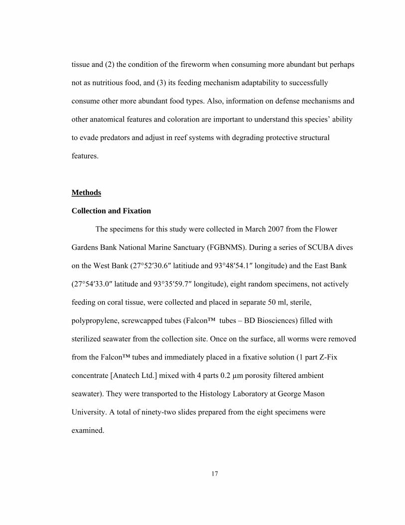
Trimming Tissue
Each fireworm was removed from the fixative and placed on a dissecting board.
All specimens were trimmed with a scalpel in sections according to Figure 2.1. Section I
was the head cut, two segments past the hard section. It was cut along the midsagittal
plane into a left lateral and a right lateral section. Section II had two sections IIA
(anterior cross section) and IIP (posterior cross section). These sections were cut
perpendicular to the axis into two 3-mm thick pieces. Section III was a longitudinal cut, 1
cm in length, along the midsagittal plane. Section IV was similar to section II and had
two sections, IVA (anterior cross section) and IVP (posterior cross section). These
sections were cut along the axial plane into two 3 mm pieces. Section V was a
longitudinal cut of the final posterior section. This section of tissue was cut into left
lateral and right lateral sections along the midsagittal plane. Due to small body lengths,
three specimens had sections I-III and did not have sections IV-V. Table 2.1 shows the
number of slides per section.
Trimmed tissue sections were individually wrapped in lens paper, placed in a
labeled round cassette (side of tissue for microscopic viewing face down), and stored in a
plastic tub filled with fixative (1 part Z-Fix concentrate [Anatech Ltd.] mixed with 4 parts
0.2 µm porosity filtered ambient seawater water).
18
19
Figure 2.1 A pictorial re s. The dotted lines indicate the parasaggita Table 2.1 Total num ections IVA-V were prepared for longer worm
Section Total # of S
(HandE and Cason’s)
presentation of the trimmed tissue sectionl sections.
ber of slides analyzed for each section. Ss only.
lides Samples
I 14 All except 07-053
IIA 16 All
IIP 16 All
III 16 All
IVA 10 07-056, 07-057, 07-058, 07-059, 07-060
IVP 10 07-056, 07-057, 07-058, 07-059, 07-060
V 10 07-056, 07-057, 07-058, 07-059, 07-060
anteriorSection I
Section II
Section III
Section IV
Section Vposterior

Processing and Staining Sections
The cassettes were removed from the fixative and washed in running tap water for
15 min and then placed in a tissue processor with fresh reagents according to Peters et al.
(2005). Each processor treatment step was set for 45 min.
After the completion of processing, the basket of cassettes was placed in a beaker
of molten Paraplast Extra at 56 °C and transferred into a vacuum oven kept at 60 °C.
Vacuum infiltration was performed manually by pumping out the air from the oven until
it reached 21-22 inches (in) Hg, and vacuum was held for 1 m. Air was returned to the
chamber slowly, then the vacuum pumping started again. Air pressure in the oven
remained at 21-22 in Hg for 10-min before venting and removing the beaker and
transferring specimens to an embedding center. The specimens were then embedded in
Paraplast Xtra and sectioned at 4-µm thickness using disposable microtome blades. One
section from the ribbon was placed on a labeled slide, after smoothing it on the surface of
a water bath (45 °C). The slide was placed vertically for several minutes to drain water
away from the wax and then placed horizontally on a slide warmer set at 45 °C for
drying. This procedure was repeated for each block to obtain two slides per block.
For this study, two staining procedures were used: Mayer’s hematoxylin and eosin
(H&E) and Cason's aniline blue. Both used the same methodology for removing the
paraffin and rehydrating the tissue. The paraffin was removed and the sections were
rehydrated and dehydrated according to standard protocols (Peters et al. 2005).
After rehydration, the slides were moved to a dish containing Mayer’s
hematoxylin solution for 15 min followed by a water rinse for 2 min in deionized water.
20
For Cason’s procedure, the slides were placed in Bouin’s fixative as a mordant for the
aniline blue dye to bind to collagen for 1 h followed by Cason’s stain for 5 min and a
water rinse for 3-5 s. The post-staining dehydration steps were the same for both staining
procedures. The stained slides were coverslipped with Permount.
Results
Integumentary System
Epidermis and Cuticle
H. carunculata has an epidermis composed of pseudostratified columnar cells
overlaid by a cuticle (Figure 2.2). The base of the epidermis rests on collagenous
connective tissue (basement membrane). Melanocytes are found throughout the
epidermis, varying by individual fireworm in color and range of colors (Figure 2.3). The
pigment granules in Cason’s-stained sections range from green, orange, red, purple, and
pink. Interestingly, there are a few specimens where clusters of green granules are found
in the same orientation in each segment throughout the worm (Figure 2.4). There is a
uniform film lining the external surface of the collagenous cuticle (Figure 2.5). This film
is a simple layer of cells resembling, and of the size of, bacteria. Circular and longitudinal
muscles are found at the base of both the ventral and dorsal epidermis (Figure 2.3 and
2.4). The dorsal epidermis has a rigid appearance with frequent and small grooves while
the ventral epidermis is smoother with longer grooves (compare dorsal epidermis in
Figure 2.4 to ventral in Figure 2.3).
21
22
Parapodia
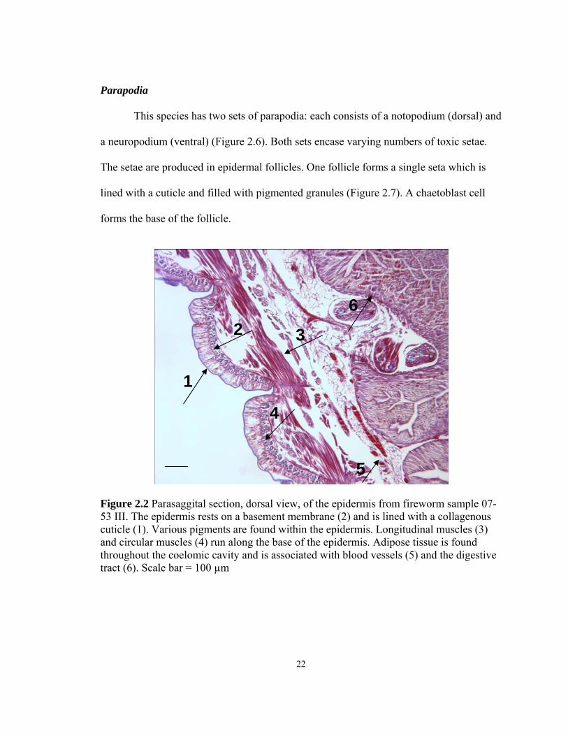
This species has two sets of parapodia: each consists of a notopodium (dorsal) and
a neuropodium (ventral) (Figure 2.6). Both sets encase varying numbers of toxic setae.
The setae are produced in epidermal follicles. One follicle forms a single seta which is
lined with a cuticle and filled with pigmented granules (Figure 2.7). A chaetoblast cell
forms the base of the follicle.
Figure 2.2 Parasaggital section, dorsal view, of the epidermis from fireworm sample 07-53 III. The epidermis rests on a basement membrane (2) and is lined with a collagenous cuticle (1). Various pigments are found within the epidermis. Longitudinal muscles (3) and circular muscles (4) run along the base of the epidermis. Adipose tissue is found throughout the coelomic cavity and is associated with blood vessels (5) and the digestive tract (6). Scale bar = 100 µm
1
2 3
4
6
5

23
Figure 2.3 granular pigm
Circulatory
each segm 2).
Adipose tissue encom
1
2
Parasaggital section, ventral view, of the fireworm sample 07-60 V. Brown ents (1) are found in the dorsal epidermis. The clusters of rings are
transverse sections through the bases of the hollow setae (2). Scale bar = 100 µm
system
There are several blood vessels found in this species. The main vessel is the
longitudinal dorsal blood vessel (Figure 2.11). There are also ventral vessels that
surround the nerve cord. Some dorsal cross-sectional blood vessels have been observed in
ent. Blood vessels can also be found surrounding the gut cavity (Figure 2.
passes blood vessels (Figure 2.9).

24
Figure 2.4 Parasaggital section, dorsal view, of the fireworm sample 07-53 III. The epidermis in this specimen has green granular pigments (black arrows) at the posterior end of each segment throughout the body. Scale bar = 100 µm
Figure 2.5 Cross-section, ventral view, of fireworm sample 07-56 IVA. Layer of cells (black arrow) are uniformly distributed on the surface of the cuticle. Scale bar = 50 µm

25
Figure 2.6 Side view of Hermodice carunculata. The two red arrows indicate the parapodia. The notopodium (1) is dorsal and the neuropodium (2) is located ventrally.
Figure 2.7 Cross-section of fireworm sample 07-58 IVP. The toxic setae have cuticle coverings and are filled with red cells (black arrow). Scale bar = 100 µm
1
2

26
Figure 2.8 Parasaggital section, dorsal view, of fireworm sample 07-59 I. The eye (1) is a simple photoreceptor which is surrounded by brown-pigmented cells. The dorsal cirri (2) is another sensory receptor. The photoreceptor (1) and the nuchal organ are connected to the cerebral ganglion (3). Scale bar = 100 µm Nervous System
This species has a two-lobed ventral nerve cord that runs anterior to posterior
(Figure 2.12). The cord is connected to the epidermis and the pigmented granules at this
connection are at a higher density than anywhere else in the body (Figure 2.12). Also,
granules at this connection penetrated the cuticle (Figure 2.13). Blood vessels are found
on both sides of the cord (Figure 2.11).
A cerebral ganglion is noticeable ventral and posterior to the eye (Figure 2.8). The
ganglion is connected to the dorsal nuchal organ (Gardiner 1992), also known as the
caruncle (Fauchald 1977) through nerve fibers (Figures 2.8 and 2.9). The cerebral
1
2
3

27
Figure 2.9 Parasaggital section, dorsal view, from fireworm sample 07-59 I. Sensory nerve fibers (black arrow) connect the nuchal organ to the cerebral ganglion. Scale bar = 100 µm
Figure 2.10 Dorsal view of Hermodice carunculata. The red arrow indicates the location of the buccal region, located at the most anterior part of the body.

28
Figure 2.11 Cross-section view of fireworm sample 07-58 IVP. The main blood vessel (2) is dorsal. Several blood vessels (4) are located around the ventral nerve cord (5). Notice the notopodium (1) and the neuropodium (3). Scale bar = 40 mm ganglion consists of neurons and a neuropil of nueraxons and dendrites. The connection
from the cerebral region and the ventral nerve cord was not observed.
Sensory Structures
H. carunculata has two eyes and each has photoreceptor cells surrounded by
brown- pigmented (Cason’s and H&E stains) granules (Figure 2.8). Nerve fibers are
found around the pigmented granules and neurons are attached to the pigmented areas.
This area may reflect light to increase photosensitivity. A second feature of this species’
sensory system is the nuchal organ which is located dorsally at the anterior end of the
body (Figure 2.9 and 2.10). It is a complex folded structure with the typical

29
Figure 2.12 Cross-section, ventral view, from fireworm sample 07-59 IIA. The nerve cord is two lobed (1 and 2) and is connected to the epidermis (3). Scale bar = 40 µm
epidermis and covering cuticle layer. It is immediately posterior to the photoreceptor. The
sensory cells connect the photoreceptor and the nuchal organ to the nerve cells (Figure
2.8) posterior to the eye.
Excretory System
A well defined excretory system was not observed in these specimens. However,
literature sources (Fauchald 1977, Gardiner 1992) state that the metanephridium (which

30
Figure 2.13 Cross-section of fireworm sample 07-59 IVP. The pigmented cells in the epidermis are at highest density where the nerve cord connects to the epidermis (1). Some of the granules from the pigmented cells penetrate the cuticle (2). Scale bar = 50 µm
includes the nephrostome, nephridial tubule, and the nephridiopore) in polychaetes may
be associated with the nerve cord. Therefore, further analysis will be needed to better
understand the function and association of the nerve cord with the excretory system of H.
carunculata.
Digestive System
The digestive tract runs longitudinally from anterior to posterior and is a
composed of pseudostratified columnar ciliated epithelial cells on a basement membrane
(Figures 2.18-2.20).
1
2

31
Figure 2.14 Parasaggital section of the foregut from fireworm sample 07-56 I. The foregut is lined with a thick collagenous cuticle (1), which stains blue with aniline blue. Circular muscles line the foregut (4). Granular secretory cells and mucus-secreting cells (mucocytes) are part of the pseudostratified epithelium (2 and 3). Scale bar = 50 µm Foregut
Like many polychaetes, H. carunculata has an eversible proboscis. Numerous layers of
longitudinal and circular muscles (varying in thickness) are observed in this anterior
region which supports its ability to extract food in the absence of jaws. Also, the
pseudostratified columnar epithelium in this region is lined with a thick cuticle which
may function to protect this region during eversion of the pharynx when feeding (Figure
2.14). The proboscis has large, dense pockets of granular secretory cells ranging in colors
from red to orange, purple, and yellow (Cason’s stain; purple and red in H&E stain)

Figure 2.15 Parasaggital image of the foregut from fireworm sample 07-057 I. The proboscis contains thick clustering of red, pink, orange, and purple-staining (with Cason’s procedure) granular secretory cells, possibly used during feeding. The cuticle is very thin in the sections. The black box indicates the section captured at higher magnification in Figure 2.16. Scale bar = 100 µm
(Figure 2.15 and 2.16). These cells are not found anywhere else in the body. These
secretory cells may take part in the feeding mechanism to extract food.
The foregut has a cuticle lining the epithelium (Figure 2.14). The cuticle thins and
disappears posterior in sections IIA and IIP. Secretory cells are present; however, there
appears to be no interaction between food particles and the epithelium in this region.
Given its location in the most anterior portion of the gut, this cuticle-lined epithelium is
probably the pharynx. A cuticle-lined epithelium in this section of the gut will function as
protection during feeding when this region is exposed and potentially vulnerable to
foreign objects.
32

Figure 2.16 Close-up image of Figure 1.15. The black arrow indicates the release of the secretory granules into the gut cavity. These granules may function to break down food during feeding and after ingestion. Scale bar = 50 µm
Figure 2.17 Parasaggital section from fireworm sample 07-56 I. The black arrow indicates a specialized tissue. It is surrounded by a cuticle and has blood vessels and pigmented cells (Cason’s stain shown). Its function is unclear. Scale bar = 100 µm
33

34
Interestingly, there is a specialized tissue in the anterior section of the epithelium
(Figure 2.17). The structure is lined with a cuticle and comes out into the gut cavity from
the epithelium on a stalk. It is located within the foregut near the proboscis and is filled
with brown and red cells (Cason’s stain). In the H&E stained sections, the cells are purple
and pink. Blood vessels are present and epithelium cells are absent. Its function is
unknown.
Figure 2.18 Cross-section of the foregut (1) and more posterior digestive tract (2) from fireworm sample 07-59IIA. The foregut is lined with a thick blue cuticle and the epithelium contains red granular secretory cells and mucocytes. The digestive tract epithelium (2) is not lined with a cuticle and there is an absence of cilia and the large secretory cells found in the foregut and proboscis. The black arrows indicate a longitudinal muscle. Scale bar = 100 µm
2
1

35
Figure 2.19 sample
secretory cells are p
Midgut
The m
microvillous
lume s III
Cells are ciliated with varying degrees throughout the gut. The longer, more pronounced
cilia are found in the posterior epithelium, which also lacks granular secretory cells
(Figure 2.20). Shorter cilia are found in the anterior portion of the gut, where there is also
1
2
Parasaggital section of the digestive tract posterior from fireworm07-055 III. Cilia are present on the apical surfaces of these columnar cells (1). The
resent in the epithelium and granules are released into the gut cavity as indicated by the black arrow (2). Scale bar = 50 µm
idgut is anterior to the foregut and contains the intestine which has a
appearance. The intestine epithelium contains different cells, including
gland cells that appear to produce zymogen granules, which are discharged into the
n (Figures 2.16 and 2.19). Within the epithelium, mucocytes are found in section
and IV. Both circular and longitudinal muscle layers encompass the wall.

36
Figure 2.20 Parasaggital section of the most posterior gut cavity from fireworm sample 07-053 III. The epithelium is unlined and the length and density of cilia is higher in this region of the digestive tract than in other regions. Also, few secretory cells are present. Scale bar = 50µm
a higher density of granular secretory cells (Figure 2.19). Furthermore, higher densities of
granular secretory cells are found within the gut cavity in the anterior section of the gut
(sections I-II) (Figure 2.16). The disparity between anterior and posterior epithelium in
relation to cilia and suspected zymogens may be an indication that the main site of
digestion is the anterior intestine. The long cilia in the posterior epithelium function to
transport the remaining materials to the rectum.
The feeding behavior for these specimens was not observed at the time of
collection. Also, these specimens were collected at different sites on different days.
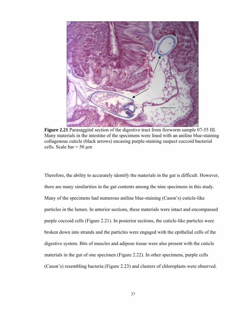
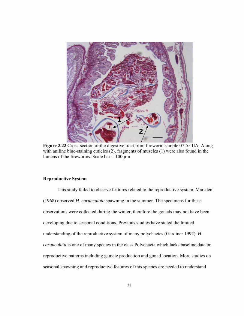
Figure 2.21 Parasaggital section of the digestive tract from fireworm sample 07-55 III. Many materials in the intestine of the specimens were lined with an aniline blue-staining collagenous cuticle (black arrows) encasing purple-staining suspect coccoid bacterial cells. Scale bar = 50 µm
Therefore, the ability to accurately identify the materials in the gut is difficult. However,
there are many similarities in the gut contents among the nine specimens in this study.
Many of the specimens had numerous aniline blue-staining (Cason’s) cuticle-like
particles in the lumen. In anterior sections, these materials were intact and encompassed
purple coccoid cells (Figure 2.21). In posterior sections, the cuticle-like particles were
broken down into strands and the particles were engaged with the epithelial cells of the
digestive system. Bits of muscles and adipose tissue were also present with the cuticle
materials in the gut of one specimen (Figure 2.22). In other specimens, purple cells
(Cason’s) resembling bacteria (Figure 2.23) and clusters of chloroplasts were observed.
37
38
Figure 2.22 Cross-section of the digestive tract from fireworm sample 07-55 IIA. Along with aniline blue-staining cuticles (2), fragments of muscles (1) were also found in the lumens of the fireworms. Scale bar = 100 µm Reproductive System
This study failed to observe features related to the reproductive system. Marsden
(1968) observed H. carunculata spawning in the summer. The specimens for these
observations were collected during the winter, therefore the gonads may not have been
developing due to seasonal conditions. Previous studies have stated the limited
understanding of the reproductive system of many polychaetes (Gardiner 1992). H.
carunculata is one of many species in the class Polychaeta which lacks baseline data on
reproductive patterns including gamete production and gonad location. More studies on
seasonal spawning and reproductive features of this species are needed to understand
21
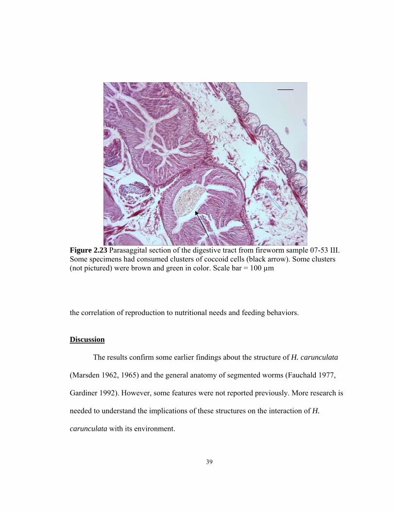
Figure 2.23 Parasaggital section of the digestive tract from fireworm sample 07-53 III. Some specimens had consumed clusters of coccoid cells (black arrow). Some clusters (not pictured) were brown and green in color. Scale bar = 100 µm
the correlation of reproduction to nutritional needs and feeding behaviors.
Discussion
The results confirm some earlier findings about the structure of H. carunculata
(Marsden 1962, 1965) and the general anatomy of segmented worms (Fauchald 1977,
Gardiner 1992). However, some features were not reported previously. More research is
needed to understand the implications of these structures on the interaction of H.
carunculata with its environment.
39
Integumentary System
The epidermis and outer collagenous cuticle of H. carunculata is typical of the
Order Amphinomida (Gardiner 1992). Furthermore, the toxic setae in the notopodium
and neuropodium have also been reported for H. carunculata previously (Marsden 1962).
The pigment granules in the epidermis were in different orientations, densities, and
variation of colors unique to individual specimens. This histological observation is
consistent with field observations of various color schemes for this species (Lewis,
unpublished data).
A thin layer of coccoid cells, perhaps bacteria, was found lining the outside of the
cuticle on all organisms. There was no evidence of exchange between these cells and the
epidermis. This thin layer could be another type of defense mechanism, perhaps a
“biofilm.” If these cells are microbes, their presence may function to protect the cuticle.
Conversely, the cells could feed on the cuticle or the biofilm could pick up microbes
known to cause coral disease. Sussman et al. (2003) showed the Vibrio shilio cells
penetrated the epidermis, but did not mention the existence of a collagenous cuticle nor
how the bacteria were able to pass through it. This outer layer of cells has not been
previously noted in literature for this species and therefore more research should be done
to understand the role and biological nature of this outer cuticle coating.
40
Circulatory System
The circulatory system consists of various blood vessels including the main blood
vessels located dorsally. Blood vessels were also associated with the gut cavity. No new
or previously unreported features of the circulatory system were noted in this study.
Nervous System
H. carunculata has multiple features that function to find prey and avoid
predators. This species has primitive eyes in the form of two photoreceptor lenses located
at the most anterior section of the body. The eyes are connected via nerve fibers to a
cerebral ganglion. The cerebral ganglion also receives information from the nuchal organ
in the caruncle region located on the dorsal side of the body. The epidermis of the
caruncle region is intensely folded and supported on connective tissue, with nerve fibers
apparently connecting to epidermal cells, although further study with transmission
electron microscopy will be necessary to determine whether the ends of the fibers are
modified in some manner to support chemoreception or mechanoreception. A large
surface area in this region would increase sensory capability. Therefore, this observation
suggests the caruncle region serves to pick up cues from the environment and may serve
to locate prey and/or to identify predators.
The most obvious defense mechanism of the species which has been mentioned is
previous studies (Marsden 1962) is the toxic setae found in high density in both
parapodia. The setae are lined cuticles filled with red granules (H&E stain). Nuclei and
other organelles (H&E stain) were not present in these granules. Muscles surround the
41

base of the setae. The setae appear to grow out of a follicle in the epidermis, which
indicates the site of regeneration of broken setae.
An unreported observation of the nervous system is the connectivity of the two-
lobed nerve cord to the epidermis. The nerve cord runs ventrally along the body and is
connected to the epidermis through a thin connective tissue layer. At the point of contact
with the nerve cord, the epidermis has pigmented granules uniquely dense in this area and
some cells penetrate the cuticle. Furthermore, the segment in this region is uniquely
shaped with a central peak, similar to a keel on a boat, at the site of penetrating granules.
The nerve cord may receive information from the external environment via these
pigmented cells. This interface outside the body shows it has a sensory function which
could be for finding prey as it moves along substrate. An alternative function of these
penetrating pigmented granules could also have a mucous function and seep through
pores in the cuticle. Marsden (1968) noted a dark mid-ventral line which contained dark
material similar to fecal excretions. This region is in the same location as the mid-ventral
line and could also serve as another site of waste removal. More observations on this
feature are needed to fully understand its function.
Digestive System
The structural features used for consuming prey were examined to understand the
ability of H. carunculata to forage on various types of organisms. The proboscis is made
up of numerous layers of muscles which would aid in extracting materials from a solid
substrate. The epithelium of the foregut and the pharynx is lined with a thick cuticle. This
42
feature may protect the epithelium when exposed to the environment. Though this
invertebrate is not equipped with jaws similar to other marine invertebrates (Gardiner
1992), it uses its powerful pharynx muscles for effective predation. Bruckner (2001)
documented H. carunculata enlarging its pharynx to completely engulf the tips of A.
cervicornis and removing the coral tissue.
Once food has been consumed, it passes through the foregut and into the digestive
tract (Marsden 1963b). The epithelium posterior to the foregut is not lined with a cuticle.
Mucocytes and granular secretory cells embedded in the epithelium secrete compounds to
work on the materials in the lumen for digestion (Marsden 1966). The materials move
through the gut aided by the muscles lining the digestive tract and apical cilia of the thin
columnar epithelial cells. The cilia become longer and denser in the posterior section of
the gut. Secretory cells and mucocytes become less numerous in the epithelium inverse to
the length and density of the cilia. These observations are consistent with previous studies
on the digestive tract of this species (Marsden 1966).
Gut content was examined to extrapolate diet composition of this species. Cuticles
of various shapes and sizes were found consistently in specimens. In some instances, the
cuticles were found with clusters of coccoid cells resembling bacteria. Also, in one
specimen, cuticles were found in the same region of the gut as fragmented muscle cells.
Clusters of green cells resembling chloroplasts (H&E), brown cells (H&E), and purple
cells resembling bacteria were found independent of other materials. The materials in the
gut varied which is consistent with previous studies (Marsden 1962, 1963a).
43

Two features of the foregut, not previously reported, were found in the course of
this investigation: clusters of secretory cells in the proboscis region and a specialized
tissue within the digestive tract. The orientation of these secretory cells suggests they are
released when the pharynx is everted during feeding to breakdown the tissue and aid in
consuming materials. However, previous studies have not documented the release of
digestive enzymes on the feeding site. This feeding technique, specifically on coral
colonies, may induce tissue loss after the predation episode is over. The impact of
digestive enzymes on coral tissue needs to be understood to help diagnose causal agents
of coral tissue loss in field.
The second new feature is the specialized tissue within the digestive system. It is
lined with a cuticle similar to the surrounding epithelium. However, the tissue is not
directly attached to the epithelium and comes out into the digestive cavity via a stalk. Its
function is unclear and more studies are needed to ascertain the role of this tissue.
Marsden (1963a) identified coral tissue by the presence of nematocysts and
clusters of cells that appeared to be zooxanthellae. However, her findings were not
coordinated with active coral feeding observations. The specimens collected for this
study were also not actively feeding on coral and evidence of coral tissue, including the
presence of nematocysts, based on Marsden’s criteria, was missing. H. carunculata can
take up to 9 hours to fully digest materials (Marsden 1968). Nevertheless, these
corallivores did not have evidence of coral tissue in their gut which would indicate these
specimens were feeding on alternative prey in the presence of coral tissue. Due to the
lack of documented studies on identifying coral materials in gut contents, this study can
44
not draw conclusions about the presence or absence of coral tissue in the digestive tract.
Future studies are needed to coordinate feeding observationswith histological and
physiological analysis to obtain a more complete picture of the appearance of coral tissue
in the gut and document the passage of coral through the digestive system.
H. carunculata has not been researched extensively. To date, most studies have
focused on the digestive tract (Marsden 1962, 1963), on foraging cycles (Lewis and
Crooks 1996), or on field observations at one site (Miller and Williams 2007). Sussman
et al. (2003) found this species to be a vector for the bleaching pathogen V. shioli but did
not provide information on the length of time the bacteria were retained in the specimens’
bodies. Despite these studies, baseline information on diet, feeding preferences, and
trophodynamics is lacking for this species. The results from this study indicate this
species has features, like a “biofilm,” a ventral sensory feature, and secretory cells in the
proboscis, which may be key elements in its interactions with its prey and benthic habitat.
Also, the observations, at least from this sample of worms, indicate coral tissue is not a
vital part of this species’ diet. Since detailed information on H. carunculata’s diet
composition is lacking, it is difficult to determine the impact of predation either on coral
or on its alternative prey. The presence of secretory cells in the proboscis suggests this
species may use digestive enzymes to aid in consumption of food. Furthermore, the
pharynx has tremendous elasticity to enlarge and is supported by layers of muscles to
consume prey. These features will aid H. carunculata to adapt its feeding behavior to
consume different, more abundant, types of prey.
45

As available coral tissue declines worldwide, the loss of coral reef biological and
physical structures are impacting the survivorship of coral reef inhabitants. More research
is needed to better understand H. carunculata’s role in coral reef ecosystems to predict
the impact of coral reef decline on this species and its prey. Studies on the adaptability of
these coral reef inhabitants are needed to understand their ability to survive in a changing
environment.
46
Literature Cited
Adams AJ, Ebersole JP (2002) Use of back-reef and lagoon habitats by coral reef fishes. Mar Ecol-Prog Ser 228:213-226
Alvarez-Filip L, Gil I (2006) Effects of Hurricanes Emily and Wilma on coral reefs in
Cozumel, Mexico. Coral Reefs 25:583-583 Aronson RB, MacIntyre IG, Wapnick CM, O'Neill MW (2004) Phase shifts, alternative
states, and the unprecedented convergence of two reef systems. Ecology 85:1876-1891
Baums IB, Miller MW, Szmant AM (2003a) Ecology of a corallivorous gastropod,
Coralliophila abbreviata, on two scleractinian hosts. 1: Population structure of snails and corals. Mar Biol 142:1083-1091
Baums IB, Miller MW, Szmant AM (2003b) Ecology of a corallivorous gastropod,
Coralliophila abbreviata, on two scleractinian hosts. II. Feeding, respiration and growth. Mar Biol 142:1093-1101
Bell JD, Galzin R (1984) Influence of live coral cover on coral-reef fish communities.
Mar Ecol-Prog Ser 15:265-274 Bellwood DR, Hoey AS, Ackerman JL, Depczynski M (2006) Coral bleaching, reef fish
community phase shifts and the resilience of coral reefs. Glob Change Biol 12:1587-1594
Berumen ML, Pratchett MS, McCormick MI (2005) Within-reef differences in diet and
body condition of coral-feeding butterflyfishes (Chaetodontidae). Mar Ecol-Prog Ser 287:217-227
Booth DJ, Wellington G (1998) Settlement preferences in coral-reef fishes: Effects on
patterns of adult and juvenile distributions, individual fitness and population structure. Blackwell Science, p 274-279
Bruckner AW (2001) Coral health and mortality. Recognizing the signs of coral diseases
and predators. In: Humann P, Deloach N (eds) Reef Coral Identification: Florida, Caribbean, Bahamas, Including Marine Plants, p 240-271
Cabaitan PC, Gomez ED, Alino PM (2008) Effects of coral transplantation and giant
clam restocking on the structure of fish communities on degraded patch reefs. J Exp Mar Biol Ecol 357:85-98
47

Carr M, Hixon M (1997) Artificial reefs: the importance of comparisons with natural reefs. Mar Ecol-Prog Ser 22:28-33
Cheal A, Wilson SK, Emslie MJ, Dolman A, Sweatman H (2008) Responses of reef fish
sommunities to coral declines on the Great Barrier Reef. Mar Ecol-Prog Ser 372:211-223
Connell SD, Kingsford MJ (1998) Spatial, temporal and habitat related variation in the
abundance of large predatory fish at One Tree Reef, Australia. Coral Reefs 17:49-57
Done TJ (1992) Phase shifts in coral reef communities and their ecological significance
Hydrobiologia 247:121-132 Fauchald K (1977) The Polychaete Worms: Definitions and Keys to the Orders, Families
and Genera, Vol 28. Natural History Museum of Los Angeles County, Los Angeles
Gardiner S (1992) Annelida. In: Harrison F (ed) Microscopic Anatomy of Invertebrates,
Vol 7. Wiley-Liss, New York Hayes JA (1990) Prey preference in a Caribbean corallivore, Coralliophila abbreviata
(Lamark) (Gastropoda, Coralliophilidae). . Bull Mar Sci 47:557-560 Hoegh-Guldberg O, Mumby PJ, Hooten AJ, Steneck RS, Greenfield P, Gomez E, Harvell
CD, Sale PF, Edwards AJ, Caldeira K, Knowlton N, Eakin CM, Iglesias-Prieto R, Muthiga N, Bradbury RH, Dubi A, Hatziolos ME (2007) Coral reefs under rapid climate change and ocean acidification. Science 318:1737-1742
Hughes TP, Rodrigues MJ, Bellwood DR, Ceccarelli D, Hoegh-Guldberg O, McCook L,
Moltschaniwskyj N, Pratchett MS, Steneck RS, Willis B (2007) Phase shifts, herbivory, and the resilience of coral reefs to climate change. Curr Biol 17:360-365
Hughes TP, Tanner JE (2000) Recruitment failure, life histories, and long-term decline of
Caribbean corals. Ecology 81:2250-2263 Jones GP, McCormick MI, Srinivasan M, Eagle JV (2004) Coral decline threatens fish
biodiversity in marine reserves. Proc Natl Acad Sci U S A 101:8251-8253 Kokita T, Nakazono A (2001) Rapid response of an obligately corallivorous filefish
Oxymonacanthas longirostris (Monacanthidae) to a mass coral bleaching event. Coral Reefs 20:155-158
48

Lewis JB, Crooks RE (1996) Foraging cycles of the amphinomid polychaete Hermodice carunculata preying on the calcareous hydrozoan Millepora complanata. Bull Mar Sci 58:853-856
Lindahl U, Ohman MC, Schelten CK (2001) The 1997/1998 mass mortality of corals:
Effects on fish communities on a Tanzanian coral reef. Mar Pollut Bull 42:127-131
Lizama J, Blanquet RS (1975) Predation on sea anemones by the amphinomid
polychaete, Hermodice carunculata. Bull Mar Sci 25:442-443 Marsden JR (1962) A coral-eating polyachete. Nature 193:598 Marsden JR (1963a) A preliminary report on digestive enzymes of Hermodice
carunculata. Canadian Journal of Zoology/Revue Canadien de Zoologie 41:159-164
Marsden JR (1963b) The digestive tract of Hermodice carunculata. (Pallas). Polychaeta:
Amphinomidae. Canadian Journal of Zoology/Revue Canadien de Zoologie 41:165-184
Marsden JR (1966) The coelomocytes of Hermodice carunculata (Polychaeta:
Amphimomidae) in relation to digestion and excretion. Canadian Journal of Zoology/Revue Canadien de Zoologie 44:377-389
Martin A, Losada F (1991) Regeneration of Injuries caused by the Amphinomid
polychaete Hermodice carunculata on scleractinian corals, hydrocorals and octocorals (Cnidaria). Ecotropicos 4:18-26
Menge BA, Lubchenco J (1981) Community organization in temperate and tropical rocky
intertidal habitats - prey refuges in relation to consumer pressure gradients Ecol Monogr 51:429-450
Miller MW, Williams DE (2007) Coral disease outbreak at Navassa, a remote Caribbean
island. Coral Reefs 26:97-101 Motta PJ (1988) Functional morphology of the feeding apparatus of 10 species of pacific
butterflyfishes (Perciformes, Chaetodontidae) – an ecomorphological approach. Environ Biol Fishes 22:39-67
Mumby PJ, Dahlgren CP, Harborne AR, Kappel CV, Micheli F, Brumbaugh DR, Holmes
KE, Mendes JM, Broad K, Sanchirico JN, Buch K, Box S, Stoffle RW, Gill AB (2006) Fishing, trophic cascades, and the process of grazing on coral reefs. Science 311:98-101
49

Newman MJH, Paredes GA, Sala E, Jackson JBC (2006) Structure of Caribbean coral
reef communities across a large gradient of fish biomass. Ecol Lett 9:1216-1227 Ott B, Lewis JB (1972) The importance of the gastropod Coralliophila abbreviata
(Lamarck) and the polychaete Hermodice carunculata (Pallas) as coral reef predators. Can J Zool/Rev Can Zool 50:1651-1656
Pandolfi JM, Jackson JBC (2006) Ecological persistence interrupted in Caribbean coral
reefs. Ecol Lett 9:818-826 Pandolfi JM, Jackson JBC, Baron N, Bradbury RH, Guzman HM, Hughes TP, Kappel
CV, Micheli F, Ogden JC, Possingham HP, Sala E (2005) Ecology - Are US coral reefs on the slippery slope to slime? Science 307:1725-1726
Pratchett MS, Munday PL, Wilson SK, Graham NAJ, Cinner JE, Bellwood DR, Jones
GP, Polunin NVC, McClanahan TR (2008) Effects of climate-induced coral bleaching on coral-reef fishes - Ecological and economic consequences. In: Oceanography and Marine Biology: an Annual Review, Vol 46, Vol 46. Crc Press-Taylor and Francis Group, Boca Raton, p 251-296
Pratchett MS, Wilson SK, Baird AH (2006) Long-term monitoring of the Great Barrier
Reef. J Fish Biol 69:1269-1280 Pratchett MS, Wilson SK, Berumen ML, McCormick MI (2004) "Sublethal effects of
coral bleaching on an obligate coral feeding butterflyfish". Coral Reefs 23:352-356
Rotjan RD, Lewis SM (2005) Selective predation by parrotfishes on the reef coral Porites
astreoides. Mar Ecol-Prog Ser 305:193-201 Rotjan RD, Lewis SM (2008) Impact of coral predators on tropical reefs. Mar Ecol-Prog
Ser 367:73-91 Samways MJ (2005) Breakdown of butterflyfish (Chaetodontidae) territories associated
with the onset of a mass coral bleaching event. Aquatic Conservation: Marine and Freshwater Ecosystems 15:S101-S107
Sano M, Shimizu M, Nose Y (1984) Changes in structure of coral-reef fish communities
by destruction of hermatypic corals - observational and experimental views Pacific Science 38:51-79
50

Smith JE, Shaw M, Edwards RA, Obura D, Pantos O, Sala E, Sandin SA, Smriga S, Hatay M, Rohwer FL (2006) Indirect effects of algae on coral: algae-mediated, microbe-induced coral mortality. Ecol Lett 9:835-845
Smith TB, Nemeth RS, Blondeau J, Calnan JM, Kadison E, Herzlieb S (2008) Assessing
coral reef health across onshore to offshore stress gradients in the US Virgin Islands. Mar Pollut Bull 56:1983-1991
Sussman M, Loya Y, Fine M, Rosenberg E (2003) The marine fireworm Hermodice
carunculata is a winter reservoir and spring-summer vector for the coral-bleaching pathogen Vibrio shiloi. Environmental Microbiology, 2003 Apr, 5(4):250-5
Syms C, Jones GP (2000) Disturbance, habitat structure, and the dynamics of a coral-reef
fish community. Ecology 81:2714-2729 Tricas TC (1989) Determinants of feeding territory size in the corallivorous butterflysih,
Chaetodon multicinctus Anim Behav 37:830-841 Wilkinson C (ed) (2004) Status of coral reefs of the world, Vol 1. Global Coral Reef
Monitoring Network and Australian Institute of Marine Science, Townesville, Australia
Wilson SK, Graham NAJ, Pratchett MS, Jones GP, Polunin NVC (2006) Multiple
disturbances and the global degradation of coral reefs: are reef fishes at risk or resilient? Glob Change Biol 12:2220-2234
Witman JD (1988) Effects of predation by the fireworm Hermodice carunculata on
Milleporid hydrocorals. Bull Mar Sci 42:446-458 Yap HT (2004) Differential survival of coral transplants on various substrates under
elevated water temperatures. Mar Pollut Bull 49:306-312 Yap HT (2009) Local changes in community diversity after coral transplantation. Mar
Ecol-Prog Ser 374:33-41
51

3. The effect of a Millepora complanata feeding regime on the body weight of
Hermodice carunculata
Abstract
As coral reef species densities and distributions shift in response to environmental
changes, impacts on benthic coral reef communities need to be well understood. Dietary
habitats and preferences are important considerations when determining the
consequences of coral reef decline on corallivores and their prey. However, the effects of
decreased available coral tissue on the nutrient intake of direct coral consumers, like the
invertebrate corallivore Hermodice carunculata, has not been addressed extensively in
previous studies. H. carunculata is commonly found on tropical reefs including off
Barbados. Barbados reefs have experienced high percentage of coral colony mortality
over the past 30 years. The hydrocoral Millepora complanata is now found in higher
densities than previously abundant coral species like Montastraea annularis. Ex situ
experiments were conducted to examine the weight change of H. carunculata organisms
under a feeding regime of M. complanata as compared with a feeding regime of a
previously abundant species M. annularis. This study showed a M. complanata feeding
regime did not have a substatantal impact on the body weight of H. carunculata over
time.
52

Introduction
Coral populations are declining worldwide (Knowlton 2001, Gardner et al. 2003,
Hughes et al. 2003, Pandolfi et al. 2003, Bellwood et al. 2004). The causes have been
attributed to various factors both human-induced and biotic, including overfishing
(Jackson et al. 2001, Myers and Worm 2003), diseases (Pandolfi et al. 2003, Sutherland
et al. 2004), storms (Bythell et al. 1993, Hughes 1994, Knowlton 1992, Alvarez-Filip and
Gil 2006), and elevated sea temperatures (Runnalls and Coleman 2003, Edmunds 2004,
Rotjan and Lewis 2008). Coral mortality has been particularly widespread throughout the
Caribbean (Connell 1997, Green et al. 2008, Mumby et al. 2007, Pandolfi et al. 2003). It
has been estimated that Caribbean corals have declined by 80% in the last 30 years
(Richardson and Voss 2005). Coral reefs off of Barbados have undergone disturbances
similar to the wider Caribbean region (Runnalls and Coleman 2003, Macintyre et al.
2007). Coral cover has decreased and once dominate coral species, like Acropora
palmata, have disappeared (Aronson and Precht 2001, Aronson et al. 2004, Macintyre et
al. 2007). In place of the previously abundant scleractinian corals like Montastraea and
Acropora, species with high fecundity like Porites (Chornesky and Peters 1987, McGuire
1998) have become dominant in the community structure through increase in percentage
cover (Green et al. 2008).
Mass mortality and phase shift of coral species can have implications for reef
biodiversity (May 1977, Chapin et al. 2000, Lokrantz et al. 2008) and specifically impact
marine species which rely on coral reefs for all or part of their life cycle (Bellwood et al.
2003, Folke et al. 2004). While short-term and long-term factors continue to lead to coral
53
loss (Richardson and Voss 2005, Mumby et al. 2007), coral reef management plans must
evaluate the impact of newly dominant coral species on community ecology (Chapin et
al. 2000, Pandofli et al. 2003, Lokrantz et al. 2008). More specifically, the decline of
coral populations can have an important impact on direct consumers of coral tissue,
corallivores (Baums et al. 2003a, Rotjan and Lewis 2008).
The role of corallivores in reef communities is important and complex (Glynn
2004, Knowlton 1992, Rotjan et al. 2006, Rotjan and Lewis 2008). Coral predators total
more than 160 known species representing 11 families of fishes (Motta 1988) and 5
invertebrate phyla (Dalton and Godwin 2006, Bak and Steward-Van Es 1980, Lester and
Bak 1985, Oren et al. 1997). Corallivores practice two types of feeding mode: obligate
feeders, which eat exclusively live coral, and facultative feeders, which eat mixed diets
including coral. Corallivores use different feeding strategies which have varying levels of
impact on coral prey (Rotjan and Lewis 2008). There are four categories used to classify
the feeding techniques of most corallivores: browsers, excavators, mucus-feeders, and
scrapers.
‘Browsers’ generally remove live coral tissue without removing the underlying
skeleton (Hiatt and Strasburg 1960). ‘Excavators’ and ‘scrapers’ remove both tissue and
skeleton (Bellwood and Choat 1990). However, ‘excavators’ remove a considerable
amount of skeleton while ‘scrapers’ only take a small portion of the skeleton with the
attached tissue. ‘Mucus-feeders’ consume the coral mucus without removing tissue
and/or skeleton (Rotjan and Lewis 2008).
54

Corallivores can be found in the vertebrate and invertebrate groups. There are
reportedly 114 species of vertebrate corallivores (Rotjan and Lewis 2008). About half the
total corallivorous vertebrates are butterfly fishes and exhibit all four feeding strategies
described above (Motta 1988). The other piscine corallivores are either scrapers or
excavators and include damselfish, filefish, triggerfish, and wrasses (Bellwood and Choat
1990). Acropora, Montipora, Pocillopora, and Porites are the corals most commonly
grazed on by these fishes (Rotjan and Lewis 2008).
Annelids, anthropods, echinoderms, and mollusks make up the 51 species of
invertebrate corallivores (Rotjan and Lewis 2008). All four feeding strategies are used by
at least one species. Due to their rate of tissue consumption (Moran 1986, Colgan 1987,
Turner 1994) and periodic population outbreaks (Birkeland and Lucas 1990, Glynn 1990,
Turner 1994, Houk et al. 2007), two influential invertebrate corallivores are Acanthaster
planci (145 cm2 d-1) (Morton et al. 2002) and Drupella spp. (137-229 bites m-1) (Rotjan
and Lewis 2008). Localized surges in their population densities can result in rapid loss of
coral tissue (Rotjan and Lewis 2008) and reduced coral survival (Colgan 1987).
Food preferences for invertebrate corallivores vary but mostly consist of
Acropora, Millepora, Montipora, Pocillopora, and Porites (Morton et al. 2002). Diet has
been shown to have a major influence on the population structure of one invertebrate
corallivore, the corallivorous gastropod, Coralliophila abbreviata (Baums et al. 2003a).
Snails feeding on A. palmata grew faster than snails residing on M. annularis colonies
even though the tissue of M. annularis provides more nitrogen (N) and carbon (C) per
unit area than A. palmata (Baums et al. 2003b). Baums et. al (2003b) found feeding rates
55
depended on the nutritional value of the coral prey (M. annularis: 10.9±0.5 C:N ratio;
0.13-0.88 cm2 day-1; A. palmata: 6.3±0.3 C:N ratio; 0.44-3.28 cm2 day-1).
Population aggregations and parameters of invertebrate corallivores can be
affected by coral species diversity and abundance (Ott and Lewis 1972, Glynn 1990,
Hayes 1990, Baums et al. 2003a). However, more studies are needed to understand
invertebrate corallivory feeding preference and consequential tissue loss impact (Martin
and Losada 1991, Baums et al. 2003b, Williams and Miller 2005, Rotjan and Lewis
2008).
Hermodice carunculata is an invertebrate corallivore which has not been studied
extensively despite recognition of its important role in coral mortality (Ott and Lewis
1972, Knowlton 1992, Lewis and Crooks 1996) and etiology of coral disease (Sussman et
al. 2003, Miller and Williams 2007). H. carunculata is a segmented polychaete from the
Family Amphinomidae. Its dorsal parapodia contain toxic setae which are used as a
defense mechanism (Marsden 1963). It is widely distributed in the tropical and
subtropical areas of the Atlantic Ocean, the Southern Atlantic Ocean, and the
Mediterranean Sea (Martin and Losada 1991). H. carunculata is a facultative browser
(Rotjan and Lewis 2008) and is known to consume Acropora, Millepora, Montastraea,
Oculina, and Porites coral species (Glynn 1963, Ott and Lewis 1972, Witman 1988)
along with sponges and other invertebrates (Marsden 1963, Lizama and Blanquet 1975).
The majority of the few studies conducted on this species have focused on the
Barbados populations of H. carunculata and their predation on the hydrozoan fire corals
of the genus Millepora. Witman (1988) calculated H. carunculata consumption rate of
56
Millepora to be 12.9cm2 d-1. A separate study (Martin and Losada 1991) found the tissue
regeneration percentage and rate for Millepora was 44% and 0.06 cm2/day, respectively,
after predation by H. carunculata. Lewis and Crooks (1996) monitored a large Millepora
colony to determine the foraging cycle of H. carunculata was mostly during the late
afternoon.
Similar to Porites’ ability to dominate disturbed systems, fire corals have been
successful and spatially dominant in shallow coral reef systems (de Weerdt 1981,
Witman 1992, Lewis 2006), locally abundant and important reef-framework builders
(Lewis 1989, Edmunds 1999). As rapid growers and with the use of both asexual and
sexual reproduction, they are often the first to recover from local disturbances like
bleaching (Lewis 2006). There are some general similarities between these hydrocorals
and scleractinian corals, such as having calcareous skeletons and symbiotic
dinoflagellates (Sheppard 1982, Lewis 1992). However, fire corals differ from
scleractinians in their morphological strategy (Edmunds 1999) and energy consumption
(Lewis 1992). They can grow thin encrusting bases which can spread across substrate and
produce dense branches (Wahle 1980, Witman 1992). One benefit to this structural
characteristic is the ability to survive wave stress (Lewis 1989). Despite fire corals
protruding polyps which contain highly toxic nematocysts (Lewis 2006), a variety of
predators and burrowing animals rely on this hydrocoral (Lewis 2006), and, hence, fire
corals are also important to the community ecology of reef ecosystems (Edmunds 1999).
M. complanata and M. alcicornis can be found in large densities in the Caribbean
(Edmunds et al. 1990). In contrast, M. annularis has become reduced in density on most
57

Caribbean reefs (Tomascik and Sander 1987, Hughes and Tanner 2000, Edmunds and
Elahi 2007). Since Millepora is abundant in the shallow reef systems while M. annularis
has experienced a decrease in density, this project addressed the impact of increased M.
complanata spatial dominance on the body weight of the corallivore H. carunculata
through ex situ experiments. The physiological effect of a Millepora feeding regime on
the body weight of H. carunculata was compared to the body weight of H. carunculata
under a M. annularis feeding regime.
Field observations in Barbados indicate H. carunculata feeds on fire coral
frequently (Ott and Lewis 1972, Witman 1988, Lewis and Crooks 1996). Thus, this
observed feeding behavior would indicate that M. complanata is a substantial food source
and H. carunculata body weight will be greater in the presence of a M. complanata
feeding regime than when feeding on M. annularis. The working hypothesis is if H.
carunculata feeds on fire coral (M. complanata), its body weight will be greater than its
body weight when it has been feeding on stony corals. The null hypothesis for this study
is if H. carunculata feeds on fire coral (M. complanata), its body weight will not be
greater than its body weight when it has been feeding on stony corals.
Methods
The effect of a M. complanata feeding regime on the body weight of H.
carunculata was tested through ex situ experiments in the laboratories of the Bellairs
Research Institute in Barbados in November and December 2005 and February–April
2006. The specimens were randomly selected from the population of H. carunculata on
58
the west coast shallow fringing reefs primarily Batts Rock Reef which had high densities
of M. complanata (S. Lewis, unpublished data). The wet weight of the specimens were
measured at several intervals and compared with the wet weight of specimens
experiencing a shift in feeding regimes to M. annularis.
Experiment Set-up:
The laboratory feeding experiments were conducted in five custom-made glass
aquaria (4 x 2 x 3 ft) using a flow-through seawater system in the wet laboratory at the
Bellairs Research Institute in Barbados. Prior to the start of the experiments, the aquaria
were filled with water to leach any toxins from the adhesives used on the tanks. After one
week, the tanks were emptied and scrubbed clean. The flow-through seawater system ran
for three days to further flush out the tanks. Prior to the start of the experiments, sand was
gathered from the nearshore beach and sifted through a #3 mesh screen. The sifted sand
was then placed in the bottom of each tank to create an 8 cm-thick layer.
To limit the food resources of the specimens to only the coral colonies provided in
the tank (i.e., reduce availability of phytoplankton and zooplankton, larvae, bacteria,
etc.), the tanks were scrubbed clean and the water was drained to 10 cm above the sand
and refilled completely with the flow-through seawater system each week. During the
experiments, the flow-through seawater system ran for 8 hours each day for 4 consecutive
days per week. A large aerator was placed in each tank and ran continuously.
59
Measurements:
The body weight was determined by measuring wet weight on a Fisher Scientific
digital bench scale. To prevent physical stress to the specimens, seawater from one of the
five aquaria was poured into the scale tray to a depth appropriate for the specimens to be
completely immersed (around 4cm). The scale was tared with the water present. Each
specimen was placed in the tray and the weight recorded. After the weight was recorded
and video images were captured, the specimen was returned to the designated aquarium
and the next specimen was measured. All statistical analyses of the results were done
using SPSS17.0 software program. The tests were evaluated within a 90% confidence
interval (p≤0.10 is statistically significant).
Experiments:
Two experiments were performed with this equipment and general set-up. The
first experiment (1FCE) ran for twenty-six days and all specimens were exposed to a
feeding regime of M. complanata the entire length of the experiment. For 1FCE, each
tank was divided into three sections using mesh screens attached to PVC frames
immersed in the tanks. Each section housed an individual coral colony (15 sections of a
M. complanata sample in total) and a H. carunculata specimen (n =15), which were
collected at random from nearshore reefs. The wet weight of each worm was measured
six times throughout the experiment (days 0, 3, 5, 11, 13, and 26). On day 11, the
colonies were removed and replaced with new M. complanata colonies on day 13. Thus,
60

the worms experienced a starving period for three days. After the completion of 1FCE,
the worms were removed from the tanks.
In the second experiment (2FCE), the mesh screens were removed from the tanks.
The 2FCE ran for 109 days and involved two feeding regimes at different times. For a
total of 94 days, 13 specimens (two escaped from the tanks during the experiment) were
provided with M. complanata colonies. The first round of colonies was removed on day
79 and the worms experienced starvation for the next 6 days. New M. complanata
colonies were introduced into the tanks on day 87. The worms continued to feed on M.
complanata colonies until day 94 when these fire coral colonies were immediately
replaced with M. annularis colonies. The experiment with the new feeding regime of M.
annularis (2FCE M. ann) ran for 15 days. The wet weight of each worm was measured
nine times throughout the experiment (days 0, 11, 28, 53, 79, 87, 94, 103, and 109).
Results:
Analysis of overall weight change for each experiment (1FCE, 2FCE, 2FCE M. ann)
The initial average weight of the specimens in 1FCE (n = 15) was 1.07 g ± 0.502.
The end average weight of specimens in 1FCE (n = 15) was 0.947 g ± 0.407. Although
the average weight decreased during this time, a Mann-Whitney U test showed this
difference was not significant: U = 100.5; exact p = 0.624.
The initial average weight measurements for the 2FCE specimens (n=14) was
1.61 g±0.548. At the end of the Millepora complanata feeding regime, the average
weight of specimens in 2FCE (n=12) was 1.36 g±0.511. Similar to 1FCE, the mean
61

weight for the specimens decreased. A Mann-Whitney U test showed this difference was
also not insignificant: U=48.0; exact p=0.178.
These end weights for 2FCE are also the initial measurements of the 2FCE
specimens at the beginning of the M. annularis feeding regime (2FCE M. ann). After 15
days under the new feeding regime, the average weight of specimens (n=12) in 2FCE was
1.13 g±0.551. Compared with the average weight 1.36 g±0.511, the specimens’ weight
decreased. However, a Mann-Whitney U test showed this difference was not significant:
U=61.5; p=0.551.
Even though the average weight decreased in all three experiments, none of the
differences were significant. To examine the change in body weight over time during the
experiment, the average weight change for each group after each measurement was
calculated to analyze the difference between the means (Mann-Whitney U test) and the
covariance (Pearson’s correlation) of body weight over time between groups (1FCE,
2FCE, 2FCE M. ann).
Analysis of change in weight over time for the Millepora complanata experiments
(1FCE, 2FCE)
The trend in body weight was determined from the average change in group
weight between measurement intervals (5 in 1FCE and 8 in 2FCE). At each measurement
interval, the last weight of each worm was subtracted from the new weight. The average
weight change was calculated across each group (1FCE, 2FCE, 2FCE M. ann). The
62
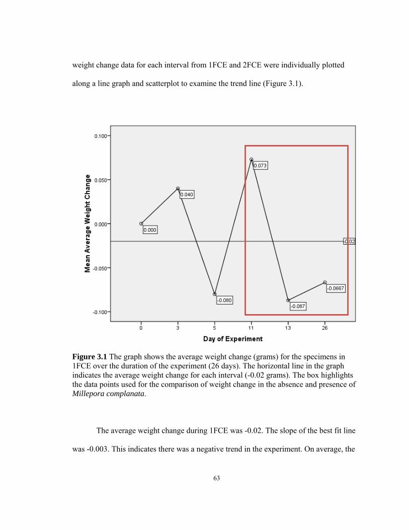
weight change data for each interval from 1FCE and 2FCE were individually plotted
along a line graph and scatterplot to examine the trend line (Figure 3.1).
Figure 3.1 The graph shows the average weight change (grams) for the specimens in 1FCE over the duration of the experiment (26 days). The horizontal line in the graph indicates the average weight change for each interval (-0.02 grams). The box highlights the data points used for the comparison of weight change in the absence and presence of Millepora complanata.
The average weight change during 1FCE was -0.02. The slope of the best fit line
was -0.003. This indicates there was a negative trend in the experiment. On average, the
63

worms in 1FCE lost weight during the experiment however, there were oscillations in
positive and negative weight changes. The greatest decrease in weight occurred between
days 11 and 13 which encompassed the starvation time period. This comparison is tested
for correlation and significance in the next section.
The slope in the 2FCE graph was also negative. The mean average weight change
was -0.04 g (Figure 3.2) which is a slightly larger decrease in weight than 1FCE. The
slope of the line was -0.0008. As in 1FCE, the greatest increase and decrease in weight
occurs during a period of food removal and reintroduction.
The average weight change for 2FCE M. ann was -0.08 g (Figure 3.3). When the
data points were graphed linearly, the slope was 0.007. The three weight change
measurements for this experiment were: day 94= -0.108 g; day 103=-0.15; and day
109=0.017. Therefore, only one time interval was positive. A pattern of negative growth
was evident in all three data sets. In order to test whether the patterns were significant,
difference in the means and the correlation between the data sets were analyzed.
Comparison of covariance and difference in the means between Millepora complanata
experiments
The average weight change data sets from 1FCE and 2FCE were compared for
correlation (Pearson’s correlation) and difference in the means (t-test). The first statistical
test was run with the full data sets, including the two data points from a different food
regime in 2FCE.
The Pearson’s correlation r-value was 0.006 with a p-value of 0.992. This
indicates the correlation is not significant between 1FCE and 2FCE. The Levene’s test
64
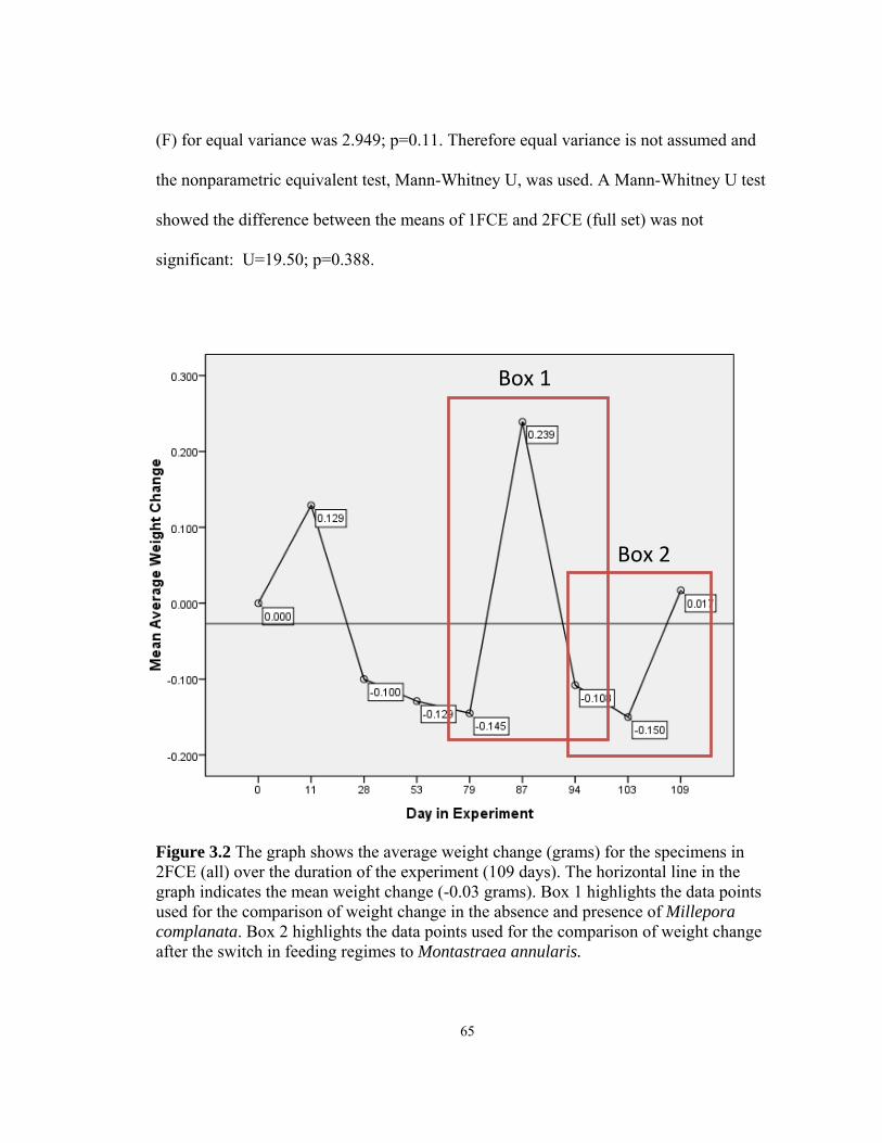
65
(F) for equal variance was 2.949; p=0.11. Therefore equal variance is not assumed and
the nonparametric equivalent test, Mann-Whitney U, was used. A Mann-Whitney U test
showed the difference between the means of 1FCE and 2FCE (full set) was not
significant: U=19.50; p=0.388.
Figure 3.2 The graph shows the average weight change (grams) for the specimens in 2FCE (all) over the duration of the experiment (109 days). The horizontal line in the graph indicates the mean weight change (-0.03 grams). Box 1 highlights the data points used for the comparison of weight change in the absence and presence of Millepora complanata. Box 2 highlights the data points used for the comparison of weight change after the switch in feeding regimes to Montastraea annularis.
Box 1
Box 2

The second statistical test was with 1FCE and 2FCE without the M. ann data
points (day 103 and day 109). The Levene’s test (F) for equal variance was 3.837;
p=0.08. The equal variance is assumed and the parametric t-test can be used to determine
the association between the means. The t-value was -0.168 with a p-value of 0.875.
Although there was a negative correlation, the association was not statistically
significant. The Pearson’s correlation value for this association was also not significant
(r=-0.665; p=0.537).
Comparison of body weight change between Millepora complanata removal and
reintroduction
At one point during experiments 1FCE (Box in Figure 3.1) and 2FCE (Box 1 in
Figure 3.2), the M. complanata colonies were removed from the tanks for several days
before new M. complanata colonies were reintroduced. Pearson’s correlation (r) was used
to compare the linear relationship between three variables (average weight with food
(WWF), weight without food (WOF), and weight after introduction of new food (WNF)).
Since the Levene’s test was not significant for these data sets, a Mann-Whitney U test
was used to calculate the significance of the difference in means between the treatments.
66
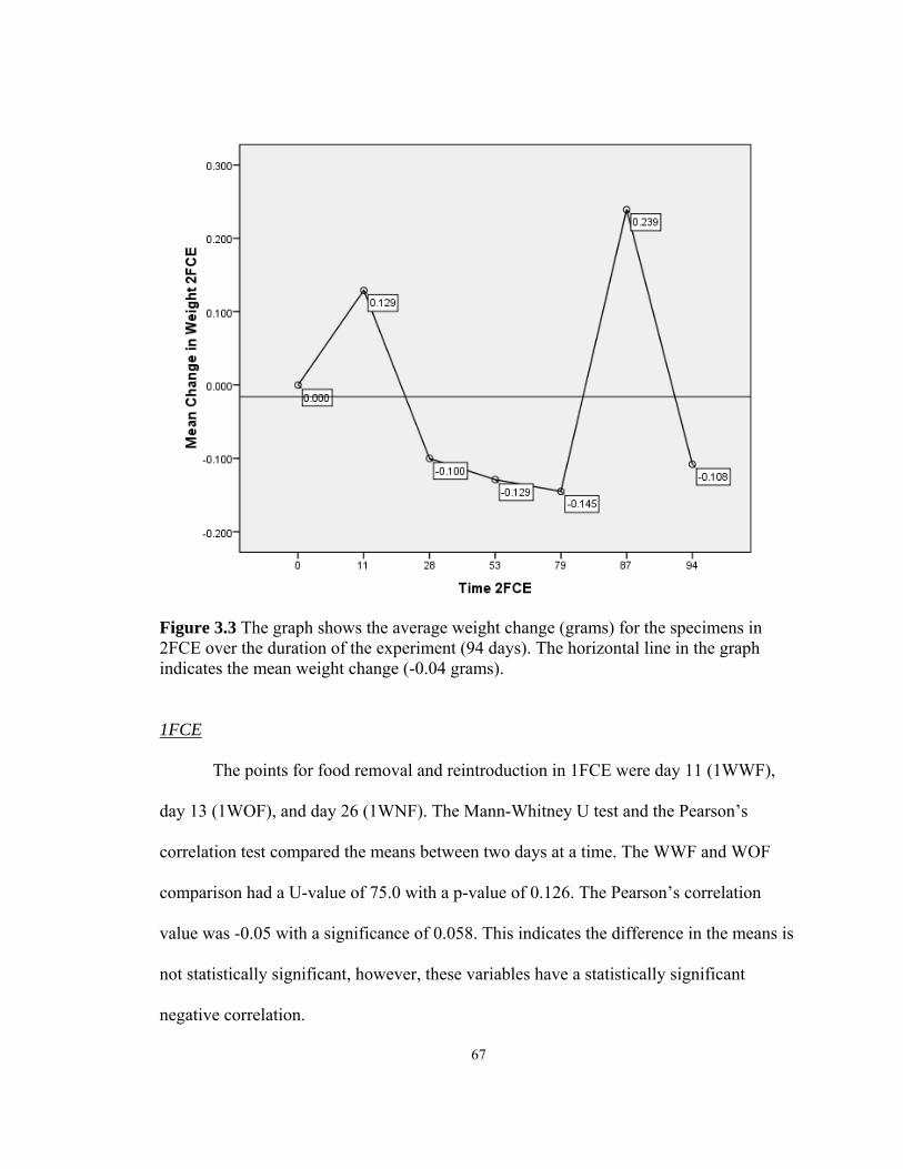
Figure 3.3 The graph shows the average weight change (grams) for the specimens in 2FCE over the duration of the experiment (94 days). The horizontal line in the graph indicates the mean weight change (-0.04 grams).
1FCE
The points for food removal and reintroduction in 1FCE were day 11 (1WWF),
day 13 (1WOF), and day 26 (1WNF). The Mann-Whitney U test and the Pearson’s
correlation test compared the means between two days at a time. The WWF and WOF
comparison had a U-value of 75.0 with a p-value of 0.126. The Pearson’s correlation
value was -0.05 with a significance of 0.058. This indicates the difference in the means is
not statistically significant, however, these variables have a statistically significant
negative correlation.
67

The means of 1WWF and 1WNF measurements have a U-value of 93.5 with a p-
value of 0.436 and an r-value of -.0.036 with a p-value of 0.898. This indicates the
difference in means and the correlation between the variables are not statistically
significant.
The means of 1WOF and 1WNF have a U-value of 94.0 with a p-value of 0.461.
This indicates the mean difference is not statistically significant. However, the Pearson’s
correlation value (r) was -0.044 with a significance of 0.10. Thus, the two variables
(1WOF and 1WNF) have a negative correlation which is statistically significant.
2FCE
During 2FCE, the time points were day 79 (2WWF), day 87 (2WOF), and day 94
(2WNF). The means of 2WWF and 2WOF have a U- value of 16.0 with a p-value of
0.01. The Pearson’s correlation value was -0.503 with a significance of 0.096. The mean
difference is statistically significant and the negative correlation between the variables is
statistically significant.
The 2WWF and 2WNF measurements have a U-value of 58.0 with a p-value of
0.414. This indicates the mean difference is not statistically significant. The Pearson’s
correlation r-value was 0.53 with a significance of 0.076. The correlation between these
variables is a statistically significant positive relationship.
The 2WOF and 2WNF variables have a U-value of 30.0 with a p-value of 0.014.
This indicates the mean difference between the day 87 and day 94 measurements is
statistically significant. The Pearson’s correlation value was -0.138 with a significance of
0.668. The correlation between these variables is not statistically significant.
68

69
Figure 3.4 The graph shows the average weight change (grams) for the specimens in 2FCE M. ann over the duration of the experiment (15 days). The horizontal line in the graph indicates the mean weight change (-0.08 grams).
Comparing the body weight change between the feeding regimes Millepora complanata
and Montastraea annularis
During a period in 2FCE (Figure 3.4 and Box 2 in Figure 3.2), M. complanata
colonies were removed and immediately replaced with M. annularis colonies. The
difference in body weight during three points (day 94: before introduction of M.
annularis (2MA); day 103: after introduction of M. annularis (2AMA); and day 109: 15
days after introduction of M. annularis (2BMA)) were compared for variance and
difference between the means.
Comparison of the 2MA and 2AMA means gives a U-value of 66.5 with a p-value
of 0.755. This indicates the difference between the mean weights between the beginning
and end of the experiment is not statistically significant. The Pearson’s correlation value
was -0.661 with a significance of 0.019. Therefore, the two variables have a statistically
significant negative relationship.
The 2MA and 2BMA variable have a U-value of 51.0 with a p-value of 0.242.
This mean difference is not statistically significant. The Pearson’s correlation value was -
0.032 with a significance of 0.921. The correlation between the variables is also not
statistically significant.
The 2AMA and 2BMA variables have a U-value of 61.0 with a p-value of 0.551.
The Pearson’s correlation r-value was -0.225 with a significance of 0.481. These two
measurements did not have statistically significantly different means or were correlated.
Summary of Results:
The statistically significant tests are listed in Tables 3.1 and 3.2. The difference in
means (U-value) and the correlation (r-value) were both statistically significant in one
series: 2FCE, weight before removal of food and weight after removal of food; (-0.503,
moderately negative correlation). The U-value was statistically significant in one series
where the r-value was not statistically significant:
70

1. 2FCE, weight after removal of M. complanata and weight after
reintroduction of M. complanata.
The Pearson’s correlation was statistically significant when the difference in the
means was not statistically significant in four series:
1. 1FCE, weight before removal of M. complanata and weight after removal
of M. complanata; (-0.5, moderately positive correlation)
2. 1FCE, weight after removal of M. complanata and weight after
reintroduction of M. complanata; (-0.004, weak negative correlation)
3. 2FCE, weight before removal of M. complanata and weight after
reintroduction of M. complanata; (0.53, moderately positive correlation)
4. 2FCE M.ann, weight before switch to M. annularis and weight 9 days
after switch; (-0.661, moderately negative correlation)
Discussion:
Periodic phenomena or long-term shifts in ocean conditions can affect coral reef
community ecology (May 1977, Knowlton 1992, Pandolfi et al. 2003) including the
abundance and biodiversity of coral species (Bythell et al. 1993, Hughes 1994, Aronson
and Precht 2001). The Caribbean is a region which has experienced local extinction and
phase shifts in coral species (Hughes 1994, Macintyre et al. 2007). Important reef-
building coral species like M. annularis have been replaced by dominate and fast-
growing species like P. astreoides (Green et al. 2008). Moreover, critical habitat species
like A. palmata and A. cervicornis are now in such low densities they are listed as
71

“threatened species” under the Endangered Species Act (Aronson et al. 2004). Similarly,
the reef systems off of Barbados have seen a reduction in M. annularis and A. cervicornis
(Macintyre et al. 2007). In contrast to the decrease in scleractinian corals, the hydrocoral
M. complanata remains in high densities in the shallow reefs (Lewis 2006).
Table 3.1 The chart shows the Pearson’s correlation tests which were statistically significant: 1FCE (days 11,13 and days 13,26); 2FCE (days 79,87 and days 79,94); 2FCE M. ann (94,103).
Pearson's Correlations: Statistically significant
Experiment Days r-value p-value
1FCE 11,13 -0.5 0.058
13,26 -0.044 0.099
2FCE 79,87 -0.503 0.096
79,94 0.053 0.076
2FCE M. ann 94, 103 -0.661 0.019
Table 3.2 The chart shows the Mann-Whitney U tests which were statistically significant: 2FCE (days 79,87 and days 87,94).
Mann-Whitney U tests: Statistically significant
Experiment Days U-value p-value
2FCE 79,87 16 0.001
87,94 30 0.014
72

Phase shifts and mass mortality of corals can impact sessile and benthic marine
organisms including the consumers of coral tissue, corallivores (Rotjan and Lewis 2008).
The reduction in nutritional value and availability of corals as a food resource could have
a detrimental effect on species (Baums et al. 2003b). If the new dominant coral species
are less nutritious per tissue area, this may increase the area of coral tissue loss per
feeding event (Baums et al. 2003b). Also, feeding behavior of corallivores (e.g. scrapers
versus grazers) may not be conducive to the newly dominant coral species morphology
(Rotjan and Lewis 2008). If factors are not favorable for consumption of the new coral
species or if coral tissue becomes unavailable (i.e., complete coral mortality), facultative
corallivores may utilize alternate food resources in larger quantities, which could have
compounding impacts on the reef community (Rotjan and Lewis 2008).
Given the availability of M. complanata and the decreased density of other coral
species as food resources for H. carunculata on Barbados reefs, this study evaluated the
impact of consuming M. complanata on the body weight of the invertebrate corallivore
H. carunculata. Two ex situ experiments (1FCE and 2FCE) were conducted to measure
the body weight change of H. carunculata in an isolated feeding regime of .M.
complanata. The average body weight at the conclusion of both experiments (1FCE and
2FCE) with M. complanata was less than the initial average weight. However, the
difference in the means for both experiments was not statistically significant. These
results indicate the body weight did not fluctuate significantly in the presence of M.
complanata.
73

When the feeding regime of the second group (2FCE) was changed to the stony
coral, M. annularis,(2FCE M. ann) the average weight decreased in 9 days from 1.36 g to
1.21 g (mean weight change = -0.15 g) followed by an increase after 15 days (1.26 g,
mean weight change = 0.017 g). However, the difference in the weight means between
the presence of M. complanata and M. annularis as the food resources was not
statistically significant. Since the body weight of H. carunculata was not greater in the
presence of M. complanata compared with the body weight in M. annularis, the null
hypothesis is accepted.
Although the U-tests for all three comparisons were not statistically significant,
there was a significant negative correlation between the weight in the presence of M.
complanata and the weight 9 days after the feeding regime was switched to M. annularis.
The results indicate the specimens’ weight decreased in the presence of the new food (M.
annularis). However, this is one data point and afterwards the weight increased. An
explanation for the initial negative correlation in weight could be a result of conditioning
to one type of food (M. annularis). The specimens in 2FCE and 2FCE M. ann were the
same and 2FCE ran for 94 days before the food was changed suddenly. In 2FCE M. ann,
the specimens increased in weight after 6 more days in the M. annularis feeding regime
which supports the idea that the worms were conditioned to feed on M. complanata and it
took time to adjust to the new food. A control group was not used, therefore, one must be
cautious in drawing conclusions from these data.
This study assumed the frequency of H. carunculata predation on M. complanata
in field observations (Ott and Lewis 1972, Witman 1988, Lewis and Crooks 1996)
74
indicated there was a benefit to this feeding behavior. When the body weight was
compared before and after starvation (1FCE and 2FCE separately), there was a
statistically significant difference in the means and a negative correlation between
average weights in the presence and absence of M. complanata. Since body weight
decreased in the absence of M. complanata more than when colonies were available, M.
complanata colonies provided a positive level of sustenance. However, after 26 days
(1FCE) and 94 days (2FCE) of feeding, the specimens did not gain significant weight
over time which indicates feeding on M. complanata does not provide a sustainable level
of nutrition for life longevity. Regardless of the results, it is difficult to draw conclusions
when comparing body weight in 1FCE and 2FCE since a control group was not used.
It is important for coral managers to know the impact of a declining resource (i.e.,
coral tissue) on consumers (i.e., corallivores). The experiments in this study attempted to
examine the body weight of a corallivore, H. carunculata, in the presence of an abundant
fire coral species, M. complanata from a system of declining scleractinian coral cover.
The lack of control groups hinders the ability to draw conclusions about the results.
The experiments should have included negative and positive controls. The
negative control would have tested the effect of no food. A positive control would have
involved a mixed diet and a different coral species like Porites. There was a period of
negative control for both 1FCE and 2FCE when the food was removed. However, a
proper control would have occurred at the same time as the experiment with M.
complanata. Also, in order to compare body weight under different feeding regimes (e.g.
M. complanata versus M. annularis), length of experiments and number of replications of
75
the different food conditions should be similar. Furthermore, wet weight may not be the
most comprehensive measurement to use when assessing the impact of a feeding regime
on fireworm specimens. Other measurements like dry weight, length, and volume may a
better set of information to determine organism growth over time.
Nevertheless, the outcome of the feeding experiments suggests H. carunculata
does not gain weight in the presence of M. complanata as the only food resource despite
the frequency of predation from field observations (Lewis and Crooks 1996). The same
results of decreased average weight occurred during the M. annularis feeding regime
even though the length of time under the M. annularis feeding regime was shorter. These
results could have been from various factors including tank affects, water quality affects,
seasonal growth rates, species population parameters, or nutritional value of M.
complanata.
Life cycle affects, specifically reproduction and age, could be contributing factors
to little or no weight gain. H. carunculata spawns in the summer months (Marsden 1962)
and the experiments took place during the winter (1FCE) and early spring (2FCE).
Therefore, weight gain may have been stagnant prior to the spawning season. Also, the
age of the specimens may have contributed to the weight trend observed in the
experiments. However, baseline information on the life cycle and growth rate as it related
to life cycle of this species in not known or documented (Ritchie personal
communication).
Also, body length parameters for this species are also poorly understood.
Unpublished data by this author suggested the population sampled for this experiment
76

were smaller in average length than historic data. The specimens were taken from the
population on the Batts Rock Reef off the west coast of Barbados. In 2002, this author
found the average length of this population to be 4.52 cm which is a reduction from 8cm,
the average size of fireworms found off the west coast of Barbados in 1962 observed by
Marsden (personal communication). Comparing historic data to this unpublished data
suggested this population has reduced in average growth size. The shift in average body
size suggests the use of length as an indication of age would be an inaccurate
measurement for this population. Therefore, more information on the life cycle of H.
carunculata and tools to measure age are needed to determine if nutritional requirements
change with age.
In a M. complanata feeding regime, H. carunculata did not increase in weight but
the results from starvation periods suggest the specimens were consuming M. complanata
and that “fire corals” does provide a certain level of nutrition. However, fire corals did
not provide sufficient nutrients for growth. These data suggest H. carunculata needs
other sources for sustenance and M. complanata may not be the ideal food source.
Previous studies discussed the diverse diet composition of H. carunculata (Lewis and
Crooks 1996) and Rotjan and Lewis (2008) defined this species as a facultative
corallivore. If M. complanata is more abundant than other coral species but does not
provide enough nutrients, this may affect the amount of tissue consumed per feeding
event and also affect the consumption rate of alternative food sources in order for H.
carunculata organisms to maintain nutritional requirements.
77

Given the declining state of reefs in the Caribbean and worldwide, more research
needs to be done on the consequences for corallivores as well as their prey. Facultative
corallivores, like H. carunculata, are less affected by a reduction in coral tissue than
obligate corallivores (Rotjan and Lewis 2008). However, the consequences for alternative
prey like sponges and anemones may be affected as preferred coral species, like
scleractinian corals, become less abundant. Coral reef management plans should address
how decline and diversity changes in scleractinian or fire corals will affect the physiology
and feeding behavior of corallivores to understand the impacts on overall coral reef food
webs. The first step is collecting the necessary and fundamental information on coral
predators including food preference, foraging time, and diet composition.
78

Literature Cited
Alvarez-Filip L, Gil I (2006) Effects of Hurricanes Emily and Wilma on coral reefs in
Cozumel, Mexico. Coral Reefs 25:583-583 Aronson RB, MacIntyre IG, Wapnick CM, O'Neill MW (2004) Phase shifts, alternative
states, and the unprecedented convergence of two reef systems. Ecology 85:1876-1891
Aronson RB, Precht WF (2001) White-band disease and the changing face of Caribbean
coral reefs. Hydrobiologia 460:25-38 Bak RPM, Steward-Van Es Y (1980) Regeneration of superficial damage in the
scleractinian corals Agaricia agaricites, f. purpurea and Porites astreoides. Bull Mar Sci 30:883-887
Baums IB, Miller MW, Szmant AM (2003a) Ecology of a corallivorous gastropod,
Coralliophila abbreviata, on two scleractinian hosts. 1: Population structure of snails and corals. Mar Biol 142:1083-1091
Baums IB, Miller MW, Szmant AM (2003b) Ecology of a corallivorous gastropod,
Coralliophila abbreviata, on two scleractinian hosts. II. Feeding, respiration and growth. Mar Biol 142:1093-1101
Bellwood DR, Choat JH (1990) A functional analysis of grazing in parrotfishes (Family
Scaridae) – the ecological implications. Environ Biol Fishes 28:189-214 Bellwood DR, Hoey AS, Choat JH (2003) Limited functional redundancy in high
diversity systems: resilience and ecosystem function on coral reefs. Ecol Lett 6:281-285
Bellwood DR, Hughes TP, Folke C, Nystrom M (2004) Confronting the coral reef crisis.
Nature 429:827-833 Birkeland C, Lucas J (1990) Acanthaster planci: major management problem of coral
reefs, Vol. CRC Press Bythell JC, Gladfelter EH, Bythell M (1993) Chronic and catastrophic natural mortality
of 3 common Caribbean reef corals. Coral Reefs 12:143-152 Chapin FS, Zavaleta ES, Eviner VT, Naylor RL, Vitousek PM, Reynolds HL, Hooper
DU, Lavorel S, Sala OE, Hobbie SE, Mack MC, Diaz S (2000) Consequences of changing biodiversity. Nature 405:234-242
79

Chornesky EA, Peters EC (1987) Sexual reproduction and colony growth in the
scleractinian coral Porities astreoides. Biol Bull 172:161-177 Colgan MW (1987) Coral reef recovery on Guam (Micronesia) after catastrophic
predation by Acanthaster planci. Ecology 68:1592-1605 Connell JH (1997) Disturbance and recovery of coral assemblages. Coral Reefs 16:S101-
S113 Dalton SJ, Godwin S (2006) Progressive coral tissue mortality following predation by a
corallivorous nudibranch (Phestilla sp.). Coral Reefs 25:529-529 De Weerdt Wd (1981) Transplantation experiments with Caribbean Millepora species
(Hyrdozoa, Coelenterata), including some ecological observations on growth forms. Bijdr Dierk 51:1-19
Edmunds PJ (1999) The role of colony morphology and substratum inclination in the
success of Millepora alcicornis on shallow coral reefs. Coral Reefs 18:133-140 Edmunds PJ (2004) Juvenile coral population dynamics track rising seawater temperature
on a Caribbean reef. Mar Ecol-Prog Ser 269:111-119 Edmunds PJ, Elahi R (2007) The demographics of a 15-year decline in cover of the
Caribbean reef coral Montastraea annularis. Ecol Monogr 77:3-18 Edmunds PJ, Roberts DA, Singer R (1990) Reefs of the northeastern Caribbean. 1.
Scleractinian populations. Bull Mar Sci 46:780-789 Folke C, Carpenter S, Walker B, Scheffer M, Elmqvist T, Gunderson L, Holling CS
(2004) Regime shifts, resilience, and biodiversity in ecosystem management. Annu Rev Ecol Evol Syst 35:557-581
Gardner TA, Cote IM, Gill JA, Grant A, Watkinson AR (2003) Long-term region-wide
declines in Caribbean corals. Science 301:958-960 Glynn P (1990) Feeding ecology of selected coral-reef macroconsumers: patterns and
effects on coral community structure. In: Z Dubinsky (ed) Ecosystems of the world 25: coral reefs. Elsevier, New York, p 365-400
Glynn PW (1963) Hermodice carunculata and Mithraculus sculptus, two hermatypic
coral predators. Association of Island Marine Laboratories 4th Meeting:16-17
80
Glynn PW (2004) High complexity food webs in low-diversity eastern Pacific reef-coral communities. Ecosystems 7:358-367
Green DH, Edmunds PJ, Carpenter RC (2008) Increasing relative abundance of Porites
astreoides on Caribbean reefs mediated by an overall decline in coral cover. Mar Ecol-Prog Ser 359:1-10
Hayes JA (1990) Prey preference in a Caribbean corallivore, Coralliophila abbreviata
(Lamark) (Gastropoda, Coralliophilidae). Bull Mar Sci 47:557-560 Hiatt R, Strasburg D (1960 ) Ecological relationships of the fish fauna on coral reefs of
the Marshall Islands. Ecol Monogr 30:65-127 Houk P, Bograd S, van Woesik R (2007) The transition zone chlorophyll front can trigger
Acanthaster planci outbreaks in the Pacific Ocean: Historical confirmation. J Oceanogr 63:149-154
Hughes TP (1994) Catastrophes, phase shifts, and large scale degradation of a Caribbean
coral reef. Science 265:1547-1551 Hughes TP, Baird AH, Bellwood DR, Card M, Connolly SR, Folke C, Grosberg R,
Hoegh-Guldberg O, Jackson JBC, Kleypas J, Lough JM, Marshall P, Nystrom M, Palumbi SR, Pandolfi JM, Rosen B, Roughgarden J (2003) Climate change, human impacts, and the resilience of coral reefs. Science 301:929-933
Hughes TP, Tanner JE (2000) Recruitment failure, life histories, and long-term decline of
Caribbean corals. Ecology 81:2250-2263 Jackson JBC, Kirby MX, Berger WH, Bjorndal KA, Botsford LW, Bourque BJ, Bradbury
RH, Cooke R, Erlandson J, Estes JA, Hughes TP, Kidwell S, Lange CB, Lenihan HS, Pandolfi JM, Peterson CH, Steneck RS, Tegner MJ, Warner RR (2001) Historical overfishing and the recent collapse of coastal ecosystems. Science 293:629-638
Knowlton N (1992) Thresholds and multiple stable states in coral reef community
dynamics. American Zoologist 32:674-682 Knowlton N (2001) The future of coral reefs. Natl Acad Sciences, p 5419-5425 Lester RT, Bak RPM (1985) Effects of environment on regeneration rate of tissue lesions
in the reef coral Montastrea annularis (Scleractinia). Mar Ecol-Prog Ser 24:183-185
Lewis JB (1989) The ecology of Millepora - A review. Coral Reefs 8:99-107
81

Lewis JB (1992) Heterotrophy in corals – zooplankton predation by the hydrocoral
Millepora complanata. Mar Ecol-Prog Ser 90:251-256 Lewis JB (2006) Biology and ecology of the hydrocoral Millepora on coral reefs. In:
Advances in Marine Biology, Vol 50, Vol 50. Academic Press Ltd, London, p 1-55
Lewis JB, Crooks RE (1996) Foraging cycles of the amphinomid polychaete Hermodice
carunculata preying on the calcareous hydrozoan Millepora complanata. Bull Mar Sci 58:853-856
Lizama J, Blanquet RS (1975) Predation on sea anemones by the amphinomid
polychaete, Hermodice carunculata. Bull Mar Sci 25:442-443 Lokrantz J, Nystrom M, Thyresson M, Johansson C (2008) The non-linear relationship
between body size and function in parrotfishes. Coral Reefs 27:967-974 Macintyre IG, Glynn PW, Toscano MA (2007) The demise of a major Acropora palmata
bank-barrier reef off the southeast coast of Barbados, West Indies. Coral Reefs 26:765-773
Marsden JR (1963) The digestive tract of Hermodice carunculata. (Pallas). Polychaeta:
Amphinomidae. Can J Zool/Rev Can Zool 41:165-184 Marsden JR (2001) personal communication, unpublished data. Martin A, Losada F (1991) Regeneration of injuries caused by the Amphinomid
polychaete Hermodice carunculata on scleractinian corals, hydrocorals and octocorals (Cnidaria). Ecotropicos 4:18-26
May RM (1977) Thresholds and multiple stable states in coral reef community dynamics.
Nature 269:471-477 McGuire MP (1998) Timing of larval release by Porites astreoides in the northern
Florida Keys. Coral Reefs 17:369-375 Miller MW, Williams DE (2007) Coral disease outbreak at Navassa, a remote Caribbean
island. Coral Reefs 26:97-101 Moran PJ (1986) The Acanthaster phenomenon. Oceanogr Mar Biol 24:379-480 Morton B, Blackmore G, Kwok CT (2002) Corallivory and prey choice by Drupella
rugosa (Gastropoda : Muricidae) in Hong Kong. J Molluscan Stud 68:217-223
82

Motta PJ (1988) Functional morphology of the feeding apparatus of 10 species of pacific
butterflyfishes (Perciformes, Chaetodontidae) – an ecomorphological approach. Environ Biol Fishes 22:39-67
Mumby PJ, Hastings A, Edwards HJ (2007) Thresholds and the resilience of Caribbean
coral reefs. Nature 450:98-101 Myers RA, Worm B (2003) Rapid worldwide depletion of predatory fish communities.
Nature 423:280-283 Oren U, Benayahu Y, Loya Y (1997) Effect of lesion size and shape on regeneration of
the Red Sea coral Favia favus. Mar Ecol-Prog Ser 146:101-107 Ott B, Lewis JB (1972) The importance of the gastropod Coralliophila abbreviata
(Lamarck) and the polychaete Hermodice carunculata (Pallas) as coral reef predators. Can J Zool/Rev Can Zool 50:1651-1656
Pandolfi JM, Bradbury RH, Sala E, Hughes TP, Bjorndal KA, Cooke RG, McArdle D,
McClenachan L, Newman MJH, Paredes G, Warner RR, Jackson JBC (2003) Global trajectories of the long-term decline of coral reef ecosystems. Science 301:955-958
Richardson LL, Voss JD (2005) Changes in a coral population on reefs of the northern
Florida Keys following a coral disease epizootic. Mar Ecol-Prog Ser 297:147-156 Ritchie K (2009) personal communication, unpublished data. Rotjan RD, Dimond JL, Thornhill DJ, Leichter JJ, Helmuth B, Kemp DW, Lewis SM
(2006) Chronic parrotfish grazing impedes coral recovery after bleaching. Coral Reefs 25:361-368
Rotjan RD, Lewis SM (2008) Impact of coral predators on tropical reefs. Mar Ecol-Prog
Ser 367:73-91 Runnalls LA, Coleman ML (2003) Record of natural and anthropogenic changes in reef
environments (Barbados West Indies) using laser ablation ICP-MS and sclerochronology on coral cores. Coral Reefs 22:416-426
Sheppard CRC (1982) Coral populations on reef slopes and their major controls. Mar
Ecol-Prog Ser 7:83-115 Sussman M, Loya Y, Fine M, Rosenberg E (2003) The marine fireworm Hermodice
carunculata is a winter reservoir and spring-summer vector for the coral-
83
bleaching pathogen Vibrio shiloi. Environmental microbiology, 2003 Apr, 5(4):250-5
Sutherland KP, Porter JW, Torres C (2004) Disease and immunity in Caribbean and
Indo-Pacific zooxanthellate corals. Mar Ecol-Prog Ser 266:273-302 Tomascik T, Sander F (1987) Effects of eutrophication on reef-building corals. 2.
structure of scleractinian coral communities on fringing reefs, Barbados, West Indies. Mar Biol 94:53-75
Turner SJ (1994) The biology and population outbreaks of the corallivorous gastropod
Drupella on Indo-Pacific reefs. In: Oceanography and Marine Biology, Vol 32, Vol 32. U C L Press Ltd, London, p 461-530
Wahle CM (1980) Detection, pursuit, and overgrowth of tropical gorgonians by
Milleporid hydrocorals - Perseus and Medusa revisited. Science 209:689-691 Williams DE, Miller MW (2005) Coral disease outbreak: pattern, prevalence and
transmission in Acropora cervicornis. Mar Ecol-Prog Ser 301:119-128 Witman JD (1988) Effects of predation by the fireworm Hermodice carunculata on
Milleporid hydrocorals. Bull Mar Sci 42:446-458 Witman JD (1992) Physical disturbance and community structure of exposed and
protected reefs - a case study from St. John, US Virgin Islands. American Zoologist 32:641-654
84

4. The use of molecular techniques to investigate the feeding behavior of Hermodice
carunculata
Abstract
The invertebrate corallivore Hermodice carunculata was associated with the
tissue loss margin of coral colonies during the 2005, 2006, and 2007 winter outbreaks of
a suspect white plague (WP) in the Flower Garden Banks National Marine Sanctuary.
However, the gross signs of denuded tissue were inconsistent with WP-associated
lesions. During the winter outbreak in 2007, actively feeding H. carunculata organisms
were foraging on tissue loss margins. The tissue loss margins of colonies without actively
feeding exhibited patterns of tissue loss inconsistent with WP. Lesions caused by H.
carunculata also result in white denuded skeleton. However, specific information on
predation patterns and scars is unavailable for H. carunculata. Diagnosis techniques and
protocols are needed to distinguish H. carunculata-induced tissue loss from microbial-
induced tissue loss. This study tested the ability to detect trace amounts of H. carunculata
DNA on known predation sites by developing specific H. carunculata primers to amplify
its DNA using Polymerase Chain Reactions. Under the conditions of this study, the
H.carunculata DNA was not detectable. However, appropriately adjusted techniques
should be useful to develop comprehensive and accurate field diagnostic tools of coral
tissue mortality.
85

Introduction
Coral diseases caused by infectious microorganisms are considered the most
important factor contributing to the decline of live coral tissue and coral population
densities (Harvell et al. 1999, Wilkinson 2004, Bruckner and Bruckner 2006, Weil and
Croquer 2009). Over the past 30 years, coral diseases have increased in range (Weil et al.
2002, Sutherland et al. 2004, Raymundo et al. 2005) and prevalence (Bruno et al. 2003,
Sutherland et al. 2004), subsequently damaging reefs worldwide (Harvell et al. 1999,
Porter et al. 2001, Rosenberg and Loya 2004). It has been more than 30 years since field
observers first noticed unusual lesions on corals (Weil et al. 2006), but the identities of
most pathogens remain unknown (Richardson 1998, Harvell et al. 2007, Luna et al. 2007,
Weil and Croquer 2009).
Of the 29 proposed coral diseases (Remily and Richardson 2006) that lead to
partial or complete colony mortality, only five microbial causal agents have been
implicated through Koch’s postulates (Sutherland et al. 2004, Harvell et al. 2007): white
plague type II, white pox, aspergillosis, Vibrio shiloi-induced bleaching, and Vibrio
coralliilyticus-induced bleaching and disease (Table 4.1). The causative agent of white
plague II (Aurantimonas coralicida) was not been confirmed since Richardson et al
(1998c). Several coral diseases (black band disease (BB), shut-down reaction (SD),
skeletal anomalies (SA), aspergillosis (AS), white pox (WP), dark spots (DS), Vibrio
shiloi-induced bleaching (VS), Vibrio coralliilyticus-induced bleaching and disease (VC),
yellow band (YB), and fungal-protozoan syndrome (FP)) have also been correlated with
various abiotic stressors including elevated sea temperature, eutrophication,
86

sedimentation, pollution, fecal contamination, solar UV radiation, and poor water quality
(Table 4.2).
Table 4.1 Coral diseases associated with biota which have been proven through Koch’s postulates listed by disease, biota, and literature reference. Disease Biota Reference
White Plague Type II Aurantimonas coralicida
Richardson et al. 1998a, Denner et al. 2003
White Pox Serratia marcescens Patterson et al. 2002 Aspergillosis Aspergillus sydowii Smith and Weil 2004, Geiser et
al. 1998 Vibrio shiloi-induced Bleaching
Vibrio shiloi Kushmaro et al., 1997, Rosenberg et al. 1998
Vibrio coralliilyticus-induced Bleaching
Vibrio coralliilyticus Ben-Haim and Rosenberg 2002, Ben-Haim et al. 2003
Due to the plethora of phenomena contributing to coral reef decline, it is difficult
to determine the importance of each factor in the survivorship, abundance, and
biodiversity of coral species (Bythell et al. 1993, Hughes 1994, Aronson and Precht 2001,
Knowlton 2001, Gardner et al. 2003, Edmunds and Elahi 2007). The ability of coral
colonies to recover from detrimental events, like epizootics or long-term shifts in ocean
conditions, is based on various intrinsic factors including species body size (Bak and
Meesters 1998, Kramarsky-Winter and Loya 2000), morphology (Nagelkerken and Bak
1998), and age (Henry and Hart 2005). Extrinsic factors which also limit regeneration
capabilities include wound characteristics (Henry and Hart 2005), water temperatures
(Kramarsky-Winter and Loya 2000), food availability (Nagelkerken et al. 1999), and
disturbance history (Cumming 2002).
87

Table 4.2 Coral diseases associated with abiotic stressors listed by disease, stressor, and literature reference. Disease Abiotic Stressor Reference Black Band Disease Elevated
Temperatures Eutrophication Sedimentation Pollution Fecal contamination
Richardson and Kuta 2003 Kuta and Richardson 2002 Frias-Lopez et al. 2002 Al-Moghrabi 2002 Frias-Lopez et al. 2002
Shut-Down Reaction Elevated Temperatures Sedimentation
Antonius 1977 Antonius 1977
Skeletal Anomalies Solar UV Radiation Coles and Seapy 1998 Aspergillosis Elevated
Temperatures Sedimentation Poor water quality
Alker et al. 2001 Shinn et al. 2000, Smith and Weil 2004 Kim and Harvell 2002
White Pox Elevated Temperatures Fecal contamination
Patterson et al. 2002 Patterson et al. 2002
Dark Spots Elevated Temperatures
Gil-Agudelo and Garzon-Ferreira 2001
Vibrio shiloi-induced bleaching
Elevated Temperatures
Israely et al. 2001
Vibrio coralliilyticus-induced bleaching and disease
Elevated Temperatures
Ben-Haim et al. 2003
Yellow Band Elevated Temperatures
Riegl 2002
Fungal-Protozoan Syndrome Elevated Temperatures
Cerrano et al. 2000
To understand the role of coral disease in the state of coral reef ecosystems, other
threats to tissue loss, in particular predation, need to be better understood and
distinguished from disease-induced tissue loss (Miller and Williams 2007). However,
using field observations to determine causal agents of coral tissue loss is understood to be
88
a major challenge (Zorpette 1995, Bruckner 2006, Ainsworth et al. 2007). Coral tissue
consumed by corallivores such as Hermodice carunculata (bearded fireworm),
Coralliophila abbreviata spp., Drupella spp. (corallivorous gastropod), and Acanthaster
planci (crown-of-thorns starfish) leave denuded skeleton lesions similar in appearance to
those attributed to pathogenic microorganisms (Witman 1988, Sussman et al. 2003,
Miller and Williams 2007) and the lesions can easily be mistaken as microbial disease
(Sutherland et al. 2004). Thus, effective and accurate diagnostic tools are needed to
distinguish disease-induced tissue loss from corallivory (Miller and Williams 2007).
Identifying predation patterns is also important to understand the impact of coral
predators on an already declining system (Bruckner 2001, Rosenberg and Loya 2004,
Rotjan and Lewis 2008). The diet composition of corallivores has a 100% coral species
overlap (including many scleractinians), which could exacerbate the decline of the
preferred coral species (Rotjan and Lewis 2008). Chronic corallivory may cause the
affected colonies to redirect energy for repair which would otherwise be used for
reproduction and growth (Henry and Hart 2005). Furthermore, predation can also
increase susceptibility to colonization of algal turfs which can inhibit coral recovery and
increase incidences of disease (Rotjan and Lewis 2005). Field studies have reported
progressive coral tissue loss after corallivores leave foraging sites which suggests the
predator might transmit pathogenic bacteria to healthy corals (Sussman et al. 2003,
Williams and Miller 2005, Dalton and Godwin 2006). The role of corallivores in coral
tissue loss needs further study because of this distinct possibility that corallivores are
89
disease vectors, as well as being predators threatening coral survival (Rosenberg and
Loya 2004, Sutherland et.al. 2004, Williams and Miller 2005).
This study focused on one of the 116 known corallivorous species (Rotjan and
Lewis 2008), the amphinomid polychaete H. carunculata (Marsden 1963). As a
facultative corallivore (Rotjan and Lewis 2008), this species eats scleractinian corals and
also grazes on zoanthids, anemones, gorgonians, and hydrocorals (Glynn 1963, Marsden
1963, Ott and Lewis 1972, Lizama and Blanquet 1975, Martin and Losada 1991). Field
observations of Miller and Williams (2007) and others (S. Lewis, G. Cook, R. Jonas,
unpub. observ.) contradict literature reports that fireworms feed exclusively during late
afternoon and early evening (Marsden 1962, Lewis and Crooks 1996, Witman 1988).
Wittman (1988) found H. carunculata population densities differed regionally, but were
consistent within each region. This species locates its prey through a chemoreceptor
located immediately dorsal to its mouth region, the nuchal organ (Martin and Losada
1991). H. carunculata mechanically removes coral tissue by everting its muscular
pharynx over the prey (Marsden 1962, Miller and Williams 2007). Through the use of its
strong muscular buccal region (Ott and Lewis 1972), the organism sucks the tissue into
the esophagus and may excrete digestive enzymes to aid in removal of tissue (Marsden
1962, Marsden 1963, Ott and Lewis 1972, Witman 1988). This species has been observed
to consume 12.9 cm2 d-1of coral tissues (Rotjan and Lewis 2008) and have two separate
feeding episodes before retreating into reef cervices (Lewis and Crooks 2006). Once
ingested, particles can remain in the pharynx for up to nine hours (Marsden 1968). Larger
lesions are correlated with longer foraging times and larger body size (Ott and Lewis
90

1972). A foraging event is apparent by the noticeable enlargement of the organism’s
buccal region and its sedentary orientation (Marsden 1962, S. Lewis, unpublished data).
It is difficult to determine the correlation of H. carunculata with other pathogens through
field observations, because gross patterns of H. carunculata tissue removal from
scleractinian coral species have not been documented in the literature either
photographically or descriptively (Miller and Williams 2007). Ex situ experiments have
shown H. carunculata to be a vector for the Vibrio shiloi-induced bleaching pathogen
(Sussman et al. 2003).
The feeding behavior of H. carunculata on reefs of the Flower Garden Banks
National Marine Sanctuary (FGBNMS) was investigated in relation to outbreaks of the
white syndrome most often referred to as white plague (WP). Preliminary unpublished
field studies on H. carunculata by this author indicated the lesions of rapid tissue loss in
suspect WP are similar to the gross patterns of tissue removal resulting from predation by
the invertebrate corallivore H. carunculata (Figure 4.1). All suspect WP-type lesions
exhibit a smooth margin along apparently healthy, but receding, coral tissue and stark
white, recently denuded skeleton (Bruckner 2001, Bythell et al. 2004) (Figure 4.2).
Lesions caused by H. carunculata also result in white denuded skeleton (Ott and Lewis
1972, Miller and Williams 2007). At least 38 coral species are susceptible to WP or WP-
like diseases (Bruckner 2006, Bythell et al. 2004, Sutherland et al. 2004). Dustan (1977)
initially described this as a slow progressing, lethal disease of stony corals and referred to
it as WP (now WP type I), but no etiological agent was described. Since then, two more
damaging, faster progressing forms of this disease have been identified: WP types II and
91

92
III (WPII and WPIII) (Richardson et al.1998a). The diagnostic criteria for WP have been
complicated by inconsistent results of identification of bacterial etiological agents,
including finding different bacteria associated with the diseased margin (Denner et al.
2003, Pantos et al. 2003). The inconclusive findings of causal agents of WP (Ainsworth
et al. 2007, Richardson et al. 1998a, Rosenberg and Loya 2004) and the recent predation
correlations with suspect white plague outbreaks (Miller and Williams 2007) indicates
more studies are needed on WP and predation at the tissue loss margin.
Figure 4.1. H. carunculata feeding on a Montastraea colony denuded skeleton margin in FGBNMS in winter 2007. The red circle highlights recently denuded tissue from this foraging event. Photo by R.B. Jonas.
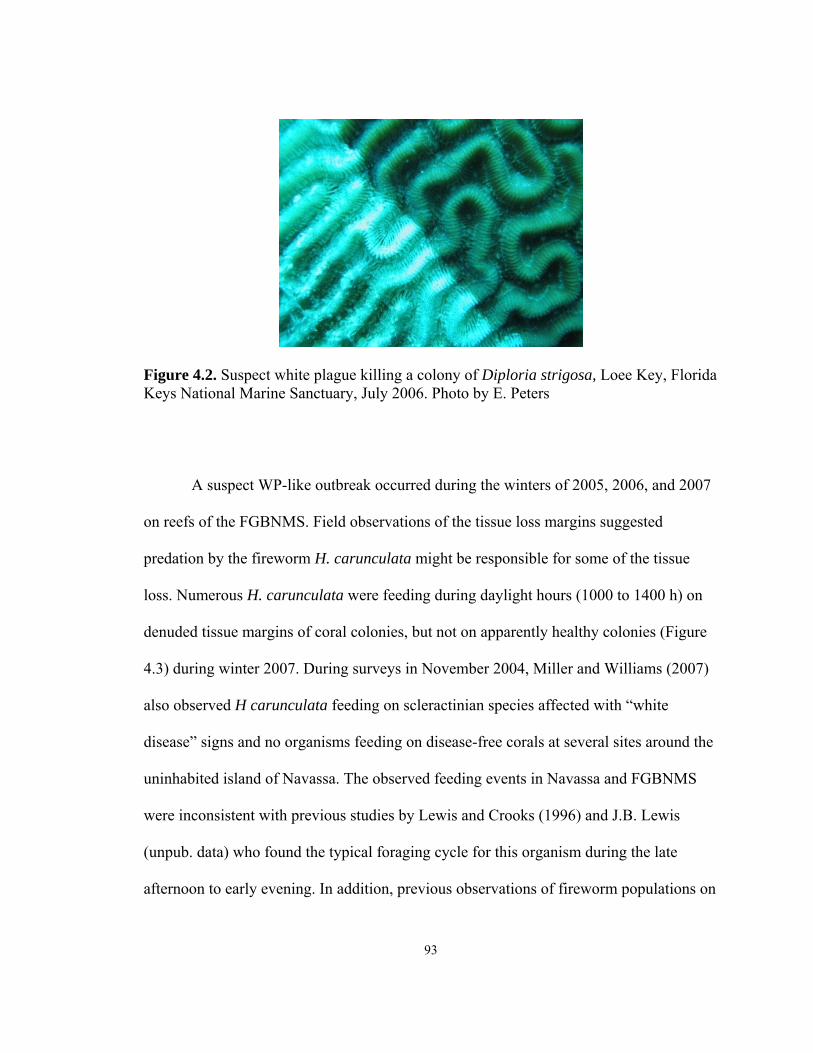
93
Diploria strigosa, Loee Key, Florida July 2006. Photo by E. Peters
during the winters of 2005, 2006, and 2007
of the tissue loss margins suggested
ght be responsible for some of the tissue
loss. Numerous H. carunculata were feeding during daylight hours (1000 to 1400 h) on
denuded tissue margins of coral colonies, but not on apparently healthy colonies (Figure
4.3) during winter 2007. During surveys in November 2004, Miller and Williams (2007)
also observed H carunculata feeding on scleractinian species affected with “white
disease” signs and no organisms feeding on disease-free corals at several sites around the
uninhabited island of Navassa. The observed feeding events in Navassa and FGBNMS
were inconsistent with previous studies by Lewis and Crooks (1996) and J.B. Lewis
(unpub. data) who found the typical foraging cycle for this organism during the late
afternoon to early evening. In addition, previous observations of fireworm populations on
Figure 4.2. Suspect white plague killing a colony of Keys National Marine Sanctuary,
A suspect WP-like outbreak occurred
on reefs of the FGBNMS. Field observations
predation by the fireworm H. carunculata mi

94
similar reefs (Witman 1988, Lewis and Crooks 1996) are inconsistent with the densities
of foraging H. carunculata during the white syndrome outbreaks in Navassa and the
FGBNMS, which suggests a dramatic increase in the populations. Population explosions
(or outbreak) of other corallivores, including the crown-of-thorns starfish (Acanthaster)
in the Indo-Pacific, have proven devastating to coral reefs (Volger et al. 2008). On closer
examination in FGBNMS and Navassa, the tissue loss patterns were not completely
consistent with those of WP (R.B. Jonas personal communication, Miller and Williams
2007). Furthermore, the white syndrome outbreaks occurred during the colder seawater
temperatures when most microbial coral diseases are not active (Bruno et al. 2007).
Figure 4.3. Two H. carunculata specimens (indicated by red circles) feeding on a Montastraea colony at the tissue loss margin during the white syndrome outbreak in FGBNMS in winter 2007. Photo by M/V Spring staff.

Diagnostic tools are needed to determine when tissue loss is resulting from H.
carunculata predation (and by extension, by other predators or pathogens). This study
assessed the feeding behavior of H. carunculata in relation to the white syndrome event
in the FGBNMS during the winter of 2007. Using known H. carunculata foraging sites,
samples were collected to determine the accuracy of molecular markers as diagnostic
tools of H. carunculata-induced lesions. Given the direct contact of predator to prey, I
hypothesized that H. carunculata leaves a trace amount of DNA at feeding sites, which
can be identified through DNA amplification. The null hypothesis was H. carunculata
does not leave a trace molecular marker at foraging sites.
Methods
Sample Collection
The collection sites were the East Bank (FE) (27°54′33.0″ latitude and
93°35′59.7″ longitude) and West Bank (FW) (27°52′30.6″ latitiude and 93°48′54.1″
longitude) of the Flower Garden Banks National Marine Sanctuary (FGBNMS) from
March 4-9, 2007. On March 5, a dive took place on buoy 5 (FW511) at a depth of 71 ft
and the water temperature of 68 °F. A dive was conducted on March 6 at a depth of 71ft
with the water temperature was 68 °F on the West Bank at buoy 5 (FW501). On March 7,
2007, a 67 foot dive at a temperature of 67 °F was conducted on buoy 4 (FE401). On
March 8, 2007, a dive at a depth of 65ft with 68°F water temperature was conducted on
the East Bank at buoy 2 (FE201). As the coral species most commonly exhibiting signs of
apparently rapid tissue loss, Montastraea annularis coral colonies with active predation
95

were targeted for collection. First, H. carunculata actively feeding on coral tissue
margins were removed from the feeding site, swabbed, and collected for laboratory
analysis. Then swabs and cores were taken from the tissues of the same coral colonies
exhibiting tissue loss and active predation by H. carunculata. The samples were labeled
according to the collection site (FE201, FE401, FW501, or FW511). When possible,
three cores and swabs were taken along the tissue loss margin: 10cm on both sides of
feeding site along the tissue loss margin and one sample from the foraging site. Three
cores and swabs (control) were taken from seemingly healthy tissue on the diseased
colony.
Swabbing Technique:
Sterile, synthetic-topped swabs were used to obtain cells and external or internal
secretions (e.g., mucus) from both actively feeding fireworms and the foraging sites on
corals prior to collecting coral core samples. To prepare swabs for sampling, the ends
were trimmed to fit into a 9-mL sterilized, air-filled glass tube (one swab per tube). Once
the collection site had been identified, divers put on surgical, latex gloves. The designated
swab was pulled from the inverted tube for immediate sampling. All swabs were
promptly returned to their mostly air-filled glass tube. Exposure of the sterile swab to
seawater was minimized by injecting small volumes of compressed air into the inverted
test tube.
First, the targeted fireworm was removed from its foraging site using sterilized
forceps and its mouth was immediately swabbed. The fireworm was then placed into a
96

50-mL tube (Falcon™ tubes – BD Biosciences) with ambient seawater and transported to
the surface. The swabs intended for coral sampling were removed from their tube and
rubbed vigorously over the surface tissue for 4 seconds. Swab samples were collected
from coral tissue at the site of active feeding, 10 cm on each side of the site along the
tissue loss margin, and from the surface of an apparently healthy tissue area on the
affected coral.
Coring Technique:
Coral tissue and the associated skeleton was collected using sterilized, seamless,
stainless steel corers carried to the sample site in sterile, screwcapped, 50-mL tubes
(Falcon™ tubes – BD Biosciences) filled with 0.2-µm filtered seawater. After sampling
with swabs, the divers removed the designated corer from the sterile tube. The corer was
placed sharpened end down on the surface of the coral colony. Using a sledge hammer,
the diver penetrated the coral colony with the corer (see Figure 4.4). The corer was
tapped on both sides to ensure a clean break from the skeleton and gently pulled out of
the colony. After each sample had been acquired, the stainless steel corer containing the
coral tissue and skeleton was immediately returned to its dedicated 50-mL Falcon™
transport tube and the steps were repeated for each core sample. The coral disk remained
in the core tube until processing could commence on the surface. The small divots
created by the coring procedure were plugged with waterproof Roma Plastalina No. 2
clay immediately after sampling.
97
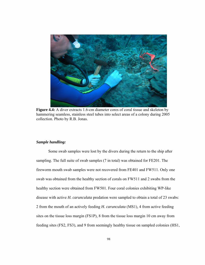
98
Figure 4.4: A diver extracts 1.6-cm diameter cores of coral tissue and skeleton by
collection. P
ter
samp
fireworm
the
healthy section were obtained from FW501. Four coral colonies exhibiting WP-like
disease with active H. carunculata predation were sampled to obtain a total of 23 swabs:
2 from the mouth of an actively feeding H. carunculata (MS1), 4 from active feeding
sites on the tissue loss margin (FS1P), 8 from the tissue loss margin 10 cm away from
feeding sites (FS2, FS3), and 9 from seemingly healthy tissue on sampled colonies (HS1,
hammering seamless, stainless steel tubes into select areas of a colony during 2005 hoto by R.B. Jonas.
Sample handling:
Some swab samples were lost by the divers during the return to the ship af
ling. The full suite of swab samples (7 in total) was obtained for FE201. The
mouth swab samples were not recovered from FE401 and FW511. Only one
swab was obtained from the healthy section of corals on FW511 and 2 swabs from

HS2, HS3). The summary of swab samples is provided in Table 4.3. Swab stems were
trimmed to fit and then placed into dedicated cryovials, frozen at liquid nitrogen
temperature (-196 °C), and returned to the laboratory in a thermal-regulated dry shipper.
The colonies used for the swab sampling were also cored for a total of 16 cores: 4
from active feeding sites on the tissue loss margin (FC1P), 4 from the tissue loss margin
10 cm away from feeding sites (FC2, FC3), and 8 from seemingly healthy tissue on
sampled colonies (HC1, HC2, HC3). The summary of core samples is provided in Table
4.4. Weather conditions prevented collecting a full suite of core samples from FE201 and
FE401. Only two cores were collected from these sites: one at the feeding site and one
from the apparently healthy surfaces of the coral colonies. On the ship, the cores were
removed from the Falcon™ tubes and trimmed with a diamond blade on a dremel tool to
reduce the quantity of skeleton. The samples were placed in individual cryovials and
frozen at liquid nitrogen temperature (-196 °C) for return to the laboratory using a
thermal-regulated dry shipper.
The actively feeding fireworms were taken to the ship in individual Falcon™
tubes containing ambient seawater. Each one was removed from the tube and dissected
immediately into three parts: anterior (head), trunk, and posterior. Each part was further
cut sagittally and placed in a labeled cryovial, then frozen at liquid nitrogen temperature
(-196 °C) for return to the laboratory in the thermal-regulated dry shipper.
99
Analysis
DNA Extraction and test for eukaryotic 18S ribosomal RNA
The genomic DNA of fireworms and coral core and swab samples was extracted
using a refined protocol based on the Bio101 FastDNA Extraction kit for tissue. Prior to
extraction, the H. carunculata samples were removed from the freezer and allowed to
thaw completely in the refrigerator. Approximately 200 mg tissue was cut from each
collected H. carunculata specimens and placed into individual lysing tubes for DNA
extraction. Each frozen coral core samples was crushed using a sterilized mortar and
pestle and allowed to thaw afterwards in the refrigerator. Once thawed, an approximate
50/50 mix of liquid and solid portions (total of 200 µl) of each sample was pipetted into
the designated lysing buffer tube for extraction. To prepare the swab samples for DNA
extraction, each sample was placed in its designated lysing tube and soaked in 1 mL of
lysing buffer for 1 hour.
After extraction, an aliquot of sample DNA was amplified with a universal protist
18S primer to determine whether eukaryotic DNA was present. Fam_Prot18SF was used
as the protist primer to amplify the 18S rRNA of eukaryotic organisms. The reverse
primer was DA443R. DNA extracted from American Type Culture Collection protist
CCMP768 (Rhodomonas sp.) was used as a positive control during the initial PCR runs.
Each PCR reaction went through 35 cycles of the PCR using Taq Gold polymerase with
an annealing temperature of 54 °C. PCR products were run on a 1% agarose
electrophoresis gel to quantify and size the amplification product.
100
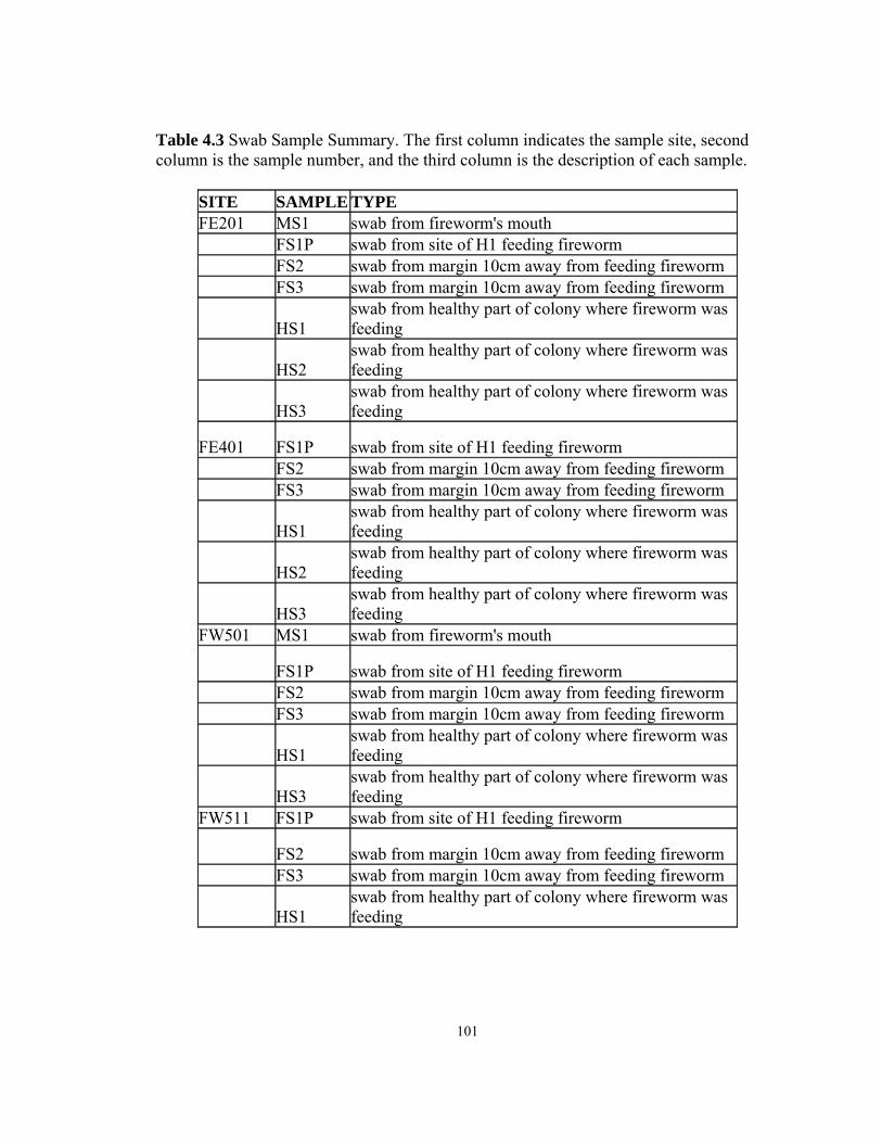
Table 4.3 Swab Sample Summary. The first column indicates the sample site, second column is the sample number, and the third column is the description of each sample.
SITE SAMPLE TYPE FE201 MS1 swab from fireworm's mouth FS1P swab from site of H1 feeding fireworm FS2 swab from margin 10cm away from feeding fireworm FS3 swab from margin 10cm away from feeding fireworm
HS1 swab from healthy part of colony where fireworm was feeding
HS2 swab from healthy part of colony where fireworm was feeding
HS3 swab from healthy part of colony where fireworm was feeding
FE401 FS1P swab from site of H1 feeding fireworm FS2 swab from margin 10cm away from feeding fireworm FS3 swab from margin 10cm away from feeding fireworm
HS1 swab from healthy part of colony where fireworm was feeding
HS2 swab from healthy part of colony where fireworm was feeding
HS3 swab from healthy part of colony where fireworm was feeding
FW501 MS1 swab from fireworm's mouth
FS1P swab from site of H1 feeding fireworm FS2 swab from margin 10cm away from feeding fireworm FS3 swab from margin 10cm away from feeding fireworm
HS1 swab from healthy part of colony where fireworm was feeding
HS3 swab from healthy part of colony where fireworm was feeding
FW511 FS1P swab from site of H1 feeding fireworm
FS2 swab from margin 10cm away from feeding fireworm FS3 swab from margin 10cm away from feeding fireworm
HS1 swab from healthy part of colony where fireworm was feeding
101
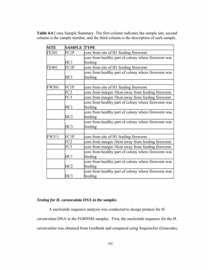
Table 4.4 Cores Sample Summary. The first column indicates the sample site, second column is the sample number, and the third column is the description of each sample.
SITE SAMPLE TYPE FE201 FC1P core from site of H1 feeding fireworm
HC1 core from healthy part of colony where fireworm was feeding
FE401 FC1P core from site of H1 feeding fireworm
HC1 core from healthy part of colony where fireworm was feeding
FW501 FC1P core from site of H1 feeding fireworm FC2 core from margin 10cm away from feeding fireworm FC3 core from margin 10cm away from feeding fireworm
HC1 core from healthy part of colony where fireworm was feeding
HC2 core from healthy part of colony where fireworm was feeding
HC3 core from healthy part of colony where fireworm was feeding
FW511 FC1P core from site of H1 feeding fireworm FC2 core from margin 10cm away from feeding fireworm FC3 core from margin 10cm away from feeding fireworm
HC1 core from healthy part of colony where fireworm was feeding
HC2 core from healthy part of colony where fireworm was feeding
HC3 core from healthy part of colony where fireworm was feeding
Testing for H. carunculata DNA in the samples
A nucleotide sequence analysis was conducted to design primers for H.
carunculata DNA in the FGBNMS samples. First, the nucleotide sequence for the H.
carunculata was obtained from GenBank and compared using Sequencher (Gencodes,
102

Ann Arbor, MI) with the nucleotide sequence of M. annularis to select a sequence of
bases that did not have similarities to M. annularis and were highly selective for H.
carunculata. Two reverse primers were designed using Primer Select (DNAStar,
Madison, WI) and paired with two forward protist primers to create a long product of 526
base pairs using Prot18SF (5' GGTTGATCCTGCCAGTAGTCATATGCTTG 3', Tm =
61 °C) and HC737RA (5' AGTTAAGAGCACCAAGGGAAAACCGG 3', Tm = 60 °C)
and a short product of 260 base pairs using Prot397F (5' CCGGAGAGGGAGCCTGA
3', Tm = 54 °C) and HC737RB (5' TAAGAGCACCAAGGGAAAACCGG 3', Tm=57 °C).
The four primers lacked self-complementary sequences or hairpins.
Table 4.5 Standard PCR Protocol with Taq Gold DNA Polymerase used for all the PCR reactions.
Reagents Volume
(µl)
DEPC water 7.9
10X PCR Gold Buffer 2
25mM Magnesium mix 2
dNTPs (2mM each) 2
0.1% BSA 2
Forward Primer 1
Reverse Primer 1
Taq DNA Polymerase 0.1
Master Mix Volume 18
DNA from Sample 2
Final Volume 20
103
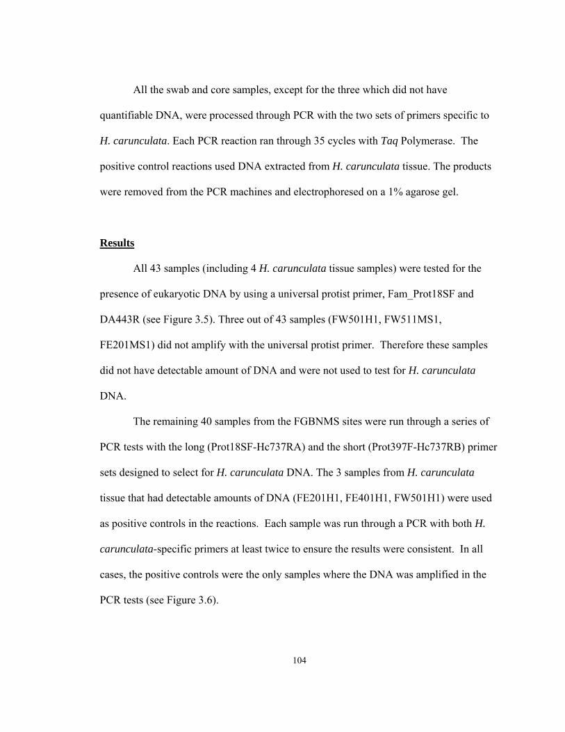
All the swab and core samples, except for the three which did not have
quantifiable DNA, were processed through PCR with the two sets of primers specific to
H. carunculata. Each PCR reaction ran through 35 cycles with Taq Polymerase. The
positive control reactions used DNA extracted from H. carunculata tissue. The products
were removed from the PCR machines and electrophoresed on a 1% agarose gel.
Results
All 43 samples (including 4 H. carunculata tissue samples) were tested for the
presence of eukaryotic DNA by using a universal protist primer, Fam_Prot18SF and
DA443R (see Figure 3.5). Three out of 43 samples (FW501H1, FW511MS1,
FE201MS1) did not amplify with the universal protist primer. Therefore these samples
did not have detectable amount of DNA and were not used to test for H. carunculata
DNA.
The remaining 40 samples from the FGBNMS sites were run through a series of
PCR tests with the long (Prot18SF-Hc737RA) and the short (Prot397F-Hc737RB) primer
sets designed to select for H. carunculata DNA. The 3 samples from H. carunculata
tissue that had detectable amounts of DNA (FE201H1, FE401H1, FW501H1) were used
as positive controls in the reactions. Each sample was run through a PCR with both H.
carunculata-specific primers at least twice to ensure the results were consistent. In all
cases, the positive controls were the only samples where the DNA was amplified in the
PCR tests (see Figure 3.6).
104
105
Figure 4.5 l in this picture shows the PCR reactio e in the
n the reaction ples
(FE201H1 and FE401H1) am er.
Despite d species, the DNA on the mouth
511MS2), was not amplified by the H.
carunculata- H. carunculata tissue
samples, we sults may be caused by
an inhibition of H. carunculata by coral DNA. To preclude this problem, a PCR was run
with a different ratio of H. carunculata tissue DNA and coral DNA extracted from
healthy coral cores (1:1, 1:2, 1:10) (see Figure 3.7). The PCR products demonstrated that
even in the highest ratio of coral DNA (1:10), H. carunculata DNA was amplified.
PCR with Universal Protist Primer. The agarose gen using the protist primer FAM_Prot18SF (DA443R). The positiv
reaction was the protist CCMP768 and the negative was absent of DNA itube. The DNA in the core (FE201FC1P and HC1, FE401FC1P) and tissue sam
plified in the presence of the prim
irect contact with the H. carunculata
swabs (FE201MS1, FE401MS1, FW501MS1, FW
specific primers. Since all the samples, except for the
re in the direct contact with coral, the negative PCR re

Figure 4.6 PCR with H. carunculata-specific primer Hc737RA. The agarose gel in this picture show er (Hc737RA). The positive in the reac (FE201H1, well 6). The negative control was the pr ples (FW511H1, FW plified in the presence of the
Discussion
s the PCR reaction using the H. carunculata primtion was tissue sample from H. carunculata
otist CCMP768 (well 7). The DNA in the 5 other sam501FC1P, FW501HC1, FW501FS1P, FW501HS1) was not am
H. carunculata-specific primer.
line of knowledge to be used in future
developm predation from other agents of coral tissue
loss. Since visual observations are not sufficient to diagnose a causal agent of tissue loss
(Zorpette 1995, Bruckner 2006, Ainsworth et al. 2007) this approach provides
information on potential analytical tools to understand the feeding behavior and
mechanisms of this coral reef predator.
This study took place during the third year (2007) of consecutive winter outbreaks
of apparently rapid tissue loss (aka white syndrome) in the FGBNMS. The corallivore H.
carunculata was found consuming coral tissue at the margin of M. annularis colonies
The results of this study provide a base
ent of diagnostic tools to distinguish
106
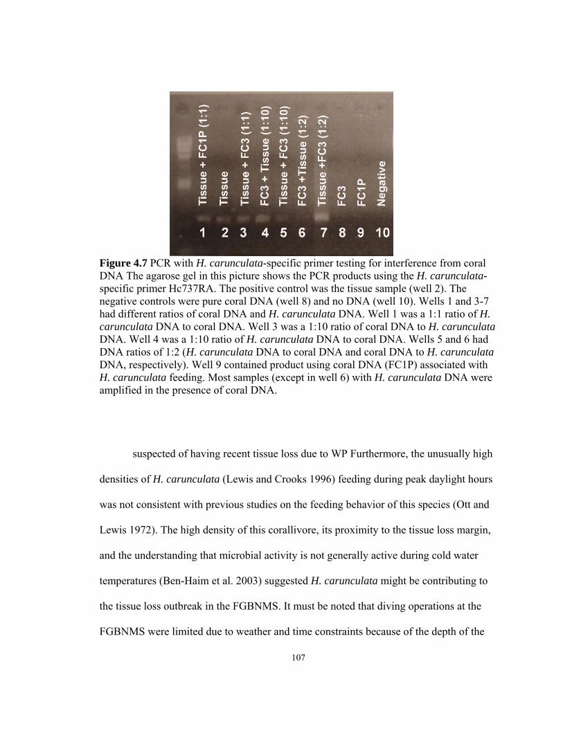
107
Figure 4.7 r interference from coral DNA The ag H. carunculata- specific prim le (well 2). The negative cont 10). Wells 1 and 3-7
DNA. Well 1 was a 1:1 ratio of H. carunculata H. carunculata
ells 5 and 6 had DNA ratios of 1:2 ( ral DNA to H. carunculata DNA, respectively). W ith H. carunculata H. carunculata DNA were am
suspected of having recent tissue loss due to WP Furthermore, the unusually high
densities of H. carunculata (Lewis and Crooks 1996) feeding during peak daylight hours
was not consistent with previous studies on the feeding behavior of this species (Ott and
Lewis 1972). The high density of this corallivore, its proximity to the tissue loss margin,
and the understanding that microbial activity is not generally active during cold water
temperatures (Ben-Haim et al. 2003) suggested H. carunculata might be contributing to
the tissue loss outbreak in the FGBNMS. It must be noted that diving operations at the
FGBNMS were limited due to weather and time constraints because of the depth of the
PCR with H. carunculata-specific primer testing foarose gel in this picture shows the PCR products using the er Hc737RA. The positive control was the tissue samprols were pure coral DNA (well 8) and no DNA (well
had different ratios of coral DNA and H. carunculata DNA to coral DNA. Well 3 was a 1:10 ratio of coral DNA to
DNA. Well 4 was a 1:10 ratio of H. carunculata DNA to coral DNA. WH. carunculata DNA to coral DNA and co
ell 9 contained product using coral DNA (FC1P) associated wfeeding. Most samples (except in well 6) with
plified in the presence of coral DNA.

reefs, thus it was impossible to determine whether and how tissue loss occurred prior to
finding the fireworms at the tissue margin and to assess whether the tissue loss continued
after the fireworms removed during this study. However, the limited information
available about the predation scars of H. carunculata makes is difficult to identify H.
carunculata-induced tissue loss in field observations (Miller and Williams 2007, Rotjan
and Lewis 2008).
To address the question of whether H. carunculata feeding behavior leaves
behind a molecular marker unique to the fireworm, actively feeding specimens, coral
cores, and swabs were taken from coral colonies at tissue loss margins at FGBNMS in
March 2007. Genomic DNA was extracted from the samples. All samples with
eukaryotic DNA were run through PCRs with primers designed to be highly selective for
H. carunculata. Of the 40 samples with eukaryotic DNA, only the three samples
extracted from H. carunculata tissue (positive controls) were amplified by the H.
carunculata-specific primers. Interestingly, the swabs sampled directly from the mouths
of the specimens were not amplified by the specific primers. More tests were conducted
with the specific primers to investigate a potential interference from coral DNA when
mixed with H. carunculata DNA. Even in the products with the largest ratio of coral
DNA (1.8 µl) to H. carunculata DNA (0.2 µl), H. carunculata DNA was amplified.
These results indicate that H. carunculata DNA is not detectable at feeding sites through
molecular analytical techniques. Therefore, the working hypothesis is rejected and the
null hypothesis is accepted. Hermodice carunculata does not leave a molecular marker at
foraging sites, at least under the conditions of this study.
108

The need for H. carunculata research is apparent given the observations of their
foraging on tissue margins in the FGBNMS and Navassa during outbreaks of suspect
WP. However, molecular techniques may not be effective tools to use for detecting
predation sites. The H. carunculata DNA that might have been left at feeding sites could
be affected by digestive enzymes secreted by the organism to aid in tissue extraction.
Residual digestive enzymes on the coral tissue or swabs might break down, interfere, or
damage some of the H. carunculata DNA on the sample swabs before they could be
frozen or after thawing.
Another possible explanation for H. carunculata DNA not amplifying in this
series of PCRs could be the nculata
tissue. Future mo a different
region of amplification. Also, plicon
Length Heterogeneity (LH-PCR) to obtain molecular fingerprints. This approach will
allow a more thorough understanding of the DNA present in the samples, including the
presence of H. carunculata DNA.
Corallivores and their population controls have been overlooked in previous
studies assessing the impact of coral tissue loss on reef ecosystems (Ott and Lewis 1972,
Birkeland and Lucas 1990, Sussman et al. 2003, Dalton and Godwin 2006, Miller and
Williams 2007). Extensive research is needed to understand the role of H. carunculata
and other corallivores in coral tissue loss and the etiology of coral diseases (Rotjan and
Lewis 2008). The field observations in this study and other documented coral predator
events indicate corallivory may be an important part in suspect disease outbreaks and
primers. The selective primers amplified pure H. caru
lecular studies should use a new set of primers prepared from
molecular techniques should include the use of Am
109

therefore should always be considered when investigating causal agents of coral tissue
loss.
110
Literature Cited Ainsworth TD, Kramasky-Winter E, Loya Y, Hoegh-Guldberg O, Fine M (2007) Coral
disease diagnostics: What's between a plague and a band? Appl Environ Microbiol 73:981-992
Alker AP, Smith GW, Kim K (2001) Characterization of Aspergillus sydowii (Thom et
Church), a fungal pathogen of Caribbean sea fan corals. Hydrobiol 460:105-111 Al-Moghrabi S (2001) Unusual black band disease (BBD) outbreak in the northern tip of
the Gulf of Aqaba (Jordan). Coral Reefs 19:330-331 Antonius A (1977) Coral mortality in reefs: a problem for science and management 3rd
International Coral Reef Symposium, Miami, p 617-623 Aronson RB, Precht WF (2001) White-band disease and the changing face of Caribbean
coral reefs. Hydrobiol 460:25-38 Bak RPM, Meesters EH (1998) Coral population structure: the hidden information of
colony size-frequency distributions. Mar Ecol-Prog Ser 162:301-306 Ben-Haim Y, Rosenberg E (2002) A novel Vibrio sp. pathogen of the coral Pocillopora
damicornis. Mar Biol 141:47-55 Ben-Haim Y, Zicherman-Keren M, Rosenberg E (2003) Temperature-regulated bleaching
and lysis of the coral Pocillopora damicornis by the novel pathogen Vibrio coralliilyticus. Appl Environ Microbiol 69:4236-4242
Birkeland C, Lucas J (eds) (1990) Acanthaster planci: major management problem of
coral reefs, Vol. CRC Press Bruckner AW (2001) Coral health and mortality. Recognizing the signs of coral diseases
and predators. In: Humann P, Deloach N (eds) Reef Coral Identification: Florida, Caribbean, Bahamas, Including Marine Plants, pp 240-271
Bruckner AW (2006) Progress in Understanding Coral Diseases in the Caribbean Coral
Health and Disease in the Pacific: Vision for Action, Honolulu, Hawaii Bruckner AW, Bruckner RJ (1997) Outbreak of coral disease in Puerto Rico. Coral Reefs
16:260 Bruckner AW, Bruckner RJ (2006) Consequences of yellow band disease (YBD) on
Montastraea annularis (species complex) populations on remote reefs off Mona Island, Puerto Rico. Dis Aquat Org 69:67-73
111
Bruno JF, Petes LE, Harvell CD, Hettinger A (2003) Nutrient enrichment can increase
the severity of coral diseases. Ecol Lett 6:1056-1061 Bruno JF, Selig ER, Casey KS, Page CA, Willis BL, Harvell CD, Sweatman H, Melendy
AM (2007) Thermal stress and coral cover as drivers of coral disease outbreaks. PLoS Biol 5:1220-1227
Bythell JC, Gladfelter EH, Bythell M (1993) Chronic and catastrophic natural mortality
of 3 common Caribbean reef corals. Coral Reefs 12:143-152 Bythell JC, Pantos O, Richardson LL (2004) White plague, white band, and other “white”
diseases. In: Rosenberg E, Loya Y (eds) Coral Health and Disease. Springer-Verlag, Berlin, p 351-366
Cerrano C, Bavestrello G, Bianchi CN, Cattaneo-vietti R, Bava S, Morganti C, Morri C,
Picco P, Sara G, Schiaparelli S, Siccardi A, Sponga F (2000) A catastrophic mass-mortality episode of gorgonians and other organisms in the Ligurian Sea (Northwestern Mediterranean), summer 1999. Ecol Lett 3:284-293
Coles SL, Seapy DG (1998) Ultraviolet absorbing compounds and tumorous growths on
acroporid corals from Bandar Khayran, Gulf of Oman, Indian Ocean. Coral Reefs 17:195-198
Cumming RL (2002) Tissue injury predicts colony decline in reef-building corals. Mar
Ecol-Prog Ser 242:131-141 Dalton SJ, Godwin S (2006) Progressive coral tissue mortality following predation by a
corallivorous nudibranch (Phestilla sp.). Coral Reefs 25:529-529 Denner EBM, Smith GW, Busse HJ, Schumann P, Narzt T, Polson SW, Lubitz W,
Richardson LL (2003) Aurantimonas coralicida gen. nov., sp. nov., the causative agent of white plague type II on Caribbean scleractinian corals. Int J Syst Evol Microbiol 53:1115-1122
Dustan P (1977) Vitality of reef coral populations off Key Largo, Florida: recruitment
and mortality. Environ Geol 2:51-58 Edmunds PJ, Elahi R (2007) The demographics of a 15-year decline in cover of the
Caribbean reef coral Montastraea annularis. Ecol Monogr 77:3-18 Frias-Lopez J, Zerkle AL, Bonheyo GT, Fouke BW (2002) Partitioning of bacterial
communities between seawater and healthy, black band diseased, and dead coral surfaces. Appl Environ Microbiol 68:2214-2228
112
Gardner TA, Cote IM, Gill JA, Grant A, Watkinson AR (2003) Long-term region-wide
declines in Caribbean corals. Science 301:958-960 Geiser DM, Taylor JW, Ritchie KB, Smith GW (1998) Cause of sea fan death in the
West Indies. Nature 394:137-138 Gil-Agudelo DL, Garzon-Ferreira J (2001) Spatial and seasonal variation of Dark Spots
Disease in coral communities of the Santa Marta area (Colombian Caribbean). Bull Mar Sci 69:619-629
Glynn PW (1963) Hermodice carunculata and Mithraculus sculptus, two hermatypic
coral predators. Association of Island Marine Laboratories 4th Meeting pp 16-17 Harvell CD, Kim K, Burkholder JM, Colwell RR, Epstein PR, Grimes DJ, Hofmann EE,
Lipp EK, Osterhaus A, Overstreet RM, Porter JW, Smith GW, Vasta GR (1999) Review: Marine ecology - Emerging marine diseases - Climate links and anthropogenic factors. Science 285:1505-1510
Harvell D, Jordan-Dahlgren E, Merkel S, Rosenberg E, Raymundo L, Smith G, Weil E,
Willis B (2007) Coral disease, environmental drivers, and the balance between coral and microbial associates. Oceanogr 20:172-195
Henry LA, Hart M (2005) Regeneration from injury and resource allocation in sponges
and corals - A review. Int Rev Hydrobiol 90:125-158 Hughes TP (1994) Catastrophes, phase shifts, and large scale degradation of a Caribbean
coral reef. . Science 265:1547-1551 Hughes TP, Tanner JE (2000) Recruitment failure, life histories, and long-term decline of
Caribbean corals. Ecology 81:2250-2263 Israely T, Banin E, Rosenberg E (2001) Growth, differentiation and death of Vibrio shiloi
in coral tissue as a function of seawater temperature. Aquat Microb Ecol 24:1-8 Kim K, Harvell C (2002) Aspergillosis of sea fan corals: disease dynamics in the Florida
Keys. In: Porter J, Porter K (eds) The Everglades Florida Bay and coral reefs of the Florida Keys: an ecosystem sourcebook. CRC Press, New York, p 813-824
Knowlton N (2001) The future of coral reefs. Natl Acad Sci, pp 5419-5425 Kramarsky-Winter E, Loya Y (2000) Tissue regeneration in the coral Fungia granulosa:
the effect of extrinsic and intrinsic factors. Mar Biol 137:867-873
113

Kushmaro A, Rosenberg E, Fine M, Loya Y (1997) Bleaching of the coral Oculina patagonica by Vibrio AK-1. Mar Ecol-Prog Ser 147:159-165
Kuta KG, Richardson LL (2002) Ecological aspects of black band disease of corals:
relationships between disease incidence and environmental factors. Coral Reefs 21:393-398
Lewis JB, Crooks RE (1996) Foraging cycles of the amphinomid polychaete Hermodice
carunculata preying on the calcareous hydrozoan Millepora complanata. Bull Mar Sci 58:853-856
Lizama J, Blanquet RS (1975) Predation on sea anemones by the amphinomid
polychaete, Hermodice carunculata. Bull Mar Sci 25:442-443 Luna GM, Biavasco F, Danovaro R (2007) Bacteria associated with the rapid tissue
necrosis of stony corals. Environ Microbiol 9:1851-1857 Marsden JR (1962) A coral-eating polychaete. Nature 193:598 Marsden JR (1963) The digestive tract of Hermodice carunculata. (Pallas). Polychaeta:
Amphinomidae. Can J Zool/Rev Can Zool 41:165-184 Martin A, Losada F (1991) Regeneration of injuries caused by the amphinomid
polychaete Hermodice carunculata on scleractinian corals, hydrocorals and octocorals (Cnidaria). Ecotropicos 4:18-26
Miller MW, Williams DE (2007) Coral disease outbreak at Navassa, a remote Caribbean
island. Coral Reefs 26:97-101 Nagelkerken I, Bak RPM (1998) Differential regeneration of artificial lesions among
sympatric morphs of the Caribbean corals Porites astreoides and Stephanocoenia michelinii. Mar Ecol-Prog Ser 163:279-283
Nagelkerken I, Meesters EH, Bak RPM (1999) Depth-related variation in regeneration of
artificial lesions in the Caribbean corals Porites astreoides and Stephanocoenia michelinii. J Exp Mar Biol Ecol 234:29-39
Ott B, Lewis JB (1972) The importance of the gastropod Coralliophila abbreviata
(Lamarck) and the polychaete Hermodice carunculata (Pallas) as coral reef predators. Can J Zool/Rev Can Zool 50:1651-1656
Pandolfi JM, Bradbury RH, Sala E, Hughes TP, Bjorndal KA, Cooke RG, McArdle D,
McClenachan L, Newman MJH, Paredes G, Warner RR, Jackson JBC (2003) Global trajectories of the long-term decline of coral reef ecosystems. Science
114

301:955-958 Pantos O, Cooney RP, Le Tissier MDA, Barer MR, O'Donnell AG, Bythell JC (2003)
The bacterial ecology of a plague-like disease affecting the Caribbean coral Montastraea annularis. Environ Microbiol 5:370-382
Patterson KL, Porter JW, Ritchie KB, Polson SW, Mueller E, Peters EC, Santavy DL,
Smith GW (2002) The etiology of white pox, a lethal disease of the Caribbean elkhorn coral, Acropora palmata. Nat Acad Sci 99:8725-8730
Porter JW, Dustan P, Japp WC, Patterson KL, Kosmynin V, Meier OW, Patterson ME,
Parsons M (2001) Patterns and spread of coral disease in the Florida Keys. Hydrobiol. 460:1-24
Raymundo LJ, Rosell KB, Reboton CT, Kaczmarsky L (2005) Coral diseases on
Philippine reefs: genus Porites is a dominant host. Dis Aquat Org 64:181-191 Remily ER, Richardson LL (2006) Ecological physiology of a coral pathogen and the
coral reef environment. Microb Ecol 51:345-352 Richardson LL (1998a) Coral diseases: what is really known? Trends Ecol Evol 13:438-
443 Richardson LL, Goldberg WM, Carlton RG, Halas JC (1998b) Coral disease outbreak in
the Florida Keys: Plague type II. Rev Biolog Tropical 46:187-198 Richardson LL, Goldberg WM, Kuta KG, Aronson RB, Smith GW, Ritchie KB, Halas
JC, Feingold JS, Miller SL (1998c) Florida's mystery coral-killer identified. Nature 392:557- 558
Richardson LL, Kuta KG (2003) Ecological physiology of the black band disease
cyanobacterium Phormidium corallyticum. FEMS Microbiol Ecol 43:287-298 Riegl B (2002) Effects of the 1996 and 1998 positive sea-surface temperature anomalies
on corals, coral diseases and fish in the Arabian Gulf (Dubai, UAE). Mar Biol 140:29-40
Ritchie KB, Smith GW (1998) Type II white-band disease. Revista De Biologia Tropical
46:199-203 Rosenberg E, Ben-Haim Y, Toren A, Banin E, Kushmaro A, Fine M, Loya Y (1998)
Effect of temperature on bacterial bleaching of corals. In: Rosenberg E (ed) Current perspectives in microbial ecology. ASM Press, Washington, D.C., pp 242-254
115

Rosenberg E, Loya Y (2004) Coral Health and Disease, Vol. Springer-Verlag, Berlin Rotjan RD, Lewis SM (2005) Selective predation by parrotfishes on the reef coral Porites
astreoides. Mar Ecol-Prog Ser 305:193-201 Rotjan RD, Lewis SM (2008) Impact of coral predators on tropical reefs. Mar Ecol-Prog
Ser 367:73-91 Shinn EA, Smith GW, Prospero JM, Betzer P, Hayes ML, Garrison V, Barber RT (2000)
African dust and the demise of Caribbean coral reefs. Geophys Res Lett 27:3029-3032
Smith G, Weil E (2004) Aspergillosis in gorgonians. In: Rosenberg E, Loya Y (eds)
Coral Health and Disease. Springer Verlag, pp 279-288 Sussman M, Loya Y, Fine M, Rosenberg E (2003) The marine fireworm Hermodice
carunculata is a winter reservoir and spring-summer vector for the coral-bleaching pathogen Vibrio shiloi. Environ Microbiol 5(4):250-255
Sutherland KP, Porter JW, Torres C (2004) Disease and immunity in Caribbean and
Indo-Pacific zooxanthellate corals. Mar Ecol Prog Ser 266:273-302 Vogler C, Benzie J, Lessios H, Barber P, Worheide G (2008) A threat to coral reefs
multiplied? Four species of crown-of-thorns starfish. Biol Lett 4:696-699 Weil E, Croquer A (2009) Spatial variability in distribution and prevalence of Caribbean
scleractinian coral and octocoral diseases. I. Community-level analysis. Dis Aquat Org 83:195-208
Weil E, Smith G, Gil-Agudelo DL (2006) Status and progress in coral reef disease
research. Dis Aquat Org 69:1-7 Weil E, Urreiztieta I, Garzón-Ferreira J (2002) Geographic variability in the incidence of
coral and octocoral diseases in the wider Caribbean. 9th International Coral Reef Symposium, Bali, Indonesia, pp 1231–1237
Wilkinson C (ed) (2004) Status of coral reefs of the world, Vol 1. Global Coral Reef
Monitoring Network and Australian Institute of Marine Science, Townesville, Australia
Williams DE, Miller MW (2005) Coral disease outbreak: pattern, prevalence and
transmission in Acropora cervicornis. Mar Ecol-Prog Ser 301:119-128
116
Witman JD (1988) Effects of predation by the fireworm Hermodice carunculata on Milleporid hydrocorals. Bull Mar Sci 42:446-458
Zorpette G (1995) More coral trouble. Sci Am 273:36-37
117
5. Overall Conclusions
1. Gut content varied in Hermodice carunculata specimens. The most common
materials were collagenous cuticle and coccoid cells. In Chapter 3, H. carunculata
specimens lost weight while feeding on Millepora complanata. These results suggest
H. carunculata obtains a sustainable nutritional value from consuming prey in
addition to coral tissue. A reduction in live coral tissue may not have a direct impact
on H. carunculata survivorship but may increase H. carunculata’s foraging time on
alternative prey. Also, the availability of alternative prey may be influenced by
changes in physical and biological structures of coral reefs which may also affect H.
carunculata nutritional intake. Research is needed on H. carunculata species’ diet
composition, diet requirements, and the nutritional value of its food to qualify the
direct and indirect impacts of coral tissue loss on H. carunculata and its prey.
2. Through histological observations, a layer of coccoid cells was found on the outer
cuticle and a several clusters within the digestive tract and foregut of H. carunculata.
Also, H. carunculata was associated with tissue loss margins of suspect white plague
in the Flower Garden Banks National Marine Sanctuary. Bacteria associated with this
organism may be transmitted during movement and/or during foraging activities. A
layer of bacteria on the outer cuticle may serve as protection from potentially harmful
118

microbes or other organisms. This layer of cells may also provide opportunities for
microbes to attach to H. carunculata organisms for transportation to other coral
colonies. Bacteria in the foregut may be transmitted when H. carunculata everts its
pharynx during feeding which, if pathogenic, could exacerbate tissue loss or induce
disease. Bacteria staining and microscopic analysis in needed to determine if the
coccoid cells on the outer cuticle and with the digestive system are bacteria. Field
observations are needed at foraged sites to see if tissue margin continues after a
predation episode. More research on the ability to uptake and distribute bacteria either
from the outer cuticle or during feeding is needed to understand the vector
capabilities of this species.
3. H. carunculata has primitive eyes, a dorsal nuchal organ, and a ventral nerve cord
which may have sensory functions to find prey. These features may pick up
environmental cues as chemoreceptors and photoreceptors. H. carunculata has
feeding features including digestives enzymes in the muscular proboscis and a
flexible and expandable buccal region which help to consume prey of various sizes
and types. The ability to eat various prey is an important adaptative characteristic for
inhabitants of a changing environment like coral reefs. Digestive enzymes may help
consume prey through breakdown and removal of tissue or materials from various
substrate. The ventral cord may interact with the substrate through pigmented cells.
Observations and studies examining the function the sensory features are needed to
119

understand how H. carunculata receives environmental cues to find prey and avoid
predators.
4. H. carunculata has various defense mechanisms. Tufts of setae will become brighter
in color when an object is near the organism. The setae are toxic and they break off or
are released when H. carunculata is handled. Histological observations confirm field
observations on the different coloration patterns of this species. Individuals can be
various patterns of green, red, yellow, and brown. The color schemes may help the
organism blend into the substrate to disguise from potential predators. Though
specific predators have not been identified in literature or observations, the presence
of biological structures which deter predators suggests there is an evolutionary
pressure to develop these features. A driving force for defense mechanism may be H.
carunculata’s feeding behavior. Its pharynx is everted and the organism is stationary
on the reef surface during the predation episode. Therefore, it may be vulnerable to
predators when consuming coral tissue and moving along the coral substrate in search
of prey.
5. H. carunculata is a poorly understood coral predator and reef dweller. Histological
studies in Chapter 2 found new features which may have ecological implications.
Results from the feeding experiments in Chapter 3 suggest coral tissue does not
provide enough nutrients for survivorship. Field observations on H. carunculata
feeding behavior associated with tissue loss margins of suspect-WP and the presence
120
of coccoid cells resembling bacteria in the body of H. carunculata suggests this
species has vector capabilities. This organism and its role in coral tissue loss through
feeding and transmission of pathogenic bacteria need to be well understood.
Documentation of predation scars and patterns are needed to distinguish H.
carunculata-induced lesions. Also, questions about the vulnerability of coral colonies
after predation episodes to future foraging by the same organism or other corallivores
or the vulnerability of coral tissue to pathogenic microbes after predation need to be
answered to determine this species’ contribution to overall coral tissue loss.
121

122
CURRICULUM VITAE
Staci Lewis received her Bachelors of Science degree from Salem College in 2002. She started studing Hermodice carunculata in Barbados as a research assistant under Dr. Suzanne Dorsey. From 2003-2005, she worked as the laboratory and projects manager for Dr. Rich Aronson at the Dauphin Island Sea Lab in Alabama and worked on various coral reef research project, She conducted studies on Hermodice carunculata during her Fulbright Fellowship in Barbados from 2005-2006.