Sabir, H., Wood, T. R., Gill, H., Liu, X., Dingley, J...
19
Sabir, H., Wood, T. R., Gill, H., Liu, X., Dingley, J., & Thoresen, M. (2016). Xenon depresses aEEG background voltage activity whilst maintaining cardiovascular stability in sedated healthy newborn pigs. Journal of the Neurological Sciences, 363, 140-144. https://doi.org/10.1016/j.jns.2016.02.051 Peer reviewed version Link to published version (if available): 10.1016/j.jns.2016.02.051 Link to publication record in Explore Bristol Research PDF-document This is the author accepted manuscript (AAM). The final published version (version of record) is available online via Elsevier at http://www.sciencedirect.com/science/article/pii/S0022510X16301162 University of Bristol - Explore Bristol Research General rights This document is made available in accordance with publisher policies. Please cite only the published version using the reference above. Full terms of use are available: http://www.bristol.ac.uk/pure/about/ebr-terms
Transcript of Sabir, H., Wood, T. R., Gill, H., Liu, X., Dingley, J...

Sabir, H., Wood, T. R., Gill, H., Liu, X., Dingley, J., & Thoresen, M. (2016).Xenon depresses aEEG background voltage activity whilst maintainingcardiovascular stability in sedated healthy newborn pigs. Journal of theNeurological Sciences, 363, 140-144.https://doi.org/10.1016/j.jns.2016.02.051
Peer reviewed version
Link to published version (if available):10.1016/j.jns.2016.02.051
Link to publication record in Explore Bristol ResearchPDF-document
This is the author accepted manuscript (AAM). The final published version (version of record) is available onlinevia Elsevier at http://www.sciencedirect.com/science/article/pii/S0022510X16301162
University of Bristol - Explore Bristol ResearchGeneral rights
This document is made available in accordance with publisher policies. Please cite only the publishedversion using the reference above. Full terms of use are available:http://www.bristol.ac.uk/pure/about/ebr-terms
1
Xenon depresses aEEG background voltage activity whilst maintaining
cardiovascular stability in sedated healthy newborn pigs
Hemmen Sabir1,2, Tommy Wood3, Hannah Gill1, Xun Liu1, John Dingley4, Marianne
Thoresen1,3
1 School of Clinical Sciences, University of Bristol, St Michael’s Hospital, Bristol, United
Kingdom;
2 Departments of General Pediatrics, Neonatology and Pediatric Cardiology, University
Children's Hospital, Düsseldorf, Heinrich-Heine University Düsseldorf, Germany;
3 Department of Physiology, Institute of Basic Medical Sciences, University of Oslo, Oslo,
Norway;
4 College of Medicine, Swansea University, United Kingdom
Sources of Funding: This study was supported by Sport Aiding Medical Research for Kids
(SPARKS (UK)), the Laerdal Foundation for Acute Medicine (Norway) and the Norwegian
Research Council.
Author´s key words: Xenon, amplitude-integrated encephalography, inhalation
anaesthetics, newborn
Abbreviated Title: Xenon reduces aEEG activity in healthy pigs
Address for correspondence
Marianne Thoresen MD PhD.
Department of Physiology, Institute of Basic Medical Sciences, University of Oslo,
Visiting address: Room 2366, Domus Medica, Sognsvannsveien 9, 0372 Oslo, Norway
Postal address: Postal address: PB 1103 Blindern
0317 Oslo, Norway
Direct line: +47 22851568, fax: +47 228 512

2
Abstract
Background: Changes in electroencephalography (EEG) voltage range are used to monitor
the depth of anaesthesia, as well as predict outcome after hypoxia-ischaemia in neonates.
Xenon is being investigated as a potential neuroprotectant after hypoxic-ischaemic brain
injury, but the effect of Xenon on EEG parameters in children or neonates is not known. This
study aimed to examine the effect of 50% inhaled Xenon on background amplitude-
integrated EEG (aEEG) activity in sedated healthy newborn pigs.
Methods: Five healthy newborn pigs, receiving intravenous fentanyl sedation, were
ventilated for 24h with 50%Xenon, 30%O2 and 20%N2 at normothermia. The upper and lower
voltage-range of the aEEG was continuously monitored together with cardiovascular
parameters throughout a 1h baseline period with fentanyl sedation only, followed by 24h of
Xenon administration.
Results: The median (IQR) upper and lower aEEG voltage during 1h baseline was 48,0µV
(46,0 – 50,0) and 25,0µV (23,0 – 26,0), respectively. The median (IQR) aEEG upper and
lower voltage ranges were significantly depressed to 21,5µV (20,0 – 26,5) and 12,0µV (12,0
– 16,5) from 10min after the onset of 50% Xenon administration (p=0.002). After the initial
Xenon induced depression in background aEEG voltage, no further aEEG changes were
seen over the following 24h of ventilation with 50% xenon under fentanyl sedation. Mean
arterial blood pressure and heart rate remained stable.
Conclusion: Mean arterial blood pressure and heart rate were not significantly influenced by
24h Xenon ventilation. 50% Xenon rapidly depresses background aEEG voltage to a steady
~50% lower level in sedated healthy newborn pigs. Therefore, care must be taken when
interpreting the background voltage in neonates also receiving Xenon.

3
Abbreviations
aEEG amplitude-integrated electroencephalography
EEG electroencephalography
HT therapeutic hypothermia
i.v. intravenous
MAC minimal alveolar concentration
N2 nitrogen
NMDA N-methyl-D-aspartate
O2 oxygen
Trec Rectal temperature
Xe Xenon

4
Introduction
Monitoring the depth of sedation in newborns and children is of great interest, particularly as
the use of sedatives and anaesthetics in this group of patients is increasing.[1] Most of the
drugs that are frequently used in the paediatric population affect the characteristics of the
electroencephalogram (EEG), and several calculated EEG parameters have been used to
monitor the depth of anaesthesia in both children [2-4] and neonates.[5] The amplitude-
integrated EEG (aEEG) is a bedside monitoring tool widely used in newborns to continuously
monitor brain activity and detect seizures.[6, 7] Following neonatal encephalopathy, the
degree and timing of aEEG voltage depression and aEEG recovery also predicts outcome
after perinatal asphyxia in both cooled and non-cooled newborns.[8, 9] However, the ability
of the aEEG to predict outcome may be altered by any concurrent anaesthetic or sedative
drug regimen that the patient is receiving. One example is the noble gas Xenon (Xe), which
has both anaesthetic and analgesic actions.[10] We have previously shown that prolonged
administration of 50% inhaled Xe to the developing brain is safe.[11, 12] Xenon also
augments neuroprotection when combined with therapeutic hypothermia (HT) after hypoxia-
ischaemia in newborn animal studies.[13-15] Xenon significantly reduces EEG background
voltage activity in healthy adults,[16] and reports using EEG parameters to assess depth of
anaesthesia in adult patients inhaling Xe have been published.[17-20]
However, the anaesthetic potency of Xe, or MAC-Xe (the minimal alveolar concentration of
Xe preventing purposeful movement in 50% of subjects in response to a standardised painful
stimulus), appears to be highly variable and age-dependent.[21] For instance, in eight
healthy, newborn pigs, individual MAC-Xe ranged from 60 – 120%.[22] Though MAC-Xe for
newborn humans has not been measured, its lower range is likely to be above 50%. This is
because the MAC value for most inhalational anaesthetics is higher in newborn mammals
compared to adults, and the MAC-Xe in adult humans was originally found to be 71%.[23]
Although 50% Xe is a proven neuroprotective concentration experimentally when used in
combination with therapeutic hypothermia (HT), 50% Xe alone does not provide adequate
sedation to tolerate being ventilated while undergoing HT. Therefore, additional sedation

5
such as opioid administration is required. Importantly, the use of high doses of intravenous
fentanyl, both with and without co-administration of 50% Xe, did not induce neuroapoptosis in
the newborn pig brain, and is therefore likely to be safe also for humans.[11]
We, as well as others, have previously shown that Xe administration reduces aEEG
background voltage activity and suppresses seizures in asphyxiated cooled newborns.[12,
24] Xenon, in combination with HT and infusion of opioids, is currently being investigated in
clinical trials to reduce brain injury after neonatal encephalopathy.[12, 24, 25] Therefore, it is
important to assess the effect of Xe on aEEG voltage changes, as this might change the
ability of the aEEG to predict outcome during treatment in these patients. As the sedative
effect of Xe cannot be assessed reliably in infants with concurrent encephalopathy, due to
varying degrees of cerebral depression, the current study was carried out.
This current study aimed to examine the effect of 50% inhaled Xe plus fentanyl infusion on
background aEEG background activity, as well as cardiovascular parameters, in healthy
newborn pigs.
6
Material and Methods
Conduct of Experiment
All experiments were conducted according to the United Kingdom Home Office license
guidelines, and were approved by the University of Bristol Ethical Review Panel (Bristol,
United Kingdom). This was a sub study of our original experiment, where we reported the
safety of 50% Xe-ventilation in healthy newborn pigs, showing that ventilation with 50% Xe
does not cause cellular injury in the newborn brain.[11] The current study uses data from five
healthy newborn pigs (aged <24h) receiving intravenous fentanyl sedation, whilst being
mechanically ventilated for 24h with 50% inhaled Xe, and maintained at normothermia (rectal
temperature 38.5°C).
Animals and Experimental groups
Five crossbred landrace/large white pigs born at term (four male) with a median (range) age
and weight of 10h (4 - 15h) and 1.7kg (1.2 - 1.9kg) were used. After a 1h baseline period on
fentanyl sedation, all pigs received 24h of 50% inhaled Xe at normothermia. Intravenous
fentanyl sedation was continued throughout.
Animal Preparation, Baseline data and Management of pigs
All animals were initially prepared as published in our original experiment.[11]. In brief, after
initial intubation and insertion of umbilical arterial and venous catheters, continuous
monitoring of mean arterial blood pressure and heart rate was enabled. During the 60min
baseline period and the first 30min after Xe administration, mean arterial blood pressure and
heart rate data were collected every 5min. For the following 23.5h of xenon administration,
mean arterial blood pressure and heart rate data was collected every 30min, and the median
was calculated for each pig hourly.
Intensive care management of the animals followed standard procedures.[11] Pigs received
intravenous maintenance fluid (5% dextrose/0.45% saline) at 10ml/kg/h. Tracheal suction
was performed every 8h or as clinically indicated, and blood glucose, lactate, and pH values

7
were maintained between 3.0 and 8.0mmol/L, <3.5mmol/L, and pH 7.35 and 7.47 (analyzed
at actual body temperature), respectively. Temperature measurements were undertaken with
a rectal probe (reusable YSI 400 series, CritiCool, MTRE, Yavne, Israel) inserted 6cm into
the rectum, and a skin probe (CritiCool, MTRE, Yavne, Israel) sited on the ear lobe. Both
probes were calibrated before use within ±0.1°C, over a temperature range of 20 to 40°C,
against a certified mercury-in-glass thermometer (BS593; Zeal, London, United Kingdom).
Rectal temperature (Trec) was maintained at 38.5°C ± 0.2°C using a servo-controlled
(CritiCool, MTRE, Yavne, Israel) body-wrap containing circulating water. Ventilation settings
and inspired oxygen fraction were adjusted to maintain transcutaneous oxygen saturation
between 95 and 98%, and the end-tidal carbon dioxide between 4.0 and 6.0kPa.
Fentanyl Sedation and Xenon inhalation
After animal preparation, the 60min baseline aEEG recording period was initiated. An i.v.
fentanyl infusion of 1µg/kg/h was started with a bolus of 10µg/kg to ensure appropriate
sedation during ventilation. The fentanyl infusion was adjusted to achieve the sedation
required to ventilate a healthy newborn pig.
After 60min of baseline measurements during ventilation, a xenon/oxygen/nitrogen mixture
(Xe 50%/O2 30%/N2 20%) was delivered using an automated servo-controlled version of a
previously described closed-circuit delivery system, for a period of 24h.[26] Target Xe
concentration was achieved within 10min of onset, and was maintained at a concentration of
50% throughout the whole 24h experiment.
Amplitude-integrated EEG (aEEG) recording and analysis
Cross brain single-channel aEEG and raw EEG (Olympic CFM 6000, Natus Medical
Incorporated, Seattle, USA) was recorded from 3 subdermal needle electrodes (0.4mm
(27G), Viasys Healthcare, Madison, USA) and stored digitally. Inter-electrode distance was
3cm. The aEEG recording was started after animal preparation, and continued throughout
the 60min baseline and 24h Xe-treatment periods during which all pigs received i.v. fentanyl
8
sedation. The recording was concluded after 24h of ventilation with 50%Xe and the animal
was sacrificed.
The aEEG background voltage changes were continuously recorded, and voltage criteria [27]
were used to measure the voltage level of the upper and lower margin of the time-
compressed (6cm/hour) aEEG traces. For each semi-logarithmic aEEG trace the bandwidth
representing the upper and lower voltage margin was read from the digital screen. The level
for the upper band was 90% of the peak amplitude and the lower band was 10%. During the
60min baseline period and the first 60min after Xe administration the bandwidth was read
every 2min. The median of 30 baseline readings and 25 early xenon readings for each pig
was calculated (excluding the 10min when xenon increased from 0 - 50%). These medians
were compared. For the following 23h of xenon administration the last 10min period of each
hour was read every 2min and the median of the 5 readings was calculated.
Statistical Analysis
Statistical analyses were performed with SPSS version 22 (SPSS Inc., Chicago, IL, USA).
For both the upper and lower voltage margin separately, Wilcoxon signed rank test for paired
samples were used comparing the 60min baseline period with the first 20min period after
stable 50% xenon had been obtained. Mean arterial blood pressure and heart rate were
continuously recorded and median values during baseline, Xe onset and Xe maintenance
were compared visually. Two-sided testing with p <0.05 was considered statistically
significant.
9
Results
Physiological data
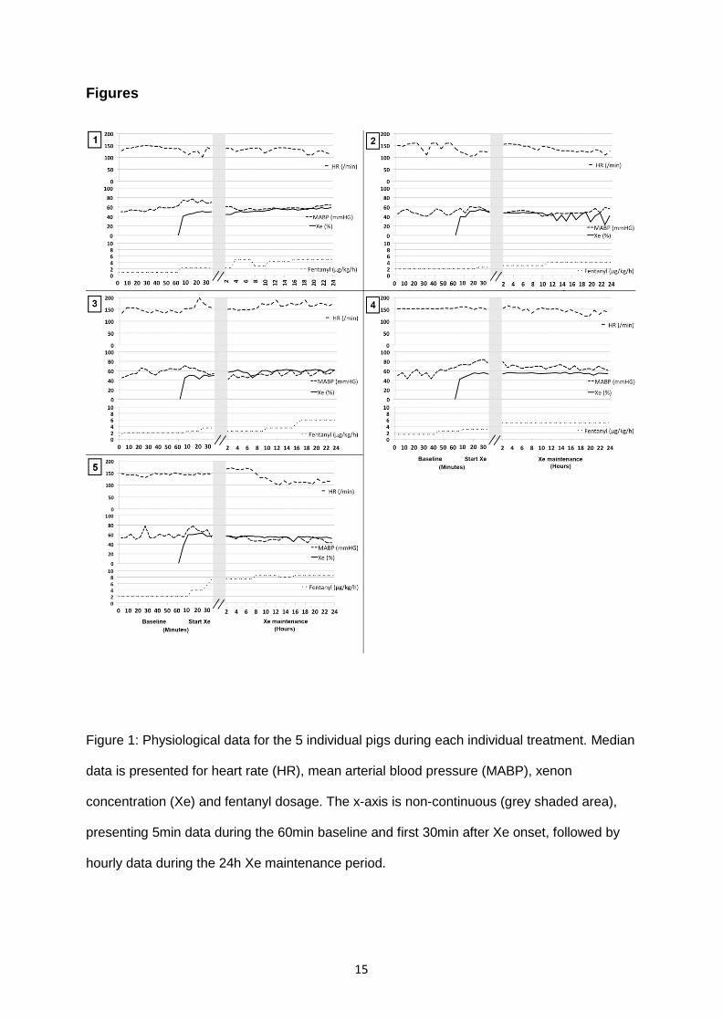
Figure 1 presents the physiological parameters for each individual pig before Xe
administration, during the onset of Xe and during the 24h Xe treatment period. Median
(range) heart rate during baseline, Xe onset and Xe maintenance were 146/min (110 – 160),
144/min (101 – 199) and 140/min (100 – 189), respectively. Median (range) arterial blood
pressure during baseline, Xe onset and Xe maintenance were 54mmHg (40 –78), 69mmHg
(47 – 84) and 54mmHg (40 – 79), respectively.
Analyzing each pig’s individual response, we found that in 3 pigs (Pig 2, 4 and 5) there was a
decrease in heart rate during the 24h Xe administration, whilst mean arterial blood pressure
increased (Pig 2 and 4), or remained stable (Pig 5).
Median (range) fentanyl dose was constant for each pig during the baseline period,
(2microgram/kg/h; 0.9 – 2.5). However, increased fentanyl delivery was necessary after
onset of xenon, increasing to a median of 2.5microgram/kg/h (2 – 7.5) during Xe onset and
4.9microgram/kg/h (2.4 – 8.5) during Xe maintenance. Background aEEG voltage remained
unchanged during the baseline period before Xe administration.
aEEG changes with xenon
Figure 2 shows an example of an individual (Pig 2) aEEG response to the onset of Xe
followed by 24h of continuous ventilation with 50% Xe. For the 5 pigs, during the 1h baseline
period, the median (IQR) upper aEEG voltage was 48,0µV (46,0 – 50,0). From 10min after
the onset of 50% Xenon administration when the concentration had reached 50%, the
median (IQR) aEEG upper voltage range was significantly depressed to 21,5µV (20,0 – 26,5)
(p=0.002). The corresponding lower aEEG voltage during baseline was 25,0µV (23,0 – 26,0),
reducing to 12,0µV (12,0 – 16,5) (p<0.002) with 50% Xe. Figure 3 shows the median
(interquartile range) upper and lower voltage margin during the whole treatment period.
During steady Xe ventilation the upper and lower voltage level (aEEG bandwidth) did not
significantly change.

10
Discussion
This study shows that background aEEG voltage in sedated healthy newborn pigs is rapidly
reduced upon administration of 50% inhaled Xe, and remains suppressed at the same level
throughout prolonged Xe delivery. This is consistent with clinical findings in encephalopathic
newborns.[12, 24] As these sedated healthy pigs did not receive an hypoxic insult, this
suggests a direct pharmacological effect of Xe. Additionally, prolonged Xe delivery did not
significantly alter cardiovascular parameters.
In pediatric anaesthesia, volatile anaesthetics are commonly used, as they have a
predictable onset and offset of action, and are assumed to be safe. Importantly, an adequate
anaesthetic depth can be achieved, and rapidly tailored to clinical need, while in most cases
maintaining hemodynamic stability.[28] However, there is evidence that inhalational
anaesthetics might be associated with increased apoptosis in the developing brain,[29-31]
which is a cause for concern. Xenon, a non-competitive inhibitor of the N-methyl-D-aspartate
(NMDA) receptor, has been shown to double neuroprotection when combined with
therapeutic hypothermia following hypoxic-ischaemic brain injury in multiple preclinical
models.[13-15] This has led to two clinical trials using Xe as an additional neuroprotective
agent during cooling in newborns suffering neonatal encephalopathy of hypoxic-ischaemic
origin; TOBY-Xe and CoolXenon.[12, 25] One of the two trials (TOBY-Xe) has been
completed, which showed no additional short-term neuroprotection when combining cooling
with 30% inhaled Xe (starting 10h after birth), compared to cooling only as the standard
treatment.[25] There are two important differences between the two study protocols;
CoolXenon aims to start Xe administration earlier (within 5h of birth), and uses a higher
concentration of inhaled Xe (50%). Data from the CoolXenon trial are therefore eagerly
awaited. However, recruitment is estimated to end in December 2016, followed by 18 months
of follow-up.
Amplitude-integrated EEG is widely used in neonatal units and is an established method for
prognosticating outcome in both normothermic and cooled asphyxiated newborns.[8, 9]
However, in order to accurately interpret aEEG monitoring during clinical Xe administration,

11
the effects of Xe on the neonatal aEEG must be investigated. As the sedative effect of Xe
cannot be assessed reliably in infants with concurrent encephalopathy, due to varying
degrees of cerebral depression, the current study was carried out. The proven
neuroprotective Xe dose for newborn animals (50%) is considered to be a sub-anaesthetic
dose, as it is below the MAC-Xe of healthy newborn pigs (previously found to be 60 –
120%).[22, 31] We show that 50% Xe significantly reduces aEEG background voltage activity
by around 50% in sedated healthy newborn pigs. The reduction was small for one pig (pig 5)
and significant for the remaining four. Since MAC-Xe varies over a large range (60 - 120%),
one could expect 50% xenon to exert a variable depression on the aEEG. We have
previously shown that there is large inter-individual variation in MAC values for Xe in healthy
newborn pigs.[22] Importantly, MAC-Xe and aEEG changes were correlated, as pigs with low
MAC-Xe levels had greater aEEG depression at 50% Xe.
Xenon has also been intensively studied by several groups with regards to safety and
hemodynamic stability in neonatal models.[11, 15, 31, 32] Xenon has a rapid onset and
offset of action regardless of duration of administration, as it does not accumulate within the
body.[18] As with most inhalational anaesthetics, Xe reduces EEG background voltage level
in healthy adults[16], and monitoring has been developed using bispectral analysis of EEG
traces to monitor depth of anaesthesia.[17-20]
In the clinical setting of neonatal encephalopathy, Azzopardi et al. have shown that Xe
reduces seizure activity, with seizures reoccurring when Xe was stopped.[24] In our clinical
feasibility study, using Xe as an add-on treatment during therapeutic hypothermia in
asphyxiated newborns, we found that Xe depressed aEEG background voltage activity in
some newborns.[12] Of interest, here we show that after a significant reduction in aEEG
voltage in piglets after the onset of Xe, no further changes were seen on the aEEG trace
throughout the remaining treatment period. This suggests that there is no adaptation or
tolerance to Xe treatment over a therapeutic time window. As Xe is thought to be non-toxic to
the neonatal brain, an anaesthetic such as Xe that can be administered for long periods of
time without needing to increase the dose is of great potential use clinically. There is also

12
potential benefit for the injured newborn brain, as an anaesthesia-induced reduction in brain
activity might positively alter the clearance of metabolic waste in the central nervous
system.[34]
As shown in Figure 1, fentanyl doses had to be increased over time during the 24-hour
experiment. The increased need of fentanyl during continuous intravenous administration
has been described in neonates before and might be associated with the uniform
development of opioid tolerance.[35] Interestingly, we have previously demonstrated that Xe
reduces the need for inotropic support in newborn pigs,[32] and despite a high level of
fentanyl administration in all pigs in the current study, there was no need for inotropic
support. In three of the five pigs (Pig 2, 4 and 5) heart rate decreased over time, whilst mean
arterial blood pressure increased (Pig 2 and Pig 4) or maintained stable (Pig 5). In theory,
this might be due to a waning sedative effect of Xe during 24h administration. However, we
believe that in pig 5 this was due to high fentanyl levels, whereas in pig 2 and 4,
cardiovascular stability increased.
There are some limitations to our study. Firstly, the total number of animals is low. However,
as the effect of aEEG voltage change after Xe administration was immediate and significant,
it is unlikely that further experiments would have added any additional information. Secondly,
we cannot prove that fentanyl alone administered over a 24h treatment period will not alter
aEEG background voltage activity, though after 60min of baseline fentanyl only sedation,
aEEG did not change in any of the pigs. However, opioids may also influence the aEEG
voltage.[36] Measuring fentanyl blood levels at certain time points might give further insight
into the cumulative dosages each individual pig received, however we were not able to
analyze blood levels in this current study. Thirdly, the aEEG was analyzed at specific time
points rather than using a continuous spectral analysis. Though analysis of the continuous
effect of Xe has potential benefits, using exact time points allowed us to identify a specific
temporal effect of Xe on the aEEG background voltage, and later time points showed no
further changes after this initial suppression of the aEEG by Xe.

13
In summary, as Xe depresses background aEEG voltage, care must be taken when using
the aEEG to predict the outcome of newborns receiving Xe as an additive neuroprotective
therapy after neonatal encephalopathy.

14
Acknowledgement
We thank Professor Lars Walløe for advice on statistical analysis.
15
Figures
Figure 1: Physiological data for the 5 individual pigs during each individual treatment. Median
data is presented for heart rate (HR), mean arterial blood pressure (MABP), xenon
concentration (Xe) and fentanyl dosage. The x-axis is non-continuous (grey shaded area),
presenting 5min data during the 60min baseline and first 30min after Xe onset, followed by
hourly data during the 24h Xe maintenance period.

16
Figure 2: Example of an individual response to the onset of Xe from one aEEG trace
recorded during the 24h of Xe ventilation (pig number 2). The Y-axis shows the semi-
logarithmic voltage (μV) scale and the x-axis shows time frames (each small grey box
indicating a 10min period).
Figure 3: Median (interquartile range) upper and lower voltage margin for all five pigs. The x-
axis represents data analysed every 2min during baseline period and during the first hour of
Xe onset. Thereafter (grey shaded area), for the following 23h of xenon administration the
last 10min period of each hour was analysed every 2min to best represent the voltage
margins during Xe maintenance. The y-axis shows the semi-logarithmic voltage (μV) scale.

17
References
1. Hall, R.W., Anesthesia and analgesia in the NICU. Clin Perinatol, 2012. 39(1): p. 239-54. 2. Davidson, A.J., et al., Performance of entropy and Bispectral Index as measures of
anaesthesia effect in children of different ages. Br J Anaesth, 2005. 95(5): p. 674-9. 3. Gamble, C., et al., Bispectral analysis during procedural sedation in the pediatric emergency
department. Pediatr Emerg Care, 2012. 28(10): p. 1003-8. 4. McKeever, S., L. Johnston, and A.J. Davidson, An observational study exploring amplitude-
integrated electroencephalogram and spectral edge frequency during paediatric anaesthesia. Anaesth Intensive Care, 2012. 40(2): p. 275-84.
5. Werther, T., et al., Bispectral index and lower margin amplitude of the amplitude-integrated electroencephalogram in neonates. Neonatology, 2015. 107(1): p. 34-41.
6. Shah, D.K. and A. Mathur, Amplitude-integrated EEG and the newborn infant. Curr Pediatr Rev, 2014. 10(1): p. 11-5.
7. Mastrangelo, M., et al., Acute neonatal encephalopathy and seizures recurrence: a combined aEEG/EEG study. Seizure, 2013. 22(9): p. 703-7.
8. Thoresen, M., et al., Effect of hypothermia on amplitude-integrated electroencephalogram in infants with asphyxia. Pediatrics, 2010. 126(1): p. e131-9.
9. Hellstrom-Westas, L., I. Rosen, and N.W. Svenningsen, Predictive value of early continuous amplitude integrated EEG recordings on outcome after severe birth asphyxia in full term infants. Arch Dis Child Fetal Neonatal Ed, 1995. 72(1): p. F34-8.
10. Franks, N.P., et al., How does xenon produce anaesthesia? Nature, 1998. 396(6709): p. 324. 11. Sabir, H., et al., Neither xenon nor fentanyl induces neuroapoptosis in the newborn pig brain.
Anesthesiology, 2013. 119(2): p. 345-57. 12. Dingley, J., et al., Xenon ventilation during therapeutic hypothermia in neonatal
encephalopathy: a feasibility study. Pediatrics, 2014. 133(5): p. 809-18. 13. Hobbs, C., et al., Xenon and hypothermia combine additively, offering long-term functional
and histopathologic neuroprotection after neonatal hypoxia/ischemia. Stroke, 2008. 39(4): p. 1307-13.
14. Chakkarapani, E., et al., Xenon enhances hypothermic neuroprotection in asphyxiated newborn pigs. Ann Neurol, 2010. 68(3): p. 330-41.
15. Faulkner, S., et al., Xenon augmented hypothermia reduces early lactate/N-acetylaspartate and cell death in perinatal asphyxia. Ann Neurol, 2011. 70(1): p. 133-50.
16. Hartmann, A., et al., Effect of stable xenon on regional cerebral blood flow and the electroencephalogram in normal volunteers. Stroke, 1991. 22(2): p. 182-9.
17. Stoppe, C., et al., aepEX monitor for the measurement of hypnotic depth in patients undergoing balanced xenon anaesthesia. Br J Anaesth, 2012. 108(1): p. 80-8.
18. Stuttmann, R., et al., Assessing the depth of hypnosis of xenon anaesthesia with the EEG. Biomed Tech (Berl), 2010. 55(2): p. 77-82.
19. Fahlenkamp, A.V., et al., Bispectral index monitoring during balanced xenon or sevoflurane anaesthesia in elderly patients. Eur J Anaesthesiol, 2010. 27(10): p. 906-11.
20. Fahlenkamp, A.V., et al., Evaluation of bispectral index and auditory evoked potentials for hypnotic depth monitoring during balanced xenon anaesthesia compared with sevoflurane. Br J Anaesth, 2010. 105(3): p. 334-41.
21. Goto, T., et al., Minimum alveolar concentration-awake of Xenon alone and in combination with isoflurane or sevoflurane. Anesthesiology, 2000. 93(5): p. 1188-93.
22. Liu, X., et al., Minimum alveolar concentration (MAC) for sevoflurane and xenon at normothermia and hypothermia in newborn pigs. Acta Anaesthesiol Scand, 2013. 57(5): p. 646-53.
23. Cullen, S.C., et al., Observations on the anesthetic effect of the combination of xenon and halothane. Anesthesiology, 1969. 31(4): p. 305-9.
24. Azzopardi, D., et al., Anticonvulsant effect of xenon on neonatal asphyxial seizures. Arch Dis Child Fetal Neonatal Ed, 2013. 98(5): p. F437-9.

18
25. Azzopardi, D., et al., Moderate hypothermia within 6 h of birth plus inhaled xenon versus moderate hypothermia alone after birth asphyxia (TOBY-Xe): a proof-of-concept, open-label, randomised controlled trial. Lancet Neurol, 2015.
26. Chakkarapani, E., et al., A closed-circuit neonatal xenon delivery system: a technical and practical neuroprotection feasibility study in newborn pigs. Anesth Analg, 2009. 109(2): p. 451-60.
27. al Naqeeb, N., et al., Assessment of neonatal encephalopathy by amplitude-integrated electroencephalography. Pediatrics, 1999. 103(6 Pt 1): p. 1263-71.
28. Dale MM, R.H., Dale MM, Rang & Dale`s pharmacology. 2007. 6th edition(Chapter 41): p. 503.
29. Loepke, A.W. and S.G. Soriano, An assessment of the effects of general anesthetics on developing brain structure and neurocognitive function. Anesth Analg, 2008. 106(6): p. 1681-707.
30. Creeley, C.E. and J.W. Olney, The young: neuroapoptosis induced by anesthetics and what to do about it. Anesth Analg, 2010. 110(2): p. 442-8.
31. Liu, J., et al., Toxic and protective effects of inhaled anaesthetics on the developing animal brain: systematic review and update of recent experimental work. Eur J Anaesthesiol, 2014. 31(12): p. 669-77.
32. Chakkarapani, E., et al., Xenon offers stable haemodynamics independent of induced hypothermia after hypoxia-ischaemia in newborn pigs. Intensive Care Med, 2012. 38(2): p. 316-23.
33. Morris, L.E., J.R. Knott, and C.B. Pittinger, Electro-encephalographic and blood gas observations in human surgical patients during xenon anesthesia. Anesthesiology, 1955. 16(3): p. 312-9.
34. Xie, L., et al., Sleep drives metabolite clearance from the adult brain. Science, 2013. 342(6156): p. 373-7.
35. Arnold, J.H., et al., Tolerance and dependence in neonates sedated with fentanyl during extracorporeal membrane oxygenation. Anesthesiology, 1990. 73(6): p. 1136-40.
36. Bernet, V., et al., Effect of sedation and analgesia on postoperative amplitude-integrated EEG in newborn cardiac patients. Pediatr Res, 2010. 67(6): p. 650-5.