s3-eu-west-1.amazonaws.com · Web viewDifferent therapeutic modalities are available for treating...
44
Running head: NEUROGENESIS MEDIATING ANTIDEPRESSANT ACTION 1 Neurogenesis: a factor mediating the therapeutic effects of antidepressant treatments Dima Obari American University of Beirut
Transcript of s3-eu-west-1.amazonaws.com · Web viewDifferent therapeutic modalities are available for treating...

Running head: NEUROGENESIS MEDIATING ANTIDEPRESSANT ACTION 1
Neurogenesis: a factor mediating the therapeutic effects of antidepressant treatments
Dima Obari
American University of Beirut

NEUROGENESIS MEDIATING ANTIDEPRESSANT ACTION 2
Abstract
Different therapeutic modalities are available for treating major depressive disorder, and
most have been shown to significantly alleviate depressive symptoms. However, their
mechanism of action remains poorly understood. In the current review, we will present
neurogenesis as a possible underlying mechanism of action, common to several treatment
modalities. For this matter, we will first present the neurogenic hypothesis of depression
along with several lines of evidence supporting it. Next we will consider several forms of
depression treatment separately, including antidepressant drugs, electroconvulsive
therapy, repeated transcranial magnetic stimulation, deep brain stimulation, vagus nerve
stimulation and exercise therapy. We will see how each of these affect the levels of
neuroegenesis, along with several of its regulatory molecular mechanisms.

NEUROGENESIS MEDIATING ANTIDEPRESSANT ACTION 3
Major depressive disorder (MDD) is a psychological illness affecting more than
350 million people worldwide (World Health Organization, 2012), and expected to
become the second leading cause of disability after cardiovascular disease, by year 2020
(Murray & Lopez, 1996). MDD is also characterized by high rates of disability and
morbidity, and is the most common illness among those who commit suicide
(Cavanaugh, Carson &Sharpe, 2003). Several treatment modalities exist for treating
depression, however their mechanism of action remains poorly understood, and about one
third of subjects with MDD who undergo treatment do not attain full remission, and 15%
stay significantly depressed (Gebhart et al., 2013).
Recently, several findings have pointed towards the increase in adult hippocampal
neurogenesis seen subsequent to antidepressant treatment as a potential mechanism
underlying the alleviation of depressive symptoms. Indeed, PubMed citations
comprehending the search items “neurogenesis” and “depression” have augmented 14-
fold in just three years (Eisch & Petrik, 2012).
In the current review our aim is to find molecular mechanisms that could link the
therapeutic effects of various antidepressant treatments to an increase in neurogenesis.
For this matter, we will start by summarizing current knowledge of neurogenesis, and the
rationale behind the neurogenic hypothesis of depression. Next, we will consider the
efficacy of several treatment modalities in treating depression, and their effect on
neurogenesis and molecular mechanisms thought to regulate it. Understanding these
mechanisms could shed some light on the processes by which antidepressants work, and,

NEUROGENESIS MEDIATING ANTIDEPRESSANT ACTION 4
ultimately, lead to the development of more rapid and efficient treatments for depression,
along with many other psychological disorders.
Neurogenesis in the adult brain
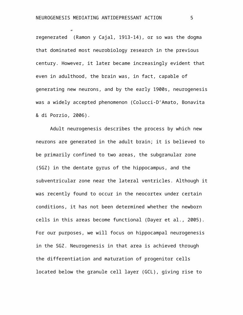
In the adult brain, “the nerve paths are something fixed, ended, immutable.
Everything may die, nothing may be regenerated” (Ramon y Cajal, 1913-14), or so was
the dogma that dominated most neurobiology research in the previous century. However,
it later became increasingly evident that even in adulthood, the brain was, in fact, capable
of generating new neurons, and by the early 1900s, neurogenesis was a widely accepted
phenomenon (Colucci-D’Amato, Bonavita & di Porzio, 2006).
Adult neurogenesis describes the process by which new neurons are generated in
the adult brain; it is believed to be primarily confined to two areas, the subgranular zone
(SGZ) in the dentate gyrus of the hippocampus, and the subventricular zone near the
lateral ventricles. Although it was recently found to occur in the neocortex under certain
conditions, it has not been determined whether the newborn cells in this areas become
functional (Dayer et al., 2005). For our purposes, we will focus on hippocampal
neurogenesis in the SGZ. Neurogenesis in that area is achieved through the differentiation
and maturation of progenitor cells located below the granule cell layer (GCL), giving rise
to an approximation of 9000 neurons per day. These will then migrate into the GCL, and
become physiologically and morphologically identical to mature neurons (van Praag &
Christie, 2002).
Neurogenic hypothesis of depression
NEUROGENESIS MEDIATING ANTIDEPRESSANT ACTION 5
However the causal mechanisms underlying depression remain poorly understood
to date. One suggested explanation is the adult hippocampal neurogenesis hypothesis of
MDD, which points to the decreased rate of adult-generated neurons in the hippocampus
found in patients suffering from major depression, as causal factor underlying the
expression of the disorder (Ernst et al., 2006).This hypothesis finds support in several
lines of evidence.
The hippocampus is smaller in depressed populations
First, several anomalies in limbic-related structures have been associated with
major depressive disorder, the most consistent of which concerning the hippocampus.
Indeed, numerous magnetic resonance imaging (MRI) studies have shown the
hippocampus to be reduced in size in subjects with major depressive disorder. In a meta-
analysis of 12 MRI studies, the left and right hippocampi were found to be reduced by
8% and 10%, respectively (Sheline et al.,1999). Additionally, there was a significant
correlation between the reduction of the right hippocampus volume, and the number and
duration of depressive episodes (Videbech & Ravnkilde, 2004). Furthermore, no sign of
accelerated neural degeneration in the hippocampus of patients with MDD was found to
date (Ernst et al., 2006). This suggests a causal relationship between depression and
hippocampal atrophy. Postmortem studies were also conducted using brain tissue from
depressed subjects and these revealed more peculiarities such as a decrease in the number
of neurons and glial cells, and an increase in their density by 30-35% in the dentate gyrus
of the hippocampus. Moreover, when other brain structures were analyzed for
comparison (frontal lobe, temporal love, caudate, amygdala), they showed no significant
NEUROGENESIS MEDIATING ANTIDEPRESSANT ACTION 6
volume reduction, relative to controls who were not depressed (Bremner et al., 2000),
thus the volume reduction is specific to the hippocampus. These findings are relevant as
the hippocampus is a major site for neurogenesis, and thus a decreased rate of
neurogenesis in this region may be partially responsible for the loss in volume.
Antidepressants increase neurogenesis
Second is the finding that neurogenesis levels, while decreased by stress and
depression, are normalized by effective antidepressant treatments (Boldrini et al., 2009).
Indeed, stress is an important risk factor for depression, as it is capable of
triggering depressive episodes or worsening them. Stress was shown to exert a potent
inhibitory effect on neurogenesis. Indeed, using the resident-intruder paradigm, Tanapat
and colleagues (1998) rat pups exposed to the odor of an adult male rat, a psychosocial
stressor. They found that a single exposure led to elevated levels of corticosterone and
diminished levels of newborn cells in hippocampus. This indicates that a higher
activation of the HPA axis could be contributing to a decrease in cell proliferation in the
dentate gyrus of depressed populations. Additionally, several studies reported
neurogenesis to be necessary for the therapeutic effect to occur. Indeed, in an animal
model of depression, Santarelli and colleagues (2003) demonstrated that ablation of
neurogenesis through irradiation of the hippocampus eliminated the antidepressant effects
of fluoxetine. This effect was replicated in a subsequent study by Perera and colleagues
(2011).
Time lapse of therapeutic effects is the same than that that for neuronal maturation

NEUROGENESIS MEDIATING ANTIDEPRESSANT ACTION 7
Third, the onset of therapeutic effects for several treatment modalities, notably
antidepressant drugs (Wong & Licinio, 2001) and electroconvulsive therapy (Segman et
al., 1995), is delayed by a period of 4 weeks. This suggests gradual changes in the
structure and neurochemistry of the brain. In fact, this delay closely matches the time
needed for newly generated neurons to become functionally integrated in the
hippocampus (Ernst et al., 2006).
Antidepressant drugs
Chronic antidepressant treatment increases cell proliferation and survival
Several classes of antidepressant drugs have been shown to alleviate behavioral
symptoms of depression, but to also reverse the deleterious effects on the hippocampus
volume depressed populations. Indeed, studies attesting for the serotonin reuptake
inhibitor fluoxetine’s antidepressant effects on behavior also found that it reversed the
volume loss seen in the temporal lobes of depressed populations, along with inducing a
significant increase in cell proliferation in the adult hippocampus by 60%. However, this
effect was only present after chronic administration of the drug for 11 or 28 days, as
opposed to acute administrations of 5 days which did not have any significant effects
(Santarelli et al., 2003). This suggests that a certain period is needed for antidepressants
to work; because this period corresponds to the time needed for neuronal maturation, it
may be that neurogenesis underlies antidepressant action.
Using adult male rats, Malberg and colleagues (2000) investigated the effects of
two antidepressant types in addition to fluoxetine: reboxetine (a norepinephrine slective
reuptake inhibitor) and tranylcypromine (a monoamine oxidase inhibitor). They also used

NEUROGENESIS MEDIATING ANTIDEPRESSANT ACTION 8
two additional groups for comparison; one was administered an antipsychotic
(haloperidol) and the other a placebo. All antidepressants resulted in a significant increase
in BrdU-labeled cells in the dentate gyrus compared to the control group, indicating an
increase in the rate of neurogenesis. Thus these antidepressants may share the
upregulation of neurogenesis as a common mechanism of action. In accordance with
Santarelli et al.’s findings, this effect was only present when the drugs were administered
for either 14 or 28 days, but not 1 or 5 days. Also, there was no difference in the rate of
neurogenesis between the rats that were treated for 14 days, and those treated for 28 days,
indicating the possibility of a plateau effect after 2 weeks of drug administration.
In contrast, administration of haloperidol had no effect on neurogenesis. Eisch and
colleagues (2002) also showed that administration of morphine has an opposite effect on
neurogenesis, as it decreases it. Thus the increase in neurogenesis is specific to certain
classes of drugs. Additionally, neurogenesis upregulation is also specific to certain brain
regions. Indeed, when the subventricular zone (SVZ) near the lateral ventricles was
examined subsequent to fluoxetine administration, no increase in BrdU-positive cells was
found compared to controls.
The increase in neurogenesis may be due to alterations in cell proliferation,
differentiation or survival of the granule cells. As the BrdU-positive cells increased just 2
hours after injection of BrdU, we already know that antidepressants increase cell
proliferation. As for the cell differentiation stage, Malberg and colleagues (2000) also
used neuronal (NeuN) and glial markers (GFAP), and found that 75% of the newborn
cells differentiated into neurons in both rats treated with antidepressants, and rats in the
control group. Thus administration of antidepressants has no significant effect on the
NEUROGENESIS MEDIATING ANTIDEPRESSANT ACTION 9
differentiation of granule cells in the dentate gyrus. However, antidepressants do increase
survival of the new granule cells, when administered for longer periods. Indeed, in a
separate experiment, the rats were injected with BrdU before the treatment with
fluoxetine, to see whether the labeled cells would survive longer. They found that in
comparison with the 14 days treatment, the 28 days treatment resulted in a 25% increase
in BrdU-positive cells (Malberg et al., 2004). Thus although treatment with fluoxetine for
periods longer than 14 days does not affect cell proliferation, it does seem to increase
survival. Similar findings were obtained for rolipram, a phosphodiesterase-IV inhibitor,
which, in addition to increasing cell proliferation, increased survival after 3 weeks of
administration (Nakagawa et al., 2002). Thus chronic treatment (14 days) with chemical
antidepressants increases the number of granule cells in the dentate gyrus by increasing
their proliferation, and, when administered for longer periods of time (28 days), their
survival. This effect however seems to be restricted to the hippocampus.
Antidepressant neuorgenic action is mediated by upregulation of neurotrophic factors
In Santarelli and colleagues’ experiment, fluoxetine also significantly increased
the expression of brain-derived neurotrophic factor (BDNF) in the dentate gyrus of the
hippocampus. This protein that has been strongly implicated in the regulation of
neurogenesis by numerous studies; one such study noted a superior level of BDNF in
postmortem examinations of the dentate gyrus of depressed patients who received
treatment, in comparison to those who did not and to controls who were not depressed
(Chen et al., 2001). In another study, using genetically modified mice with either lower
levels of BDNF, or altered functioning of trkb, the receptor for BDNF, had lower levels

NEUROGENESIS MEDIATING ANTIDEPRESSANT ACTION 10
of neurogenesis, which were, however, normalized after chronic treatment with
fluoxetine. It was concluded that BDNF may be required for survival and proliferation of
the newly generated cells, and that antidepressant drugs may exert their effect on
neurogenesis indirectly by increasing the levels of BDNF (Sairanen et al., 2005). This
hypothesis is supported by the finding that in vitro addition of BDNF to granule cells
cultured from the dentate gyrus, and direct injection of BDNF into the rat hippocampus
both increased the newly generated cells’ survival &differentiation.
Some studies also reported an increase in other growth factors. For instance, the
vascular endothelial growth factor (VEGF) is a cellular mitogen which was initially
recognized for its role in angiogenesis, but also mediates cytogenesis and therapeutic
effects of antidepressants on behavior. It is increased subsequent to administration of
chemical antidepressants, and its injection directly into the lateral ventricles is sufficient
to upregulate neurogenesis in the dentate gyrus. In contrast, blocking VEGF signaling
blocks both serotonin and norepinephrine-induced neurogenesis. Thus VEGF signaling
may be required for chemical antidepressants to increase neurogenesis. In addition,
VEGF was found to stimulate endothelial cells, therby upregulating the release of other
regulatory proteins such as BDNF. Similarly, fibroblast growth factor-2 (FGF2), is
thought to augment the number of both neurons and glial cells, and in postmortem
studies, it was found to be decreased in the hippocampus of depressed populations (Evans
et al., 2004). Insulin-like growth factor (IGF1) was also shown to contribute to the
neurogenic action of antidepressants (Warner-Schmidt & Dunman, 2007), . Therefore,
different classes of chemical antidepressants increase proliferation and survival of the
granule cells in the dentate gyrus of the hippocampus by altering the signaling of several

NEUROGENESIS MEDIATING ANTIDEPRESSANT ACTION 11
regulatory proteins (BDNF, VGEF, FGF2, IGF1).
Serotonin and Norepinephrine induce neurogenesis through different mechanisms
Moreover, knock-out (KO) mice in which the HT1a serotonin receptor was
selectively suppressed did not show any behavioral effect or increase in neurogenesis
subsequent to fluoxetine administration; additionally, administration of 8-OH-DPAT, a
5-HT1a-selective agonist, resulted in behavioral and neurogenic effects similar to those
associated with fluoxetine. Together these two findings point to the HT1a serotonin
receptor as site of action for fluoxetine. HT1a has indeed been previously reported to
have an important role in mood and anxiety disorders, and thus it warrants further
investigation with regards to the pharmacotherapies of depression.
However this does not hold true for other antidepressant drugs such as
imipramine, a tricyclic antidepressant (TCA) which acts upon the neurotransmitter
norepinephrine (NE). Indeed, the antidepressant effects of imipramine were not affected
by the suppression of this serotonin receptor 5-HT1a. This suggests that norepinephrine
induces neurogenesis through a mechanism which is qualitatively different from the one
used by serotonin. That said, both SSRIs and TCAs were found to induce similar
increases of neurogenesis, and in a postmortem analysis, the dentate gyrus of patients
treated with either medication was significantly larger than that in controls who did not
suffer from depression.
Neurogenesis is required for antidepressant action
NEUROGENESIS MEDIATING ANTIDEPRESSANT ACTION 12
Finally, a major finding in the last decade was the fact that neurogenesis not only
accompanies therapeutic effects on behavior, but may also be required for them to occur.
Santarelli and colleagues (2003) used a low dose of x-irradiation on adult rats to trigger
apoptosis in neural progenitor cells, thereby eliminating neurogenesis. Indeed, the
irradiation resulted in an 85% decrease in BrdU-labeled cells in the subgranular zone of
the hippocampus for a period of 8 weeks, without affecting other areas such as the
subventricular zone. They then treated the rats with either fluoxetine or imipramine (a
tricyclic antidepressant), and tested them on an animal model of depression, the Novelty-
suppressed feeding (NSF). Neither antidepressants altered the rats’ response on this test;
thus we conclude that ablation of neurogenesis through irradiation of the hippocampus
blocked the antidepressants’ therapeutic effects on behavior. This finding was replicated
by Perera and colleagues (2011) on nonhuman primates (NHPs), some of which had their
temporal lobes irradiated. Next, all NHPs underwent repeated social separation stress,
while being administered with fluoxetine. The antidepressant prevented depressive-like
behavioral symptoms such as anhedonia and lower hierarchy scores from appearing, only
in the NHPs whose temporal lobes were not irradiated. This demonstrates, once again,
that neurogenesis is required for antidepressant action, as neurogenesis ablation blocks
the antidepressants’ therapeutic effects.
ECT/ECS
ECS upregulates cell proliferation in the hippocampus
Certain instances of severe depression do not show improvement subsequent to
drug therapy; however, patients with treatment-resistant therapy can consider a variety of
NEUROGENESIS MEDIATING ANTIDEPRESSANT ACTION 13
somatic therapies. One such treatment is electroconvulsive therapy (ECT); it is the most
powerful treatment for depression, and produces the most rapid and the largest increase in
neurogenesis, compared to other forms of treatment. However, its mechanism of action
remains largely unknown to date (Malberg et al., 2000).
In the first study linking ECS, the equivalent form of ECT in animals, to
neurogenesis, Madsen and colleagues (2000) found that the increase in neurogenesis was
dose-dependent; that is, while administration of ECS in the form of a single seizure was
sufficient to increase neurogenesis, along with synaptic density in the CA1 filed of the
hippocampus and the volume of the dentate gyrus overall, this effect was even more
apparent when a series of seizures was applied. Also, the increased neurogenesis rate was
still apparent 3 months after the treatment, as the number of BrdU-positive cells did not
decline. Moreover, the new cells were fully functional, and there was no sign of increased
apoptosis (programmed cell death) during this period. Similar conclusions were reached
by Hellsten et. al (2002), who found that a 75% reduction in neurogenesis in rats induced
by chronic administration of corticosterone, was counteracted by just one
electronconvulsive shock.
ECS produces an increase in regulatory proteins
Moreover, ECS was found to increase the amount of several regulatory proteins,
notably B-cell chronic lymphocytic lymphoma 2 (BCL2), which protect newborn cells
and promote their differentiation. The BCL2 were produced by local mature cells which
were thereby enhancing the new cells’ survival (Kuhn et al., 2005).
Similarly, research has implicated the growth factor in the antidepressant effect of ECS.

NEUROGENESIS MEDIATING ANTIDEPRESSANT ACTION 14
Warner-Schmidt and colleagues (2008) irradiated the hippocampus of rats, thereby
decreasing neurogenesis; this procedure also reduced hippocampal levels of VEGF
mRNA and protein. VGEF levels were almost completely reversed with administration of
ECS, without affecting the subventricular zone. Effectively, the increase in cell
proliferation was detectable for months post treatment. ECS was found to affect different
types of cells in the granule cell layer (GCL), as it increased the numbers of neurons,
endothelial cells, astrocytes and ogliodendrocytes. Specifically, while irradiation
decreased the number of new neuron in the GCL by 98%, ECS restored this number to
40% of the number found in control rats. This is in line with previous findings by Madsen
and colleagues (2000). ECS also increase cell survival in the granule cell layer (GCL) in
both irradiated rats sham-treated controls. Thus ECS successfully restored some, but not
all of the neural progenitor cells’ proliferative capacity in the GCL; this increase in
VEGF could constitute one of the mechanisms by which ECS stimulates neurogenesis.
ECS was also found to cause a far greater rise in the levels of BDNF, compared to the
chemical antidepressant tranylcypromine. This is consistent with the fact that ECS also
has more powerful therapeutic effects than antidepressant drugs. Thus the sharp increase
in BDNF mRNA and protein subsequent to ECS may explain this treatment modality’s
advantage in alleviating symptoms in treatment-resistant patients (Altara et al., 2003).
ECT alleviates depressive symptoms in humans
As for the ECT’s behavioral effects in humans, patients with MDD recovered 72
days after the treatment was completed (Weeks et al, 1980), with a remission rate of 87%,
and significant decrease in expressed suicidal intent (Petrides et. al, 2001).
NEUROGENESIS MEDIATING ANTIDEPRESSANT ACTION 15
However, it is important to note that ECT, unlike other treatment modalities, induces
generalized seizures which affect all of the central nervous system. Furthermore, ECT,
again, in contrast to other antidepressant treatments, is also used in the treatment of
mania, psychosis and Parkinson’s disease. These two factors strongly suggest that ECT
acts by mechanisms other than the regionally selective neurogenesis, and which remain to
be explained (Ongur & Heckers, 2004).
Repetitive Transcranial Magnetic Stimulation
Repetitive transcranial magnetic simulation (rTMS) is a noninvasive therapeutic
tool which has been demonstrated to attenuate the activity of the hypothalamic-pituitary-
adrenal (HPA) axis in patients undergoing high levels of stress. Chronic psychosocial
stress, a risk factor for depression, is known to suppress neurogenesis in the dentate
gyrus. However, although clinical trials have previously found rTMS to augment the
levels of BDNF and neurogenesis in the dentate gyrus (Muller et al., 2000), a subsequent
study by Czeh and colleagues (2001) showed rTMS to attenuate the increase in stress-
induces rise in glucocorticoids, however, with very mild effects on neurogenesis.
Additionally, the rate of survival of BRdU-positive cells was even decreased by the
treatment, a finding inconsistent with previous literature on rTMS treatment.
This emphasizes the little understanding we currently have of the mechanisms underlying
the effects of rTMS, and suggests that additional cellular mechanisms should be
investigated for this treatment modality.
Deep Brain Stimulation

NEUROGENESIS MEDIATING ANTIDEPRESSANT ACTION 16
Deep brain stimulation (DBS) is a powerful neuromodulation therapy used for a
variety of neurological, psychiatric, and movement disorders, and successful in the
treatment of patients with TRD with large rates of remission (Anderson et al., 2012). It
consists in the application of a continuous electrical current to specific neuroanatomical
structures, and has the advantage of being a non-ablative, highly adjustable procedure
with both long-term and acute therapeutic effects, in contrast to other invasive somatic
treatments (e.g. anterior capsulotomy, limbic leucotomy). This treatment is of significant
interest to us, as its applicability to specific neural circuits can be used to elucidate
relationships between these circuits (Schlaepfer et al., 2011).
In a recent clinical study, Encinas and colleagues (2011) investigated the effect of
stimulation of the anterior thalamic nuclei (ATN) on neurognesis in the dentate gyrus,
and found that the number of BrdU-positive cells was 45% higher in mice subjected to
the high frequency-stimulation, compared to mice who were not stimulated. Specifically,
their study showed that DBS increases the amount of symmetric divisions of transient
amplifying neural progenitors, which translates into a larger number of neurons in the
GCL of the dentate gyrus. These findings draw attention to structures of the limbic
system which could, upon stimulation, cause an increased rate of neurogenesis in the
dentate gyrus, and so far have been used to treat patients with epilepsy by stimulation of
the ANS. Further studies on the limbic system’s relation to neurogenesis could help
improve treatment of depression by deep brain stimulation.
With regards to the trophic factors discussed in other treatment modalities, these
remain to be investigated as little research on the use of DBS in TRD patietns is available
at the current time.

NEUROGENESIS MEDIATING ANTIDEPRESSANT ACTION 17
Vagus Nerve Stimulation
Vagus nerve stimulation (VNS) was investigated by Rush and colleagues (2005)
as an alternative to current pharmacotherapies for treating both severe depression and
treatment-resistant depression (TRD). This investigation was motivated by the need for a
more efficient treatment, with lower rates of relapse and faster onset of effects.
They tested the treatment in clinical trials on TRD patients; these showed variable
response rates for more than two months but nevertheless continued to improve in the
following year.
In another study on rats, researchers pointed to possible molecular and neurochemical
events underlying the effectiveness of VNS; they found, similarly to research on
antidepressant drugs, an increased rate in adult hippocampal neurogenesis, as well as
higher levels of BDNF and FGF levels in the hippocampus by 26.3% and 22,3%
respectively, and of higher levels norepinephrine by 70% in the prefrontal cortex.
However these effects were observed following a briefer period of time and thus VNS
offers an advantage in comparison to other treatment modalities (Follesa et al., 2007).
Recently, Gebhardt and colleagues (2013) conducted the first study that tests VNS effects
on behavior in an animal model of depression, the bilateral olfactory bulbectomy (OBX).
3 weeks after treatment with VNS, OBX rats improved significantly on the disturbed one-
way active avoidance task, which attests to the treatment’s beneficial effects.
Furthermore, these rats also showed increased BrdU labeling, and, 8 weeks later, they
demonstrated levels of neurogenesis similar to control rats. This effect was dependent on
the intensity of the stimulation, and this relationship could be described by an inverted U-
NEUROGENESIS MEDIATING ANTIDEPRESSANT ACTION 18
shape. They also found a region-dependent increase in growth factors, notably BDNF,
and basic epidermal growth factor (BEGF). The latter molecule stimulates neurogenesis
as it binds to its receptor.
However in this particular study, control rats didn't show an increased neurogenesis after
VNS, which is in contrast to other studies. This may be due a different length of the
stimulation period.
Exercise Therapy
In addition to the traditional antidepressant treatments, exercise therapy is also
found to significantly attenuate symptoms of depression in humans, mice and rats.
Each year, brain tissue in older adults shrinks by 1-2%. However, Erikson and
colleagues (2011) showed that exercise was a neuroprotective factor which, under certain
conditions, could increase the volume of the hippocampus by 2% in just about a year,
thereby reversing age-related volume loss by 1 to 2 years. This effect was confined to the
caudal hippocampus and did not affect the thalamus and the caudate nucleus, indicating
that exercise acts on regionally-dependent molecular pathways rather than the whole
brain.
This study also showed the temporal lobe to be significantly larger in more active
adults, which could be explained by an increase in neurogenesis, dendritic branching and
complexity, as well as vascularization in the dentate gyrus.
Due to the difficulties associated with analyzing cellular mechanisms underlying
therapeutic effects in premortem human brain tissue, many studies were also conducted
on animals using approved animal models of depression. In their study, Huang and

NEUROGENESIS MEDIATING ANTIDEPRESSANT ACTION 19
colleagues (2012) used three different paradigms to demonstrate the effects of exercise on
behavior. In all three models, both rats that exercised daily and rats that were chronically
treated with fluoxetine displayed less depressive-like patterns of behaviors compared to
other conditions (enriched environment) or drugs (Clozapine, Haloperidol). Similarly, the
biological effects induced by exercise in rats closely matched those induced by
fluoxetine. Indeed, both exercise and fluoxetine acted by increasing neurogenesis and
spinal density in the CA1 field of the hippocampus, a finding that was not shared by the
rats in the other groups.
Exercise was also found to increase levels of ß-endorphins, VEGF, BDNF as well
as serotonin, and ultimately, to an increase in neurogenesis. This increase in cell
proliferation may have been mediated by the increase in BDNF (Eisch, 2002). However,
another study by Bjornebekk, Mather and Brene (2005) suggested that exercise increases
cell proliferation independently of the levels of BDNF, as the latter, in contrast to cell
proliferation, did not correlate with exercise’s antidepressant effect.

NEUROGENESIS MEDIATING ANTIDEPRESSANT ACTION 20
Conclusion
In conclusion, different treatment modalities exist for treating major depressive
disorder, and most represent efficient ways of alleviating and even suppressing symptoms
associated with depression. Although little is known about their underlying mechanism of
action, they all seem to influence the rate of neurogenesis in the adult hippocampus, and
the delayed onset of their therapeutic effects is a close match to the time needed for
neuronal maturation (4 weeks). Additionally, reduction of symptoms is also associated
with a reversal of brain anomalies seen in patients with MDD, specifically in the
hippocampus which plays a critical role in many cognitive functions affected by
depression.
Thus neurogenesis, which occurs primarily in this region, may constitute a
mediator for antidepressant effects. Indeed, this is line with previous findings implicating
neurogenesis in hippocampal functions such as transient place recognition and trace fear
conditioning, which are associated with depression. Thus suppression of neurogenesis in
the hippocampus could be responsible for the inaccurate representations of the external
world &ultimately the cognitive distortions seen in patients with MDD (Perera et al,
2011).
Additionally, the role of neurogenesis as a mediator finds additional support in the
fact that, as shown in this review, many of the molecular mechanisms associated with
neurogenesis (BDNF, VEGF, FGF2) are also affected by antidepressant treatments.
That said, this does not eliminate the possibility that the increased neurogenesis is no
more than an epiphenomenon, unrelated to antidepressant effects. Indeed, it could be that
the cellular mechanisms discussed regulate many factors in addition to neurogenesis, and

NEUROGENESIS MEDIATING ANTIDEPRESSANT ACTION 21
as such, these factors would be the ones mediating the therapeutic effects of
antidepressants, and not neurogenesis. Another limitation concerns the fact that although
major depressive disorder is a human condition, it has been mostly studied in rodents, due
to the difficulty of analyzing cellular mechanisms in the human brain premortem. Finally,
other mechanisms have emerged as alternatives to neurogenesis in explaining
antidepressant effects, such as the neuronal remodeling suggested by Bessa and
colleagues (2009); however, further investigation of these will be required in attempt to
determine their validity.

NEUROGENESIS MEDIATING ANTIDEPRESSANT ACTION 22
References
Altara, CA, Whiteheada, RE, Chena, R, Wörtweinb, G, Madsenb, TM (2003). Effects of
electroconvulsive seizures and antidepressant drugs on brain-derived neurotrophic
factor protein in rat brain, Biological Psychiatry, Vol 54, 703–709,
doi:10.1016/S0006-3223(03)00073-8
Anderson, RJ, Frye, MA, Abulseoud, OA, Lee, KH, McGillivray, JA, Berk, M, Tye, SJ
(2012). Deep brain stimulation for treatment-resistant depression: Efficacy, safety
and mechanisms of action, Neuroscience and Biobehavioral Reviews, Vol 36,
1920-1933, doi:10.1016/j.neubiorev.2012.06.001
Bessa, J.M., Ferreira, D., Melo, I., Marques, F., Cerqueira, J.J., Palha, J.A., Almeida,
O.F., and Sousa, N. (2009). The mood improving actions of antidepressants do
not depend on neurogenesis but are associated with neuronal remodeling. Mol.
Psychiatry 14, 764–773, 739, doi: 10.1038/mp.2008.119
Boldrini, M, Underwood, MD, Hen, R, Rosoklija, GB, Dwork, AJ, Mann, JM, Arango, V
(2009). Antidepressants increase neural progenitor cells in the human
hippocampus, Neuropsychopharmacology, 2376-89, doi: 10.1038/npp.2009.75
Cavanagh JT, Carson AJ, Sharpe M. Psychological autopsy studies of suicide: A
systematic review. Psychological Medicine, Vol 33, 395-405. doi:
10.1017/S0033291702006943
Colucci-D'Amato, L, Bonavita, V, di Porzio, U (2006). The end of the central dogma of
neurobiology: stem cells and neurogenesis in adult CNS. Neurol Sci. Vol 27, 266-
70. doi:10.1007/s10072-006-0682-z
Czeh, B, Michaelis, T, Watanabe, T, Frahm, J, de Biurrun, G, van Kampen M,

NEUROGENESIS MEDIATING ANTIDEPRESSANT ACTION 23
Bartolomucci, A, Fuchs, E. (2001). Stress-induced changes in cerebral
metabolites, hippocampal volume, and cell proliferation are prevented by
antidepressant treatment with tianeptine. The National Academy of Sciences, Vol
98, 12796-801. doi: 10.1073/pnas.211427898
Eisch (2002). Adult neurogenesis: implications for psychiatry, Progress in Brain
Research, Vol 138, 317-344. doi:10/1016/S0079-6123(02)38085-3
Eisch, AJ, Petrik, D (2012). Depression and Hippocampal Neurogenesis: A Road to
Remission? Science, Vol 138, 315-342. doi: 10.1126/science.1222941
Encinas, JM, Hamani, C, Lozano, AM, Enikolopov, E (2011). Neurogenic Hippocampal
Targets of Deep Brain Stimulation, Journal of Comparative Neurology, Vol 519,
6–20. doi:10.1002/cne.22503
Erikson, KI, Voss, MW, Prakash, RS, Basak, C, Szabo, A, Chaddock, L, Kim, JS, Heo,
S, Alves, H, White, SM, Wojcicki, TR, Mailey, E, Vieira, VJ, Martin, SA, Pence,
BD, Woods, JA, McAuley, E, Kramer, AF (2011). Exercise training increases size
of hippocampus and improves memory, The National Academy of Sciences, Vol
108, 3017-3022, doi/10.1073/pnas.1015950108
Ernst, C, Olson, AK (2006). Antidepressant effects of exercise: Evidence for an adult-
neurogenesis hypothesis? Journal of Psychiatry & Neuroscience, Vol 31, 84-92.
Retrieved from:
http://www.cma.ca/multimedia/staticContent/HTML/N0/I2/jpn/vol-31/issue-2/pdf/
pg84.pdf
Hellsten, J, Wennstro¨m, M, Mohapel P, Ekdahl, CT, Bengzon, J, Tingstro¨m, A (2002).
Electroconvulsive seizures increase hippocampal neurogenesis after chronic

NEUROGENESIS MEDIATING ANTIDEPRESSANT ACTION 24
corticosterone treatment. Eur J Neurosci, Vol16, 283–290, doi:10.1046-
9568.2002.02093.x
Huang, GJ, David, EB, Piella, AT, Edwards, A, Flint, J, Shifman, S (2012).
Neurogenomic Evidence for a Shared Mechanism of the Antidepressant Effects of
Exercise and Chronic Fluoxetine in Mice, PLoS ONE, Vol 7, e35901.
doi:10.1371/journal.pone.0035901
Kuhn, HG, Biebl, M, Wilhelm, D, Li, M, Friedlander, RM, Winkler, J (2005). Increased
generation of granule cells in adult Bcl-2 overexpressing mice: a role for cell
death during continued hippocampal neurogenesis. Eur J Neurosci, Vol 22, 1907–
1915. doi:10.1111/j.1460-9568.2005.04377.x
Madsen TM, Treschow A, Bengzon J, Bolwig TG, Lindvall O, Tingstro¨m A (2000)
Increased neurogenesis in a model of electroconvulsive therapy. Biol Psychiatry,
Vol 47, 1043–1049. doi:10.1073/pnas.0710858105
Malberg, JE, Eisch, AJ, Nestler, EJ, Duman, RS (2000). Chronic Antidepressant
Treatment Increases Neurogenesis in Adult Rat Hippocampus, J. Neurosci, Vol
20, 9104. Retrieved from:
http://www.jneurosci.org/content/2-/24/9104.full.pdf+html
Malberg, JE (2004). Implications of adult hippocampal neurogenesis in antidepressant
action, J Psychiatry Neurosci Vol 29, 196-205. Retrieved from:
http://www.cma.ca/multimedia/staticContent/HTML/N0/I2/jpn/vol-29/issue-3/
pdf/pg196.pdf
Muller, MB, Toschi, N, Kresse, AE, Post, A, Keck, ME (2000): Long-term repetitive
transcranial magnetic stimulation increases the expression of brain-derived
NEUROGENESIS MEDIATING ANTIDEPRESSANT ACTION 25
neurotrophic factor and cholecystokinin mRNA, but not neuropeptide tyrosine
mRNA in specific areas of rat brain, Neuropsychopharmacology, Vol 23, 205–
215. doi:10.1016/S0893-133X(00)00099-3
Murray, CJ, Lopez AD (1996). Evidence-based health policy –Lessons from the Global
Burden of Disease Study, Science, Vol 274, 740–743. doi:
10.1126/science.274.5288.740
Nakagawa, S, Kim, JE, Lee, R, Chen, J, Fujioka, T, Malberg, J et al. Localization of
phosphorylated cAMP response elementbinding protein in immature neurons of
adult hippocampus. J Neurosci, Vol 22, 9868-76. Retrieved from:
http://www.jneurosci.org/content/22/22/9868.full.pdf
Ongur, D, Heckers, S (2004). A Role for Glia in the Action of Electroconvulsive
Therapy, Harv Rev Psychiatry. doi: 10.1080/10673220490886185
Perera, TD, Dwork, AJ, Keegan, KA, Thirumangalakudi, L, Lipira, CM, Joyce, N, Lange,
C, Higley, JD, Rosoklija, G, Hen, R, Sackeim, HA, Coplan, JD (2011). Necessity
of Hippocampal Neurogenesis for the Therapeutic Action of Antidepressants in
Adult Nonhuman Primates, Plos One, Vol 6. doi:10.1371/journal.pone.0017600.
Petrides, G, Fink, M, Husain, MM et al (2001). ECT remission rates in psychotic vs.
nonpsychotic depressed patients: a report from CORE. J ECT, Vol 17, 244–253.
Rush AJ, Sackeim HA, Marangell LB, George MS, Brannan SK, Davis SM, et al. (2005).
Effects of 12 months of vagus nerve stimulation in treatment-resistant depression:
a naturalistic study. Biol Psychiatry, Vol 58, 355-63. Retrieved from:
http://digitalcommons.unl.edu/cgi/viewcontent.cgi?
article=1068&context=veterans
NEUROGENESIS MEDIATING ANTIDEPRESSANT ACTION 26
Santarelli L, Saxe M, Gross C, Surget A, Battaglia F, Dulawa S et al. (2003).
Requirement of hippocampal neurogenesis for the behavioral effects of
antidepressants. Science, Vol 301, 805–809. doi:10.1126/science.1083328
Sairanen, M, Lucas, G, Ernfors, P, Castre´n, M, Castre´n1, E (2005). Brain-Derived
Neurotrophic Factor and Antidepressant Drugs Have Different But Coordinated
Effects on Neuronal Turnover, Proliferation, and Survival in the Adult Dentate
Gyrus, J Neurosci., Vol 25, 1089-94. doi:10.1523/jneurosci.3741-04.2005
Schlaepfer, TE, Bewernick, B, Kayser, S, Lenz, D (2011). Modulating affect, cognition,
and behavior – prospects of deep brain stimulation for treatment-resistant
psychiatric disorders, Frontiers in Integrative Neuroscience, Vol 5, 29
Sheline, YI, Sanghavi, M, Mintun, MA, Gado, MH (1999). Depression duration but not
age predicts hippocampal volume loss in medically healthy women with recurrent
major depression. The Journal of Neuroscience, Vol 19, 5034-5043.
doi:10.3389/fnint.2011.00029
Van Praag H, Schinder AF, Christie BR, et al. Functional neurogenesis in the adult
hippocampus. Nature, Vol 415, 1030-4. doi:10.1038/4151030a
Videbech P, Ravnkilde B (2004). Hippocampal volume and depression: A meta-analysis
of MRI studies. Am J Psychiatry; Vol 161, 1957–1966. doi:
10.1176/appi.ajp.161.11.1957
Wong ML, Licinio J (2001) Research and treatment approaches to depression. Nat Rev
Neurosci, Vol 2, 343–351. doi:10.1038/35072566
World Health Organization (2012). Retrieved from:
http://www.who.int/mediacentre/factsheets/fs369/en/index.html

NEUROGENESIS MEDIATING ANTIDEPRESSANT ACTION 27
Warner-Schmidt, J.L., and Duman, R.S. (2007). VEGF is an essential mediator of the
neurogenic and behavioral actions of antidepressants. The National Academy of
Sciences, 104, 4647–4652. doi:10.1073/pnas.0610282104
Warner-Schmidt, JL, Madsen, TM, Duman, RS (2008). Electroconvulsive seizure
restores neurogenesis and hippocampus-dependent fear memory after disruption
by irradiation, European Journal of Neuroscience, Vol 27, 1485–1493. doi:
10.1111/j.1460-9568.2008.06118.x