Ruiz, G., Verrot, L., Laloy, E., & Benchekroun, G. (2017 ... · 58 from the formalin jars. 59 The...
28
Ruiz, G., Verrot, L., Laloy, E., & Benchekroun, G. (2017). Diagnostic contribution of cytological specimens obtained from biopsies during gastrointestinal endoscopy in dogs and cats. Journal of Small Animal Practice, 58(1), 17–22. https://doi.org/10.1111/jsap.12597 Peer reviewed version Link to published version (if available): 10.1111/jsap.12597 Link to publication record in Explore Bristol Research PDF-document This is the accepted author manuscript (AAM). The final published version (version of record) is available online via Wiley at DOI: 10.1111/jsap.12597. Please refer to any applicable terms of use of the publisher. University of Bristol - Explore Bristol Research General rights This document is made available in accordance with publisher policies. Please cite only the published version using the reference above. Full terms of use are available: http://www.bristol.ac.uk/pure/about/ebr-terms
-
Upload
nguyentuyen -
Category
Documents
-
view
212 -
download
0
Transcript of Ruiz, G., Verrot, L., Laloy, E., & Benchekroun, G. (2017 ... · 58 from the formalin jars. 59 The...

Ruiz, G., Verrot, L., Laloy, E., & Benchekroun, G. (2017). Diagnosticcontribution of cytological specimens obtained from biopsies duringgastrointestinal endoscopy in dogs and cats. Journal of Small AnimalPractice, 58(1), 17–22. https://doi.org/10.1111/jsap.12597
Peer reviewed version
Link to published version (if available):10.1111/jsap.12597
Link to publication record in Explore Bristol ResearchPDF-document
This is the accepted author manuscript (AAM). The final published version (version of record) is available onlinevia Wiley at DOI: 10.1111/jsap.12597. Please refer to any applicable terms of use of the publisher.
University of Bristol - Explore Bristol ResearchGeneral rights
This document is made available in accordance with publisher policies. Please cite only the publishedversion using the reference above. Full terms of use are available:http://www.bristol.ac.uk/pure/about/ebr-terms

Diagnostic contribution of cytological specimens obtained from biopsies during
gastrointestinal endoscopy in dogs and cats
Guillaume Ruiza, *, Lucas Verrota, Eve Laloyb, Ghita Benchekrouna
a Internal Medicine Department, Université Paris-Est, Ecole Nationale Vétérinaire d’Alfort, 7
avenue du général de Gaulle, 94704 Maisons-Alfort Cedex, France
b Pathology Department, Université Paris-Est, Ecole Nationale Vétérinaire d’Alfort, 7 avenue
du général de Gaulle, 94704 Maisons-Alfort Cedex, France
* Corresponding author (tel. +44 7774720958). Current address for corresponding author is:
Langford Veterinary Services-Small Animal Referral Hospital, University of Bristol,
Langford House, Langford, BS40 5DU, United Kingdom
Email addresses
Guillaume Ruiz, DVM, MRCVS: [email protected]
Lucas Verrot, DVM: [email protected]
Eve Laloy, DVM, dip-ECVP, PhD: [email protected]
Ghita Benchekroun, DVM, dip-ECVIM-CA: [email protected]
Acknowledgements
The authors wish to thank Mr Loic Desquilbet for his assistance in doing the statistical
analyses of the results.
Preliminary results were presented as an Abstract at the 25th European College of Veterinary
Internal Medicine-Companion Animals (ECVIM-CA) Congress, Lisbon, 10-12 September
2015.

1
Diagnostic contribution of cytological specimens obtained from biopsies during 1
gastrointestinal endoscopy in dogs and cats 2
3
Summary 4
5
Objectives: The aims of this study were to compare cytological samples obtained from endoscopic 6
biopsies using ‘imprint’ and ‘squash’ techniques, and to evaluate the potential value of cytology 7
compared to histology in reaching the diagnosis. 8
Methods: Eighteen dogs and five cats undergoing endoscopy for chronic gastrointestinal signs were 9
prospectively included. Imprint and squash samples were obtained from one biopsy and then 10
analysed. Comparison between cytology and histology was performed using Cohen’s kappa 11
coefficient. 12
Results: Appropriate samples for cytological evaluation were most often obtained with the squash 13
technique (96% of the cases vs. 68% with the imprint technique). The diagnoses obtained with 14
cytological samples and by histology, considered as the gold standard, were compared. The same 15
diagnosis was obtained with the squash technique in 65% of the cases. Furthermore, cytology was 16
considered complementary to histology for gastric spiral organisms and mast cells identification. 17
Clinical significance: These results suggest that squash cytology obtained from endoscopic 18
biopsies of the gastrointestinal tract could provide relevant and additional information to histology 19
in dogs and cats. Further studies are needed to confirm these findings. 20
21
Keywords: cytology; endoscopy; gastrointestinal tract; imprint; squash 22

2
Introduction 23
Endoscopy is commonly used in veterinary gastroenterology to assess macroscopic lesions of the 24
mucosa and to obtain targeted samples (Washabau et al. 2010). Histology remains the current gold 25
standard to achieve a definitive diagnosis for infiltrative and structural diseases. However, 26
histological results are usually only available a few working days after endoscopy due to the time 27
required for laboratory processing. Moreover, some abnormal findings such as organisms present 28
within the surface mucus can be lost during the actual process and therefore misdiagnosed (Jergens 29
et al. 1998). 30
Cytology is commonly used in veterinary medicine, and different techniques have been described 31
(Cohen et al. 2003, Bonfanti et al. 2006, Ballegeer et al. 2007). However, lesions differ in their 32
ability to exfoliate, thus creating a discrepancy between ’exfoliative lesions’ (which often lead to a 33
reliable cytological diagnosis) and ‘poorly exfoliative lesions’ (which are generally non-diagnosed 34
with cytology) (Cohen et al. 2003). 35
Only a few studies have focused on the diagnostic contribution of cytological samples obtained 36
from the digestive tract of dogs and cats (Tobey et al. 1988, Jergens et al. 1998, Bonfanti et al. 37
2006, Riondato et al. 2014, Mangelsdorf et al. 2015). The aims of this prospective study were to 38
compare the abilities of ‘imprint’ and ‘squash’ techniques to provide valid cytological samples from 39
biopsies obtained during gastrointestinal endoscopy in dogs and cats, and to evaluate the potential 40
value of cytology compared to histology in reaching the definitive diagnosis. 41
42
Materials and methods 43
This prospective study included dogs and cats presented for gastrointestinal symptoms and which 44
underwent a gastrointestinal endoscopy between March 2012 and March 2013 at XXX Hospital. 45
Cases were included only if biopsies, concurrent squash and imprint preparations for cytology, and 46
a conclusive histopathology report, were available. Signalment and clinical signs were recorded. 47
48

3
The endoscopy was performed under general anaesthesia with a GIF-160 Olympus gastroscope, 49
using FB-240K Olympus forceps for biopsies. At least five biopsies from each area of interest (i.e., 50
stomach and proximal duodenum for upper alimentary tract, distal ileum and colon for lower 51
alimentary tract) were collected in separate cassettes, fixed in 10% formalin and then routinely 52
processed for histopathological analysis. An additional biopsy was used to obtain cytological 53
specimens with two techniques. First, the ‘imprint’ specimen was obtained by imprinting the biopsy 54
sample on a glass slide several times, using a 25 G needle to gently hold the sample. Then the 55
‘squash’ specimen was obtained by crushing the biopsy between two slides, and pulling them apart 56
without smearing. The slides were then sent to the laboratory for cytological analysis, separately 57
from the formalin jars. 58
The biopsies were stained with haematoxylin, eosin and saffron (HES), which are the routine stain 59
used for all biopsies in our laboratory. They were reviewed by board certified pathologists from the 60
Pathology Department of XXX, and interpreted according to the current guidelines of the WSAVA 61
International Gastrointestinal Standardization Group (IGSG) described elsewhere (Day et al. 2008). 62
The presence of organisms was also recorded. Biopsy samples of less than 3mm or without lamina 63
propria were considered of insufficient quality and the cases were excluded. 64
Cytological samples were stained with May-Grünwald-Giemsa. All the slides were reviewed by a 65
single ECVP-diplomate (XXX) in a blinded manner at the end of the recruitment process. The 66
pathologist had access to the submission form (history and clinical signs) but was blinded to the 67
histological results. The cytological samples were analyzed using a method adapted from Jergens et 68
al. (1998) and Andreasen et al. (2009). The following categories were identified: inflammatory 69
cells, atypical and neoplastic cells, epithelial clusters, gastric spiral organisms (GSO), bacterial 70
flora, haemorrhage, debris/ingesta, and mucus. A cell count mean was calculated for each category 71
from at least 10 fields in well spread areas of the slide, and was then classified in a grading system 72
similar to that of Jergens et al. (1998) (Table 1). Samples with a grade 2 or less for epithelial 73
clusters were considered non-representative of the organ and were thus classified as ‘non 74

4
diagnostic’. Samples presenting poor preservation or numerous artefacts that precluded further 75
interpretation - such as crushed cells - were also excluded from further analysis. 76
For each case, the diagnostic conclusions for both the cytological and histological analyses were 77
classified into one of the categories described in Table 2, based on the predominant pathological 78
findings, in order to allow comparison of the cytological and histological diagnoses. The cytological 79
diagnosis obtained by either the imprint or squash technique, as compared with histopathological 80
analysis (considered as the gold standard) was assessed by statistical analysis. The agreement 81
between cytology and histology was determined by Cohen’s Kappa coefficient. The techniques 82
were considered in agreement if both concluded to the exact same diagnosis, as per Table 2. The 83
different kappa () values were scored as follows: very poor for <0, poor for 0<<0.2, fair for 84
0.21<<0.4, moderate for 0.41<<0.6, good for 0.61<<0.8 and excellent for 0.81<<1 (Landis & 85
Koch 1977). 86
87
Results 88
Twenty-three cases, including 18 dogs and five cats, met the inclusion criteria. The ages ranged 89
from one to 16 years (from one to 13 years in dogs and from four to 16 years in cats). There were 90
eight females (seven dogs and one cat) and 15 males (11 dogs and four cats). All the cats were 91
domestic shorthairs, whereas the dogs were of various breeds: three French Bulldogs, three Jack 92
Russell Terriers, three crossbreeds, two German Shepherds, one Labrador Retriever, one Rhodesian 93
Ridgeback, one Boxer, one Weimaraner, one Parson Terrier, one Lhasa Apso and one West 94
Highland White Terrier. 95
Clinical presentation was variable but included at least one of the following signs: vomiting (17 96
cases), diarrhoea (seven cases), inappetence (five cases), weight loss (five cases), lethargy (four 97
cases), haematochezia (two cases), retching (two cases), melaena (one case), tenesmus (one case), 98
and constipation (one case). 99

5
Eighteen cases had biopsies taken from the stomach (fundus in 11 cases, antrum in one case, and 100
both fundus and antrum in six cases). Seventeen cases had biopsies taken from the small intestine 101
(duodenum only in 13 cases, ileum only in three cases and both duodenum and ileum in one case). 102
Six cases had biopsies taken from the large intestine (colon in five cases and rectum in one case). In 103
total, 48 sites were collected during the course of the study. Histopathological analysis resulted in 104
24 diagnoses because one case was diagnosed with two distinct conditions; they are presented in 105
Table 3 (see online). 106
For the cytological evaluation, 95 specimens were examined (48 slides obtained with the squash 107
technique, and 47 slides with the imprint technique - one slide of small intestine was lost during the 108
laboratory processing). Only 4% of the cytological samples obtained by squash technique (2/48) 109
were considered as ‘non diagnostic’, based on an insufficient number of epithelial cell clusters, as 110
described above. For the imprint technique, 32% of the samples (15/47) were considered as ‘non 111
diagnostic’. All diagnostic specimens obtained with the imprint technique (32/47) were 112
also diagnostic with the squash technique. All ‘non diagnostic’ samples were excluded from further 113
statistical evaluation, in order to focus on the agreement between cytology and histopathology when 114
sample quality was good enough to reach a diagnosis. 115
Squash cytology and histology gave the same results in 65% of the cases (30/46). Agreement 116
between the two techniques was considered ‘moderate’ (=0.48 [0.32; 0.65]). This agreement was 117
‘fair’ for the imprint technique (=0.39 [0.2; 0.58]). Squash and imprint techniques led to the same 118
cytological result in 84% of the 32 specimens for which imprint cytology was diagnostic. The 119
conclusion reached for the remaining specimens (5/32) with the imprint technique was ‘within 120
normal limits’, whereas it was ‘abnormal’ with the squash technique. The overall agreement 121
between the two cytological techniques was considered ‘good’ (=0.75 [0.52; 0.98]). The 122
agreements calculated for each part of the digestive tract are summarized in Table 4. 123

6
Organisms were observed in nine cases. Gastric spiral organisms were found in two cases on both 124
the histological and squash samples, in three cases on histological samples only, and in three cases 125
on squash samples only (Fig. 1A and 1B). They were also observed in three cases on imprint 126
samples, and the squash technique was also diagnostic for each of them. In one dog, the histological 127
analysis revealed amastigotes forms of Leishmania spp. in the stomach, ileum and colon, whereas 128
the two cytology techniques only revealed them in colonic samples. 129
The cases with gastric samples were retrospectively reviewed for presence of mast cells. These cells 130
were never observed on the HES-stained histological samples, as gastrointestinal mast cells can be 131
more difficult to identify with conventional HES-stain (Ramsay et al. 2010). Conversely, mast cells 132
were found on cytological samples obtained from 4/5 cases where GSO had been diagnosed by 133
cytology (at a level of one cell per field at x400 magnification) (Fig. 1A). Mast cells were also 134
found in another case for which GSO had only been detected by histology. Mast cells were never 135
found in cases other than those diagnosed with GSO. 136
137
Discussion 138
In 1998, Jergens et al. described perendoscopic techniques (cytobrush and biopsy imprint) to obtain 139
cytological smears. They reported that the accuracy of cytology was satisfactory and they 140
recommended taking samples for cytology in adjunction to biopsies during endoscopy. Eight years 141
later, Bonfanti et al. (2006) reported that direct impression smears of biopsies obtained from 142
gastrointestinal lesions appeared to be more accurate than ultrasound-guided FNA in diagnosing 143
digestive tumours by cytological examination. More recently, Riondato et al. (2014) demonstrated 144
the reliability of cytological examination of squash preparations from endoscopic gastric biopsies to 145
diagnose canine gastric adenocarcinomas. 146

7
Our study demonstrated that the cytological smears obtained by ‘squash’ technique were of better 147
quality than those obtained by ‘imprint’ technique (4% vs. 32% of non diagnostic samples due to 148
insufficient quality, respectively). Furthermore, in all cases for which the imprint samples agreed 149
with the histological diagnosis, the same diagnosis was obtained with the squash technique. The 150
good quality of squash samples for cytology has also been reported in another recent study 151
(Mangelsdorf et al. 2015). The discrepancy between the two might be explained by the actual 152
technique. When an imprint smear is prepared, the biopsy sample is applied several times on a slide, 153
but leads only to the deposition of superficial cells from the sample. With the squash technique, the 154
sample is crushed on the slide, allowing cells from deeper layers of the biopsy to be deposited on 155
the slide. However, the clinician must pay attention to the thickness of the smear and avoid cell 156
mounds that would be uninterpretable for the pathologist. 157
The overall agreement between cytology (with the squash technique) and histopathology was 65%, 158
which was considered moderate (κ=0.48 [0.32; 0.65]) and was consistent with a previous 159
comparative study of cytology and histology techniques (Cohen et al. 2003). The terminology 160
associated with kappa’s values is derived from a study by Landis and Koch (1977), but this 161
terminology could be misleading to the clinician. Due to the small number of cases included in our 162
study, it is hard to say whether some diseases are more likely to be identified than others with 163
cytology. Nevertheless, only one case led to a significant disagreement, where the conclusion with 164
squash cytology was neutrophilic gastritis, whereas it was diagnosed as lymphoma with 165
histopathology, and the imprint technique was inconclusive. Although lymphoma can usually be 166
identified by cytology due to the ability of neoplastic cells to exfoliate, in our case, the squash 167
sample might only have caught the inflammatory reaction associated with the tumour (Bonfanti et 168
al. 2006). Regarding other cases of disagreement, cytology appeared normal or the conclusion was 169
an inflammatory process, as with histopathology, but a different cell population was identified 170
(eosinophilic instead of lymphoplasmacytic, lymphoplasmacytic instead of neutrophilic, and 171
lymphoplasmacytic instead of eosinophilic, respectively with cytology and histology, in one case 172

8
each). In conclusion, squash cytology appears interesting as a first diagnostic approach, but 173
histopathology remains necessary. Considering that cytology is generally faster than histology to 174
obtain results, squash samples may help the clinician to initiate an appropriate treatment more 175
rapidly, especially in the case of neoplastic disease such as lymphoma. 176
For the small intestine, histology and squash cytology led to the same diagnosis in 82% of the 17 177
cases. Cytology also gave the best diagnostic agreement at this site in another study (Jergens et al. 178
1998). Paradoxically, the agreement between the two techniques in this particular location, as 179
calculated by the kappa coefficient, was poor (=0.15 [-0.07; 0.38]). In our study lymphocytic-180
plasmacytic enteritis, as demonstrated in Fig. 2A and 2B, was the condition most commonly 181
diagnosed by histology (n=13/17) and was often correctly identified by cytology (n= 9/12 for the 182
imprint technique and n=13/13 for the squash technique) (Table 3 – see online). However other less 183
commonly encountered conditions (such as eosinophilic enteritis or non inflammatory fibrosis) 184
were not correctly identified by cytology (n= 0/4 for both imprint and squash techniques). The 185
disagreement between cytology and histology in diagnosing such conditions explains the low value 186
of the kappa coefficient. A future study focusing on inflammatory bowel disease could be useful to 187
confirm the diagnostic contribution of a cytological examination using squash preparations obtained 188
from gastro-intestinal biopsies in this particular situation. 189
In three cases, gastric spiral organisms were only observed in squash cytology, and were not 190
observed in the corresponding histological slides. It is unlikely that these organisms were initially 191
absent from the histological samples because both biopsies for cytological and histological 192
examinations were taken from the same area of the stomach. The potential of cytology in detecting 193
GSO has already been reported in other studies, and different explanations can be proposed 194
(Happonen et al. 1996, Jergens et al. 1998). First, spiral bacteria are found in the surface mucus of 195
the stomach. This mucus is likely to be eliminated during manipulation of the histological sample, 196
leading to organism loss. Secondly, special stains (such as Warthin-Starry stain) are generally 197

9
required to recognize GSO because spiral organisms are poorly stained with HES. Conversely, they 198
are easily seen with the standard cytological stain (May, Grunwald and Giemsa). To conclude and 199
providing this finding is confirmed in a larger cohort of cases, it appears that cytology can help in 200
detecting Helicobacter infection in the cases where these organisms are missed on histology. 201
In our study, mast cells were observed in 5 cases of gastritis. They were only detected by cytology 202
and never by histology. This was expected, as mast cells in the gastrointestinal tract are usually well 203
stained with conventional cytological stains whereas their identification by histopathology can be 204
more challenging with conventional HE stains and requires specific stains (e.g., Giemsa, toluidine 205
blue) (Ramsay et al. 2010). More interestingly, we noticed a close association between the presence 206
of mast cells and GSO. In human medicine, mast cells are often seen in the gastric mucosa in 207
association with Helicobacter pylori, and recent studies have documented their involvement in the 208
initiation and promotion of mucosal oedema, attraction of neutrophils within the mucosa and 209
epithelial cells apoptosis (Nakajima et al. 2004, Hofman et al. 2007, Caruso et al. 2011). Further 210
studies focusing on this association are needed, as they could contribute to a better understanding of 211
the pathologic role - if any - of GSO in chronic gastritis in dogs and cats. 212
Neoplastic processes were diagnosed in five cases by histopathology (high-grade lymphoma in four 213
cases, as demonstrated in Fig 3B, and colonic adenoma in one case) (Table 3 – see online). 214
However, only two lymphomas were identified by cytology (both imprint and squash techniques, as 215
demonstrated in Fig 3A). For the other cases, cytological analysis was either not conclusive or led 216
to a false diagnosis (neutrophilic gastritis in one case, and carcinoma in another case). This was 217
unexpected, as round cell tumours such as lymphomas are usually considered as ‘well-exfoliating’ 218
lesions. Similarly, the sensitivity of cytology (squash smear technique) to diagnose low-grade 219
alimentary lymphoma was low in a recent study (Mangelsdorf et al. 2015). However, a better 220
sensitivity is expected to diagnose high-grade alimentary lymphoma. Indeed, in the study by 221
Bonfanti et al. (2006), both sensitivity and specificity were scored as 100% when diagnosis of 222
gastrointestinal lymphoma was based on cytological examination of impression smears (obtained 223

10
from surgical or post-mortem biopsies). In our study, cytological samples were obtained from a 224
single endoscopic biopsy. This probably explains the poor diagnostic contribution of cytology in 225
cases of lymphoma in our study. A further study, focused on lymphoma cases and using more 226
biopsies to obtain squash preparations, will be required to confirm this hypothesis. 227
Our study has several limitations. The small number of cases and the diversity of the diagnoses 228
make generalization difficult. In particular, further studies involving a larger number of patients 229
could be useful to see if some diseases are more likely to be diagnosed by cytology than others. 230
Only one single biopsy was collected for both cytological techniques, and the squash specimen was 231
always obtained after performing the imprint specimen. It is possible that this had a negative 232
influence on the quality of the squash specimen. Different pathologists were involved for 233
interpretation of histopathological slides, and although they all followed the current published 234
guidelines (Day et al. 2008), this limited the standardization of the results. 235
The primary aim of our study was to obtain preliminary results regarding the interest of cytology 236
compared to histology. For this reason, we performed only one slide for each cytology technique to 237
limit the number of additional biopsies taken from the patients, whereas histological diagnosis was 238
based on at least five biopsy samples from each area. Obviously, increasing the number of 239
cytological samples is likely to improve the agreement rate between cytology and histology and this 240
could be investigated further based on the results of the current preliminary study. 241
Our study included no ‘control population’ of healthy dogs and cats, for ethical reasons. This is 242
certainly a limiting factor in comparing the sensitivity and specificity of cytology and histology of 243
the gastrointestinal tract. However, the purpose of this study was mainly to identify the value of 244
cytology as an adjunct to histology, considered as the gold standard to achieve a definitive diagnosis 245
for structural diseases. 246
247

11
Conclusions 248
This pilot prospective study shows that cytological examination of squash preparation obtained 249
from endoscopic gastrointestinal biopsies can be of interest in gastrointestinal disease investigation. 250
The technique is easy, quick and cost-effective to perform, and provides preliminary results which 251
can help the clinician to initiate a treatment pending the histopathological analysis. Furthermore, 252
cytology may be a useful adjunct for finding organisms (i.e., GSO). Although these are only 253
preliminary results, further studies focusing on the squash technique and a larger number of cases 254
would be of interest to determine more accurately the place of cytology in gastrointestinal disease 255
diagnosis in dogs and cats. 256
257
Conflict of interest statement 258
The authors disclose no conflict of interest. None of the authors has any financial or personal 259
relationships that could inappropriately influence or bias the content of the paper. 260

12
References 261
Andreasen, C.B., Jergens, A.E., Meyer, D.J. (2009) Oral cavity, gastrointestinal tract, and 262
associated structures. In: Canine and feline cytology, A color atlas and interpretation guide. 2nd 263
edn. Eds R.E. Raskin and D.J. Meyer. Saunders, Philadelphia. pp 207-229 264
Ballegeer, E.A., Forrest, L.J., Dickinson, R.M., et al. (2007) Correlation of ultrasonographic 265
appearance of lesions and cytologic and histologic diagnoses in splenic aspirates from dogs and 266
cats: 32 cases (2002-2005). Journal of American Veternary Medical Association 230, 690-696 267
Bonfanti, U., Bertazzolo, W., Bottero, E., et al. (2006) Diagnostic value of cytologic examination of 268
gastrointestinal tract tumors in dogs and cats: 83 cases (2001-2004). Journal of American 269
Veterinary Medical Association 229, 1130-1333 270
Caruso, R.A., Parisi, A., Crisafulli, C., et al. (2011) Intraepithelial infiltration by mast cells in 271
human Helicobacter pylori active gastritis. Ultrastructural Pathology 35, 251-255 272
Cohen, M., Bohling, M.W., Wright, J.C., et al. (2003) Evaluation of sensitivity and specificity of 273
cytologic examination: 269 cases (1999-2000). Journal of American Veterinary Medical 274
Association 222, 964-967 275
Day, M.J., Bilzer, T., Mansell, J., et al. (2008) Histopathological standards for the diagnosis of 276
gastrointestinal inflammation in endoscopic biopsy samples from the dog and cat: a report from the 277
World Small Animal Veterinary Association Gastrointestinal Standardization Group. Journal of 278
Comparative Pathology 138, S1-S43 279
Happonen, I., Saari, S., Castren, L., et al. (1996) Comparison of diagnostic methods for detecting 280
gastric Helicobacter-like organisms in dogs and cats. Journal of Comparative Pathology 115, 117-281
127 282

13
Hofman, V., Lassalle, S., Selva, E., et al. (2007) Involvement of mast cells in gastritis caused by 283
Helicobacter pylori: a potential role in epithelial cell apopotosis. Journal of Clinical Pathology 60, 284
600-607 285
Jergens, A.E., Andreasen, C.B., Hagemoser, W.A., et al. (1998) Cytologic examination of 286
exfoliative specimens obtained during endoscopy for diagnosis of gastrointestinal tract disease in 287
dogs and cats. Journal of American Veterinary Medical Association 213, 1755-1759 288
Landis, J.R., Koch, G.G. (1977) The measurement of observer agreement for categorical data. 289
Biometrics 33, 159-174 290
Mangelsdorf, S., Teske, E., v. Bomhard, W., et al. (2015) Cytology of endoscopically obtained 291
biopsies for the diagnosis of chronic intestinal diseases in cats. Tierärztliche Praxis Kleintiere 43, 292
15-22 293
Nakajima, S., Bamba, N., Hattori, T. (2004) Histological aspects and role of mast cells in 294
Helicobacter pylori-infected gastritis. Alimentary Pharmacology and Therapeutics 20, 165-170 295
Ramsay, D., Stephen, S., Borum, M., et al. (2010) Mast cells in gastrointestinal disease. 296
Gastroenterology & hepatology 6, 772-777 297
Riondato, F., Miniscalco, B., Berio, E., et al. (2014) Diagnosis of canine gastric adenocarcinoma 298
using squash preparation cytology. The Veterinary Journal 201, 390-394 299
Tobey, J.C., Willard, M.D., Krehbiel, J.D. (1988) Comparison of cytologic and histopathologic 300
evaluations of duodenal biopsies (abstr). Veterinary Clinical Pathology 18, 13 301
Washabau, R. J., Day, M. J., Willard, M. D., et al. (2010) Endoscopic, biopsy, and histopathologic 302
guidelines for the evaluation of gastrointestinal inflammation in companion animals. Journal of 303
Veterinary Internal Medecine 24, 10-26 304

14
Figure legends 305
Fig. 1A. Stomach, dog. Gastric spiral organisms (arrowheads) and a mast cell (arrow). The strands 306
of eosinophilic material (chromatin) in the background are artefacts. Cytology, squash preparation, 307
May-Grünwald-Giemsa stain. Bar = 20 µm. Insert: detail of spiral organisms at higher 308
magnification. 309
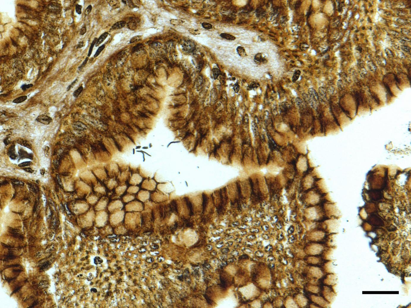
Fig. 1B. Mucosa, stomach, same dog as Fig. 1A. Gastric spiral organisms in the lumen of a crypt. 310
Histology, Warthin-Starry stain. Bar = 20 µm. 311
312
Fig. 2A. Stomach (pylorus), dog. Several small lymphocytes (arrowheads) close to epithelial cell 313
clusters (arrows): chronic lymphocytic gastritis. Cytology, squash preparation, May-Grünwald-314
Giemsa stain. Bar = 20 µm. 315
316
Fig. 2B. Mucosa, stomach (fundus), same dog as in Fig. 2A. Increased number of lymphocytes and 317
plasma cells in the lamina propria: chronic lymphoplasmacytic gastritis. Histology, haematoxylin-318
eosin-saffron stain. Bar = 20 µm. 319
320
Fig. 3A. Stomach (pylorus), dog. Large lymphoblasts predominate: gastric lymphoma. Cytology, 321
squash preparation, May-Grünwald-Giemsa stain. Bar = 20 µm. 322
323
Fig. 3B. Mucosa, stomach (pylorus), same dog as in Fig. 3A. Large lymphoblasts infiltrate the 324
lamina propria and the lamina muscularis: gastric lymphoma. Histology, haematoxylin-eosin-325
saffron stain. Bar = 20 µm. 326

Table 1
Grading system used for cytological analysis. Adapted from Jergens et al. (1998) and
Andreasen et al. (2009).
Categories Description Definition of grades used
Inflammatory cells Neutrophils, eosinophils, lymphocytes,
plasma cells, macrophages, mast cells
Grade 0: no cell
Grade 1: 1 cell
Grade 2: 2 cells
...
Grade 7: ≥7 cells
Per field at
40x-
Objective Atypical/neoplastic cells
Gastric Spiral Organisms
(GSO)
Grade 0: absence
Grade 1 to 2: presence of
mild amount
Grade 3 to 4: presence of
moderate amount
Grade 5 to 7: presence of
marked amount
Bacterial flora Rods and cocci
Haemorrhage Presence of peripheral blood
Debris/ingesta Plant material, darkly pigmented
particulate matter
Mucus Diffusely basophilic mucinous
material or rounded mucinous globules
Epithelial clusters Grade 0: none
Grade 1: 1 to 2 clusters
Grade 2: 3 to 4 clusters
Grade 3: 4 to 5 clusters
Grade 4: 6 to 7 clusters
Grade 5: 7 to 8 clusters
Grade 6: 9 to 10 clusters
Grade 7: > 10 clusters
Per field at
10x-
Objective

Table 2
Diagnostic categories defined for both cytological and histological analyses to allow
comparison of the techniques.
Normal
Inflammation
(defined by the predominant cell population)
Lymphoplasmacytic
Eosinophilic
Neutrophilic
Histiocytic
Non inflammatory fibrosis
Neoplasia Lymphoid
Epithelial
Mesenchymal
Neuroendocrine

Table 3
Histological and cytological results obtained for the 23 cases included in the study.
Species Breed Sex Age Histological diagnosis Cytological diagnosis
Imprint Squash
Cat DSH1 Male 11 Gastric lymphoma Gastric lymphoma Gastric lymphoma
DSH Male 11 Rectal lymphoma Non conclusive Non conclusive
DSH Male 16 Gastric lymphoma Non conclusive Neutrophilic gastritis
DSH Female 12 Lymphoplasmacytic
gastroenteritis
Eosinophilic gastritis
and
lymphoplasmacytic
enteritis
Eosinophilic gastritis and
lymphoplasmacytic
enteritis
DSH Male 4 Lymphoplasmacytic
gastritis + GSO2
Non conclusive Normal stomach + GSO
Dog West
highland
white terrier
Male 13 Lymphoplasmacytic
enteritis
Slide not available Lymphoplasmacytic
enteritis
French
bulldog
Female 3 Neutrophilic gastritis
+ GSO, normal
duodenum
Normal stomach,
lymphoplasmacytic
enteritis
Normal stomach,
lymphoplasmacytic
enteritis
Lhasa apso Female 5 Normal stomach,
lymphoplasmacytic
enteritis, normal
colon
Normal Normal stomach,
lymphoplasmacytic
enteritis, normal colon
Jack Russell
terrier
Male 10 Gastric lymphoma,
lymphoplasmacytic
enteritis
Gastric lymphoma,
lymphoplasmacytic
enteritis
Gastric lymphoma,
lymphoplasmacytic
enteritis
Rhodesian
ridgeback
Female 10 Colonic adenoma Non conclusive Colonic carcinoma
1 DSH: domestic shorthair b GSO: gastric spiral organisms

Parson
terrier
Male 12 Lymphoplasmacytic
gastroenteritis
Stomach non
conclusive,
lymphoplasmacytic
enteritis
Normal stomach + GSO,
lymphoplasmacytic
enteritis
Labrador Female 1 Lymphoplasmacytic
gastroenteritis +
Leishmania spp.
amastigotes
Stomach non
conclusive,
lymphoplasmacytic
enteritis +
Leishmania spp.
amastigotes
Normal stomach,
lymphoplasmacytic
enteritis + Leishmania spp.
amastigotes
Labrador Male 4 Lymphoplasmacytic
gastroenteritis + GSO
Normal stomach,
lymphoplasmacytic
enteritis
Lymphoplasmacytic
gastroenteritis
German
shepherd
Male 9 Lymphoplasmacytic
enteritis
Normal Lymphoplasmacytic
enteritis
Boxer Male 10 Normal stomach,
lymphoplasmacytic
enteritis (small and
large intestine)
Stomach non
conclusive,
lymphoplasmacytic
enteritis, normal
colon
Stomach non conclusive,
lymphoplasmacytic
enteritis (small and large
intestine)
Crossbred Male 3 Lymphoplasmacytic
gastritis + duodenal
fibrosis
Normal stomach +
GSO,
lymphoplasmacytic
enteritis
Lymphoplasmacytic
gastroenteritis + GSO
German
shepherd
Male 7 Normal stomach,
eosinophilic enteritis
Normal stomach and
duodenum
Normal stomach and
duodenum
Weimaraner Female 12 Normal stomach,
lymphoplasmacytic
enteritis
Stomach non
conclusive,
lymphoplasmacytic
enteritis
Normal stomach,
lymphoplasmacytic
enteritis

Crossbred Female 11 Eosinophilic enteritis
+ colonic fibrosis
Ileum non
conclusive, normal
colon
Lymphoplasmacytic
enteritis, normal colon
French
bulldog
Male 3 Normal stomach
+GSO,
lymphoplasmacytic
enteritis
Normal stomach +
GSO,
lymphoplasmacytic
enteritis
Normal stomach + GSO,
lymphoplasmacytic
enteritis
French
bulldog
Female 7 Neutrophilic gastritis
+ GSO and
lymphoplasmacytic
enteritis
Stomach non
conclusive, normal
duodenum
Normal stomach,
lymphoplasmacytic
enteritis
Jack Russell
terrier
Male 11 Lymphoplasmacytic
gastroenteritis
Normal stomach,
lymphoplasmacytic
enteritis
Normal stomach,
lymphoplasmacytic
enteritis
Jack Russell
terrier
Male 12 Lymphoplasmacytic
gastritis
Lymphoplasmacytic
gastritis + GSO
Lymphoplasmacytic
gastritis + GSO

Table 4
Agreement between cytological techniques and histology for the different parts of the
digestive tract.
Organ Technique Number of
specimens
Number of
specimens
considered
‘diagnostic’1
Number of
agreements
between
cytology and
histology2
Kappa value
Stomach Imprint 24 13 7 0.42 [0.18; 0.65]
Squash 24 23 12 0.37 [0.18; 0.56]
Small
intestine
Imprint 173 15 10 0.04 [-0.3; 0.37]
Squash 18 18 14 0.15 [-0.07; 0.38]
Large
intestine
Imprint 6 4 2 0.27 [-0.24; 0.78]
Squash 6 5 4 0.72 [0.23; 1.22]
Total Imprint 47 32 19 0.39 [0.2; 0.58]
Squash 48 46 30 0.48 [0.32; 0.65]
1 Number of specimens of sufficient quality to be interpretable 2 Amongst ‘diagnostic specimens’ 3 One of the 18 imprint specimens was lost during laboratory process