Root Fractures in Children and Adolescents Diagno
32
Root Fractures in Children and Adolescents: Diagnostic Considerations Julie Robinson Molina A thesis submitted to the faculty of the University of North Carolina at Chapel Hill in partial fulfillment of the requirements for the degree of Masters of Science in the School of Dentistry (Pediatrics). Chapel Hill 2007 Approved by: William F. Vann, Jr. Jessica Y. Lee Judy McIntyre Martin Trope
description
Root Fractures in Children and Adolescents: Diagnostic ConsiderationsJulie Robinson MolinaA thesis submitted to the faculty of the University of North Carolina at Chapel Hill in partialfulfillment of the requirements for the degree of Masters of Science in the School ofDentistry (Pediatrics).Chapel Hill2007
Transcript of Root Fractures in Children and Adolescents Diagno

Root Fractures in Children and Adolescents: Diagnostic Considerations
Julie Robinson Molina
A thesis submitted to the faculty of the University of North Carolina at Chapel Hill in partialfulfillment of the requirements for the degree of Masters of Science in the School ofDentistry (Pediatrics).
Chapel Hill2007
Approved by:
William F. Vann, Jr.
Jessica Y. Lee
Judy McIntyre
Martin Trope

ii
ABSTRACT
JULIE MOLINA: Root Fractures in Children and Adolescents: Diagnostic Considerations(Under the direction of William F. Vann, Jr.)
Purpose: To (1) characterize epidemiological trends in anterior permanent tooth trauma (2)
examine the relationship of crown fractures and root fractures to determine if crown fractures
are protective against root fractures (3) determine the diagnostic value of obtaining three
periapical radiographic projections to assess potential root fractures.
Methods: Two dentists served as expert examiners for the radiographic assessments. Kappa
statistics, Pearson’s Chi-Square tests and logistic regression analyses were employed.
Results: The final sample included 185 teeth. Expert examiners detected: 22 root fractures,
10% of the teeth exhibited root fractures when no crown fractures was documented, and 14%
of the teeth had both crown fractures and root fractures as separate entities.
Conclusions: Crown fractures were not protective against root fractures. As radiographic
projections increased, root fractures were identified more often. Our data support obtaining
multiple radiographic projections at different vertical angulations to rule out root fractures in
children and adolescents.

iii
ACKNOWLEDGEMENTS
To the casual observer, a master’s thesis may appear to be solitary work. However, tocomplete a project of this magnitude requires a network of support, and I am indebted tomany people. I am most grateful to my principal advisor, Dr. William F. Vann, Jr. for all hissupport and guidance through this journey.

iv
TABLE OF CONTENTS
LIST OF TABLES………………………………………………………………………........vi
LIST OF FIGURES…………………………………………………………………….........vii
Chapter
I. INTRODUCTION…………………………………………………………….1
Diagnosis of Root Fractures in Children and Adolescents……………………1
Treatment and Prognosis of Root Fractures…………………………………...2
II. MATERIALS AND METHODS……………………………………………...5
Specific Aims………………………………………………………………….5
Sample Size Estimations………………………………………………………5
Sample Characterization…………………………………………………...….6
Root Fracture Assessment by Expert Examiners…………………………...…6
Why Expert Examiners?....................................................................................7
Statistical Analyses……………………………………………………………8
Human Subject Assurances……………………………………………………8
III. RESULTS…………………………………………………………………....10
Demographic Findings……………………………………………………….10
Epidemiological Findings……………………………………………………10
Inter- and Intra- Examiner Reliability……………………………………..…10
Radiographic Assessment Findings………………………………………….11
v
Findings Related to the Number of Radiographic Images…………………...11
Characteristics of the Root Fractures………………………………………...11
IV. DISCUSSION………………………………………………………………..12
Epidemiology………………………………………………………………...12
Intra- and Inter- Examiner Reliability………………………………………..12
The Challenge of Root Fracture Diagnosis………………………………......13
Are Coronal Fractures Protective Against Root Fractures?.............................14
How Many Radiographic Projections are Needed?.........................................14
V. STRENGTHS AND POTENTIAL LIMITATIONS………………………...17
VI. CONCLUSIONS…………………………………………………………….18
Moving Toward Evidence-Based Practice Guidelines………………………18
Conclusions and Implications for Clinical Practice………………………….18
REFERENCES……………………………………………………………………....25

vi
LIST OF TABLES
Table
1. Epidemiology and demographics…………………………………………………21
2. Relationship between crown fractures and expert examiners’ radiographicdiagnosis of root fractures………………………………………………………...22
3. Root fracture assessment by expert examiners…………………………………...23

vii
LIST OF FIGURES
Figure
1. Epidemiology of Maxillary Anterior Trauma in Children and Adolescents…..…24
CHAPTER 1
INTRODUCTION
Root fractures are relatively uncommon, comprising only 0.5-7% of all dental injuries
in the permanent dentition.1 The most common age range for root fractures involving the
permanent dentition in children is between 11-20 years1 with 75% affecting maxillary central
incisors.2 Typically the mechanism of root fractures is a frontal impact that creates
compression zones labially and lingually.1
Root fractures often present clinically as a slightly extruded tooth, often lingually
displaced. Such a tooth is often mobile but the degree of mobility is determined by the
fracture site. Root fractures are more commonly found in the middle third of the root.1
Without radiographic examination, usually it is impossible to distinguish between
displacement due to a luxation injury or root fracture.3 The authors clinical experience
suggest that in children and adolescents, coronal fractures may be a protective and mitigating
factor against root fractures but to date this clinical perception is untested.
Diagnosis of Root Fractures in Children and Adolescents
Correct diagnosis of root fractures is essential to ensure proper treatment to achieve
the best possible prognosis.2 A correct diagnosis will aid the clinician in decisions about
immediate treatment and splinting strategies as well as the timing of follow-up examinations,
radiographs, and sensitivity testing.

2
A root fracture can be seen only if the radiographic beam is directed through the
plane of fracture. Many authorities argue that one radiograph often will not lead to optimal
disclosure of root fractures. One protocol to diagnose or rule-out root fractures in children
and adolescents is advocated by the guidelines4 established by the International Association
of Dental Traumatology (IADT). This protocol includes four periapical radiographs: an
occlusal, a periapical central angle, periapical mesial and distal excentric projections.4
Currently, there are no published data to support this recommendation.
A multi-directional approach using a conventional periapical exposure and two
additional vertical periapical projections that vary +/- 15-20° from the central beam has been
advocated by Andreasen and Andreasen1, Wilson3, Degering5, Bender6, Berman7, and
Herweijer8. Both Andreasen and Andreasen1 and Degering5 have published eloquent
illustrations involving artificial root fractures in human incisors, adding a weight of
documentation to this radiographic protocol. At The University of North Carolina at Chapel
Hill (UNC-CH) School of Dentistry, the protocol for examining permanent incisor root
fractures in children and adolescents entering the Pediatric Dentistry or Endodontic Clinics is
to obtain three periapical radiographic projections at different vertical angulations1,3,5-8 for
dental-related tooth trauma wherein root fracture is possible. This protocol has been the
standard of care since the mid-1990s. While many authorities recommend a multi-projection
clinical protocol for diagnosis of root fractures, it should be noted that there are no clinical
trials to support these recommendations.
Treatment and Prognosis of Root Fractures
Treatment and prognosis of root fractures in permanent teeth is dependent on a
variety of factors including stage of root development, type of healing and optimal

3
repositioning of coronal fragments and/or splinting of teeth. Both the IADT4 and Andreasen
and Andreasen1 recommend repositioning the coronal fragment if displaced and
immobilizing the tooth with a splint with follow-up in 3-4 weeks. Recent research suggests
that optimal repositioning may be more critical than splinting. With dislocation of coronal
fragments, optimal repositioning enhances the likelihood of both pulp healing and hard tissue
repair in mature and immature teeth.9 Cvek and colleagues9 found no difference in the
frequency of healing between splinted and non-splinted teeth.
One exception to the concept of repositioning root fractures involves teeth with
incomplete fractures with immature roots wherein fractures have been found to heal
spontaneously.10 Similar findings have been reported by Freely and colleagues11 as well as
Cvek and colleagues9, both of whom found good root healing to occur most often in teeth
with incomplete root development.
Based on the current literature, root fractures in children and adolescents have a good
prognosis if a proper diagnosis is made at the time of the traumatic injury and if the proper
treatment is undertaken. Radiographic information is a key component for making such
diagnosis, and rendering the proper treatment. Accordingly, our findings should offer
important insights for clinicians to consider in making a radiographic diagnosis of root
fractures. These insights may lead to the development of new clinical standards of care that
may save time, money and unneeded radiation exposure.

4
ENDNOTES
1Andreasen JO, Andreasen FM. Textbook and color atlas of traumatic injuries to the teeth.3rd edn. Copenhagen: Munksgaard; 1994: p. 151-8, 171-5, 279-81.
2Majorana A, Pasini S, Bardellini E, Keller E. Clinical and epidemiological study oftraumatic root fractures. Dent Traumatol 2002;18:77-80.
3Wilson C. Management of trauma to primary and developing teeth. Dental Clinics of NorthAmerica 1995;39:133-67.
4Flores MT, Andreasen JO, Bakland LK. Guidelines for the Evaluation and Management ofTraumatic Dental Injuries. Dent Traumatol 2001;17:97-102.
5Degering C. Radiography of dental fractures: an experimental evaluation. Oral Surg1970;30:213-9.
6Bender IB, Freedland JB: Clinical considerations in the diagnosis and treatment of intra-alveloar root fractures, J Am Dent Assoc 1983;107:595-600.
7Berman L. A Clinical Guide to Dental Traumatology. Chapter 4 Intraalveolar root fractures.2006: p. 51-71.
8Herweijer JA, Torabinejad M, Bakland L: Healing of horizontal root fractures, J Endod1992;18(3):118-122.
9Cvek M, Andreasen JO, Borum MK. Healing of 208 intraalveolar root fractures in patientsaged 7-17years. Dent Traumatol 2001;17:53-62.
10Andreasen FM, Andreasen JO, Bayer T. Prognosis of root-fractured incisors: prediction ofhealing modalities. Endod Dent Traumatol 1989;5:11-22.
11Feely L, Mackie IC, Macfarlane T. An investigation of root-fractured permanent incisorteeth in children. Dent Traumatol 2003;19:52-4.

CHAPTER 2
MATERIALS AND METHODS
Specific Aims
1. To characterize epidemiological trends in anterior permanent tooth trauma in
a sample of children and adolescents aged 6-18.
2. To examine the relationship of root fractures with and without concomitant crown
fractures to answer the question: are crown fractures protective against
root fractures in children and adolescents?
3. To carefully examine our patient population for radiographic evidence of root
fractures and determine the diagnostic and clinical value of obtaining three vertical
periapical radiographic projections to assess maxillary anterior root fractures in
children and adolescents aged 6 -18.
We tested two null hypotheses for children and adolescents aged 6-18: 1) crown fractures are
not protective against root fractures and 2) three vertical periapical radiographs at different
angles are not necessary for the diagnosis of root fractures.
Sample Size Estimations
For sample size estimates, we used reported prevalence scores from a recent study of
dental trauma in children12 in which children (7-18 years of age) had a reported crown and
root fracture prevalence of 32.1% and 2.1%, respectively. Assuming the same prevalence
levels with an alpha error of 0.05 and beta of 0.80, we estimated a sample size of 134 cases
6
would be needed to detect a difference using simple parametric tests such as chi-square tests.
Because our inclusion criteria required three clearly diagnostic radiographs, we expected that
some patient records would be hard to access, some radiographs would be missing from
some records, and others would not meet our strict standard of being clearly diagnostic.
Sample Characterization
We reviewed our eight-year emergency registry and carefully selected only the cases
involving permanent maxillary incisor trauma for which three diagnostic vertical periapical
radiographs were available in each patient’s record. All cases were then categorized by
diagnostic category with tooth/bone-related trauma: uncomplicated, complicated, root
fracture, crown-root fracture, alveolar fracture and luxation-related trauma: concussion,
subluxation, luxation, intrusion, extrusion and avulsion. In classifying the type of trauma, we
relied upon the diagnosis given by the treating dentists at the time of trauma.
We classified each trauma case according to ethnicity, gender, age, and etiology. The
etiology was classified into seven groups: falls during free-play, sports-related accidents,
bicycle accidents, automobile accidents, ATV/motorbike accidents, child abuse, and “other.”
For athletic injuries, we recorded whether an athletic mouthguard was in use by the child at
the time of the injury.
Root Fracture Assessment by Expert Examiners
Two experienced dentists with expertise in dental trauma served as expert examiners
for root fracture assessment. The examiners were trained in two consensus-building
calibration sessions using a sub-sample of trauma cases that included three periapical
radiographs obtained at different vertical angulations. All radiographic interpretations were
accomplished using view boxes in a dark room. The purpose of each session was to 1)
7
review the definition of root fractures 2) complete independent reviews of selected
radiographs from sample cases including some with root fractures and 3) to debrief all
reviews to achieve calibration and build examiner consensus.
The current literature included no data on the degree of examiner agreement
achievable. Our goal was to achieve a Kappa score of at least 0.80 for both intra- and inter-
examiner reliability. After training and calibration, the examiners independently assessed the
case-study radiographs for root fractures in a final, structured session under the supervision
of the Principal Investigators. A random sample group of twenty cases were re-examined
unknowingly by the examiners to provide data for the determination of intra-examiner
reliability. Following a review of all cases, the examiners discussed those cases for which
there were diagnostic disagreements and reached a diagnostic consensus. Kappa statistics
were performed to determine the level of agreement for intra- and inter-examiner reliability.
Why Expert Examiners?
Considering that the patients in this study had undergone a comprehensive dental
trauma examination and many had subsequent follow-up care during which root fractures
could have been detected and diagnosed, the purpose of deploying expert examiners was to
cross-examine the study sample to determine whether there were cases diagnosed with root
fractures with actual clinical/radiographic assessments and follow-up care that went
undetected by the expert examiners. Further, we also wanted to examine if the expert
examiners would detect any occult fractures not detected by clinical/radiographic assessment
and follow-up care. Finally, we wanted to generate data for intra- and inter-examiner
reliability to illuminate the ease or difficulty in the radiographic diagnosis of root fractures in
children and adolescents.
8
Statistical Analysis
All analyses were conducted using STATA 9.0 (STATA Corp. College Station,
Texas). We examined the relationship between crown fractures and root fractures using a
Likelihood Ratio Chi-square test with the level of significance set at an alpha of 0.05. We
determined the diagnostic value of obtaining three vertical periapical radiographs using
inspection of our examiner-derived positive root fractures.
Human Subject Assurances
This study was approved by the Institution Review Boards of the Schools of Dentistry
and Medicine at The University of North Carolina at Chapel Hill.

9
ENDNOTES
12Skaare AB, Jacobsen I. Dental Injuries in Norwegians aged 7-18 years. Dent Traumatol2003;19:67-71.

CHAPTER 3
RESULTS
Demographic Findings
During the study time-frame (1997-2004) the total emergency visits ranged from 400-
700 annually with an estimated 125 permanent anterior tooth trauma cases each year.
A total of 114 patients experienced dental trauma to the permanent maxillary incisors for
which three radiographs were obtained at the initial emergency visit. Relevant demographic
data for the patients are found in Table 1.
Epidemiologic Findings
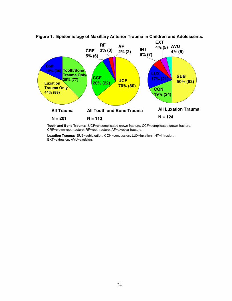
Our 114 patients experienced a total of 201 traumatized maxillary incisor teeth. The
epidemiological data are illustrated in Figure 1. From our sample size of 201 traumatized
teeth, using our strict inclusion criteria of three clearly diagnostic radiographs, 185 teeth met
our inclusion criteria. The expert examiners assessed these images to generate data for root
fractures.
Intra- and Inter-Examiner Reliability
The radiographic assessment session for the study data generation yielded Kappa
scores of 81% for inter-examiner reliability and 100% consensus for those cases wherein
examiners at first disagreed. The intra-examiner reliability Kappa scores were 0.80 and 0.75
respectively for examiners 1 and 2.

11
Radiographic Assessment Findings
The expert examiners focused on the 185 teeth that met our strict inclusion criteria for
the availability of three clearly diagnostic radiographs having been obtained at the initial
trauma visit. The expert examiners assessed these images to generate data for root fractures
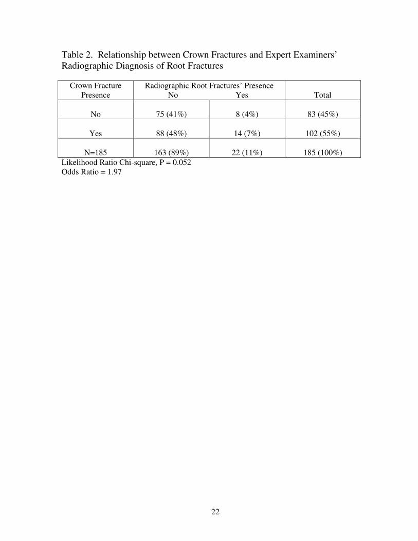
as illustrated in Table 2. Crown fractures were not protective against root fractures; indeed,
teeth with crown fractures were two times as likely to have a root fracture as those without
crown fractures.
Findings Related to the Number of Radiographic Images
Our expert examiners reached a consensus on a total of 22 root fractures in the
sample. Three root fractures (13%) were seen on only one of three images, 14 (64%) were
seen on two of the three images and five (23%) were seen on all three images.
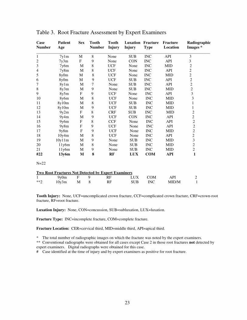
Characteristics of the Root Fractures
Table 3 illustrates specific details of the root fractures in our study. The prevalence
of the root fractures diagnosed by the clinicians at the time of injury was 1.6% (3 out of 185).
The prevalence of root fractures identified by the expert examiners was 11.9 % (22 out of
185). The patients in our study for whom root fractures were identified were in the age range
of 7y1m to 13y6m with a median of 8y10m. The prevalence slightly favored males.
Table 3 illustrates concomitant tooth/bone and luxation injury relationships. Of
special interest are location and fracture type categories. Note that half of the occult fractures
were found in the mid-root location and half in the apical location. A more dramatic finding
is that 95% of the occult fractures were incomplete while only 5% (one fracture) was
complete.
CHAPTER 4
DISCUSSION
Epidemiology
It should be recognized that ours is a select sample of trauma to only permanent
maxillary incisors; however, relative to gender and etiology our epidemiological findings are
very similar to those reported by Rajab13 and Andreasen and Andreasen.1 One difference in
our patient population was the age of those patients. In our sample of 185 teeth, patients’
ages ranged from 7y1m to 13y6m. Our patients are clearly younger and this is in contrast to
root fractures reported by Rajab13 that occurred in older children (10-15) and those reported
by Andreasen and Andreasen1 with ages ranging from 11-20.
None of our trauma cases involved mouthguard use, including the 14 children with
sports-related injuries. Also, we were surprised that sports injuries were not higher among
our study sample. Mouthguard use among school-aged athletes is relatively high in our
community and we speculate that this phenomenon might have reduced the prevalence of
sports-related trauma during the time-frame of the study.
Intra- and Inter-Examiner Reliability
Our examiners were experienced clinicians with expertise in dental taumatology and
both were active in the field. While the Kappa scores for intra-examiner reliability were
acceptable, in general the findings suggest that radiographic diagnosis of root fractures in
children and adolescents is difficult, even under the most ideal conditions.
13
The Challenge of Root Fracture Diagnosis
It should be noted that our sample included only three root fractures that were
diagnosed on the basis of the clinical/radiographic data at the time of injury and no additional
diagnoses were made during trauma follow-up. Our examiners correctly identified only one
of those three fractures. The examiners found an additional 21 occult fractures that were not
detected by the treating dentists at the time of injury. These 21 fractures were either
undiagnosed or not recorded by the attending dentist following the traumatic injury. The
difficulty of root fracture diagnosis is highlighted by the results that even calibrated “experts”
in dental trauma had difficulty detecting root fractures. This further emphasizes the
importance of the additional clinical exam, as well as the radiographic exam, to accurately
detect or suspect root fractures; again, our experts did not have any information related to the
clinical exams that had taken place. It is evident how even after “sensitizing” examiners to
detect root fractures, two remained undiagnosed.
It is interesting that the large prevalence of occult root fractures detected were
incomplete and Andreasen and Andresean 10 point out that such fractures heal with
subsequent hard tissue formation and have an excellent prognosis. The one complete fracture
was located in the apical third, which authorities1 suggest have an excellent prognosis with
no treatment required.
One could also justify that it is not important to diagnose root fractures as long as
repositioning is performed. Studies show optimal repositioning leads to better healing and a
more favorable prognosis.9 These findings underscore the challenge of detecting root
fractures in children and adolescents and also suggest that the clinical examination is an
important adjunct to supplementing radiographic assessment for diagnosis at the time of

14
injury. These results also emphasize the need to examine radiographs very carefully, using a
dark room and close inspection. It is important to diagnose a root fracture because if it is
missed, the diagnosis may be a severe luxation injury which would typically necessitate root
canal treatment. However, if a root fracture is correctly diagnosed, root canal treatment
should NOT be performed and will eventually only be needed about 25% of the time. 1
Are Coronal Fractures Protective Against Root Fractures?
Clinical experience had suggested to us that coronal fractures seemed to be protective
against root fractures in children and adolescents. The rationale is that injury to the tooth
occurs at the site of impact and if a tooth has a coronal fracture, this is the focus of impact
and the remaining tooth should remain sound. Yet, the results from the present investigation
indicated that crown fractures were not protective against root fractures. In fact, teeth with
crown fractures were almost twice as likely to have a root fracture.
These findings suggest clinicians should be more suspicious of a root fracture in those
teeth with uncomplicated crown fractures or no trauma to the coronal aspect of the tooth
because these teeth were more likely to have an accompanying root fracture. Table 3
illustrates this point. This finding should heighten clinicians’ awareness when evaluating and
diagnosing dental trauma.
How Many Radiographic Projections are Needed?
The literature supports that multiple radiographic projections are needed to increase
the likelihood of diagnosing a root fracture.1,3,5-8 Degering5 looked at radiographs of
experimental root fractures of the anterior teeth to find which angulations provided the most
diagnostic information. His study revealed fractures were diagnostic in a latitude of +/- 15 to
20 degrees of vertical angulation relative to the fracture plane. Two additional radiographs
15
should be obtained of the questionable area with a +15 degree and -15 degree vertical
angulation in relation to the original tube position.5
Our findings indicate that multiple radiographic projections are needed to increase the
likelihood of diagnosing a root fracture. Under the conditions of our study, we were not able
to say definitively that three radiographs is the best protocol. However we hypothesize that a
root fracture that can be detected in more than one image increases the clinician’s confidence
in the diagnosis and is more likely to be recorded and treated by the clinician as a true root
fracture. Without obtaining more than one film, the root fracture may be overlooked or
disregarded as a defect in root development, an artifact, or bone trabeculae/bony
trabeculation.

16
ENDNOTES
1Andreasen JO, Andreasen FM. Textbook and color atlas of traumatic injuries to the teeth.3rd edn. Copenhagen: Munksgaard; 1994: p. 151-8, 171-5, 279-81.
3Wilson C. Management of trauma to primary and developing teeth. Dental Clinics of NorthAmerica 1995;39:133-67.
5Degering C. Radiography of dental fractures: an experimental evaluation. Oral Surg1970;30:213-9.
6Bender IB, Freedland JB: Clinical considerations in the diagnosis and treatment of intra-alveloar root fractures, J Am Dent Assoc 1983;107:595-600.
7Berman L. A Clinical Guide to Dental Traumatology. Chapter 4 Intraalveolar root fractures.2006: p. 51-71.
8Herweijer JA, Torabinejad M, Bakland L: Healing of horizontal root fractures, J Endod1992;18(3):118-122.
9Cvek M, Andreasen JO, Borum MK. Healing of 208 intraalveolar root fractures in patientsaged 7-17years. Dent Traumatol 2001;17:53-62.
10Andreasen FM, Andreasen JO, Bayer T. Prognosis of root-fractured incisors: prediction ofhealing modalities. Endod Dent Traumatol 1989;5:11-22.
13Rajab LD. Traumatic dental injuries in children presenting for treatment at the Departmentof Pediatric Dentistry, Faculty of Dentistry, University of Jordan, 1997-2000. DentTraumatol 2003;19:6-11.
CHAPTER 5
STRENGTHS AND POTENTIAL LIMITATIONS
This study included all children and adolescents who presented with a traumatic
dental injury over an eight-year period of time. Because we relied on patient records, we
were not able to monitor root fracture outcomes over time because some patients did not
return for follow-up. An ideal study design would be a prospective, randomized controlled
trial; however, such a study for a population of this size for eight consecutive years would be
strategically challenging and enormously costly. Our sample offered us an opportunity to
study a relatively large cohort with relative ease, and at a fraction of the cost. At the same
time, it would not be ethical to conduct a prospective, randomized controlled trial assessing
one versus multiple projections under a scenario where the latter is the standard of care.
By using a research design that included two calibrated and “sensitized” examiners,
our findings yielded new information about the diagnostic challenge of radiographic
interpretation of root fractures in children and adolescents.

CHAPTER 6
CONCLUSIONS
Moving toward Evidence-Based Practice Guidelines
One area for which more evidence-based study is needed is in the realm of diagnosis
and clinical management of dental trauma in children and adolescents. A 20-year (1985-
2005) Medline search revealed 102 published studies on this subject but only 20 were
focused on children and adolescents and only three of these were scientific investigations and
none established clinical guidelines or recommendations. These results were confirmed by a
search in the Cochrane Collaboration systematic review database that revealed no studies on
this topic.14 Our study sought seeks to fill a gap through the generation of evidenced-based
clinically relevant guidelines for the diagnosis of root fractures in children and adolescents.
Conclusions and Implications for Clinical Practice
Under the conditions of this study examining children and adolescents 6-18 years of
age with anterior permanent tooth trauma:
1) Crown fractures were not protective against root fractures. Teeth with crown fractures
were almost twice as likely to sustain root fractures.
2) Radiographic root fractures were very difficult to detect. Radiographic images aimed at
detection of root fractures should be reviewed carefully under ideal conditions of
illumination to make a proper diagnosis.
3) Root fractures in children in the pre-teen years are likely to be incomplete and located in
the apical or middle third of the root.
19
4) Our data would suggest there is no reason to suspect a complete root fracture in pre-teen
children unless the tooth exhibits clinical signs such as luxation or excessive mobility; in
short, obtaining three radiographic images to examine for root fractures for all tooth trauma
in this age group seems unnecessary.
5) When root fractures are suspected, multiple radiographic projections at different vertical
angulations will increase the diagnostic precision for making a root fracture diagnosis.

20
ENDNOTES
14Cochrane Collaboration. Available at: http://www.cochrane.org/index0.htm. AccessedFebruary 10, 2006.

21
Table 1. Epidemiology and Demographics N=114
Variables _________n Percent________________________________________________GenderMale 72 63%Female 42 37%________________________________________________AgeSix 5 4%Seven 20 18%Eight 27 24%Nine 22 19%Ten 14 12%Eleven 7 6%Twelve 14 12%Thirteen 0 0%Fourteen 0 0%Fifteen 3 3%Sixteen 2 2%Seventeen 0 0%Eighteen 0 0%________________________________________________EtiologyFalls 38 34%Bicycle Accident 22 20%Playing Sports 14 12%ATV/motorbike Accident 2 2%Automobile Accident 1 1%Child Abuse 0 0%Other 37 31%(i.e., free play, random accidents, etc)
_________________________________________________Mouthguard Use 0 0%_________________________________________________
22
Table 2. Relationship between Crown Fractures and Expert Examiners’Radiographic Diagnosis of Root Fractures
Crown FracturePresence
Radiographic Root Fractures’ PresenceNo Yes Total
No 75 (41%) 8 (4%) 83 (45%)
Yes 88 (48%) 14 (7%) 102 (55%)
N=185 163 (89%) 22 (11%) 185 (100%)Likelihood Ratio Chi-square, P = 0.052Odds Ratio = 1.97
23
Table 3. Root Fracture Assessment by Expert Examiners
Case Patient Sex Tooth Tooth Luxation Fracture Fracture RadiographicNumber Age Number Injury Injury Type Location Images *______________________________________________________________________________________1 7y1m M 8 None SUB INC API 32 7y3m F 9 None CON INC API 33 7y6m M 8 UCF None INC MID 24 7y8m M 8 UCF None INC API 25 8y0m M 8 UCF None INC MID 26 8y0m M 9 UCF SUB INC API 27 8y1m M 7 None SUB INC API 28 8y3m M 9 None SUB INC MID 29 8y5m F 9 UCF None INC API 310 8y6m M 8 UCF None INC MID 311 8y10m M 8 UCF SUB INC MID 112 8y10m M 9 UCF SUB INC MID 113 9y2m F 8 CRF SUB INC MID 214 9y4m M 9 UCF CON INC API 215 9y6m F 8 CCF None INC API 216 9y8m F 9 UCF None INC API 217 9y8m F 9 UCF None INC MID 218 10y4m M 8 UCF None INC API 219 10y11m M 9 None SUB INC MID 320 11y6m M 8 None SUB INC MID 221 11y6m M 9 None SUB INC MID 2#22 13y6m M 8 RF LUX COM API 1
N=22
Two Root Fractures Not Detected by Expert Examiners1 9y0m F 9 RF LUX COM API 2**2 10y3m M 8 RF SUB INC MID/M 1
Tooth Injury: None, UCF=uncomplicated crown fracture, CCF=complicated crown fracture, CRF=crown-rootfracture, RF=root fracture.
Luxation Injury: None, CON=concussion, SUB=subluxation, LUX=luxation.
Fracture Type: INC=incomplete fracture, COM=complete fracture.
Fracture Location: CER=cervical third, MID=middle third, API=apical third.
* The total number of radiographic images on which the fracture was noted by the expert examiners.** Conventional radiographs were obtained for all cases except Case 2 in those root fractures not detected byexpert examiners. Digital radiographs were obtained for this case.# Case identified at the time of injury and by expert examiners as positive for root fracture.
24
Figure 1. Epidemiology of Maxillary Anterior Trauma in Children and Adolescents.
Tooth/BoneTrauma Only38% (77)
Both18% (36)
LuxationTrauma Only44% (88)
All Trauma
N = 201
All Tooth and Bone Trauma
N = 113
All Luxation Trauma
N = 124
SUB50% (62)
CON19% (24)
LUX17% (21)
INT6% (7)
EXT4% (5) AVU
4% (5)
UCF70% (80)
CCF20% (22)
CRF5% (6)
RF3% (3)
AF2% (2)
Tooth and Bone Trauma: UCF=uncomplicated crown fracture, CCF=complicated crown fracture,CRF=crown-root fracture, RF=root fracture, AF=alveolar fracture.
Luxation Trauma: SUB=subluxation, CON=concussion, LUX=luxation, INT=intrusion,EXT=extrusion, AVU=avulsion.
25
REFERENCES
1Andreasen JO, Andreasen FM. Textbook and color atlas of traumatic injuries to the teeth.3rd edn. Copenhagen: Munksgaard; 1994: p. 151-8, 171-5, 279-81.
2Majorana A, Pasini S, Bardellini E, Keller E. Clinical and epidemiological study oftraumatic root fractures. Dent Traumatol 2002;18:77-80.
3Wilson C. Management of trauma to primary and developing teeth. Dental Clinics of NorthAmerica 1995;39:133-67.
4Flores MT, Andreasen JO, Bakland LK. Guidelines for the Evaluation and Management ofTraumatic Dental Injuries. Dent Traumatol 2001;17:97-102.
5Degering C. Radiography of dental fractures: an experimental evaluation. Oral Surg1970;30:213-9.
6Bender IB, Freedland JB: Clinical considerations in the diagnosis and treatment of intra-alveloar root fractures, J Am Dent Assoc 1983;107:595-600.
7Berman L. A Clinical Guide to Dental Traumatology. Chapter 4 Intraalveolar root fractures.2006: p. 51-71.
8Herweijer JA, Torabinejad M, Bakland L: Healing of horizontal root fractures, J Endod1992;18(3):118-122.
9Cvek M, Andreasen JO, Borum MK. Healing of 208 intraalveolar root fractures in patientsaged 7-17years. Dent Traumatol 2001;17:53-62.
10Andreasen FM, Andreasen JO, Bayer T. Prognosis of root-fractured incisors: prediction ofhealing modalities. Endod Dent Traumatol 1989;5:11-22.
11Feely L, Mackie IC, Macfarlane T. An investigation of root-fractured permanent incisorteeth in children. Dent Traumatol 2003;19:52-4.
12Skaare AB, Jacobsen I. Dental Injuries in Norwegians aged 7-18 years. Dent Traumatol2003;19:67-71.
13Rajab LD. Traumatic dental injuries in children presenting for treatment at the Departmentof Pediatric Dentistry, Faculty of Dentistry, University of Jordan, 1997-2000. DentTraumatol 2003;19:6-11.
14Cochrane Collaboration. Available at: http://www.cochrane.org/index0.htm. AccessedFebruary 10, 2006.