Rom J Morphol Embryol 2014, 55(3 Suppl):1191–1196 R J M E ... · Rom J Morphol Embryol 2014, 55(3...
6
Rom J Morphol Embryol 2014, 55(3 Suppl):1191–1196 ISSN (print) 1220–0522 ISSN (on-line) 2066–8279 CASE REPORT In vivo imaging techniques for psoriatic lesions CONSTANTIN CĂRUNTU 1) , DANIEL BODA 2) , ANA CĂRUNTU 3) , MARIA ROTARU 4) , FLAVIA BADERCA 5) , SABINA ZURAC 6) 1) Department of Physiology, “Carol Davila” University of Medicine and Pharmacy, Bucharest, Romania 2) Dermatology Research Laboratory, “Carol Davila” University of Medicine and Pharmacy, Bucharest, Romania 3) “Prof. Dr. Dan Theodorescu” Oral and Maxillofacial Surgery Hospital, Bucharest, Romania 4) Department of Dermatology, “Victor Papilian” Faculty of Medicine, “Lucian Blaga” University of Sibiu, Romania 5) Department of Histology, “Victor Babeş” University of Medicine and Pharmacy, Timisoara, Romania 6) Department of Pathology, "Carol Davila” University of Medicine and Pharmacy, Bucharest, Romania; Colentina University Hospital, Bucharest, Romania Abstract Psoriasis is an inflammatory skin disease, with a polymorphic clinical appearance. Modern techniques for in vivo imaging of skin structure, such as reflectance confocal microscopy (RCM) and dermoscopy, could be very useful tools for diagnosis and monitoring of psoriatic lesions. We report a case of psoriasis vulgaris in which we identified certain dermoscopic features suggestive for psoriasis and RCM enabled us to identify in vivo most of the histological features of psoriatic lesions. Keywords: psoriasis, skin, reflectance confocal microscopy, dermoscopy. Introduction Psoriasis is a common chronic inflammatory skin disorder with a prevalence of 2% worldwide characterized by an increased turnover rate of epidermal cells and hyper- proliferation of epidermal keratinocytes [1–7]. Psoriasis may occur at any age but the age of onset has two peaks, one between the age of 16–22 years and another between 57–60 years [8]. The precise cause of psoriasis is still unknown but the interaction between genetic predispo- sition, immunologic factors and environmental triggers, such as local trauma, infections, alcohol, drugs, psycho- logical stress plays a major role in this complex skin disorder [3, 4, 9–15]. The clinical presentation of psoriasis is polymorphic but the most common form is psoriasis vulgaris [16, 17]. Typical psoriatic lesions are well-demarcated erythema- tous plaques covered by white scales, which can have different shapes and sizes and are most commonly located on elbows, knees, scalp, arms and lumbar area but may be located all over the body [4, 16, 18, 19]. Even though psoriatic lesions usually have a charac- teristic clinical appearance and they are easily identified and diagnosed, unusual presentations may be mistaken for other erythematosquamous diseases, such as seborrheic dermatitis, nummular eczema, pityriasis rosea, tinea, pityriasis rubra pilaris, and in those cases skin biopsy with histological evaluation is required to confirm diag- nosis [4–6, 16, 17, 20]. Histopathological representation of psoriatic lesions include increased thickness of the stratum corneum (hyper- keratosis), incomplete keratinocyte maturation with pre- sence of nuclei in cornified layer (parakeratosis), focal absence or reduction of granular layer, epidermal thicke- ning (acanthosis) associated with supra-papillary epidermal thinning and elongation of dermal papillae (papillomatosis) with tortuous, dilated capillaries. In both dermis and epidermis, there is a variable degree of mixed inflam- matory response with lymphocytic and neutrophilic infil- tration. Trafficking of inflammatory cells through epidermis leads to formation of spongiform pustules (micropustules of Kogoj) with collections of neutrophils in spinous layer or aggregates of neutrophils within parakeratotic areas of the stratum corneum (microabscesses of Munro), which are highly specific for psoriasis [4, 5, 7, 21]. Even though histopathological examination can confirm clinical diagnosis of psoriasis, it is often not performed due to its invasiveness. Moreover, skin biopsy has its limitations in the early phases of disease because the changes may be minor. Also, when psoriatic lesions must be monitored it does not allow assessment of the same skin area over time [5, 7, 16]. Thus, the use of modern techniques for in vivo imaging of skin structure may be of real interest for the evaluation of psoriatic lesions. A non-invasive diagnostic technique that enables in vivo observation of skin lesions is dermoscopy and recently have been described certain dermoscopic features that could be of real help for the diagnosis and therapeutic follow up of psoriatic lesions [5, 6, 22–26]. Another novel technique is reflectance confocal micro- scopy (RCM), which allows a harmless, morphological and functional evaluation of skin lesions with a resolution comparable to conventional light microscopy [27, 28] and recent studies showed that RCM facilitates in vivo identification of most of the histological features in psoriasis [5, 7, 17, 29, 30]. We present the case of a patient with multiple erythe- matosquamous lesions for which an accurate diagnosis was facilitated by dermoscopic and RCM examination. R J M E Romanian Journal of Morphology & Embryology http://www.rjme.ro/
Transcript of Rom J Morphol Embryol 2014, 55(3 Suppl):1191–1196 R J M E ... · Rom J Morphol Embryol 2014, 55(3...

Rom J Morphol Embryol 2014, 55(3 Suppl):1191–1196
ISSN (print) 1220–0522 ISSN (on-line) 2066–8279
CCAASSEE RREEPPOORRTT
In vivo imaging techniques for psoriatic lesions
CONSTANTIN CĂRUNTU1), DANIEL BODA2), ANA CĂRUNTU3), MARIA ROTARU4), FLAVIA BADERCA5), SABINA ZURAC6)
1)Department of Physiology, “Carol Davila” University of Medicine and Pharmacy, Bucharest, Romania 2)Dermatology Research Laboratory, “Carol Davila” University of Medicine and Pharmacy, Bucharest, Romania 3)“Prof. Dr. Dan Theodorescu” Oral and Maxillofacial Surgery Hospital, Bucharest, Romania 4)Department of Dermatology, “Victor Papilian” Faculty of Medicine, “Lucian Blaga” University of Sibiu, Romania 5)Department of Histology, “Victor Babeş” University of Medicine and Pharmacy, Timisoara, Romania 6)Department of Pathology, "Carol Davila” University of Medicine and Pharmacy, Bucharest, Romania; Colentina University Hospital, Bucharest, Romania
Abstract Psoriasis is an inflammatory skin disease, with a polymorphic clinical appearance. Modern techniques for in vivo imaging of skin structure, such as reflectance confocal microscopy (RCM) and dermoscopy, could be very useful tools for diagnosis and monitoring of psoriatic lesions. We report a case of psoriasis vulgaris in which we identified certain dermoscopic features suggestive for psoriasis and RCM enabled us to identify in vivo most of the histological features of psoriatic lesions.
Keywords: psoriasis, skin, reflectance confocal microscopy, dermoscopy.
Introduction
Psoriasis is a common chronic inflammatory skin disorder with a prevalence of 2% worldwide characterized by an increased turnover rate of epidermal cells and hyper-proliferation of epidermal keratinocytes [1–7]. Psoriasis may occur at any age but the age of onset has two peaks, one between the age of 16–22 years and another between 57–60 years [8]. The precise cause of psoriasis is still unknown but the interaction between genetic predispo-sition, immunologic factors and environmental triggers, such as local trauma, infections, alcohol, drugs, psycho-logical stress plays a major role in this complex skin disorder [3, 4, 9–15].
The clinical presentation of psoriasis is polymorphic but the most common form is psoriasis vulgaris [16, 17]. Typical psoriatic lesions are well-demarcated erythema-tous plaques covered by white scales, which can have different shapes and sizes and are most commonly located on elbows, knees, scalp, arms and lumbar area but may be located all over the body [4, 16, 18, 19].
Even though psoriatic lesions usually have a charac-teristic clinical appearance and they are easily identified and diagnosed, unusual presentations may be mistaken for other erythematosquamous diseases, such as seborrheic dermatitis, nummular eczema, pityriasis rosea, tinea, pityriasis rubra pilaris, and in those cases skin biopsy with histological evaluation is required to confirm diag-nosis [4–6, 16, 17, 20].
Histopathological representation of psoriatic lesions include increased thickness of the stratum corneum (hyper-keratosis), incomplete keratinocyte maturation with pre-sence of nuclei in cornified layer (parakeratosis), focal absence or reduction of granular layer, epidermal thicke-ning (acanthosis) associated with supra-papillary epidermal
thinning and elongation of dermal papillae (papillomatosis) with tortuous, dilated capillaries. In both dermis and epidermis, there is a variable degree of mixed inflam-matory response with lymphocytic and neutrophilic infil-tration. Trafficking of inflammatory cells through epidermis leads to formation of spongiform pustules (micropustules of Kogoj) with collections of neutrophils in spinous layer or aggregates of neutrophils within parakeratotic areas of the stratum corneum (microabscesses of Munro), which are highly specific for psoriasis [4, 5, 7, 21].
Even though histopathological examination can confirm clinical diagnosis of psoriasis, it is often not performed due to its invasiveness. Moreover, skin biopsy has its limitations in the early phases of disease because the changes may be minor. Also, when psoriatic lesions must be monitored it does not allow assessment of the same skin area over time [5, 7, 16].
Thus, the use of modern techniques for in vivo imaging of skin structure may be of real interest for the evaluation of psoriatic lesions.
A non-invasive diagnostic technique that enables in vivo observation of skin lesions is dermoscopy and recently have been described certain dermoscopic features that could be of real help for the diagnosis and therapeutic follow up of psoriatic lesions [5, 6, 22–26].
Another novel technique is reflectance confocal micro-scopy (RCM), which allows a harmless, morphological and functional evaluation of skin lesions with a resolution comparable to conventional light microscopy [27, 28] and recent studies showed that RCM facilitates in vivo identification of most of the histological features in psoriasis [5, 7, 17, 29, 30].
We present the case of a patient with multiple erythe-matosquamous lesions for which an accurate diagnosis was facilitated by dermoscopic and RCM examination.
R J M ERomanian Journal of
Morphology & Embryologyhttp://www.rjme.ro/

Constantin Căruntu et al.
1192
Materials and Methods
The study was performed in Dermato-oncology Research Laboratory of “Carol Davila” University of Medicine and Pharmacy, Bucharest, Romania in accordance with the guidelines of local ethics committee and after obtaining informed consent from the patient.
A clinically significant skin lesion and one distant non-lesional skin site, both located on the right forearm, were examined and minimal pressure was applied during dermoscopic and RCM investigation.
We used a digital videodermoscopy system (FotoFinder, Teachscreen, Germany) to acquire dermoscopic images.
In vivo RCM was performed with a reflectance-mode confocal laser scanning microscope (Vivascope 1500, Lucid Inc., Rochester, NY, USA) with wavelength of 785 nm, power of the laser source lower than 30 mW, lateral resolution of 1 μm and axial resolution less than 5 μm that allows the assessment of skin structures up to a depth of 200–350 μm with a resolution similar to classical histological examination. The area of each confocal image is 500×500 μm but the system allows joining of multiple images acquired at a similar depth, resulting a mosaic of maximum 8×8 mm. RCM images are recorded with a frame rate of nine frames per second, which enables a real-time micromorphological and funct-ional investigation of skin structures [27, 28, 31–34].
For in vivo RCM analysis, full-resolution individual images and a minimum of four mosaics of 6×6 mm to 8×8 mm were acquired at four different skin levels (corni-fied layer, spinous layer, dermo-epidermal junction, and papillary dermis) were obtained for every skin site.
After RCM imaging, tissue samples were obtained by skin biopsy of the lesion previously investigated.
For histopathological examination, tissue fragments were fixed in 10% buffered formalin and routinely auto-matically processed using a Tissue Processor STP 420D (ThermoScientific/Microm, Germany) and then embedded in paraffin blocks; paraffin blocks were cut into 3 μm thick sections with a semi-automated Rotary Microtome RM2245 (Leica, Germany). Slides were stained with Hematoxylin–Eosin (HE), Giemsa and Periodic Acid–Schiff (PAS). Sections were analyzed using a Nikon Eclipse 80i microscope with a digital camera attached to a computer.
Results
A 16-year-old woman presented to our clinic with red scaly skin lesions located on the trunk and limbs. Lesions first appeared about one year ago and in the last few months have increased in number and size despite occasional treatment with topical emollients and moistu-rizers.
Clinical examination revealed multiple sharply demar-cated erythematosquamous plaques and papules, located mainly on the forearms, arms, calves, thighs and chest (Figure 1A).
We used dermoscopy and RCM for in vivo imaging of a clinically significant lesion and one non-lesional site, located on the same skin area.
Dermoscopy showed dotted vessels arranged in both central part and periphery of the lesion and superficial
white scales distributed over a light red background (Figure 1B).
Figure 1 – Correlation between clinical and dermo-scopic images: (A) Clinical image showing sharply demarcated erythematous plaques, covered by white scales located on the right forearm; (B) Dermoscopy image with dotted vessels and superficial white scales regularly distributed over a light red background.
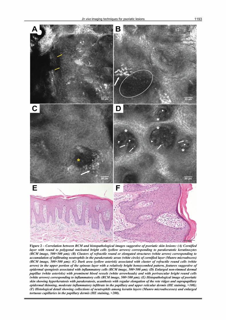
RCM examination of epidermis (Figure 2, A–C) exhibited a thick cornified layer with round or polygonal nucleated bright cells corresponding to parakeratotic keratinocytes, sometimes adjacent to highly refractile, elongated structures clustered together, corresponding to accumulation of infiltrating neutrophils in the cornified layer (Munro microabscesses). Epidermal thickness was increased but the granular layer was reduced or even absent in some areas. In the upper portion of the spinous layer with a relatively bright honeycombed pattern we detected dark areas associated with clusters of refractile round cells, which had no corresponding structure in conventional histopathology examination. Also, in certain lesional regions the dermal papillae were visible in the upper part of epidermis.
In comparison to healthy skin, lesional dermal papillae were significantly enlarged without the bright papillary ring and separated by thinner epidermal tissue (Figure 3). Lesional dermal papillae are filled with dilated and tortuous capillaries with increased blood flow in real time imaging. Around the enlarged vessels are visible bright round cells corresponding to perivascular inflammatory cell infiltration (Figures 3 and 2D).
Blood count and serum chemistry were within normal limits.
Clinical appearance, dermoscopy features and RCM findings were suggestive of psoriasis. A biopsy of the skin lesion previously investigated was performed.
Histopathology analysis confirmed the diagnosis of psoriasis vulgaris and revealed the presence of hyper-keratosis and parakeratosis with sparse collections of neutrophils among keratin layers, acanthosis with elongated rete ridges uniformly thickened and fused in the basal part, suprapapillary epidermal thinning, enlarged tortuous capillaries in the papillary dermis and moderate inflam-matory infiltrate in the papillary and upper reticular dermis (Figure 2, E and F). Special stains have not shown microbial (Giemsa stain) or fungal (PAS stain) colonies.
Topical treatment with corticosteroids, emollients and moisturizers was recommended, and psoriatic lesions cleared almost completely within the next four weeks.
In vivo imaging techniques for psoriatic lesions
1193
Figure 2 – Correlation between RCM and histopathological images suggestive of psoriatic skin lesions: (A) Cornified layer with round to polygonal nucleated bright cells (yellow arrows) corresponding to parakeratotic keratinocytes (RCM image, 500×500 μm); (B) Clusters of refractile round or elongated structures (white arrow) corresponding to accumulation of infiltrating neutrophils in the parakeratotic areas (white circle) of cornified layer (Munro microabscess) (RCM image, 500×500 μm); (C) Dark area (yellow asterisk) associated with cluster of refractile round cells (white arrow) in the upper portion of the spinous layer with a relatively bright honeycombed pattern, features suggestive of epidermal spongiosis associated with inflammatory cells (RCM image, 500×500 μm); (D) Enlarged non-rimmed dermal papillae (white asterisks) with prominent blood vessels (white arrowheads) and with perivascular bright round cells (white arrows) corresponding to inflammatory cells (RCM image, 500×500 μm); (E) Histopathological image of psoriatic skin showing hyperkeratosis with parakeratosis, acanthosis with regular elongation of the rete ridges and suprapapillary epidermal thinning, moderate inflammatory infiltrate in the papillary and upper reticular dermis (HE staining, ×100); (F) Histological detail showing collections of neutrophils among keratin layers (Munro microabscesses) and enlarged tortuous capillaries in the papillary dermis (HE staining, ×200).

Constantin Căruntu et al.
1194
Figure 3 – RCM images of the dermo-epidermal junction from lesional and healthy skin. (A) RCM mosaic of 1.5×1.5 mm from lesional skin revealing enlarged dermal papillae without the bright papillary ring and separated by thinner epidermal tissue; (B) Detail of previous RCM mosaic showing dermal papillae (white asterisks) filled with dilated and tortuous capillaries (white arrowheads) and bright round cells around the enlarged vessels, corresponding to inflammatory cells (white arrows); (C) RCM mosaic of 1.5×1.5 mm from healthy skin showing rimmed dermal papillae with normal diameter; (D) Detail of previous RCM mosaic showing dermal papillae (white asterisks) with small capillary loops visible as dark round structures (white arrowheads).
Discussion
Psoriasis is an inflammatory skin disease, with a polymorphic clinical appearance that often requires a histopathological evaluation to confirm the diagnosis. Modern techniques for in vivo imaging of skin structure, such as RCM and dermoscopy, could be very useful tools for diagnosis and monitoring of psoriatic lesions.
In the present case, dermoscopy showed the presence of dotted vessels and superficial white scales regularly distributed over a light red background, previous studies showing that this dermoscopic pattern can reliably predict the diagnosis of psoriasis vulgaris [5, 6, 22, 23, 25, 26].
In vivo RCM allows the non-invasive, high-resolution examination of the skin structure. Recent studies demons-trated that RCM examination allows the identification of the main histological features of psoriatic lesions and
revealed a high degree of correlation between RCM findings and routine histology [5, 7, 17, 29, 30, 35]. In the present case, RCM examination of epidermis showed hyperkeratotic stratum corneum, parakeratosis, accumu-lation of infiltrating neutrophils in the parakeratotic areas of cornified layer (Munro microabscesses), acanthosis with normal honeycomb pattern of epidermis and reduction/ absence of granular layer.
Dermal papillae were visible in the upper part of epi-dermis corresponding to suprapapillary epidermal thinning. At the dermo-epidermal junction, psoriatic lesion showed the enlargement of dermal papillae compared to unin-volved skin, the tortuosity and enlargement of blood vessels and perivascular inflammatory cell infiltration.
Moreover, RCM revealed some micromorphological aspects without corresponding features in conventional histopathological examination. At the dermo-epidermal
In vivo imaging techniques for psoriatic lesions
1195
junction, the bright papillary rings of dermal papillae were reduced or absent. Previous studies explained this feature by epidermal hyperproliferation and upward spread of melanocytes [5], inhibition of melanogenesis and/or increased melanocyte apoptosis [16, 36]. Another aspect highlighted only by RCM examination was the presence of dark areas with adjacent clusters of refractile round cells in the upper portion of the spinous layer, described by previous studies as epidermal spongiosis associated with inflammatory cells [16, 36], features suggestive of small spongiform pustules of Kogoj. This might be due to tissue processing and fixation in routine histopathology that could mask the presence of spongiosis [16]. Another explanation is that unlike the vertical sections of con-ventional histology, the images obtained by RCM are in horizontal plane and allow a full investigation of cutaneous tissue at a given depth in a wide skin area.
Conclusions
Our results highlight the possibility of using modern imaging techniques for in vivo diagnosis of psoriasis. There are specific dermoscopic features suggestive for psoriasis and RCM enables identification of most of the histological features of psoriatic lesions, and both dermoscopy and RCM allow a real time non-invasive examination of skin lesions offering the possibility to perform serial determinations.
Acknowledgments Research funded by grant PN-II-RU-TE-2011-3-0249
from the National University Research Council, Romania. Authors wish to thank Dr. Harillaq Kaleshi from Department of Dermatology, “Nicolae Paulescu” National Institute of Diabetes, Nutrition and Metabolic Diseases, Bucharest for his support during the course of this work.
References [1] Raychaudhuri SP, Farber EM, The prevalence of psoriasis in
the world, J Eur Acad Dermatol Venereol, 2001, 15(1):16–17. [2] Christophers E, Psoriasis – epidemiology and clinical spectrum,
Clin Exp Dermatol, 2001, 26(4):314–320. [3] Naldi L, Epidemiology of psoriasis, Curr Drug Targets Inflamm
Allergy, 2004, 3(2):121–128. [4] Schön MP, Boehncke WH, Psoriasis, N Engl J Med, 2005,
352(18):1899–1912. [5] Wolberink EA, van Erp PE, Teussink MM, van de Kerkhof PC,
Gerritsen MJ, Cellular features of psoriatic skin: imaging and quantification using in vivo reflectance confocal microscopy, Cytometry B Clin Cytom, 2011, 80(3):141–149.
[6] Lallas A, Kyrgidis A, Tzellos TG, Apalla Z, Karakyriou E, Karatolias A, Lefaki I, Sotiriou E, Ioannides D, Argenziano G, Zalaudek I, Accuracy of dermoscopic criteria for the diagnosis of psoriasis, dermatitis, lichen planus and pityriasis rosea, Br J Dermatol, 2012, 166(6):1198–1205.
[7] Ardigò M, Agozzino M, Longo C, Conti A, Di Lernia V, Berardesca E, Pellacani G, Psoriasis plaque test with confocal microscopy: evaluation of different microscopic response pathways in NSAID and steroid treated lesions, Skin Res Technol, 2013, 19(4):417–423.
[8] Henseler T, Christophers E, Psoriasis of early and late onset: characterization of two types of psoriasis vulgaris, J Am Acad Dermatol, 1985, 13(3):450–456.
[9] Naldi L, Parazzini F, Peli L, Chatenoud L, Cainelli T, Dietary factors and the risk of psoriasis. Results of an Italian case-control study, Br J Dermatol, 1996, 134(1):101–106.
[10] Tsankov N, Kazandjieva J, Drenovska K, Drugs in exacer-bation and provocation of psoriasis, Clin Dermatol, 1998, 16(3):333–351.
[11] Naldi L, Chatenoud L, Linder D, Belloni Fortina A, Peserico A, Virgili AR, Bruni PL, Ingordo V, Lo Scocco G, Solaroli C, Schena D, Barba A, Di Landro A, Pezzarossa E, Arcangeli F, Gianni C, Betti R, Carli P, Farris A, Barabino GF, La Vecchia C, Cigarette smoking, body mass index, and stressful life events as risk factors for psoriasis: results from an Italian case-control study, J Invest Dermatol, 2005, 125(1):61–67.
[12] Brauchli YB, Jick SS, Curtin F, Meier CR, Association between beta-blockers, other antihypertensive drugs and psoriasis: population-based case-control study, Br J Dermatol, 2008, 158(6):1299–1307.
[13] Mak RK, Hundhausen C, Nestle FO, Progress in understanding the immunopathogenesis of psoriasis, Actas Dermosifiliogr, 2009, 100(Suppl 2):2–13.
[14] Roberson ED, Bowcock AM, Psoriasis genetics: breaking the barrier, Trends Genet, 2010, 26(9):415–423.
[15] Căruntu C, Grigore C, Căruntu A, Diaconeasa A, Boda D, The role of stress in skin diseases, Medicina Internă (Internal Medicine), 2011, 8(3):73–84.
[16] Ardigo M, Cota C, Berardesca E, González S, Concordance between in vivo reflectance confocal microscopy and histology in the evaluation of plaque psoriasis, J Eur Acad Dermatol Venereol, 2009, 23(6):660–667.
[17] Zhong LS, Wei ZP, Liu YQ, Sensitivity and specificity of Munro microabscess detected by reflectance confocal microscopy in the diagnosis of psoriasis vulgaris, J Dermatol, 2012, 39(3):282–283.
[18] Wright V, Roberts MC, Hill AG, Dermatological manifestations in psoriatic arthritis: a follow-up study, Acta Derm Venereol, 1979, 59(3):235–240.
[19] Mease PJ, Menter MA, Quality-of-life issues in psoriasis and psoriatic arthritis: outcome measures and therapies from a dermatological perspective, J Am Acad Dermatol, 2006, 54(4): 685–704.
[20] Ianoşi S, Stoicescu I, Ianoşi G, Neagoe D, Georgescu CV, The study of CD20 and CD45.Ro antibodies in the inflam-matory infiltrate involved in acne and seborrheic dermatitis, Rom J Morphol Embryol, 2007, 48(3):285–289.
[21] Toussaint S, Kamino H, Noninfectious erythematous, papular, and squamous diseases. In: Elder D, Elenitsas R, Jaworsky C, Johnson B Jr (eds), Lever’s histopathology of the skin, 8th edition, Lippincott–Raven, Philadelphia, 1997, 151–184.
[22] Vázquez-López F, Manjón-Haces JA, Maldonado-Seral C, Raya-Aguado C, Pérez-Oliva N, Marghoob AA, Dermoscopic features of plaque psoriasis and lichen planus: new obser-vations, Dermatology, 2003, 207(2):151–156.
[23] Vázquez-López F, Zaballos P, Fueyo-Casado A, Sánchez-Martín J, A dermoscopy subpattern of plaque-type psoriasis: red globular rings, Arch Dermatol, 2007, 143(12):1612.
[24] Vázquez-López F, Kreusch J, Marghoob AA, Dermoscopic semiology: further insights into vascular features by screening a large spectrum of nontumoral skin lesions, Br J Dermatol, 2004, 150(2):226–231.
[25] Zalaudek I, Argenziano G, Dermoscopy subpatterns of inflammatory skin disorders, Arch Dermatol, 2006, 142(6):808.
[26] Moscarella E, Longo C, Zalaudek I, Argenziano G, Piana S, Lallas A, Dermoscopy and confocal microscopy clues in the diagnosis of psoriasis and porokeratosis, J Am Acad Dermatol, 2013, 69(5):e231–e233.
[27] Diaconeasa A, Boda D, Neagu M, Constantin C, Căruntu C, Vlădău L, Guţu D, The role of confocal microscopy in the dermato-oncology practice, J Med Life, 2011, 4(1):63–74.
[28] Căruntu C, Boda D, Evaluation through in vivo reflectance confocal microscopy of the cutaneous neurogenic inflam-matory reaction induced by capsaicin in human subjects, J Biomed Opt, 2012, 17(8):085003.
[29] González S, Rajadhyaksha M, Rubinstein G, Anderson RR, Characterization of psoriasis in vivo by reflectance confocal microscopy, J Med, 1999, 30(5–6):337–356.
[30] Wolberink EA, van Erp PE, de Boer-van Huizen RT, van de Kerkhof PC, Gerritsen MJ, Reflectance confocal microscopy: an effective tool for monitoring ultraviolet B phototherapy in psoriasis, Br J Dermatol, 2012, 167(2):396–403.
[31] Rajadhyaksha M, González S, Zavislan JM, Anderson RR, Webb RH, In vivo confocal scanning laser microscopy of human skin II: advances in instrumentation and comparison with histology, J Invest Dermatol, 1999, 113(3):293–303.

Constantin Căruntu et al.
1196
[32] Esmaeili A 4th, Scope A, Halpern AC, Marghoob AA, Imaging techniques for the in vivo diagnosis of melanoma, Semin Cutan Med Surg, 2008, 27(1):2–10.
[33] Calzavara-Pinton P, Longo C, Venturini M, Sala R, Pellacani G, Reflectance confocal microscopy for in vivo skin imaging, Photochem Photobiol, 2008, 84(6):1421–1430.
[34] Altintas AA, Altintas MA, Ipaktchi K, Guggenheim M, Theodorou P, Amini P, Spilker G, Assessment of micro-circulatory influence on cellular morphology in human burn wound healing using reflectance-mode-confocal microscopy, Wound Repair Regen, 2009, 17(4):498–504.
[35] Archid R, Patzelt A, Lange-Asschenfeldt B, Ahmad SS, Ulrich M, Stockfleth E, Philipp S, Sterry W, Lademann J, Confocal laser-scanning microscopy of capillaries in normal and psoriatic skin, J Biomed Opt, 2012, 17(10):101511.
[36] Ardigo M, Malizewsky I, Dell’anna ML, Berardesca E, Picardo M, Preliminary evaluation of vitiligo using in vivo reflectance confocal microscopy, J Eur Acad Dermatol Venereol, 2007, 21(10):1344–1350.
Corresponding author Daniel Boda, MD, PhD, Dermatology Research Laboratory, “Carol Davila” University of Medicine and Pharmacy, 22–24 Grigore Manolescu Street, Sector 1, 0111234 Bucharest, Romania; Phone +40757–079 117, Fax +4021–222 13 10, e-mail: [email protected] Received: March 15, 2014
Accepted: November 25, 2014


![Rom J Morphol Embryol 2011, 52(1):69–74 R J M E … · Rom J Morphol Embryol 2011, 52(1) ... blished by the World Health Organization (WHO) Classification [1], ... rehydrated in](https://static.fdocuments.in/doc/165x107/5b6443407f8b9a687e8d1c3f/rom-j-morphol-embryol-2011-5216974-r-j-m-e-rom-j-morphol-embryol-2011.jpg)