Role of the Heat-Shock Response in the Life and Death of...
13
Role of the Heat-Shock Response in the Life and Death of Proteins ANU MATHEW AND RICHARD I. MORIMOTO a Department of Biochemistry, Molecular Biology and Cell Biology, Rice Institute for Biomedical Research, Northwestern University, Evanston, Illinois 60208 USA Changes in protein levels and the folded states of proteins are sensed by molecular chaperones and the degradative machinery that function to maintain cellular pro- tein homeostasis. The importance of these systems is highlighted by their high degree of functional and structural conservation across evolution, from prokaryotes to eukaryotes, and their essential roles in a variety of cellular functions. 1–3 The chap- erone network, proteases, and components of the degradative machinery are also linked by common genetic regulatory pathways. For example, many of the proteins involved in protein folding and degradation are also heat-shock proteins whose expression is induced when cells are stressed and accumulate denatured or mal- folded proteins. 1,3– 6 The family of heat-shock proteins (HSPs) encompasses a num- ber of functionally related proteins that are expressed constitutively and/or at elevated levels upon exposure of cells to a variety of stress conditions including ele- vated temperature, arsenite, heavy metals, amino acid analogues, and oxidants. 1,4,6 The chaperones and proteolytic systems often function together to determine the ultimate fates of proteins and are often coordinately regulated, as outlined in FIGURE 1. The heat-shock response regulates proteolysis by enhancing the expres- sion of proteolytic factors and proteases, as well as of chaperones which may func- tion as cofactors for proteolysis. 2,3,5 The stress response in both prokaryotes and eukaryotes is, in turn, regulated by the proteolytic machinery 7–10 (also A. Mathew, S. K. Mathur, and R. I. Morimoto, unpublished observations). This review will address the chaperone requirements in eukaryotic proteolysis and the regulation of eukaryotic chaperone expression by the ubiquitin–proteasome proteolytic system. A number of recent reviews have also addressed chaperone activities associated with proteolysis in prokaryotes and eukaryotes. 3,11,12 BIOCHEMICAL PROPERTIES OF MOLECULAR CHAPERONES Chaperones are involved at various stages in protein biogenesis, regulating their structure and function under normal physiological conditions, as well as dur- ing and following stress which results in protein unfolding and misfolding. The family of heat-shock proteins and molecular chaperones includes Hsp100, Hsp90, Hsp70 (dnaK), Hsp60 (GroEL), Hsp40 (dnaJ), and the small HSPs. These chaper- ones are abundant and ubiquitous and function constitutively in protein synthesis and folding, protein translocation into membrane compartments, and the assembly and disassembly of oligomers. 2,13 Chaperones often function in concert with other 99 a Address for correspondence: Dr. Richard I. Morimoto, Department of Biochemistry, Molecular and Cell Biology, Northwestern University, 2153 North Campus Drive, Evanston, IL 60208. Telephone: 847-491-3340; Fax: 847-491-4461; e-mail: [email protected]
Transcript of Role of the Heat-Shock Response in the Life and Death of...

Role of the Heat-Shock Response in theLife and Death of Proteins
ANU MATHEW AND RICHARD I. MORIMOTOa
Department of Biochemistry, Molecular Biology and Cell Biology, RiceInstitute for Biomedical Research, Northwestern University, Evanston,Illinois 60208 USA
Changes in protein levels and the folded states of proteins are sensed by molecularchaperones and the degradative machinery that function to maintain cellular pro-tein homeostasis. The importance of these systems is highlighted by their highdegree of functional and structural conservation across evolution, from prokaryotesto eukaryotes, and their essential roles in a variety of cellular functions.1–3 The chap-erone network, proteases, and components of the degradative machinery are alsolinked by common genetic regulatory pathways. For example, many of the proteinsinvolved in protein folding and degradation are also heat-shock proteins whoseexpression is induced when cells are stressed and accumulate denatured or mal-folded proteins.1,3–6 The family of heat-shock proteins (HSPs) encompasses a num-ber of functionally related proteins that are expressed constitutively and/or atelevated levels upon exposure of cells to a variety of stress conditions including ele-vated temperature, arsenite, heavy metals, amino acid analogues, and oxidants.1,4,6
The chaperones and proteolytic systems often function together to determinethe ultimate fates of proteins and are often coordinately regulated, as outlined inFIGURE 1. The heat-shock response regulates proteolysis by enhancing the expres-sion of proteolytic factors and proteases, as well as of chaperones which may func-tion as cofactors for proteolysis.2,3,5 The stress response in both prokaryotes andeukaryotes is, in turn, regulated by the proteolytic machinery7–10 (also A. Mathew,S. K. Mathur, and R. I. Morimoto, unpublished observations). This review willaddress the chaperone requirements in eukaryotic proteolysis and the regulationof eukaryotic chaperone expression by the ubiquitin–proteasome proteolytic system.A number of recent reviews have also addressed chaperone activities associated withproteolysis in prokaryotes and eukaryotes.3,11,12
BIOCHEMICAL PROPERTIES OF MOLECULAR CHAPERONES
Chaperones are involved at various stages in protein biogenesis, regulatingtheir structure and function under normal physiological conditions, as well as dur-ing and following stress which results in protein unfolding and misfolding. Thefamily of heat-shock proteins and molecular chaperones includes Hsp100, Hsp90,Hsp70 (dnaK), Hsp60 (GroEL), Hsp40 (dnaJ), and the small HSPs. These chaper-ones are abundant and ubiquitous and function constitutively in protein synthesisand folding, protein translocation into membrane compartments, and the assemblyand disassembly of oligomers.2,13 Chaperones often function in concert with other
99
aAddress for correspondence: Dr. Richard I. Morimoto, Department of Biochemistry,Molecular and Cell Biology, Northwestern University, 2153 North Campus Drive, Evanston,IL 60208. Telephone: 847-491-3340; Fax: 847-491-4461; e-mail: [email protected]

protein-folding activities such as protein disulfide isomerases and peptidylprolylisomerases (immunophilins).13 Molecular chaperones including Hsp90, Hsp70,Hip, p23, and immunophilins regulate steroid hormone aporeceptors and kinaseactivities.14–18 The activities of the Hsp70 and Hsp60 chaperones are also modu-lated by their associations with cochaperones.13 The prokaryotic cochaperonesDnaJ and GrpE stimulate the ATPase activity of Hsp70 (DnaK) and promote effi-cient nucleotide release, respectively. In eukaryotes multiple DnaJ homologues areexpressed in addition to other Hsp70 regulatory proteins such as Hip andBag1.19–20a Similarly, the ATPase activity of GroEL (bacterial homologue of hsp60)is modulated by GroES.13
Under conditions of stress, heat-shock proteins prevent inappropriate inter-and intramolecular interactions, which may lead to protein misfolding or aggre-gation.1,2,13,21,22 Prolonged association with chaperones may occur in the event thatproteins are irreparably defective in conformation or oligomer assembly.3 Variousactivities of the HSP family members are outlined in FIGURE 2. In vitro studies toelucidate chaperone activities have shown that Hsp90, Hsp70, Hsp60, and thesmall HSPs are effective in maintaining unfolded proteins in soluble nonnativestates conducive to refolding to native state.13,23,24 The Hsp70 and Hsp60 chaper-ones, together with their respective cochaperones and ATP, have the ability torefold nonnative intermediates to the final native state13 whether the substrate is anascent polypeptide25 or thermally unfolded.26,27 Malfolded proteins not rescuedby the action of these chaperones are likely to form aggregates. The Hsp100 chap-erones have the unique ability to disaggregate protein aggregates in an ATPdependent manner.28
100 ANNALS NEW YORK ACADEMY OF SCIENCES
FIGURE 1. Coordinate regulation of expression and activity of chaperones and proteolyticfactors.

THE UBIQUITIN–PROTEASOME SYSTEM OF PROTEIN DEGRADATION
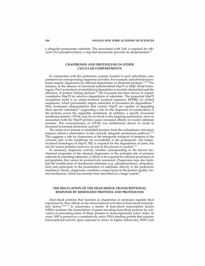
Denatured and misfolded proteins that appear during heat shock and otherstresses that cannot be refolded by the chaperone machinery must be rapidlyremoved by proteolysis. The ubiquitin–proteasome machinery is the major routeof removal of soluble cytosolic and nuclear proteins in higher eukaryotes and con-sequently has a central role in a variety of regulatory events of the cell.29–34 Thesteps involved in the ubiquitin–proteasome-dependent degradation of proteinsare summarized in FIGURE 3. Many of the proteins targeted to this system fordegradation by the proteasome (a multicatalytic protease complex) are modifiedby polyubiquitination. However, it must be noted that not all proteasome sub-strates are ubiquitinated35–37 and not all ubiquitinated proteins are metabolicallyunstable5 or targeted to the proteasome (some are targeted to the lysosome/vac-uole).38– 40 The 26S proteasome32,34,41,42 comprises a cylindrical 20S catalytic coreassembled from four heptameric rings. Deletion of even one of its subunits islethal to the cell, and, furthermore, mutations in these subunits often result in sen-sitivity to stress.43 A 19S particle that associates with each end of the 20S cylinderhas been suggested to participate in the unfolding of the substrate and facilitatingthe translocation of the substrate into the core.42 The composition of the 19S“mouth” particle is complex and contains a ubiquitin-binding site, severalATPases, and isopeptidases (to cleave and release ubiquitin chains and moi-eties),32,41 which all probably contribute to the selective nature of the proteasome.
Ubiquitination is mediated by a cascade of enzyme activities, namely, the ubiquitin-activating enzyme, E1, ubiquitin-conjugating enzymes (E2), and theubiquitin protein ligases (E3), as outlined in FIGURE 3.5 Ubiquitin is activated to anactive thiol ester intermediate by the action of E1. The activities of the E2 proteinsfacilitate covalent attachment of the activated ubiquitin to the lysine residues of
MATHEW & MORIMOTO: HEAT-SHOCK RESPONSE 101
FIGURE 2. Functions of the chaperones. Proteins in their native state (1) undergo unfold-ing as a result of stress. This transient unfolded state (2), can be prevented from aggregat-ing by association with the Hsp70, Hsp60, Hsp90, and small HSP proteins (3). Onceaggregate formation occurs, the activity of the Hsp100 proteins may facilitate disaggrega-tion (4). The transient unfolded intermediate of the protein can also be refolded by theactions of Hsp60, Hsp70, and their cochaperones, in the presence of ATP (5). The latter fac-tors mediate the folding of nascent polypeptides as well (6).

target protein substrates by formation of an isopeptide bond, which may or maynot be associated with an E3 activity. Polyubiquitin chains are normally added tothose substrates destined for degradation. The abundant protein ubiquitin isexpressed constitutively in all cells and expression is upregulated upon exposureto stress. At least two yeast E2 proteins, UBC4 and UBC5, are also heat-shockinduced and are responsible for most of the ubiquitin-conjugating activity duringstress.44 Exposure of yeast cells to cadmium results in significantly elevatedexpression of an alternate pair of E2 proteins, namely, UBC5 and UBC7.45 The exis-tence of multiple E2 and E3 enzymes that respond to different stresses, togetherwith the ability to form alternative ubiquitin chain conformations, may provide ahigh degree of substrate specificity.5,46 The fate of proteins that accumulate duringexposure of cells to stress is dependent on the interplay of chaperones and theubiquitin–proteasome system.3,12 The stress signal, in turn, leads to the transcrip-tional activation of genes encoding components for proteolysis and chaperoneactivity1–7,8a,10 (also, A. Mathew, S. K. Mathur, and R. I. Morimoto, unpublishedobservations).
102 ANNALS NEW YORK ACADEMY OF SCIENCES
FIGURE 3. The ubiquitin–proteasome pathway of protein degradation.

ROLE OF MOLECULAR CHAPERONES IN PROTEIN DEGRADATION
Molecular chaperones are required for normal energy-dependent proteindegradation in various prokaryotic systems, and furthermore, the levels of certainchaperones are often rate limiting to proteolysis.3,47– 49 Mutations in genes encodingchaperones result in both a general decrease in protein degradation and the con-stitutive activation of the heat-shock response, which leads to the elevated expres-sion of chaperones and proteases.50 Chaperones may function both in the refoldingof misfolded proteins and in the presentation of substrates to proteolytic activitiesif the substrate cannot be refolded. The fate of chaperone-bound substrates can bealtered by other chaperones or modulators of chaperone activities; for example,the affinity of GroEL for substrates may be increased by either its association withtrigger factor or by heat-shock-induced phosphorylation.51,52
Several lines of genetic evidence point to a link between the chaperone activitiesand ubiquitin–proteasome-mediated protein degradation. A ubiquitin-processingenzyme was identified as a suppressor of an Hsp70 mutation,21 suggesting an inter-action between Hsp70 and the ubiquitination machinery. A more direct role forchaperones in the degradative process has been suggested from yeast mutations inHsp70, which result in a general reduction in ubiquitin–proteasome-mediateddegradation of proteins. The phenotype observed for these Hsp70 mutations is sim-ilar to that described for mutations in ubc4/ubc5.1,44 Likewise, mutation of the yeastDnaJ homologue, Ydj1, affects ubiquitination of abnormal and short-lived proteins,whereas mutation of the other yeast DnaJ homologue, Sis, primarily affects protea-somal digestion of ubiquitinated proteins.3,53
Exposure of cultured mammalian cells to proteasome inhibitors leads to theaccumulation of polyubiquitinated proteins that are found to be associated withHsp90 and Hsp70. Coassociated polyubiquitinated proteins are released fromboth Hsp90 and Hsp70 immunoprecipitates upon ATP hydrolysis, which is anindication of the specificity of this interaction (A. Mathew, S. K. Mathur, and R. I.Morimoto, unpublished observations). The association of polyubiquitinated pro-teins with Hsp70 and Hsp90 is lost upon removal of the proteasome inhibitors,which reveals that the interaction is transient and reversible and that the chaper-one-associated substrates are either degraded or refolded. Direct chaperone asso-ciation may be required for presentation of specific substrates to theubiquitination machinery; however, the evidence for such a requirement isderived from in vitro experiments using lysates. Immunodepletion of Hsc70 froman in vitro degradation assay revealed a requirement for this chaperone in ubiqui-tination of some, not all, protein substrates.54 Chaperone association with specificubiquitin–proteasome substrates has been detected and proposed to be requiredto expose sites for ubiquitin modification.54 Association of Hsp90, Hsp70, andother components of the Hsp90 heterocomplex with a denatured substrate resultsin either refolding or degradation; prolonged association with Hsp90 favored thelatter.55 Specific inhibition of Hsp90 release (and consequently of the other mem-bers of the chaperone complex) with the drug herbimycin A also results inincreased degradation of other proteins known to normally associate with Hsp90(Raf-1 and transmembrane receptor tyrosine kinases).55,56 Specific Hsp70-associatedproteins shown to be degraded by the proteasome include CFTR56a,56b and theapolipoprotein B100.56c Experiments in yeast provide additional support for theroles of the chaperones Ydj1 and Hsp70 homologues in proteolysis. Both chaper-ones associate with the G1 cyclin, Cln3, an important yeast cell cycle regulator and
MATHEW & MORIMOTO: HEAT-SHOCK RESPONSE 103
a ubiquitin–proteasome substrate. The association with Ydj1 is required for effi-cient Cln3 phosphorylation, a step that necessarily precedes its ubiquitination.57
CHAPERONES AND PROTEOLYSIS IN OTHER CELLULAR COMPARTMENTS
In conjunction with the proteolytic systems located in each subcellular com-partment are corresponding chaperone activities. For example, mitochondrial pro-teases require chaperones for efficient degradation of abnormal proteins.11,58,59 Forinstance, in the absence of functional mitochondrial Hsp70 or Mdj1 (DnaJ homo-logue), Pim1 proteolysis of misfolded polypeptides is severely diminished and theefficiency of protein folding declines.59 The lysosome has been shown to requireconstitutive Hsp70 for selective degradation of substrates. The purported Hsp70recognition motif is an amino-terminal localized sequence, KFERQ (or relatedsequences), which presumably targets substrates to lysosomes for degradation.60
Only lysosomal subpopulations that contain Hsp70 are capable of degradingthese specific substrates,61 supporting a role for the chaperone in translocation ofthe proteins across the organellar membrane. In addition, a specific lysosomalmembrane protein, LPG96, may be involved in this targeting mechanism, since itsassociation with the Hsp70 protein causes increased affinity for model substrateproteins. The overexpression of LPG96 was furthermore shown to result inincreased lysosomal proteolytic activity.62
The removal of mutant or misfolded proteins from the endoplasmic reticulumrequires selective elimination via the cytosolic ubiquitin–proteasome pathway.37,63
This suggests a role for chaperones in the retrograde transport of proteins to thecytosolic side of the membrane for accessibility to the proteasome. The lumen-localized homologue of Hsp70, BiP, is required for the degradation of some, butnot all, lumen proteins; however, its role in this process is unclear.3,13
In summary, chaperone activity, whether corresponding to the known bio-chemical properties of the classical chaperones or the potential role of proteasesubunits in unfolding substrates, is likely to be required for efficient proteolysis ofpolypeptides that cannot be productively renatured. Chaperones may also facili-tate the modification of obsolescent substrates (e.g., phosphorylation, ubiquitina-tion) and participate in the presentation of substrates directly to the proteolyticmachinery. Hence, chaperones constitute a major factor in the protein quality con-trol mechanism, which has recently been described as a triage system.11
THE REGULATION OF THE HEAT-SHOCK TRANSCRIPTIONALRESPONSE BY MISFOLDED PROTEINS AND PROTEOLYSIS
Heat-shock proteins that function as chaperones or proteases regulate theirexpression by their effects on the stress-induced activities of heat-shock transcrip-tion factors.8,9,64– 66 In eukaryotes, a family of heat-shock transcription factors(HSFs) mediates the transcription of genes encoding heat-shock proteins by acti-vation of preexisting forms of these proteins to transcriptionally active states. Inyeast, HSF is present as a constitutively active DNA-binding protein that acquirestranscriptional activity upon exposure to stress. In higher eukaryotes, HSFs exist
104 ANNALS NEW YORK ACADEMY OF SCIENCES

in a non-DNA-bound state, which upon heat-shock or other forms of stress under-goes oligomerization and binds DNA. HSF is trimeric in its active state and bindsto a consensus target sequence, the heat-shock element (HSE), present in promoterregions of heat-shock genes.67 The attenuation of HSF activity is likely to involvenegative regulation by Hsp70 without affecting the level of HSF.65,66 This is likelyto be at least partly due to repression of transcriptional activity by direct chaper-one association with the HSF transactivation domain, as has been shown forHSF1.67a Because the level of free Hsp70 seems to be critical and Hsp70 interactswith stress-damaged proteins, the processes by which such damaged proteins aredisposed of may be important to this homeostatic control mechanism. Of the fourHSFs in vertebrates, HSF1 corresponds to the ubiquitous heat-shock-induced fac-tor; HSF2 activity has been detected in conditions of differentiation; HSF3 exhibitsthe properties of an extreme stress-induced factor activated under non-stress con-ditions by association with c-Myb, and the expression of which is required forHSF1 activation in avian systems; and HSF4 lacks the activity of a positive activa-tor and may be a negative regulator of heat-shock gene expression.65,66,67b,67c
MATHEW & MORIMOTO: HEAT-SHOCK RESPONSE 105
FIGURE 4. Regulation of chaperone expression by proteasome activity. Malfolded,mutant, and short-lived proteins are polyubiquitinated (1) and normally degraded by theproteasome (2). Inhibition of proteasome activity by the addition of MG132, lactacystin, orhemin results in accumulation of polyubiquitinated substrates (3). HSF2 is activated fromthe inert dimer to DNA-binding trimer in response to this accumulation (4). HSF2 inducesexpression of molecular chaperones such as Hsp70, Hsp90, and Hdj-1 (5). These molecularchaperones associate with the nonnative polyubiquitinated substrates (6) and are suggestedto prevent their aggregation (7), maintaining the polyubiquitinated proteins in an interme-diate folded state primed for degradation upon resumption of proteasome activity.
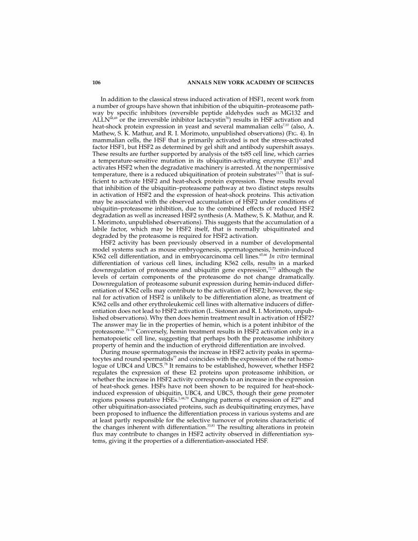
In addition to the classical stress induced activation of HSF1, recent work froma number of groups have shown that inhibition of the ubiquitin–proteasome path-way by specific inhibitors (reversible peptide aldehydes such as MG132 andALLN68,69 or the irreversible inhibitor lactacystin70) results in HSF activation andheat-shock protein expression in yeast and several mammalian cells7,10 (also, A.Mathew, S. K. Mathur, and R. I. Morimoto, unpublished observations) (FIG. 4). Inmammalian cells, the HSF that is primarily activated is not the stress-activatedfactor HSF1, but HSF2 as determined by gel shift and antibody supershift assays.These results are further supported by analysis of the ts85 cell line, which carriesa temperature-sensitive mutation in its ubiquitin-activating enzyme (E1)71 andactivates HSF2 when the degradative machinery is arrested. At the nonpermissivetemperature, there is a reduced ubiquitination of protein substrates11,71 that is suf-ficient to activate HSF2 and heat-shock protein expression. These results revealthat inhibition of the ubiquitin–proteasome pathway at two distinct steps resultsin activation of HSF2 and the expression of heat-shock proteins. This activationmay be associated with the observed accumulation of HSF2 under conditions ofubiquitin–proteasome inhibition, due to the combined effects of reduced HSF2degradation as well as increased HSF2 synthesis (A. Mathew, S. K. Mathur, and R.I. Morimoto, unpublished observations). This suggests that the accumulation of alabile factor, which may be HSF2 itself, that is normally ubiquitinated anddegraded by the proteasome is required for HSF2 activation.
HSF2 activity has been previously observed in a number of developmentalmodel systems such as mouse embryogenesis, spermatogenesis, hemin-inducedK562 cell differentiation, and in embryocarcinoma cell lines.65,66 In vitro terminaldifferentiation of various cell lines, including K562 cells, results in a markeddownregulation of proteasome and ubiquitin gene expression,72,73 although thelevels of certain components of the proteasome do not change dramatically.Downregulation of proteasome subunit expression during hemin-induced differ-entiation of K562 cells may contribute to the activation of HSF2; however, the sig-nal for activation of HSF2 is unlikely to be differentiation alone, as treatment ofK562 cells and other erythroleukemic cell lines with alternative inducers of differ-entiation does not lead to HSF2 activation (L. Sistonen and R. I. Morimoto, unpub-lished observations). Why then does hemin treatment result in activation of HSF2?The answer may lie in the properties of hemin, which is a potent inhibitor of theproteasome.74–76 Conversely, hemin treatment results in HSF2 activation only in ahematopoietic cell line, suggesting that perhaps both the proteasome inhibitoryproperty of hemin and the induction of erythroid differentiation are involved.
During mouse spermatogenesis the increase in HSF2 activity peaks in sperma-tocytes and round spermatids77 and coincides with the expression of the rat homo-logue of UBC4 and UBC5.78 It remains to be established, however, whether HSF2regulates the expression of these E2 proteins upon proteasome inhibition, orwhether the increase in HSF2 activity corresponds to an increase in the expressionof heat-shock genes. HSFs have not been shown to be required for heat-shock-induced expression of ubiquitin, UBC4, and UBC5, though their gene promoterregions possess putative HSEs.1,44,79 Changing patterns of expression of E280 andother ubiquitination-associated proteins, such as deubiquitinating enzymes, havebeen proposed to influence the differentiation process in various systems and areat least partly responsible for the selective turnover of proteins characteristic ofthe changes inherent with differentiation.33,81 The resulting alterations in proteinflux may contribute to changes in HSF2 activity observed in differentiation sys-tems, giving it the properties of a differentiation-associated HSF.
106 ANNALS NEW YORK ACADEMY OF SCIENCES

Changes in the flux of proteins in the cell, whether induced by normal physio-logical signals such as differentiation, or by stress conditions that lead to the accu-mulation of oxidatively modified proteins (which affect proteasome function82) canbe immediately transmitted to the chaperone network by activation of HSF2/HSF,resulting in increased expression of heat-shock proteins. During inhibition of pro-teasome activity, heat-shock protein synthesis remains elevated, but is rapidlyreversed upon relief of the inhibition (A. Mathew, S. K. Mathur, and R. I. Morimoto,unpublished observations). As illustrated in FIGURE 4, a consequence of diminishedproteasome activity is the accumulation of polyubiquitinated proteins that may beprone to aggregation. The HSPs would serve to prevent aggregation, facilitate pro-tein repair/refolding, participate in ubiquitination/deubiquitination, and preparefor presentation to the proteasome for degradation. In support of such a scenario,HSP association with polyubiquitinated proteins is observed when proteasomeactivity is inhibited, and once proteasome activity is restored this association is lost.
REFERENCES
1. LINDQUIST, S. & E. A. CRAIG. 1988. The heat-shock proteins. Annu. Rev. Genet. 22:631–677.
2. MORIMOTO, R. I., A. TISSIERES & C. GEORGOPOULOS. 1994. Progress and perspectives on thebiology of heat shock proteins and molecular chaperones. In The Biology of Heat ShockProteins and Molecular Chaperones. R. I. Morimoto, A. Tissieres & C. Georgopoulos,Eds.: 1–30. Cold Spring Harbor Laboratory Press. Cold Spring Harbor, NY.
3. SHERMAN, M. Y. S. & A. L. GOLDBERG. 1996. Involvement of molecular chaperones inintracellular protein breakdown. In Stress-Inducible Cellular Responses. U. Feige, R.I. Morimoto, I. Yahara & B. Polla, Eds.: 57–78. Birkhauser Verlag. Boston.
4. CRAIG, E. A. & C. A. GROSS. 1991. Is Hsp70 the cellular thermometer? Trends Biochem.Sci. 16: 135–140.
5. JENTSCH, S. 1992. The ubiquitin-conjugation system. Annu. Rev. Genet. 26: 179–207.6. MORIMOTO, R. I., K. D. SARGE & K. ABRAVAYA. 1992. Transcriptional regulation of heat
shock genes. A paradigm for inducible genomic responses. J. Biol. Chem. 267:21987–21990.
7. BUSH, K. T., A. L. GOLDBERG & S. K. NIGAM. 1997. Proteasome inhibition leads to a heat-shock response, induction of endoplasmic reticulum chaperones, and thermotoler-ance. J. Biol. Chem. 272: 9086–9092.
8. HERMAN, C., D. THEVENET, R. D’ARI & P. BOULOC. 1995. Degradation of σ32, the heatshock regulator in Escherichia coli, is governed by HflB. Proc. Natl. Acad. Sci. USA 92:3516–3520.
8a. LEE, D. H. & A. L. GOLDBERG. 1998. Proteasome inhibitors cause induction of heat shockproteins and trehalose, which together confer thermotolerance in Saccharomyces cerevisiae. Mol. Cell. Biol. 18: 30–38.
9. TOMOYASU, T., J. GAMER, B. BUKAU, M. KANEMORI, H. MORI, A. J. RUTMAN, A. B.OPPENHEIM, T. YURA, K. YAMANAKA, H. NIKI, S. HIRAGA & T. OGURA. 1995. Escherichiacoli FtsH is a membrane-bound, ATP-dependent protease which degrades the heat-shock transcription factor σ32. EMBO J. 14: 2551–2560.
10. ZHOU, M., X. WU & H. N. GINSBERG. 1996. Evidence that a rapidly turning over protein,normally degraded by proteasomes, regulates Hsp72 gene transcription in HepG2cells. J. Biol. Chem. 271: 24769–24775.
11. GOTTESMAN, S., S. WICKNER & M. R. MAURIZI. 1997. Protein quality control: Triage bychaperones and proteases. Genes Dev. 11: 815–823.
12. HAYES, S. A. & J. F. DICE. 1996. Roles of molecular chaperones in protein degradation. J. CellBiol. 132: 255–258.
MATHEW & MORIMOTO: HEAT-SHOCK RESPONSE 107

13. GETHING, M. J. & J. SAMBROOK. 1992. Protein folding in the cell. Nature 355: 33–45.14. CHEN, S., V. PRAPAPANICH, R. A. RIMERMAN, B. HONORE & D. F. SMITH. 1996. Interactions
of p60, a mediator of progesterone receptor assembly, with heat shock proteins Hsp90and Hsp70. Mol. Endocrinol. 10: 682–693.
15. PRATT, W. B. 1993. The role of heat shock proteins in regulating the function, folding,and trafficking of the glucocorticoid receptor. J. Biol. Chem. 268: 21455–21458.
16. PRATT, W. B. & M. J. WELSH. 1994. Chaperone functions of the heat shock proteins asso-ciated with steroid receptors. Semin. Cell Biol. 5: 83–93.
17. SMITH, D. F. 1993. Dynamics of heat shock protein 90-progesterone receptor binding andthe disactivation loop model for steroid receptor complexes. Mol. Endocrinol. 7:1418–1429.
18. VAN DER STRATEN, A., C. ROMMEL, B. DICKSON & E. HAFEN. 1997. The heat shock protein83 (Hsp83) is required for Raf-mediated signalling in Drosophila. EMBO J. 16:1961–1969.
19. CAPLAN, A. J., D. M. CYR & M. G. DOUGLAS. 1993. Eukaryotic homologues of Escherichiacoli dnaJ: A diverse protein family that functions with Hsp70 stress proteins. Mol. Biol.Cell. 4: 555–563.
20. HOHFELD, J., Y. MINAMI & F. U. HARTL. 1995. Hip, a novel cochaperone involved in theeukaryotic Hsc70/Hsp40 reaction cycle. Cell 83: 589–598.
20a. TAKAYAMA, S., D. N. BIMSTON, S. MATSUZAWA, B. C. FREEMAN, C. AIME-SEMPE, Z. XIE, R. I.MORIMOTO & J. C. REED. 1997. BAG-1 modulates the chaperone activity ofHsp70/Hsc70. EMBO J. 16: 4887–4896.
21. CRAIG, E. A., B. K. BAXTER, J. BECKER, J. HALLADAY & T. ZIEGELHOFFER. 1994. CytosolicHsp70s of Saccharomyces cerevisiae: Roles in protein synthesis, protein translocation,proteolysis, and regulation. In The Biology of Heat Shock Proteins and MolecularChaperones. R. I. Morimoto, A. Tissieres & C. Georgopoulos, Eds.: 31–52. Cold SpringHarbor Laboratory Press. Cold Spring Harbor, NY.
22. ROTHMAN, J. E. 1989. Polypeptide chain binding proteins: Catalysts of protein foldingand related processes in cells. Cell. 59: 591–601.
23. FREEMAN, B. C. & R. I. MORIMOTO. 1996. The human cytosolic molecular chaperonesHsp90, Hsp70 (hsc70) and hdj-1 have distinct roles in recognition of a non-native pro-tein and protein refolding. EMBO J. 15: 2969–2979.
24. JAKOB, U. & J. BUCHNER. 1994. Assisting spontaneity: The role of Hsp90 and small Hspsas molecular chaperones. Trends Biochem. Sci. 19: 205–211.
25. HARTL, F. U. 1996. Molecular chaperones in cellular protein folding. Nature 381:571–579.
26. SKOWYRA, D., C. GEORGOPOULOS & M. ZYLICZ. 1990. The E. coli dnaK gene product, theHsp70 homolog, can reactivate heat-inactivated RNA polymerase in an ATP hydroly-sis-dependent manner. Cell 62: 939–944.
27. ZIEMIENOWICZ, A., D. SKOWYRA, R. J. ZEILSTRA, O. FAYET, C. GEORGOPOULOS & M. ZYLICZ.1993. Both the Escherichia coli chaperone systems, GroEL/GroES and DnaK/DnaJ/GrpE,can reactivate heat-treated RNA polymerase. Different mechanisms for the same activ-ity. J. Biol. Chem. 268: 25425–25431.
28. SCHIRMER, E. C., J. R. GLOVER, M. A. SINGER, & S. LINDQUIST. 1996. HSP100/Clp proteins:A common mechanism explains diverse functions. Trends Biochem. Sci. 19: 87–89.
29. CIECHANOVER, A. 1994. The ubiquitin-proteasome proteolytic pathway. Cell 79: 13–21.30. GOLDBERG, A. L., M. GACZYNSKA, E. GRANT, M. MICHALEK & K. L. ROCK. 1995. Functions
of the proteasome in antigen presentation. Cold Spring Harbor Symp. Quant. Biol. 60:479–490.
31. HERSHKO, A. & A. CIECHANOVER. 1992. The ubiquitin system for protein degradation.Annu. Rev. Biochem. 61: 761–807.
32. HILT, W. & D. H. WOLF. 1996. Proteasomes: Destruction as a programme. TrendsBiochem. Sci. 21: 96–102.
33. HOCHSTRASSER, M. 1995. Ubiquitin, proteasomes, and the regulation of intracellular pro-tein degradation. Curr. Opin. Cell Biol. 7: 215–223.
34. RECHSTEINER, M., L. HOFFMAN & W. DUBIEL. 1993. The multicatalytic and 26 S proteases.J. Biol. Chem. 268: 6065–6068.
108 ANNALS NEW YORK ACADEMY OF SCIENCES

35. JARIEL, E. I., M. PARIAT, F. MARTIN, S. CARILLO, C. SALVAT & M. PIECHACZYK. 1995.Ubiquitinylation is not an absolute requirement for degradation of c-Jun protein bythe 26 S proteasome. J. Biol. Chem. 270: 11623–11627.
36. MURAKAMI, Y., S. MATSUFUJI, T. KAMEJI, S. HAYASHI, K. IGARASHI, T. TAMURA, K. TANAKA &A. ICHIHARA. 1992. Ornithine decarboxylase is degraded by the 26S proteasome with-out ubiquitination. Nature 360: 597–599.
37. WERNER, E. D., J. L. BRODSKY & A. A. MCCRACKEN. 1996. Proteasome-dependent endo-plasmic reticulum-associated protein degradation: An unconventional route to afamiliar fate. Proc. Natl. Acad. Sci. USA 93: 13797–13801.
38. HICKE, L. & H. RIEZMAN. 1996. Ubiquitination of a yeast plasma membrane receptor sig-nals its ligand-stimulated endocytosis. Cell 84: 277–287.
39. ROTH, M. G., C. DOYLE, J. SAMBROOK & M. J. GETHING. 1986. Heterologous transmem-brane and cytoplasmic domains direct functional chimeric influenza virus hemagglu-tinins into the endocytic pathway. J. Cell Biol. 102: 1271–1283.
40. STROUS, G. J., K. P. VAN, R. GOVERS, A. CIECHANOVER & A. L. SCHWARTZ. 1996. The ubiq-uitin conjugation system is required for ligand-induced endocytosis and degradationof the growth hormone receptor. EMBO J. 15: 3806–3812.
41. COUX, O., K. TANAKA & A. L. GOLDBERG. 1996. Structure and functions of the 20S and 26Sproteasomes. Annu. Rev. Biochem. 65: 801–847.
42. GOLDBERG, A. L. 1995. Functions of the proteasome: The lysis at the end of the tunnel[comment]. Science 268: 522–523.
43. HEINEMEYER, W., J. A. KLEINSCHMIDT, J. SAIDOWSKY, C. ESCHER & D. H. WOLF. 1991.Proteinase yscE, the yeast proteasome/multicatalytic-multifunctional proteinase:Mutants unravel its function in stress induced proteolysis and uncover its necessityfor cell survival. EMBO J. 10: 555–562.
44. SEUFERT, W. & S. JENTSCH. 1990. Ubiquitin-conjugating enzymes UBC4 and UBC5 medi-ate selective degradation of short-lived and abnormal proteins. EMBO J. 9: 543–550.
45. JUNGMANN, J., H. A. REINS, C. SCHOBERT & S. JENTSCH. 1993. Resistance to cadmium medi-ated by ubiquitin-dependent proteolysis. Nature 361: 369–371.
46. SEUFERT, W., B. FUTCHER & S. JENTSCH. 1995. Role of a ubiquitin-conjugating enzyme indegradation of S- and M-phase cyclins. Nature 373: 78–81.
47. STRAUS, D., W. WALTER & C. A. GROSS. 1990. DnaK, DnaJ, and GrpE heat shock proteinsnegatively regulate heat shock gene expression by controlling the synthesis and sta-bility of σ32. Genes Dev. 4: 2202–2209.
48. STRAUS, D. B., W. A. WALTER & C. A. GROSS. 1988. Escherichia coli heat shock gene mutantsare defective in proteolysis. Genes Dev. 2: 1851–1858.
49. TILLY, K., J. SPENCE & C. GEORGOPOULOS. 1989. Modulation of stability of the Escherichiacoli heat shock regulatory factor sigma. J. Bacteriol. 171: 1585–1589.
50. GROSS, C. 1996. Function and regulation of the heat shock proteins. In Escherichia coliand Salmonella typhimurium. F. C. Neidhart, R. Curtiss III, J. L. Ingraham, et al., Eds.:1382–1399. American Society for Microbiology. Washington, DC.
51. KANDROR, O., M. SHERMAN, R. MOERSCHELL & A. L. GOLDBERG. 1997. Trigger factor asso-ciates with GroEL in vivo and promotes its binding to certain polypeptides. J. Biol.Chem. 272: 1730–1734.
52. KANDROR, O., M. SHERMAN, M. RHODE & A. L. GOLDBERG. 1995. Trigger factor is involvedin GroEL-dependent protein degradation in Escherichia coli and promotes binding ofGroEL to unfolded proteins. EMBO J. 14: 6021–6027.
53. LEE, D. H., M. Y. SHERMAN & A. L. GOLDBERG. 1996. Involvement of the molecular chap-erone Ydj1 in the ubiquitin-dependent degradation of short-lived and abnormal pro-teins in Saccharomyces cerevisiae. Mol. Cell. Biol. 16: 4773–4781.
54. BERCOVICH, B., I. STANCOVSKI, A. MAYER, N. BLUMENFELD, A. LASZLO, A. L. SCHWARTZ & A.CIECHANOVER. 1997. Ubiquitin-dependent degradation of certain protein substrates invitro requires the molecular chaperone Hsc70. J. Biol. Chem. 272: 9002–9010.
55. SCHNEIDER, C., L. L. SEPP, E. NIMMESGERN, O. OUERFELLI, S. DANISHEFSKY, N. ROSEN &F. U. HARTL. 1996. Pharmacologic shifting of a balance between protein refoldingand degradation mediated by Hsp90. Proc. Natl. Acad. Sci. USA 93: 14536–14541.
MATHEW & MORIMOTO: HEAT-SHOCK RESPONSE 109

56. SEPP, L. L., Z. MA, D. E. LEBWOHL, A. VINITSKY & N. ROSEN. 1995. Herbimycin A inducesthe 20 S proteasome- and ubiquitin-dependent degradation of receptor tyrosinekinases. J. Biol. Chem. 270: 16580–16587.
56a. WARD, C. L., S. OMURA & R. R. KOPITO. 1995. Degradation of CFTR by the ubiquitin–proteasome pathway. Cell 83: 121–127.
56b. YANG, Y., S. JANICH, J. A. COHN & J. M. WILSON. 1993. The common variant of cystic fibro-sis transmembrane conductance regulator is recognized by Hsp70 and degraded in apre-Golgi nonlysosomal compartment. Proc. Natl. Acad. Sci. USA 90: 9480–9484.
56c. FISHER, E. A., M. ZHOU, D. M. MITCHELL, X. WU, S. OMURA, H. WANG, A. L. GOLDBERG &H. N. GINSBERG. 1997. The degradation of apolipoprotein B100 is mediated by theubiquitin–proteasome pathway and involves heat shock protein 70. J. Biol. Chem. 272:20427–20434.
57. YAGLOM, J. A., A. L. GOLDBERG, D. FINLEY & M. Y. SHERMAN. 1996. The molecular chap-erone Ydj1 is required for the p34CDC28-dependent phosphorylation of the cyclinCln3 that signals its degradation. Mol. Cell. Biol. 16: 3679–3684.
58. STUART, R. A., D. M. CYR, E. A. CRAIG & W. NEUPERT. 1994. Mitochondrial molecularchaperones: Their role in protein translocation. Trends Biochem. Sci. 19: 87–92.
59. WAGNER, I., H. ARLT, D. L. VAN, T. LANGER & W. NEUPERT. 1994. Molecular chaperonescooperate with PIM1 protease in the degradation of misfolded proteins in mitochon-dria. EMBO J. 13: 5135–5145.
60. DICE, J. F., F. AGARRABERES, M. KIRVEN-BROOKS, L. J. TERLECKY & S. R. TERLECKY. 1994.Heat shock 70-kD proteins and lysosomal proteolysis. In The Biology of Heat ShockProteins and Molecular Chaperones. R. I. Morimoto, A. Tissieres & C. Georgopoulos,Eds.: 137–151. Cold Spring Harbor Laboratory Press. Cold Spring Harbor, NY.
61. CUERVO, A. M., J. F. DICE & E. KNECHT. 1997. A population of rat liver lysosomes respon-sible for the selective uptake and degradation of cytosolic proteins. J. Biol. Chem. 272:5606–5615.
62. CUERVO, A. M. & J. F. DICE. 1996. A receptor for the selective uptake and degradation ofproteins by lysosomes. Science 273: 501–503.
63. HILLER, M. M., A. FINGER, M. SCHWEIGER & D. H. WOLF. 1996. ER degradation of a mis-folded luminal protein by the cytosolic ubiquitin-proteasome pathway. Science 273:1725–1728.
64. GAMER, J., G. MULTHAUP, T. TOMOYASU, J. S. MCCARTY, S. RUDIGER, H. J. SCHONFELD,C. SCHIRRA, H. BUJARD & B. BUKAU. 1996. A cycle of binding and release of theDnaK, DnaJ and GrpE chaperones regulates activity of the Escherichia coli heatshock transcription factor σ32. EMBO J. 15: 607–617.
65. MORIMOTO, R. I., D. A. JURIVICH, P. E. KROEGER, S. K. MATHUR, S. P. MURPHY, A. NAKAI,K. SARGE, K. ABRAVAYA & L. T. SISTONEN. 1994. Regulation of heat shock gene tran-scription by a family of heat shock factors. In The Biology of Heat Shock Proteins andMolecular Chaperones. R. I. Morimoto, A. Tissieres & C. Georgopoulos, Eds.:417–455. Cold Spring Harbor Laboratory Press. Cold Spring Harbor, NY.
66. WU, C. 1995. Heat shock transcription factors: Structure and regulation. Annu. Rev. ofCell. Dev. Biol. 11: 441–469.
67. FERNANDES, M., T. O’BRIEN & J. T. LIS. 1994. Structure and regulation of heat shock genepromoters. In The Biology of Heat Shock Proteins and Molecular Chaperones. R. I.Morimoto, A. Tissieres & C. Georgopoulos, Eds.: 375–393. Cold Spring HarborLaboratory Press. Cold Spring Harbor, NY.
67a. SHI, Y., D. D. MOSSER & R. I. MORIMOTO. 1998. Molecular chaperones as HSF1-specifictranscriptional repressors. Genes Dev. 12: 654–666.
67b. KANEI-ISHII, C., J. TANIKAWA, A. NAKAI, R. I. MORIMOTO & S. ISHII. 1997. Activation of heatshock transcription factor 3 by c-Myb in the absence of cellular stress. Science 277:246–248.
67c. TANABE, M., Y. KAWAZOE, S. TAKEDA, R. I. MORIMOTO, K. NAGATA & A. NAKAI. 1998.Disruption of the HSF3 gene results in the severe reduction of heat shock gene expres-sion and loss of thermotolerance. EMBO J. 17: 1750–1758.
110 ANNALS NEW YORK ACADEMY OF SCIENCES

68. PALOMBELLA, V. J., O. J. RANDO, A. L. GOLDBERG & T. MANIATIS. 1994. The ubiquitin–pro-teasome pathway is required for processing the NF-kappa B1 precursor protein andthe activation of NF-kappa B. Cell 78: 773–785.
69. ROCK, K. L., C. GRAMM, L. ROTHSTEIN, K. CLARK, R. STEIN, L. DICK, D. HWANG & A. L.GOLDBERG. 1994. Inhibitors of the proteasome block the degradation of most cell pro-teins and the generation of peptides presented on MHC class I molecules. Cell 78:761–771.
70. FENTEANY, G., R. F. STANDAERT, W. S. LANE, S. CHOI, E. J. COREY & S. L. SCHREIBER. 1995.Inhibition of proteasome activities and subunit-specific amino-terminal threoninemodification by lactacystin. Science 268: 726–731.
71. FINLEY, D., A. CIECHANOVER & A. VARSHAVSKY. 1984. Thermolability of ubiquitin-activat-ing enzyme from the mammalian cell cycle mutant ts85. Cell 37: 43–55.
72. SHIMBARA, N., E. ORINO, S. SONE, T. OGURA, M. TAKASHINA, M. SHONO, T. TAMURA, H. YASUDA,K. TANAKA & A. ICHIHARA. 1992. Regulation of gene expression of proteasomes (multi-pro-tease complexes) during growth and differentiation of human hematopoietic cells. J. Biol.Chem. 267: 18100–18109.
73. SHIMBARA, N., C. SATO, M. TAKASHIMA, T. TANAKA, K. TANAKA & A. ICHIHARA. 1993.Down-regulation of ubiquitin gene expression during differentiation of humanleukemia cells. Febs Lett. 322: 235–239.
74. ETLINGER, J. D. & A. L. GOLDBERG. 1980. Control of protein degradation in reticulocytesand reticulocyte extracts by hemin. J. Biol. Chem. 255: 4563–4568.
75. HAAS, A. L. & I. A. ROSE. 1981. Hemin inhibits ATP-dependent ubiquitin-dependentproteolysis: Role of hemin in regulating ubiquitin conjugate degradation. Proc. Natl.Acad. Sci. USA 78: 6845–6848.
76. VIERSTRA, R. D. & M. L. SULLIVAN. 1988. Hemin inhibits ubiquitin-dependent proteoly-sis in both a higher plant and yeast. Biochemistry 27: 3290–3295.
77. SARGE, K. D., S. O. PARK, J. D. KIRBY, K. E. MAYO & R. I. MORIMOTO. 1994. Expression ofheat shock factor 2 in mouse testis: Potential role as a regulator of heat-shock proteingene expression during spermatogenesis. Biol. Reprod. 50: 1334–1343.
78. WING, S. S., N. BEDARD, C. MORALES, P. HINGAMP & J. TRASLER. 1996. A novel rat homologof the Saccharomyces cerevisiae ubiquitin-conjugating enzymes UBC4 and UBC5 withdistinct biochemical features is induced during spermatogenesis. Mol. Cell. Biol. 16:4064–4072.
79. GRAHAM, R. W., D. JONES & E. P. CANDIDO. 1989. UbiA, the major polyubiquitin locus inCaenorhabditis elegans, has unusual structural features and is constitutively expressed.Mol. Cell. Biol. 9: 268–277.
80. WEFES, I., L. D. MASTRANDREA, M. HALDEMAN, S. T. KOURY, J. TAMBURLIN, C. M. PICKART& D. FINLEY. 1995. Induction of ubiquitin-conjugating enzymes during terminal ery-throid differentiation. Proc. Natl. Acad. Sci. USA 92: 4982–4986.
81. LAM, Y. A., W. XU, G. N. DEMARTINO & R. E. COHEN. 1997. Editing of ubiquitin conju-gates by an isopeptidase in the 26S proteasome. Nature 385: 737–740.
82. FRIGUET, B., E. R. STADTMAN & L. I. SZWEDA. 1994. Modification of glucose-6-phosphatedehydrogenase by 4-hydroxy-2-nonenal. Formation of cross-linked protein thatinhibits the multicatalytic protease. J. Biol. Chem. 269: 21639–21643.
MATHEW & MORIMOTO: HEAT-SHOCK RESPONSE 111