ROLE OF LIPIDS IN PREVENTION OF MENTAL DISEASES
18
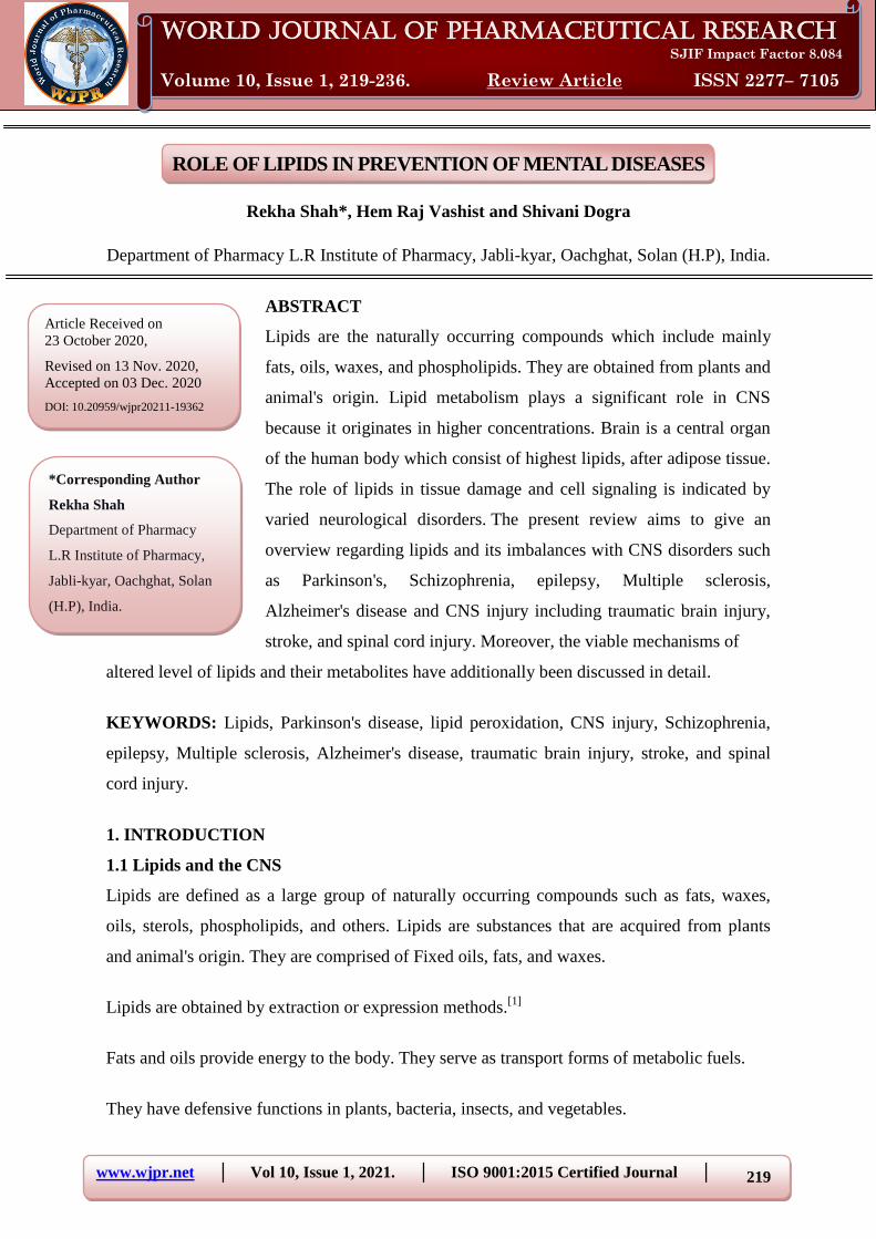
www.wjpr.net │ Vol 10, Issue 1, 2021. │ ISO 9001:2015 Certified Journal │ 219 ROLE OF LIPIDS IN PREVENTION OF MENTAL DISEASES Rekha Shah*, Hem Raj Vashist and Shivani Dogra Department of Pharmacy L.R Institute of Pharmacy, Jabli-kyar, Oachghat, Solan (H.P), India. ABSTRACT Lipids are the naturally occurring compounds which include mainly fats, oils, waxes, and phospholipids. They are obtained from plants and animal's origin. Lipid metabolism plays a significant role in CNS because it originates in higher concentrations. Brain is a central organ of the human body which consist of highest lipids, after adipose tissue. The role of lipids in tissue damage and cell signaling is indicated by varied neurological disorders. The present review aims to give an overview regarding lipids and its imbalances with CNS disorders such as Parkinson's, Schizophrenia, epilepsy, Multiple sclerosis, Alzheimer's disease and CNS injury including traumatic brain injury, stroke, and spinal cord injury. Moreover, the viable mechanisms of altered level of lipids and their metabolites have additionally been discussed in detail. KEYWORDS: Lipids, Parkinson's disease, lipid peroxidation, CNS injury, Schizophrenia, epilepsy, Multiple sclerosis, Alzheimer's disease, traumatic brain injury, stroke, and spinal cord injury. 1. INTRODUCTION 1.1 Lipids and the CNS Lipids are defined as a large group of naturally occurring compounds such as fats, waxes, oils, sterols, phospholipids, and others. Lipids are substances that are acquired from plants and animal's origin. They are comprised of Fixed oils, fats, and waxes. Lipids are obtained by extraction or expression methods. [1] Fats and oils provide energy to the body. They serve as transport forms of metabolic fuels. They have defensive functions in plants, bacteria, insects, and vegetables. World Journal of Pharmaceutical Research SJIF Impact Factor 8.084 Volume 10, Issue 1, 219-236. Review Article ISSN 2277– 7105 Article Received on 23 October 2020, Revised on 13 Nov. 2020, Accepted on 03 Dec. 2020 DOI: 10.20959/wjpr20211-19362 *Corresponding Author Rekha Shah Department of Pharmacy L.R Institute of Pharmacy, Jabli-kyar, Oachghat, Solan (H.P), India.
Transcript of ROLE OF LIPIDS IN PREVENTION OF MENTAL DISEASES
www.wjpr.net │ Vol 10, Issue 1, 2021. │ ISO 9001:2015 Certified Journal │
Rekha et al. World Journal of Pharmaceutical Research
219
ROLE OF LIPIDS IN PREVENTION OF MENTAL DISEASES
Rekha Shah*, Hem Raj Vashist and Shivani Dogra
Department of Pharmacy L.R Institute of Pharmacy, Jabli-kyar, Oachghat, Solan (H.P), India.
ABSTRACT
Lipids are the naturally occurring compounds which include mainly
fats, oils, waxes, and phospholipids. They are obtained from plants and
animal's origin. Lipid metabolism plays a significant role in CNS
because it originates in higher concentrations. Brain is a central organ
of the human body which consist of highest lipids, after adipose tissue.
The role of lipids in tissue damage and cell signaling is indicated by
varied neurological disorders. The present review aims to give an
overview regarding lipids and its imbalances with CNS disorders such
as Parkinson's, Schizophrenia, epilepsy, Multiple sclerosis,
Alzheimer's disease and CNS injury including traumatic brain injury,
stroke, and spinal cord injury. Moreover, the viable mechanisms of
altered level of lipids and their metabolites have additionally been discussed in detail.
KEYWORDS: Lipids, Parkinson's disease, lipid peroxidation, CNS injury, Schizophrenia,
epilepsy, Multiple sclerosis, Alzheimer's disease, traumatic brain injury, stroke, and spinal
cord injury.
1. INTRODUCTION
1.1 Lipids and the CNS
Lipids are defined as a large group of naturally occurring compounds such as fats, waxes,
oils, sterols, phospholipids, and others. Lipids are substances that are acquired from plants
and animal's origin. They are comprised of Fixed oils, fats, and waxes.
Lipids are obtained by extraction or expression methods.[1]
Fats and oils provide energy to the body. They serve as transport forms of metabolic fuels.
They have defensive functions in plants, bacteria, insects, and vegetables.
World Journal of Pharmaceutical Research SJIF Impact Factor 8.084
Volume 10, Issue 1, 219-236. Review Article ISSN 2277– 7105
Article Received on
23 October 2020,
Revised on 13 Nov. 2020,
Accepted on 03 Dec. 2020
DOI: 10.20959/wjpr20211-19362
*Corresponding Author
Rekha Shah
Department of Pharmacy
L.R Institute of Pharmacy,
Jabli-kyar, Oachghat, Solan
(H.P), India.

www.wjpr.net │ Vol 10, Issue 1, 2021. │ ISO 9001:2015 Certified Journal │
Rekha et al. World Journal of Pharmaceutical Research
220
It serves as pigment (carotene), hormones (VitA & D), detergent, cofactors (Vitamin E, K),
and signaling molecules (steroids).[2]
1.2 Classification
Lipids are broadly classified into the following types:
Table 1: Classification of lipids.
Simple Compound Derived
1.. Fats and oils 1.. Phospholipids 1.. Fatty acids
2.. Waxes 2.. Glycolipids 2.. Alcohol (glycerol, sterol)
3.. Lipoproteins
1.2.1 Simple lipids
Simple lipids are esters of fatty acids with various types of an alcohol group. E.g. Fats, oils,
and waxes.
Fats and oils are stored forms of energy. They are also known as unsaturated fats. Fixed oils
are a long chain of fatty acids, alcohol, and glycerol. Waxes are the esters of fatty acid with
an alcohol.
Utilizations of the fixed oils and fats
1. Utilized in soap manufacturing
2. In suppositories and tablet coating
3. Also in dietary enhancements
4. Utilized as emulsifying agents
5. Utilized in manufacture of paints, varnishes, and lubricants
6. Therapeutic uses (castor oil).
Waxes
Waxes are esters of a long chain alcohol and a fatty acid. Waxes are found in nature as
coatings on leaves and stems.
Table 2: The major types of waxes.
Type Examples
1. Natural Waxes
1.1 Animal Waxes Beeswax, Lanolin, Tallow
1.2 Vegetable Waxes Carnauba, Candelilla, Soy
1.3 Mineral Waxes
1.3.1 Fossil or Earth Ceresin, Montan

www.wjpr.net │ Vol 10, Issue 1, 2021. │ ISO 9001:2015 Certified Journal │
Rekha et al. World Journal of Pharmaceutical Research
221
1.3.2 Petroleum
1.3.2.1 Paraffin Slack, Scale Wax, Refined Paraffin
1.3.2.2 Microcrystalline
1.3.2.3 Petrolatum
2. Synthetic Waxes
2.1 Ethylene Polymers Polyethylene, polyol ether-esters
USES OF WAX
1. Wax is used in pharmacy to prepare lip balm.
2. The technical uses of waxes, e.g. in shoe polishes and car waxes.
3. Waxes are also used in making soft ointments.
1.2.2 Compound lipids
Compound lipids are esters of fatty acids with a group of alcohol.
E.g. phospholipids, glycolipids, and lipoproteins.
Phospholipids contain phosphoric acid.
1.2.3 Derived lipids
These include fatty acids, steroids, other alcohols, etc.
Derived lipids are the substances derived from simple and compound lipids by hydrolysis.
Fatty acids are the simplest form of lipids. Fatty acids are also known as acyl group when it is
a part of ester. Fatty acids are further divided into saturated and unsaturated fatty acids.[3]
1.3 Role of Lipids in the Central Nervous System
Lipid metabolism plays a significant role for CNS because lipid originates in higher
concentration, second to adipose tissue. Lipids have been beneficial for the brain. Lipids also
participate in the brain for the maintenance and regulation of its activities. However, if the
concentration of lipids is altered in the brain, it leads to worsening of the condition.
Brain disorders, CNS traumas, stroke, multiple diseases are the most issues within the clinical
field.[4]
However, there is no cure for these CNS injuries and disorders.
The role of lipids in tissue damage and cell signaling is diagrammatic by the varied
neurological disorders like
Alzheimer's disease,
Parkinson's disease,

www.wjpr.net │ Vol 10, Issue 1, 2021. │ ISO 9001:2015 Certified Journal │
Rekha et al. World Journal of Pharmaceutical Research
222
Multiple disorders,
Schizophrenia,
Epilepsy,
CNS injury (Stroke, traumas, brain injury, and funiculus injury).
Lipids in the brain are concerned with several metabolic pathways. The most important role
of lipids in the brain is proliferation, cell growth, and neuroprotection.[5]
Some lipids show
their action by binding with receptors like sphingomyelin, G-protein coupled receptors. These
lipids are called “neurolipids”.
1.3.1 Sources of lipids to CNS
Blood-brain barrier considerably inhibits the entry of harmful substances in the CNS.
Therefore, all the lipids found in CNS should be synthesized at intervals CNS. E.g. fatty
acids, cholesterol of these are helpful for neurological performance.
Peroxisomal fatty acids reaction is very important within the brain because the brain contains
very-long-chain fatty acids and open-chain fatty acids.
2. Lipids in CNS disorders
Table 3: Lipid systems affected by the CNS disorders and injuries.[6]
Disorder/ injury Symptoms/pathologic
features
CNS region(s)
affected
Mechanism of
damage
Possible
Treatments
Alzheimer
Disease
> Memory loss
> Difficulty in
communicating
> Problems learning,
thinking, reasoning
> Difficulty with
familiar tasks
> Disorientation
> Amyloid plaques
and tau protein
aggregation
> Amygdala
> Hippo-campus
> Cerebral
cortical areas
controlling
reasoning,
learning, and
language
> Altered
cholesterol and
lipid
homeostasis
> APP
cleavage in
lipid rafts
> DHA levels
↓; upregulation
of PLA2,
increased lipid
peroxidation
sPLA2-IIA
expression
increased
> Ganglio-side
treatments prevent
neuronal death.
> ApoE protects
against oxidative
injury by mediating
Aβ
> Statins

www.wjpr.net │ Vol 10, Issue 1, 2021. │ ISO 9001:2015 Certified Journal │
Rekha et al. World Journal of Pharmaceutical Research
223
Disorder/ injury Symptoms/pathologic
features
CNS region(s)
affected
Mechanism of
damage
Possible
Treatments
Parkinson’s
Diseases
> Movement disorder
> Resting tremors
> Muscle rigidity
> Bradykinesia (slow
movemets)
> Impaired posture,
balance, coordination
> Lewy bodies/α-
synuclein aggregate
> Although a
direct role of
PLA2 in PD is
not yet clearly
demonstrated,
cPLA2 knock
out mice
showed
protection
against MPTP
toxicity.
> PUFAs
promote α -
synuclein
aggregation
> PLA2 inhibition
Ganglioside
treatments
Quinacrine
Multiple
Sclerosis-
Experimental
Autoimmune
Encephalomyelitis
(MS-EAE)
> Unpredictable and
varies from person to
person
> Loss of balance
> Loss of muscle
coordination, resulting
in tremors, bladder
problems and slurred
speech.
> Problems with
memory, attention,
cognitive functions.
Demyelination of
axons
> Lipid
peroxidation
products from
ROS.
> cPLA2 is
highly
expressed in
EAE.
>sPLA2 levels
increased prior
to onset of
symptoms.
> T cells and
auto-antibodies
to Lipids
> DHA levels
↓
> Antioxidants
>sPLA2 inhibition
by CHEC-9 blocks
inflammation.
> Ganglioside
treatments prevent
neuronal death.
Schizophrenia
> Disturbances in
thinking, emotional
reactions, and social
behaviour
> Dorsalateral
prefrontal cortex
> Altered lipid
metabolism
may be
responsible for
defects in
neurological
development
> Antipsychotic
drugs
> Eicosapentaenoic
acid
supplementation
Epilepsy > Wide range of
severity
> Focal cortical
area, later
transferred
> DHA levels
↓
Ketogenic diet
Phenytoin
> Violent convulsions
> Loss of
consciousness
> Minimal or no
to the Thalamus
> Second
generation
antiepileptic drugs

www.wjpr.net │ Vol 10, Issue 1, 2021. │ ISO 9001:2015 Certified Journal │
Rekha et al. World Journal of Pharmaceutical Research
224
Disorder/ injury Symptoms/pathologic
features
CNS region(s)
affected
Mechanism of
damage
Possible
Treatments
movements
Stroke
> Sudden weakness on
one side of the body,
loss of balance and
coordination, trouble
with cognition
> Cerebral
cortical Areas
> Striatum
> Activation of
Phospholipases
(A2, C, D),
increased
sPLA2
>
cPLA2 knock
out mice
showed
protection [4
and references
Cited therein]
> DHA levels
↓
> CDP-choline
attenuated sPLA2
> Neuroprotectin
D1 reduces infarct
in MCAO model
> sPLA2 inhibitors
Traumatic Brain
Injury
> Loss of
CA3 hippocampal
neurons
> Aβ
deposition, tau
pathology
> Corticosteroids
> apoE mimetic
peptide showed
benefit in
experimental TBI
Spinal Cord
Injury
> Weakness and
sensory loss; paralysis
> Activation of
PLA2,
COX/LOX
pathways.
Corticosteroids
inhibit These
activations
> High-dose
methylprednisolone
in clinical use ;
DHA treatment is
beneficial
2.1 Parkinson's disease
Parkinson's disease may be a progressive nervous system disorder that affects movement,
usually together with tremors. The disorder also causes stiffness or reduces speed of
movement. There is no etiology of Parkinson's disease (PD). The most pathological
characteristics of PD are necrobiosis within the brain's basal ganglia and also the presence of
Lewy bodies in several of the remaining neurons.[7]
This loss of neurons during the death of
astrocytes (star-shaped glial interstitial cells) and a major increase within the range of
neuroglia (another sort of glial cell) within the neural structure. Shockingly, a few
investigations have additionally detailed that there is no affiliation between advancement of
PD and cholesterol levels. Additionally, low degrees of complete cholesterol (TC), HDL-C
and LDL-C have been seen in the PD patients.[8]
Reaction stress and lipid peroxidation plays
a vital role in pathogenesis of PD.

www.wjpr.net │ Vol 10, Issue 1, 2021. │ ISO 9001:2015 Certified Journal │
Rekha et al. World Journal of Pharmaceutical Research
225
2.1.1 Treatment of PD
No therapy to delay the loss of dopamine neurons in PD has been demonstrated. The
dopamine prodrug levodopa continues to be the gold standard for treating PD. Long-term
levodopa treatment does, however, contribute to dyskinesia progressing. However,
pallidotomy and thalamotomy may be performed in selected patients, deep brain stimulation
is that the operation for PD patients.
Some medicines such as Carbidopa and Levodopa are utilized in the treatment of Parkinson’s
disease. Levodopa is the most effective medication for the treatment. It is continually given
within the combination with carbidopa to stop decarboxylation. It is used as a symptomatic
treatment for early PD. Monoamine neurotransmitter agonists stimulate monoamine
neurotransmitter receptors directly and also the second most potent category of
medication.[8]
Amantadine's mechanism of action remains unknown; however, in addition to
acting as an antagonist to the NMDA receptor, it has been reported to have anticholinergic
properties to enhance dopamine release and prevent its reuptake. However, these procedures
remain based on symptomatic relief while reducing the adverse effects.
2.1.2 PD, oxidative stress & lipid peroxidation
In Parkinson's disease (PD), the metabolism of monoamine neurotransmitters by the
monoamine-oxidase-B may lead to excessive reactive oxygen species formation. A role for
oxidative stress in PD was denoted by increase in 8-hydroxy-2`-deoxyguanosine, a hydroxyl
radical-damaged guanine nucleotide commonly used to evaluate oxidative DNA damage.
Further, there are several markers of lipid peroxidation that were found to be enlarged in PD
brain regions.[9]

www.wjpr.net │ Vol 10, Issue 1, 2021. │ ISO 9001:2015 Certified Journal │
Rekha et al. World Journal of Pharmaceutical Research
226
Fig 1: Lipid metabolism in Parkinson's disease.[10]
2.2 Schizophrenia & epilepsy
Schizophrenia is a disorder that affects a person's ability to assume or behave. It is a heavy
disturbance and will result in hallucinations, very disordered thinking, and behavior. It
impacts thinking, emotions, speech, and alterations areas of life. Schizophrenia may be a
fairly uncommon condition, moving around 0.25% to 0.64% of individuals within the United
States, consistent with the National Institute of Mental Health (NIMH). Schizophrenia
disorder may be a lifelong condition however effective treatment can help to overcome the
situation.[11]
Latest theories on the Schizophrenia have focused on abnormalities in lipid metabolism, in
particular increased activity of PLA2 enzymes, and reduced activity of the system which
includes PUFAs into phospholipids.[11]
These abnormalities lead to changes in the structure
of the membrane and so the function of membrane-bound proteins and also the behavior of
neurotransmitter systems. Therefore, lipid metabolism has a vital role in neuronal

www.wjpr.net │ Vol 10, Issue 1, 2021. │ ISO 9001:2015 Certified Journal │
Rekha et al. World Journal of Pharmaceutical Research
227
growth.[11,12]
It's been found that schizophrenia is associated with lipid transport proteins and
membrane phospholipid composition.
From a restorative point of view, various reports demonstrate that in any event a bit of
schizophrenic patients have decreased degrees of PUFAs, especially ArAc and DHA, in red
cell phospholipids, with low levels especially connected with negative indications. ArAc,
DHA and eicosapentaenoic corrosive (EPA) are significant for monoaminergic
neurotransmission, mental health, and synaptic working.[12]
This recommends
supplementation with basic unsaturated fats could ease indications of schizophrenia. In starter
examines, in any case, DHA basically had no impact and ArAc seemed to compound
indications in some schizophrenia patients.[11]
Latest findings indicate the function of oxidative stress-induced abnormalities and changes of
membrane phospholipids and fatty acids in etiopathogenic pathways in schizophrenia where
the oxidative metabolites derived from membrane lipids, including prostaglandins and
isoprostanes, had been identified.[13]
In addition, schizophrenia may be associated with
altered metabolism of polyunsaturated fatty acids (PUFA), particularly arachidonic acid and
cell membrane phospholipids.[11,13]
Epilepsy
Epilepsy is a neurological disorder that causes unverified, repeated seizures. A seizure may
be an explosive rush of electrical activity within the brain. Seizures can also lead to the death
of brain cells.[14]
Epilepsy is also called a seizure disorder. Epilepsy is treated with a medicine
called ant-epileptic drugs. Phenytoin is the widely used anti-epileptic medicine.[15]
The
essential site of action appears to be the motor cortex, where the spread of seizure activity is
inhibited by advancing sodium efflux from neurons.
The ketogenic diet is an efficient non-pharmacological symptomatic treatment for
epilepsy.[16]
The ketogenic diet is a very high fat, low carbohydrate, controlled
macromolecule diet. This diet is used since 1920s for the treatment of epilepsy.[17]
Despite its
use for several years, there is still a tidy discussion over how the ketogenic diet works.
Elevated cholesterol is involved in several initiating disorders of both neurological and
neurodegenerative type. Cholesterol is degraded by an enzyme named as cholesterol 24-
Hydroxylase, encoded by the gene CYP46A1. Apparently, by the inhibition of this catabolic

www.wjpr.net │ Vol 10, Issue 1, 2021. │ ISO 9001:2015 Certified Journal │
Rekha et al. World Journal of Pharmaceutical Research
228
enzyme, cholesterol level in the neurons of hippocampus increases. In turn this increased
cholesterol outcomes in the neuronal cell death and deviant hippocampus synchronies.[18]
2.3 Alzheimer's disease
Alzheimer's disease (AD) is a progressive disease that destroys memory and alternative
mental functions. Neuron connections and also the cells themselves degenerate and die,
eventually destroying memory and alternative mental functions. Alzheimer’s disease is
broadly divided into early-onset AD (occurring in persons under age 65 years, 5-10% of AD)
and late-onset (90-95% of AD).
Fig. 2: Amyloid Plaque Formation: Enzymes act on the APP (Amyloid precursor
protein) and cut it into fragments of protein, one of which is called beta-amyloid and it’s
crucial in the formation of senile plaques in AD.[19]
One of the hallmarks of AD is the overproduction of a 4-kDa peptide, amyloid peptide
resulting in the formation of plaques. The other hallmark of AD is the formation of
neurofibrillary tangles because of the hyperphosphorylation of tau proteins. Whereas the
etiology of AD is unknown, notable risk factors for the disease include increasing age,
positive family history. Theories relating to AD specialize in the abnormalities of the brain
involving the nervous system. There is evidence that cholesterol is of importance in the
development and progression of the disease.[20]
Apolipoprotein (Apo) E is one of the major
Apos in plasma and the principal cholesterol carrier protein in the brain.
2.3.1 AD, oxidative stress & lipid peroxidation
A number of studies demonstrating increased amount of lipid peroxidation in AD provide
mounting evidence supporting a role for oxidative damage in this disorder. In recent studies,

www.wjpr.net │ Vol 10, Issue 1, 2021. │ ISO 9001:2015 Certified Journal │
Rekha et al. World Journal of Pharmaceutical Research
229
increased levels of hydroxynonenal (HNE) and acrolein,[21]
in the brain tissue from patients
affected by early AD, shows that lipid peroxidation occurs early in the pathogenesis of
AD.[21]
Acrolein, the strongest electrophile among all -unsaturated aldehydes, reacts with
DNA bases such as guanine, adenine, cytosine, and thymidine to form cyclic adducts, the
main cyclic adducts is acrolein-deoxyguanosine. Increased levels of acrolein-deoxyguanosine
adducts were denoted in brain tissue from AD patients. Reactive oxygen species (ROS) may
also play a role in amyloid deposition in AD as oxidizing conditions cause protein cross-
linking and aggregation of Aβ peptides; Aβ aggregation has been shown to induce ROS
accumulation, which may result in cyclical or self-propagating oxidative damage.[21]
This
indicates that oxidative stress plays an important pathological function in the development of
disease.
2.4 Multiple sclerosis
Multiple sclerosis (MS) may be a potentially disabling disease of the brain and spinal cord. In
MS, the immune system attacks the protective sheath that covers the nerve fibers and causes
communication problems between the brain and the body. Eventually, this disease can cause
permanent damage or deterioration of the nerves.
It is predominantly a T-lymphocyte-mediated disorder and cytokines have a key role in the
pathogenesis of the disease. Multiple sclerosis is the only neurological disorder where
therapeutic manipulation of the cytokine system influences the disease. Thiobarbituric acid
substances and F2-isoprostane levels were shown in MS patients, and HNE was indicative
that lipid peroxidation also occurs in multiple sclerosis. The metabolism of lipids in the body
may have direct and indirect effects on multiple sclerosis disability and disease progression
due to the fact they are essential for regulating inflammatory responses and for re-
myelination and repair in the CNS and disruption of lipid homeostasis can affect myelin
integrity and modulate neurodegeneration.

www.wjpr.net │ Vol 10, Issue 1, 2021. │ ISO 9001:2015 Certified Journal │
Rekha et al. World Journal of Pharmaceutical Research
230
Fig. 3: Immune‐mediated destruction of myelin components in multiple sclerosis.[22]
In Marrie and colleagues study, the presence of hypercholesterolemia at any time throughout
the disorder course was associated with 35% increased risk of early gait disability, 33%
increased risk of unilateral walking assistance and 24% increased risk of bilateral walking
assistance. From these it is clear studies which affect cholesterol levels increase in
impairment levels. On the whole, evidence from the studies indicates a negative effect of high
TC, LDL and triglycerides on acute inflammatory activity, disease course in patients with MS
and a useful impact of higher HDL levels on MS.[23]
3. Lipids in CNS injury
3.1 Stroke
A stroke occurs when the blood supply to part of the brain is interrupted or severely reduced,
depriving brain tissue of oxygen and food. Stroke is the abrupt onset of a neurological deficit.
Stroke is also known as Focal cerebral ischemia.
For the treatment of ischemic stroke, tissue plasminogen activator (tPA) is the only drug
approved by the US FDA.
The primary event in ischemia stroke is energy failure, resulting in excessive release of the
neurotransmitters (dopamine & glutamate).
In many epidemiological units, there is a direct relationship between cholesterol levels and
ischemic stroke. The relationship of lipids to ischemic stroke varies by stroke subtypes.

www.wjpr.net │ Vol 10, Issue 1, 2021. │ ISO 9001:2015 Certified Journal │
Rekha et al. World Journal of Pharmaceutical Research
231
Eventually, there is an increased risk of intracerebral haemorrhage and small vessel disease at
low levels of cholesterol.
Excessive stimulation of glutamate receptors results in elevated intracellular Ca2+ and
activation of phospholipases A2, C, and D. Stimulation of these phospholipases causes
hydrolysis of membrane phospholipids and release of second messengers. The nature of the
inflammatory response after stroke suggests that cytokines affect phospholipid metabolism
and free radicals that enhance brain injury. Several studies have reported the impaired fatty
acids metabolism with the increased risk of disease. Namely, low level of linoleic acid in
platelets, erythrocytes, adipose tissue and blood are related with the increased risk of
Ischemia Stroke and total stroke. In a case study, it was found that FAs alteration is involved
in the progression of various subtypes of stroke. Like low level of linoleic acid can accelerate
the stroke whereas, high levels of serum SFAs and ω-3 PUFAs have been found to be related
with haemorrhagic stroke.[24]
Fig. 4: Lipid peroxidation in stroke.[25]
3.1.1 Stroke, ROS & lipid peroxidation
As yet, it has been believed that the oxidative metabolism of ArAc through COX causes
prostaglandins and ROS. Few studies have mentioned that COX-2 generates tyrosyl radicals
on the protein and carbon-centred radicals on the substrate ArAc, but does not produce ROS.

www.wjpr.net │ Vol 10, Issue 1, 2021. │ ISO 9001:2015 Certified Journal │
Rekha et al. World Journal of Pharmaceutical Research
232
It has appeared that ROS production was raised in a stroke model but that COX-2 inhibition
did not reduce ROS production. Although, ROS generation was not reduced in COX-2
deficient mice. These studies revealed that NADPH oxidase was an important source of ROS
in the stroke model.[4]
The role of ROS is in stimulating various signaling pathways including
matrix metaloproteinases, NF-kB, and stroke injury has been reviewed.[26]
The time course of variations in lipid metabolism and formation of lipid metabolites and lipid
peroxidation products after transient cerebral ischemia are conferred.[27]
3.2 Traumatic brain injury
Traumatic brain injury (TBI) is the leading cause of death and disability in children and
adults between the ages of 1 to 44. TBI is a non-degenerative, non-congenital insult in the
brain from an external mechanical force, with an associated altered state of consciousness.
TBI may be divided into primary and secondary injuries.
The pathways of secondary injury that take place after the primary trauma present targets for
therapeutic interventions are correlative to stroke.[28]
Corticosteroids have been determined as
therapies to reduce the secondary injuries following Traumatic Brain Injury (TBI).
Corticosteroids inhibit the PLA2/COX/LOX pathways, thus limiting ArAc release and
metabolism, down-regulating pro-inflammatory cytokines and increasing the inflammatory
response.
Fig. 5: Mechanisms of traumatic brain injury.[29]
www.wjpr.net │ Vol 10, Issue 1, 2021. │ ISO 9001:2015 Certified Journal │
Rekha et al. World Journal of Pharmaceutical Research
233
3.3 Spinal cord injury
Spinal cord injury (SCI) is damage to any part of the spinal cord or nerves at the end of the
spinal canal- often causes permanent changes in strength, sensation and other body functions
below the site of the injury. SCI, as with acute stroke, is a dynamic process. SCI is the result
of an initial physical trauma followed by a secondary degenerative process. The majority of
SCI do not involve physical transaction of the spinal cord; instead, the cord is injured as a
result of contusive, compressive, or stretch injury. The primary event after SCI is
depolarization and opening of voltage depending ion channels, and consequent massive
release of neurotransmitters, like glutamate. This results in to accumulation of intracellular
calcium, initiating a variety of damaging events: mitochondrial dysfunction, activation of
nitric oxide synthase (NOS) and PLA2.[30]
The glucocorticoid steroids dexamethasone and methylprednisolone are used in the clinical
treatment of SCI. The preliminary rationale was that, since these compounds reduced brain
edema in brain tumor patients, they would also reduce edema in SCI. It is believed that
inhibition of lipid peroxidation is the principle neuroprotective mechanism of high dose
methylprednisolone and that glucocorticoid receptor-mediated anti-inflammatory effects have
only a minor part.[30]
The levels of apolipoprotein-A1 were found to be decreased and the
concentrations of apolipoprotein-B increased in people with SCI.
4. CONCLUSION
The group of lipids plays an important role in the cell and tissue. The concept of the review
has been introduced to describe the role of lipids in the brain and CNS disorders. Several
types of researches recommend that many lipids are involved in the maintenance of the
regulation of inflammation and pain, energy metabolism, and development of the brain in the
nervous system. More profound information on the nature of lipid signaling will promote our
comprehensive of the role of lipid metabolism in different CNS disorders, opening new doors
for improvement and treatments for neurological diseases. The connection behind the altered
lipid metabolism and brain functions depends on the beginning of various brain disorders
such as Schizophrenia, AD, PD, epilepsy, multiple sclerosis as well as brain injuries such as
stroke, trauma, and spinal cord injury. Besides, in this review, the relationship between lipid
metabolism and neurological diseases has been discussed in detail.

www.wjpr.net │ Vol 10, Issue 1, 2021. │ ISO 9001:2015 Certified Journal │
Rekha et al. World Journal of Pharmaceutical Research
234
Ethical Approval
It is not applicable.
CONFLICT OF INTEREST
The authors confirm that this article has no conflict of interest.
ACKNOWLEDGEMENT
I am very thankful to my teacher Dr. Hem Raj Vashist for his constant support and guidance.
REFERENCES
1. Kokate C.K., A. P. Purohit, Gokhale S.B., Pharmacognosy 13th edition, Nirali Prakashan,
Page. 10.1-10.84. Jain P, Surana SJ (2015) A review of Indian medicinal plants with
hypolipidemic activity and their medicinal importance. World Journal of Pharmacy and
Pharmaceutical Sciences, 2002; 4(3): 1477-1493.
2. Lipid Library: Lipid Chemistry, Biology, Technology & Analysis
http://lipidlibrary.aocs.org/.
3. Fahy E, Subramaniam S, Brown HA et al.: A comprehensive classification system for
lipids. J. Lipid Res., 2005; 46(5): 839–862.
4. Adibhatla RM, Hatcher JF, Dempsey RJ: Lipids and lipidomics in brain injury and
diseases. AAPS J., 2006; 8(2): E314–E321.
5. Wenk MR: The emerging field of lipidomics. Nat. Rev. Drug Discover, 2005; 4(7): 594–
610. Excellent comprehensive review on various aspects of lipidomics and
bioinformatics. 2A comprehensive classification system for lipids. J. Lipid Res., 2005;
46(5): 839–862.
6. Adibhatla RM, Hatcher JF, Dempsey RJ. Lipids and lipidomics in brain injury and
diseases. AAPS J., 2006; 8(2): E314–E321.
7. Hauser RA, Zesiewicz TA: Advances in the pharmacologic management of early
Parkinson disease. Neurologist, 2007; 13(3): 126–132.
8. Samadi P, Grégoire L, Rouillard C et al.: Docosahexaenoic acid reduces levodopainduced
dyskinesia in 1-methyl-4-phenyl1, 2, 3, 6-tetrahydropyridine monkeys. Ann. Neurol,
2006; 59(2): 282–288.
9. Hauser RA, Zesiewicz TA: Advances in the pharmacologic management of early
Parkinson disease. Neurologist, 2007; 13(3): 126–132.
10. Xicoy H, Wieringa B, Martens GJ: Lipids in Parkinson disease. Cells, 2019; 8(1): 27.
11. Horrobin D: The lipid hypothesis of schizophrenia. In: Brain Lipids and Disorders in
www.wjpr.net │ Vol 10, Issue 1, 2021. │ ISO 9001:2015 Certified Journal │
Rekha et al. World Journal of Pharmaceutical Research
235
Biological Psychiatry. Skinner ER (Ed.), Elsevier Science, Amsterdam, the Netherlands,
2002; 39–52.
12. Berger GE, Smesny S, Amminger GP: Bioactive lipids in schizophrenia. Int. Rev.
Psychiatry, 2006; 18(2): 85–98.
13. Fendri C, Mechri A, Khiari G, Othman A, Gaha L: Oxidative stress improvement n
schizophrenia pathophysiology. Encephale, 2006; 32: 244-252.
14. Bialer M, Johannessen SI, Kupferberg HJ, Levy RH, Perucca E, Tomson T: Progress
report on new antiepileptic drugs: a summary of the Eighth Eilat Conference (EILAT
VIII). Epilepsy Research, 2007; 73(1): 1–52 Review of the new generation anti-epileptic
drugs.
15. Klotz U: The role of pharmacogenetics in the metabolism of antiepileptic drugs:
pharmacokinetic and therapeutic implications. Clin. Pharmacokinetic, 2007; 46(4):
271–279.
16. Papandreou D, Pavlou E, Kalimeri E, Mavromichalis I: The ketogenic diet in children
with epilepsy. Br. J. Nutr, 2006; 95(1): 5–13.
17. Bough KJ, Rho JM: Anticonvulsant mechanisms of the ketogenic diet. Epilepsy, 2007;
48(1): 43–58.
18. Gasior M, Rogawski MA, Hartman AL: Neuroprotective and disease-modifying effects of
the ketogenic diet. Behave. Pharmacol, 2006; 17(5–6): 431–439.
19. https://upload.wikimedia.org/wikipedia/commons/f/fb/Amyloid-plaque_formation-big.jpg
20. Mandavilli A: The amyloid code. Nat. Med, 2006; 12(7): 747–751. Provocative
commentary on the current amyloid theories in Alzheimer’s disease (AD).
21. Williams TI, Lynn BC, Markesbery WR, & Lovell MA: Increased levels of 4-
hydroxynonenal and acrolein, neurotoxic markers of lipid peroxidation, in the brain in
mild cognitive impairment and early Alzheimer’s disease. Neurobiol. Aging, 2006; 27(8):
1094–1099.
22. Ramirez‐Ramirez V, Macias‐Islas MA, Ortiz GG, Pacheco‐Moises F, Torres‐Sanchez
ED, et al. Efficacy of fish oil on serum of TNF‐α, IL‐1 β, and IL‐6 oxidative stress
markers in multiple sclerosis treated with interferon beta‐1b. Oxid Med Cell Longev,
2013; 2013: 709493.
23. Marrie RA, Rudick R, Horwitz R et al. Vascular comorbidity is associated with more
rapid disability progression in multiple sclerosis. Neurology, 2010; 74: 1041–1047.
24. Adibhatla RM, Dempsey RJ, Hatcher JF: Integration of cytokine biology and lipid

www.wjpr.net │ Vol 10, Issue 1, 2021. │ ISO 9001:2015 Certified Journal │
Rekha et al. World Journal of Pharmaceutical Research
236
metabolism in stroke. Front Neurosurg. Res. (under the aegis of Front Biosci). (In Press),
2007.
25. Ugidos IF, Pérez-Rodríguez D, Fernández-López A. A role for lipids as agents to
alleviate stroke damage: the neuroprotective effect of 2-hydroxy arachidonic acid. Neural
Regen Res [cited 2020 Jul 18], 2017; 12: 1273-5.
26. Adibhatla RM, Hatcher JF: Phospholipase A2, reactive oxygen species, and lipid
peroxidation in cerebral ischemia. Free Radic. Biol. Med, 2006; 40(3): 376–387.
27. Liu KJ, Rosenberg GA: Matrix metaloproteinases and free radicals in cerebral ischemia.
Free Radic. Biol. Med, 2005; 39(1): 71–80.
28. Rigg JL, Zafonte RD: Corticosteroids in TBI: is the story closed? J. Head Trauma Rehab,
2006; 21(3): 285–288.
29. https://www.google.com/imgres?imgurl=https%3A%2F%2Fimage.slidesharecdn.com%2
Ftbi-140824231703-phpapp02%2F95%2Ftraumatic-brain-injury-8-.
30. Hall ED, Springer JE: Neuroprotection and acute spinal cord injury: a reappraisal.
NeuroRx, 2004; 1(1): 80–100.