Role of Inflammatory Mediators and their Genetics in ...
113
MIIA VIRTA Role of Inflammatory Mediators and their Genetics in Epstein-Barr Virus Infection, Febrile Seizures and Atopy ACADEMIC DISSERTATION To be presented, with the permission of the Faculty of Medicine of the University of Tampere, for public discussion in the Auditorium of Finn-Medi 1, Biokatu 6, Tampere, on October 16th, 2009, at 12 o’clock. UNIVERSITY OF TAMPERE
Transcript of Role of Inflammatory Mediators and their Genetics in ...
MIIA VIRTA
Role of Inflammatory Mediators and their Genetics in Epstein-Barr Virus Infection,
Febrile Seizures and Atopy
ACADEMIC DISSERTATIONTo be presented, with the permission of
the Faculty of Medicine of the University of Tampere,for public discussion in the Auditorium of Finn-Medi 1,
Biokatu 6, Tampere, on October 16th, 2009, at 12 o’clock.
UNIVERSITY OF TAMPERE

Reviewed byDocent Riitta KarttunenUniversity of OuluFinlandDocent Johannes SavolainenUniversity of TurkuFinland
DistributionBookshop TAJUP.O. Box 61733014 University of TampereFinland
Tel. +358 3 3551 6055Fax +358 3 3551 7685 [email protected]/tajuhttp://granum.uta.fi
Cover design byJuha Siro
Acta Universitatis Tamperensis 1452ISBN 978-951-44-7835-2 (print)ISSN-L 1455-1616ISSN 1455-1616
Acta Electronica Universitatis Tamperensis 885ISBN 978-951-44-7836-9 (pdf )ISSN 1456-954Xhttp://acta.uta.fi
Tampereen Yliopistopaino Oy – Juvenes PrintTampere 2009
ACADEMIC DISSERTATIONUniversity of Tampere, Medical SchoolTampere Graduate School in Biomedicine and Biotechnology (TGSBB)Finland
Supervised byProfessor Mikko HurmeUniversity of TampereFinlandDocent Merja HelminenUniversity of TampereFinland

3
To my parents
4
Contents
CONTENTS...................................................................................................4
LIST OF ORIGINAL COMMUNICATIONS ..............................................7
ABBREVIATIONS .......................................................................................8
ABSTRACT................................................................................................ 10
TIIVISTELMÄ ........................................................................................... 12
INTRODUCTION ...................................................................................... 14
REVIEW OF THE LITERATURE ............................................................ 16
1. Interleukin1B promoter polymorphism and febrile seizures ............................161.1. Interleukin1 ..........................................................................................16
1.1.1. IL1 family ................................................................................161.1.2. Function of IL1 ......................................................................161.1.3. IL1Ra/ IL1 ratio....................................................................181.1.4. IL1B gene polymorphisms ........................................................18
1.2. IL1B511C>T polymorphism and febrile seizures ................................211.2.1. Febrile seizure ...........................................................................211.2.2. IL1 and febrile seizures ...........................................................221.2.3. Associations between IL1B511 and febrile seizures ...............23
2. Interleukin10 promoter polymorphisms and EpsteinBarr virus infection.......232.1. Interleukin10 ........................................................................................23
2.1.1. Function of IL10 ......................................................................232.1.2. Role of IL10 in disease ............................................................242.1.3. IL10 gene polymorphisms.........................................................26
2.2. IL10 promoter haplotype and EBV .......................................................282.2.1. EBV infection ...........................................................................282.2.2. Role of IL10 in EpsteinBarr virus infection...........................282.2.3. Associations between IL10 gene promoterpolymorphisms and EpsteinBarr virus...............................................29
3. Interleukin 4 promoter polymorphism and atopy .............................................30

5
3.1. Interleukin4 ..........................................................................................303.1.1. Function of IL4 ........................................................................303.1.2. IL4 gene polymorphisms...........................................................31
3.2. IL4590C>T polymorphism and atopy..................................................343.2.1. Atopy.........................................................................................343.2.2. IL4 and atopy...........................................................................353.2.3. IL4590C>T polymorphism, Helicobacter pylori andatopy....................................................................................................36
4. CD14 promoter polymorphism and IgE ............................................................374.1. CD14......................................................................................................37
4.1.1. Function of CD14......................................................................374.1.2. CD14 gene polymorphisms.......................................................39
4.2. CD14159C>T polymorphism and serum total IgE ..............................394.2.1. IgE.............................................................................................394.2.2. CD14 and IgE............................................................................414.2.3. CD14159C>T polymorphism, Helicobacter pylori andserum total IgE....................................................................................414.2.4. Effect of geneenvironment interactions on serum totalIgE and atopy ......................................................................................42
AIMS OF THE STUDY ............................................................................. 44
SUBJECTS AND METHODS ................................................................... 45
1. Subjects ..............................................................................................................451.1. Studies I and II.......................................................................................451.2. Study III.................................................................................................461.3. Study IV.................................................................................................461.4. Study V..................................................................................................46
2. Methods..............................................................................................................492.1. Measurement of cytokine plasma levels (Studies II, III) ......................492.2. EBV, H.pylori and T.gondii serology (Studies I, IV, V).......................492.3. Analysis of IL1B, IL4, IL10, CD14 and TLR4 genepolymorphisms (Studies I, III, IV, V) ..........................................................492.4. Skin prick test (Study IV)......................................................................512.5. Measurement of serum total IgE (Studies IVV) ..................................512.6. Statistical analyses (Studies IV)...........................................................522.7. Ethics (Studies IV) ...............................................................................52
RESULTS ................................................................................................... 53
1. Effect of IL1B511 gene polymorphism on febrile seizures (Study I)...............53
2. Plasma and cerebrospinal fluid cytokines and febrile seizures (Study II) .........54

6
2.1. Plasma cytokines and febrile seizures ...................................................542.2. Cerebrospinal fluid cytokines in febrile seizures ..................................552.3. IL1B511C>T polymorphism and plasma cytokines in febrileseizures .........................................................................................................55
3. Effect of IL10 promoter haplotype on EpsteinBarr virus infection(Study III)...............................................................................................................57
4. Effect of IL4590 C>T polymorphism and Helicobacter pylori on skinprick test positivity (Study IV) ..............................................................................58
4.1. Associations...........................................................................................584.2. Interactions ............................................................................................59
5. Effect of CD14159 C>T polymorphism and Helicobacter pylori onserum total IgE (Study V) ......................................................................................59
5.1. Associations...........................................................................................595.2. Interactions ............................................................................................61
DISCUSSION............................................................................................. 63
1. IL1 and febrile seizures ..................................................................................631.1. Association between IL1B511C>T polymorphism and febrileseizures .........................................................................................................631.2. Associations between cytokines and febrile seizures ............................64
2. Association between IL10 promoter haplotype and EBV infection ..................64
3. Association between IL4590C>T polymorphism, Helicobacter pyloriand skin prick test ..................................................................................................65
4. Association between CD14159C>T polymorphism, Helicobacter pyloriand serum total IgE ................................................................................................67
5. Candidate gene studies.......................................................................................68
CONCLUSIONS ........................................................................................ 70
ACKNOWLEDGEMENTS........................................................................ 71
REFERENCES ........................................................................................... 73
ORIGINAL PUBLICATIONS ................................................................... 92

7
List of original communications
This dissertation is based on the following original communications, which arereferred to in the text by their Roman numerals (IV).
I Virta M, Hurme M, Helminen M (2002): Increased frequency ofinterleukin1 (511) allele 2 in febrile seizures. Pediatr Neurol 26: 192195.
II Virta M, Hurme M, Helminen M (2002): Increased plasma levelsof pro and antiinflammatory cytokines in patients with febrile seizures.Epilepsia 43: 920923.
III Helminen M, Kilpinen S, Virta M, Hurme M (2001): Susceptibilityto primary EpsteinBarr virus infection is associated with interleukin10 genepromoter polymorphism. J Infect Dis 184: 777780.
IV Pessi T, Virta M, Ådjers K, Karjalainen J, Rautelin H, Kosunen T,Hurme M (2005): Genetic and environmental factors in the immunopathogenesisof atopy: Interaction of Helicobacter pylori infection and IL4 genetics. Int ArchAllergy Immunol 137: 282288.
V Virta M, Pessi T, Helminen M, Seiskari T, Kondrashova A, KnipM, Hyöty M, Hurme M (2008): Interaction between CD14159C>Tpolymorphism and Helicobacter pylori is associated with serum total IgE. Clinexp allergy 38: 19291934.
In addition, this dissertation contains unpublished data.
8
Abbreviations
APC antigen presenting cellbp base pair (only with numbers)cDNA complementary deoxyribonucleic acidCI confidence intervalCNS central nervous systemCSF cerebrospinal fluidDNA deoxyribonucleic acidEBV EpsteinBarr virusETS environmental tobacco smokeFEV1 forced expiratory volume in one secondFS febrile seizureHIV human immunodefiency virushIL10 human IL10IBD inflammatory bowel diseaseIFN interferonIg immunoglobulinIL interleukin e.g. interleukin1IL1RN IL1 receptor antagonist geneIM infectious mononucleosisIU international unitsLPS lipopolysaccharidemCD14 membrane CD14NK natural killerNS nonsignificantOR odds ratioPAGE polyacrylamide gelPBMC peripheral blood mononuclear cellPCR polymerase chain reactionPEF peak expiratory flow ratePGE prostaglandin ER receptorRA rheumatoid arthritisRa receptor antagonist (e.g. IL1Ra)RFLP restriction fragment length polymorphismrhIL4 recombinant human IL4s soluble (e.g. sCD14)SLE systemic lupus erythematosusSNP single nucleotide polymorphism
9
SPT skin prick testTh T helper cellTLE temporal lobe epilepsyTLE+HS temporal lobe epilepsy with hippocampal sclerosisTLR Toll like receptorTNF tumor necrosis factorTreg T regulatory cellUNCT undifferentiated carcinoma of nasopharyngeal typeUV ultravioletvIL10 viral IL10
Abbreviations are defined at first mention in the abstract and review of theliterature and used only for concepts that occur more than twice.

10
Abstract
Inflammatory reactions are mediated by several molecules including cytokines,which can be divided into proinflammatory and antiinflammatory depending ontheir main inflammatory functions. Cytokines interact with many otherimmunomodulators like innate immunity receptors, including CD14, in a verycomplex network. These inflammatory mediators are essential for normal hostdefense against pathogens, but they also participate in the pathogenesis ofdiseases. Genetic variations in inflammatory mediator genes can alter thefunction of the gene thereby possibly affecting susceptibility to or severity ofseveral diseases.
This study was undertaken in order to investigate the role of Interleukin(IL)1B, IL4, IL10 and CD14 gene polymorphisms in three clinical conditions inwhich inflammatory mediators have an important role: EpsteinBarr virus (EBV)infection, febrile seizures (FSs) and atopy.
Associations between IL1B511C>T polymorphism and FSs were studiedamong Finnish FS patients. The IL1B511C>T allele T carriage was significantlyincreased in 35 FS patients compared to 400 adult blood donors (P=0.03).Relationships between plasma cytokines and FSs were also analyzed in Finnishchildren. Increased plasma IL1Ra levels (P=0.0005), IL6 levels (P=0.005) andIL1Ra/IL1β ratio (P<0.0001) and were found in 55 children with FSscompared to 20 control children with febrile illness without convulsions.However, there was no statistically significant association between IL1B511C>T polymorphism and plasma cytokine levels in FS patients (n=35).
The relationship between IL10 promoter 1082A>G/819C>T/592A>Chaplotype and early EBV infection was investigated in 115 yearold Finnishchildren (n=116) and in adult blood donors (n=400). In children IL10 promoterhaplotype ATA was associated with EBV seronegativity (P=0.035). However, inadult blood donors this haplotype was not associated with EBV seronegativity(P=0.98).
Associations between Helicobacter pylori seropositivity, IL4590C>Tpolymorphism and sensitization measured by skin prick test (SPT) were studiedin Finnish asthmatic (n=245) and nonasthmatic (n=405) adults. H.pyloriseronegativity was associated with increased risk of SPT positivity in bothasthmatic and nonasthmatic groups (OR 2.28 95%CI 1.353.85 and OR 1.5995%CI 1.062.39 respectively). When subjects were further divided intosubgroups according to the number of positive SPT results, the number ofsubjects with more than one postitive SPT reaction was lower in H.pyloriseropositive group compared to seronegative in both asthmatics and controls(P=0.0005 and P=0.004). This association between H.pylori and sensitization
11
was not seen among subjects with only one postitive reaction in SPT. There wasno association between IL4590C>T polymorphism and sensitization in thesepopulations. The IL4590 allele T was related to dimished risk of H.pyloriseropositivity, but only in asthmatics (OR 0.485 95%CI 0.2870.819). Geneenvironment interactions between IL4590 polymorphism and H.pylori infectionhaving effect on sensitization or serum total IgE were also analyzed in bothasthmatics and nonasthmatics, but no interactions were found.
In Russian Karelian schoolchildren (n=264) associations between H.pyloriseropositivity, Toxoplasma gondii seropositivity, CD14159C>T polymorphism,TLR4+896A>G polymorphism and serum total IgE were investigated. In thispopulation serum total IgE median was 76.1 IU/L (interquartile range 30.9236.0). Serum total IgE levels were increased in T.gondii seropositive childrencompared to seronegative (P=0.036). H.pylori seropositivity was not associatedwith serum total IgE. CD14159 and TLR4+896 polymorphisms did not have anyeffect on serum total IgE. However, a significant interaction between H.pyloriseropositivity and CD14159 allele T carrier status on serum total IgE was found(P=0.004). In this population H.pylori seronegative children who were CD14159 allele T noncarriers had higher serum total IgE levels than allele T carrierswhereas in H.pylori seropositive children allele T noncarriers had lower IgElevels than allele T carriers. No other interactions were found.
As a conclusion, it seems that IL10 promoter haplotype may be associatedwith delayed EBV infection and IL1B511 polymorphism with FSs, whereas IL4590, CD14159 and TLR4+896 polymorphisms do not seem to be associatedwith sensitization or serum total IgE according to our results. However,candidate gene studies are known to have many limitations such as conflictingand unreplicable results especially in small populations. In addition, a singlepolymorphism rarely makes a remarkable contribution to the susceptibility orseverity of multifactorial diseases like EBV infection, FSs and atopy. From thispoint of view approaches taking into account other factors in addition to a singlepolymorphism, like geneenvironment interactions, could be more relevant.Therefore the scope of our investigations was widened from a candidate geneapproach to geneenvironment interactions. According to our results CD14159polymorphism and H.pylori seem to have interaction which is associated withserum total IgE in Russian Karelian children whereas IL4590 polymorphism didnot interact with H.pylori on sensitization or serum total IgE in Finnish adults.However, even though geneenvironment interactions may explain some of theconficting results of candidate gene studies, caution should be exercised, becausethe interpretation of geneenvironment interactions is very difficult due to thecomplex nature of these interactions.

12
Tiivistelmä
Tulehdusreaktioiden välittäjäaineina toimivat monenlaiset molekyylit kutensytokiinit, jotka voidaan jakaa tulehdusta aiheuttaviin proinflammatorisiin jatulehdusta estäviin eli antiinflammatorisiin sytokiineihin. Sytokiinit toimivatmonimutkaisessa vuorovaikutusverkostossa monien muidenpuolustusjärjestelmän välittäjäaineiden, kuten synnynnäisen immuniteetinreseptoreiden (esim. CD14), kanssa. Nämä tulehdusvälittäjäaineet ovatvälttämättömiä puolustusjärjestelmän normaalille toiminnalle, mutta nevaikuttavat myös monien sairauksien kehittymiseen. Näiden välittäjäaineidengeeneissä on variaatiokohtia, jotka saattavat muuttaa geenien toimintaa, mikäpuolestaan voi vaikuttaa sairauksien puhkeamiseen tai vaikeusasteeseen.
Tämän työn tarkoituksena oli tutkia Interleukiini(IL)1B, IL4, IL10 ja CD14geenien variaatiokohtien yhteyttä EpsteinBarr virusinfektioihin,kuumekouristuksiin sekä atopiaan, joissa kaikissa tulehdusvälittäjäaineilla ontärkeä rooli.
IL1B511C>T polymorfian ja kuumekouristusten välistä yhteyttä tutkittiinsuomalaisilla lapsilla. IL1B511C>T alleeli T:n kantajuus oli merkitsevästilisääntynyt 35 kuumekouristajalla verrattuna 400 aikuiseen verenluovuttajaan(P=0.03). Suomalaisilla lapsilla tutkittiin myös plasman sytokiinitasojen yhteyttäkuumekouristuksiin. Kuumekouristajilla (n=55) plasman IL1Ra ja IL6 tasotsekä IL1Ra/IL1 suhde olivat kohonneet verrattuna 20 lapseen, joilla olikuumesairaus ilman kouristuksia (P=0.0005, P=0.005 ja P<0.0001).Kuumekouristajien (n=35) IL1B511 C>T polymorfian ja plasmansytokiinitasojen välillä ei kuitenkaan löytynyt yhteyttä.
IL10 geenin promootterialueen 1082A>G/819C>T/592A>C haplotyypin javarhaisen EBV infektion välistä yhteyttä tutkittiin suomalaisilla 115 vuotiaillalapsilla (n=116) ja aikuisilla verenluovuttajilla (n=400). Lapsilla IL10promoottorin haplotyyppi ATA liittyi EBV seronegatiivisuuteen (P=0.035),mutta aikuisilla verenluovuttajilla tätä assosiaatiota ei löytynyt (P=0.98).
Helicobacter pylori seropositiivisuuden, IL4590C>T polymorfian ja Pricktesteillä määritetyn allergeeneille herkistymisen välisiä yhteyksiä tutkittiinsuomalaisilla astmaa sairastavilla (n=245) ja astmaa sairastamattomilla (n=405)aikuisilla. H.pylori seronegatiivisuus oli yhteydessä lisääntyneeseen Pricktestipositiivisuuteen niin astmaa sairastavilla kuin sairastamattomilla (OR 2.2895%CI 1.353.85 ja OR 1.59 95%CI 1.062.39). H.pylori seropositiivistenjoukossa useammalle kuin yhdelle allergeenille herkistyminen Pricktestillämitattuna oli vähäisempää kuin H.pylori seronegatiivisilla niin astmaasairastavien kuin sairastamattomien ryhmissä (P=0.0005 ja P=0.004). H.pylori eikuitenkaan vaikuttanut vain yhdelle allergeenille herkistymisen riskiin. IL4
13
590C>T polymorfian ja herkistymisen välillä ei löytynyt yhteyttä tässätutkimuksessa. IL4590 alleeli T liittyi kuitenkin pienentyneeseen H.pyloriseropositiivisuuden riskiin astmaatikoilla (OR 0.485 95%CI 0.2870.819). Tässätutkimuksessa analysoitiin myös geenin ja ympäristötekijän vuorovaikutusta,mutta IL4590 polymorfian ja H.pylorin välillä ei löytynyt allergeeneilleherkistymiseen tai seerumin kokonaisIgE tasoon vaikuttavaa vuorovaikutusta.
Venäjänkarjalaisilla lapsilla (n=264) tutkittiin H.pylori seropositiivisuuden,Toxoplasma gondii seropositiivisuuden, CD14159C>T polymorfian,TLR4+896A>G polymorfian ja seerumin kokonaisIgE:n välisiä yhteyksiä.Näillä lapsilla seerumin kokonaisIgE:n mediaani oli 76.1 IU/L (kvartiiliväli30.9236.0). Seerumin kokonaisIgE tasot olivat korkeammat T.gondiiseropositiivisilla lapsilla verrattuna seronegatiivisiin (P=0.036). H.pyloriseropositiivisuudella sekä CD14159 ja TLR4+896 polymorfioilla ei ollutvaikutusta seerumin kokonaisIgE tasoihin. H.pylori seropositiivisuuden jaCD14159 alleeli T:n kantajuuden välillä löytyi kuitenkin vuorovaikutus, jokavaikutti seerumin kokonaisIgE tasoon (P=0.004). H.pylori seronegatiivisillalapsilla, jotka eivät olleet CD14159 alleeli T:n kantajia, seerumin kokonaisIgEtasot olivat korkeampia kuin alleeli T:n kantajilla, kun taas H.pyloriseropositiivisilla lapsilla, jotka eivät olleet alleeli T:n kantajia, seeruminkokonaisIgE tasot olivat matalampia kuin alleeli T:n kantajilla. Tutkittujentekijöiden välillä ei löytynyt muita vuorovaikutuksia, joilla olisi ollut yhteyttäseerumin kokonaisIgE tasoihin.
Väitöskirjan tulosten mukaan IL10 promoottorialueen haplotyypillä saattaaolla yhteyttä primaariin EBV infektioon ja IL1B511 polymorfiallakuumekouristuksiin. IL4590, CD14159 ja TLR4+896 polymorfioilla ei sensijaan näytä olevan yhteyttä allergeeneille herkistymiseen tai seerumin kokonaisIgE tasoihin. Kandidaattigeenitutkimuksiin tiedetään kuitenkin liittyvän moniaongelmia kuten ristiriitaiset tulokset erityisesti pienissä aineistoissa. Tämänlisäksi yksittäisellä polymorfialla on harvoin merkittävää vaikutustamonitekijäisten tautien, joihin EBV infektiot, kuumekouritukset ja atopiakuuluvat, alttiuteen ja vaikeusasteeseen. Nämä seikat huomioden muuttutkimusmenetelmät, jotka tarkastelevat geenien lisäksi sairauteen liittyviäympäristötekijöitä, voisivat olla merkityksellisempiä kuinkandidaattigeenitutkimukset. Tämän vuoksi väitöskirjan tutkimuksialaajennettiin kandidaattigeenitutkimuksista geenien ja ympäristön vuorovaikutustutkimuksiin. Venäjänkarjalaisilla lapsilla löytyi CD14159 polymorfian jaH.pylori välillä merkittävä vuorovaikutus, joka näyttää vaikuttavan seeruminkokonaisIgE tasoihin, mutta IL4590 polymorfian ja H.pylorin välillä eipuolestaan löytynyt vuorovaikutusta, jolla olisi ollut vaikutusta allergeeneilleherkistymiseen tai seerumin kokonaisIgE:hen. Geenien ja ympäristön välistävuorovaikutusta analysoivien tutkimusten tulosten arvioinnissa pitää kuitenkinottaa huomioon se, että näiden tulosten tulkinta on erittäin vaikeaavuorovaikutusten monimutkaisuuden vuoksi.

14
Introduction
Inflammation is a complex biological process that occurs in response to tissueinjury, microbial or allergen exposure. Inflammation is characterized by rapidactivation of leukocytes including monocytes, macrophages and neutrophils. Thecontact of antigen or allergen with antigen presenting cells (APCs), like dendriticand monocyte/magrophage lineage cells, induces an inflammatory responsemediated by proinflammatory cytokines such as interleukin (IL)1, IL6, IL12,interferon (IFN) and tumor necrosis factor (TNF) . The immune response iscontrolled, for example, by the negative feedback mechanism of antiinflammatory cytokines like IL1 receptor antagonist (IL1Ra) and IL10.
Innate immunity receptors, such as CD14 and Toll like receptors (TLRs) areessential to the host defense against microbes. Engagement of lipopolysaccharide(LPS) or other microbial components with CD14 results via TLRs in activationof APCs and subsequent release of proinflammatory cytokines and otherinflammatory mediators (Koppelman et al. 2003). In allergy, allergens are takenup by dendritic cells and presented to T cells, which triggers specific IgEproduction by B cells to that allergen. This reaction is encouraged by T helper(Th) type 2 cells. Repeated exposure to allergen leads to binding of the allergento the allergenspecific IgE on the surface of mast cells resulting in mast celldegranulation and release of numerous mediators such as histamines,prostaglandins and cytokines that triggers a secondary inflammatory reaction(Cookson 2004).
Cytokines are signaling proteins participating in cellcell communication.Usually they act in a paracrine fashion affecting the adjacent cells, but they alsohave an effect on more distant cells (endocrine function) or the producing cellitself (autocrine function) (Callard et al. 1999). Cytokines are involved in everyaspect of inflammatory reactions. Assessment of the function of an individualcytokine is complicated because the role of the cytokine may vary depending onthe cellular source, target and phase of the immune reaction. Numerouscytokines have been shown to have both pro and antiinflammatory functions(Commins et al. 2008).
Variations in cytokine levels have been associated with disease susceptibilityand progression, but in many cases the results have been contradictory. Manyfactors like local production, timing of the sample taking and measurementmethod affect cytokine levels and therefore the genetic background of the hostcould be more relevant in disease associations. Studies of cytokines and theirgenetics suggest that some of the interindividual differences in cytokine profilescould be explained by allelic polymorphism within regulatory or coding regionsof the cytokine genes (Bidwell et al. 1999).

15
There are several different types of polymorphisms in the genome includingsingle nucleotide polymorphisms (SNPs), deletions, insertions and repeatpolymorphism. SNPs are very common appearing in every 5001000 base pairsin the human genome. Some of the SNPs alter the amino acids in the protein andsome of them affect the protein indirectly, for example, by changing the functionof regulatory sequences that control gene expression. Associations betweenSNPs and diseases have been widely analyzed (Hollegaard et al. 2006).However, the results have not been unambiguous.
Most common diseases like EpsteinBarr virus (EBV) infection, atopy andfebrile seizures (FSs) studied in this dissertation have a multifactorial origin.Therefore a single polymorphism can explain only a fraction of the etiology ofthese diseases, which makes candidate gene studies challenging and quite oftenthe results cannot be repeated (Zhang et al. 2008). It seems that environmentalfactors strongly influence the associations between single SNPs and diseases. Forexample, the same allele can be associated with either increased or decreasedrisk of disease depending on the environment the subject is exposed to (Vercelli2006). Therefore the susceptibility or severity of disease may be better explainedby interaction between genes and the environment.
In this dissertation inflammatory mediators and their genetics were studied inthree different clinical conditions in which inflammatory mediators have animportant role: EBV infection, FSs, and atopy. The first two studies focused onassociations between polymorphisms and diseases. In the two last studiesenvironmental factors and polymorphism were concomitantly investigated toascertain the possibly geneenvironment interactions having an effect on atopicphenotypes. Associations between FSs and pro and antiinflammatory cytokineswere also analyzed.
16
Review of the literature
1. Interleukin1B promoter polymorphism and febrileseizures
1.1. Interleukin1
1.1.1. IL1 family
The Interleukin1 (IL1) cytokine family was originally discoveredindependently in several institutes in the late 1970’s, but the search for IL1 wasstarted as early as in the 1940’s, when the factors causing fever were sought. Thecomplementary deoxyribonucleic acid (cDNA) for human IL1 and mouse IL
were cloned in 1984 (Dinarello 1996a). The socalled classic IL1 familyconsists of two agonists IL1 and IL1 and of the specific IL1 receptorantagonist (IL1Ra). IL18 has subsequently been accepted as the fourth memberof the IL1 superfamily, since its gene structure and tertiary protein structure arevery similar to those of IL1 and IL1Ra (Bazan et al. 1996, Dinarello 2002).Recently seven other members of the IL1 family (IL1F510 and IL33) havebeen identified by different research groups on the basis of sequence homology,threedimensional structure, gene location and receptor binding. The exactfunctions of these novel members are still under investigation. The nomenclatureof the IL1 family has been revised and IL1 , IL1 , IL1Ra, IL18 and IL33are also known as IL1F1, IL1F2, IL1F3, IL1F4 and IL1F11 respectively(Sims et al. 2001). An IL1 receptor (IL1R) family, which consists of at leastnine receptors (IL1R1IL1R9), has also been described. The function of someof these receptors is well known (e.g. IL1R types I and II) whereas some arestill under investigation (Sims et al. 2001, Dinarello 2002, Barksby et al. 2007).
1.1.2. Function of IL1
IL1 is a pleiotropic inflammatory mediator. It affects nearly every cell type.There are two forms of IL1 called IL1 and IL1 , which share a similarfunction profile. IL1 is the mainly secreted form of IL1 whereas IL1remains primary cell associated and acts as an intracellular transcriptionalregulator (Dinarello 1996a). The principal cellular sources of IL1 are

17
monocytes, specialized tissue macrophages like Langerhans cells, endothelialcells, mast cells, chondrocytes, alveolar and synovial macrophages, fibroblasts,astrocytes and glia cells (Rosenwasser 1998). Many proinflammatory mediators,like pathogenassociated molecule patterns (PAMPs) such as LPS, andproinflammatory cytokines, like TNF , IFN , IFN and IL1 itself, stimulateproduction of IL1 whereas cytokines with antiinflammatory functions, like IL4 and IL10, and glucocorticoids have inhibitory effect on IL1 production(Rothwell et al. 2000, Barksby et al. 2007).
IL1 is primarily synthesized as an immature and inactive 31kDa proteincalled proIL1 . ProIL1 is cleaved to the 17 kDa active form intracellularlyby IL1 converting enzyme also known as caspase1 (Dinarello 1996a).Caspase1 is normally presented as an inactive precursor procaspase1 in restingcells. It has been postulated that the initial stimulus, like LPS, causes largeaccumulation of proIL1 in the cytosol. However, a second stimulus byextracellular adenosine triphosphate (ATP) via the P2X7R receptor causingprocaspase1 activation is needed for further IL1 processing and secretion(Ferrari et al. 2006). IL1 precursor can also be cleaved by some extracellularproteases like matrix metalloproteases 2 and elastase (Dinarello 2002).
The biological effect of IL1 as well as IL1 results from their ability tomodulate gene expression in target cells. IL1 has a variety of local and systemiceffects. For example, IL1 induces cyclooxygenase type 2, type 2 phospholipaseA and inducible nitric oxide synthase. This accounts for the production ofprostaglandinE2 (PGE2), platelet activation factor and nitric oxide, whichenhances inflammatory reactions. IL1 also increases the expression of othercytokines, chemokines, adhesion molecules and vascular endothelial growthfactor. It also acts as an adjuvant during antibody production and stimulates bonemarrow stem cells for differentiation (Dinarello 2002). In central nervous systemIL1 participates, for example, in the production of fever, lethargy, slowwavesleep and anorexia. IL1 has also been found to promote oligodendrocyte celldeath through glutamate excitotoxity (Rosenwasser 1998, Takahashi et al. 2003).
The effect of IL1 and IL1 is mediated by type I IL1 receptor (IL1RI).Binding of IL1 (or IL1 ) to IL1RI induces association of the receptor withIL1 receptoraccessory protein (IL1RacP), which initiates signal transductionevents. The effect of IL1 can be blocked by IL1Ra binding to the IL1RI.There is also type II receptor (IL1RII), which binds IL1 and IL1 but doesnot induce signal transduction events (Colotta et al. 1993, Sims et al. 1993).
The systemic effects of IL1 as well as IL1 have been studied in animalmodels and also in humans. Intravenous injection of only a few hundrednanograms of IL1 (or IL1 ) into humans causes chills, fever, hypotension, anincrease in cortisol levels, a fall in serum glucose, an increase inadenocorticotropic hormone and thyroid stimulating hormone and a decrease intestosterone. IL1 also stimulates production of other cytokines, like IL6, whichin turn induce the synthesis of hepatic acute phase proteins, like Creactiveprotein (Dinarello 1998).
Due to its many biological effects, IL1 has been shown to have a role inmany diseases like rheumatoid arthritis (RA), inflammatory bowel disease

18
(IBD), cancers, atherosclerotic vascular disease, craftversushost disease,allergic diseases, psoriasis and central nervous system (CNS) degenerativediseases (Rosenwasser 1998, Rothwell et al. 2000).
1.1.3. IL1Ra/ IL1 ratio
Nearly all the cell types that produce IL1 and IL1 also produce IL1Ra. Thishighly specific and naturally occurring receptor antagonism is quite unique incytokine biology. After adequate stimulus, like LPS, plasma IL1 levels havebeen seen to rise in a couple of hours followed by a peak of IL1Ra levels a fewhours later (Granowitz et al. 1991). 100fold or greater levels of IL1Ra over IL1 are needed to inhibit the effects of IL1 on target cells even though IL1 andIL1Ra have almost similar affinity to IL1RI. It has been speculated that theneed for excess IL1Ra could result from high responsiveness to small amountsof IL1 because maximal biological responses have been seen even when lessthan 5% of available receptors are occupied by IL1 (Arend et al. 1990).
The delicate balance between IL1 and IL1Ra has an important role in thenormal physiology of various organs and tissues including the CNS and thefemale reproductive system. This balance between IL1 and IL1Ra also has aprofound effect on the pathogenesis of many inflammatory diseases like RA,IBD, kidney diseases, graftversushost disease, leukemia, osteoporosis, diabetesand arterial diseases (Arend 2002). Modification of impaired balance betweenIL1/ILRa has provided a target for pharmacological intervention and forexample recombinant IL1Ra protein has been developed and proved to beefficient in treatment of RA (Arend 2002, Dinarello 2002).
1.1.4. IL1B gene polymorphisms
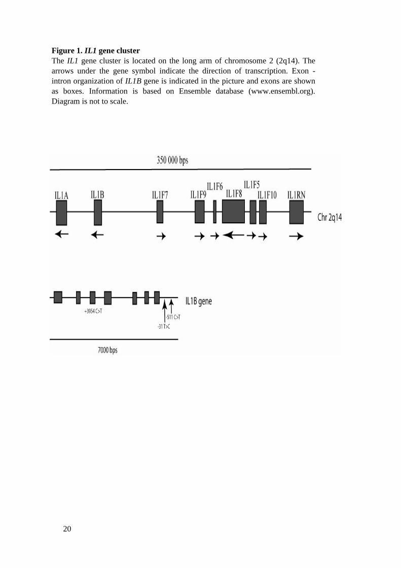
The IL1 gene cluster including IL1B gene is located on the long arm ofchromosome 2 (2q14) as seen in Figure 1. IL1B gene is about 7.0 kbp in lengthcontaining seven exons and six introns. The intronexon organization of IL1 genecomplex genes suggests duplications of a common gene some 350 million yearsago. IL1B regulatory regions are distributed over several thousand base pairsupstream and a few base pairs downstream from the transcriptional start site(Dinarello 2002, Barksby et al. 2007).
IL1B gene has several polymorphic sites. 83 SNPs have been listed in theIL1B gene region determined by NCBI (http://www.ncbi.nlm.nih.gov). However,many of these SNPs contain only one genotype, suggesting that these areartefacts of the database and thereby the number of SNPs in this gene area couldbe smaller. There are also many SNPs reported without allele frequencies andSNPs with very low minor allele frequency (<0.05). The structure of IL1B geneand the most studied polymorphisms, IL1B511C>T (rs16944), IL1B31T>C(rs1143627) and IL1B+3954C>T (rs1143633) are shown in Figure 1.
19
This dissertation focuses on IL1B promoter region C to T baseexchangepolymorphism at position 511 from the transcription start site. Thispolymorphism was first described by di Giovine and colleagues in 1992 (diGiovine et al. 1992). The ILIB511 polymorphism is in nearcomplete linkagedisequilibrium with the TATA box polymorphism IL1B31 in Caucasianpopulation so that the IL1B511 allele T is in linkage with the IL1B31 allele C(ElOmar et al. 2000, ElOmar et al. 2001).
The exact biological role of IL1B511 polymorphism is under investigationand the results have so far been somewhat confusing. The IL1B511 allele T hasbeen shown to increase the transcriptional activity more than allele C (Chen et al.2006). The IL1B511 TT genotype has been associated with higher gastricmucosa IL1 levels compared to other genotypes in H.pylori positive Japaneseadult population (Hwang et al. 2002). However, the IL1B511 CC genotype hasbeen associated with increased production of IL1Ra and IL1 in LPSstimulated PBMCs (Reich et al. 2002, Iacoviello et al. 2005). According torecent IL1B promoter haplotype studies, it seems that the functional role of IL1B511 polymorphism may depend on the IL1B promoter region haplotype context.The IL1B511 allele T has strongly enhanced transcription with the IL1B31allele C, whereas the enhancement was significantly lower in the context ofIL1B31 allele T (Chen et al. 2006). In addition, IL1B promoter haplotype 511T/31C has been associated with 23 fold increase in LPS induced IL1secretion (Hall et al. 2004). However, another three locus haplotype 1470G/511C/31T have been shown to produce more IL1 after LPS stimulationcompared to 1470C/511T/31C haplotype. The former haplotype was alsotranscriptionally more active (Wen et al. 2006). IL1B511 polymorphism mayalso have epistatic effects on protein production with other gene polymorphismslike IL1RN (Hurme et al. 1998, Hwang et al. 2002).
Associations between IL1B511 polymorphism and diseases have beenwidely studied in recent years and over one hundred reports have been published.Associations have been found, for example, with Alzheimer’s disease, asthma,chronic hepatitis C, gastric cancer, severe gastric inflammation, ischemic stroke,myocardial infarction, allergic rhinitis, EBV infection, Parkinson’s disease,multiple sclerosis, bone marrow transplantation, schizophrenia, major depressivedisorder, psoriasis, FSs, localization related epilepsy etc. (Hollegaard et al.2006). The role of genotypes seems to differ in various diseases. For example,the IL1B511 genotype TT has been associated with an increased risk for gastricinflammation whereas this genotype has been associated with decreased risk ofischemic stroke and myocardial infarction at young age (Hwang et al. 2002,Iacoviello et al. 2005). In several studies no associations between IL1B511polymorphism and diseases have been found (Hollegaard et al. 2006).
20
Figure 1. IL1 gene clusterThe IL1 gene cluster is located on the long arm of chromosome 2 (2q14). Thearrows under the gene symbol indicate the direction of transcription. Exon intron organization of IL1B gene is indicated in the picture and exons are shownas boxes. Information is based on Ensemble database (www.ensembl.org).Diagram is not to scale.
21
1.2. IL1B511C>T polymorphism and febrile seizures
1.2.1. Febrile seizure
FS has been defined as a seizure occurring in childhood associated with febrileillness not caused by CNS infection, without previous neonatal or unprovokedseizures and not meeting the criteria of other acute symptomatic seizures. FSs arethe most common type of convulsions in childhood affecting approximately 25% of children living in western countries. FSs usually occur between 6 monthsand 5 years of age with the peak incidence at 18 months (Waruiru et al. 2004,Fetveit 2008).
FSs have been classified as simple and complex. Simple FS has been definedas a selflimiting tonicclonic seizure of short duration (usually less than 15minutes), not recurring within 24 hours and without postictal pathology.Complex FSs have focal onset or focal features during seizure, are followed byneurological deficit, last over 15 minutes or recur during the same febrile illnesswithin 24 hours. Most of the FSs are simple and the incidence of complexseizure is 935% of all FSs (Waruiru et al. 2004, Fetveit 2008).
Risk factors for the first FS are high fever and positive genetic backgroundin first degree relatives. 2540% of children having FSs have a positive familyhistory of febrile convulsions (Berg AT et al. 1995). The rate of temperature risehas commonly been believed to be a risk factor for FS, but it has not beenassociated with FSs in every study (Berg AT et al. 1995, Waruiru et al. 2004).Other factors related to FSs are, for instance, daycare, exposure to passivesmoking prenatally and immunization, but the results have been contradictory(Berg AT et al. 1995, Waruiru et al. 2004, Sillanpää et al. 2008). Both viral andbacterial infection causing fever can provoke FSs (Waruiru et al. 2004).
FSs are usually benign in nature. The risk of recurrence of febrile seizure isabout 2935%. Risk factors for recurrent FS are, for example, young age at thetime of the first FS (<18 months), a positive family history of FS, relatively lowfever during the first FS and short duration of fever before the first febrileseizure (Shinnar et al. 2002). The risk of developing epilepsy after simple FSs is12.4% and 4.16% after complex FSs. Yet 1015% of people with epilepsy orunprovoked seizures have a history of FSs. Risk factors for developing epilepsyafter FSs are positive family history of epilepsy, complex features of FSs and thepresence of early onset neurodevelopmental abnormalities (Waruiru et al. 2004,Fetveit 2008). It has been speculated that the associations between FSs andepilepsy may demonstrate a genetic link between these two diseases rather than acausal relationship, because the evidence of causality is not unambiguous(Waruiru et al. 2004, Fetveit 2008).
22
1.2.2. IL1 and febrile seizures
Cytokines are important immunomodulators in CNS and they have beenassociated with FSs (Rothwell et al. 2000). However, the importance of feverinducing cytokines, like IL1, in FSs is disputed. Helminen and Vesikari firstreported increased IL1 production in LPS stimulated mononuclear cells isolatedfrom FS patients (Helminen et al. 1990). This finding has been repeated inanother study (Straussberg et al. 2001). Increased IL1 production has also beenseen in doublestranded ribonucleic acid stimulated leukocytes obtained fromchildren with positive history of FSs (Matsuo et al. 2006). Elevated plasma IL1levels have been found in acute phase of FS, but cerebrospinal fluid (CSF) IL1levels were not associated with FSs in this study (Tütüncüoglu et al. 2001).Interestingly, in another study elevated CSF IL1 levels were seen in FSchildren, but no association between plasma IL1β levels and FSs was found(Haspolat et al. 2002). In some studies no associations between plasma or CSFIL1 levels and FSs have been found (Lahat et al. 1997, Ichiyama et al. 1998,Tomoum et al. 2007).
The relationship between IL1 and seizures has been studied in animalmodels. Expression of messenger RNA (mRNA) of many cytokines, like IL1β,IL6 and TNF , has been reported in the brain after kainic acid induced seizures(Minami et al. 1991). Intrahippocampally administrated kainic acid has beenshown to induce IL1 production in hippocampus whereas intrahippocampallyadministrated IL1 increased the duration of kainic acid induced seizure activityand this effect was blocked by IL1Ra (Vezzani et al. 1999). FSs have also beenstudied in experimental seizure model in mice with IL1 receptor deficiency. TheIL1R deficient mice were more resistant to FS than wild type mice (Dube et al.2005). In this study high IL1 doses were able to induce seizures even withoutrise of temperature, but only in IL1 receptorexpressing mice (Dube et al.2005).
In addition to a putative role as a seizure inducing factor, IL1 may alsoparticipate in the pathogenesis of FSs by regulating fever, which is the maintrigger of FSs. IL1 produced during infection triggers IL1 receptors on thehypothalamic vascular network resulting in synthesis of cyclooxygenase type 2,which elevates brain PGE2 levels leading to activation of the thermoregulatorycenter (Dinarello 1996b, Mackowiak et al. 1997, Davidson et al. 2001, Dinarello2005). IL1 can also cause fever by interacting with other proinflammatorycytokines, like IL6 and TNF , which induce fever (Dinarello 1996b). Antiinflammatory cytokines, such as IL1Ra, may downregulate the effect of proinflammatory cytokines during the febrile response and therefore the balancebetween proand antiinflammatory cytokines may contribute to the level offever and could have a role in the pathogenesis of FSs (Opp et al. 1991, Miller etal. 1997, Fukuda et al. 2009). Hyperthermia itself may induce an excitatoryeffect in the brain especially in immature hippocampus (Schiff et al. 1985,Thompson et al. 1985, Moser et al. 1993, Liebregts et al. 2002, Baulac et al.2004).
23
1.2.3. Associations between IL1B511 and febrile seizures
Genetic predisposition to FSs has been shown in family and twin studies (Baulacet al. 2004, Nakayama et al. 2006). FSs most probably have multifactorial origini.e. both genetic and environmental factors have a role in their pathogenesis. FSshave been reported to be linked to many genetic loci including 2q, 5q, 6q, 8q 18pand 19. However, it seems that simple sporadic FSs differ genetically fromcomplex and familial FSs and most of the loci mentioned above do not have arole in simple FSs except for the chromosome 5 locus reported in Japanesepopulation (Nakayama J et al. 2000, Waruiru et al. 2004).
Genes, like IL1B, encoding proteins involved in the regulation ofinflammatory reaction and fever are also considered to be plausible candidategenes in the pathogenesis of FSs (Kauffman et al. 2008). Increased carriage ofthe IL1B511 allele T has been found in TLE patients with hippocampal sclerosis(TLE+HS) and in localizationrelated epilepsy patients (Peltola et al. 2001,Kanemoto et al. 2003). According to these results IL1B511 polymorphism mayhave a role in the pathogenesis of convulsions. However, the results of recentassociation studies between IL1B511 and FSs have been contradictory and inmost studies no association has been found (Kauffman et al. 2008).
2. Interleukin10 promoter polymorphisms and EpsteinBarr virus infection
2.1. Interleukin10
2.1.1. Function of IL10
IL10 is considered to be an antiinflammatory multifunctional cytokine. It wasfirst described as a cytokine synthesis inhibitory factor (CSIF) when the Th2clones were shown to produce a factor that inhibited proliferation and cytokineproduction by activated mouse Th1 clones (Fiorentino et al. 1989). Human IL10cDNA was demonstrated in 1991 (Vieira et al. 1991). IL10 is produced by manycells such as T cells, B cells (especially EBV infected or CD5+B cells),monocytes and keratinocytes (O'Garra et al. 2008).
IL10 has an important role in the regulation of immune responses and affectsmany cell types. For example, IL10 inhibits cytokine production andproliferation of T cells responding to antigens and IFN production by naturalkiller (NK) cells. Most of the inhibitory effect of IL10 on T cell cytokineproduction seems to be caused indirectly via suppressing crucial antigenpresenting cell (ACP) functions. IL10 is able to downregulate HLA IIexpression and antigen presentation of APC. IL10 also inhibits, for example, theexpression of CD80 and CD86 surface molecules on APCs. These molecules are

24
ligands for CD28 and CTLA4 on T cells and mediate costimulatory signalsaffecting T cell activation (Moore et al. 2001). IL10 has also been shown tohave a direct inhibitory effect on T cell activation by suppressing the expressionof the Tcell costimulatory molecules CD28 and ICOS (Taylor et al. 2007).Additionally, IL10 inhibits a number of inflammatory functions of monocytesand macrophages by inhibiting the synthesis of many cytokines (e.g. IL1, TNF
, IL6, IL10 itself), chemokines and PGE2. IL10 has been shown to promoteB cell activation and differentiation and induces immunoglobulin synthesis andautoantibody production (Llorente et al. 1995, Moore et al. 2001). IL10 has alsobeen found to be a potent suppressor of both total and specific IgE production,while it simultaneously increases IgG4 production (Blaser et al. 2004). Inaddition, IL10 has shown direct inhibitory effects on mast cells and basophils(Pierkes et al. 1999, Royer et al. 2001).
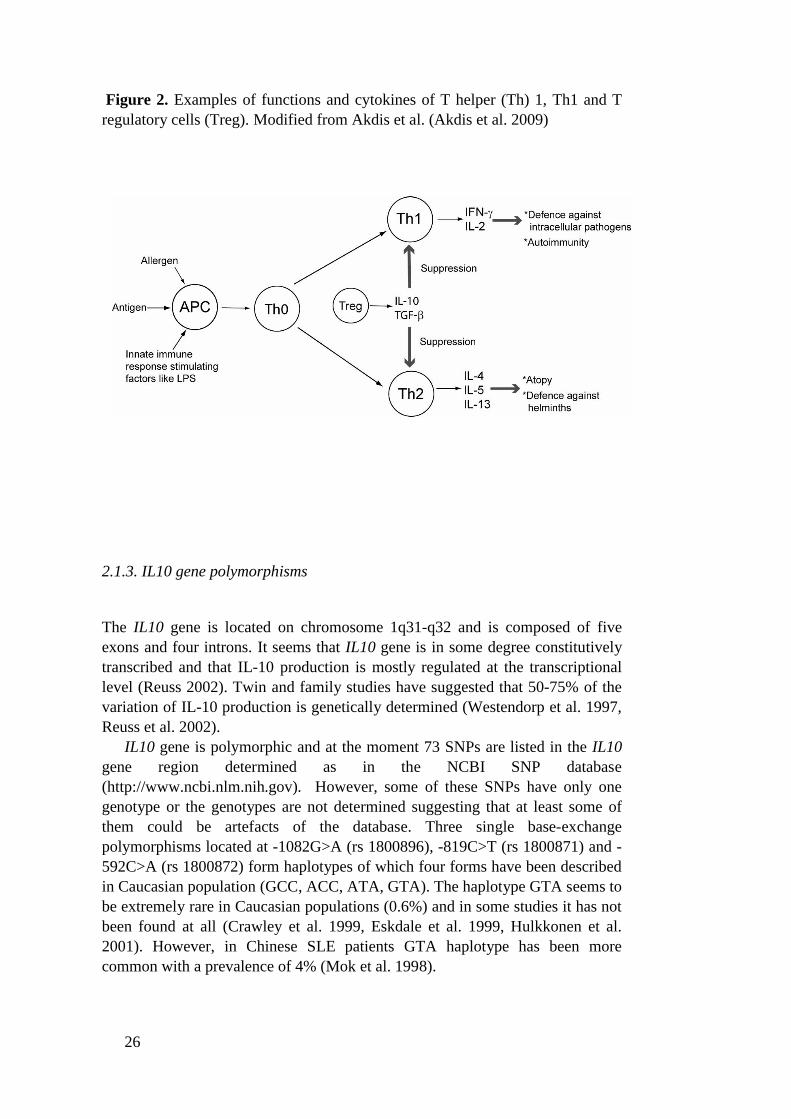
Maintenance of peripheral tolerance has been associated with regulatory Tcells (Tregs). Suppressive effects of inducible Tregs on Th1 and Th2 reactionsare, at least partly, mediated by IL10 and TGF (Vignali et al. 2008).Downregulation of T helper (Th)1 and Th2 responses by Tregs and IL10 arepresented in Figure 2. IL10 has been shown to induce the differentiation ofTregs and also thereby mediate tolerence (Groux et al. 1997). In addition, IL10has been associated with inducing of T cell anergy (Groux et al. 1996). IL10also modulates the influence of TGF on T cells via regulation of the expressionof TGF receptor (Cottrez et al. 2001).
The functions of IL10 are mediated by IL10 receptor (IL10R), which iscomposed of two subunits, the ligandbinding IL10R1 and the accessory subunitIL10R2. These subunits are members of the IFN receptor family (Moore et al.2001). IL10R1 is mainly expressed by hemopoietic cells and IL10R2 in mostcells and tissues studied (Liu et al. 1994, Moore et al. 2001, Wolk et al. 2002).
IL10 has closely related homologs in several virus genomes of which thehomology to EBV IL10 (ebvIL10) gene was found first (Moore et al. 1990,Moore et al. 2001). IL10 superfamily has been described and includes IL10,viral gene homologs of IL10, IL19, IL20, IL22, IL24, IL26, IL28 and IL29. These cytokines share genetic similarity through exonintron gene structure,share receptors and have conserved signal cascades. However, the effects thatcellular IL10 family cytokines mediate differ significantly from immunesuppression to enhancing antiviral activity (Commins et al. 2008).
2.1.2. Role of IL10 in disease
The major function of IL10 seems to be limitation of host’s harmful immuneresponses during infection and inflammation. IL10 downregulates both Th1 andTh2 immune responses and thereby it seems to have important role inautoimmune and atopic diseases (von Hertzen et al. 2009). The role of IL10 ininfections and systemic inflammations has been studied in both humans andanimals (Moore et al. 2001).

25
IL10 deficient mice have been very susceptible to LPSinduced shock andadministration of IL10 has a protective effect against LPSinduced shock (BergDJ et al. 1995). However, in human studies high IL10 levels have beenassociated with fatal outcome in meningococcal disease (Lehmann et al. 1995,Westendorp et al. 1997). IL10 / mice have been shown to develop chronicenterocolitis (Berg et al. 1996). In human studies elevated IL10 levels have beenfound in autoimmune diseases including systemic lupus erythematosus (SLE),Sjögren syndrome and RA patients (Llorente et al. 1995, Hulkkonen et al. 2001).IL10 has also been associated with many other diseases like EBV infection,psoriasis, cancer, diabetes and graftversushost disease (Moore et al. 2001).
Associations between atopic phenotypes and IL10 have also been found. Inanimal studies Treg cells have been shown to inhibit allergen specific IgEresponse in mice and this effect was at least partly mediated by IL10 (Cottrez etal. 2000). Treg cells have also been shown to regulate airway hyperreactivity inTGF and IL10 dependent manner in mice (Joetham et al. 2007). In addition,diminished IL10 concentrations in the lungs and genetically determined low IL10 production have been associated with asthma in humans (Borish et al. 1996,Lim et al. 1998). Allergen specific immunotherapy has been related withincreased IL10 production in vitro in many studies (Jutel et al. 2003, Savolainenet al. 2004, Hawrylowicz et al. 2005, von Hertzen et al. 2009).
IL10 has been considered for therapeutic use because of its antiinflammatory functions. The early studies of recombinant IL10 treatmentshowed promising results, but in larger studies the results have beencontradictory and side effects have also been reported (Moore et al. 2001,O'Garra et al. 2008) Other ways of using IL10 for treatment are underinvestigation. For example, gene therapy approaches to target local expression ofIL10 by adenoviral vector expressing IL10 have been studied in animal models(Scumpia et al. 2005, O'Garra et al. 2008).
26
Figure 2. Examples of functions and cytokines of T helper (Th) 1, Th1 and Tregulatory cells (Treg). Modified from Akdis et al. (Akdis et al. 2009)
2.1.3. IL10 gene polymorphisms
The IL10 gene is located on chromosome 1q31q32 and is composed of fiveexons and four introns. It seems that IL10 gene is in some degree constitutivelytranscribed and that IL10 production is mostly regulated at the transcriptionallevel (Reuss 2002). Twin and family studies have suggested that 5075% of thevariation of IL10 production is genetically determined (Westendorp et al. 1997,Reuss et al. 2002).
IL10 gene is polymorphic and at the moment 73 SNPs are listed in the IL10gene region determined as in the NCBI SNP database(http://www.ncbi.nlm.nih.gov). However, some of these SNPs have only onegenotype or the genotypes are not determined suggesting that at least some ofthem could be artefacts of the database. Three single baseexchangepolymorphisms located at 1082G>A (rs 1800896), 819C>T (rs 1800871) and 592C>A (rs 1800872) form haplotypes of which four forms have been describedin Caucasian population (GCC, ACC, ATA, GTA). The haplotype GTA seems tobe extremely rare in Caucasian populations (0.6%) and in some studies it has notbeen found at all (Crawley et al. 1999, Eskdale et al. 1999, Hulkkonen et al.2001). However, in Chinese SLE patients GTA haplotype has been morecommon with a prevalence of 4% (Mok et al. 1998).

27
These IL10 promoter haplotypes seem to be functionally relevant, becausethey have been associated with IL10 production. The ATA haplotype has beenassociated with lower transcriptional activity than GCC haplotype and theATA/ATA haplotype combination with lower IL10 production in LPSstimulated whole blood cultures compared to other haplotypes (Crawley et al.1999). The GCC haplotype has been shown to have 20% higher transcriptionalactivity compared to ACC and ATA haplotype in luciferase reporter gene assay(Reuss et al. 2002). In vivo the GCC haplotype has been associated with elevatedplasma IL10 levels in primary Sjögren’s syndrome patients (Hulkkonen et al.2001). However, there are also controversial results and ATA haplotype carriershave been reported to have higher IL10 levels than noncarriers among healthyadults (Kilpinen et al. 2002).
It has been postulated that IL101082, IL10819 and IL10592polymorphisms are located within important regulatory regions and may alter thestructure of transcription factor binding sites. The IL101082 polymorphism islocated within E26 transformation specific (ETS) transcription factorbindingsite and allele A has been shown to confer a higher binding affinity to thetranscription factor PU.1, which inhibits gene expression and leads to decreasedIL10 expression (Reuss et al. 2002). In addition, the IL101082 allele A carriershave been associated with lower IL10 production compared to allele A noncarriers (i.e. GG genotype) in Con A stimulated PBMC cultures (Turner et al.1997). The IL10819 is located within a putative positive regulatory region andthe IL10592 polymorphism is situated within a possible STAT3 binding site anda negative regulatory region, but the exact transcription factors have not beenfound (Kube et al. 1995, Reuss et al. 2002).
Almost one hundred reports on an association between IL10 promoterhaplotype 1082/819/592 and diseases have been published. For example, theputatively low producing haplotype ATA has been found to be associated withasthma severity, SLE with renal disease, susceptibility to herpes zoster, a severeform of malaria, aggressive periodontitis and susceptibility to melanoma yet atthe same time with better survival in advanced melanoma (Lim et al. 1998, Moket al. 1998, Haanpää et al. 2002, Vuoristo 2007, Ouma et al. 2008, Reichert et al.2008). The possibly high producing haplotype GCC has in turn been associatedwith Sjögren’s syndrome, poor response to IFN therapy in hepatits C and SLE(EdwardsSmith et al. 1999, Hulkkonen et al. 2001, Rosado et al. 2008). In somestudies no associations between diseases and IL10 promoter haplotype have beenfound (Hollegaard et al. 2006).

28
2.2. IL10 promoter haplotype and EBV
2.2.1. EBV infection
EBV belongs to the human herpes virus family. EBV infection is very commonand approximately only 5% of adults are seronegative for EBV. The primaryEBV infection usually occurs within the first years of life, when the symptomsare mild or the infection is asymptomatic. In industrialized countries with a highstandard of living many children are protected from early infection and usuallycontract EBV infection during adolescence and even in adulthood, when up to3050% of EBV infections can be presented as acute infectious mononucleosis(IM) with fever, tonsillitis, lymphadenopathy, hepatitis and splenomegaly. Insome cases IM has potentially lifethreatening manifestations like meningoencephalitis, myocarditis and pneumonia. EBV has also been associated withmany malignancies including Burkitt’s lymphoma, Hodgkin’s disease,nasopharyngeal carcinoma and gastric carcinoma (Crawford 2001).
EBV is mainly transmitted through the salivary contact. After oraltransmission, EBV replicates in a permissive cell type in the oropharynx. Thesecells are probably specialized epithelial cells that bind virus directly or acquirevirus by transfer from the surface of adjacent B cells. The virus infects mucosalB cells and initiates a latent infection simultaneously (ShannonLowe et al.2006). Cell mediated immunity and cytokines seem to be crucial to the host’sdefense against infection. Immune responses are able to control the primaryinfection, but they do not eliminate the virus. The EBV infected B cellsconstitute the site of latency and after the primary infection the virus remains inthe body for life (Crawford 2001).
EBV infection seldom occurs after 20 years of age and 8595% of 20yearolds are already EBV seropositive. The reason why some people remainseronegative for EBV is not clear, but it seems that seronegative adults differfrom seropositive adults in some immunologic functions. Higher percentage ofmonocytes in the peripheral blood and increased IFN and IL6 levels in culturesupernatants of seronegative adults have been reported. However, the expressionlevels of the EBV receptor CD21 on peripheral B cells have not differed betweenEBV negative and positive subjects (Jabs et al. 1996, Jabs et al. 1999). It has alsobeen hypothesized that EBV seronegative individuals may have immunogeneticdifferences compared to seropositive (Helminen et al. 1999).
2.2.2. Role of IL10 in EpsteinBarr virus infection
IL10 plays a central role in the establishment and persistence of EBV infection.Elevated levels of circulating IL10 have been found in people with acute andchronic acute EBV infection and it has been speculated that IL10 maycontribute to disease pathogenesis by inhibiting host immunity and allowing thedevelopment of latency (Taga et al. 1995, Kanegane et al. 1997). During EBV

29
latency EBV gene products are able to enhance humanIL10 (hIL10) transcriptionand production of hIL10, thus supporting the persistence of latent infection(Marshall et al. 2003).
EBV codes for a cellular homolog of IL10 called viral IL10 (ebvIL10, latervIL10). The amino acid sequences of hIL10 and vIL10 are 84% identical andvIL10 shares similar properties with human IL10 including both cellproliferative and antiimmune functions. The effect of vIL10 may depend on theduration of exposure, because vIL10 has been shown to have a stimulatoryeffect on T cells after longterm vIL10 secretion, whereas short exposure to vIL10 has shown inhibitory effects (Müller et al. 1999). vIL10 is 3 to 10fold lesspotent than hIL10 and has at least 100 to 1000fold lower affinity to IL10receptor than hIL10 (Moore et al. 2001). Both vIL10 and hIL10 have beenable to induce expression of EBV latent membrane protein 1 in EBV infected Bor NK cells and thus IL10 may have a role in the establishment of latency (Kiset al. 2006). ebvIL10 gene (BCRF1) seems to be well conserved among the EBVstrains, which emphasizes the importance of vIL10 in EBV infection (Kanai etal. 2007).
2.2.3. Associations between IL10 gene promoter polymorphisms and EpsteinBarr virus
Associations between IL10 gene polymorphisms and EBV or EBV associateddiseases have been investigated in many studies. High IL10 gene expression hasbeen reported among the IL101082 allele G carriers in EBVtransformedlymphoblastoid cells lines of fullterm healthy infants (Capasso et al. 2007).IL101082 polymorphism has also been shown to have an effect on both thesusceptibility and severity of EBV infection in Finnish adults (Helminen et al.1999). IL101082 allele G carriers were more often seronegative for EBVwhereas IL101082 allele A carriers had more severe EBV infection leading tohospitalization. IL101082 allele G has been associated with higher IL10producing capability and therefore it has been speculated that the possibly lowerproducing capability associated with IL101082 allele A makes people moresusceptible to severe EBV infection (Helminen et al. 1999). In addition, IL10819 CC genotype has been associated with increased risk of elevated EBV IgGantibody titers in Japanese women (Yasui et al. 2008). However, there is onlyone report concerning an association between IL10 promoter haplotype1082G>A/819C>T/592C>A and EBV infection. In this study ATA haplotypewas associated with diminished risk of early EBV infection (Helminen et al.2001).
EBV has been associated with several malignancies including gastriccarcinoma, nasopharyngeal carcinoma and Hodgkin’s lymphoma. Therefore therelationships between IL10 promoter polymorphisms and these diseases havebeen investigated. IL101082 allele G frequency has been increased in EVBnegative gastric carcinoma patients compared to controls (Wu et al. 2002).However, IL101082 allele G was extremely rare, especially among EBV
30
seropositive gastric carcinoma patients, so the results cannot be generalized. Aparallel association has been found in patients with undifferentiated carcinoma ofnasopharyngeal type (UCNT) (Pratesi et al. 2006). In this study IL10 1082/819/592 haplotype was not related to UCNT, but the IL101082 allele G wasassociated with EBVnegative UCNT (Pratesi et al. 2006). In addition, thepossibly high producing IL101082 GG genotype has been associated with EBVpositive Hodgkin’s lymphoma development, whereas IL10819 and IL10592polymorphisms were not related to this disease. The threelocus haplotype wasnot analyzed in this study (da Silva et al. 2007). The IL101082 GG genotype hasalso been associated with decreased risk of lateonset EBVassociated posttransplant lymphoproliferative disorder in solid organ recipients (Babel et al.2007). However, this association has not been seen in children (Lee TC et al.2006).
3. Interleukin 4 promoter polymorphism and atopy
3.1. Interleukin4
3.1.1. Function of IL4
Interleukin4 is a pleiotropic cytokine produced by activated T cells, mast cellsand basophils. It was already functionally characterized in 1982 as a T cellderived B cell growth factor distinct from IL2 (Howard et al. 1982).
IL4 plays an important role in the regulation of B and T cell mediatedimmune reactions. It promotes immunoglobulin synthesis and directs theimmunoglobulin class switching into the synthesis of IgE and IgG4 in activatedB lymphocytes (Pene et al. 1988). It also enhances the antigen presentingcapacity of B cells and upregulates IgE receptors on B lymphocytes, mast cellsand basophils thereby enhancing the activation of these cells during allergicchallenge. Differentiation of precursor Th cells into the Th2 subset is alsoregulated by IL4. IL4 stimulates the production of Th2 cytokines including IL5, IL9, IL13 and IL4 itself. IL4 also has antiinflammatory effects byinhibiting the production of proinflammatory cytokines like IL1, TNF andIL6 and stimulating IL1Ra production. IL4 also activates the expression ofadhesion molecules like vascular cell adhesion molecule 1 and chemokines likeeotaxin and thereby directing the migration of cells to the inflammatory site andpromoting eosinophilic inflammation (Paul 1991, Romagnani 2004, Andrews etal. 2006).
IL4 exerts its biological effects by binding to IL4 receptor (IL4R)complex, which consists of two subunits: IL4R , which is a high affinity IL4binding site shared with IL13, and cchain (type I receptor) or IL13R 1 (typeII receptor). Type I receptor binds only IL4 and requires IL4R for assembly
31
with cchain whereas type II receptors binds both IL4 and IL13 and requireassembly of IL4R with IL13R 1subunit. Because IL4 and IL13 share thesame receptor they have many similar functions. IL4Rs are expressed on bothhematopoietic and nonhematopoietic cells like epithelial, endothelial, muscle andliver cells (KellyWelch et al. 2003, Steinke 2004). There is also a soluble formof IL4R (sIL4R) (Andrews et al. 2006).
Due to its many functions IL4 has been associated with various diseases,especially with atopy, which will be discussed in detail later. IL4 has alsoshown a potent antitumor activity (Paul 1991). However, the results ofrecombinant human IL4 (rhIL4) in cancer treatment have been disappointing,because rhIL4 has shown only low antitumor activity (Vokes et al. 1998,Whitehead et al. 2002). Furthermore, autocrine IL4 production has been seen inmany kinds of cancer cells and it seems that colon cancer stem cells resistapoptosis by producing IL4 (Todaro et al. 2007).
Th2 cells are important in immune reactions against many parasites. Highlevels of IL4 have been found in parasite infected mice (Paul 1991). IL4 seemsto be important in host protection against parasites like Trichinella spiralis(Finkelman et al. 2004). In bacterial infections the role of IL4 is somewhatconfusing. In mouse models IL4 has been shown to enhance pulmonaryclearance of Pseudomonas aeruginosa, but in case of Staphylococcus aureus IL4 has been associated with increased risk of septic arthritis (Hultgren et al. 1998,JainVora et al. 1998).
3.1.2. IL4 gene polymorphisms
IL4 gene is located on chromosome 5q31, which in many studies has been linkedwith atopic phenotypes (Marsh et al. 1994, Palmer et al. 1998, Ober et al. 2000).Many other atopy related genes like IL5, IL9, IL13 and CD14 are located in theadjacent area as seen in Figure 2. IL4 gene has 4 exons and 3 introns and is about9 kb in length. At the moment 104 SNPs have been listed in IL4 gene regiondefined by NCBI (http://www.ncbi.nlm.nih.gov). However, Sakagami and coworkers sequenced 25.6 kb genomic region including both IL13 and IL4 genesand found 45 SNPs in IL4 gene region (from 600 to +8500 bps fromtranscription start site) (Sakagami et al. 2004). Sakagami and colleaguesclassified 14 of these 45 SNPs in IL4 gene as common SNPs (minor allelefrequency at least 0.10 in two populations), which were used for further analysis.A strong linkage disequilibrium across the IL4 gene was found. Two majorhaplotypes accounted for >80% of haplotypes in European Americans andJapanese. However, the haplotype frequencies differed substantially betweenthese populations and therefore it was speculated that natural selection has acteddifferently on IL4 haplotypes in separate populations (Sakagami et al. 2004).
This dissertation focuses on IL4 gene promoter baseexchange polymorphismC to T at position 590 from the open reading frame (rs 2243250). Thispolymorphism was first described in 1994 and belongs to the most studiedpolymorphisms of IL4 gene (Borish et al. 1994). The IL4590 allele T has been
32
associated with stronger transcription of IL4 and has also shown a greaterbinding activity to nuclear transcription factors than allele C (Rosenwasser et al.1995, Nakashima et al. 2002).
The IL4590 polymorphism seems to be functional and therefore its role indiseases has been studied extensively in recent years. The IL4590 allele T hasbeen associated, for example, with human immunodefiency virus 1 (HIV1)syncytiuminducing phenotype, elevated antibody levels against malaria, severityof respiratory syncytial virus infection and H.pylori cagA positive infections(Nakayama EE et al. 2000, Luoni et al. 2001, Hoebee et al. 2003, Zambon et al.2008). An association between IL4590 polymorphism and diphtheria andtetanus vaccine responses in Australian children has also been reported. Thisassociation was modified by parental tobacco smoking: among childrenunexposed to parental tobacco smoking the IL4590 allele T carriers had morestronger diphtheria and tetanus antibody responses than did allele T noncarrierswhereas in exposed children tetanus antibody responses were decreased in alleleT carriers (Baynam et al. 2007). IL4590 polymorphism has also been associatedwith many atopic phenotypes, which will be discussed in more detail later.
The role of the IL4590 allele T seems to vary in different diseases. It hasbeen associated, for example, with susceptibility to subacute sclerosingpanencephalitis in Japanese children, RA in Columbian patients and Crohn’sdisease in Caucasian population (Klein et al. 2001, Inoue et al. 2002, Moreno etal. 2007). However, the IL4590 allele T has also been associated withdiminished risk of autoimmune thyroid diseases, myocardial infarction at youngage, severity of Sjögren’s syndrome, survival in colorectal cancer in Caucasianpopulations and in minimal change nephritic syndrome in Japanese children(Hunt et al. 2000, Kobayashi et al. 2003, Pertovaara et al. 2006, Paffen et al.2008, Wilkening et al. 2008).

33
Figure 3. IL4 and CD14 gene regions
IL4 and CD14 genes are located on chromosome 5 at chromosome band 5q31.This area also contains many other atopy associated genes like IL5, IL9 and IL13as seen in the diagram. The arrows under the gene symbol indicate the directionof transcription. The exon intron organization of common IL4 and commonCD14 gene transcripts (ENST00000302014 and ENST00000231449respectively) are indicated in the lower part of the picture. Exons are shown asboxes. The sites of IL4590C>T and CD14159C>T polymorphisms are markedin the diagram. Information is based on Ensemble Database (www.ensembl.org).The diagram is not to scale.
34
3.2. IL4590C>T polymorphism and atopy
3.2.1. Atopy
Atopy is defined by the European Academy of Allergology and ClinicalImmunology as a personal or familial tendency to produce IgE antibodies inresponse to low dose of allergens and to develop typical symptoms such asasthma, rhinoconjunctivitis or eczema/dermatitis (Johansson et al. 2001). Thepresence of specific IgE is usually studied by either skin prick tests (SPT) orserum assay. The results of SPTs are generally in line with anamnestic data ofatopic symptoms, but not all sensitized individuals develop an atopic disease.Elevated serum total IgE levels have also been associated with atopic phenotypes(Burrows et al. 1989). However, many patients suffering from atopic diseases donot have allergen specific IgE and their serum total IgE levels are normal(Cookson 2004).
Atopic diseases run in families and, for example, asthma heritability has beenestimated in twin studies to vary from 36% to 87% (Nieminen et al. 1991,Laitinen et al. 1998). In addition to genetics, many environmental factors seem tohave a role in the etiology of atopy. Epidemiological evidence has shown thatchildhood infectious diseases, especially gastrointestinal infections such asHelicobacter pylori, Toxoplasma gondii and hepatitis A associated with poorlevel of hygiene, have been associated with decreased risk of atopic diseases(Matricardi et al. 2000, Seiskari et al. 2007). Other environmental factorsassociated with atopy and serum total IgE include, for example, growing up on afarm, number of siblings, tobacco smoke and socioeconomic status (von Mutius2000).
The prevalence of atopic diseases has increased markedly in developedcountries in recent decades. This change has been explained, at least partly, bydecrease of infections due to improved standard of living and better hygiene.This so called “hygiene hypothesis” was first introduced by Strachan in 1989(Strachan 1989). This theory has evolved a great deal in recent years and thereare at least four dimensions that need be considered when assessing thishypothesis: extensive variety of allergic phenotypes, diversity of environmentalexposures, timing of the exposure and the individual’s genetic susceptibility toreact to these exposures (von Mutius 2007). In light of many studies it seems thatinfections occurring during the first years of life could be the most important,because they may have an effect on the maturation of immune system and thedevelopment of Th1/Th2 balance (Matricardi et al. 2000, von Mutius 2007).
The hygiene hypothesis has also been explained on the cellular level. Th cellshave been classified at least as Th1 and Th2 cells according to their mainfunctions and cytokine production (Mosmann et al. 1989). Th1 cells are mainlyresponsible for cellmediated immunity while Th2 cells participate in humoral

35
immune reactions, especially in IgE formation. Th1 cells produce IL2, IFNand TNF whereas Th2 cells produce IL4, IL5, IL9 and IL13 (Mosmann etal. 1989). The differentiation of Th cells is known to be modulated by microbialfactors and by the cytokines they induce. For example, in the presence of LPSthe differentiation of Th cells tends towards Th1 whereas Th2 direction isfavored in the absence of this kind of stimulus. This immune deviation theoryhas been widely accepted (Romagnani 2004). However, it is coming clear thatTreg cells and IL10 and TGF have an important role in the balance betweenperipheral tolerance and allergy. In atopy disturbed balance between Treg andTh2 cells has been shown in both animal and human studies (Cottrez et al. 2000,Akdis et al. 2004, Ling et al. 2004).
The role of allergen exposure in the development of atopic disease is stillunder debate. It has been postulated that the dose, type and timing of allergenexposure may be critical. For example, in asthmatic children early exposure tocat allergen has been shown to increase sensitization to cat, but it had no effecton asthma risk, whereas exposure to dog did not sensitize to dog allergen butprotected against asthma and sensitization to airborne allergens (Almqvist et al.2003). The negative association between pet exposure and sensitization has alsobeen reported in other studies (Karjalainen et al. 2005).
3.2.2. IL4 and atopy
IL4 is considered to be a key cytokine in allergic inflammation because, forexample, it induces IgE synthesis and promotes Th cell differentiation in Th2direction (Romagnani 2004). The role of IL4 in atopy has been widely studiedin both animal models and human.
In IL4 knockout mice sensitization, development of bronchialhyperresponsiveness and anaphylactic shock did not occur and similar effectshave been seen in mice missing a functional IL4 receptor (Brusselle et al. 1995,Grunewald et al. 1998). IL4 has also been shown to induce mucin geneexpression and hypersecretion of mucus in the airways of mice (Dabbagh et al.1999).
In allergic individuals serum IL4 levels have been increased, likewise thenumber of IL4 messenger RNA positive cells in bronchoalveolar lavage(Robinson et al. 1992, Daher et al. 1995). Indivuduals with atopic dermatitishave been reported to have more IL4 producing T cells than nonatopicindividuals (Chan et al. 1996). Inhaled rhIL4 has shown increased airwayhyperresponsiveness associated with eosinophilia in sputum (Shi et al. 1998).However, there is also evidence that genetic risk for atopy could be associatedwith decreased production of both Th1 and Th2 cytokines, because the cordblood IL4 and IFN have been inversely associated with the development ofasthma and atopy and TNF inversely associated with atopy at age 6 (Macaubaset al. 2003).
Because IL4 seems to have an important role in atopy, blocking the effectsof IL4 has been investigated for treatment of atopic diseases. In animal models

36
sIL4R has efficiently inhibited IgE production and therefore recombinant sIL4R has been evaluated as asthma treatment, however in larger studies it was notclinically efficient (Steinke 2004). IL4R antagonist (IL4 mutein), antiIL4monoclonal antibody and antiIL4R monoclonal antibody are underinvestigation for asthma treatment, but the results have not been too promising sofar (Steinke 2004, Andrews et al. 2006).
3.2.3. IL4590C>T polymorphism, Helicobacter pylori and atopy
Associations between IL4590C>T polymorphism and various atopic phenotypeshave been studied widely and over 50 investigations had been published byJanuary 2009 according to the Pubmed Database (www.pubmed.com). IL4590polymorphism has been associated with many atopic phenotypes like asthma,serum total IgE, atopic dermatitis, rhinitis and sensitization determined by skinprick test or specific IgE (Rosenwasser et al. 1995, Burchard et al. 1999, Zhu etal. 2000, Söderhall et al. 2002, Liu et al. 2004, Chiang et al. 2007, Li et al. 2008).However, the results have been inconsistent, and in many studies no associationbetween IL4590 polymorphism and atopic phenotypes has been found (Walleyet al. 1996, Dizier et al. 1999, Elliott et al. 2001).
H. pylori is one example of environmental factors which have beenassociated with decreased risk of atopic phenotypes in many studies (Kosunen etal. 2002, McCune et al. 2003, von Hertzen et al. 2006, Seiskari et al. 2007,Konturek et al. 2008). However, this association has not been seen in every study(Bodner et al. 2000, Uter et al. 2003, Law et al. 2005). In addition, a positiveassociation between food allergy and H.pylori has even been reported (Corradoet al. 1998). H.pylori is a common gastrointestinal pathogen infecting gastricmucosa usually early in life and often leading to lifelong chronic gastritis(Brown 2000, Suerbaum et al. 2002). The prevalence of H.pylori is stronglyrelated with socioeconomic status. H.pylori is positively associated with lowsocioeconomic class, poor living conditions including absence of fixed hot watersupply, contaminated water and household overcrowding (Mendall et al. 1992,Malaty et al. 1994, McCallion et al. 1996). H.pylori infection has been suggestedto influence the development of the immune system by LPS binding with theCD14 receptor which results in increased production of IL12 and IFN . Thiscould drive the immune responses towards the Th1 pathway and thereby have aprotective effect against the development Th2 polarized diseases like atopy (vonMutius 2000). It has also been proposed that negative association betweenH.pylori and atopy may be due to increased IL10 expression associated with thisinfection in some studies (Bontems et al. 2003, Maciorkowska et al. 2005,Oderda et al. 2007), because IL10 is considered to be crucial in the developmentand maintenance of immunological homeostasis in allergy (Hawrylowicz et al.2005).
In animal models IL4 has been shown to limit H.pylori associated gastricinflammation (Smythies et al. 2000). In children with H.pylori infection elevatedIL4 levels have been found in gastric and duodenal mucosa (Bontems et al.
37
2003, Maciorkowska et al. 2005). Because IL4590 polymorphism may have aneffect on IL4 production, associations between this polymorphism and H.pylorihave been investigated. The IL4590 allele T has been shown to enhance the riskfor cagA positive H.pylori infection in gastric cancer patients (Zambon et al.2008). However, in another study no association between IL4590 polymorphismand H.pylori was found (GarcíaGonzález et al. 2007).
There is increasing evidence that genegene and geneenvironmentinteractions may have an effect on atopic phenotypes. Several reports of genegene interactions including IL4590 polymorphism have been published. Forexample, the IL4590 allele T has been shown to increase the risk of asthma inFinnish females when combined with the TLR4+896 allele G. However, thiscombination was not associated with atopy defined by SPT (Ådjers et al. 2005).The IL4590 allele T has also been associated with development of atopicdermatitis and SPT positivity at age of 24 months when combined with theIL13Arg130Gln allele 130Gln. However, this genegene interaction was notassociated with probable asthma diagnosis or rhinitis (He et al. 2003).
There are only few reports of geneenvironment interactions on atopyincluding IL4590 polymorphism and the results have been contradictory.Interaction between exposure to environmental tobacco smoke (ETS) and IL4590 polymorphism has been associated with wheezing without cold in the firstyear of life in African Americans, but not in nonAfrican Americans (Smith et al.2008). Additionally, in Australian children no interaction between IL4590polymorphism and ETS on atopy defined as SPT positivity was found (Baynamet al. 2007). In another study the effect of IL4590 polymorphism onsensitization to mite analyzed by specific IgE has been shown to be modulatedby Dermatophagoides pteronyssinus (Der p 1) allergen levels. In this study theIL4590 allele T was a risk for mite senzitisation only when the Der p 1 allergenlevels were high (Liu et al. 2004).
4. CD14 promoter polymorphism and IgE
4.1. CD14
4.1.1. Function of CD14
CD14 is an important innate immunity receptor belonging to socalled patternrecognition receptors, which participate in starting the innate immune responses.CD14 acts as an accessory receptor for many TLRs including TLR2, TLR3,TLR4 (Schmitz et al. 2002, Lee HK et al. 2006). cDNA of CD14 was describedin 1988 and the functional role of CD14 in LPS recognition was found in 1990(Ferrero et al. 1988, Wright et al. 1990).

38
CD14 recognizes a vast variety of microbial ligands, for example, LPS ofgramnegative bacteria, lipoteichoic acids of grampositive bacteria,mycobacterial glycolipids and mannans from yeast (Vercelli 2002). CD14 has animportant role in endotoxin signaling by facilitating endotoxin responses throughTLR4MD2 system, which leads to the activation of innate hostdefensemechanisms by releasing cytokines, such as IL12, which are regarded asobligatory signals for the differentiation of naïve T cells into Th1 cells. TherebyCD14 also has a role in directing adaptive immunity reactions (Vercelli et al.2001, Martinez 2007). In addition to microbe recognition, CD14 has also beenshown to have an important role in the recognition and clearance of apoptoticcells by macrophages (Devitt et al. 1998). There is also evidence that sCD14may regulate T and B lymphocyte activation and function (Rey Nores et al.1999, Arias et al. 2000). sCD14 also contributes to lipid metabolism as sCD14transports lipids and LPS to high density lipoprotein (HDL) (Wurfel et al. 1995).
CD14 is expressed on the surfaces of monocytes, macrophages andneutrophils as membrane bound molecule (mCD14). There is also a soluble formin the sera (sCD14), which is recognized, for example, by dendritic cells when itis coupled with LPS (LeVan et al. 2001). sCD14 is thought to be derived fromboth proteasedependent shedding of mCD14 on myeloid cells and proteaseindependent release from intracellular compartments. There is accumulatingevidence that sCD14 is also produced by hepatinocytes (LeVan et al. 2001, Zhaoet al. 2007).
Expression of CD14 is regulated by many factors. Bacterial cell wallcomponents including LPS have been shown to upregulate CD14 (Landmann etal. 1996). In addition, maternal exposure to a farming environment rich inmicrobial compounds has led to increased upregulation of innate immunereceptors, including CD14, in childhood (Ege et al. 2006). IL4 and IL13 havebeen shown to downregulate the expression of CD14 (Lauener et al. 1990,Cosentino et al. 1995). In addition to microbial products many otherenvironmental factors like alcohol consumption and use of nonsteroidal antiinflammatory analgetics, have been associated with increased sCD14 levels(Karhukorpi et al. 2002, Campos et al. 2005).
Innate immunity receptors, including CD14, participate in the host defenseagainst harmful pathogens and at the same time influence the etiology of severaldiseases. CD14 has been shown to be an important inflammatory mediator ingramnegative sepsis and increased sCD14 levels in the beginning of this diseasehave been associated with high mortality (Landmann et al. 1995). Consistentwith this finding, CD14 knockout mice have been more resistant to LPS inducedshock than wild type controls (Haziot et al. 1996). Elevated sCD14 levels havealso been associated with many other diseases including HIV, malaria, atopicdermatitis and extensive tissue damage in trauma and severe burns (LeVan et al.2001). AntiCD14 therapies for sepsis treatment are under investigation, but sofar they have achieved limited success (Axtelle et al. 2003)

39
4.1.2. CD14 gene polymorphisms
CD14 gene is located on chromosome 5q31 near IL5, IL9, IL13 and IL4 genes asseen in Figure 2. This area has been connected with the regulation of serum totalIgE and atopy in many linkage studies as mentioned earlier (Marsh et al. 1994,Palmer et al. 1998, Ober et al. 2000, Xu et al. 2000). CD14 gene is about 1.6 kbin length and has 2 exons. CD14 encodes two protein forms: mCD14 and sCD14,which lacks the anchor of mCD14 (LeVan et al. 2001).
There are 21 SNPs reported in CD14 gene area defined by NCBI(http://www.ncbi.nlm.nih.gov). In a recent study CD14 genomic area from 6000up till +2500 bps from the transcription site was sequenced and 17 SNPs werefound. 15 of these 17 SNPs were reported to have minor allele frequency >10%(LeVan et al. 2008). This dissertation focuses on one of the most studied CD14promoter region single baseexchange polymorphisms C to T at position 159from transcription start site (260 from the translation start site, rs 2569190),which was first described in 1999 (Baldini et al. 1999). The CD14159 allele Thas been associated with decreased affinity of DNA/ protein interactions at a GCbox containing binding sites for transcription factors Sp1, Sp2, and Sp3. Anincrease in CD14 gene expression has been seen in CD14159 C to T change sothat allele T is transcriptionally more active (LeVan et al. 2001, Zhao et al.2007). The CD14159 allele T has also been associated with elevated sCD14levels in many studies (Zhang et al. 2008). The CD14159 TT genotype has beenassociated with increased IL10 and IL1 levels after endotoxin stimulation andlower IL4 levels after concanavalin A stimulation (Keskin et al. 2006).
Associations between CD14159 polymorphism and diseases have beenstudied abundantly and over 150 reports had been published by January 2009.Many of these studies investigated the relationship between CD14159polymorphism and serum total IgE or other atopic phenotypes, which will bediscussed in more detail later. A wide variety of other diseases, like IBD,coronary artery disease, type I diabetes, severity of Streptococcus pneumoniaeinfection and H.pylori related gastric carcinoma have also been associated withCD14159 polymorphism (ArroyoEspliguero et al. 2005, Baumgart et al. 2007,Zhao et al. 2007, Dezsöfi et al. 2008, Yuan et al. 2008).
4.2. CD14159C>T polymorphism and serum total IgE
4.2.1. IgE
IgE is an important mediator in allergic reactions and in immune defense againstparasites. IgE was discovered as early as 1960’s (Ishizaka et al. 1966, Johansson1967). The total serum IgE levels have been shown to be markedly heritable indifferent populations whereas it seems that specific IgE response is mainlydetermined by environmental factors (Hopp et al. 1984, Hanson et al. 1991,Miller et al. 2005).
40
IgE is produced by B cells after the immunoglobulin isotype switching to IgEsynthesis mediated by IL4, IL13 and costimulatory signals from CD4+ T cells(Vercelli 2001). IgE acts through two receptors, high affinity IgE receptor Fc RI,and low affinity IgE receptor FRc II also called CD23. Fc RI is mainlyexpressed in mast cells and basophils. FRc II has two forms: FRc IIa, which isnormally expressed on B cells and FRc IIb, which is inducible on T cells, Bcells, monocytes and macrophages by IL4 and IL13 (Corry et al. 1999). Itseems that IgE upregulates the expression of Fc RI at least on basophils byinteracting through Fc RI (MacGlashan et al. 1998). Exposure to allergen in asensitized individual leads to cross linking of IgE/Fc RI complexes, whichcauses mastcell degranulation and the initiation of allergic inflammation(Cookson 1999).
IgE production is regulated by stimulatory signals, such as IL4, IL5, IL9and IL13, provided by Th2 cells and inhibitory signals like IL2 and IFNproduced by Th1 cells (Pene et al. 1988, Nakanishi et al. 1995, Corry et al.1999). IL12 and IL18 have also been shown to suppress IgE secretion probablythrough IFN induction (Yoshimoto et al. 1998). However, IL18 has also beenshown to increase IgE production (Yoshimoto et al. 2000). IL10 seems toinfluence the IL4 induced isotype switching from IgE to IgG4 which maypromote tolerance instead of allergic reactions (Jeannin et al. 1998). At very highIgE levels CD23 may have an inhibitory effect on IgE synthesis (Corry et al.1999). The role of sCD14 in regulation of IgE is not clear, which will bediscussed more in the next chapter.
Parasites, especially heminths, are known to upregulate IgE production(Ramaswamy et al. 1994). Smoking, alcohol consumption and exposure to dieselexhaust have also been associated with increased serum total IgE (Beeh et al.2000, Campos et al. 2006). Many other factors like age, gender, ethnicity andsocioeconomic status have been found to affect serum total IgE, but the resultshave been inconsistent and in some studies no associations have been found(Burrows et al. 1989, Beeh et al. 2000, Litonjua et al. 2005b).
Elevated serum total IgE levels have been strongly associated with bronchialhyperresponsiveness, sensitization, atopic dermatitis and both atopic and nonatopic asthma (Burrows et al. 1989, Beeh et al. 2000, Matricardi et al. 2000,Gern et al. 2004). In addition to atopic phenotypes, IgE has been suggested to belinked with chronic urticaria, myeloma, IBD, HIV infection and Job’s syndrome(hyper IgE syndrome), malaria and helminth infections (Ramaswamy et al. 1994,Beeh et al. 2000, Johansson et al. 2001, SekaSeka et al. 2004, Vonakis et al.2008). Because IgE has a key role in atopic reactions, antiIgE therapy has beendeveloped for the treatment of allergic disease. It has shown efficacy in asthmaand allergic rhinitis patients (Holgate et al. 2005). There are also promisingresults of antiIgE therapy in atopic eczema (Belloni et al. 2007).
41
4.2.2. CD14 and IgE
CD14 has been called a bridge between innate immunity and adaptive immuneresponses because CD14 seems to influence the effects of bacterial componentson the development of Th1/Th2 balance and thereby possibly affect regulation ofIgE (Vercelli et al. 2001). Therefore genetic alterations in CD14 production mayhave an effect on serum total IgE (Vercelli et al. 2001).
However, the relationship between CD14 and IgE is not clear. In somestudies CD14 has been shown to have an inhibitory effect on IgE production.Interaction between sCD14 and B cells has resulted in higher levels of IgG1 anddecreased IgE production and inhibition of IL4 and IL6 secretion (Arias et al.2000). sCD14 has also been associated with IL4 inhibition in other studies(Baldini et al. 1999, Rey Nores et al. 1999). However, the effect of sCD14 onTh1 cytokine IFN has differed from inhibition to stimulation (Baldini et al.1999, Rey Nores et al. 1999). In addition, the CD14 mediated stimulus onmonocytes/macrophages has resulted in the secretion of cytokines, such as IL6,that potently amplify IgE synthesis (Jabara et al. 1994). Thus activation of theCD14 pathway could have an inhibitory or enhancing effect on IgE expression(Vercelli 2003). In human studies, reverse correlation between high serumsCD14 levels and low serum total IgE have been reported (Baldini et al. 1999,Tan et al. 2006). However, this association has not been repeated consistently(Kabesch et al. 2004).
4.2.3. CD14159C>T polymorphism, Helicobacter pylori and serum total IgE
Baldini and colleagues first reported an association between CD14159polymorphism and serum total IgE. In their study atopic children with the TTgenotype had higher sCD14 levels and lower serum IgE levels than allele Ccarriers (Baldini et al. 1999). This finding has received a lot of attention and overfifty studies concerning associations between CD14159 polymorphism andserum total IgE or atopic phenotypes has been published between the first reportand January 2009.
The original finding of Baldini and colleagues has been repeated in manystudies in different ethnic groups (Gao et al. 1999, Koppelman et al. 2001, Leunget al. 2003). However, there are also opposite findings. The CD14159 allele Thas been associated, for example, with atopy in a rural population, eczema andelevated serum total IgE in children and food allergy and nonatopic asthma inadults (Ober et al. 2000, Woo et al. 2003, Litonjua et al. 2005a). In severalstudies no association has been found (Sengler et al. 2003, Kabesch et al. 2004,Liang et al. 2006, Nishimura et al. 2006, Zhang et al. 2008). These conflictingresults have been explained, for instance, by ethnic differences, lack of power insome studies and genegene interactions (Zhang et al. 2008).
The suggested mechanism by which CD14159 polymorphism could modifyserum total IgE is via CD14. A genetically determined increase in CD14expression could result in enhanced responsiveness to pathogen products in early

42
life and modify immune reactions into Th1 direction thus protecting againstatopy (Vercelli 2003, Martinez 2007). The carriers of the transcriptionally moreactive CD14159 allele T have had higher sCD14 levels than allele T noncarriers in many studies (Baldini et al. 1999, Leung et al. 2003, Kabesch et al.2004). However, in some studies no association was found (Liang et al. 2006,Zhang et al. 2008).
Many environmental factors, including H.pylori, have been associated withserum total IgE and atopy as already discussed in Chapter 3. The exactmechanism behind association between H.pylori and atopy is not known.However, LPS of H.pylori is able to bind to mCD14 on monocytes and tostimulate monocytes to secrete cytokines and chemokines (Bliss et al. 1998).Therefore genetically defined changes in H.pylori LPS responses through CD14could have an effect on the Th1/Th2 balance and atopy risk. In Finnish adultsincreased sCD14 levels have been found to be associated with H.pylori infection.This association was modified by CD14159 polymorphism so that sCD14 levelsdiffered significantly more between H.pylori positive and negative individualswith the CD14159 CC homozygotes compared to other genotypes (Karhukorpiet al. 2002). However, not every study found an association between H.pyloriand CD14159 polymorphism (Park et al. 2006).
4.2.4. Effect of geneenvironment interactions on serum total IgE and atopy
The results of association studies concerning CD14159 polymorphism andserum total IgE and other atopic phenotypes are contradictory, as discussedearlier. It is well known that serum total IgE is regulated by both environmentaland genetic factors and therefore geneenvironment interactions may be morerelevant than CD14159 polymorphism alone in the regulation of serum totalIgE.
The concomitant effect of endotoxin and CD14159 polymorphism on IgEand atopy has been investigated in many studies. In five studies the CD14159CC genotype has been associated with diminished risk of atopy and high serumtotal/specific IgE when exposure to endotoxin has been high, whereas at lowendotoxin levels this genotype has been an increased risk for these phenotypes(Eder et al. 2005, ZambelliWeiner et al. 2005, Simpson et al. 2006, Williams etal. 2006, Williams et al. 2008). Marginally significant interactions betweenendotoxin exposure and CD14159 polymorphism on serum total IgE or atopicsensitization were found in four of these five studies (Eder et al. 2005, Simpsonet al. 2006, Williams et al. 2006, Williams et al. 2008).
Other environmental factors have also been included in interaction studies.Eder and colleagues found a significant interaction between CD14159polymorphism and animal exposure related to specific and total serum IgE: theCD14159 allele C was associated with lower levels of both total and specificIgE in children with regular contact with stable animals, whereas this allele wasa risk factor for elevated IgE levels in children with regular contact with furrypets (Eder et al. 2005). Similar interactions between CD14159 polymorphism
43
and pet exposure associated with serum total IgE, sensitization and atopicdermatitis have been reported in children (Gern et al. 2004, Bottema et al. 2008).However, conflicting results concerning exposure to farming environmentincluding stable animals has been reported (Leynaert et al. 2006). Alcohol andETS have been reported to have interaction with CD14159 polymorphism onIgE or atopy (Choudhry et al. 2005, Campos et al. 2006). However, in someother studies no interaction between ETS and CD14159 polymorphism on atopyphenotypes has been found (Simpson et al. 2006, Bottema et al. 2008).
The exact mechanisms behind these interactions are not known. According tothe endotoxin switch model environmental endotoxin modulates the Th1/Th2balance so that low and very high endotoxin exposure directs this balance in Th2direction and with intermediate endotoxin exposure Th1 responses will develop(Vercelli 2003). Polymorphisms of innate immunity genes, like CD14 crucial inhost/environment interface, may modulate immune responses by changing theendotoxin switch so that the carriers of the probably high producing CD14159allele T require less environmental endotoxin to switch in Th1 responses, andthat thereby this the CD14159 allele T could protect from allergy (Vercelli2003). However, when the effects of CD14159 polymorphism on serum totalIgE and atopy have been concomitantly investigated with environmentalendotoxin exposure, the CD14159 allele C has been suggested to be a risk factorfor atopic phenotypes at low levels of endotoxin or microbial exposure, whereasthe CD14159 allele T seemed to be a risk factor at high levels of exposure(Martinez 2007). It has also been speculated that there could be a difference inconstitutive and induced CD14 synthesis so that constitutive CD14 expressionmay be higher in CD14159 TT homozygotes than in other genotypes, whereasinduced synthesis of CD14 could be higher in the CD14159 allele C carrieswhen the doses of CD14 agonist, like LPS, are high (Martinez 2007).
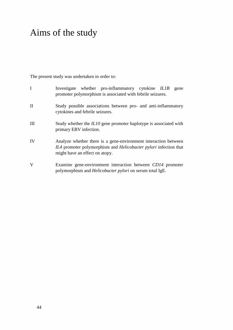
44
Aims of the study
The present study was undertaken in order to:
I Investigate whether proinflammatory cytokine IL1B genepromoter polymorphism is associated with febrile seizures.
II Study possible associations between pro and antiinflammatorycytokines and febrile seizures.
III Study whether the IL10 gene promoter haplotype is associated withprimary EBV infection.
IV Analyze whether there is a geneenvironment interaction betweenIL4 promoter polymorphism and Helicobacter pylori infection thatmight have an effect on atopy.
V Examine geneenvironment interaction between CD14 promoterpolymorphism and Helicobacter pylori on serum total IgE.

45
Subjects and methods
1. Subjects
1.1. Studies I and II
For studies I and II blood samples were obtained from 55 children with FStreated in the Department of Pediatrics at the Tampere University Hospitalbetween October 1997 and March 2000. The inclusion criteria for FS patientswere age 6 months to 5 years, no other identifiable cause for the seizure likeencephalitis, meningitis, raised intracranial pressure, disturbance in electrolyticequilibrium, epilepsy, poisoning or trauma, and temperature at least 38.5ºC. Dataregarding the family history for FSs, earlier FSs, duration of the seizure, andduration of fever before seizure were obtained from the parents by questionnaire.Family history was regarded positive when seizure was reported in a firstdegreerelative. Whole blood was available only from 35 patients out of 55 (64%) andplasma from all 55 children. CSF samples were obtained from 16 out of 55(29%) children based on the clinical judgment of the attending pediatrician. Forstatistical analysis infections were divided into viral and bacterial infections.Viral infection was defined as an infection with fever, low Creactive protein andno need for antibiotic treatment. Bacterial infections were treated with antibioticsand included both focal and septic infections defined as bacterial growth in bloodsample. The blood and CSF samples were taken immediately upon arrival at thehospital.
In Study II plasma samples were obtained from 20 control children treated atthe pediatric department of Tampere University Hospital between February 1999and March 2000 with febrile illness without convulsions and no history of FSaccording to parents and medical records. The control group was matched forage and temperature with the FS group. Laboratory and clinical data wereobtained from the medical records in both the FS and the control group.
Healthy blood donors were used as controls in Study I because the likelihoodof febrile seizure in this group is small (26%). Blood samples (buffy coats) fromthese 400 healthy adults (1860 years old) were obtained from the Finnish RedCross Blood Transfusion Centre, Tampere. The blood donors did not have anybloodtransmitted diseases or any signs of other infections during a 2weekperiod prior to the blood donation according to information acquired byquestionnaire. The ethnic background of the patients and controls was FinnishCaucasian. Characteristics of study subjects are presented in Table 1.

46
1.2. Study III
The blood samples were obtained from 116 children aged between 9 months and15 years, attending to the Tampere University Hospital for pediatric consultationbetween November 1999 and May 2000. Cord blood was collected from theumbilical veins of 50 healthy, fullterm newborns after normal vaginal deliveryin Tampere University Hospital. The 400 healthy adult blood donors from StudyI were used as controls in Study III. All cases and controls were of the sameethnic origin, Finnish Caucasian. Characteristics of study subjects are presentedin Table 1.
1.3. Study IV
In Study IV 245 asthmatic and 405 control subjects were participants in aFinnish population based casecontrol study aimed at identifying risk factors andpredictors of the outcome of adult asthma. Inclusion criteria for asthmaticsubjects were entitlement to reimbursement for asthma medication from theSocial Insurance Institution of Finland and age over 30 years. The entitlement toasthma medication was granted during the course of the study if the subjectfulfilled the criteria for persistent asthma confirmed by a chest specialist. Typicalhistory, clinical features and course of asthma needed to be documented. At leastone of the following physiological criteria was required for diagnosis: a variationof 20% in diurnal peak expiratory flow rate (PEF) recording (reference tomaximal value) or an increase of 15% in PEF or forced expiratory volume inone second (FEV1) with 2 agonist or a decrease of 15% in PEF or FEV1 inexercise test. A period of at least 6 months of continuing use of antiasthmaticdrugs must have elapsed by the time of the decision. This method of caseascertainment has been described in detail and evaluated elsewhere (Karjalainenet al. 2001). Per study case one or two controls with no asthma or chronicobstructive pulmonary disease were initially selected from a registry coveringthe whole population of Finland. Controls were matched with subjects for age,sex and area of residence. The ethnic background of the patients and controlswas Finnish Caucasian. Characteristics of study subjects are presented in Table1.
1.4. Study V
In Study V the cohort of 266 Russian Karelian children from our earlier studywas used as a study population (Seiskari et al. 2007). The Finnish child cohortfrom the earlier study was excluded from this study, because seropositivities forH.pylori and T.gondii were too rare for statistical analyses among Finnishschoolchildren (5% and 2% respectively).
The Russian Karelian child population was recruited as a part of the type 1diabetesrelated EPIVIR Project (EU INCOCopernicus Programme, contract

47
number IC15CT980316, Coordinator Professor Hyöty). In the EPIVIR Projectwhole blood and serum samples were collected from a total of 1998 randomlyselected schoolchildren in Russian Karelia during the period 19971999. Fromthis group all children with both parents of Finnish or Karelian ethnicity wereincluded in Study V (n=266). However, two of these children were excludedfrom the study because their DNA samples were accidentally confused in thelaboratory so the final number of subjects was 264 in Study V. The studychildren were not selected according to possible allergic or other diseases.Characteristics of study subjects are presented in Table 1.
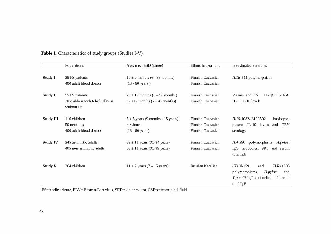
48
Table 1. Characteristics of study groups (Studies IV).
Populations Age: mean±SD (range) Ethnic background Investigated variables
Study I 35 FS patients400 adult blood donors
19 ± 9 months (6 36 months)(18 60 years )
Finnish CaucasianFinnish Caucasian
IL1B511 polymorphism
Study II 55 FS patients20 children with febrile illnesswithout FS
25 ± 12 months (6 – 56 months)22 ±12 months (7 – 42 months)
Finnish CaucasianFinnish Caucasian
Plasma and CSF IL1 , IL1RA,IL6, IL10 levels
Study III 116 children50 neonates400 adult blood donors
7 ± 5 years (9 months 15 years)newborn(18 60 years)
Finnish CaucasianFinnish CaucasianFinnish Caucasian
IL101082/819/592 haplotype,plasma IL10 levels and EBVserology
Study IV 245 asthmatic adults405 nonasthmatic adults
59 ± 11 years (3184 years)60 ± 11 years (3189 years)
Finnish CaucasianFinnish Caucasian
IL4590 polymorphism, H.pyloriIgG antibodies, SPT and serumtotal IgE
Study V 264 children 11 ± 2 years (7 – 15 years) Russian Karelian CD14159 and TLR4+896polymorphisms, H.pylori andT.gondii IgG antibodies and serumtotal IgE
FS=febrile seizure, EBV= EpsteinBarr virus, SPT=skin prick test, CSF=cerebrospinal fluid
49
2. Methods
2.1. Measurement of cytokine plasma levels (Studies II, III)
In Studies II and III IL10 cytokine plasma levels were measured using acommercially available enzymelinked immunosorbent assay (ELISA) kitaccording to manufacturer’s instructions (ELISA; CLB, PeliKine Compacthuman IL10 ELISA kit, Amsterdam, The Netherlands). The detection limit ofthe assay was 1.2 pg /ml. In Study II IL1 , IL1RA, IL6 and TNF cytokineplasma and CSF levels were measured using commercially available ELISA kitsaccording to manufacturer’s instructions (ELISA; CLB, PeliKine Compacthuman IL1 , IL6 and TNF ELISA kits, Amsterdam, The Netherlands and forIL1RA RD systems Quantikine kit, Minneapolis, MN, U.S.A.). The detectionlimits of the assays were 0.4 pg/ml for IL1 , 46.9 pg/ml for IL1Ra, 0.6 pg/mlfor IL6 and 1.4 pg/ml for TNF .
2.2. EBV, H.pylori and T.gondii serology (Studies I, IV, V)
In Study III EBV antibodies were measured by enzyme immunoassay accordingto manufacturer’s instructions (Enzygnost antiEBV/IgG, Behring, Marburg,Germany). H. pylori IgG antibodies were measured by Pyloriset EIAG III,(Orion Diagnostica, Espoo, Finland) in Study IV and by Enzygnost AntiHelicobacter pylori/IgG Assay (Dade Behring, Marburg, Germany) in Study V.Both assays were used according to manufacturer’s instructions. T. gondii IgGantibodies were measured by Enzygnost Toxoplasmosis IgG Assay (DadeBehring, Marburg, Germany) according to manufacturer’s instructions in StudyV.
2.3. Analysis of IL1B, IL4, IL10, CD14 and TLR4 genepolymorphisms (Studies I, III, IV, V)
Genomic deoxyribonucleic acid (DNA) was extracted from buffy coats or wholeblood using the salting out method described earlier (Miller et al. 1988) (StudiesI, III and IV) or the QIAamp DNA blood Mini Kit (QIAGEN Inc., USA) (StudyV).
IL1B promoter region single baseexchange C to T polymorphism at position511 (rs 16944) was amplified by polymerase chain reaction (PCR) in 50 µlreaction containing 100 ng of template DNA, 20 pmol of each primer, 0.1mMdNTPmix (Pharmacia Biotech), 1 mM MgCl2, 1x PCR buffer for DyNAzyme(Finnzymes, Espoo, Finland) and 1U of DyNAzyme polymerase (Finnzymes,

50
Espoo, Finland) using the primers earlier described (di Giovine et al. 1992). ThePCR conditions were as follows: 95 ºC for 2 min, then 36 cycles of 95 ºC for 1min, 55 ºC for 1 min and 74 ºC for 1 min, and finally 74 ºC for 4 min. Afteramplification the PCR products were digested for 3 hours in +37 ºC with AvaIrestriction enzyme (New England Biolabs inc., Boston, USA) in 50 µl reactioncontaining 25µl of the PCR product, 6U of AvaI and 1x NEbuffer 4 (NewEngland Biolabs inc., Boston, USA). After digestion the fragments wereseparated by electrophoresis in 9% polyacrylamide gel (PAGE) and visualizedwith ethidium bromide staining under ultraviolet (UV) light.
Detection of the IL4 gene promoter region polymorphic site at position 590(rs 2243250) was done by PCR and restriction fragment length polymorphism(RFLP) using primers and restriction enzyme AvaII as earlier described(Noguchi et al. 2001). The PCR reaction mix of total volume 50 µl contained400 ng of template DNA, 20 pmol of each primer, 0.1mM dNTPmix (PharmaciaBiotech), 20 mM PCR buffer of (NH4)2SO4 and 0.1% Tween 20, 5mM MgCl2,2% DMSO and 1,25U of Taq DNA polymerase (Fermentas, International Inc.,Burlington, Canada). The PCR conditions were as follows: 94 ºC for 2 min, then35 cycles of 94 ºC for 40 sec, 58 ºC for 40 sec and 72 ºC for 50 sec, and finally72 ºC for 10 min. After amplification the PCR products were digested overnightin +37 ºC with AvaII restriction enzyme (Fermentas, International Inc.,Burlington, Canada) in 25 µl reaction containing 12.5 µl of the PCR product, 5Uof AvaII and 1x BufferR+ (Fermentas, International Inc., Burlington, Canada).Fragments were analyzed by electrophoresis on 3.5% agarose gel stained withethidium bromide under UV light.
The IL10 promoter region polymorphisms at positions 592 (rs 1800872) and819 (rs 1800871) were detected by PCR and RFLP using primers and restrictionenzymes as described earlier (Mok et al. 1998, EdwardsSmith et al. 1999). Thecomposition of PCR mixture contained 100 ng template DNA, 20 pmol of eachprimer, 0.1 mM dNTP mix (Pharmacia Biotech), 1x PCR buffer for DyNAzyme(Finnzymes, Espoo, Finland), 2.5 mM MgCl2 and 1U of DNA Polymerase(Finnzymes, Espoo, Finland) in total volume 50 µl. The PCR conditions usedwere as follows: : 94 ºC for 2 min, then 35 cycles of 94 ºC for 30 sec, 60 ºC for45 sec and 72 ºC for 1 min, and finally 74 ºC for 10 min. In case of the IL10592polymorphism, the PCR product was digested for 3 hours at 37 ºC in a 50 µlreaction containing 30 µl of the PCR product, 1x BufferY+/TANGOTM
(Fermentas, International Inc., Burlington, Canada) and 5U of RsaI restrictionenzyme (Fermentas, International Inc., Burlington, Canada). In case of the IL10819 polymorphism, the PCR product was digested for 3 hours at 55 ºC in a 50 µlreaction containing 15 µl of the PCR product, 1x Mae III buffer (RocheDiagnostics, GmbH, Mannheim, Germany) and 2U MaeIII restriction enzyme(Roche Diagnostics, GmbH, Mannheim, Germany). After digestion thefragments were separated by electrophoresis in 9% PAGE and visualized withethidium bromide staining under UV light.
Amplification of the IL10 promoter polymorphism at position 1082 (rs1800896) was performed using the HotStarTaq kit (Qiagen, Melbourne,Australia), and 20 pmol of each primer and Qsolution was included in the PCR

51
mixture as earlier described (EdwardsSmith et al. 1999). The PCR conditionswere 15 min at 95 ºC followed by 35 cycles of 1 min at 94 ºC, 1 min at 60 ºC and1 min at 72 ºC and last 7 min at 72 ºC. The PCR products were digested for 3hours at 37 ºC in 50 µl solution containing 30 µl of the PCR product,1x BufferG+ (Fermentas, International Inc., Burlington, Canada), 1mM BSA and 10U ofMmlI restriction enzyme (Fermentas, International Inc., Burlington, Canada).The fragments were visualized on 3% agarose gel in UV light after ethidiumbromide staining.
The CD14 gene polymorphism at position 159 (rs 2569190) and the TLR4gene polymorphism at position +896 (rs 4986790) were genotyped withTaqMan® chemistry using the ABI PRISM 7000 Sequence Detection System(Applied Biosystems, CA, USA) for both PCR and allelic discrimination.Designed unlabeled PCR primers and fluorogenic TaqMan® minor groovebinding (MGB) probes were used (Assay by Design, ABI, CA, USA). Theuniversal PCR thermal cycling conditions from ABI were followed: first 50 ºCfor 2 min and 95 ºC for 10 min, and then 40 cycles at 95 ºC for 15 sec and 60 ºCfor 1 min. The PCR reaction was done in 25 µl reaction containing TaqMan®Universal PCR Master Mix with AmpErase® UNG (ABI, CA, USA), 1x AssayMix (primers and probes, ABI, CA, USA) and 10100ng of template DNA. Thegenotypes were selected manually from the allelic discrimination tab.
2.4. Skin prick test (Study IV)
Skin prick tests (SPT) were performed by specially trained nurses with a panel of22 common allergen extracts (ALK A7S, Copenhagen, Denmark) in Study IV.The allergens used were birch, alder, timothy grass, meadow foxtail, mugworth,dog, cat, horse, cow dander, Dermatophagoides farina, Dermatophagoidespteronyssinus, Acarus siro, Tyrophagus putrescentiae, Lepidoglyphus destructor,Aterbaria alernata, Cladosporium herbarum, Aspergillus fumigates, oats, barley,barley flour, wheat flour and rye flour. The test sites were placed on the volarside of the arm. A test was considered to be positive when the diameter of theweal was at least 3 mm larger than the negative control (saline). Theseprocedures were done in line with Position Paper of European Academy ofAllergology and Clinical Immunology (EAACI 1989). The patient wasconsidered prick test positive if at least one allergen gave a positive result.Allergy testing by the skin prick method was carried out on 99.1% of theasthmatic and 99.3% of control subjects. Positive reactions to specific allergensand differences between asthmatics and controls have been published earlier(Karjalainen et al. 2002).
2.5. Measurement of serum total IgE (Studies IVV)
In Study IV serum total IgE determinations were carried out by theimmunoluminometric method (Ciba Corning Diagnostics, Halsted, U.K.)
52
according to manufacturer’s instructions. Serum total IgE was measured usingthe ImmunoCAP® fluoroenzyme immunoassay (Phadia Diagnostics, Uppsala,Sweden) according to manufacturer’s instructions in Study V.
2.6. Statistical analyses (Studies IV)
For skewed continuous variables nonparametric statistics (MannWhitney Utest, KruskallWallis test) were used. Frequencies were compared using the chisquare test. Student’s ttest was used to analyze differences in mean values inStudy III. Logistic regression analysis was used to define relationships betweenfactors in Studies II and III. Logistic regression was also used to analyzeinteraction between H.pylori serology and IL4 genetics on the risk of atopy andasthma in Study IV. Multiway ANOVA was used for modeling geneenvironment interactions on serum total IgE levels, which were logarithmicallytransformed, in Studies IV and V. Bonferroni correction was used for pvalues ofplasma cytokine levels in study II and for association analyses with serum totalIgE in Study V. Statistical calculations were carried out on Statistica software(ver Win 5.1D, StatSoft, Tulsa, OK, USA) and SPSS software (ver. 6.0, 9.0,10.1., 11.5, 12.0 and 14.0, SPSS inc., Chicago, IL, USA). To test the fit ofgenotype frequencies with the HardyWeinberg equilibrium the Arlequinprogram (ver. 1.1 and 2.0, Genetics and Biometry Laboratory, Geneva,Switzerland) was used. In Study IV odds ratios and 95% confidence intervalswere calculated using CIA software (ver 1.1., copyrighted by M.J. Gardner andBritish Medical Journal 1989). P values <0.05 were considered statisticallysignificant.
2.7. Ethics (Studies IV)
The ethical committee of the Tampere University Hospital approved the studyplans for Studies IIV. The ethics board of the Finnish Red Cross BloodTransfusion Centre gave their approval for human blood use in Studies I and III.The study protocol for Study V was approved by the Ministry of Health,Karelian Republic Russia and the collection of samples was organized by theDepartment of Pediatrics, University of Petrozavodsk. Written informed consentwas obtained from subjects’ parents for Studies IIII, from the participants inStudy IV and from both parents and participants in Study V.

53
Results
1. Effect of IL1B511 gene polymorphism on febrileseizures (Study I)
Interleukin1 is a well known inducer of fever, which is a risk for FSs (Berg ATet al. 1995). IL1B511 polymorphism seems to be functional possibly affectingIL1 production as discussed in the review of the literature. Therefore wewanted to ascertain whether there is an association between FSs and IL1B511polymorphism.
Thirtyfive out of 55 FS patients with DNA samples obtained were includedin this study. The mean age of the 35 FS patients was 19.2 months (range 6months to 36 months). Seventeen (49%) patients were under 18 months old.Fortynine percent of the children were male (n= 17). The mean temperature was39.6 ºC (95% CI 39.439.9) when hospitalized. Twentythree (66%) children hadseizure duration less than 5 minutes and only two children (5.5%) had seizureslasting over 15 minutes. Nine children (26%) had a positive family history of FSin firstdegree relatives and 4 children (11%) had a previous history of FS. Mostof the children had no signs of bacterial infection and their diseases wereclassified as viral infections (n=23).
The genotype frequencies of IL1B511 polymorphism did not significantlydiffer between FS patients and healthy blood donors (p=0.1). However, thefrequency and carriage of allele T (=allele 2) of IL1B511 was significantlyincreased in FS patients compared to controls (0.54 vs. 0.41, p = 0.03, 0.80 vs.0.64, p=0.05 respectively). The genotype frequencies followed the HardyWeinberg equation.
Allele frequencies and allele T carrier status of IL1B511 did not differbetween children with or without positive family history of FS. Type of infection(viral vs. bacterial) was not associated with IL1B511 allele frequencies or theallele T carrier status in FS children. Due to the small number of FS children,analyses of genotype distributions according to family history of FS and type ofinfection could not be conducted.
54
2. Plasma and cerebrospinal fluid cytokines and febrileseizures (Study II)
The balance between pro and antiinflammatory cytokines is crucial in thepathogenesis of many diseases and, it for example, has a role in the regulation offever (Dinarello 2005). Therefore disturbances in this balance may have an effecton FSs. In Study II we wanted to investigate whether pro and antiinflammatorycytokines and the balance between them have an effect on FSs.
Fiftyfive FS patients were included in this study. Fifteen (27%) children hada positive family history of FSs in firstdegree relatives and eight (15%) had apositive history of FSs. Thirtyseven (67%) children had seizure duration lessthan 5 minutes and only two (3.6%) children had FS lasting over 15 minutes.Children with FSs did not differ from the twenty control children with febrileillness without convulsions and without history of FS by gender, age, type ofinfection, duration of fever before the blood sample, fever when hospitalized orlaboratory data.
2.1. Plasma cytokines and febrile seizures
FS patients had significantly higher plasma IL1Ra levels, plasma IL1Ra/IL1βratio and plasma IL6 levels than control children, as shown in Table 3. Therewas a trend for lower plasma IL1β levels in FS children compared to controls.However, when multiple testing was taken into account by Bonferroni correction(=multiplying by the number of cytokines measures (n=5)), this difference wasnot statistically significant (P=0.1). Differences in IL1Ra and IL6 levels wereso remarkable that they remained significant even after Bonferroni correction.There was no difference in plasma IL10 or plasma TNFα levels between FSand control patients. The cytokine plasma levels and the plasma IL1Ra/IL1βratio are presented in Table 3.
Logistic regression analysis was used to identify the most significant factorsassociated with FSs. Cytokine plasma levels, age and fever at the time ofhospitalization were divided into two groups using median value as a cutoffpoint. In the univariate logistic regression analysis we included the plasmacytokines studied, age, sex, type of infection, fever at the time of hospitalizationand duration of fever before the blood sample. Significant associations werefound between FSs and high plasma IL1Ra levels (odds ratio (OR) 6.5, 95%confidence interval (CI) 1.922.0), FSs and high plasma IL1Ra/IL1β ratio (OR36.0, 95% CI 4.5289.9) and FSs and high plasma IL6 levels (OR 4.2, 95% CI1.413.8). These significant variables were included in the multivariate logisticregression analysis. In this analysis the high plasma IL1RA/IL1β ratio was themost significant factor associated with FSs (OR 41.5, 95% CI 4.9352.8). Highplasma IL6 levels were also significantly associated with FSs (OR 5.3, 95% CI1.420.3), but in this model plasma IL1Ra did not have a discrete role.

55
2.2. Cerebrospinal fluid cytokines in febrile seizures
CSF samples were available of 16 FS patients. The CSF samples had cell count <5/µl and normal protein level 0.24 0.5 g/l. Unfortunately, the volume of somesamples was not enough for all cytokine analyses. IL6 levels were detectable inall 16 FS children studied (median 9.4 pg/ml, interquartile range 5.716.2 pg/ml),IL10 levels in 10 of 16 (median 7.2 pg/ml, interquartile range 019.5 pg/ml) andIL1Ra levels in nine of 12 FS children (median 170.0 pg/ml interquartile range0213.0 pg/ml). Only one out of 10 children had measurable IL1 levels andTNFα was undetectable in all the 15 children studied. No significantcorrelations between IL1Ra, IL6 and IL10 plasma and CSF levels were foundamong FS children (unpublished data).
2.3. IL1B511C>T polymorphism and plasma cytokines in febrileseizures
Plasma IL1 levels were not associated with IL1B511 genotype (p=0.7) orallele T carrier status (p=0.4) in Finnish FS children, nor were there anyassociations between IL1Ra, IL6, TNF , IL10 levels and IL1B511polymorphism in this population (unpublished data).
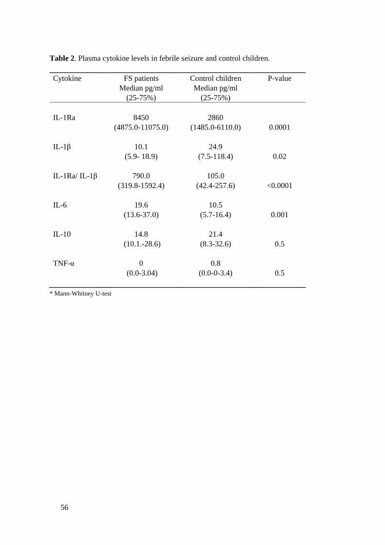
56
Table 2. Plasma cytokine levels in febrile seizure and control children.
Cytokine FS patientsMedian pg/ml
(2575%)
Control childrenMedian pg/ml
(2575%)
Pvalue
IL1Ra 8450(4875.011075.0)
2860(1485.06110.0) 0.0001
IL1 10.1(5.9 18.9)
24.9(7.5118.4) 0.02
IL1Ra/ IL1 790.0(319.81592.4)
105.0(42.4257.6) <0.0001
IL6 19.6(13.637.0)
10.5(5.716.4) 0.001
IL10 14.8 (10.1.28.6)
21.4(8.332.6) 0.5
TNF 0 (0.03.04)
0.8(0.003.4) 0.5
* MannWhitney Utest

57
3. Effect of IL10 promoter haplotype on EpsteinBarrvirus infection (Study III)
IL10 promoter polymorphism at position 1082 has been associated withsusceptibility to EBV infection (Helminen et al. 1999). In Study III we wanted toascertain, whether IL10 promoter haplotype 1082/819/592 influences EBVseroconversion. Mean age of the 116 study children was 6.7±4.9 years rangingfrom 1 year to 15 years. Fortyfive (39%) children were under 2 years of age, 41(35%) children between 2 and 10 years and 30 (26%) children over 10 years ofage. Thirtyeight percent (44/116) of the children were seropositive for EBV. Asexpected, seropositivity was more common among older children: 13.3 % (n=6)of the children under 2 years old, 46.3% (n=19) of the children between 2 and 10years and 63.3% (n=19) of the children over 10 years were seropositive for EBV.Among adult blood donors 95% (380/400) were seropositive for EBV.
The IL10 1082/819/592 promoter haplotype frequencies did not differbetween study populations and were GCC 0.44, ACC 0.36, ATA 0.20 inchildren, GCC 0.51, ACC 0.32 and ATA 0.17 in neonates and GCC 0.43, ACC0.35 and ATA 0.22 in blood donors. Among children IL10 haplotype ATA wasmore common in EBV seronegative compared to seropositive subjects (44% vs.25%, p=0.035). In further analysis by logistic regression, ATA positivity wassignificantly associated with EBV seronegativity when controlled by age (OR2.6, 95% CI 1.046.7, P=0.04). No significant association between carriage oftwo other haplotypes GCC and ACC and EBV seropositivity was found.
ACC haplotype carriage was significantly more common in the EBVseropositive adults compared to seronegative adults (61% vs. 25% respectively,p=0,004). In blood donors neither GCC nor ATA haplotype carrier status wasassociated with EBV seropositivity. However, the seronegative adults were moreoften homozygous for the GCC haplotype (GCC/GCC) compared to seropositive(55% vs. 17% respectively, p<0.01).
In both adults and neonates the IL10 haplotype ATA carriers had higherplasma IL10 levels than noncarriers. In adults the ACC and the GCC haplotypecarrier statuses were not associated with plasma IL10 levels, whereas inneonates GCC haplotype carriers had significantly lower IL10 levels than GCChaplotype noncarriers (p=0.001). In neonates ACC haplotype did not have effecton plasma IL10 levels.
58
4. Effect of IL4590 C>T polymorphism andHelicobacter pylori on skin prick test positivity (StudyIV)
4.1. Associations
H. pylori and IL4590C>T polymorphism have both been associated with atopy.IL4, which induces IgE production, has also been associated with H.pyloriinfection (Smythies et al. 2000). However, no studies investigating theinteraction between these two factors on atopy risk are available. Thus wewanted to investigate in Study IV whether H.pylori and IL4590 polymorphismhave interactions that have an effect on sensitization defined by SPT.
The 245 asthmatics and 405 nonasthmatic controls did not differ by age,gender or history of smoking. Fiftyfive percent of asthmatics had at least onepositive reaction in SPTs whereas only 38% of controls had positive SPT results.This difference was significant, as reported earlier (p<0.001) (Karjalainen et al.2002). Controls were more often seropositive for H.pylori than asthmatics (60%vs. 47%), but this difference was not statistically significant p=0.3). Sixasthmatics and 9 controls were excluded from further analysis due to lack ofresults from all measurements needed (=SPT, H.pylori antibodies and IL4590polymorphism).
An association between H.pylori seropositivity and SPT positivity was foundin Finnish adults. H.pylori seronegativity was associated with increased SPTpositivity in both asthmatic and control groups (OR 2.28, 95%CI 1.353.85 andOR 1.59, 95%CI 1.062.39 respectively). Most of the SPT positive subjects inboth asthmatic and control groups had more than one positive reaction (78.4%and 66.2% respectively). When SPT positive subjects were divided intomonosensitized (=one positive SPT reaction) and plurisensitized (=more thanone positive SPT reaction), H.pylori seropositivity was associated with dimishedSPT positivity only in plurisensitized subjects in both asthmatics (p=0.0005) andcontrols (p=0.004).
IL4590 genotype frequencies and allele T carrier status did not differbetween asthmatics and controls (p=0.3 and p=0.1 respectively). When thesubjects were divided into subgroups according to H.pylori seropositivity, IL4590 allele T carrier status was associated with asthma only in the H.pyloriseronegative group (p=0.03). This association was not seen in H.pyloriseropositive subjects (p=0.9). Neither IL4590 genotypes nor the IL4590 alleleT carrier status was statistically associated with SPT positivity even when SPTpositive subjects were divided into mono and plurisensitized. However, theasthmatic IL4590 allele T carriers had decreased risk for H.pylori seropositivity(OR 0.49, 95%CI 0.290.82). This effect was independent of atopic status andwas not seen in controls. IL4590 genotype frequencies followed the HardyWeinberg equation in both asthmatics and controls.

59
4.2. Interactions
Interaction between IL4590 polymorphism and H.pylori seropositivity on SPTpositivity was analyzed in the logistic regression model, but no interaction wasfound in either asthmatics or controls, nor was there any interaction betweenH.pylori and IL4590C>T polymorphism on serum total IgE studied by multiwayANOVA model. In these study populations serum total IgE was not associatedwith IL4590 polymorphism or H.pylori seropositivity (unpublished data).
5. Effect of CD14159 C>T polymorphism andHelicobacter pylori on serum total IgE (Study V)
5.1. Associations
CD14159C>T polymorphism has been associated in many studies with serumtotal IgE levels, but the results have been inconsistent. There is some evidencethat environmental factors like exposure to endotoxin, tobacco smoke, farmingenvironment and animals may modulate this association (Choudhry et al. 2005,Eder et al. 2005, Leynaert et al. 2006, Williams et al. 2006). However, the effectof microbes on this association has not been studied. Therefore we wanted toinvestigate in Study V if there were interactions between CD14159polymorphism and H.pylori or between CD14159 polymorphism and T.gondiiaffecting serum total IgE in Russian Karelian children. TLR4 has an importantrole in the same functional pathway in endotoxin response as CD14 andtherefore we included asthma associated TLR4 polymorphism +896A>G, in thisstudy (Fagerås Böttcher et al. 2004).
The Russian Karelian children were 7.115.0 years old (mean age 11.4 years)and 43% (n=114) of them were male. The median of serum total IgE was 76.1IU/L (interquartile range 30.9236.0 IU/L). Sex did not have an effect on serumtotal IgE levels (p=0.8). In these Russian Karelian children seropositivity forH.pylori and T.gondii was common: 73% (n=193) of children were seropositivefor H.pylori, 24% (n=63) for T.gondii and 20% for both these microbes.
T.gondii seropositive children had significantly higher serum total IgE levelsthan seronegative (median 114.0 IU/L, interquartile range 44.0393.0 vs median68.5 IU/L, interquartile range 28.3174.3 respectively, p=0.009), as we havereported earlier (Seiskari et al. 2007). This difference was statistically significanteven when multiple testing was taken into account using Bonferroni correction(p=0.036). H.pylori seropositivity did not have an effect on serum total IgE (inthe seropositive group IgE median 76.8 IU/L and in seronegative median 68.2IU/L, p=0.86) as reported earlier (Seiskari et al. 2007). Because most of theT.gondii seropositive subjects were also H.pylori seropositive further analysisbetween H.pylori and T.gondii seropositive and negative subgroups was done byKruskallWallis ANOVA. A significant difference between groups was found
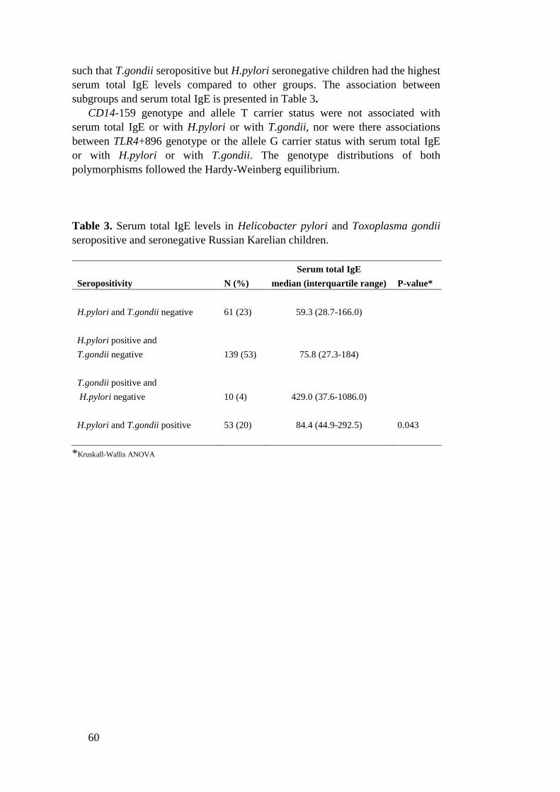
60
such that T.gondii seropositive but H.pylori seronegative children had the highestserum total IgE levels compared to other groups. The association betweensubgroups and serum total IgE is presented in Table 3.
CD14159 genotype and allele T carrier status were not associated withserum total IgE or with H.pylori or with T.gondii, nor were there associationsbetween TLR4+896 genotype or the allele G carrier status with serum total IgEor with H.pylori or with T.gondii. The genotype distributions of bothpolymorphisms followed the HardyWeinberg equilibrium.
Table 3. Serum total IgE levels in Helicobacter pylori and Toxoplasma gondiiseropositive and seronegative Russian Karelian children.
Serum total IgESeropositivity N (%) median (interquartile range) Pvalue*
H.pylori and T.gondii negative 61 (23) 59.3 (28.7166.0)
H.pylori positive andT.gondii negative 139 (53) 75.8 (27.3184)
T.gondii positive andH.pylori negative 10 (4) 429.0 (37.61086.0)
H.pylori and T.gondii positive 53 (20) 84.4 (44.9292.5) 0.043
*KruskallWallis ANOVA
61
5.2. Interactions
Geneenvironment and genegene interactions on serum total IgE were analyzedby a multiway ANOVA model. A statistically significant interaction betweenH.pylori seropositivity and the CD14159 allele T carrier status on serum totalIgE was found (p=0.004). Those H.pylori seronegative children who were CD14159 allele T noncarriers (i.e. genotype CC) had higher serum total IgE levelsthan allele T carriers (i.e. genotypes CT and TT). However, in H.pyloriseropositive children allele T noncarriers had lower IgE levels than allele Tcarriers (Figure 3). There was a trend for interaction between T.gondiiseropositivity and the CD14159 allele T carrier status on serum total IgE, butthis interaction was not statistically significant (Figure 3). There were nostatistically significant interactions between TLR4+896 and H.pylori orTLR4+896 and T.gondii on serum total IgE in this population. No statisticallysignificant interaction between CD14159 allele T carrier status and TLR4+896allele G carrier status on serum total IgE was found. Moreover, there was nointeraction between T.gondii and H.pylori on serum total IgE.
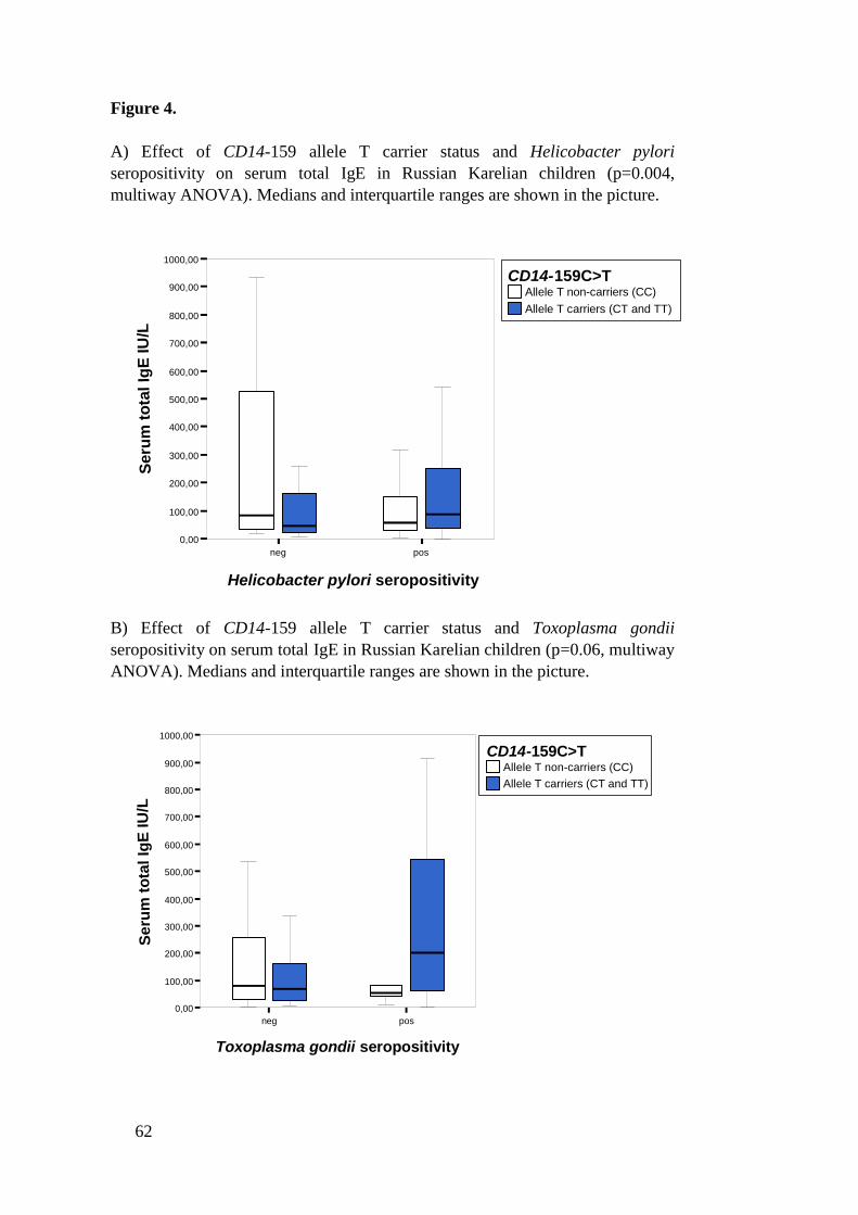
62
Figure 4.
A) Effect of CD14159 allele T carrier status and Helicobacter pyloriseropositivity on serum total IgE in Russian Karelian children (p=0.004,multiway ANOVA). Medians and interquartile ranges are shown in the picture.
Allele T noncarriers (CC)Allele T carriers (CT and TT)
CD14159C>T
Outliers are hiddenExtreme values are hidden
neg pos
Helicobacter pylori seropositivity
0,00
100,00
200,00
300,00
400,00
500,00
600,00
700,00
800,00
900,00
1000,00
Seru
m to
tal I
gE IU
/L
B) Effect of CD14159 allele T carrier status and Toxoplasma gondiiseropositivity on serum total IgE in Russian Karelian children (p=0.06, multiwayANOVA). Medians and interquartile ranges are shown in the picture.
Allele T noncarriers (CC)Allele T carriers (CT and TT)
CD14159C>T
Outliers are hiddenExtreme values are hidden
neg pos
Toxoplasma gondii seropositivity
0,00
100,00
200,00
300,00
400,00
500,00
600,00
700,00
800,00
900,00
1000,00
Seru
m to
tal I
gE IU
/L
63
Discussion
1. IL1 and febrile seizures
1.1. Association between IL1B511C>T polymorphism and febrileseizures
An association between the IL1B511C>T allele T and FSs was reported for thefirst time in our study (Study I). This finding has only been replicated in oneJapanese study, in which IL1B511 allele T and IL1B31 allele C were associatedwith sporadic simple FSs (Kira et al. 2005). In another Japanese study prolongedFSs strengthened the association between the IL1B511 allele T and TLE+HS,but there was no association between FSs and IL1B511 polymorphism in thisstudy (Kanemoto et al. 2003). In addition, no association between FSs and IL1B511 polymorphism was found in German, Taiwanese, Turkish and Japanesestudies (Tilgen et al. 2002, Chou et al. 2003, Haspolat et al. 2005, Matsuo et al.2006). Some of the contradictory results might be explained by differences insampling, definition of FSs and ethnic backgrounds. Another significant reasonbehind these inconsistent results could be the small sample size in every study,which causes low power to detect genetic effects that most probably are small inmultifactorial diseases like FSs. In a recent metaanalysis no statisticallysignificant relationship between IL1B511 polymorphism and FS or TLE riskwas found. However, TLE+HS risk was found to be increased among IL1B511allele T homozygous individuals in this metaanalysis, suggesting that thispolymorphism may play some role in convulsions (Kauffman et al. 2008).
The major limitation of Study I was the small sample size, which increasesthe risk of positive findings by chance. It is also possible that some otherpolymorphisms which are in linkage disequilibrium with IL1B511, like IL1B31, are actually more relevant in the pathogenesis of FSs than IL1B511polymorphism. Because simple and complex FSs seem to have different geneticbackground (Baulac et al. 2004, Waruiru et al. 2004), it would have beeninteresting to compare IL1B511 genotype and allele frequencies between simpleand complex FSs, but it was not statistically possible due to the small number ofstudy subjects in Study I.
64
1.2. Associations between cytokines and febrile seizures
The relationship between IL1 and FSs has been investigated in recent yearsbecause IL1 is a well known pyrogen and may therefore play a role in FSs. Insome studies an association has been found (Helminen et al. 1990, Tütüncüogluet al. 2001, Haspolat et al. 2005, Matsuo et al. 2006). However, the results havebeen inconsistent and in many studies no association between IL1 and FSs hasbeen seen (Lahat et al. 1997, Ichiyama et al. 1998, Tomoum et al. 2007). InFinnish children plasma IL1Ra and IL6 levels as well as IL1Ra/IL1 ratiowere higher in FS patients than in children with febrile illness withoutconvulsions (Study II). Plasma IL1 levels did not differ between FS andcontrol children in Study II. However, high IL1Ra plasma levels in FS patientsmay indicate preceding high IL1 production at the beginning of the infection,because production of IL1Ra is stimulated by IL1 and a strong initial IL1response could lead to high IL1Ra levels later during infection (Dinarello1996a).
Plasma IL1Ra/IL1 ratio was the most significant factor associated withFSs in Finnish children (Study II). The mechanism behind this finding is notknown. However, inflammatory reactions, including fever, are regulated by bothpro and antiinflammatory cytokines (Mackowiak et al. 1997, Dinarello 2005).Therefore the balance between IL1Ra and IL1 could be more important than asingle cytokine in regulating fever during infection. It could be speculated thatthe vast excess of IL1Ra seen in FS patients could be produced to neutralize theeffect of IL1 produced earlier. However, increased IL1Ra levels in FS patientscould also be a consequence of seizure (Eriksson et al. 1998).
In earlier studies the authors have speculated that the intrathecal presence ofcytokines could be used to differentiate FSs from CNS infections (Azuma et al.1997, Ichiyama et al. 1998). According to our results, detectable IL6, IL10 orIL1RA CSF levels cannot be used for this purpose, because these cytokineswere also found in FS children without any signs of CNS infection. However, thesample size was too small to permit firm conclusions so larger studies are neededto ascertain the role of CSF cytokines in differentiation of CNS infections fromharmless FSs.
2. Association between IL10 promoter haplotype andEBV infection
The IL10 promoter haplotype ATA consisting of three SNPs at positions 1082(A>G), 819 (C>T) and 592 (A>C) was associated with EBV seronegativity inFinnish children, but not in adult blood donors, among whom increasedfrequency of GCC/GCC haplotype was seen in EBV seronegative subjectsinstead (Study III). According to these results it could be speculated that eventhough ATA haplotype seems to be associated with delayed primary EVB
65
infection it does not seem to be associated with EBV seronegativity itself. Itwould have been interesting to investigate whether this haplotype is associatedwith IM, because IL101082 allele A involved in ATA haplotype has beenassociated with more severe forms of IM in Finnish adults (Helminen et al.1999). However, the study population was not suitable for this investigation.
The IL10 ATA haplotype was associated with increased plasma IL10 levelsin healthy Finnish blood donors and neonates. Even though IL10 is consideredan antiinflammatory cytokine, it has been shown to enhance the activation ofNK cells by EBV transformed B cells, to increase NK cell activity and decreaseviral replication and thereby to enhance immune response against EBV (Stewartet al. 1992, Kurilla et al. 1993). Therefore it could be speculated that the highspontaneous IL10 plasma levels seen in IL10 ATA haplotype carriers couldresult in a strong initial antiviral effect thus delaying the onset age of primaryEBV infection. However, ATA haplotype has also been associated withdecreased IL10 levels or IL10 production in some studies, which does notsupport this hypothesis (Crawley et al. 1999, EdwardsSmith et al. 1999,Hulkkonen et al. 2001). It would have been interesting to analyze whetherassociation between IL10 levels and IL10 haplotype is influenced by acute EBVinfection, but it was not possible because samples were not taken during acuteillness.
The most significant weakness of this study was the relatively small studypopulation for purpose of genetic analyses. It should also be noted thatsocioeconomic and other environmental aspects were not investigated in thisstudy, because the subgroups would have been too small for statistical analysis.However, it is well known that these factors influence the onset age of EBVinfection (Crawford 2001, Junker 2005).
3. Association between IL4590C>T polymorphism,Helicobacter pylori and skin prick test
In Finnish asthmatic and nonasthmatic adults H.pylori seropositivity wasassociated with SPT positivity so that among H.pylori seropositive subjects SPTpositivity to more than one allergen (=plurisensitizatoin) was decreased (StudyIV). This finding is in line with other studies, in which H.pylori has been shownto be associated with decreased risk of atopy (McCune et al. 2003, von Hertzenet al. 2006, Seiskari et al. 2007, Konturek et al. 2008). This result also supportsthe hygiene hypothesis according to which exposure to pathogens may reducethe risk of atopy (Strachan 1989, von Mutius 2007). However, an associationbetween H.pylori and decreased risk of atopic phenotypes was not seen in everystudy (Bodner et al. 2000, Uter et al. 2003, Law et al. 2005).
The exact mechanism by which H.pylori may affect the risk of atopy is notknown, but it has been speculated that it could be related to the increased Th1activity associated with H.pylori infection (von Mutius 2000). Increased IL10expression associated with H.pylori in some studies has been suggested to be

66
another possible mechanism by which H.pylori might be related to reduced riskof atopy, because there is mounting evidence that IL10 has an important role indownregulation of atopic response (von Hertzen et al. 2006, Oderda et al. 2007,von Hertzen et al. 2009). However, because H.pylori has been associated withgrowing up in a rural area, low socioeconomic status and poor living conditionslike absence of fixed hot water supply, contaminated water and domesticovercrowding in childhood, it is possible that H.pylori is only a surrogate markerof environmental factors associated with poor hygiene and low socioeconomicstatus (Mendall et al. 1992, Malaty et al. 1994, Brown 2000). Therefore thisassociation between H.pylori and reduced risk of atopy may, at least partly, bemediated by other infectious agents, like parasites, or by other environmentalfactors associated with low socioeconomic status mentioned above.
The IL4590 allele T associated with increased IL4 production has beenrelated to atopic asthma in a current metaanalysis (Li et al. 2008). The IL4590allele T has also been associated with other atopic phenotypes like elevatedserum total IgE, atopic dermatitis, rhinitis and sensitization defined by SPTpositivity (Rosenwasser et al. 1995, Zhu et al. 2000, Söderhall et al. 2002).However, in many studies, including our study on Finnish asthmatic and nonasthmatic adults (Study IV), IL4590 polymorphism has not been associated withatopy (Walley et al. 1996, Elliott et al. 2001). However, when the Finnish adultswere further divided into subgroups according to H.pylori seropositivity, the IL4590 allele T was positively associated with asthma, but only in H.pyloriseronegative subjects. According to these results it could be speculated that theassociation between IL4590 polymorphism and atopic phenotypes may be somodest that the association is only seen when environmental factors affectingsusceptibility are absent.
In addition, in Finnish asthmatics the IL4590 allele T was associated withdecreased H.pylori seropositivity. However, this association was not seen in thenonasthmatic group. The significance of this finding is not known and it isinconsistent with other studies in which the IL4590 allele T has been shown toincrease the risk of cagA positive H.pylori infection or in which no associationbetween H.pylori and IL4590 polymorphism has been found (GarcíaGonzálezet al. 2007, Zambon et al. 2008). However, it could be speculated that IL4590polymorphism could modify the relationship between infectious agent and atopyby affecting the host susceptibility to microbes, which for one might modulatethe susceptibility to atopic diseases. Nevertheless, the association between theIL4590 allele T and H.pylori seropositivy seen in Study IV could also be a falsepositive finding.
Geneenvironment interactions may be more relevant than a single SNP inatopy because many environmental factors, like infections, seem to be associatedwith atopy risk in addition to genetics (Beghé et al. 2003). However, in Finnishadults no interaction between IL4590 polymorphism and H.pylori seropositivityon SPT positivity was found, suggesting that association between H.pylori andSPT positivity is not influenced by this polymorphism.
67
4. Association between CD14159C>T polymorphism,Helicobacter pylori and serum total IgE
Serum total IgE is regulated by both genetic and environmental factors. AmongRussian Karelian children neither CD14159 polymorphism nor H.pyloriseropositivity was associated with serum total IgE (Study V). The results ofearlier studies analyzing associations between CD14159C>T polymorphism andserum total IgE and atopy have been conflicting because the same geneticvariants have been associated with both increased and decreased risk of atopy indifferent environments (Baldini et al. 1999, Ober et al. 2000, Eder et al. 2005). Ithas been speculated that the effect of CD14159 polymorphism on atopy is quitemodest and dependent on coexisting environmental risk factors. Thus recentlyinvestigated geneenvironment interactions could be more relevant and explainsome of the inconsistent results (Martinez 2007, Zhang et al. 2008).
Endotoxin exposure, contact with animals in childhood, exposure to ETS andfarming environment in early life have been reported to interact with CD14159polymorphism in the modulation of serum total IgE (Choudhry et al. 2005, Ederet al. 2005, Leynaert et al. 2006, Williams et al. 2006). In Russian Karelianchildren a statistically significant interaction between the CD14159 allele Tcarrier status and H.pylori seropositivity associated with serum total IgE wasfound (Study V). This was the first report of geneenvironment interactionbetween specific microbe and CD14159 polymorphism on serum total IgE. Inthis population H.pylori seronegative CD14159 allele T noncarriers had higherserum total IgE than allele T carriers. Among H.pylori seropositive childrenallele T noncarriers had lower IgE levels than allele T carriers. This resultsupports the hypothesis that the same genotype may increase, decrease or haveno effect on risk of a certain phenotype, in this case serum total IgE, dependingon the environmental factors, such as microbes, to which the population isexposed (Martinez 2007). Our result emphasizes the role of geneenvironmentinteraction in the regulation of serum total IgE and suggests that H.pylori may beone of the microbes that modulate the genetic regulation of serum total IgE.However, caution should be exercised when interpreting interactions betweenmicrobes and CD14159 polymorphism on serum total IgE, because, forexample, H.pylori may be only a surrogate marker for poor hygiene and largemicrobial load as discussed in the preceding chapter. Therefore the interactionsdetected could either reflect interaction between socioeconomic factorsassociated with H.pylori or the whole microbial burden and CD14159polymorphism as well as the interaction between a specific microbe and thepolymorphism.
A limitation of this study was that specific IgE could not be used in geneenvironment interaction analyses because the number of atopic subjects in thestudy population was too small for statistical analyses (n=16). Many factors likeallergens and parasite infections influence serum total IgE levels and thereforethe interpretation of the results is difficult. In our earlier study we found thatserum total IgE levels were significantly higher in Russian Karelian children

68
than in Finnish children even though the sensitization to common allergensmeasured by allergen specific IgE was lower in Russian Karelian children. Thissuggests that other factors, like parasite infections, could modulate serum totalIgE in this population (Seiskari et al. 2007).
Multiple testing also complicates the interpretation of the results. However,the interaction between the CD14159 allele T carrier status and H.pyloriseropositivity on serum total IgE was so significant that it most probably wouldnot disappear even though multiple testing were done (i.e. remaining significantafter Bonferroni correction). The major limitation of our study was the smallnumber of study subjects; therefore these geneenvironment analyses should berepeated in larger populations.
5. Candidate gene studies
Studies I, III, IV andV are based on the candidate gene approach. This approachhas been widely used to identify alleles, which may have a role in thepathogenesis of different diseases even though this approach has manylimitations. Four criteria for candidate gene studies of complex diseases havebeen suggested: consistent results, location of the gene in a chromosomal area oflinkage, change in protein level or function by the mutation and biologicalplausibility of the gene for the disease (Hall 1999). Similar issues have beenemphasized in other articles (NatureGenetics 1999, Tabor et al. 2002). However,these criteria are rarely fulfilled. The main problem with candidate gene studiesseems to be conflicting and unreplicable results. In addition, the variety ofphenotype definitions and ethnic differences between the populations makesinterpretation very challenging in the candidate gene studies. Another difficultyin the candidate gene approach is that a single gene or SNP usually makes only asmall contribution to the susceptibility or severity of multifactorial disease(Zhang et al. 2008).
Considering our results according to these recommendations, somereservations are called for. All the genotypes IL1B511C>T, IL4590C>T,CD14159C>T and the IL10 promoter haplotype 1082/819/592 studied havebeen shown to modulate protein production. However, the data of the exactfunctional changes that these polymorphisms cause is still quite modest. Thereare biologically plausible relationships between inflammatory mediators and theclinical conditions investigated in this study as seen in the review of theliterature. In addition, the roles of IL10 in EBV infection, IL1 in FSs, IL4 andCD14 in atopy have been seen in many studies. Therefore functionalpolymorphisms in their genes may be associated with these diseases. However,the results of association studies concerning the polymorphisms and clinicalconditions investigated in this dissertation are not unequivocal, as discussedearlier. The small sample size in Studies I, III, IV and V also challenges theinterpretation of the results, because the small study population increases the riskof both positive findings by chance and on the other hand diminishes the power
69
of the study to identify alleles with small effects (Tabor et al. 2002). The resultsof this study should be considered taking these shortcomings into account.
Several ways to improve the candidate gene approach have been proposed.For example, genegene and geneenvironment interactions might be morerelevant than simple candidate gene in the pathogenesis of complex diseases(Martinez 2007). Therefore the scope of investigations in this dissertation wasexpanded from a candidate gene approach into geneenvironment interactions inStudies IV and V. However, even though geneenvironment interactions mayexplain some of the inconsistent results of the simple candidate gene approachstudies, the interpretation of these results is even more difficult because of thecomplex nature of geneenvironment interactions. For example, it has beenproposed that disease phenotypes are a result of genetically determinedinadequate responses to a complex variety of environmental exposures and thatthe genes and environmental factors may predispose to disease, protect againstdisease or be neutral depending the context of the interaction (Martinez 2007).Therefore further research is needed to ascertain the mechanisms behind theseinteractions.

70
Conclusions
IL1 is a well known pyrogen and therefore it has been speculated that IL1Bgene polymorphisms may be associated with the susceptibility or severity of FS.In Finnish children an association between IL1B511 polymorphism and FSs wasobserved. However, in light of recent investigations it seems unlikely that thispolymorphism plays a role in the etiology of FSs.
Plasma IL1Ra, IL6 and IL1Ra/IL1 ratio were increased in children with FScompared to children with febrile illness without seizure. This findingemphasizes the relationship between inflammatory cytokines and FSs reported inother studies. However, it was not possible to ascertain whether these cytokineswere predisposing factors for FSs or more likely a consequence of seizureactivity.
The IL10 promoter 1082/819/592 haplotype ATA was associated withincreased frequency of EBV seronegativity in childhood, but not in adult blooddonors, suggesting that this haplotype may be more likely associated withdelayed EBV infection rather than with EBV seronegativity.
In Finnish asthmatic and nonasthmatic adults H.pylori seropositivity wasassociated with decreased risk of SPT positivity, but IL4590 polymorhism wasnot associated with SPT. In addition, no interaction between H.pyloriseropositivy and IL4590 polymorhism having an effect on SPT positivity wasfound. According to this result it might be speculated that the associationbetween H.pylori and sensitization measured by SPT is not influenced by thispolymorphism, at least in Finns.
In Russian Karelian children neither H.pylori seropositivity nor CD14159polymorphism were associated with serum total IgE alone. However, asignificant interaction between H.pylori seropositivity and CD14159 on serumtotal IgE was found, which further emphasizes the role of geneenvironmentinteraction in the regulation of serum total IgE.
71
Acknowledgements
This work was carried out at the Medical School, University of Tampere, at theDepartment of Microbiology and Immunology, during the years 19982008. Thestudy was financially supported by Tampere Graduate School in Biomedicineand Biotechnology (TGSBB), the Medical Research Fund of the TampereUniversity Hospital, the Tampere Tuberculosis Foundation, the Finnish AntiTuberculosis Association Foundation and the Väinö and Laina Kivi Foundation.They all are gratefully acknowledged.
I express my deepest gratitude to my supervisor, Professor Mikko Hurme, MD,PhD, for granting me the opportunity to prepare this dissertation in his group.His patient guidance has been a great support throughout these years.
I am deeply grateful to my other supervisor Merja Helminen, MD, PhD forclinical and scientific guidance and support during this project. Thank you forbelieving in me at the times when I did not believe in my own ability to completethis dissertation.
My sincere thanks go to Docent Riitta Karttunen, MD, PhD and DocentJohannes Savolainen, MD, PhD for their evaluation and constructive commentsof this manuscript.
I wish to thank my coauthors Sanna Kilpinen, Tanja Pessi, Kati Ådjers, JussiKarjalainen, Hilpi Rautelin, Timo Kosunen, Tapio Seiskari, Anita Kondrashova,Mikael Knip and Heikki Hyöty for their valuable contribution to the originalcommunications. I want especially thank Sanna and Tanja for their help andsupport.
I express my sincere thanks to Heini Huhtala, MSc, for advising me with thestatistical matters and making them more understandable.
I am grateful to Virginia Mattila, M.A. for careful revision of the Englishlanguage of the manuscript.
I wish to thank my post and present colleagues at the Department ofMicrobiology and Immunology: Nina Lahdenpohja, Janne Hulkkonen, CaritaEklund, Tanja Pessi, Annika Raitala, Kati Ådjers, Marja Pertovaara, MaaritOikarinen, Leena Teräväinen, Juulia Jylhävä, Atte Haarala, Petri Niinisalo,Tapio Kotipelto, Anita Vuorenmaa and Minna Vittaniemi. I want especially

72
thank Janne and Sanna for statistical advice and guidance at the beginning of thisdissertation process, Carita for interesting discussions and support during thehardest times in writing this dissertation. In addition, my warmest thanks go toSinikka RepoKoskinen and Eija Spåre for excellent technical laboratoryassistance and good sense of humor; we have shared many cheerful momentsduring past years.
I wish to thank my friends and colleagues Miia, Hanna, Marika, Karita, Teea,Anniina, Kirsi, Noora, Aino and AnnaKatriina for the opportunity to exchangeviews on scientific work, clinical work and also other aspects of life; it has meanta great deal to me.
My warmest thanks go to my dear childhood friends Hanna, Kati, Virve, Ritaand Satu and their families. We have shared many things during the past twentyseven years. Thank you for standing by me throughout this dissertation, I knowthat it has not always been easy. Special thanks to Hanna, who always had timeto listen to my troubles. I also want to thank all my other friends for theirsupport.
I warmly thank my grandmother Sirkka Laakso, godmother Sirpa LaaksoVarisand her husband Aatos Varis, Aunt Riitta Paajanen and her family and AuntTiina Laakso. You have offered me encouragement during this project and triedto stop me from doing too many things at the same time.
My warmest thanks go to Jarmo, who has shared my happy moments and thedeepest despair during the writing of this dissertation. Thank you for yourpatience and positive attitude!
I want to express my deepest gratitude to my parents Irma and Kari Virta. Youhave always believed in me and loved me as I am with or without academicachievements. This thesis would never have been completed without yoursupport. Thank you!
Tampere, August 2009
Miia Virta

73
References
Akdis CA, Akdis M (2009): Mechanisms and treatment of allergic disease in thebig picture of regulatory T cells. J Allergy Clin Immunol 123:735746.
Akdis M, Verhagen J, Taylor A, Karamloo F, Karagiannidis C, Crameri R,Thunberg S, Deniz G, Valenta R, Fiebig H, Kegel C, Disch R, SchmidtWeber CB, Blaser K, Akdis CA (2004): Immune responses in healthyand allergic individuals are characterized by a fine balance betweenallergenspecific T regulatory 1 and T helper 2 cells. J Exp Med199:15671575.
Almqvist C, Egmar AC, Hedlin G, Lundqvist M, Nordvall SL, Pershagen G,Svartengren M, van HageHamsten M, Wickman M (2003): Direct andindirect exposure to pets risk of sensitization and asthma at 4 years in abirth cohort. Clin Exp Allergy 33:11901197.
Andrews AL, Holloway JW, Holgate ST, Davies DE (2006): IL4 receptor alphais an important modulator of IL4 and IL13 receptor binding:implications for the development of therapeutic targets. J Immunol176:74567461.
Arend WP (2002): The balance between IL1 and IL1Ra in disease. CytokineGrowth Factor Rev 13:323340.
Arend WP, Welgus HG, Thompson RC, Eisenberg SP (1990): Biologicalproperties of recombinant human monocytederived interleukin 1receptor antagonist. J Clin Invest 85:16941697.
Arias MA, Rey Nores JE, Vita N, Stelter F, Borysiewicz LK, Ferrara P, LabétaMO (2000): Cutting edge: human B cell function is regulated byinteraction with soluble CD14: opposite effects on IgG1 and IgEproduction. J Immunol 164:34803486.
ArroyoEspliguero R, ElSharnouby K, VázquezRey E, Kalidas K, Jeffery S,Kaski JC (2005): CD14 C(260)T promoter polymorphism andprevalence of acute coronary syndromes. Int J Cardiol 98:307312.
Axtelle T, Pribble J (2003): An overview of clinical studies in healthy subjectsand patients with severe sepsis with IC14, a CD14specific chimericmonoclonal antibody. J Endotoxin Res 9:385389.
Azuma H, Tsuda N, Sasaki K, Okuno A (1997): Clinical significance of cytokinemeasurement for detection of meningitis. J Pediatr 131:463465.
Babel N, Vergopoulos A, Trappe RU, Oertel S, Hammer MH, Karaivanov S,Schneider N, Riess H, PappVary M, Neuhaus R, Gondek LP, Volk HD,Reinke P (2007): Evidence for genetic susceptibility towardsdevelopment of posttransplant lymphoproliferative disorder in solidorgan recipients. Transplantation 84:387391.
Baldini M, Lohman IC, Halonen M, Erickson RP, Holt PG, Martinez FD (1999):A Polymorphism* in the 5' flanking region of the CD14 gene isassociated with circulating soluble CD14 levels and with total serumimmunoglobulin E. Am J Respir Cell Mol Biol 20:976983.
Barksby HE, Lea SR, Preshaw PM, Taylor JJ (2007): The expanding family ofinterleukin1 cytokines and their role in destructive inflammatorydisorders. Clin Exp Immunol 149:217225.
Baulac S, GourfinkelAn I, Nabbout R, Huberfeld G, Serratosa J, Leguern E,Baulac M (2004): Fever, genes, and epilepsy. Lancet Neurol 3:421430.

74
Baumgart DC, Büning C, Geerdts L, Schmidt HH, Genschel J, Fiedler T, GentzE, Molnar T, Nagy F, Lonovics J, Lochs H, Wiedenmann B, Nickel R,Witt H, Dignass A (2007): The c.1260C>T promoter variant of CD14but not the c.896A>G (p.D299G) variant of tolllike receptor 4 (TLR4)genes is associated with inflammatory bowel disease. Digestion 76:196202.
Baynam G, Khoo SK, Rowe J, Zhang G, Laing I, Hayden C, Kusel M, DeKlerkN, Sly P, Goldblatt J, Holt P, LeSouef P (2007): Parental smokingimpairs vaccine responses in children with atopic genotypes. J AllergyClin Immunol 119:366374.
Bazan JF, Timans JC, Kastelein RA (1996): A newly defined interleukin1?Nature 379:591.
Beeh KM, Ksoll M, Buhl R (2000): Elevation of total serum immunoglobulin Eis associated with asthma in nonallergic individuals. Eur Respir J 16:609614.
Beghé B, Barton S, Rorke S, Peng Q, Sayers I, Gaunt T, Keith TP, Clough JB,Holgate ST, Holloway JW (2003): Polymorphisms in the interleukin4and interleukin4 receptor alpha chain genes confer susceptibility toasthma and atopy in a Caucasian population. Clin Exp Allergy 33:11111117.
Belloni B, Ziai M, Lim A, Lemercier B, Sbornik M, Weidinger S, Andres C,Schnopp C, Ring J, Hein R, Ollert M, Mempel M (2007): Lowdose antiIgE therapy in patients with atopic eczema with high serum IgE levels. JAllergy Clin Immunol 120:12231225.
Berg AT, Shinnar S, Shapiro ED, Salomon ME, Crain EF, Hauser WA (1995):Risk factors for a first febrile seizure: a matched casecontrol study.Epilepsia 36:334341.
Berg DJ, Davidson N, Kühn R, Müller W, Menon S, Holland G, ThompsonSnipes L, Leach MW, Rennick D (1996): Enterocolitis and colon cancerin interleukin10deficient mice are associated with aberrant cytokineproduction and CD4(+) TH1like responses. J Clin Invest 98:10101020.
Berg DJ, Kühn R, Rajewsky K, Müller W, Menon S, Davidson N, Grünig G,Rennick D (1995): Interleukin10 is a central regulator of the response toLPS in murine models of endotoxic shock and the Shwartzman reactionbut not endotoxin tolerance. J Clin Invest 96:23392347.
Bidwell J, Keen L, Gallagher G, Kimberly R, Huizinga T, McDermott MF,Oksenberg J, McNicholl J, Pociot F, Hardt C, D'Alfonso S (1999):Cytokine gene polymorphism in human disease: online databases. GenesImmun 1:319.
Blaser K, Akdis CA (2004): Interleukin10, T regulatory cells and specificallergy treatment. Clin Exp Allergy 34:328331.
Bliss CM, Jr., Golenbock DT, Keates S, Linevsky JK, Kelly CP (1998):Helicobacter pylori lipopolysaccharide binds to CD14 and stimulatesrelease of interleukin8, epithelial neutrophilactivating peptide 78, andmonocyte chemotactic protein 1 by human monocytes. Infect Immun66:53575363.
Bodner C, Anderson WJ, Reid TS, Godden DJ (2000): Childhood exposure toinfection and risk of adult onset wheeze and atopy. Thorax 55:383387.
Bontems P, Robert F, Van Gossum A, Cadranel S, Mascart F (2003):Helicobacter pylori modulation of gastric and duodenal mucosal T cellcytokine secretions in children compared with adults. Helicobacter 8:216226.
Borish L, Aarons A, Rumbyrt J, Cvietusa P, Negri J, Wenzel S (1996):Interleukin10 regulation in normal subjects and patients with asthma. JAllergy Clin Immunol 97:12881296.
Borish L, Mascali JJ, Klinnert M, Leppert M, Rosenwasser LJ (1994): SSCpolymorphisms in interleukin genes. Hum Mol Genet 3:1710.
75
Bottema RW, Reijmerink NE, Kerkhof M, Koppelman GH, Stelma FF, GerritsenJ, Thijs C, Brunekreef B, van Schayck CP, Postma DS (2008):Interleukin 13, CD14, pet and tobacco smoke influence atopy in threeDutch cohorts: the allergenic study. Eur Respir J 32:593602.
Brown LM (2000): Helicobacter pylori: epidemiology and routes oftransmission. Epidemiol Rev 22:283297.
Brusselle G, Kips J, Joos G, Bluethmann H, Pauwels R (1995): Allergeninducedairway inflammation and bronchial responsiveness in wildtype andinterleukin4deficient mice. Am J Respir Cell Mol Biol 12:254259.
Burchard EG, Silverman EK, Rosenwasser LJ, Borish L, Yandava C, Pillari A,Weiss ST, Hasday J, Lilly CM, Ford JG, Drazen JM (1999): Associationbetween a sequence variant in the IL4 gene promoter and FEV(1) inasthma. Am J Respir Crit Care Med 160:919922.
Burrows B, Martinez FD, Halonen M, Barbee RA, Cline MG (1989):Association of asthma with serum IgE levels and skintest reactivity toallergens. N Engl J Med 320:271277.
Callard R, George AJ, Stark J (1999): Cytokines, chaos, and complexity.Immunity 11:507513.
Campos J, GonzalezQuintela A, Quinteiro C, Gude F, Perez LF, Torre JA,Vidal C (2005): The 159C/T polymorphism in the promoter region ofthe CD14 gene is associated with advanced liver disease and higherserum levels of acutephase proteins in heavy drinkers. Alcohol Clin ExpRes 29:12061213.
Campos J, Gude F, Quinteiro C, Vidal C, GonzalezQuintela A (2006): Gene byenvironment interaction: the 159C/T polymorphism in the promoterregion of the CD14 gene modifies the effect of alcohol consumption onserum IgE levels. Alcohol Clin Exp Res 30:714.
Capasso M, Avvisati RA, Piscopo C, Laforgia N, Raimondi F, de Angelis F,Iolascon A (2007): Cytokine gene polymorphisms in Italian preterminfants: association between interleukin10 1082 G/A polymorphism andrespiratory distress syndrome. Pediatr Res 61:313317.
Chan SC, Brown MA, Willcox TM, Li SH, Stevens SR, Tara D, Hanifin JM(1996): Abnormal IL4 gene expression by atopic dermatitis Tlymphocytes is reflected in altered nuclear protein interactions with IL4transcriptional regulatory element. J Invest Dermatol 106:11311136.
Chen H, Wilkins LM, Aziz N, Cannings C, Wyllie DH, Bingle C, Rogus J, BeckJD, Offenbacher S, Cork MJ, RafieKolpin M, Hsieh CM, Kornman KS,Duff GW (2006): Single nucleotide polymorphisms in the humaninterleukin1B gene affect transcription according to haplotype context.Hum Mol Genet 15:519529.
Chiang CH, Tang YC, Lin MW, Chung MY (2007): Association between the IL4 promoter polymorphisms and asthma or severity ofhyperresponsiveness in Taiwanese. Respirology 12:4248.
Chou IC, Tsai CH, Hsieh YY, Peng CT, Tsai FJ (2003): Association betweenpolymorphism of interleukin1beta511 promoter and susceptibility tofebrile convulsions in Taiwanese children. Acta Paediatr 92:1356.
Choudhry S, Avila PC, Nazario S, Ung N, Kho J, RodriguezSantana JR, CasalJ, Tsai HJ, Torres A, Ziv E, Toscano M, Sylvia JS, Alioto M, Salazar M,Gomez I, Fagan JK, Salas J, Lilly C, Matallana H, Castro RA, Selman M,Weiss ST, Ford JG, Drazen JM, RodriguezCintron W, Chapela R,Silverman EK, Burchard EG (2005): CD14 tobacco geneenvironmentinteraction modifies asthma severity and immunoglobulin E levels inLatinos with asthma. Am J Respir Crit Care Med 172:173182.
Colotta F, Re F, Muzio M, Bertini R, Polentarutti N, Sironi M, Giri JG, DowerSK, Sims JE, Mantovani A (1993): Interleukin1 type II receptor: a decoytarget for IL1 that is regulated by IL4. Science 261:472475.

76
Commins S, Steinke JW, Borish L (2008): The extended IL10 superfamily: IL10, IL19, IL20, IL22, IL24, IL26, IL28, and IL29. J Allergy ClinImmunol 121:11081111.
Cookson W (2004): The immunogenetics of asthma and eczema: a new focus onthe epithelium. Nat Rev Immunol 4:978988.
Corrado G, Luzzi I, Lucarelli S, Frediani T, Pacchiarotti C, Cavaliere M, Rea P,Cardi E (1998): Positive association between Helicobacter pyloriinfection and food allergy in children. Scand J Gastroenterol 33:11351139.
Corry DB, Kheradmand F (1999): Induction and regulation of the IgE response.Nature 402:B1823.
Cosentino G, Soprana E, Thienes CP, Siccardi AG, Viale G, Vercelli D (1995):IL13 downregulates CD14 expression and TNFalpha secretion innormal human monocytes. J Immunol 155:31453151.
Cottrez F, Groux H (2001): Regulation of TGFbeta response during T cellactivation is modulated by IL10. J Immunol 167:773778.
Cottrez F, Hurst SD, Coffman RL, Groux H (2000): T regulatory cells 1 inhibit aTh2specific response in vivo. J Immunol 165:48484853.
Crawford DH (2001): Biology and disease associations of EpsteinBarr virus.Philos Trans R Soc Lond B Biol Sci 356:461473.
Crawley E, Kay R, Sillibourne J, Patel P, Hutchinson I, Woo P (1999):Polymorphic haplotypes of the interleukin10 5' flanking regiondetermine variable interleukin10 transcription and are associated withparticular phenotypes of juvenile rheumatoid arthritis. Arthritis Rheum42:11011108.
da Silva GN, Bacchi MM, Rainho CA, de Oliveira DE (2007): EpsteinBarrvirus infection and single nucleotide polymorphisms in the promoterregion of interleukin 10 gene in patients with Hodgkin lymphoma. ArchPathol Lab Med 131:16911696.
Dabbagh K, Takeyama K, Lee HM, Ueki IF, Lausier JA, Nadel JA (1999): IL4induces mucin gene expression and goblet cell metaplasia in vitro and invivo. J Immunol 162:62336237.
Daher S, Santos LM, Solé D, De Lima MG, Naspitz CK, Musatti CC (1995):Interleukin4 and soluble CD23 serum levels in asthmatic atopic children.J Investig Allergol Clin Immunol 5:251254.
Davidson J, Abul HT, Milton AS, Rotondo D (2001): Cytokines and cytokineinducers stimulate prostaglandin E2 entry into the brain. Pflugers Arch442:526533.
Devitt A, Moffatt OD, Raykundalia C, Capra JD, Simmons DL, Gregory CD(1998): Human CD14 mediates recognition and phagocytosis ofapoptotic cells. Nature 392:505509.
Dezsöfi A, Szebeni B, Hermann CS, Kapitány A, Veres G, Sipka S, Körner A,Madácsy L, KorponaySzabó I, Rajczy K, Arató A (2008): Frequenciesof genetic polymorphisms of TLR4 and CD14 and of HLADQgenotypes in children with celiac disease, type 1 diabetes mellitus, orboth. J Pediatr Gastroenterol Nutr 47:283287.
di Giovine FS, Takhsh E, Blakemore AI, Duff GW (1992): Single basepolymorphism at 511 in the human interleukin1 beta gene (IL1 beta).Hum Mol Genet 1:450.
Dinarello CA (1996a): Biologic basis for interleukin1 in disease. Blood87:20952147.
Dinarello CA (1996b): Thermoregulation and the pathogenesis of fever. InfectDis Clin North Am 10:433449.
Dinarello CA (1998): Interleukin1 beta, interleukin18, and the interleukin1beta converting enzyme. Ann N Y Acad Sci 856:111.
Dinarello CA (2002): The IL1 family and inflammatory diseases. Clin ExpRheumatol 20:S113.

77
Dinarello CA (2005): Interleukin1beta. Crit Care Med 33:S460462.Dizier MH, Sandford A, Walley A, Philippi A, Cookson W, Demenais F (1999):
Indication of linkage of serum IgE levels to the interleukin4 gene andexclusion of the contribution of the (590 C to T) interleukin4 promoterpolymorphism to IgE variation. Genet Epidemiol 16:8494.
Dube C, Vezzani A, Behrens M, Bartfai T, Baram TZ (2005): Interleukin1betacontributes to the generation of experimental febrile seizures. Ann Neurol57:152155.
EAACI (1989): Skin tests used in type I allergy testing Position paper. SubCommittee on Skin Tests of the European Academy of Allergology andClinical Immunology. Allergy 44 Suppl 10:159.
Eder W, Klimecki W, Yu L, von Mutius E, Riedler J, BraunFährlander C,Nowak D, Martinez FD (2005): Opposite effects of CD 14/260 on serumIgE levels in children raised in different environments. J Allergy ClinImmunol 116:601607.
EdwardsSmith CJ, Jonsson JR, Purdie DM, Bansal A, Shorthouse C, Powell EE(1999): Interleukin10 promoter polymorphism predicts initial responseof chronic hepatitis C to interferon alfa. Hepatology 30:526530.
Ege MJ, Bieli C, Frei R, van Strien RT, Riedler J, Üblagger E, SchramBijkerkD, Brunekreef B, van Hage M, Scheynius A, Pershagen G, Benz MR,Lauener R, von Mutius E, BraunFährlander C (2006): Prenatal farmexposure is related to the expression of receptors of the innate immunityand to atopic sensitization in schoolage children. J Allergy ClinImmunol 117:817823.
ElOmar EM, Carrington M, Chow WH, McColl KE, Bream JH, Young HA,Herrera J, Lissowska J, Yuan CC, Rothman N, Lanyon G, Martin M,Fraumeni JF, Jr., Rabkin CS (2000): Interleukin1 polymorphismsassociated with increased risk of gastric cancer. Nature 404:398402.
ElOmar EM, Carrington M, Chow WH, McColl KE, Bream JH, Young HA,Herrera J, Lissowska J, Yuan CC, Rothman N, Lanyon G, Martin M,Fraumeni JF, Jr., Rabkin CS (2001): The role of interleukin1polymorphisms in the pathogenesis of gastric cancer. Nature 412:99.
Elliott K, Fitzpatrick E, Hill D, Brown J, Adams S, Chee P, Stewart G, FulcherD, Tang M, Kemp A, King E, Varigos G, Bahlo M, Forrest S (2001): The590C/T and 34C/T interleukin4 promoter polymorphisms are notassociated with atopic eczema in childhood. J Allergy Clin Immunol108:285287.
Eriksson C, Winblad B, Schultzberg M (1998): Kainic acid induced expressionof interleukin1 receptor antagonist mRNA in the rat brain. Brain ResMol Brain Res 58:195208.
Eskdale J, Keijsers V, Huizinga T, Gallagher G (1999): Microsatellite alleles andsingle nucleotide polymorphisms (SNP) combine to form four majorhaplotype families at the human interleukin10 (IL10) locus. GenesImmun 1:151155.
Fagerås Böttcher M, HmaniAifa M, Lindström A, Jenmalm MC, Mai XM,Nilsson L, Zdolsek HA, Björkstén B, Söderkvist P, Vaarala O (2004): ATLR4 polymorphism is associated with asthma and reducedlipopolysaccharideinduced interleukin12(p70) responses in Swedishchildren. J Allergy Clin Immunol 114:561567.
Ferrari D, Pizzirani C, Adinolfi E, Lemoli RM, Curti A, Idzko M, Panther E, DiVirgilio F (2006): The P2X7 receptor: a key player in IL1 processingand release. J Immunol 176:38773883.
Ferrero E, Goyert SM (1988): Nucleotide sequence of the gene encoding themonocyte differentiation antigen, CD14. Nucleic Acids Res 16:4173.
Fetveit A (2008): Assessment of febrile seizures in children. Eur J Pediatr167:1727.

78
Finkelman FD, SheaDonohue T, Morris SC, Gildea L, Strait R, Madden KB,Schopf L, Urban JF, Jr. (2004): Interleukin4 and interleukin13mediated host protection against intestinal nematode parasites. ImmunolRev 201:139155.
Fiorentino DF, Bond MW, Mosmann TR (1989): Two types of mouse T helpercell. IV. Th2 clones secrete a factor that inhibits cytokine production byTh1 clones. J Exp Med 170:20812095.
Fukuda M, Suzuki Y, Ishizaki Y, Kira R, Kikuchi C, Watanabe S, Hino H,Morimoto T, Hara T, Ishii E (2009): Interleukin1beta enhancessusceptibility to hyperthermiainduced seizures in developing rats.Seizure 18:211214.
Gao PS, Mao XQ, Baldini M, Roberts MH, Adra CN, Shirakawa T, Holt PG,Martinez FD, Hopkin JM (1999): Serum total IgE levels and CD14 onchromosome 5q31. Clin Genet 56:164165.
GarcíaGonzález MA, Lanas A, Quintero E, Nicolás D, ParraBlanco A, StrunkM, Benito R, Angel Simón M, Santolaria S, Sopeña F, Piazuelo E,Jiménez P, Pascual C, Mas E, Irún P, Espinel J, Campo R, Manzano M,Geijo F, Pellisé M, GonzálezHuix F, Nieto M, Espinós J, Titó L,Bujanda L, Zaballa M (2007): Gastric cancer susceptibility is not linkedto proand antiinflammatory cytokine gene polymorphisms in whites: aNationwide Multicenter Study in Spain. Am J Gastroenterol 102:18781892.
Gern JE, Reardon CL, Hoffjan S, Nicolae D, Li Z, Roberg KA, Neaville WA,CarlsonDakes K, Adler K, Hamilton R, Anderson E, GilbertsonWhiteS, Tisler C, Dasilva D, Anklam K, Mikus LD, Rosenthal LA, Ober C,Gangnon R, Lemanske RF, Jr. (2004): Effects of dog ownership andgenotype on immune development and atopy in infancy. J Allergy ClinImmunol 113:307314.
Granowitz EV, Santos AA, Poutsiaka DD, Cannon JG, Wilmore DW, Wolff SM,Dinarello CA (1991): Production of interleukin1receptor antagonistduring experimental endotoxaemia. Lancet 338:14231424.
Groux H, Bigler M, de Vries JE, Roncarolo MG (1996): Interleukin10 induces alongterm antigenspecific anergic state in human CD4+ T cells. J ExpMed 184:1929.
Groux H, O'Garra A, Bigler M, Rouleau M, Antonenko S, de Vries JE,Roncarolo MG (1997): A CD4+ Tcell subset inhibits antigenspecific Tcell responses and prevents colitis. Nature 389:737742.
Grunewald SM, Werthmann A, Schnarr B, Klein CE, Bröcker EB, Mohrs M,Brombacher F, Sebald W, Duschl A (1998): An antagonistic IL4 mutantprevents type I allergy in the mouse: inhibition of the IL4/IL13 receptorsystem completely abrogates humoral immune response to allergen anddevelopment of allergic symptoms in vivo. J Immunol 160:40044009.
Haanpää M, Nurmikko T, Hurme M (2002): Polymorphism of the IL10 gene isassociated with susceptibility to herpes zoster. Scand J Infect Dis 34:112114.
Hall IP (1999): Genetics and pulmonary medicine 8: asthma. Thorax 54:6569.Hall SK, Perregaux DG, Gabel CA, Woodworth T, Durham LK, Huizinga TW,
Breedveld FC, Seymour AB (2004): Correlation of polymorphic variationin the promoter region of the interleukin1 beta gene with secretion ofinterleukin1 beta protein. Arthritis Rheum 50:19761983.
Hanson B, McGue M, RoitmanJohnson B, Segal NL, Bouchard TJ, Jr.,Blumenthal MN (1991): Atopic disease and immunoglobulin E in twinsreared apart and together. Am J Hum Genet 48:873879.
Haspolat S, Baysal Y, Duman O, Coskun M, Tosun O, Yegin O (2005):Interleukin1alpha, interleukin1beta, and interleukin1Rapolymorphisms in febrile seizures. J Child Neurol 20:565568.
79
Haspolat S, Mihci E, Coskun M, Gümüslü S, Özben T, Yegin O (2002):Interleukin1beta, tumor necrosis factoralpha, and nitrite levels in febrileseizures. J Child Neurol 17:749751.
Hawrylowicz CM, O'Garra A (2005): Potential role of interleukin10secretingregulatory T cells in allergy and asthma. Nat Rev Immunol 5:271283.
Haziot A, Ferrero E, Köntgen F, Hijiya N, Yamamoto S, Silver J, Stewart CL,Goyert SM (1996): Resistance to endotoxin shock and reduceddissemination of gramnegative bacteria in CD14deficient mice.Immunity 4:407414.
He JQ, ChanYeung M, Becker AB, DimichWard H, Ferguson AC, Manfreda J,Watson WT, Sandford AJ (2003): Genetic variants of the IL13 and IL4genes and atopic diseases in atrisk children. Genes Immun 4:385389.
Helminen M, Lahdenpohja N, Hurme M (1999): Polymorphism of theinterleukin10 gene is associated with susceptibility to EpsteinBarr virusinfection. J Infect Dis 180:496499.
Helminen M, Vesikari T (1990): Increased interleukin1 (IL1) production fromLPSstimulated peripheral blood monocytes in children with febrileconvulsions. Acta Paediatr Scand 79:810816.
Helminen ME, Kilpinen S, Virta M, Hurme M (2001): Susceptibility to primaryEpsteinBarr virus infection is associated with interleukin10 genepromoter polymorphism. J Infect Dis 184:777780.
Hoebee B, Rietveld E, Bont L, Oosten M, Hodemaekers HM, Nagelkerke NJ,Neijens HJ, Kimpen JL, Kimman TG (2003): Association of severerespiratory syncytial virus bronchiolitis with interleukin4 andinterleukin4 receptor alpha polymorphisms. J Infect Dis 187:211.
Holgate ST, Djukanovic R, Casale T, Bousquet J (2005): AntiimmunoglobulinE treatment with omalizumab in allergic diseases: an update on antiinflammatory activity and clinical efficacy. Clin Exp Allergy 35:408416.
Hollegaard MV, Bidwell JL (2006): Cytokine gene polymorphism in humandisease: online databases, Supplement 3. Genes Immun 7:269276.
Hopp RJ, Bewtra AK, Watt GD, Nair NM, Townley RG (1984): Geneticanalysis of allergic disease in twins. J Allergy Clin Immunol 73:265270.
Howard M, Farrar J, Hilfiker M, Johnson B, Takatsu K, Hamaoka T, Paul WE(1982): Identification of a T cellderived b cell growth factor distinctfrom interleukin 2. J Exp Med 155:914923.
Hulkkonen J, Pertovaara M, Antonen J, Lahdenpohja N, Pasternack A, Hurme M(2001): Genetic association between interleukin10 promoter regionpolymorphisms and primary Sjogren's syndrome. Arthritis Rheum44:176179.
Hultgren O, Kopf M, Tarkowski A (1998): Staphylococcus aureusinducedseptic arthritis and septic death is decreased in IL4deficient mice: roleof IL4 as promoter for bacterial growth. J Immunol 160:50825087.
Hunt PJ, Marshall SE, Weetman AP, Bell JI, Wass JA, Welsh KI (2000):Cytokine gene polymorphisms in autoimmune thyroid disease. J ClinEndocrinol Metab 85:19841988.
Hurme M, Santtila S (1998): IL1 receptor antagonist (IL1Ra) plasma levels arecoordinately regulated by both IL1Ra and IL1beta genes. Eur JImmunol 28:25982602.
Hwang IR, Kodama T, Kikuchi S, Sakai K, Peterson LE, Graham DY, YamaokaY (2002): Effect of interleukin 1 polymorphisms on gastric mucosalinterleukin 1beta production in Helicobacter pylori infection.Gastroenterology 123:17931803.
Iacoviello L, Di Castelnuovo A, Gattone M, Pezzini A, Assanelli D, Lorenzet R,Del Zotto E, Colombo M, Napoleone E, Amore C, D'Orazio A, PadovaniA, de Gaetano G, Giannuzzi P, Donati MB (2005): Polymorphisms of theinterleukin1beta gene affect the risk of myocardial infarction and

80
ischemic stroke at young age and the response of mononuclear cells tostimulation in vitro. Arterioscler Thromb Vasc Biol 25:222227.
Ichiyama T, Nishikawa M, Yoshitomi T, Hayashi T, Furukawa S (1998): Tumornecrosis factoralpha, interleukin1 beta, and interleukin6 incerebrospinal fluid from children with prolonged febrile seizures.Comparison with acute encephalitis/encephalopathy. Neurology 50:407411.
Inoue T, Kira R, Nakao F, Ihara K, Bassuny WM, Kusuhara K, Nihei K,Takeshita K, Hara T (2002): Contribution of the interleukin 4 gene tosusceptibility to subacute sclerosing panencephalitis. Arch Neurol59:822827.
Ishizaka K, Ishizaka T, Hornbrook MM (1966): Physicochemical properties ofhuman reaginic antibody. IV. Presence of a unique immunoglobulin as acarrier of reaginic activity. J Immunol 97:7585.
Jabara HH, Vercelli D (1994): Engagement of CD14 on monocytes inhibits thesynthesis of human Igs, including IgE. J Immunol 153:972978.
Jabs WJ, Paulsen M, Wagner HJ, Kirchner H, Klüter H (1999): Analysis ofEpsteinBarr virus (EBV) receptor CD21 on peripheral B lymphocytes oflongterm EBV adults. Clin Exp Immunol 116:468473.
Jabs WJ, Wagner HJ, Neustock P, Klüter H, Kirchner H (1996): Immunologicproperties of EpsteinBarr virusseronegative adults. J Infect Dis173:12481251.
JainVora S, LeVine AM, Chroneos Z, Ross GF, Hull WM, Whitsett JA (1998):Interleukin4 enhances pulmonary clearance of Pseudomonas aeruginosa.Infect Immun 66:42294236.
Jeannin P, Lecoanet S, Delneste Y, Gauchat JF, Bonnefoy JY (1998): IgEversus IgG4 production can be differentially regulated by IL10. JImmunol 160:35553561.
Joetham A, Takeda K, Taube C, Miyahara N, Matsubara S, Koya T, Rha YH,Dakhama A, Gelfand EW (2007): Naturally occurring lungCD4(+)CD25(+) T cell regulation of airway allergic responses dependson IL10 induction of TGFbeta. J Immunol 178:14331442.
Johansson SG (1967): Raised levels of a new immunoglobulin class (IgND) inasthma. Lancet 2:951953.
Johansson SG, Hourihane JO, Bousquet J, BruijnzeelKoomen C, Dreborg S,Haahtela T, Kowalski ML, Mygind N, Ring J, van Cauwenberge P, vanHageHamsten M, Wüthrich B (2001): A revised nomenclature forallergy. An EAACI position statement from the EAACI nomenclaturetask force. Allergy 56:813824.
Junker AK (2005): EpsteinBarr virus. Pediatr Rev 26:7985.Jutel M, Akdis M, Budak F, AebischerCasaulta C, Wrzyszcz M, Blaser K,
Akdis CA (2003): IL10 and TGFbeta cooperate in the regulatory T cellresponse to mucosal allergens in normal immunity and specificimmunotherapy. Eur J Immunol 33:12051214.
Kabesch M, Hasemann K, Schickinger V, Tzotcheva I, Bohnert A, Carr D,Baldini M, Hackstein H, Leupold W, Weiland SK, Martinez FD, MutiusE, Bein G (2004): A promoter polymorphism in the CD14 gene isassociated with elevated levels of soluble CD14 but not with IgE oratopic diseases. Allergy 59:520525.
Kanai K, Satoh Y, Yamanaka H, Kawaguchi A, Horie K, Sugata K, HoshikawaY, Sata T, Sairenji T (2007): The vIL10 gene of the EpsteinBarr virus(EBV) is conserved in a stable manner except for a few point mutationsin various EBV isolates. Virus Genes 35:563569.
Kanegane H, Wakiguchi H, Kanegane C, Kurashige T, Tosato G (1997): Viralinterleukin10 in chronic active EpsteinBarr virus infection. J Infect Dis176:254257.

81
Kanemoto K, Kawasaki J, Yuasa S, Kumaki T, Tomohiro O, Kaji R, NishimuraM (2003): Increased frequency of interleukin1beta511T allele inpatients with temporal lobe epilepsy, hippocampal sclerosis, andprolonged febrile convulsion. Epilepsia 44:796799.
Karhukorpi J, Yan Y, Niemelä S, Valtonen J, Koistinen P, Joensuu T, Saikku P,Karttunen R (2002): Effect of CD14 promoter polymorphism and H.pylori infection and its clinical outcomes on circulating CD14. Clin ExpImmunol 128:326332.
Karjalainen A, Kurppa K, Martikainen R, Klaukka T, Karjalainen J (2001):Work is related to a substantial portion of adultonset asthma incidence inthe Finnish population. Am J Respir Crit Care Med 164:565568.
Karjalainen J, Hulkkonen J, Pessi T, Huhtala H, Nieminen MM, Aromaa A,Klaukka T, Hurme M (2002): The IL1A genotype associates with atopyin nonasthmatic adults. J Allergy Clin Immunol 110:429434.
Karjalainen J, Virta M, Pessi T, Hulkkonen J, Nieminen MM, Hurme M (2005):Childhood cat exposurerelated tolerance is associated with IL1A andIL10 polymorphisms. J Allergy Clin Immunol 116:223225.
Kauffman MA, Moron DG, Consalvo D, Bello R, Kochen S (2008): Associationstudy between interleukin 1 beta gene and epileptic disorders: a HuGereview and metaanalysis. Genet Med 10:8388.
KellyWelch AE, Hanson EM, Boothby MR, Keegan AD (2003): Interleukin4and interleukin13 signaling connections maps. Science 300:15271528.
Keskin O, Birben E, Sackesen C, Soyer OU, Alyamac E, Karaaslan C, Tokol N,Ercan H, Kalayci O (2006): The effect of CD14c159T genotypes on thecytokine response to endotoxin by peripheral blood mononuclear cellsfrom asthmatic children. Ann Allergy Asthma Immunol 97:321328.
Kilpinen S, Huhtala H, Hurme M (2002): The combination of the interleukin1alpha (IL1alpha889) genotype and the interleukin10 (IL10 ATA)haplotype is associated with increased interleukin10 (IL10) plasmalevels in healthy individuals. Eur Cytokine Netw 13:6671.
Kira R, Torisu H, Takemoto M, Nomura A, Sakai Y, Sanefuji M, Sakamoto K,Matsumoto S, Gondo K, Hara T (2005): Genetic susceptibility to simplefebrile seizures: interleukin1beta promoter polymorphisms areassociated with sporadic cases. Neurosci Lett 384:239244.
Kis LL, Takahara M, Nagy N, Klein G, Klein E (2006): IL10 can induce theexpression of EBVencoded latent membrane protein1 (LMP1) in theabsence of EBNA2 in B lymphocytes and in Burkitt lymphoma and NKlymphomaderived cell lines. Blood 107:29282935.
Klein W, Tromm A, Griga T, Fricke H, Folwaczny C, Hocke M, Eitner K, MarxM, Duerig N, Epplen JT (2001): Interleukin4 and interleukin4 receptorgene polymorphisms in inflammatory bowel diseases. Genes Immun2:287289.
Kobayashi Y, Arakawa H, Suzuki M, Takizawa T, Tokuyama K, Morikawa A(2003): Polymorphisms of interleukin4related genes in Japanesechildren with minimal change nephrotic syndrome. Am J Kidney Dis42:271276.
Konturek PC, Rienecker H, Hahn EG, Raithel M (2008): Helicobacter pylori as aprotective factor against food allergy. Med Sci Monit 14:CR452458.
Koppelman GH, Postma DS (2003): The genetics of CD14 in allergic disease.Curr Opin Allergy Clin Immunol 3:347352.
Koppelman GH, Reijmerink NE, Colin Stine O, Howard TD, Whittaker PA,Meyers DA, Postma DS, Bleecker ER (2001): Association of a promoterpolymorphism of the CD14 gene and atopy. Am J Respir Crit Care Med163:965969.
Kosunen TU, HöökNikanne J, Salomaa A, Sarna S, Aromaa A, Haahtela T(2002): Increase of allergenspecific immunoglobulin E antibodies from

82
1973 to 1994 in a Finnish population and a possible relationship toHelicobacter pylori infections. Clin Exp Allergy 32:373378.
Kube D, Platzer C, von Knethen A, Straub H, Bohlen H, Hafner M, Tesch H(1995): Isolation of the human interleukin 10 promoter. Characterizationof the promoter activity in Burkitt's lymphoma cell lines. Cytokine 7:17.
Kurilla MG, Swaminathan S, Welsh RM, Kieff E, Brutkiewicz RR (1993):Effects of virally expressed interleukin10 on vaccinia virus infection inmice. J Virol 67:76237628.
Lahat E, Livne M, Barr J, Katz Y (1997): Interleukin1beta levels in serum andcerebrospinal fluid of children with febrile seizures. Pediatr Neurol17:3436.
Laitinen T, Räsänen M, Kaprio J, Koskenvuo M, Laitinen LA (1998):Importance of genetic factors in adolescent asthma: a populationbasedtwinfamily study. Am J Respir Crit Care Med 157:10731078.
Landmann R, Knopf HP, Link S, Sansano S, Schumann R, Zimmerli W (1996):Human monocyte CD14 is upregulated by lipopolysaccharide. InfectImmun 64:17621769.
Landmann R, Zimmerli W, Sansano S, Link S, Hahn A, Glauser MP, Calandra T(1995): Increased circulating soluble CD14 is associated with highmortality in gramnegative septic shock. J Infect Dis 171:639644.
Lauener RP, Goyert SM, Geha RS, Vercelli D (1990): Interleukin 4 downregulates the expression of CD14 in normal human monocytes. Eur JImmunol 20:23752381.
Law M, Morris JK, Wald N, Luczynska C, Burney P (2005): Changes in atopyover a quarter of a century, based on cross sectional data at three timeperiods. BMJ 330:11871188.
Lee HK, Dunzendorfer S, Soldau K, Tobias PS (2006): Doublestranded RNAmediated TLR3 activation is enhanced by CD14. Immunity 24:153163.
Lee TC, Savoldo B, Barshes NR, Rooney CM, Heslop HE, Gee AP, Caldwell Y,Scott JD, Goss JA (2006): Use of cytokine polymorphisms and EpsteinBarr virus viral load to predict development of posttransplantlymphoproliferative disorder in paediatric liver transplant recipients. ClinTransplant 20:389393.
Lehmann AK, Halstensen A, Sornes S, Rokke O, Waage A (1995): High levelsof interleukin 10 in serum are associated with fatality in meningococcaldisease. Infect Immun 63:21092112.
Leung TF, Tang NL, Sung YM, Li AM, Wong GW, Chan IH, Lam CW (2003):The C159T polymorphism in the CD14 promoter is associated withserum total IgE concentration in atopic Chinese children. Pediatr AllergyImmunol 14:255260.
LeVan TD, Bloom JW, Bailey TJ, Karp CL, Halonen M, Martinez FD, VercelliD (2001): A common single nucleotide polymorphism in the CD14promoter decreases the affinity of Sp protein binding and enhancestranscriptional activity. J Immunol 167:58385844.
LeVan TD, Michel O, Dentener M, Thorn J, Vertongen F, Beijer L, Martinez FD(2008): Association between CD14 polymorphisms and serum solubleCD14 levels: effect of atopy and endotoxin inhalation. J Allergy ClinImmunol 121:434440 e431.
Leynaert B, GuilloudBataille M, Soussan D, Benessiano J, Guénégou A, Pin I,Neukirch F (2006): Association between farm exposure and atopy,according to the CD14 C159T polymorphism. J Allergy Clin Immunol118:658665.
Li Y, Guo B, Zhang L, Han J, Wu B, Xiong H (2008): Association between C589T polymorphisms of interleukin4 gene promoter and asthma: a metaanalysis. Respir Med 102:984992.
Liang XH, Cheung W, Heng CK, Liu JJ, Li CW, Lim B, Wang de Y (2006):CD14 promoter polymorphisms have no functional significance and are
83
not associated with atopic phenotypes. Pharmacogenet Genomics 16:229236.
Liebregts MT, McLachlan RS, Leung LS (2002): Hyperthermia induces agedependent changes in rat hippocampal excitability. Ann Neurol 52:318326.
Lim S, Crawley E, Woo P, Barnes PJ (1998): Haplotype associated with lowinterleukin10 production in patients with severe asthma. Lancet 352:113.
Ling EM, Smith T, Nguyen XD, Pridgeon C, Dallman M, Arbery J, Carr VA,Robinson DS (2004): Relation of CD4+CD25+ regulatory Tcellsuppression of allergendriven Tcell activation to atopic status andexpression of allergic disease. Lancet 363:608615.
Litonjua AA, Belanger K, Celedón JC, Milton DK, Bracken MB, Kraft P, TricheEW, Sredl DL, Weiss ST, Leaderer BP, Gold DR (2005a):Polymorphisms in the 5' region of the CD14 gene are associated witheczema in young children. J Allergy Clin Immunol 115:10561062.
Litonjua AA, Celedón JC, Hausmann J, Nikolov M, Sredl D, Ryan L, PlattsMills TA, Weiss ST, Gold DR (2005b): Variation in total and specificIgE: effects of ethnicity and socioeconomic status. J Allergy ClinImmunol 115:751757.
Liu X, Beaty TH, Deindl P, Huang SK, Lau S, Sommerfeld C, Fallin MD, KaoWH, Wahn U, Nickel R (2004): Associations between specific serum IgEresponse and 6 variants within the genes IL4, IL13, and IL4RA inGerman children: the German Multicenter Atopy Study. J Allergy ClinImmunol 113:489495.
Liu Y, Wei SH, Ho AS, de Waal Malefyt R, Moore KW (1994): Expressioncloning and characterization of a human IL10 receptor. J Immunol152:18211829.
Llorente L, Zou W, Levy Y, RichaudPatin Y, Wijdenes J, AlcocerVarela J,MorelFourrier B, Brouet JC, AlarconSegovia D, Galanaud P, Emilie D(1995): Role of interleukin 10 in the B lymphocyte hyperactivity andautoantibody production of human systemic lupus erythematosus. J ExpMed 181:839844.
Luoni G, Verra F, Arcà B, Sirima BS, TroyeBlomberg M, Coluzzi M,Kwiatkowski D, Modiano D (2001): Antimalarial antibody levels andIL4 polymorphism in the Fulani of West Africa. Genes Immun 2:411414.
Macaubas C, de Klerk NH, Holt BJ, Wee C, Kendall G, Firth M, Sly PD, HoltPG (2003): Association between antenatal cytokine production and thedevelopment of atopy and asthma at age 6 years. Lancet 362:11921197.
MacGlashan D, Jr., McKenzieWhite J, Chichester K, Bochner BS, Davis FM,Schroeder JT, Lichtenstein LM (1998): In vitro regulation ofFcepsilonRIalpha expression on human basophils by IgE antibody. Blood91:16331643.
Maciorkowska E, Panasiuk A, Kaczmarsk M (2005): Concentrations of gastricmucosal cytokines in children with food allergy and Helicobacter pyloriinfection. World J Gastroenterol 11:67516756.
Mackowiak PA, Bartlett JG, Borden EC, Goldblum SE, Hasday JD, MunfordRS, Nasraway SA, Stolley PD, Woodward TE (1997): Concepts of fever:recent advances and lingering dogma. Clin Infect Dis 25:119138.
Malaty HM, Graham DY (1994): Importance of childhood socioeconomic statuson the current prevalence of Helicobacter pylori infection. Gut 35:742745.
Marsh DG, Neely JD, Breazeale DR, Ghosh B, Freidhoff LR, EhrlichKautzkyE, Schou C, Krishnaswamy G, Beaty TH (1994): Linkage analysis of IL4and other chromosome 5q31.1 markers and total serum immunoglobulinE concentrations. Science 264:11521156.
84
Marshall NA, Vickers MA, Barker RN (2003): Regulatory T cells secreting IL10 dominate the immune response to EBV latent membrane protein 1. JImmunol 170:61836189.
Martinez FD (2007): CD14, endotoxin, and asthma risk: actions and interactions.Proc Am Thorac Soc 4:221225.
Matricardi PM, Rosmini F, Riondino S, Fortini M, Ferrigno L, Rapicetta M,Bonini S (2000): Exposure to foodborne and orofecal microbes versusairborne viruses in relation to atopy and allergic asthma: epidemiologicalstudy. BMJ 320:412417.
Matsuo M, Sasaki K, Ichimaru T, Nakazato S, Hamasaki Y (2006): Increased IL1beta production from dsRNAstimulated leukocytes in febrile seizures.Pediatr Neurol 35:102106.
McCallion WA, Murray LJ, Bailie AG, Dalzell AM, O'Reilly DP, Bamford KB(1996): Helicobacter pylori infection in children: relation with currenthousehold living conditions. Gut 39:1821.
McCune A, Lane A, Murray L, Harvey I, Nair P, Donovan J, Harvey R (2003):Reduced risk of atopic disorders in adults with Helicobacter pyloriinfection. Eur J Gastroenterol Hepatol 15:637640.
Mendall MA, Goggin PM, Molineaux N, Levy J, Toosy T, Strachan D,Northfield TC (1992): Childhood living conditions and Helicobacterpylori seropositivity in adult life. Lancet 339:896897.
Miller AJ, Hopkins SJ, Luheshi GN (1997): Sites of action of IL1 in thedevelopment of fever and cytokine responses to tissue inflammation inthe rat. Br J Pharmacol 120:12741279.
Miller ME, Levin L, Bernstein JA (2005): Characterization of a population ofmonozygotic twins with asthma. J Asthma 42:325330.
Miller SA, Dykes DD, Polesky HF (1988): A simple salting out procedure forextracting DNA from human nucleated cells. Nucleic Acids Res 16:1215.
Minami M, Kuraishi Y, Satoh M (1991): Effects of kainic acid on messengerRNA levels of IL1 beta, IL6, TNF alpha and LIF in the rat brain.Biochem Biophys Res Commun 176:593598.
Mok CC, Lanchbury JS, Chan DW, Lau CS (1998): Interleukin10 promoterpolymorphisms in Southern Chinese patients with systemic lupuserythematosus. Arthritis Rheum 41:10901095.
Moore KW, de Waal Malefyt R, Coffman RL, O'Garra A (2001): Interleukin10and the interleukin10 receptor. Annu Rev Immunol 19:683765.
Moore KW, Vieira P, Fiorentino DF, Trounstine ML, Khan TA, Mosmann TR(1990): Homology of cytokine synthesis inhibitory factor (IL10) to theEpsteinBarr virus gene BCRFI. Science 248:12301234.
Moreno O, González CI, Saaibi DL, Otero W, Badillo R, Martin J, Ramirez G(2007): Polymorphisms in the IL4 and IL4RA genes in Colombianpatients with rheumatoid arthritis. J Rheumatol 34:3642.
Moser E, Mathiesen I, Andersen P (1993): Association between braintemperature and dentate field potentials in exploring and swimming rats.Science 259:13241326.
Mosmann TR, Coffman RL (1989): TH1 and TH2 cells: different patterns oflymphokine secretion lead to different functional properties. Annu RevImmunol 7:145173.
Müller A, Raftery M, Schönrich G (1999): T cell stimulation upon longtermsecretion of viral IL10. Eur J Immunol 29:27402747.
Nakanishi K, Yoshimoto T, Chu CC, Matsumoto H, Hase K, Nagai N, Tanaka T,Miyasaka M, Paul WE, Shinka S (1995): IL2 inhibits IL4dependentIgE and IgG1 production in vitro and in vivo. Int Immunol 7:259268.
Nakashima H, Miyake K, Inoue Y, Shimizu S, Akahoshi M, Tanaka Y, OtsukaT, Harada M (2002): Association between IL4 genotype and IL4production in the Japanese population. Genes Immun 3:107109.
85
Nakayama EE, Hoshino Y, Xin X, Liu H, Goto M, Watanabe N, Taguchi H,Hitani A, KawanaTachikawa A, Fukushima M, Yamada K, Sugiura W,Oka SI, Ajisawa A, Sato H, Takebe Y, Nakamura T, Nagai Y, IwamotoA, Shioda T (2000): Polymorphism in the interleukin4 promoter affectsacquisition of human immunodeficiency virus type 1 syncytiuminducingphenotype. J Virol 74:54525459.
Nakayama J, Arinami T (2006): Molecular genetics of febrile seizures. EpilepsyRes 70 Suppl 1:S190198.
Nakayama J, Hamano K, Iwasaki N, Nakahara S, Horigome Y, Saitoh H, AokiT, Maki T, Kikuchi M, Migita T, Ohto T, Yokouchi Y, Tanaka R,Hasegawa M, Matsui A, Hamaguchi H, Arinami T (2000): Significantevidence for linkage of febrile seizures to chromosome 5q14q15. HumMol Genet 9:8791.
NatureGenetics (1999): Freely associating. Nat Genet 22:12.Nieminen MM, Kaprio J, Koskenvuo M (1991): A populationbased study of
bronchial asthma in adult twin pairs. Chest 100:7075.Nishimura F, Shibasaki M, Ichikawa K, Arinami T, Noguchi E (2006): Failure to
find an association between CD14159C/T polymorphism and asthma: afamilybased association test and metaanalysis. Allergol Int 55:5558.
Noguchi E, NukagaNishio Y, Jian Z, Yokouchi Y, Kamioka M, YamakawaKobayashi K, Hamaguchi H, Matsui A, Shibasaki M, Arinami T (2001):Haplotypes of the 5' region of the IL4 gene and SNPs in the intergenesequence between the IL4 and IL13 genes are associated with atopicasthma. Hum Immunol 62:12511257.
O'Garra A, Barrat FJ, Castro AG, Vicari A, Hawrylowicz C (2008): Strategiesfor use of IL10 or its antagonists in human disease. Immunol Rev223:114131.
Ober C, Tsalenko A, Parry R, Cox NJ (2000): A secondgeneration genomewidescreen for asthmasusceptibility alleles in a founder population. Am JHum Genet 67:11541162.
Oderda G, Vivenza D, Rapa A, Boldorini R, Bonsignori I, Bona G (2007):Increased interleukin10 in Helicobacter pylori infection could beinvolved in the mechanism protecting from allergy. J PediatrGastroenterol Nutr 45:301305.
Opp MR, Krueger JM (1991): Interleukin 1receptor antagonist blocksinterleukin 1induced sleep and fever. Am J Physiol 260:R453457.
Ouma C, Davenport GC, Were T, Otieno MF, Hittner JB, Vulule JM, MartinsonJ, Ong'echa JM, Ferrell RE, Perkins DJ (2008): Haplotypes of IL10promoter variants are associated with susceptibility to severe malarialanemia and functional changes in IL10 production. Hum Genet 124:515524.
Paffen E, Medina P, de Visser MC, van Wijngaarden A, Zorio E, Estellés A,Rosendaal FR, España F, Bertina RM, Doggen CJ (2008): The 589C>Tpolymorphism in the interleukin4 gene (IL4) is associated with areduced risk of myocardial infarction in young individuals. J ThrombHaemost 6:16331638.
Palmer LJ, Daniels SE, Rye PJ, Gibson NA, Tay GK, Cookson WO, Goldblatt J,Burton PR, LeSöuef PN (1998): Linkage of chromosome 5q and 11qgene markers to asthmaassociated quantitative traits in Australianchildren. Am J Respir Crit Care Med 158:18251830.
Park MH, Min JY, Koh SB, Kim BJ, Park MK, Park KW, Lee DH (2006):Helicobacter pylori infection and the CD14 C(260)T genepolymorphism in ischemic stroke. Thromb Res 118:671677.
Paul WE (1991): Interleukin4: a prototypic immunoregulatory lymphokine.Blood 77:18591870.
86
Peltola J, Keränen T, Rainesalo S, Hurme M (2001): Polymorphism of theinterleukin1 gene complex in localizationrelated epilepsy. Ann Neurol50:275276.
Pene J, Rousset F, Briere F, Chretien I, Bonnefoy JY, Spits H, Yokota T, Arai N,Arai K, Banchereau J, et al. (1988): IgE production by normal humanlymphocytes is induced by interleukin 4 and suppressed by interferonsgamma and alpha and prostaglandin E2. Proc Natl Acad Sci U S A85:68806884.
Pertovaara M, Antonen J, Hurme M (2006): Th2 cytokine genotypes areassociated with a milder form of primary Sjogren's syndrome. AnnRheum Dis 65:666670.
Pierkes M, Bellinghausen I, Hultsch T, Metz G, Knop J, Saloga J (1999):Decreased release of histamine and sulfidoleukotrienes by humanperipheral blood leukocytes after wasp venom immunotherapy is partiallydue to induction of IL10 and IFNgamma production of T cells. JAllergy Clin Immunol 103:326332.
Pratesi C, Bortolin MT, Bidoli E, Tedeschi R, Vaccher E, Dolcetti R, GuidoboniM, Franchin G, Barzan L, Zanussi S, Caruso C, De Paoli P (2006):Interleukin10 and interleukin18 promoter polymorphisms in an Italiancohort of patients with undifferentiated carcinoma of nasopharyngealtype. Cancer Immunol Immunother 55:2330.
Ramaswamy K, Hakimi J, Bell RG (1994): Evidence for an interleukin 4inducible immunoglobulin E uptake and transport mechanism in theintestine. J Exp Med 180:17931803.
Reich K, Mössner R, König IR, Westphal G, Ziegler A, Neumann C (2002):Promoter polymorphisms of the genes encoding tumor necrosis factoralpha and interleukin1beta are associated with different subtypes ofpsoriasis characterized by early and late disease onset. J Invest Dermatol118:155163.
Reichert S, Machulla HK, Klapproth J, Zimmermann U, Reichert Y, Gläser CH,Schaller HG, Stein J, Schulz S (2008): The interleukin10 promoterhaplotype ATA is a putative risk factor for aggressive periodontitis. JPeriodontal Res 43:4047.
Reuss E, Fimmers R, Kruger A, Becker C, Rittner C, Höhler T (2002):Differential regulation of interleukin10 production by genetic andenvironmental factorsa twin study. Genes Immun 3:407413.
Rey Nores JE, Bensussan A, Vita N, Stelter F, Arias MA, Jones M, Lefort S,Borysiewicz LK, Ferrara P, Labéta MO (1999): Soluble CD14 acts as anegative regulator of human T cell activation and function. Eur JImmunol 29:265276.
Robinson DS, Hamid Q, Ying S, Tsicopoulos A, Barkans J, Bentley AM,Corrigan C, Durham SR, Kay AB (1992): Predominant TH2likebronchoalveolar Tlymphocyte population in atopic asthma. N Engl JMed 326:298304.
Romagnani S (2004): Immunologic influences on allergy and the TH1/TH2balance. J Allergy Clin Immunol 113:395400.
Rosado S, RuaFigueroa I, Vargas JA, GarciaLaorden MI, LosadaFernandez I,MartinDonaire T, PerezChacon G, RodriguezGallego C, NaranjoHernandez A, OjedaBruno S, Citores MJ, PerezAciego P (2008):Interleukin10 promoter polymorphisms in patients with systemic lupuserythematosus from the Canary Islands. Int J Immunogenet 35:235242.
Rosenwasser LJ (1998): Biologic activities of IL1 and its role in human disease.J Allergy Clin Immunol 102:344350.
Rosenwasser LJ, Klemm DJ, Dresback JK, Inamura H, Mascali JJ, Klinnert M,Borish L (1995): Promoter polymorphisms in the chromosome 5 genecluster in asthma and atopy. Clin Exp Allergy 25 Suppl 2:7478;discussion 9576.

87
Rothwell NJ, Luheshi GN (2000): Interleukin 1 in the brain: biology, pathologyand therapeutic target. Trends Neurosci 23:618625.
Royer B, Varadaradjalou S, Saas P, Guillosson JJ, Kantelip JP, Arock M (2001):Inhibition of IgEinduced activation of human mast cells by IL10. ClinExp Allergy 31:694704.
Sakagami T, Witherspoon DJ, Nakajima T, Jinnai N, Wooding S, Jorde LB,Hasegawa T, Suzuki E, Gejyo F, Inoue I (2004): Local adaptation andpopulation differentiation at the interleukin 13 and interleukin 4 loci.Genes Immun 5:389397.
Savolainen J, Laaksonen K, RantioLehtimaki A, Terho EO (2004): Increasedexpression of allergeninduced in vitro interleukin10 and interleukin18mRNA in peripheral blood mononuclear cells of allergic rhinitis patientsafter specific immunotherapy. Clin Exp Allergy 34:413419.
Schiff SJ, Somjen GG (1985): The effects of temperature on synaptictransmission in hippocampal tissue slices. Brain Res 345:279284.
Schmitz G, Orsó E (2002): CD14 signalling in lipid rafts: new ligands and coreceptors. Curr Opin Lipidol 13:513521.
Scumpia PO, Moldawer LL (2005): Biology of interleukin10 and its regulatoryroles in sepsis syndromes. Crit Care Med 33:S468471.
Seiskari T, Kondrashova A, Viskari H, Kaila M, Haapala AM, Aittoniemi J,Virta M, Hurme M, Uibo R, Knip M, Hyöty H (2007): Allergicsensitization and microbial loada comparison between Finland andRussian Karelia. Clin Exp Immunol 148:4752.
SekaSeka J, Brouh Y, YapoCrezoit AC, Atseye NH (2004): The role of serumimmunoglobulin E in the pathogenesis of Plasmodium falciparummalaria in Ivorian children. Scand J Immunol 59:228230.
Sengler C, Haider A, Sommerfeld C, Lau S, Baldini M, Martinez F, Wahn U,Nickel R (2003): Evaluation of the CD14 C159 T polymorphism in theGerman Multicenter Allergy Study cohort. Clin Exp Allergy 33:166169.
ShannonLowe CD, Neuhierl B, Baldwin G, Rickinson AB, Delecluse HJ(2006): Resting B cells as a transfer vehicle for EpsteinBarr virusinfection of epithelial cells. Proc Natl Acad Sci U S A 103:70657070.
Shi HZ, Deng JM, Xu H, Nong ZX, Xiao CQ, Liu ZM, Qin SM, Jiang HX, LiuGN, Chen YQ (1998): Effect of inhaled interleukin4 on airwayhyperreactivity in asthmatics. Am J Respir Crit Care Med 157:18181821.
Shinnar S, Glauser TA (2002): Febrile seizures. J Child Neurol 17 Suppl 1:S4452.
Sillanpää M, Camfield P, Camfield C, Haataja L, Aromaa M, Helenius H,Rautava P, Hauser WA (2008): Incidence of febrile seizures in Finland:prospective populationbased study. Pediatr Neurol 38:391394.
Simpson A, John SL, Jury F, Niven R, Woodcock A, Ollier WE, Custovic A(2006): Endotoxin exposure, CD14, and allergic disease: an interactionbetween genes and the environment. Am J Respir Crit Care Med174:386392.
Sims JE, Gayle MA, Slack JL, Alderson MR, Bird TA, Giri JG, Colotta F, Re F,Mantovani A, Shanebeck K, et al. (1993): Interleukin 1 signaling occursexclusively via the type I receptor. Proc Natl Acad Sci U S A 90:61556159.
Sims JE, Nicklin MJ, Bazan JF, Barton JL, Busfield SJ, Ford JE, Kastelein RA,Kumar S, Lin H, Mulero JJ, Pan J, Pan Y, Smith DE, Young PR (2001):A new nomenclature for IL1family genes. Trends Immunol 22:536537.
Smith AM, Bernstein DI, LeMasters GK, Huey NL, Ericksen M, Villareal M,Lockey J, Khurana Hershey GK (2008): Environmental tobacco smokeand interleukin 4 polymorphism (C589T) gene: environment interactionincreases risk of wheezing in AfricanAmerican infants. J Pediatr152:709715, 715 e701.

88
Smythies LE, Waites KB, Lindsey JR, Harris PR, Ghiara P, Smith PD (2000):Helicobacter pyloriinduced mucosal inflammation is Th1 mediated andexacerbated in IL4, but not IFNgamma, genedeficient mice. J Immunol165:10221029.
Steinke JW (2004): Antiinterleukin4 therapy. Immunol Allergy Clin North Am24:599614.
Stewart JP, Rooney CM (1992): The interleukin10 homolog encoded byEpsteinBarr virus enhances the reactivation of virusspecific cytotoxic Tcell and HLAunrestricted killer cell responses. Virology 191:773782.
Strachan DP (1989): Hay fever, hygiene, and household size. BMJ 299:12591260.
Straussberg R, Amir J, Harel L, Punsky I, Bessler H (2001): Pro and antiinflammatory cytokines in children with febrile convulsions. PediatrNeurol 24:4953.
Suerbaum S, Michetti P (2002): Helicobacter pylori infection. N Engl J Med347:11751186.
Söderhall C, Bradley M, Kockum I, Luthman H, Wahlgren CF, Nordenskjöld M(2002): Analysis of association and linkage for the interleukin4 andinterleukin4 receptor b;alpha; regions in Swedish atopic dermatitisfamilies. Clin Exp Allergy 32:11991202.
Tabor HK, Risch NJ, Myers RM (2002): Candidategene approaches forstudying complex genetic traits: practical considerations. Nat Rev Genet3:391397.
Taga H, Taga K, Wang F, Chretien J, Tosato G (1995): Human and viralinterleukin10 in acute EpsteinBarr virusinduced infectiousmononucleosis. J Infect Dis 171:13471350.
Takahashi JL, Giuliani F, Power C, Imai Y, Yong VW (2003): Interleukin1betapromotes oligodendrocyte death through glutamate excitotoxicity. AnnNeurol 53:588595.
Tan CY, Chen YL, Wu LS, Liu CF, Chang WT, Wang JY (2006): Association ofCD14 promoter polymorphisms and soluble CD14 levels in mite allergensensitization of children in Taiwan. J Hum Genet 51:5967.
Taylor A, Akdis M, Joss A, Akkoc T, Wenig R, Colonna M, Daigle I, Flory E,Blaser K, Akdis CA (2007): IL10 inhibits CD28 and ICOScostimulations of T cells via src homology 2 domaincontaining proteintyrosine phosphatase 1. J Allergy Clin Immunol 120:7683.
Thompson SM, Masukawa LM, Prince DA (1985): Temperature dependence ofintrinsic membrane properties and synaptic potentials in hippocampalCA1 neurons in vitro. J Neurosci 5:817824.
Tilgen N, Pfeiffer H, Cobilanschi J, Rau B, Horvath S, Elger CE, Propping P,Heils A (2002): Association analysis between the human interleukin1beta (511) gene polymorphism and susceptibility to febrile convulsions.Neurosci Lett 334:6870.
Todaro M, Alea MP, Di Stefano AB, Cammareri P, Vermeulen L, Iovino F,Tripodo C, Russo A, Gulotta G, Medema JP, Stassi G (2007): Coloncancer stem cells dictate tumor growth and resist cell death by productionof interleukin4. Cell Stem Cell 1:389402.
Tomoum HY, Badawy NM, Mostafa AA, Harb MY (2007): Plasma interleukin1beta levels in children with febrile seizures. J Child Neurol 22:689692.
Turner DM, Williams DM, Sankaran D, Lazarus M, Sinnott PJ, Hutchinson IV(1997): An investigation of polymorphism in the interleukin10 genepromoter. Eur J Immunogenet 24:18.
Tütüncüoglu S, Kütükcüler N, Kepe L, Coker C, Berdeli A, Tekgül H (2001):Proinflammatory cytokines, prostaglandins and zinc in febrileconvulsions. Pediatr Int 43:235239.
Uter W, Stock C, Pfahlberg A, GuillénGrima F, AguinagaOntoso I, BrunSandiumenge C, Krämer A (2003): Association between infections and
89
signs and symptoms of 'atopic' hypersensitivityresults of a crosssectional survey among firstyear university students in Germany andSpain. Allergy 58:580584.
Walley AJ, Cookson WO (1996): Investigation of an interleukin4 promoterpolymorphism for associations with asthma and atopy. J Med Genet33:689692.
Waruiru C, Appleton R (2004): Febrile seizures: an update. Arch Dis Child89:751756.
Wen AQ, Wang J, Feng K, Zhu PF, Wang ZG, Jiang JX (2006): Effects ofhaplotypes in the interleukin 1beta promoter on lipopolysaccharideinduced interleukin 1beta expression. Shock 26:2530.
Vercelli D (2001): Immunoglobulin E and its regulators. Curr Opin Allergy ClinImmunol 1:6165.
Vercelli D (2002): The functional genomics of CD14 and its role in IgEresponses: an integrated view. J Allergy Clin Immunol 109:1421.
Vercelli D (2003): Learning from discrepancies: CD14 polymorphisms, atopyand the endotoxin switch. Clin Exp Allergy 33:153155.
Vercelli D (2006): Mechanisms of the hygiene hypothesismolecular andotherwise. Curr Opin Immunol 18:733737.
Vercelli D, Baldini M, Stern D, Lohman IC, Halonen M, Martinez F (2001):CD14: a bridge between innate immunity and adaptive IgE responses. JEndotoxin Res 7:4548.
Westendorp RG, Langermans JA, Huizinga TW, Elouali AH, Verweij CL,Boomsma DI, Vandenbroucke JP (1997): Genetic influence on cytokineproduction and fatal meningococcal disease. Lancet 349:170173.
Vezzani A, Conti M, De Luigi A, Ravizza T, Moneta D, Marchesi F, De SimoniMG (1999): Interleukin1beta immunoreactivity and microglia areenhanced in the rat hippocampus by focal kainate application: functionalevidence for enhancement of electrographic seizures. J Neurosci19:50545065.
Whitehead RP, Lew D, Flanigan RC, Weiss GR, Roy V, Glode ML, Dakhil SR,Crawford ED (2002): Phase II trial of recombinant human interleukin4in patients with advanced renal cell carcinoma: a southwest oncologygroup study. J Immunother 25:352358.
Vieira P, de WaalMalefyt R, Dang MN, Johnson KE, Kastelein R, FiorentinoDF, deVries JE, Roncarolo MG, Mosmann TR, Moore KW (1991):Isolation and expression of human cytokine synthesis inhibitory factorcDNA clones: homology to EpsteinBarr virus open reading frameBCRFI. Proc Natl Acad Sci U S A 88:11721176.
Vignali DA, Collison LW, Workman CJ (2008): How regulatory T cells work.Nat Rev Immunol 8:523532.
Wilkening S, Tavelin B, Canzian F, Enquist K, Palmqvist R, Altieri A, HallmansG, Hemminki K, Lenner P, Försti A (2008): Interleukin promoterpolymorphisms and prognosis in colorectal cancer. Carcinogenesis29:12021206.
Williams LK, McPhee RA, Ownby DR, Peterson EL, James M, Zoratti EM,Johnson CC (2006): Geneenvironment interactions with CD14 C260Tand their relationship to total serum IgE levels in adults. J Allergy ClinImmunol 118:851857.
Williams LK, Oliver J, Peterson EL, Bobbitt KR, McCabe MJ, Jr., Smolarek D,Havstad SL, Wegienka G, Burchard EG, Ownby DR, Johnson CC(2008): Geneenvironment interactions between CD14 C260T andendotoxin exposure on Foxp3+ and Foxp3 CD4+ lymphocyte numbersand total serum IgE levels in early childhood. Ann Allergy AsthmaImmunol 100:128136.
90
Vokes EE, Figlin R, Hochster H, Lotze M, Rybak ME (1998): A phase II studyof recombinant human interleukin4 for advanced or recurrent nonsmallcell lung cancer. Cancer J Sci Am 4:4651.
Wolk K, Kunz S, Asadullah K, Sabat R (2002): Cutting edge: Immune cells assources and targets of the IL10 family members? J Immunol 168:53975402.
von Hertzen LC, Laatikainen T, Mäkelä MJ, Jousilahti P, Kosunen TU, Petäys T,Pussinen PJ, Haahtela T, Vartiainen E (2006): Infectious burden as adeterminant of atopy a comparison between adults in Finnish andRussian Karelia. Int Arch Allergy Immunol 140:8995.
von Hertzen LC, Savolainen J, Hannuksela M, Klaukka T, Lauerma A, MäkeläMJ, Pekkanen J, Pietinalho A, Vaarala O, Valovirta E, Vartiainen E,Haahtela T (2009): Scientific rationale for the Finnish AllergyProgramme 20082018: emphasis on prevention and endorsing tolerance.Allergy 64:678701.
von Mutius E (2000): The environmental predictors of allergic disease. J AllergyClin Immunol 105:919.
von Mutius E (2007): Allergies, infections and the hygiene hypothesistheepidemiological evidence. Immunobiology 212:433439.
Vonakis BM, Saini SS (2008): New concepts in chronic urticaria. Curr OpinImmunol 20:709716.
Woo JG, Assa'ad A, Heizer AB, Bernstein JA, Hershey GK (2003): The 159 C>T polymorphism of CD14 is associated with nonatopic asthma and foodallergy. J Allergy Clin Immunol 112:438444.
Wright SD, Ramos RA, Tobias PS, Ulevitch RJ, Mathison JC (1990): CD14, areceptor for complexes of lipopolysaccharide (LPS) and LPS bindingprotein. Science 249:14311433.
Wu MS, Huang SP, Chang YT, Shun CT, Chang MC, Lin MT, Wang HP, Lin JT(2002): Tumor necrosis factoralpha and interleukin10 promoterpolymorphisms in EpsteinBarr virusassociated gastric carcinoma. JInfect Dis 185:106109.
Vuoristo MS (2007): The polymorphisms of interleukin10 gene influence theprognosis of patients with advanced melanoma. Cancer Genet Cytogenet176:5457.
Wurfel MM, Hailman E, Wright SD (1995): Soluble CD14 acts as a shuttle inthe neutralization of lipopolysaccharide (LPS) by LPSbinding proteinand reconstituted high density lipoprotein. J Exp Med 181:17431754.
Xu J, Postma DS, Howard TD, Koppelman GH, Zheng SL, Stine OC, BleeckerER, Meyers DA (2000): Major genes regulating total serumimmunoglobulin E levels in families with asthma. Am J Hum Genet67:11631173.
Yasui Y, Hamajima N, Nakamura T, ElDin NS, Tajima K, Potter JD (2008):Association of EpsteinBarr virus antibody titers with a human IL10promoter polymorphism in Japanese women. J Autoimmune Dis 5:2.
Yoshimoto T, Mizutani H, Tsutsui H, NobenTrauth N, Yamanaka K, Tanaka M,Izumi S, Okamura H, Paul WE, Nakanishi K (2000): IL18 induction ofIgE: dependence on CD4+ T cells, IL4 and STAT6. Nat Immunol 1:132137.
Yoshimoto T, Nagai N, Ohkusu K, Ueda H, Okamura H, Nakanishi K (1998):LPSstimulated SJL macrophages produce IL12 and IL18 that inhibitIgE production in vitro by induction of IFNgamma production fromCD3intIL2R beta+ T cells. J Immunol 161:14831492.
Yuan FF, Marks K, Wong M, Watson S, de Leon E, McIntyre PB, Sullivan JS(2008): Clinical relevance of TLR2, TLR4, CD14 and FcgammaRIIAgene polymorphisms in Streptococcus pneumoniae infection. ImmunolCell Biol 86:268270.

91
ZambelliWeiner A, Ehrlich E, Stockton ML, Grant AV, Zhang S, Levett PN,Beaty TH, Barnes KC (2005): Evaluation of the CD14/260polymorphism and house dust endotoxin exposure in the BarbadosAsthma Genetics Study. J Allergy Clin Immunol 115:12031209.
Zambon CF, Basso D, Marchet A, Fasolo M, Stranges A, Schiavon S, NavagliaF, Greco E, Fogar P, Falda A, D'Odorico A, Rugge M, Nitti D, Plebani M(2008): IL4 588C>T polymorphism and IL4 receptor alpha[Ex5+14A>G; Ex11+828A>G] haplotype concur in selecting H. pyloricagA subtype infections. Clin Chim Acta 389:139145.
Zhang G, Goldblatt J, LeSöuef PN (2008): Does the relationship between IgEand the CD14 gene depend on ethnicity? Allergy 63:14111417.
Zhao D, Sun T, Zhang X, Guo Y, Yu D, Yang M, Tan W, Wang G, Lin D(2007): Role of CD14 promoter polymorphisms in Helicobacter pyloriinfectionrelated gastric carcinoma. Clin Cancer Res 13:23622368.
Zhu S, ChanYeung M, Becker AB, DimichWard H, Ferguson AC, Manfreda J,Watson WT, Pare PD, Sandford AJ (2000): Polymorphisms of the IL4,TNFalpha, and Fcepsilon RIbeta genes and the risk of allergic disordersin atrisk infants. Am J Respir Crit Care Med 161:16551659.
Ådjers K, Karjalainen J, Pessi T, Eklund C, Hurme M (2005): Epistatic effect ofTLR4 and IL4 genes on the risk of asthma in females. Int Arch AllergyImmunol 138:251256.

92
Original publications
The permission of Elsevier (Study I), WileyBlackwell Publishing (Studies IIand V), the University of Chicago Press (Study III) and S. Karger AG (Study IV)to reprint the original publications is gratefully acknowledged.

Increased Frequency of Interleukin-1� (�511)Allele 2 in Febrile Seizures
Miia Virta, MS*, Mikko Hurme, MD*, Merja Helminen, MD†
Febrile seizures can be the first sign of epilepsy. In arecent study, patients with temporal lobe epilepsy werereported to carry the interleukin-1� allele 2 at position�511 more often than healthy control subjects. Be-cause pro-inflammatory cytokines, such as interleu-kin-1, are well-known inducers of fever and thereforecould play an important part in the pathogenesis offebrile seizures, we have, in this study, analyzed thecytokine gene polymorphism of interleukin-1� at posi-tion �511 in children with febrile seizures and controlsubjects. We found a statistically significant increase inthe frequency and the carriage of interleukin-1�(�511) allele 2 in children with febrile seizures (n �35) compared with healthy blood donors (n � 400)(P � 0.03 and P � 0.05, respectively). In previousstudies, this allele has been connected to increased invitro production of interleukin-1. Children with febrileseizures may therefore have an increased pro-inflam-matory reaction during fever. This pro-inflammatoryreaction may also predispose some children to thedevelopment of epilepsy. © 2002 by Elsevier ScienceInc. All rights reserved.
Virta M, Hurme M, Helminen M. Increased frequency ofinterleukin-1� (�511) allele 2 in febrile seizures. PediatrNeurol 2002;26:192-195.
Introduction
Three to five percent of children between 6 months and5 years of age have febrile seizures [1]. The seizures havea tendency to run in the family, and 20-30% of childrenwith febrile seizures have a positive family history infirst-degree relatives [1,2]. Genetic analysis of certainfamilies has suggested that there are several genes thatmight be involved in the pathogenesis of febrile seizure[2,3]. However, in the majority of patients the pathogen-esis is not known. Besides family history, rapid rise of
fever and high temperature have been connected to in-creased risk of febrile seizures [1]. The genetic predispositionto febrile seizures and their association to high fever suggestthat the genetics of inflammatory mediators, cytokines, mightbe involved in the pathogenesis of this syndrome.
Cytokines are important regulators of the immunesystem during infection and inflammation [4]. The effectsobserved during infection result from a delicate balancebetween pro- and anti-inflammatory cytokines. Pro-in-flammatory cytokines, such as interleukin-1, are well-known inducers of fever [4]. Cytokines are also importantimmunomodulators in the central nervous system and areunder active research in several psychiatric and neurologicdisorders [5]. In experimental animal models studyingepilepsy, active cytokine production has been demon-strated [6,7]. In central nervous system infections, such asencephalitis and bacterial meningitis, increased levels ofboth interleukin-1 and tumor necrosis factor-� have beenpresent [8,9].
In febrile seizures, there are some conflicting reports ofcytokine production [10-13]. The high fever observed infebrile seizures suggests that cytokines may play animportant role in the pathogenesis of this syndrome.However, the short half-life of cytokines makes theanalysis of individual cytokine levels less reliable [4].Cytokine genes have several polymorphic sites, and thereis evidence that these polymorphisms may have effect onthe amount of the cytokine produced [4,14]. Thereforepolymorphisms of cytokine genes could influence thepathogenesis of febrile seizures. In this study, we haveanalyzed the genetic polymorphisms of the interleukin-1gene complex in children with febrile seizures and inhealthy blood donors.
Patients and Methods
This study was performed between October 1997 and January 1999 atthe Tampere University Hospital and University of Tampere MedicalSchool. The study was approved by the ethical committee of the hospital.
From the *Department of Microbiology and Immunology; Universityof Tampere Medical School and Tampere University Hospital; and the†Department of Pediatrics, Tampere University Hospital, Tampere,Finland.
Communications should be addressed to:Dr. Helminen; Department of Pediatrics; Tampere University Hospital;P.O. Box 2000; FIN-33521, Tampere, Finland.Received May 22, 2001; accepted September 20, 2001.
192 PEDIATRIC NEUROLOGY Vol. 26 No. 3 © 2002 by Elsevier Science Inc. All rights reserved.PII S0887-8994(01)00380-0 ● 0887-8994/02/$—see front matter
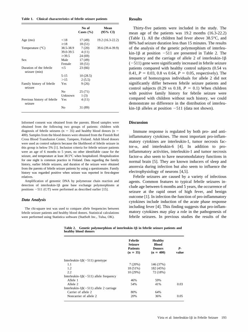
Informed consent was obtained from the parents. Blood samples wereobtained from the following two groups of patients: children withdiagnosis of febrile seizures (n � 35) and healthy blood donors (n �400). Samples from the blood donors were obtained from the Finnish RedCross Blood Transfusion Center, Tampere, Finland. Adult blood donorswere used as control subjects because the likelihood of febrile seizure inthis group is below 5% [1]. Inclusion criteria for febrile seizure patientswere an age of 6 months to 5 years, no other identifiable cause for theseizure, and temperature at least 38.5°C when hospitalized. Hospitalizationfor one night is common practice in Finland. Data regarding the familyhistory, earlier febrile seizures, and duration of the seizure were obtainedfrom the parents of febrile seizure patients by using a questionnaire. Familyhistory was regarded positive when seizure was reported in first-degreerelatives.
Amplification of genomic DNA by polymerase chain reaction anddetection of interleukin-1� gene base exchange polymorphisms atpositions �511 (C/T) were performed as described earlier [15].
Data Analysis
The chi-square test was used to compare allele frequencies betweenfebrile seizure patients and healthy blood donors. Statistical calculationswere performed using Statistica software (StatSoft Inc., Tulsa, OK).
Results
Thirty-five patients were included in the study. Themean age of the patients was 19.2 months (16.3-22.2)(Table 1). All the children had fever above 38.5°C, and80% had seizure duration less than 15 minutes. The resultsof the analysis of the genetic polymorphism of interleu-kin-1� at position �511 are presented in Table 2. Thefrequency and the carriage of allele 2 of interleukin-1�(�511) gene were significantly increased in febrile seizurepatients compared with healthy control subjects (0.54 vs0.41, P � 0.03, 0.8 vs 0.64, P � 0.05, respectively). Theamount of homozygous individuals for allele 2 did notsignificantly differ between febrile seizure patients andcontrol subjects (0.29 vs 0.18, P � 0.1) When childrenwith positive family history for febrile seizure werecompared with children without such history, we coulddemonstrate no difference in the distribution of interleu-kin-1� alleles at position �511 (data not shown).
Discussion
Immune response is regulated by both pro- and anti-inflammatory cytokines. The most important pro-inflam-matory cytokines are interleukin-1, tumor necrosis fac-tor-�, and interleukin-6 [4]. In addition to pro-inflammatory activities, interleukin-1 and tumor necrosisfactor-� also seem to have neuromodulatory functions innormal brain [5]. They are known inducers of sleep andanorexia during infection but also seem to influence theelectrophysiology of neurons [4,5].
Febrile seizures are caused by a variety of infectiousagents. Common features to typical febrile seizures in-clude age between 6 months and 5 years, the occurrence ofseizure at the rapid onset of high fever, and benignoutcome [1]. In infection the function of pro-inflammatorycytokines include induction of the acute phase responseincluding fever [4]. This finding suggests that pro-inflam-matory cytokines may play a role in the pathogenesis offebrile seizures. In previous studies the results of the
Table 1. Clinical characteristics of febrile seizure patients
No ofCases (%)
Mean(95% CI)
Age (mo) �18 17 (49) 19.2 (16.3-22.2)�18 18 (51)
Temperature (°C) 38.5-38.9 7 (20) 39.6 (39.4-39.9)39.0-39.5 4 (11)�39.5 24 (69)
Sex Male 17 (49)Female 18 (51)
Duration of the febrileseizure (min)
�5 23 (66)
5-15 10 (28.5)�15 2 (5.5)
Family history of febrileseizure
Yes 9 (26)
No 25 (71)Unknown 1 (3)
Previous history of febrileseizure
Yes 4 (11)
No 31 (89)
Table 2. Genetic polymorphism of interleukin-1� in febrile seizure patients andhealthy blood donors
FebrileSeizurePatients(n � 35)
HealthyBlood
Donors(n � 400)
P-value
Interleukin-1�(�511) genotype1.1 7 (20%) 146 (37%)1.2 18 (51%) 182 (45%)2.2 10 (29%) 72 (18%)
Interleukin-1�(�511) allele frequencyAllele 1 46% 59%Allele 2 54% 41% 0.03
Interleukin-1�(�511) allele 2 carriageCarrier of allele 2 80% 64%Noncarrier of allele 2 20% 36% 0.05
193Virta et al: Interleukin-1� in Febrile Seizure

importance of fever-inducing cytokines in febrile seizureshave been conflicting. Helminen and Vesikari [10] and,recently, Straussberg et al. [11] have demonstrated thatinterleukin-1 production of lipopolysaccharide-stimulatedmononuclear cells isolated from patients with febrileseizures is increased compared with control subjects.Lahat et al. [12] found no difference in cerebrospinal fluidand blood interleukin-1� levels between patients andcontrol subjects when measured with enzyme-linked im-munoassay. Ichiyama et al. [13] were able to demonstrateincreased levels of tumor necrosis factor-�, interleukin-1�, and interleukin-6 in cerebrospinal fluid from childrenwith acute encephalitis/encephalopathy but not from chil-dren with febrile seizures. The discrepancy in these studiesmay be explained by differing study methods. Because ofthe short half-life and numerous interactions of the cyto-kines, individual measurements also may not reflect thetrue situation [4]. The genetic make-up of the individualmay be more relevant.
The purpose of this study was to analyze the importanceof cytokine gene polymorphism in febrile seizures. Thedata indicate increased frequency and carriage of theinterleukin-1� (�511) allele 2 in children with febrileseizures compared with healthy blood donors. Interleu-kin-1 induces fever by stimulating local production ofprostaglandins which in turn raise the set point of thethermoregulatory center [16]. There are at least twopolymorphic sites within the interleukin-1� gene: at posi-tion �511 in the promoter region and at position �3953 inthe fifth exon [14,15]. In previous studies, interleukin-1�allele 2 at position �511 has been connected to increasedproduction of this cytokine [17]. Therefore the increasedoccurrence of allele 2 observed in this study may suggestthat interleukin-1 production is elevated in febrile seizures.The elevated production of interleukin-1 during febrileseizures likely explains the high fever typically observedat the onset of seizure. However, this study cannot distinguishthe impact of interleukin-1 genetics on fever and seizureactivity independently. Also, interleukin-1 genetics cannot bethe only determinant factor in febrile seizures.
The importance of interleukin-1 and its family inepilepsy has been reported previously in experimentalstudies. Increased mRNA levels of interleukin-1�, inter-leukin-1 receptor antagonist, and interleukin-1 receptorexpression have been observed by in situ hybridization inkainic acid-induced seizures [6,7]. Whether the suppos-edly increased production of interleukin-1 that was ob-served in this study is connected to the seizure or simplyreflects the high fever typically connected to febrileseizures is unknown. However, approximately 30% ofpatients suffering from temporal lobe epilepsy with hip-pocampal sclerosis have febrile seizures in their childhood[18]. In a recent study, Kanemoto et al. [19] demonstratedthat patients with temporal lobe epilepsy and hippocampalsclerosis were more often homozygotes for interleukin-1�
allele 2 at position �511 than temporal lobe epilepsypatients without hippocampal sclerosis or healthy controlsubjects, suggesting that interleukin-1� may be relevant inthe pathogenesis of this entity. The increased carriage ofinterleukin-1� allele 2 at position �511 observed in thestudy of Kanemoto et al. in temporal lobe epilepsy patientsand in this study in children with febrile seizures supportsthe theory that febrile seizures connected to increasedinterleukin-1� production may predispose to the develop-ment of epilepsy. This finding may help neurologists tofind the children with febrile seizures that are at risk ofdeveloping future seizures or epilepsy.
This work was supported by a grant from The Research Fund of TampereUniversity Hospital. The authors would like to thank Ms Mervi Salomakifor expert technical assistance.
References
[1] Berg AT, Shinnar S, Shapiro ED, Salomon ME, Crain EF,Hauser WA. Risk factors for a first febrile seizure: A matched case-control study. Epilepsia 1995;36:334-41.
[2] Johnson WG, Kugler SL, Stenroos ES, et al. Pedigree analysis infamilies with febrile seizures. Am J Med Genet 1996;61:345-52.
[3] Peiffer A, Thompson J, Charlier C, et al. A locus for febrile seizures(FEB3) maps to chromosome 2q23–24. Ann Neurol 1999;46:671-8.
[4] Dinarello CA. Biologic basis for interleukin-1 in disease. Blood1996;87:2095-147.
[5] Vitkovic L, Bockaert J, Jacque C. “Inflammatory” cytokines:Neuromodulators in normal brain. J Neurochem 2000;74:457-71.
[6] Minami M, Kuraishi Y, Satoh M. Effects of kainic acid onmessenger RNA levels of IL-1�, IL-6, TNF� and LIF in the rat brain.Biochem Biophys Res Commun 1991;176:593-8.
[7] Eriksson C, Winblad B, Schultzberg M. Kainic acid inducedexpression of interleukin-1 receptor antagonist mRNA in the rat brain.Mol Brain Res 1998;58:195-208.
[8] Aurelius E, Andersson B, Forsgren M, Skoldenberg B, Stran-negård O. Cytokines and other markers of intrathecal immune responsein patients with herpes simplex encephalitis. J Infect Dis 1994;170:678-81.
[9] Mustafa MM, Ramilo O, Saez-Llorens X, Olsen K, Magness R,McCracken G. Cerebrospinal fluid prostaglandins, interleukin 1�, and tumornecrosis factor in bacterial meningitis. Am J Dis Child 1990;144:883-7.
[10] Helminen M, Vesikari T. Increased interleukin-1 (IL-1) pro-duction from LPS-stimulated peripheral blood monocytes in childrenwith febrile convulsions. Acta Paediatr Scand 1990;79:810-6.
[11] Straussberg R, Amir J, Harel L, Punsky I, Bessler H. Pro- andanti-inflammatory cytokines in children with febrile convulsions. PediatrNeurol 2001;24:49-53.
[12] Lahat E, Livne M, Barr J, Katz Y. Interleukin-1� levels inserum and cerebrospinal fluid of children with febrile seizures. PediatrNeurol 1997;17:34-6.
[13] Ichiyama T, Nishikawa M, Yoshitomi T, Hayashi T, FurukawaS. Tumor necrosis factor-�, interleukin- 1�, and interleukin- 6 incerebrospinal fluid from children with prolonged febrile seizures. Neu-rology 1998;50:407-11.
[14] Pociot F, Mølvig J, Wogensen L, Worsaae H, Nerup J. A TaqIpolymorphism in the human interleukin-1� (IL-1�) gene correlates withIL-1� secretion in vitro. Eur J Clin Invest 1992;22:396-402.
[15] di Giovine FS, Takhsh E, Blakemore AIF, Duff GW. Singlebase polymorphism at �511 in the human interleukin- 1� gene (IL1�).Hum Mol Genet 1992;1:450.
194 PEDIATRIC NEUROLOGY Vol. 26 No. 3

[16] Mackowiak PA, Bartlett JG, Borden EC, et al. Concepts offever: Recent advantages and lingering dogma. Clin Infect Dis 1997;25:119-38.
[17] Santtila S, Savinainen K, Hurme M. Presence of the IL-1RAallele 2 (IL1RN*2) is associated with enhanced IL-1beta production invitro. Scand J Immunol 1998;47:195-8.
[18] Davies KG, Hermann BP, Dohan FC, Foley KT, Bush AJ,
Wyler AR. Relationship of hippocampal sclerosis to duration and age ofonset of epilepsy, and childhood febrile seizures in temporal lobectomypatients. Epilepsy Res 1996;24:119-26.
[19] Kanemoto K, Kawasaki J, Miyamoto T, Obayashi H, Nish-imura M. Interleukin (IL)-1�, IL-1�, and IL-1 receptor antagonist genepolymorphisms in patients with temporal lobe epilepsy. Ann Neurol2000;47:571-4.
195Virta et al: Interleukin-1� in Febrile Seizure

777
CONCISE COMMUNICATION
Susceptibility to Primary Epstein-Barr Virus Infection Is Associatedwith Interleukin-10 Gene Promoter Polymorphism
Merja E. Helminen,1 Sanna Kilpinen,2 Miia Virta,2
and Mikko Hurme2
1Department of Pediatrics, Tampere University Hospital,and 2Department of Microbiology and Immunology,
University of Tampere Medical School, Tampere, Finland
In total, 116 children were investigated to determine whether the interleukin (IL)–10 poly-morphism influences the age at primary Epstein-Barr virus (EBV) infection. The promoter ofIL-10 is polymorphic, with 3 known single base substitutions (G/A at �1082, C/T at �819,and C/A at �592), which form 3 haplotypes: GCC, ACC, and ATA. This study found thatcarriage of the ATA haplotype protects against early EBV infection. The presence of the ATAhaplotype was associated with EBV seronegativity (odds ratio, 2.6; 95% confidence interval,1.04–6.7; ), when controlled by age. To examine the effect of haplotypes on IL-10P p .04production, IL-10 plasma levels were measured in 50 newborns and 400 adults and werecorrelated with the IL-10 haplotype. The IL-10 levels were significantly higher in the ATAcarriers than in the noncarriers. These data suggest that the IL-10 ATA haplotype confersprotection against primary EBV infection and that the effect is mediated by high IL-10 levels.
Primary Epstein-Barr virus (EBV) infection usually occurswithin the first years of life. At an early age, the infection isusually asymptomatic, whereas, during adolescence and adult-hood, it can present as acute infectious mononucleosis (IM).The infection is extremely common, and 190% of adults areseropositive for EBV. The infection is spread through salivarycontact, and the mucosal epithelium of the oropharynx is con-sidered to be the first site of infection and replication. Fromthe oropharynx, the virus is transmitted to locally infiltratingB cells, where it persists for a person’s life [1].
The clinical picture of acute IM is believed to result from thehost’s immune response against the invading virus. This responseincludes cytotoxic T cells and NK cells, which provide initialcontrol of the infection. Cytokines also seem to be importantmediators in the immune response against EBV [2]. EBV infec-tion of B cells induces the proliferation of lymphocytes and pro-duction of interleukin (IL)–10 [3]. The EBV itself codes for acellular homologue of IL-10, viral IL-10, which shares propertiessimilar to cellular IL-10 [4]. Cellular IL-10 is considered to bean anti-inflammatory cytokine that induces the proliferation ofB cells and inhibits the antigen-specific activation of T cells and
Received 12 March 2001; revised 29 May 2001; electronically published7 August 2001.
The study was approved by the Tampere University Hospital ethics com-mittee and was conducted according to the hospital’s guidelines. Informedconsent was obtained from parents before study enrollment.
Financial support: Tampere University Hospital research fund.Reprints or correspondence: Dr. Merja E. Helminen, Dept. of Pediatrics,
Tampere University Hospital, PO Box 2000, 33521 Tampere, Finland ([email protected]).
The Journal of Infectious Diseases 2001;184:777–80� 2001 by the Infectious Diseases Society of America. All rights reserved.0022-1899/2001/18406-0015$02.00
the production of proinflammatory cytokines. It is produced byB cells and by monocytes and T cells [4].
The interindividual variation seen in IL-10 production is ge-netically determined. The promoter region of IL-10 contains 3base substitutions at �1082 G/A, �819 C/T, and �592 C/A,which are related to IL-10 protein production in vitro [5]. Thesealleles combine as 3 possible haplotypes, GCC, ACC, and ATA,which participate in the regulation of IL-10 gene transcription[5]. We previously showed that IL-10 genetics influence the clini-cal picture of EBV infection [6]. In this study, we analyzedwhether IL-10 genetics influence the age when EBV seroconver-sion occurs. We also measured IL-10 plasma levels in neonatesand in healthy adults and correlated these to the IL-10 genotype.
Patients, Materials, and Methods
Patients. The study was done at the Tampere University Hos-pital and University of Tampere (Tampere, Finland). Blood sam-ples were obtained from 116 children, 9 months to 15 years old,which were obtained for pediatric consultation between November1999 and May 2000. Umbilical cord blood samples were obtainedfrom 50 healthy neonates. Blood samples from 400 healthy blooddonors were obtained from the Finnish Red Cross Blood Trans-fusion Center (Tampere).
EBV serology. EBV serology was measured by EIA, accordingto the manufacturer’s instructions (Enzygnost anti-EBV/IgG; Behr-ing). The assay’s detection limit is 1.0 pg/mL.
IL-10 gene promoter polymorphism. Genomic DNA was iso-lated from the blood samples. The region of the IL-10 promoterfrom �1120 to �533 was amplified by polymerase chain reaction,and single nucleotide polymorphisms (G/A at �1082, C/T at �819,and C/A at �592) were analyzed by restriction fragment lengthpolymorphism [6].
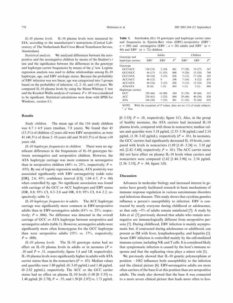
778 Helminen et al. JID 2001;184 (15 September)
Table 1. Interleukin (IL)–10 genotypes and haplotype carrier ratesand frequencies in Epstein-Barr virus (EBV)–seropositive (EBV�;
) and –seronegative (EBV�; ) adults and EBV� (n p 380 n p 20 n p) and EBV� ( ) children.44 n p 72
Genotype andhaplotype carriers
Adults Children
EBV� EBV� Pa EBV� EBV� Pa
GenotypeGCC/ACC 124 (33) 2 (10) .061 17 (39) 19 (27) .167GCC/GCC 66 (17) 11 (55) .000 9 (20) 13 (18) .749GCC/ATA 60 (16) 3 (15) .826 5 (11) 17 (24) .103ACC/ACC 46 (12) 0 .196 7 (16) 8 (12) .455ACC/ATA 60 (16) 3 (15) .826 5 (11) 13 (18) .334ATA/ATA 24 (6) 1 (5) .843 1 (1) 2 (1) .868
Haplotype carriersGCC 250 (66) 16 (80) .544 31 (70) 49 (68) .312ACC 230 (61) 5 (25) .004 29 (66) 40 (56) .836ATA 144 (38) 7 (35) .981 11 (25) 32 (44) .035
NOTE. With the exception of P values, data are no. (%) of study subjects.a
x2 Test.
IL-10 plasma levels. IL-10 plasma levels were measured byEIA, according to the manufacturer’s instructions (Central Lab-oratory of The Netherlands Red Cross Blood Transfusion Service,Amsterdam).
Statistical analysis. We analyzed differences between the sero-positive and the seronegative children by means of the Student’s ttest and the significance between the differences in the genotypeand haplotype carrier frequencies by means of the x2 test. Logisticregression analysis was used to define relationships among IL-10haplotype, age, and EBV serologic status. Because the probabilityof EBV infection was not linear, age was categorized into 3 groupsbased on the probability of infection: !2, 2–10, and 110 years. Wecompared IL-10 plasma levels by using the Mann-Whitney U testand the Kruskal-Wallis analysis of variance. was consideredP ! .05to be significant. Statistical calculations were done with SPSS forWindows, version 6.1.
Results
Study children. The mean age of the 116 study childrenwas years (median, 7.8 years). We found that 456.7 � 4.9(13.3%) of children !2 years old were EBV seropositive, as were41 (46.3%) of those 2–10 years old and 30 (63.3%) of those 110years old.
IL-10 haplotype frequencies in children. There were no sig-nificant differences in the frequencies of IL-10 genotypes be-tween seronegative and seropositive children. However, theATA haplotype carriage was more common in seronegativethan in seropositive children (44% vs. 25%, respectively; P p
). By use of logistic regression analysis, ATA positivity was.035associated significantly with EBV seronegativity (odds ratio[OR], 2.6; 95% confidence interval [CI], 1.04–6.7; )P p .04when controlled by age. No significant association was foundwith carriage of the GCC or ACC haplotypes and EBV status(OR, 0.8; 95% CI, 0.3–2.0 and OR, 0.9; 95% CI, 0.4–2.2, re-spectively; table 1).
IL-10 haplotype frequencies in adults. The ACC haplotypecarriage was significantly more common in EBV-seropositiveadults than in EBV-seronegative adults (61% vs. 25%, respec-tively; ). No difference was detected in the overallP p .004carriage of GCC or ATA haplotype between seropositive andseronegative adults (table 1). However, seronegative adults weresignificantly more often homozygous for the GCC haplotypethan were seropositive adults (55% vs. 17%, respectively;
).P p .000IL-10 plasma levels. The IL-10 genotype status had no
effect on IL-10 plasma levels in adults or in neonates (P pand , respectively; figure 1A and 1B, respectively)..10 P p .11
IL-10 plasma levels were significantly higher in adults with ATAcarrier status than in the noncarriers ( ). Median valuesP p .03and quartiles were 1.80 pg/mL (0–3.45 pg/mL) and 1.46 pg/mL(0–2.62 pg/mL), respectively. The ACC or the GCC carrierstatus had no effect on plasma IL-10 levels (1.60 [0–3.35] vs.1.48 pg/mL [0–2.70]; ; and 1.50 [0–2.87] vs. 1.71 pg/mLP p .35
[0–3.55]; , respectively; figure 1C). Also, in the groupP p .18of healthy neonates, the ATA carriers had increased IL-10plasma levels, compared with those in noncarriers; median val-ues and quartiles were 3.18 pg/mL (2.35–5.56 pg/mL) and 2.14pg/mL (1.38–3.42 pg/mL), respectively ( ). In neonates,P p .01the GCC carriers had significantly decreased IL-10 levels, com-pared with levels in noncarriers (1.89 [1.41–3.24] vs. 3.18 pg/mL [2.42–5.60], respectively; ). The ACC carrier statusP p .01did not have effect on plasma IL-10 levels when carriers andnoncarriers were compared (2.42 [1.44–3.96] vs. 2.54 pg/mL[1.56–3.53]; ; figure 1D).P p .94
Discussion
Advances in molecular biology and increased interest in ge-netics have greatly facilitated research in basic mechanisms ofimmune response regulation in various autoimmune disordersand infectious diseases. This study shows that cytokine geneticsinfluence a person’s susceptibility to infection. EBV is con-tracted by nearly everyone during childhood or adolescence,so that only ∼5% of adults remain uninfected [7]. A study byJabs et al. [7] previously showed that adults who remain sero-negative are immunologically different from seropositive per-sons [7]. During childhood, EBV infection is mainly asympto-matic but, if contracted during adolescence or adulthood, canpresent as IM with fever, lymphadenopathy, and hepatitis [1].Acute EBV infection is controlled mainly by the cell-mediatedimmune system, including NK and T cells. It is considered likelythat symptomatic infection is caused by the host’s immune re-sponse and that the replicating virus plays a minor role [1].
We previously showed that IL-10 genetic polymorphism atposition �1082 influences both susceptibility to the infectionand the clinical picture [6]. EBV-seronegative adults are moreoften carriers of the base G at this position than are seropositiveadults. The study also showed that the base A was connectedto a more severe clinical picture that leads more often to hos-
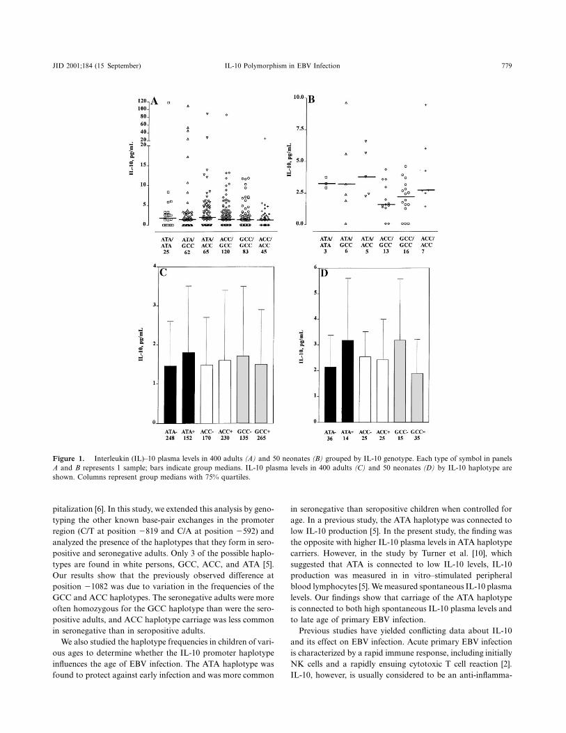
JID 2001;184 (15 September) IL-10 Polymorphism in EBV Infection 779
Figure 1. Interleukin (IL)–10 plasma levels in 400 adults (A) and 50 neonates (B) grouped by IL-10 genotype. Each type of symbol in panelsA and B represents 1 sample; bars indicate group medians. IL-10 plasma levels in 400 adults (C) and 50 neonates (D) by IL-10 haplotype areshown. Columns represent group medians with 75% quartiles.
pitalization [6]. In this study, we extended this analysis by geno-typing the other known base-pair exchanges in the promoterregion (C/T at position �819 and C/A at position �592) andanalyzed the presence of the haplotypes that they form in sero-positive and seronegative adults. Only 3 of the possible haplo-types are found in white persons, GCC, ACC, and ATA [5].Our results show that the previously observed difference atposition �1082 was due to variation in the frequencies of theGCC and ACC haplotypes. The seronegative adults were moreoften homozygous for the GCC haplotype than were the sero-positive adults, and ACC haplotype carriage was less commonin seronegative than in seropositive adults.
We also studied the haplotype frequencies in children of vari-ous ages to determine whether the IL-10 promoter haplotypeinfluences the age of EBV infection. The ATA haplotype wasfound to protect against early infection and was more common
in seronegative than seropositive children when controlled forage. In a previous study, the ATA haplotype was connected tolow IL-10 production [5]. In the present study, the finding wasthe opposite with higher IL-10 plasma levels in ATA haplotypecarriers. However, in the study by Turner et al. [10], whichsuggested that ATA is connected to low IL-10 levels, IL-10production was measured in in vitro–stimulated peripheralblood lymphocytes [5]. We measured spontaneous IL-10 plasmalevels. Our findings show that carriage of the ATA haplotypeis connected to both high spontaneous IL-10 plasma levels andto late age of primary EBV infection.
Previous studies have yielded conflicting data about IL-10and its effect on EBV infection. Acute primary EBV infectionis characterized by a rapid immune response, including initiallyNK cells and a rapidly ensuing cytotoxic T cell reaction [2].IL-10, however, is usually considered to be an anti-inflamma-
780 Helminen et al. JID 2001;184 (15 September)
tory cytokine that inhibits NK and T cell activity and thus isbelieved to control the strong inflammatory response after pri-mary infection [8]. In 1992, Stewart and Rooney [9] showedthat IL-10 enhances the activation of NK cells by EBV-trans-formed B cells and that it may actually enhance immune re-sponse against EBV. In 1993, Kurilla et al. [10], by using anSCID-mouse model, found that IL-10 actually increased NKcell activity and decreased viral replication [10]. Thus, the lateage of infection in ATA haplotype carriers could be explainedby the high spontaneous IL-10 plasma levels that could resultin a strong initial antiviral effect postponing the age of primaryEBV infection.
In children, the ATA haplotype confers protection againstearly infection, whereas seronegative adults are more likely tobe GCC haplotype carriers. It is generally believed that the fewadults who remain uninfected probably remain so for life andthat they are immunologically different from the rest of thepopulation [7]. The seronegative children probably benefit fromthe IL-10 ATA haplotype, so that EBV infection is postponedto later childhood or adolescence, but this haplotype does notprevent the infection. However, the GCC haplotype homozy-gosity or the absence of the ACC haplotype may prevent theinfection from ever taking place. A large number of study chil-dren for follow-up would be needed to verify this hypothesis.
If EBV is contracted during adolescence or early adulthood,it is more often symptomatic. EBV infection symptoms arethought to be caused by the host’s immune response. This studysuggests that the ATA haplotype of IL-10 increases the age ofprimary infection and most likely also the risk for symptomaticdisease. Previous studies concerning septic infections have sug-gested that the balance between pro- and anti-inflammatorycytokines determines the clinical picture during infection [11].The role IL-10 plays in immune response during acute EBVinfection clearly needs to be studied further. In some studies,IL-10 polymorphism was connected to differential clinical ex-pression of various diseases, such as asthma and systemic lupuserythematosus [12]. Previous studies also suggest that EBV in-fection or the age of primary EBV infection may play a rolein the pathogenesis of these diseases [13, 14]. These areas clearlyneed further study.
Acknowledgment
We thank Heini Huhtala for expert advice concerning statisticalanalysis of the data.
References
1. Rickinson AB, Kieff E. Epstein-Barr virus. In: Fields BN, Knipe DM, How-ley PM, et al, eds. Fields virology. 3d ed. Vol 2. Philadelphia: Lippincott-Raven, 1996:2397–446.
2. Andersson J. Clinical and immunological considerations in Epstein-Barrvirus–associated diseases. Scand J Infect Dis Suppl 1996;100:72–82.
3. Miyazaki BI, Cheung RK, Dosch HM. Viral interleukin-10 is critical for theinduction of B cell growth transformation by Epstein-Barr virus. J ExpMed 1993;178:439–47.
4. Moore KW, O’Garra A, de Waal Malefyt R, Viera P, Mossman TR. Inter-leukin-10. Annu Rev Immunol 1993;11:165–90.
5. Turner DM, Williams DM, Sankaran D, Lazarus M, Sinnott PJ, HutchinsonIV. An investigation of polymorphism in the interleukin-10 gene promoter.Eur J Immunogenet 1997;24:1–8.
6. Helminen M, Lahdenpohja N, Hurme M. Polymorphism of the interleukin-10 gene is associated with susceptibility to Epstein-Barr virus infection. JInfect Dis 1999;180:496–9.
7. Jabs WJ, Wagner HJ, Neustock P, Kluter H, Kirchner H. Immunologic prop-erties of Epstein-Barr virus–seronegative adults. J Infect Dis 1996;173:1248–51.
8. de Waal Malefyt R, Haanen J, Spits H, et al. IL-10 and viral IL-10 stronglyreduced antigen specific T cell proliferation by diminishing the antigenpresenting capacity of monocytes via down-regulation of class major histo-compatibility complex expression. J Exp Med 1991;174:915–24.
9. Stewart JP, Rooney CM. The interleukin-10 homolog encoded by Epstein-Barr virus enhances the reactivation of virus-specific cytotoxic T cell andHLA-unrestricted killer cell responses. Virology 1992;191:773–82.
10. Kurilla MG, Swaminathan S, Welsh RM, Kieff E, Brutkiewicz RR. Effectsof virally expressed interleukin-10 on vaccinia virus infection in mice. JVirol 1993;67:7623–8.
11. Van Dissel JT, Van Langevelde P, Westendorp RGJ, Kvappenberg K, FrolichM. Anti-inflammatory cytokine profile and mortality in febrile patients.Lancet 1998;351:950–3.
12. Lim S, Crawley E, Woo P, Barnes PJ. Haplotype associated with low inter-leukin-10 production in patients with severe asthma. Lancet 1998;352:113–5.
13. Calvani M Jr, Alessandri C, Paolone G, Rosengart L, Di Caro A, De FrancoD. Correlation between Epstein-Barr virus antibodies, serum IgE andatopic disease. Pediatr Allergy Immunol 1997;8:91–6.
14. James JA, Kaufman KM, Farris AD, Taylor-Albert E, Lehman TJA, HarleyJB. An increased prevalence of Epstein-Barr virus infection in young pa-tients suggests a possible etiology for systemic lupus erythematosus. J ClinInvest 1997;100:3019–26.

Fax +41 61 306 12 34E-Mail [email protected]
Original Paper
Int Arch Allergy Immunol 2005;137:282–288 DOI: 10.1159/000086421
Genetic and Environmental Factors in the Immunopathogenesis of Atopy: Interaction of Helicobacter pylori Infection and IL4 Genetics
T. Pessi a M. Virta a K. Ådjers a J. Karjalainen b H. Rautelin c, d T.U. Kosunen d M. Hurme a, e
a Department of Microbiology and Immunology, Medical School, University of Tampere, and b Department of Respiratory Medicine, Tampere University Hospital, Tampere ; c HUSLAB, Helsinki University Central Hospital, and d Department of Bacteriology and Immunology, Haartman Institute, University of Helsinki, Helsinki ; e Centre for Laboratory Medicine, Tampere University Hospital, Tampere , Finland
–590 allele T had a diminished risk to be H. pylori infect-ed (OR 0.485 95%CI 0.287–0.819). This effect was not seen in controls. Logistic regression analysis indicated that H. pylori and IL4 effects on atopy risk are not inter-dependent. Conclusions: This study showed that the ef-fect of H. pylori infection on atopy risk is stronger than that of IL4 genetics. There is no interaction between these factors on the pathogenesis of atopy suggesting that these factors have distinct immunopathogenetic mechanisms. However, the genetic effect may modify the role of infective agents by effecting on susceptibility to disease.
Copyright © 2005 S. Karger AG, Basel
Introduction
Both genetic and environmental factors are known to have an effect on susceptibility to atopic diseases (such as atopic eczema, rhinoconjunctivitis and asthma). Of the environmental factors, childhood infections have an important role: there is epidemiological evidence show-ing that infectious diseases (especially gastrointestinal infections) during early childhood decrease the risk of development of atopic diseases in later life, and vice ver-
Key Words Helicobacter pylori � Interleukin-4 � Genotype � Polymorphism � Atopy � Skin prick test
Abstract Background: Both genetic and environmental factors, e.g. early childhood infections, have a role in the patho-genesis of atopic diseases. Objective: To examine simul-taneously the strength and possible interactions of two known such factors, IL4 genetics and Helicobacter py-lori infection, on the risk of atopy and asthma. Methods: Gene polymorphism analyses and skin prick tests (SPT) were determined in 245 adult asthmatics and 405 non-asthmatic controls of population-based case-control study. SPTs were used as an indicator of atopy. H. py-lori infection was verifi ed by detecting anti- H. pylori IgG antibodies in sera. Results: A signifi cant negative asso-ciation was seen between the presence H. pylori antibod-ies and SPT positivity ( 6 1 positive reactions) in both asthmatics and controls (p = 0.002 and p = 0.025, respec-tively) but the effect of IL-4 polymorphism (SNP –590C/T) was nonsignifi cant in both groups (p = 0.071 and p = 0.072, respectively). However, IL4 genetics had an effect on susceptibility to H. pylori : asthmatics carrying the IL4
Received: December 21, 2004 Accepted after revision: April 7, 2005 Published online: June 17, 2005
Correspondence to: Dr. Tanja Pessi University of Tampere, Medical School FIN–33014 Tampere (Finland) Tel. +358 3 3551 11, Fax +358 3 3551 6173 E-Mail [email protected]
© 2005 S. Karger AG, Basel 1018–2438/05/1374–0282$22.00/0
Accessible online at: www.karger.com/iaa
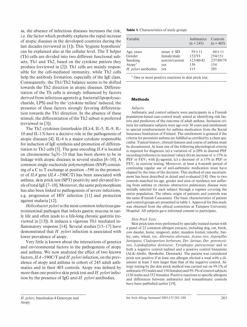
H . pylori , Interleukin-4 Genotype and Atopy
Int Arch Allergy Immunol 2005;137:282–288 283
sa, the absence of infectious diseases increases the risk, i.e. the factor which probably explains the rapid increase of atopic diseases in the developed countries during the last decades (reviewed in [1] ). This ‘hygiene hypothesis’ can be explained also at the cellular level. The T helper (Th) cells are divided into two different functional sub-sets, Th1 and Th2, based on the cytokine pattern they produce (reviewed in [2] ). Th1 cells are mainly respon-sible for the cell-mediated immunity, while Th2 cells help the antibody formation, especially of the IgE class. Consequently, the Th1/Th2 balance seems to be shifted towards the Th2 direction in atopic diseases. Differen-tiation of the Th cells is strongly infl uenced by factors derived from infectious agents (e.g. bacterial lipopolysac-charide, LPS) and by the ‘cytokine milieu’ induced, the presence of these factors strongly favoring differentia-tion towards the Th1 direction. In the absence of these stimuli, the differentiation of the Th2 subset is preferred (reviewed in [3] ).
The Th2 cytokines (interleukin (IL)-4, IL-5, IL-9, IL-10 and IL-13) have a decisive role in the pathogenesis of atopic diseases [4] . IL-4 is a major cytokine responsible for induction of IgE synthesis and promotion of differen-tiation to Th2 cells [5] . The gene encoding IL4 is located on chromosome 5q31–33 that has been shown to be in linkage with atopic diseases in several studies [6–10] . A common single nucleotide polymorphism (SNP) consist-ing of a C to T exchange at position –590 in the promot-er of IL4 gene ( IL4 –590C/T) has been associated with asthma, skin prick test (SPT) positivity and elevated lev-els of total IgE [7–10] . Moreover, the same polymorphism has also been linked to pathogenesis of severe infections, e.g. progression of HIV infection [11] and protection against malaria [12] .
Helicobacter pylori is the most common infectious gas-trointestinal pathogen that infects gastric mucosa in ear-ly life and often leads to a life-long chronic gastritis (re-viewed in [13] ). It induces a vigorous Th1 mediated in-fl ammatory response [14] . Several studies [15–17] have demonstrated that H. pylori infection is associated with lower prevalence of atopy.
Very little is known about the interactions of genetics and environmental factors in the pathogenesis of atopy and asthma. We now analyzed the effect of two known factors, IL4 –590C/T and H. pylori infection, on the prev-alence of atopy and asthma in cohort of 245 adult asth-matics and in their 405 controls. Atopy was defi ned by more than one positive skin prick test and H. pylori infec-tion by the presence of IgG anti- H. pylori antibodies.
Methods
Subjects Asthmatic and control subjects were participants in a Finnish
population-based case-control study aimed at identifying risk fac-tors and predictors of the outcome of adult asthma. Inclusion cri-teria for asthmatic subjects were age over 30 years and entitlement to special reimbursement for asthma medication from the Social Insurance Institution of Finland. The entitlement is granted if the criteria for persistent asthma are fulfi lled as certifi ed by a chest spe-cialist. Typical history, clinical features and course of asthma must be documented. At least one of the following physiological criteria is required for diagnosis: (a) a variation of 6 20% in diurnal PEF recording (reference to maximal value); (b) an increase of 6 15% in PEF or FEV 1 with � 2 -agonist; (c) a decrease of 6 15% in PEF or FEV 1 in exercise testing. Moreover, at least a 6-month period of continuing regular use of anti-asthmatic medication must have elapsed by the time of the decision. This method of case ascertain-ment has been described in detail and evaluated [18] . One to two controls matched for age, gender and area of residence not suffer-ing from asthma or chronic obstructive pulmonary disease were initially selected for each subject through a register covering the entire population. The ethnic origin of patients and controls was the same (Finnish Caucasian). The basic characteristics of patient and control groups are presented in table 1 . Approval for this study was obtained from the ethical committee at Tampere University Hospital. All subjects gave informed consent to participate.
Skin Prick Tests Skin prick tests were performed by specially trained nurses with
a panel of 22 common allergen extracts, including dog, cat, birch, cow dander, horse, mugwort, alder, meadow foxtail, timothy, bar-ley, oats, wheat, rye, Alternaria alternata , Acarus siro , Aspergillus fumigatus , Cladosporium herbarum , Der. farinae , Der. pteronyssi-nus , Lepidoglyphus destructor , Tyrophagus putrescentiae and to both a negative control (saline) and a positive control histamine (ALK-Abello, Hørsholm, Denmark). The patient was considered prick test positive if at least one allergen elicited a weal with a di-ameter at least 3 mm larger than that of the negative control. Al-lergy testing by the skin prick method was carried out on 99.1% of asthmatic (93 males and 150 females) and 99.3% of control subjects (150 males and 252 females). Positive reactions to specifi c allergens and differences between asthmatics and nonasthmatic controls have been published earlier [19] .
Table 1. Characteristics of study groups
Variable Asthmatics(n = 245)
Controls(n = 405)
Age, years mean 8 SD 59811 60811Gender female/male 152/93 254/151Smoking non/ex/current 123/80/42 237/89/79Atopy1 yes 139 154H. pylori antibodies yes 115 205
1 One or more positive reactions in skin prick test.
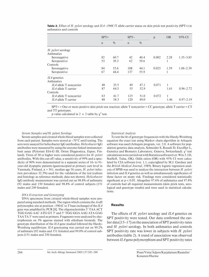
Pessi/Virta/Ådjers/Karjalainen/Rautelin/Kosunen/Hurme
Int Arch Allergy Immunol 2005;137:282–288 284
Serum Samples and H. pylori Serology Serum samples and citrated whole-blood samples were collected
from each patient. Samples were stored at –70 ° C until testing. The sera were assayed for helicobacter IgG antibodies. Helicobacter IgG antibodies were measured by using the enzyme-linked immunosor-bent assay (Pyloriset EIA-G III, Orion Diagnostica, Espoo, Fin-land). Titres of 30 or higher were considered positive for H. pylori antibodies. With this cut-off value, a sensitivity of 99% and a spec-ifi city of 90% were demonstrated in a separate series of 16- to 91-year-old dyspeptic patients (gastroscopied at primary care level in Vammala, Finland, n = 561, median age 56 years, H. pylori infec-tion prevalence 32.3%) used for the validation of the test (culture and histology as reference methods; data not shown). Helicobacter IgG antibody measurement was carried out on 98.8% of asthmatic (92 males and 150 females) and 98.8% of control subjects (151 males and 249 females).
DNA Extraction and Genotyping DNA specimens from citrated whole-blood samples were pre-
pared using standard methods. The region which contains the Ava II polymorphic site at position –590 (C to T base exchange) of the IL4 gene was amplifi ed by PCR [8] . The oligonucleotides 5 � TAA ACT TGG GAG AAC ATG GT 3 � and 5 � TGG GGA AAG ATA GAG TAA TA 3 � were used as primers. Fragments were analyzed by elec-trophoresis on 3% agarose stained with ethidium bromide. The genotype distribution of the IL4 gene studied followed the Hardy-Weinberg equilibrium. IL4 genotyping was carried out on 99.2% of asthmatic (92 males and 151 females) and 99.0% of control sub-jects (151 males and 250 females).
Statistical Analysis To test the fi t of genotype frequencies with the Hardy-Weinberg
equation the exact test using Markov chain algorithm in Arlequin software was used (Arlequin program, ver. 2.0. A software for pop-ulation genetics data analysis, Schneider S, Roessli D, Excoffi er L, Genetics and Biometry Laboratory, Geneva, Switzerland). � 2 test calculations were carried out with Statistica software (ver. Win.5.1D, StatSoft, Tulsa, OK). Odds ratios (OR) with 95% CI were calcu-lated by CIA software (ver. 1.1, copyrighted by M.J. Gardner and the British Medical Journal , 1989). Binary logistic regression anal-ysis of SPSS was used to analyze the interaction between H. pylori infection and IL4 genetics as well as simultaneously signifi cance of these factor on atopy risk. Findings were considered statistically signifi cant at p ! 0.05. Altogether 97.6% of asthmatics and 97.8% of controls had all required measurements (skin prick tests, sero-logical and genotype results) and were used in statistical calcula-tions.
Results
The effects of H. pylori serology and IL4 genetics on SPT positivity were tested. Our data confi rmed the ear-lier data [15–17] on the association of SPT positivity rates and H. pylori serology. In both asthmatics and controls SPT positivity rate was lower in subjects with H. pylori antibodies ( table 2 ). A trend of association was observed between IL4 gene polymorphism and SPT positivity rates
Table 2. Effect of H. pylori serology and IL4 –590C/T allele carrier status on skin prick test positivity (SPT+) in asthmatics and controls
SPT+ SPT– p OR 95% CI
n % n %
H. pylori serologyAsthmatics
Seronegative 82 60.7 42 40.4 0.002 2.28 1.35–3.85Seropositive 53 39.3 62 59.6 1
ControlsSeronegative 84 55.6 108 44.1 0.025 1.59 1.06–2.39Seropositive 67 44.4 137 55.9 1
IL4 geneticsAsthmatics
IL4 allele T noncarrier 48 35.5 49 47.1 0.071 1IL4 allele T carrier 87 64.5 55 52.9 1.61 0.96–2.72
ControlsIL4 allele T noncarrier 63 41.7 125 51.0 0.072 1IL4 allele T carrier 88 58.3 120 49.0 1.46 0.97–2.19
SPT+ = One or more positive skin prick test reaction; allele T noncarrier = CC genotype; allele T carrier = CT and TT genotypes.
p value calculated in 2 ! 2 table by �2 test.
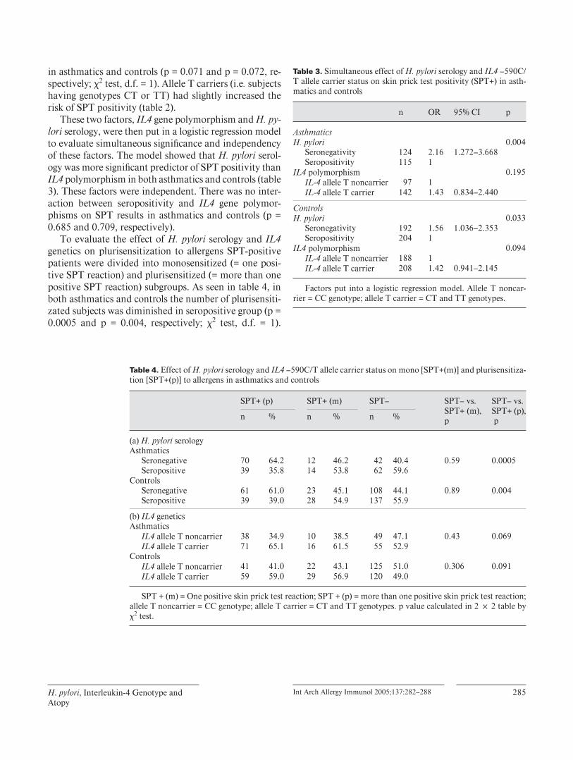
H . pylori , Interleukin-4 Genotype and Atopy
Int Arch Allergy Immunol 2005;137:282–288 285
in asthmatics and controls (p = 0.071 and p = 0.072, re-spectively; � 2 test, d.f. = 1). Allele T carriers (i.e . subjects having genotypes CT or TT) had slightly increased the risk of SPT positivity ( table 2 ).
These two factors, IL4 gene polymorphism and H. py-lori serology, were then put in a logistic regression model to evaluate simultaneous signifi cance and independency of these factors. The model showed that H. pylori serol-ogy was more signifi cant predictor of SPT positivity than IL4 polymorphism in both asthmatics and controls ( table 3 ). These factors were independent. There was no inter-action between seropositivity and IL4 gene polymor-phisms on SPT results in asthmatics and controls (p = 0.685 and 0.709, respectively).
To evaluate the effect of H. pylori serology and IL4 genetics on plurisensitization to allergens SPT-positive patients were divided into monosensitized (= one posi-tive SPT reaction) and plurisensitized (= more than one positive SPT reaction) subgroups. As seen in table 4 , in both asthmatics and controls the number of plurisensiti-zated subjects was diminished in seropositive group (p = 0.0005 and p = 0.004, respectively; � 2 test, d.f. = 1).
Table 3. Simultaneous effect of H. pylori serology and IL4 –590C/T allele carrier status on skin prick test positivity (SPT+) in asth-matics and controls
n OR 95% CI p
AsthmaticsH. pylori 0.004
Seronegativity 124 2.16 1.272–3.668Seropositivity 115 1
IL4 polymorphism 0.195IL-4 allele T noncarrier 97 1IL-4 allele T carrier 142 1.43 0.834–2.440
ControlsH. pylori 0.033
Seronegativity 192 1.56 1.036–2.353Seropositivity 204 1
IL4 polymorphism 0.094IL-4 allele T noncarrier 188 1IL-4 allele T carrier 208 1.42 0.941–2.145
Factors put into a logistic regression model. Allele T noncar-rier = CC genotype; allele T carrier = CT and TT genotypes.
Table 4. Effect of H. pylori serology and IL4 –590C/T allele carrier status on mono [SPT+(m)] and plurisensitiza-tion [SPT+(p)] to allergens in asthmatics and controls
SPT+ (p) SPT+ (m) SPT– SPT– vs.SPT+ (m),p
SPT– vs. SPT+ (p), pn % n % n %
(a) H. pylori serologyAsthmatics
Seronegative 70 64.2 12 46.2 42 40.4 0.59 0.0005Seropositive 39 35.8 14 53.8 62 59.6
ControlsSeronegative 61 61.0 23 45.1 108 44.1 0.89 0.004Seropositive 39 39.0 28 54.9 137 55.9
(b) IL4 geneticsAsthmatics
IL4 allele T noncarrier 38 34.9 10 38.5 49 47.1 0.43 0.069IL4 allele T carrier 71 65.1 16 61.5 55 52.9
ControlsIL4 allele T noncarrier 41 41.0 22 43.1 125 51.0 0.306 0.091IL4 allele T carrier 59 59.0 29 56.9 120 49.0
SPT + (m) = One positive skin prick test reaction; SPT + (p) = more than one positive skin prick test reaction; allele T noncarrier = CC genotype; allele T carrier = CT and TT genotypes. p value calculated in 2 ! 2 table by �2 test.

Pessi/Virta/Ådjers/Karjalainen/Rautelin/Kosunen/Hurme
Int Arch Allergy Immunol 2005;137:282–288 286
Whereas there was only a trend of association between IL4 gene polymorphism and plurisensitization in asth-matics and controls (p = 0.069 and p = 0.091, respective-ly; � 2 test, d.f. = 1).
There was an association (p = 0.007; � 2 test, d.f. = 1) between IL4 gene polymorphism and H. pylori seroposi-tivity among asthmatics ( table 5 ). Allele T carriers had diminished risk for H. pylori infection (OR 0.485 95% CI 0.287–0.819). The effect was independent on atopic sta-tus of the subject (data not shown). In controls, the asso-ciation between IL4 gene polymorphism and H. pylori seropositivity was not signifi cant (p = 0.220; � 2 test,d.f. = 1).
The effect of these two factors on asthma risk was also evaluated. H. pylori seropositivity rates were similar in asthmatics and controls (p = 0.370, � 2 test, d.f. = 1, see table 1 ). For the asthmatics, the frequencies were 0.41 for allele T noncarriers and 0.59 for allele T carriers at –590 of the IL4 gene. For controls respective values were 0.48 and 0.52. No difference in carrier frequencies was ob-served between asthmatics and controls (p = 0.093; � 2 test, d.f. = 1).
Discussion
In here interaction of two known atopy risk factors ( H. pylori infection and IL4 genetics) was studied. The result showed that the protective effect of H. pylori was not de-pendent on IL4 genetics. Recently McIntire et al. [20] observed that hepatitis A infection protects individuals from atopy only if they carry a certain allele of the TIM1 gene (a gene expressed on activated Th cells during dif-
ferentiation to Th2 direction and serving as a receptor for the virus). This allele did not have any effect on the infec-tion rate, but obviously modifi ed the effect of the virus on the Th cell differentiation. In here, IL4 genetics and H. pylori infection were independent risk factors. The atopy protective effect of H. pylori infection observed here may be explained by the increased Th1 activity [21, 22] . IL4 –590 T allele has been shown to be associated with a stronger transcription of IL4 than allele C [11, 23] , and consequently, the presence of this allele leads to pref-erential differentiation of the Th cells towards the Th2 direction. Our data suggest that IL4 polymorphism and H. pylori infection have distinct pathways to regulate the pathogenesis of atopy.
Our study showed that IL4 genetics had effect on in-fection rate: absence of allele T was a risk factor for this infection. IL-4 has a central role in the antibody produc-tion. H. pylori infection was evaluated by measuring spe-cifi c IgG antibodies. One possibility is that IL4 allele C, low IL4-producing allele, is involved in total IgG anti-body production, contrast to IgE promoting allele T [11, 23] . Moreover, our observation is line with the associa-tions observed with IL4 –590C/T and other infectious agents (such as malaria antigens and HIV) [11, 12] , ex-plained by this increased Th1 activity.
Rockman et al. [24] have recently observed that allele frequency variation of the IL4 SNP –590 in different pop-ulations is too great to be explained by simple genetic drift. They suggested that natural selection, e.g. various infections, have had an effect on the allele frequencies. In here the IL4 –590 polymorphism was associated with sus-ceptibility to a bacterial infection as well as there was a trend of association to atopy risk. This does not give di-
Table 5. Effect of IL4 allele carrier status on H. pylori seropositivity in asthmatics and in controls
Seropositive Seronegative p OR 95% CI
n % n %
AsthmaticsIL4 allele T noncarrier 57 49.6 40 32.3 0.007 1IL4 allele T carrier 58 50.4 84 67.7 0.485 0.287–0.819
ControlsIL4 allele T noncarrier 103 50.5 85 44.3 0.220 1IL4 allele T carrier 101 49.5 107 55.7 0.779 0.525–1.16
Allele T noncarrier = CC genotype; allele T carrier = CT and TT genotypes.p value calculated in 2 ! 2 table by �2 test.

H . pylori , Interleukin-4 Genotype and Atopy
Int Arch Allergy Immunol 2005;137:282–288 287
rect evidence for hypothesis that allergies are just by-products of the effective immune defense against mi-crobes, a capacity which has had a positive selective ad-vantage during evolution.
IL4 allele T has been linked to asthma in many studies [6–10] . However, contradictory results have been report-ed as well [25, 26] . Our study showed a weak association of IL4 genetics and asthma risk but only in seronegative patients (p = 0.03; table 5 ). This might indicate that ge-netic risk is stronger in the absence of certain environ-mental factors. Obviously, more data are needed on the interactions of these genetic and environmental factors.
We observed that the protective effect of IL4 polymor-phism on H. pylori infection was seen in asthmatics but not in controls ( table 5 ). The reason to this can only be speculated, but obviously asthmatics have some other factors modulating the effect of the H. pylori infection. One candidate is the anti-infl ammatory cytokine IL-10. It has been observed that the severity of asthma is associ-ated with the presence of a low IL10 producing haplotype [27] , which then allows uninhibited production of pro-infl ammatory cytokines and thus a more effi cient shift of the Th1/2 balance towards the protective Th1 direction.
In this study, presence of H. pylori antibodies had a clear inverse association to the incidence of atopy. This fi nding is in line with previous reports from our country [15] and from elsewhere [16, 17] , thus confi rming the role of H. pylori infection as a strong atopy-risk modulating factor (according to the hygiene hypothesis, see introduc-tion). The effect of H. pylori seropositivity was strikingly
different in subjects with one positive SPT (monoallergic) vs. several positive SPTs (pluriallergic), i.e . H. pylori in-fection only protected against plurisensitization. This might indicate that H. pylori infection does not affect the susceptibility per se but rather on the severity of the atop-ic disease. However, as H. pylori exposure is stronger in poor hygienic conditions [28, 29] , we cannot exclude the possibility that H. pylori is just a surrogate marker of, e.g. a general bacterial LPS exposure.
Acknowledgements
The authors would like to thank Prof. Markku M Nieminen, Prof. Arpo Aromaa and Docent Timo Klaukka for taking part in collection of the patient material. We also thank Mrs. Heini Huhta-la, MSc for statistical consultation as well as Mrs. Eija Spåre, Sinik-ka Repo-Koskinen and Pirjo Kosonen for expert technical assis-tance as well as Orion Diagnostica, Espoo, Finland, for the Pylori-set kits.
Funding /Support
The study was fi nancially supported by the Academy of Finland (grant no 205653), Rehabilitation Funds of the Finnish Social In-surance Institution, the Tampere Tuberculosis Foundation, the Medical Research Fund of Tampere University Hospital and the Research Funds of the University of Helsinki. The sponsors of the study had no role in study design, data collection, data analysis, data interpretation, or writing of the report.
References
1 Wills-Karp M, Santeliz J, Karp CL: The germ-less theory of allergic disease: revisiting the hy-giene hypothesis. Nat Rev Immunol 2001; 1: 69–75.
2 Romagnani S: The Th1/Th2 paradigm. Immu-nol Today 1997; 18: 263–266.
3 Perussia B, Loza MJ: Linear ‘2-0-1’ lympho-cyte development: hypotheses on cellular bases for immunity. Trends Immunol 2003; 24: 235–241.
4 Burrows B, Martinez FD, Halonen M, Barbee RA, Cline MG: Association of asthma with se-rum IgE levels and skin-test reactivity to aller-gens. N Engl J Med 1989; 320: 271–277.
5 Le Gros G, Ben-Sasson SZ, Seder R, Finkel-man FD, Paul WE: Generation of interleukin 4 (IL-4)-producing cells in vivo and in vitro: IL-2 and IL-4 are required for in vitro genera-tion of IL-4-producing cells. J Exp Med 1990; 172: 921–929.
6 Marsh DG, Neely JD, Breazeale DR, Ghosh B, Freidhoff LR, Ehrlich-Kautzky E, et al: Link-age analysis of IL4 and other chromosome 5q31.1 markers and total serum immunoglob-ulin E concentrations. Science 1994; 264: 1152–1156.
7 Noguchi E, Shibasaki M, Arinami T, Takeda K, Yokouchi Y, Kawashima T, et al: Associa-tion of asthma and the interleukin-4 promoter gene in Japanese. Clin Exp Allergy 1998; 28: 449–453.
8 Noguchi E, Nukaga-Nishio Y, Jian Z, Yokou-chi Y, Kamioka M, Yamakawa-Kobayashi K, et al: Haplotypes of the 5 � region of the IL-4 gene and SNPs in the intergene sequence be-tween the IL-4 and IL-13 genes are associated with atopic asthma. Hum Immunol 2001; 62: 1251–1257.
9 Beghe B, Barton S, Rorke S, Peng Q, Sayers I, Gaunt T, et al: Polymorphisms in the interleu-kin-4 and interleukin-4 receptor alpha chain genes confer susceptibility to asthma and atopy in a Caucasian population. Clin Exp Allergy 2003; 33: 1111–1117.
10 Liu X, Beaty TH, Deindl P, Huang SK, Lau S, Sommerfeld C, et al: Associations between to-tal serum IgE levels and the 6 potentially func-tional variants within the genes IL4, IL13, and IL4RA in German children: the German Mul-ticenter Atopy Study. J Allergy Clin Immunol 2003; 112: 382–388.
11 Nakayama EE, Hoshino Y, Xin X, Liu H, Goto M, Watanabe N, et al: Polymorphism in the interleukin-4 promoter affects acquisition of human immunodefi ciency virus type 1 syncy-tium-inducing phenotype. J Virol 2000; 74: 5452–5459.
Pessi/Virta/Ådjers/Karjalainen/Rautelin/Kosunen/Hurme
Int Arch Allergy Immunol 2005;137:282–288 288
12 Luoni G, Verra F, Arca B, Sirima BS, Troye-Blomberg M, Coluzzi M, et al: Antimalarial antibody levels and IL4 polymorphism in the Fulani of West Africa. Genes Immun 2001; 2: 411–414.
13 Go MF: Review article: natural history and epidemiology of Helicobacter pylori infection. Aliment Pharmacol Ther 2002; 16(suppl 1):3–15.
14 Smythies LE, Waites KB, Lindsey JR, Harris PR, Ghiara P, Smith PD: Helicobacter pylori-induced mucosal infl ammation is Th1 medi-ated and exacerbated in IL-4, but not IFN-gamma, gene-defi cient mice. J Immunol 2000; 165: 1022–1029.
15 Kosunen TU, Hook-Nikanne J, Salomaa A, Sarna S, Aromaa A, Haahtela T: Increase of allergen-specifi c immunoglobulin E antibodies from 1973 to 1994 in a Finnish population and a possible relationship to Helicobacter pylori infections. Clin Exp Allergy 2002; 32: 373–378.
16 Linneberg A, Ostergaard C, Tvede M, Ander-sen LP, Nielsen NH, Madsen F, et al: IgG an-tibodies against microorganisms and atopic disease in Danish adults: the Copenhagen Al-lergy Study. J Allergy Clin Immunol 2003; 111: 847–853.
17 McCune A, Lane A, Murray L, Harvey I, Nair P, Donovan J, et al: Reduced risk of atopic disorders in adults with Helicobacter pylori in-fection. Eur J Gastroenterol Hepatol 2003; 15: 637–640.
18 Karjalainen A, Kurppa K, Martikainen R, Klaukka T, Karjalainen J: Work is related to a substantial portion of adult-onset asthma inci-dence in the Finnish population. Am J Respir Crit Care Med 2001; 164: 565–568.
19 Karjalainen J, Hulkkonen J, Pessi T, Huhtala H, Nieminen MM, Aromaa A, et al: The IL1A genotype associates with atopy in nonasthmat-ic adults. J Allergy Clin Immunol 2002; 110: 429–434.
20 McIntire JJ, Umetsu SE, Macaubas C, Hoyte EG, Cinnioglu C, Cavalli-Sforza LL, et al: Im-munology: hepatitis A virus link to atopic dis-ease. Nature 2003; 425: 576.
21 Russo F, Jirillo E, Clemente C, Messa C, Chi-loiro M, Riezzo G, et al: Circulating cytokines and gastrin levels in asymptomatic subjects in-fected by Helicobacter pylori . Immunophar-macol Immunotoxicol 2001; 23: 13–24.
22 Perri F, Clemente R, Festa V, De Ambrosio CC, Quitadamo M, Fusillo M, et al: Serum tu-mour necrosis factor-alpha is increased in pa-tients with Helicobacter pylori infection and CagA antibodies. Ital J Gastroenterol Hepatol 1999; 31: 290–294.
23 Nakashima H, Miyake K, Inoue Y, Shimizu S, Akahoshi M, Tanaka Y, et al: Association be-tween IL-4 genotype and IL-4 production in the Japanese population. Genes Immun 2002; 3: 107–109.
24 Rockman MV, Hahn MW, Soranzo N, Gold-stein DB, Wray GA: Positive selection on a hu-man-specifi c transcription factor binding site regulating IL4 expression. Curr Biol 2003; 13: 2118–2123.
25 Walley AJ, Cookson WO: Investigation of an interleukin-4 promoter polymorphism for as-sociations with asthma and atopy. J Med Gen-et 1996; 33: 689–692.
26 Hijazi Z, Haider MZ: Interleukin-4 gene pro-moter polymorphism [C590T] and asthma in Kuwaiti Arabs. Int Arch Allergy Immunol 2000; 122: 190–194.
27 Lim S, Crawley E, Woo P, Barnes PJ: Haplo-type associated with low interleukin-10 pro-duction in patients with severe asthma. Lancet 1998; 352: 113.
28 Graham DY, Malaty HM, Evans DG, Evans DJ, Jr., Klein PD, Adam E: Epidemiology of Helicobacter pylori in an asymptomatic popu-lation in the United States: effect of age, race, and socioeconomic status. Gastroenterology 1991; 100: 1495–1501.
29 Patel P, Mendall MA, Khulusi S, Northfi eld TC, Strachan DP: Helicobacter pylori infection in childhood: risk factors and effect on growth. BMJ 1994; 309: 1119–1123.
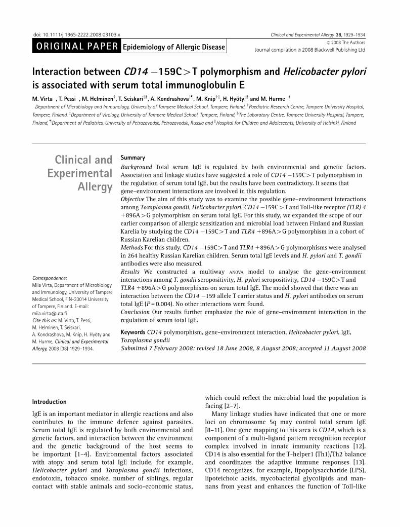
ORIGINAL PAPER
Interaction between CD14 �159C4T polymorphism and Helicobacter pyloriis associated with serum total immunoglobulin EM. Virta�, T. Pessi�, M. Helminenw, T. Seiskariz‰, A. Kondrashovazz, M. Knipwk, H. Hyotyz‰ and M. Hurme�‰�Department of Microbiology and Immunology, University of Tampere Medical School, Tampere, Finland, wPaediatric Research Centre, Tampere University Hospital,
Tampere, Finland, zDepartment of Virology, University of Tampere Medical School, Tampere, Finland, ‰The Laboratory Centre, Tampere University Hospital, Tampere,
Finland, zDepartment of Pediatrics, University of Petrozavodsk, Petrozavodsk, Russia and kHospital for Children and Adolescents, University of Helsinki, Finland
Clinical andExperimental
Allergy
Correspondence:Miia Virta, Department of Microbiologyand Immunology, University of TampereMedical School, FIN-33014 Universityof Tampere, Finland. E-mail:[email protected] this as: M. Virta, T. Pessi,M. Helminen, T. Seiskari,A. Kondrashova, M. Knip, H. Hyoty andM. Hurme, Clinical and ExperimentalAllergy, 2008 (38) 1929–1934.
Summary
Background Total serum IgE is regulated by both environmental and genetic factors.Association and linkage studies have suggested a role of CD14 �159C4T polymorphism inthe regulation of serum total IgE, but the results have been contradictory. It seems thatgene–environment interactions are involved in this regulation.Objective The aim of this study was to examine the possible gene–environment interactionsamong Toxoplasma gondii, Helicobacter pylori, CD14�159C4T and Toll-like receptor (TLR) 41896A4G polymorphism on serum total IgE. For this study, we expanded the scope of ourearlier comparison of allergic sensitization and microbial load between Finland and RussianKarelia by studying the CD14 �159C4T and TLR4 1896A4G polymorphism in a cohort ofRussian Karelian children.Methods For this study, CD14�159C4T and TLR4 1896A4G polymorphisms were analysedin 264 healthy Russian Karelian children. Serum total IgE levels and H. pylori and T. gondiiantibodies were also measured.Results We constructed a multiway ANOVA model to analyse the gene–environmentinteractions among T. gondii seropositivity, H. pylori seropositivity, CD14 �159C4T andTLR4 1896A4G polymorphisms on serum total IgE. The model showed that there was aninteraction between the CD14 �159 allele T carrier status and H. pylori antibodies on serumtotal IgE (P = 0.004). No other interactions were found.Conclusion Our results further emphasize the role of gene–environment interaction in theregulation of serum total IgE.
Keywords CD14 polymorphism, gene–environment interaction, Helicobacter pylori, IgE,Toxoplasma gondiiSubmitted 7 February 2008; revised 18 June 2008, 8 August 2008; accepted 11 August 2008
Introduction
IgE is an important mediator in allergic reactions and alsocontributes to the immune defence against parasites.Serum total IgE is regulated by both environmental andgenetic factors, and interaction between the environmentand the genetic background of the host seems tobe important [1–4]. Environmental factors associatedwith atopy and serum total IgE include, for example,Helicobacter pylori and Toxoplasma gondii infections,endotoxin, tobacco smoke, number of siblings, regularcontact with stable animals and socio-economic status,
which could reflect the microbial load the population isfacing [2–7].
Many linkage studies have indicated that one or moreloci on chromosome 5q may control total serum IgE[8–11]. One gene mapping to this area is CD14, which is acomponent of a multi-ligand pattern recognition receptorcomplex involved in innate immunity reactions [12].CD14 is also essential for the T-helper1 (Th1)/Th2 balanceand coordinates the adaptive immune responses [13].CD14 recognizes, for example, lipopolysaccharide (LPS),lipoteichoic acids, mycobacterial glycolipids and man-nans from yeast and enhances the function of Toll-like
Epidemiology of Allergic Disease
doi: 10.1111/j.1365-2222.2008.03103.x Clinical and Experimental Allergy, 38, 1929–1934
�c 2008 The Authors
Journal compilation �c 2008 Blackwell Publishing Ltd

receptors (TLR) 2 and 4, which are involved in immunedefence against T. gondii and H. pylori [12–15].
CD14 gene promoter region polymorphism �159C4Thas been associated with serum total IgE and atopy, but theresults have been contradictory. The same allele has beenshown to have opposite effects on IgE [11, 16–21]. Thegene–environment interactions in the regulation of serumtotal IgE detected in recent studies have been postulated tobe one explanation for the inconsistent results [1].Although CD14 is an important molecule for the endotoxinsignalling, other molecules such as TLR4 are needed forsignal transduction [13]. A single A4G base-exchangepolymorphism in the TLR4 gene at the position 1896 inthe fourth exon has been associated with atopic asthma,but there are also negative findings [22–24].
In our earlier study, we observed that serum total IgElevels were significantly higher in children living inRussian Karelia than in children living in Finland, eventhough sensitization to allergens was clearly less frequentamong Russian Karelian children [6]. The exact reason forthis phenomenon is not known, but one explanation forthe higher IgE levels could be parasite infections, whichare more common in Russian Karelia than in Finland [6,25]. In this study, we wanted to expand our earlierinvestigation and analyse possible gene–environmentinteractions among microbes, CD14 �159C4T and TLR41896A4G polymorphisms on serum total IgE in a childpopulation from Russian Karelia.
Materials and methods
Study population
Altogether 266 schoolchildren from Petrozavodsk, Rus-sian Karelia, having both parents of either Finnish orKarelian ethnicity participated in the present study. Twochildren were excluded, because their whole blood sam-ples were accidentally confused in the laboratory. Thestudy population has been described earlier in detail [6].Finnish schoolchildren from the earlier study were notincluded in this study due to the low prevalence ofseropositive subjects for H. pylori and T. gondii (5% and2%, respectively). The collection of samples from RussianKarelia was done as a part of the EU INCO-Copernicusprogram (contract number IC15-CT98-0316, coordinatorProfessor Hyoty) during the years 1997–1999. Collectionof samples was organized by the Department of Pediatrics,University of Petrozavodsk, and the study protocol wasapproved by the Ministry of Health in Russian Karelia.There was no local ethics board in Karelia to apply to, andthe accepted route for ethical approval for the researchstudies was via the Ministry of Health. Written consentwas obtained from all the children who took part in thestudy and from their parents. Whole blood and serumsamples were collected from each child.
Immunoglobulin E and microbial antibodies
Serum total IgE was measured using the ImmunoCAPsfluoroenzyme immunoassay (Phadia Diagnostics, Uppsala,Sweden). T. gondii IgG antibodies were measured byEnzygnost Toxoplasmosis IgG Assay and H. pylori IgGantibodies by Enzygnost Anti-H. pylori/IgG Assay ac-cording to the manufacturer’s instructions (Dade Behring,Marburg, Germany). Behring ELISA Processor III (DadeBehring, Marburg, Germany) was used for further proces-sing of the tests and for the calculation of the antibodylevels. Measurement of T. gondii antibodies succeeded in263 children of 264 (99.5%), whereas H. pylori antibodieswere measured among all the 264 (100%) study children.
Genotype analyses
DNA was extracted using standard techniques. Genotyp-ing of CD14 �159C4T polymorphism (rs 2569190) andTLR4 1896A4G (rs 4986790) was performed using theABI PRISM 7000 Sequence Detection System for both PCRand allelic discrimination. The oligonucleotides 50-CCCTTC CTT TCC TGG AAA TAT TGC A-30 and 50-CTA GATGCC CTG CAG AAT CCT T-30 were used as primers forCD14 �159C4T polymorphism, and the oligonucleotides50-TGA CCA TTG AAG AAT TCC GAT TAG CA-30 and 50-ACA CTC ACC AGG GAA AAT GAA GAA-30 for TLR41896A4G polymorphism.
Statistics
To detect a possible deviation of the genotype frequenciesfrom the Hardy–Weinberg equation, the exact test usingthe Markov chain algorithm was used (Arlequin program,ver. 2.0. A software for population genetics data analysis;Schneider S, Roessli D, Excoffier L; Genetics and BiometryLaboratory, Geneva, Switzerland). Other statistical ana-lyses were performed with SPSS for Windows version 14.0(SPSS Inc., Chigago, IL, USA). The statistical methodselected is presented in ‘Results’. Serum total IgE levelswere not normally distributed and therefore non-para-metric tests were used when serum total IgE levels wereanalysed. Serum total IgE values were logarithmicallytransformed for multiway ANOVA which was used formodelling gene–environment interactions among CD14�159 allele T carrier status, TLR4 1896 allele G carrierstatus, T. gondii seropositivity and H. pylori seropositivityon serum total IgE levels. For statistical analysis,we grouped CD14 �159 genotypes according to theallele T carrier status, because this allele has been shownto be transcriptionally more active in monocytic cell linethan allele C [26]. TLR4 1896 genotypes were groupedaccording to the allele G carrier status, because thenumber of GG genotype was too small (n = 3) for grouping
�c 2008 The AuthorsJournal compilation �c 2008 Blackwell Publishing Ltd, Clinical and Experimental Allergy, 38 : 1929–1934
1930 M. Virta et al

according to allele A. P-values o0.05 were considered tobe significant.
Results
The study cohort of 264 children included 114 boys (43%).The mean age of the children was 11.4 years (range7.1–15.0). The median total IgE in sera was 76.1 IU/L(interquartile range 30.9–236.0). There were no significantdifferences in total IgE levels between boys and girls (median75.8 and 76.4 IU/L, respectively, P= 0.8, Mann–WhitneyU-test). Among the study children, 193 out of 264 (73%)were seropositive for H. pylori and 63 out of 263 (24%) forT. gondii. Fifty-two (20%) children were seropositive forboth the microbes. Only 16% (n= 10) of T. gondii seroposi-tive children were H. pylori seronegative, whereas 72%(n= 139) of H. pylori seropositive children were T. gondiiseronegative. Serum total IgE levels were significantlyhigher in T. gondii seropositive children (P= 0.009, Mann–Whitney U-test), whereas H. pylori seropositivity did nothave any effect on serum total IgE (Table 1), as reportedearlier [6]. Association between T. gondii seropositivity andserum total IgE remained statistically significant afterBonferroni correction (P= 0.036).
The genotype frequencies of CD14�159C4T and TLR41896A4G polymorphisms as well as allele T carrierstatus of CD14 �159C4T and allele G carrier status ofTLR4 1896A4G are shown in Table 1. CD14 �159C4Tgenotype or allele T carrier status did not show any direct
association with serum total IgE, and there was noassociation between TLR4 1896A4G genotype or alleleG carrier status and serum total IgE (Table 1). Neither ofthese polymorphisms was associated with seropositivityfor H. pylori or T. gondii (data not shown). The genotypedistributions followed the Hardy–Weinberg equilibrium.
We constructed a multiway ANOVA model to investigategene–environment interactions among CD14 �159C4Tallele T carrier status, TLR4 1896A4G allele G carrierstatus, H. pylori seropositivity and T. gondii seropositivityon serum total IgE levels. In this model, we found aninteraction between H. pylori seropositivity and CD14�159 allele T carrier status on serum total IgE (P = 0.004,multiway ANOVA, Table 2a). The H. pylori seronegativechildren who were allele T non-carriers (i.e. genotype CC)had higher serum total IgE than allele T carriers (i.e.genotypes CT and TT). Among H. pylori seropositivechildren, allele T non-carriers had lower IgE levels thanallele T carriers (Table 2a). There was a trend for interac-tion between T. gondii seropositivity and CD14 �159allele T carrier status on serum total IgE, but this interac-tion was not statistically significant (Table 2b). No statis-tically significant interactions between TLR4 1896A4Gallele G carrier status and H. pylori or TLR4 1896A4Gallele G carrier status and T. gondii on serum total IgE wasfound (P = 0.95 and 0.3, respectively, multiway ANOVA). Wealso analysed gene–gene interaction between CD14�159C4T allele T carrier status and TLR4 1896A4Gallele G carrier status on serum total IgE, but no
Table 1. Associations between Toxoplasma gondii serology, Helicobacter pylori serology, CD14 �159C4T polymorphism, TLR4 1896A4Gpolymorphism and serum total IgE
N (%) IgE median (quartiles) P-value P-value�
T. gondiiSeropositive 63 (24) 114.0 (44.0–393.0)Seronegative 200 (76) 68.5 (28.3–174.3) 0.009w 0.036H. pyloriSeropositive 193 (73) 76.8 (32.7–234.0)Seronegative 71 (27) 68.2 (30.0–257.0) 0.86w NSCD14 �159C4TCC 88 (33) 66.4 (32.0–238.5)CT 139 (53) 80.9 (33.1–203.5)TT 37 (14) 62.3 (23.8–262.0) 0.9z NSAllele T carrier (CT and TT) 176 (67) 79.2 (30.8–229.5)Allele T non-carrier (CC) 88 (33) 66.4 (32.0–238.5) 0.8w NSTLR4 1896A4GAA 223 (85) 76.4 (34.1–217.0)AG 38 (14) 62.0 (21.9–266.0)GG 3 (1) 107.0 (14.1–171.0) 0.8z NSAllele G carrier (AG and GG) 41 (15) 67.5 (21.6–262.0)Allele G non-carrier (AA) 223 (85) 76.4 (34.1–217.9) 0.5w NS
�Bonferroni correction.wMann–Whitney U-test.zKruskall–Wallis ANOVA.NS, non-significant.
�c 2008 The AuthorsJournal compilation �c 2008 Blackwell Publishing Ltd, Clinical and Experimental Allergy, 38 : 1929–1934
Gene–environment interaction and serum total IgE 1931

interaction was seen (P = 0.7, multiway ANOVA). Further-more, there was no interaction between the environmentalfactors T. gondii and H. pylori on serum total IgE (P = 0.2,multiway ANOVA).
Discussion
Gene–environment interactions are very complex. It hasbeen speculated that the same genetic variants may beassociated with different phenotypes in different environ-ments, but so far, only few human studies have beenpublished [2, 3, 27]. H. pylori, T. gondii and CD14�159C4T polymorphisms have all been independentlyassociated with atopic markers and IgE. In this study, wewanted to further analyse whether there are gene–envir-onment interactions among H. pylori, T. gondii, CD14 andTLR4 polymorphisms that have an effect on serum totalIgE. To increase the sensitivity and detect possible inter-actions, we carried out the study in an environment that ischaracterized by a high exposure rate to these twomicrobes.
CD14 is part of the TLR signalling complex that facil-itates endotoxin responses through TLR4-MD2 and canalso bind with a variety of microbial TLR ligands. CD14gene (MIM158120) is located on chromosome 5 (5q31). Inmany linkage studies, this area has been connected withthe regulation of serum total IgE and atopy [8–11]. CD14promoter region single base-exchange polymorphism C toT at position �159 has been extensively studied, becauseit has been associated with the level of protein productionallele T being transcriptionally more active in monocyticcell line [17, 26, 28]. An association between CD14�159C4T polymorphism and total IgE and other atopicmarkers has been shown in the earlier studies, but theresults have been contradictory. Baldini et al. [17] reportedthat atopic children homozygous for allele T (i.e. TTgenotype) had higher sCD14 levels and lower IgE levelsin sera than allele C carriers. An association between highIgE levels or other atopic markers and the CD14 �159CCgenotype has been reported in many other studies [16, 19,20]. An opposite association has been reported by Oberet al. [11], who showed that the CD14 �159 allele T was
associated with atopy in a rural population. In somestudies, no associations have been found [18, 21].
Environmental differences between study populationsmay in part explain these conflicting results. In recentstudies, exposure to endotoxin and other environmentalfactors has been studied concomitantly with CD14�159C4T polymorphism. Eder et al.[2] reported that theCD14 �159C4T allele C was associated with lower levelsof both total and specific IgE in children in regular contactwith stable animals, whereas the result was the opposite inchildren with regular contact with furry pets. They specu-lated that one explanation for this difference could be theexposure to different kinds of microbial products [2].Williams et al. [3] found in an adult female populationthat the association between CD14�159C4T polymorph-ism and serum total IgE was modified by the level ofendotoxin exposure. At lower levels of endotoxin expo-sure, the CC genotype was associated with the highestserum total IgE levels, whereas at the highest tertile ofendotoxin exposure, individuals carrying the TT genotypehad the highest IgE levels [3]. On the other hand, in aFrench study, early-life exposure to a farming environ-ment and the CD14 �159 TT genotype together signifi-cantly reduced the risk of nasal allergy and atopy. Inaddition, there was a trend among these subjects for lowerrisk of having increased serum total IgE levels [27].
Eder et al. [2] speculated that the effect of CD14�159C4T polymorphism on IgE might be dependent onmicrobial exposure, but they did not study the specificmicrobes. In the present study, we found that interactionbetween CD14 �159C4T polymorphism and H. pyloriserology had an effect on serum total IgE. Among H.pylori seronegative children, CD14 �159 allele T non-carriers (i.e. CC genotype) had higher serum total IgE thanallele T-carriers (i.e. CT and TT genotypes), whereasamong H. pylori seropositive children, serum total IgEwas lower in allele T non-carriers. There was also a trendof gene–environment interaction between T. gondii ser-opositivity and CD14 �159C4T polymorphism. Becauseof our result, one may speculate that H. pylori, andpossibly also T. gondii, could be among the microbes thatinfluence the genetic regulation of serum total IgE, but theexact mechanism is not known.
Table 2. (a) Interaction between Helicobacter pylori serology and CD14 �159C4T allele T carrier status on serum total IgE (P = 0.004, multiway ANOVA).(b) Interaction between Toxoplasma gondii serology and CD14 �159C4T allele T carrier status on serum total IgE (P = 0.06, multiway ANOVA)
CD14 �159 C4T
Seropositive
N
Seronegative
NIgE median (quartiles) IgE median (quartiles)
(a) H. pyloriAllele T carrier (CT and TT) 87.4 (36.9–253.8) 130 44.1 (24.2–162.5) 46Allele T non carrier (CC) 56.0 (29.5–154.0) 63 84.7 (34.1–530.5) 25(b)T. gondiiAllele T carrier (CT and TT) 202.5 (59.4–550.8) 42 67.5 (26.8–160.5) 133Allele T non carrier (CC) 55.3 (39.1–81.7) 21 79.7 (29.5–272.0) 67
�c 2008 The AuthorsJournal compilation �c 2008 Blackwell Publishing Ltd, Clinical and Experimental Allergy, 38 : 1929–1934
1932 M. Virta et al

H. pylori infection has been suggested to influence thedevelopment of the immune system by LPS binding withthe CD14 receptor which results in increased production ofIL-12. This could drive the immune responses towards theTh1 pathway and thereby have a protective effect againstthe development of Th2 polarized diseases such as atopy[29]. CD14 �159 allele T carriers have been reported tohave higher sCD14 levels than allele T non-carriers andthese high sCD14 levels have been associated with lowserum total IgE [17, 28]. The exact mechanism is not known,but interaction between sCD14 and B cells has resulted inhigher levels of IgG1 and lower levels of IgE production [17,30]. Karhukorpi et al. [28] have reported that H. pyloriseropositivity has an effect on sCD14 levels in such a waythat H. pylori seropositive individuals have higher levelsthan seronegative subjects, especially if they carry the CD14�159 CC genotype. One may speculate that the CC genotypeis especially sensitive to environmental factors.
TLR4 plays a role in the same functional pathway asCD14, and both these molecules are needed for endotoxinresponsiveness. A single A4G base-exchange poly-morphism in the TLR4 gene at position 1896 has beenassociated with atopic asthma, but the results have beencontradictory [22–24]. This base exchange induces anamino acid substitution of glysine for asparagine, whichresults in a reduction in cell surface expression of TLR4and subsequent disruption of LPS-mediated signalling[31]. In the present study, TLR4 1896A4G polymorphismwas not associated with serum total IgE and there was nointeraction between this polymorphism and H. pylori orT. gondii on serum total IgE. It has been speculated thatthe combined effect of the genetic variants of CD14 andTLR4 could strengthen the associations found earlier, butno interaction between CD14 �159C4T and TLR41896A4G polymorphisms was seen in our study, whichis in line with previous findings of Sackesen et al. [24].
The major limitation of our study is the small number ofstudy subjects and therefore these gene–environmentanalyses should be repeated in a larger population.Another restriction was that we could not use specific IgEin gene–environment interaction analyses, because thenumber of atopic subjects in the study population (n = 16)was too small for statistical analyses. The problem withserum total IgE is that many factors such as allergens andparasite infections influence its levels and therefore theinterpretation of the results is challenging. Multiple test-ing also complicates the interpretation of our results.However, the interaction between CD14 �159C4T alleleT carrier status and H. pylori seropositivity on serum totalIgE was so significant that it most probably does notdisappear even though multiple testings are done (i.e.remaining significant after Bonferroni correction). Cau-tion should also be exercised when interpretinginteractions between microbes and CD14�159C4T poly-morphism on serum total IgE, because both H. pylori and
T. gondii could be surrogate markers for poor hygiene andlarge microbe load. Therefore, the interactions detectedcould either reflect interaction between the whole microbeload and CD14 �159 polymorphism or the interactionbetween specific microbe and the polymorphism.
Our study supports the hypothesis that the same geno-type may increase, decrease or have no effect on total IgEdepending on the environmental factors, such as mi-crobes, that the population is facing.
Acknowledgements
This study was supported by grants from the MedicalResearch Fund of the Tampere University Hospital, theTampere Tuberculosis Foundation, the Finnish Anti-Tu-berculosis Association Foundation, the Vaino and LainaKivi Foundation, the Paivikki and Sakari Sohlberg Foun-dation, the Academy of Finland and the EC as a part of theEPIVIR project (INCO-Copernicus Programme, contractnumber IC15-CT98-0316).
The authors thank Ms Eija Spare and Ms Sinikka Repo-Koskinen for their expert technical assistance and HeiniHuhtala MSc for expert advice on the statistical analysisof the data.
References
1 Martinez FD. CD14, endotoxin, and asthma risk: actions andinteractions. Proc Am Thorac Soc 2007; 4:221–5.
2 Eder W, Klimecki W, Yu L et al. Opposite effects of CD 14/-260 onserum IgE levels in children raised in different environments. JAllergy Clin Immunol 2005; 116:601–7.
3 Williams LK, McPhee RA, Ownby DR et al. Gene–environmentinteractions with CD14 C-260T and their relationship to totalserum IgE levels in adults. J Allergy Clin Immunol 2006;118:851–7.
4 Choudhry S, Avila PC, Nazario S et al. CD14 tobacco gene–environment interaction modifies asthma severity and immuno-globulin E levels in Latinos with asthma. Am J Respir Crit CareMed 2005; 172:173–82.
5 Matricardi PM, Rosmini F, Riondino S et al. Exposure tofoodborne and orofecal microbes versus airborne viruses inrelation to atopy and allergic asthma: epidemiological study.BMJ 2000; 320:412–7.
6 Seiskari T, Kondrashova A, Viskari H et al. Allergic sensitizationand microbial load – a comparison between Finland and RussianKarelia. Clin Exp Immunol 2007; 148:47–52.
7 von Mutius E. The environmental predictors of allergic disease. JAllergy Clin Immunol 2000; 105:9–19.
8 Marsh DG, Neely JD, Breazeale DR et al. Linkage analysis of IL4and other chromosome 5q31.1 markers and total serum immu-noglobulin E concentrations. Science 1994; 264:1152–6.
9 Xu J, Postma DS, Howard TD et al. Major genes regulating totalserum immunoglobulin E levels in families with asthma. Am JHum Genet 2000; 67:1163–73.
10 Palmer LJ, Daniels SE, Rye PJ et al. Linkage of chromosome 5qand 11q gene markers to asthma-associated quantitative traits in
�c 2008 The AuthorsJournal compilation �c 2008 Blackwell Publishing Ltd, Clinical and Experimental Allergy, 38 : 1929–1934
Gene–environment interaction and serum total IgE 1933
Australian children. Am J Respir Crit Care Med 1998; 158:1825–30.
11 Ober C, Tsalenko A, Parry R, Cox NJ. A second-generationgenomewide screen for asthma-susceptibility alleles in a founderpopulation. Am J Hum Genet 2000; 67:1154–62.
12 Schmitz G, Orso E. CD14 signalling in lipid rafts: new ligandsand co-receptors. Curr Opin Lipidol 2002; 13:513–21.
13 Vercelli D. The functional genomics of CD14 and its role in IgEresponses: an integrated view. J Allergy Clin Immunol 2002;109:14–21.
14 Jerala R. Structural biology of the LPS recognition. Int J MedMicrobiol 2007; 297:353–63.
15 Debierre-Grockiego F, Campos MA, Azzouz N et al. Activation ofTLR2 and TLR4 by glycosylphosphatidylinositols derived fromToxoplasma gondii. J Immunol 2007; 179:1129–37.
16 Gao PS, Mao XQ, Baldini M et al. Serum total IgE levels andCD14 on chromosome 5q31. Clin Genet 1999; 56:164–5.
17 Baldini M, Lohman IC, Halonen M et al. A Polymorphism� in the50 flanking region of the CD14 gene is associated with circulatingsoluble CD14 levels and with total serum immunoglobulin E. AmJ Respir Cell Mol Biol 1999; 20:976–83.
18 Kabesch M, Hasemann K, Schickinger V et al. A promoterpolymorphism in the CD14 gene is associated with elevatedlevels of soluble CD14 but not with IgE or atopic diseases.Allergy 2004; 59:520–5.
19 Leung TF, Tang NL, Sung YM et al. The C-159T polymorphism inthe CD14 promoter is associated with serum total IgE concentra-tion in atopic Chinese children. Pediatr Allergy Immunol 2003;14:255–60.
20 Koppelman GH, Reijmerink NE, Colin Stine O et al. Associationof a promoter polymorphism of the CD14 gene and atopy. Am JRespir Crit Care Med 2001; 163:965–9.
21 Sengler C, Haider A, Sommerfeld C et al. Evaluation of the CD14C �159 T polymorphism in the German Multicenter AllergyStudy cohort. Clin Exp Allergy 2003; 33:166–9.
22 Raby BA, Klimecki WT, Laprise C et al. Polymorphisms in toll-like receptor 4 are not associated with asthma or atopy-relatedphenotypes. Am J Respir Crit Care Med 2002; 166:1449–56.
23 Bottcher MF, Hmani-Aifa M, Lindstrom A et al. A TLR4 poly-morphism is associated with asthma and reduced lipopolysac-charide-induced interleukin-12(p70) responses in Swedishchildren. J Allergy Clin Immunol 2004; 114:561–7.
24 Sackesen C, Karaaslan C, Keskin O et al. The effect of polymorph-isms at the CD14 promoter and the TLR4 gene on asthmaphenotypes in Turkish children with asthma. Allergy 2005; 60:1485–92.
25 von Hertzen LC, Laatikainen T, Makela MJ et al. Infectiousburden as a determinant of atopy – a comparison between adultsin Finnish and Russian Karelia. Int Arch Allergy Immunol 2006;140:89–95.
26 LeVan TD, Bloom JW, Bailey TJ et al. A common single nucleo-tide polymorphism in the CD14 promoter decreases the affinityof Sp protein binding and enhances transcriptional activity. JImmunol 2001; 167:5838–44.
27 Leynaert B, Guilloud-Bataille M, Soussan D et al. Associationbetween farm exposure and atopy, according to the CD14 C�159T polymorphism. J Allergy Clin Immunol 2006; 118:658–65.
28 Karhukorpi J, Yan Y, Niemela S et al. Effect of CD14 promoterpolymorphism and H. pylori infection and its clinical outcomeson circulating CD14. Clin Exp Immunol 2002; 128:326–32.
29 von Mutius E, Braun-Fahrlander C, Schierl R et al. Exposure toendotoxin or other bacterial components might protect againstthe development of atopy. Clin Exp Allergy 2000; 30:1230–4.
30 Arias MA, Rey Nores JE, Vita N et al. Cutting edge: human B cellfunction is regulated by interaction with soluble CD14: oppositeeffects on IgG1 and IgE production. J Immunol 2000; 164:3480–6.
31 Arbour NC, Lorenz E, Shutte BC et al. TLR4 mutations areassociated with endotoxin hyporesponsiveness in humans. NatGenet 2000; 25:189–91.
�c 2008 The AuthorsJournal compilation �c 2008 Blackwell Publishing Ltd, Clinical and Experimental Allergy, 38 : 1929–1934
1934 M. Virta et al