Role of Ablation Therapy in Ventricular Arrhythmias€¦ · Cardiac imaging before catheter...
21
Role of Ablation Therapy in Ventricular Arrhythmias Mithilesh K. Das, MD, MRCP, FACC * , Gopi Dandamudi, MD, Hillel Steiner, MD Krannert Institute of Cardiology, 1800 North Capitol Avenue, Indianapolis, IN 46202, USA Sustained ventricular tachycardia (VT) and ventricular fibrillation (VF) are associated with a poor prognosis because of an increased risk for sudden cardiac death (SCD), particularly in pa- tients who have structural heart disease (SHD) [1]. In addition, frequent nonsustained VT (NSVT), premature ventricular complexes (PVCs), or ven- tricular couplets may cause tachycardia-induced cardiomyopathy, a rare consequence of these arrhythmias. In the present era, the implantable cardioverter defibrillator (ICD) is the mainstay therapy for primary and secondary prevention of SCD. Recurrent VT develops in 20% and 40% to 60% in patients who receive an ICD for primary and secondary prophylaxis for SCD, respectively [2]. ICDs terminate most ventricular arrhythmia (VA) episodes. ICDs do not prevent recurrence of VAs or change the underlying substrate of VA, however. In fact, there is evidence that ICDs may increase the incidence of VA. Repeated ICD shocks reduce quality of life and increase mortality. Recur- rent VAs in these patients often are treated with antiarrhythmic agents with only moderate success. Furthermore, these drugs are associated with an increased risk of proarrhythmia, systemic toxicity, and increased defibrillation threshold (especially amiodarone). Catheter ablation is the treatment of choice to cure or reduce the recurrences of VA in patients who have an ICD [3]. Catheter ablation can be life-saving for electrical storms (ES), defined as three separate episodes of VT or VF within a 24-hour period, each separated by 5 minutes. ES is an independent predictor of short-term mor- tality and occurs in 3.5% and 20% of patients who have an ICD implanted for primary and secondary prophylaxis, respectively. Catheter ablation is also the treatment of choice for symptomatic idiopathic VT or PVCs. Polymorphic VT or VF initiated by a single monomorphic PVC also can be treated with catheter ablation. A recent randomized trial showed that ablation therapy in patients who have an ICD implanted for secondary prophylaxis reduces the risk for ICD therapy by 65% during a 2-year follow up as compared with the patients who do not receive ablation therapy. Catheter ablation of VT or VF in the electro- physiology (EP) laboratory remains a challenging procedure. Patients who have SHD often have poor hemodynamic tolerance to the VA induced in the EP laboratory. Catheter ablation of VA requires a precise understanding of cardiac EP, the VA mechanism, and mapping techniques. Most VAs can be ablated endocardially. Epicar- dial ablation is needed for VTs with an epicardial circuit or focal source. The purpose of this article is to describe current mapping techniques and indications and to discuss the present status of catheter ablation for VA. Mechanisms of ventricular arrhythmia VA mechanism, like any arrhythmia, has either a focal source or a reentrant circuit. Sustained monomorphic VT (SMMVT) occurs predomi- nantly because of reentry in patients who have SHD, whereas a focal VT or a PVC occurs because of enhanced automaticity or triggered activity in patients who have normal hearts and rarely, in patients who have SHD (Box 1, Table 1). Dr. Steiner is a recipient of a Fellowship Grant from The American Physicians Fellowship for Medicine in Israel. * Corresponding author. E-mail address: [email protected] (M.K. Das). 0733-8651/08/$ - see front matter. Published by Elsevier Inc. doi:10.1016/j.ccl.2008.03.010 cardiology.theclinics.com Cardiol Clin 26 (2008) 459–479
Transcript of Role of Ablation Therapy in Ventricular Arrhythmias€¦ · Cardiac imaging before catheter...

Cardiol Clin 26 (2008) 459–479
Role of Ablation Therapy in Ventricular ArrhythmiasMithilesh K. Das, MD, MRCP, FACC*, Gopi Dandamudi, MD,
Hillel Steiner, MDKrannert Institute of Cardiology, 1800 North Capitol Avenue, Indianapolis, IN 46202, USA
Sustained ventricular tachycardia (VT) and
ventricular fibrillation (VF) are associated witha poor prognosis because of an increased risk forsudden cardiac death (SCD), particularly in pa-
tients who have structural heart disease (SHD) [1].In addition, frequent nonsustained VT (NSVT),premature ventricular complexes (PVCs), or ven-
tricular couplets may cause tachycardia-inducedcardiomyopathy, a rare consequence of thesearrhythmias. In the present era, the implantablecardioverter defibrillator (ICD) is the mainstay
therapy for primary and secondary prevention ofSCD. Recurrent VT develops in 20% and 40% to60% in patients who receive an ICD for primary
and secondary prophylaxis for SCD, respectively[2]. ICDs terminate most ventricular arrhythmia(VA) episodes. ICDs do not prevent recurrence of
VAs or change the underlying substrate of VA,however. In fact, there is evidence that ICDs mayincrease the incidence of VA.Repeated ICD shocksreduce quality of life and increasemortality. Recur-
rent VAs in these patients often are treated withantiarrhythmic agents with only moderate success.Furthermore, these drugs are associated with an
increased risk of proarrhythmia, systemic toxicity,and increased defibrillation threshold (especiallyamiodarone). Catheter ablation is the treatment
of choice to cure or reduce the recurrences of VAin patients who have an ICD [3]. Catheter ablationcan be life-saving for electrical storms (ES), defined
as three separate episodes of VT or VF withina 24-hour period, each separated by 5 minutes.
Dr. Steiner is a recipient of a Fellowship Grant
from The American Physicians Fellowship for
Medicine in Israel.
* Corresponding author.
E-mail address: [email protected] (M.K. Das).
0733-8651/08/$ - see front matter. Published by Elsevier Inc.
doi:10.1016/j.ccl.2008.03.010
ES is an independent predictor of short-term mor-
tality and occurs in 3.5% and 20% of patients whohave an ICD implanted for primary and secondaryprophylaxis, respectively. Catheter ablation is also
the treatment of choice for symptomatic idiopathicVT or PVCs. Polymorphic VT or VF initiated bya single monomorphic PVC also can be treated
with catheter ablation. A recent randomized trialshowed that ablation therapy in patients whohave an ICD implanted for secondary prophylaxisreduces the risk for ICD therapy by 65% during
a 2-year follow up as compared with the patientswho do not receive ablation therapy.
Catheter ablation of VT or VF in the electro-
physiology (EP) laboratory remains a challengingprocedure. Patients who have SHD often havepoor hemodynamic tolerance to the VA induced
in the EP laboratory. Catheter ablation of VArequires a precise understanding of cardiac EP,the VA mechanism, and mapping techniques.Most VAs can be ablated endocardially. Epicar-
dial ablation is needed for VTs with an epicardialcircuit or focal source. The purpose of this articleis to describe current mapping techniques and
indications and to discuss the present status ofcatheter ablation for VA.
Mechanisms of ventricular arrhythmia
VA mechanism, like any arrhythmia, has eithera focal source or a reentrant circuit. Sustained
monomorphic VT (SMMVT) occurs predomi-nantly because of reentry in patients who haveSHD, whereas a focal VT or a PVC occurs
because of enhanced automaticity or triggeredactivity in patients who have normal heartsand rarely, in patients who have SHD (Box 1,
Table 1).
cardiology.theclinics.com

Box 1. Classification of ventricular
arrhythmia
Monomorphic VT1. SHD
a. CAD (Post-MI):� Scar related reentry� Focal VT� His-Purkinje related VT
b. Cardiomyopathies and myocarditis:� DCM� Others� Myocarditis� Hypertrophic cardiomyopathy� ARVD/C� Infiltrative (Sarcoidosis)� Chagas disease
c. Incisional (after cardiac surgery):� VSD or TOF repair� Aortic and mitral valve repair� LVAD
d. Bundle branch reentry;� DCM� CAD� Myotonic dystrophy� Mitral and aortic valve-surgery
2. Structurally Normal Heart (Idiopathic VT):a. Idiopathic RVOT VT and its variantsb. Verapamil-sensitive left ventricular
fascicular VTc. Catecholamiergic monomorphic VT
Polymorphic VT/VF1. SHD:
a. CAD� Late post-MI� During acute myocardial ischemia
b. DCM and other cardiomyopathies2. Structurally Normal Heart (Idiopathic VT):
a. Idiopathic RVOT VT (malignant variety)b. Idiopathic VF
3. Inherited arrhythmia sydromesa. Long QT syndromeb. Short QT syndromec. Brugada syndromed. Exercise induced catecholaminergic
polymorphic VTe. Overlap syndrome (SCN5A mutation)
Abbreviations: ARVD/C, arrhythmogenic right
ventricular dysplasia cardiomyopathy; CAD, coro-
nary artery disease; DCM, dilated cardiomyopathy;
LVAD, left ventricular assist device; RVOT, right ven-
tricular outflow tract; SHD, structural heart disease;
TOF, tetralogy of Fallot; VSD, ventricular septal de-
fect; VT, ventricular tachycardia.
460 DAS et al
Ventricular arrhythmia in structural heart disease
The mechanism of VA in patients who haveSHD is mostly scar-related reentry, but rarely canbe focal in origin [4,5]. Reentry occurs at the
border zone of myocardial infarction (MI) scar,around areas of scar in nonischemic dilated car-diomyopathy (DCM), and around surgical scarsin patients who have congenital heart disease,
valvular heart disease, or implanted hardwaresuch as a left ventricular assist device (LVAD).SMMVT in patients who have an MI scar usually
is caused by myocardial reentry in the infarct bor-der zone. The reentrant circuit of scar-related VTcan be modeled as having an isthmus or corridor
bounded by two scars, or a scar and a line of an-atomic barrier or physiologic conduction block.Most of these VT circuits have surviving strandsof myocardium interlaced with interstitial fibrosis
and diminished cell-to-cell coupling with slowconduction. Wave front conduction through theseareas creates an excitation gap and promotes reen-
try. The conduction of wave fronts through thesecorridors is mapped as mid-diastolic or presystolicelectrograms (Egms). These voltages, however, are
too low to contribute to the surface ECG. TheQRS complex is inscribed when the impulse exitsthe isthmus and spreads rapidly across the rela-
tively healthier ventricular myocardium. The VTcycle length depends mainly upon the length ofisthmus and conduction velocity of the impulsewhile passing through the isthmus (Fig. 1). During
a slow VT, the isthmus can be mapped for a fewcentimeters with relatively slow conduction acrossthe isthmus. Of note, a single area of the scar bor-
der zone may be a substrate for multiple channelsgiving rise to multiple VT morphologies of vary-ing cycle lengths. Unlike coronary artery disease
(CAD), DCM is associated with diffuse myocar-dial scar, which occurs mostly in the basal leftventricle, primarily in midmyocardial and epicar-dial layers [6]. In addition, His-Purkinje system-
related reentrant arrhythmia such as bundlebranch reentry and focal SMMVT preceded byPurkinje potentials are occasionally encountered
in patients who have SHD [7,8].
Idiopathic and inherited ventricular arrhythmia
Focal SMMVT originating from right ventric-ular (or less commonly left ventricular [LV])
outflow tract (RVOT) is the most common type(approximately 90%) of VT in patients who have

Table 1
Comparisons between the electrophysiological characteristics of the focal and reentrant ventricular tachycardia
Characteristics Focal ventricular tachycardia (VT) Reentrant VT
Initiation Spontaneous or during isoproterenol
infusion
Spontaneous or programmed
stimulation
Number of VT morphologies present Single Single or multiple
Electrogram voltage around site
of successful ablation
Normal voltage in normal hearts or
low voltage around the focal source
in structural heart disease (SHD)
Low voltage area at the site and
surrounding area usually has
a protected corridor.
Electrogram (Egm) during VT 1. Shorter Egm to QRS interval
2. A lower ratio of Egm to QRS
duration and diastolic interval
1. Isolated mid to early diastolic
potential
2. Relatively higher ratio of Egm
to QRS duration and diastolic
interval
Pace map Identical to nearly identical pace-map,
short stimulus-QRS interval
(approximates Egm–QRS interval
in VT)
Identical to nearly identical pace map,
long stimulus–QRS interval
(approximates Egm–QRS interval
in VT)
Entrainment Cannot be entrained Entrainment in most stable VT
is possible.
Activation mapping (isochronal
electroanatomical map)
Focal source with a centrifugal
spread of activation
Reentrant excitation with a protected
diastolic corridor
461ABLATION THERAPY IN VENTRICULAR ARRHYTHMIAS
structurally normal hearts. The mechanism ofthese VTs is postulated to be cAMP-mediated
delayed afterdepolarizations and triggered activitycaused by an acquired somatic cell mutation in theinhibitory G protein. Idiopathic LV fascicular VT
(ILVT), the second most common SMMVT, hasa reentrant mechanism with a relatively smallcircuit in close proximity to the left posterior
fascicle (10% in close proximity to the leftanterior fascicle). Both these arrhythmias havea benign prognosis. Rarely, repetitive idiopathicPVCs from RVOT or originating from other sites
in the ventricles can initiate polymorphic VT orVF (idiopathic VF), which is associated witha high risk for SCD. The mechanism of VA
varies in inherited arrhythmia syndromes: earlyafter depolarization-induced triggered activity(long QT syndrome), delayed afterdepolariza-
tion-induced triggered activity (exercise-inducedcatecholaminergic polymorphic VT), and phase 2reentry (Brugada syndrome).
Indications for catheter ablation
Catheter ablation is indicated as a first-line
therapy for symptomatic VT in patients who havenormal hearts, and recurrent VT despite antiar-rhythmic therapy in patients who have SHD
(Table 2). Consideration of risks and benefitsshould be individualized. Procedural risks arelikely to be increased in the elderly and in patients
who have severe underlying heart disease. Inmany patients, VT recurrences are reduced
acceptably by antiarrhythmic drug therapy, andablation is not required. Recurrent PVCs or VTepisodes can result in deterioration of LV systolic
function (tachycardia-induced cardiomyopathy)and increase the risk for heart failure and SCD.Catheter ablation is indicated in these patients.
Additionally, symptomatic frequent PVCs andmonomorphic PVCs that repeatedly initiate poly-morphic VT or VF (idiopathic VF and in rarecases of Brugada syndrome and long QT syn-
drome) can be treated with catheter ablation.
Preprocedural evaluation
Patients should be risk stratified according totheir clinical status and the type of VA. Patients
who have poor LV function or advanced heartfailure may not tolerate VT induction necessaryfor precise mapping and ablation of VA. There-fore, active coronary ischemia and fluid as well as
electrolyte status should be evaluated and treatedappropriately before the procedure. Patients whopresent with polymorphic VT or VF and ischemic
symptoms should be evaluated for active myocar-dial ischemia, whereas patients who have anSMMVT often have stable CAD and usually do
not need ischemia evaluation. Therefore, evalua-tion for active coronary artery disease usually isnot needed before catheter ablation of SMMVT.

Fig. 1. Reentrant versus focal ventricular tachycardia (VT). The figure shows the timing of intracardiac recordings in
relation to the surface ECG during mapping of a reentrant VT and a focal VT. Focal VT has a centrifugal activation.
462 DAS et al
Cardiac imaging before catheter ablation
Echocardiographic evaluation is required in
patients who have SHD and poor systolic LVfunction to rule out any LV clot. It also mayprovide information regarding the location and
extent of scar in the left ventricle. Traditionalfluoroscopic mapping during EP study is a two-dimensional mapping system. Therefore, fine
anatomic details such as papillary muscles andLV aneurysm are not visible. Recently, variousmapping systems have been developed that createa three-dimensional map of the ventricles or the
area of interest. These mapping systems recreatethe geometry of the ventricles from point-by-pointsampling while providing continuous display of
the catheter position. Electrophysiological datasuch as the activation time, Egm amplitude, andimpulse propagation are color-coded for display,
which helps in localizing a focal source or a criticalisthmus. This is possible only during a hemo-dynamically stable VT and allows mapping of
multiple points during the VT. During a hemody-namically unstable VT, however, circuits cannotbe localized well and therefore, ablation is per-formed in the scar border zones using substrate
and pace mapping techniques. Scars in the leftventricle can be defined better by importingpreacquired CT or cardiac magnetic resonance
(CMR) images on mapping systems and usingthem as anatomic shells. These three-dimensionalimages can be incorporated into the three-
dimensional map acquired during the EP study,
which allows for a better understanding of thesubstrate and possible channels of VAs to be
ablated.
Ventricular tachycardia morphology:
an important guide to ablation
A careful analysis of ECGs of the clinical VT
(mainly QRS morphology, QRS axis, and R wavetransition in precordial leads) is prudent, because itis a major clue to the area of interest for mapping
and ablation [9]. These serve only as a generalguideline, however, because the QRS morphologyin a reentrant VT also depends on several other fac-tors such as the amount of scar, use of antiarrhyth-
mic drugs, and orientation of the heart in thethorax (horizontal versus vertical) or on other pa-thology in the thoracic cavity (eg, pneumonec-
tomy), causing alteration of the QRS vector.
Ventricular morphology in structural heart disease
As a general rule, in scar-based VT, all leftbundle branch block (LBBB) morphology VTs
arise from the LV septum (rarely from the rightventricle), whereas right bundle branch block(RBBB) morphology VTs can arise on the LV
septum or the LV free wall. The QRS axis is thenext major clue regarding the site of exit of a VT.A superior axis suggests an inferior wall exit site,
whereas an inferior axis suggests an anterior wallexit site. The R wave amplitude in precordial leadsis also a very important indication of the exit site

Table 2
Indications for catheter ablation of ventricular tachycardia and ventricular fibrillation according to current American
College of Cardiology/American Heart Association guidelines [26]
Indications
Level of
evidence Common scenario
Class 1 Patients who are otherwise at low risk
for sudden cardiac death (SCD) and
have sustained predominantly
monomorphic ventricular tachycardia
(VT) that is drug resistant, who are drug
intolerant, or who do not wish long-term
drug therapy
C Idiopathic VT
Patients with bundle-branch reentrant VT C Usually require an implantable cardioverter
defibrillator (ICD) implant because of
high recurrence of scar-related VT
As adjunctive therapy in patients with
an ICD who are receiving multiple shocks
as a result of sustained VT that is not
manageable by reprogramming or changing
drug therapy or who do not wish long-term
drug therapy
C Patient with recurrent VT shocks or VT
storm refractory to antiarrhythmic therapy
Patients with WPW syndrome resuscitated
from sudden cardiac arrest caused by atrial
fibrillation and rapid conduction over the
accessory pathway causing VF
B High-risk patients with WPW syndrome
Class 2a Patients who are otherwise at low risk for
SCD and have symptomatic nonsustained
monomorphic VT or monomorphic
premature ventricular complex (PVC) that
is drug-resistant, who are drug intolerant
or who do not wish long-term drug
therapy.
C Idiopathic VT, tachycardia- induced
cardiomyopathy
Class 2b Ablation of Purkinje fiber potentials may be
considered in patients who have ventricular
arrhythmia storm consistently provoked
by PVC of similar morphology
C Idiopathic VF or VF in patients with
structural heart disease (SHD) repeatedly
initiated by a single monomorphic PVC
Ablation of asymptomatic PVC may be
considered when the PVCs are very
frequent to avoid or treattachycardia-
induced cardiomyopathy.
C Tachycardia- induced cardiomyopathy
in patients with normal hearts and SHD
Class 3 Ablation of asymptomatic relatively
infrequent PVC is not indicated
C Asymptomatic PVC in patients with normal
hearts or SHD
463ABLATION THERAPY IN VENTRICULAR ARRHYTHMIAS
of a VT. Dominant R waves in precordial leads(V1 to V5) suggest a basal exit, and dominant R
waves in the midprecordial leads (V3 and V4,) sug-gest an exit site between the base and apex. ApicalVAs generate dominant S waves in all the precor-
dial leads (similar to that encountered during rightventricular apical pacing).
Idiopathic ventricular tachycardia
Various algorithms have been described tolocalize idiopathic VTs also. VT originating fromRVOT has an LBBB, inferior axis configuration
with precordial transition to more positive QRS byV4, whereas the LV outflow tract (LVOT) VTs
have RBBB inferior axis configuration [10]. A freewall origin is suggested by QRS duration greaterthan 140 milliseconds and notches in the inferior
leads (II, III, AVF). Deeper S waves in aVL thanin aVR suggest a leftward superior focus, andthis ratio decreases with sites located more right-
ward and inferior in the RVOT. VT originatingfrom the pulmonary artery and the aorta typicallyhas large R waves in the inferior leads and greaterR/S ratio in lead V2. The QRS axis depends on the
location on the annulus.

464 DAS et al
Epicardial ventricular tachycardia
VTs that originate in the subepicardium gen-erally have a slurred upstroke (pseudodelta wave).A pseudodelta wave of greater than or equal to
34 milliseconds (sensitivity: 83%, specificity:95%), an intrinsicoid deflection time of greaterthan or equal to 85 milliseconds (sensitivity: 87%,specificity: 90%), and an RS complex duration of
greater than or equal to 121 milliseconds (sensi-tivity: 76%, specificity: 85%) suggest an epicardialorigin of a VT [11].
Ablation catheters and mapping techniques
Various mapping techniques are used to locatethe circuit of a reentrant VT and the site of originof a focal VT. VT that cannot be induced during
EP study or be mapped because of hemodynamiccompromise has a lower ablation success rate thanthe VTs in which the critical isthmus or the focal
site can be localized. Several mapping techniquesare used during catheter ablation of VT, includingactivation mapping, pace mapping, entrainment
mapping, and substrate mapping (Table 3) [2,12].Most commonly, a 4 mm or 5 mm tip mappingand ablation catheter is used for endocardial ra-
diofrequency ablation (RFA). A 3.5 mm irrigatedtip catheter is also used for VT ablation, especiallyfor inflicting deeper tissue damage. Alternatively,an 8 mm catheter can be used for inducing a larger
area of tissue damage; however, mapping is lessprecise because of a relatively larger antenna ofthese catheters. An irrigated tip catheter is pre-
ferred over the regular 4 mm tip RFA catheterfor epicardial ablation. Cryoablation (4 mm to6 mm tip) is another modality of catheter abla-
tion, which inflicts tissue damage by decreasingthe local temperature to �70� (usually appliedfor approximately 4 minutes at a single site). It
has been used for VT ablation in the epicardiumand the coronary sinus.
Activation mapping
Activation mapping is a very useful techniquein patients who have hemodynamically stable VT.In focal VTs such as idiopathic RVOT VT, the
earliest site of activation usually has a localintracardiac Egm 10 to 60 milliseconds earlierthan the QRS onset. In reentrant VT in patients
who have SHD, single or multiple presystolic ormid-diastolic Egms can be recoded in the isthmusor blind loop of the circuit (see Fig. 1).
Pace mapping
Pace mapping of VT and PVCs is performedby pacing from putative sites in the ventricles toentirely replicate the 12-lead QRS morphology of
the VT induced in the EP laboratory or recordedon a standard ECG during the clinical arrhyth-mia, thereby to indicate the origin of a focal VTand critical isthmus of a reentrant VT. The QRS
morphology obtained during pace mapping de-pends, not only on the location of the catheter inrelation to the VT circuit or exit site, but also on
several other factors. These include location ofscar, catheter contact to the myocardial tissue,pacing output, unipolar versus bipolar pacing,
and interelectrode distance during bipolar pac-ing. Pace mapping should be performed at a ratesimilar to the target VT using the minimumpossible pacing output to ensure capture. Use of
high pacing output may result in a relativelylarger area of myocardial capture in the vicinityof the isthmus and may give rise to an erroneous
QRS morphology, even when pacing is per-formed in the true isthmus. The 12-lead ECGduring pace mapping should be matched care-
fully with the 12-lead ECG of the clinical VT orthe target PVCs. An ideal pace map is consideredperfect or exact if the QRS complexes in all 12
leads during pacing are identical to those of thetargeted VT (ie, superimposable) (Fig. 2). A pacemap is considered good if the QRS complexesduring pacing and VT are identical in 10 or 11
of the 12 leads. An exact pace map (match)can be obtained in 49% to 81% of VTs [13].Pace mapping at the site of fractionated Egms
in the region of the suspected isthmus results inprogressive prolongation of the S-QRS intervalas the pacing site moves along the length of the
isthmus, consistent with pacing progressively fur-ther from the exit site. Pace mapping is helpful inlocalizing the critical isthmus in up to 85% ofVTs.
Entrainment mapping
Entrainment mapping is a very useful tech-
nique for mapping an SMMVT to determine thereentrant mechanism of the VT, and it can beperformed from different sites in the ventricles.
Usually, entrainment initially is performed froma site remote from the presumed isthmus bypacing at a cycle length marginally faster than
that of the induced VT to demonstrate QRSfusion (manifest entrainment). Pacing also can beperformed at progressively faster rates from the

Table 3
Comparison of mapping techniques and success rates of focal and reentrant ventricular tachycardia ablation
Hemodynamic
status
Mechanism
of VA VT morphology
VA in structural
heart disease (SHD) Idiopathic VA
Mapping
Success rateActivation Pace Entrainment Substrate
Stable Reentrant VT SMMVT Scar related ILVT þ � þ þ High
Focal SMMVT or PVC His-Purkinje disease RVOT
VT/ PVC
þ þ – – High
Unstable Reentrant VT Sustained
monomorphic VT
Scar-related reentry – � – þ Moderate to low
Polymorphic VT/VF Scar related reentry – – – Moderate to low
Focal Polymorphic Monomorphic
PVC-induced
VT/VF
Idiopathic VT/VF,
BS, LQTS
– þ – – High
Abbreviations: BS, Brugada syndrome; ILVT, idiopathic left ventricular tachycardia; PVCs, premature ventricular complexes; SMMVT, sustained monomorphic ventric-
ular tachycardia; VA, ventricular arrhythmia; VF, ventricular fibrillation; VT, ventricular tachycardia; þ, useful; �, not useful; �, sometimes useful.
465
ABLATIO
NTHERAPY
INVENTRIC
ULAR
ARRHYTHMIA
S

Fig. 2. Pace mapping of ventricular tachycardia (VT). Pace maps 1 and 2 of the VT are overlayed on the left. Pace map 1
is an example of a ‘‘not so good’’ pace map (9/12) of reentrant VT. At first look, the match appears to be close for QRS
morphology in most of the leads but is obviously different in inferior leads (II, III and aVF), whereas pace map 2 is an
excellent match (12/12) with the VT.
466 DAS et al
same site (or other sites remote from the pre-
sumed VT circuit) to demonstrate the progressivefusion of VT morphology on the 12-lead ECG,confirming its reentrant mechanism (entrainment
with progressive fusion). Entrainment mappingthen can be performed from additional sites,comparing the difference between the interval
from the stimulus (S) artifact to the Egm at thepacing site on the first return complex of the VT(the postpacing interval, PPI) and the VT cycle
length as a indicator of proximity of the pacingsite to a critical isthmus. Finally, when entrain-ment is performed by pacing from the site ofpresumed critical isthmus, the 12-lead QRS
morphology is identical to the VT, and the PPIat that site is equal or minimally longer (less than30 milliseconds) than the VT cycle length (en-
trainment with concealed fusion) (see Fig. 2;Fig. 3).
Substrate mapping
Activation and entrainment mapping of rapidSMMVT, polymorphic VT, or VF is difficult
because of a changing wavefront propagation,rapid heart rate, and hemodynamic instability.Careful analysis of clinical VT morphology helps
to guide the regionalization of exit sites of VT.Because most of the VT circuits in CAD arelocated in the peri-infarct region (border zone),
pace mapping, along with substrate mapping, is
helpful in localizing these areas. Single or multiplelines of ablation are performed perpendicular tothe area of scar, which is extended from the scar
tissue to the normal myocardium. Recently, aT-shaped line of ablation at the border zone hasbeen used with reasonably good success [14].
Overall success rates of these types of ablationare less, because the precise circuit cannot belocalized.
Therefore, these mapping techniques comple-ment each other. All of these tools can be used tolocalize the critical isthmus of a hemodynamicallystable sustained MMVT, whereas activation map-
ping and pace mapping are the only useful tech-niques available for localizing the exit sites ofa focal VT or frequent symptomatic PVCs
[15,16]. Mapping techniques for hemodynamicallyunstable MMVTs are limited to pace mappingand substrate mapping. Pace mapping comple-
ments the findings of activation and entrainmentmapping [15]. Entrainment mapping is not helpfulfor focal VT mapping.
Ablation techniques
Most of the VT circuits are mapped andablated endocardially. Epicardial ablation isneeded for a minority of VTs associated with

Fig. 3. Pace and entrainment mapping of a reentrant ventricular tachycardia (VT). A good pace map of VT (11/12) is
displayed by pacing from the site where a presystolic potential (small arrow) was recorded by the ablation catheter (upper
panel). Pacing during VT from the same site revealed concealed entrainment with a postpacing interval (PPI) equal to the
tachycardia cycle length (TCL) (middle panel). Lower panel shows tachycardia termination during radiofrequency abla-
tion. Abbreviations: Abl D: ablation distal, Abl P: ablation proximal, RV: right ventricle.
467ABLATION THERAPY IN VENTRICULAR ARRHYTHMIAS

468 DAS et al
CAD, 30% to 40% of VTs associated with DCM,and a minority of idiopathic focal VTs [17]. InChagas disease, 70% of VTs are epicardial in or-
igin. Rarely, VT may arise from the pulmonaryartery, the aortic sinus of Valsalva, the musclebands in relation with the coronary sinus andthe middle cardiac vein. Most idiopathic RVOT
VTs and VTs associated with ARVD, congenitalheart disease, or sarcoidosis arise from the rightventricle.
Endocardial ablation
LV mapping is performed retrogradely acrossthe aortic valve or anterogradely across the mitralvalve by means of a transseptal approach. The
transseptal approach is the only method used forpatients who have a mechanical aortic valve orsevere peripheral vascular disease. LV mappingneeds continuous anticoagulation during the pro-
cedure. The right ventricle is accessed by means ofthe femoral veins. The major risks of ablationinclude cardiac tamponade, thromboembolism
including cerebral embolism (0% to 2.7%), coro-nary artery damage, incessant VT requiring mul-tiple DC shocks, damage to the aortic valve,
conduction system, or a coronary artery ostia,and rarely death. Significant vascular access com-plications, including bleeding, arterial dissection,
and femoral arteriovenous fistulas can occur inabout 2% of patients.
Epicardial ablation
Epicardial ablation is needed for VTs thatoriginate in subepicardial or deep myocardial
layers and cannot be ablated via the endocar-dium. The pericardial space is entered using anepidural needle under fluoroscopic guidance and
contrast injection, followed by placement ofa guidewire and introducer sheath for themapping catheter [18]. Epicardial ablation is as-
sociated with a risk for epicardial coronary ar-tery damage. Therefore, coronary angiographyis recommended before the application of radio-frequency energy in all cases but infarct-related
VT. Other risks include hepatic hemorrhage,phrenic nerve damage, pericardial effusion, andpericarditis. Pericarditis usually resolves in
a few weeks, but patients may need aspirin orother anti-inflammatory drugs for that period.
Ablation strategies for sustained monomorphic
ventricular tachycardia
Reentrant ventricular tachycardia
Mapping and ablation of a hemodynamically
stable reentrant VT is very rewarding because ofthe high success rate of ablation. If a VT isinducible, then the QRS morphology guides to-ward the area of interest for mapping and
ablation. Fig. 4 shows a reentrant circuit of anSMMVT with representation of different possiblechannels: critical isthmus, inner loop, outer loop,
blind loop, and a bystander pathway. Careful ma-nipulation of the mapping catheter locates the ear-liest mid-diastolic to late diastolic Egm during
activation mapping (sites 1 and 3). Entrainmentfrom these sites usually differentiates a blindloop (site 7) from the true isthmus (PPI–tachycar-dia cycle length is usually greater than 30 millisec-
onds at the blind loop). Additionally, pacemapping is performed within the isthmus. Thestimulated wave front can proceed along certain
paths, only which occur in at least two directions:the orthodromic and antidromic directions ofpropagation during the VT. Similar to the reen-
trant VT, the wave front during pace mappingonly is detected on the surface ECG when it leavesthis protected isthmus. Pacing from the exit site
should give rise to a similar QRS morphologyand a S-QRS interval that is relatively short andsimilar to the Egm to QRS (Egm–QRS) interval(sites 4 and 5). Pacing proximal to the exit sites
should give similar results, except that theS-QRS and Egm-QRS intervals will be longerbecause of slow conduction of the impulse from
those sites to the exit site [19]. Pace mappingfrom site 3 of the reentrant VT in Fig. 4 showsa long Egm-QRS (arrow) and similar S-QRS in-
tervals with pacing, and a complete replicationof each feature of each ECG lead. When the isth-mus is short, or the catheter is positioned moreproximally close to an entrance site of the critical
isthmus (site 1), the stimulated antidromic wavefront leaves the protected isthmus at the entranceand propagates to the surrounding myocardium
producing a different QRS morphology. If the or-thodromic wave front reaches the exit, a fusedQRS complex is produced that includes depolar-
izations from both antidromic and orthodromicwave fronts. Therefore, the resultant QRS mor-phology depends upon the precise location of
the pacing site relative to the reentrant circuit.As the mapping catheter is moved toward the
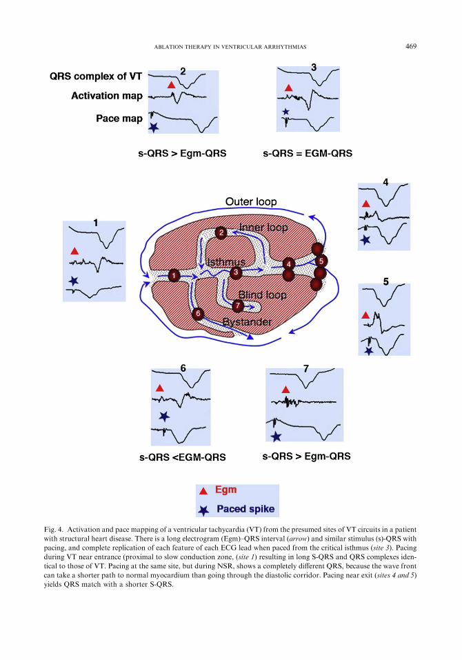
Fig. 4. Activation and pace mapping of a ventricular tachycardia (VT) from the presumed sites of VT circuits in a patient
with structural heart disease. There is a long electrogram (Egm)–QRS interval (arrow) and similar stimulus (s)-QRS with
pacing, and complete replication of each feature of each ECG lead when paced from the critical isthmus (site 3). Pacing
during VT near entrance (proximal to slow conduction zone, (site 1) resulting in long S-QRS and QRS complexes iden-
tical to those of VT. Pacing at the same site, but during NSR, shows a completely different QRS, because the wave front
can take a shorter path to normal myocardium than going through the diastolic corridor. Pacing near exit (sites 4 and 5)
yields QRS match with a shorter S-QRS.
469ABLATION THERAPY IN VENTRICULAR ARRHYTHMIAS

470 DAS et al
exit site (sites 4 and 5), pacing results in a QRSmatch of the VT but have a relatively shorterS-QRS interval. At the hypothetical bystander
site (site 6), the activation map shows a relativelylong Egm–QRS interval and a short S-QRSduration, whereas pace mapping shows a QRSmorphology different from the clinical VT. In
contrast to the entrance site of the critical isthmus(site 1), ablation at this site (site 6) will be unsuc-cessful. In the inner loop, the wavefront travels in
the same direction as the outer loop. Therefore,the impulse reaches late during diastole at site 2,resulting in a relatively short Egm-QRS interval,
whereas pacing from this site will show a relativelylonger S-QRS interval, and ablation at this sitealso will be unsuccessful.
Fig. 5 shows various scar-related macrore-
entrant circuits encountered during mapping.Presystolic or mid- to late diastolic potentials (orEgms) are identified in approximately two thirds
of patients during activation mapping of theseVTs. A complete reentry circuit is defined in lessthan 20% of VTs with catheter mapping, whereas
multielectrode and noncontact mapping systemsidentify endocardial exit regions of presystolicelectric activity in greater than 90% of VTs. It is
Fig. 5. Reentrant ventricular tachycardia (VT) circuits aroundm
different endocardial and epicardial reentrant circuits with singl
ablated at critical isthmus sites. Reentry around a ventricula
(RVOT)patch canbe ablatedby connecting the ablation line from
T-shaped line or multiple radial lines of ablation across the boar
unstable VT. Blue lines denote potential circuits, red dots represe
not imperative to do this, however, because locat-ing and ablating at the ideal site in the isthmus in-terrupts the particular circuit and terminate the
VT. The participation of a particular isthmuswith diastolic potential recorded during VT isconfirmed by entrainment and pace mapping.
Focal ventricular arrhythmia
Focal MMVT (or PVC) generally is encoun-tered in patients who have idiopathic VT, but it
rarely may occur in patients who have MI scar.The bipolar Egm is used universally for thispurpose, seeking a timing of 10 to 60 milliseconds
before the QRS onset. The timing and morphol-ogy of unipolar recording from the catheter tip arealso very helpful. At the suspected site of origin,
this should have a sharp QS deflection that timeswith the bipolar recording and precedes the QRSonset by a similar amount.
As mentioned earlier, VTs in the setting ofSHD have a reentrant mechanism in most pa-tients, but a focal source of sustained MMVT hasbeen reported also. A focal mechanism of VT has
been found in 5% to 8% of VTs in patients whohave CAD (Fig. 6). Similarly, a focal automatic
yocardial scar in structural heart disease. The figure shows
e loop or figure-of-eight wave fronts of VT, which can be
r septal defect patch or right ventricular outflow tract
the patch to thepulmonaryor the tricuspid valve.A single
der zones often are drawn in patients in hemodynamically
nt ablation points needed to interrupt the reentrant circuit.

Fig. 6. Focal monomorphic premature ventricular complexes (PVCs) initiating nonsustained polymorphic ventricular
tachycardia (VT) and ventricular fibrillation (VF). The telemetry recordings of a patient with history of myocardial
infarction who presented with recurrent VF revealed frequent monomorphic PVCs and nonsustained VTs. Ablation
of the PVC focus in the lateral left ventricle eliminated the ventricular arrhythmias.
471ABLATION THERAPY IN VENTRICULAR ARRHYTHMIAS

Fig. 7. Endocardial substrate mapping for ventricular
tachycardia (VT). Endocardial substrate and pace
mapping was performed for hemodynamically unstable
reentrant VT with an exit site in the scar border zone
of the anterolateral mid-left ventricle in a patients with
history of myocardial infarction. Note a line of ablation
was drawn across the border zone where good pace
maps were achieved.
472 DAS et al
mechanism was found to be responsible for VT in27% of patients with DCM who underwentradiofrequency ablation for VT in one study [20].
Purkinje system ventricular tachycardia
His-Purkinje system-related SMMVT has been
reported in patients who have SHD (8%) and inpatients who have normal hearts [21].
Bundle branch reentrant ventricular tachycardiaBundle branch reentrant VT occurs in 15% to
40% of patients who have DCM. It also can occur
in ischemic cardiomyopathy (6%), after mitral oraortic valve surgery, myotonic dystrophy andrarely, in conduction system disease [22]. Typically,
the VT has a LBBB pattern because of the wavefronts propagating retrogradely up the left bundleand anterogradely down the right bundle branch.
Less frequently, the circuit revolves in the oppositedirection, producing aRBBB configuration. There-fore, interruption of either of the bundles results intermination of the VT. Right bundle branch abla-
tion, however, is preferred to avoid the hemody-namic consequences of chronic left bundle branchblock (LV dyssynchrony). Usually, complete atrio-
ventricular block does not occur. An ICD often iswarranted, because of high risk of scar-related re-entrant VT in these patients.
Ventricular tachycardias with participation
of the left-sided His-Purkinje systemInterfascicular and fascicular VT occurs rarely
in patients who have SHD. It can be cured byablating the left posterior or left anterior fascicle
depending upon the site of origin of the VT. It isassociated with a considerable risk of completeheart block caused by preexisting conduction
abnormalities. Similar VTs with a narrow QRScomplex have been reported after acute or remoteMI. During the VT, Purkinje potentials are
captured orthodromically with decremental con-duction properties, whereas presystolic Purkinjepotentials are captured antidromically and appearbetween the His and QRS complex. In one study,
catheter ablation at the site exhibiting a Purkinje–QRS interval of 58 plus or minus 26 millisecondssuccessfully eliminated VTs without provoking
any conduction disturbances.
Idiopathic left ventricular tachycardiaILVT involves the left posterior fascicle (less
commonly, the anterior fascicle) and the posterior
Purkinje network. The VT has a relatively narrowQRS (RBBB, superior axis), probably because ofthe conduction system being an integral part of
the circuit. An abnormal diastolic potential withinthe posterior Purkinje network during sinusrhythm and VT can be used to guide successful
catheter ablation of ILVT.
Ablation strategies for polymorphic ventricular
tachycardia and ventricular fibrillation
Structural heart disease
For single or multiple unstable MMVTs andpolymorphic VTs or VF, voltage maps are createdfrom three-dimensional anatomic plots of low-
voltage regions (less than 1.55 mV in bipolarrecordings) to identify areas of scar and perinfarctzones. Pace mapping at the scar border zone
usually is required for a SMMVT or PVC thattriggers polymorphic VT/VF. Brief entrainment istried and is often possible, even for unstable VTs,to confirm the location of a reentry circuit. Low-
amplitude isolated potentials and late potentialsinscribed after the end of the QRS complex duringsinus rhythm also suggest potential isthmus sites.
Connecting the two scar zones or scar and a lineof conduction block/anatomic structure also isperformed empirically (Fig. 7). A radial line or
T-shaped line of ablation also is performed oftenat the scar border zones (see Fig. 5).

473ABLATION THERAPY IN VENTRICULAR ARRHYTHMIAS
Polymorphic ventricular tachycardia and idiopathicventricular fibrillation in normal hearts
Rare forms of polymorphic VT originate fromthe RVOT in structurally normal hearts and in the
left ventricle in patients who have a history of MI.These VAs are triggered by monomorphic PVCs,which usually are preceded by a Purkinje-likepotential. In idiopathic VF, activation and pace
mapping of PVCs that initiate VF can be identi-fied. Catheter ablation at these sites eliminatesthese trigger PVCs and prevents the recurrence of
malignant polymorphic VT or VF episodes witha high success rate. Although this variety of life-threatening VA is initiated by repetitive mono-
morphic PVCs, the QRS morphology changes inthe subsequent beats, and rapid pace mappingfrom the earliest site of activation also results ina change of QRS morphology similar to the
clinical polymorphic VT. It is presumed thata shift in exit site during rapid pacing is re-sponsible for the changing QRS morphology.
Thus, PVCs that trigger polymorphic VT or VFin patients who have normal hearts, or SHD canbe treated successfully with catheter ablation.
Ventricular tachycardia ablation in specific
subpopulations
Coronary artery disease
Patients who have multiple VTs generally aretried on one or more antiarrhythmic drugs (mostfrequently, amiodarone) that have a limited
response (19% to 50%). Most VTs can be ablatedendocardially with a reasonably good long-termsuccess rate of 77% to 95% for the clinical VT [2].
The epicardial approach for ablation is used forepicardial circuits or in presence of a LV throm-bus (see Fig. 5).
Nonischemic dilated cardiomyopathy
SMMVT is uncommon in DCM. It occursmostly (80%) because of scar-related reentry [20].SMMVTs caused by reentry related to low-volt-
age regions (scars) often are located adjacent toa valve annulus that forms a border for the reen-trant circuit. When a VT circuit cannot be defined
during endocardial mapping, and significant low-voltage regions are not present on the endocar-dium, it often is present in the subepicardium
(see Fig. 5). This likely contributes to the lowerreported success rate of endocardial ablation.Some physicians start with epicardial ablation if
VT morphology suggests epicardial circuits. InChagas disease, approximately 70% of VTs areepicardial in origin. Less commonly, it occurs be-cause of bundle branch reentry [20]. Rarely, focal
VTs are encountered. Of a series of 22 patientswho had VT caused by reentry associated withDCM, endocardial ablation failed in 10 patients.
Seven of these patients underwent epicardial map-ping, with successful ablation in six patients.Fig. 8 demonstrates the endocardial and epicar-
dial map of a patient with DCM in whom peri-valvular and RV epicardial scar is demonstrated,whereas endocardial scar is minimal. His clinical
VT was terminated by catheter ablation in theepicardial right ventricle medial to the proximalleft anterior descending artery.
Other cardiomyopathies
Sustained VA is one of the major causes of
SCD in cardiomyopathies such as hypertrophiccardiomyopathy, arrhythmogenic right ventricu-lar dysplasia/cardiomyopathy (ARVD/C), various
infiltrative cardiomyopathies such as sarcoidosis,and Chagas disease. ICD is indicated for most ofthese patients for secondary prophylaxis and for
primary prophylaxis in high-risk patients.
Cardiac sarcoidosisCardiac manifestations of sarcoidosis include
cardiomyopathy, AV block, and VA. Steroid
treatment improves the heart block in 62% ofpatients, while the VT is unaffected. Many casesof VT related to sarcoidosis are misdiagnosed as
idiopathic cardiomyopathy or ARVD. In a studyof 98 patients who had nonischemic cardiomyop-athy and VT, sarcoidosis was the cause in eightpatients, and most were scar-related. Ablation
abolished one or more VTs in six (75%) of eightpatients, but other VTs remained inducible in allbut one patient. After ablation, some form of
sustained VT recurred in six of eight patientswithin 6 months. During a longer follow-up(range 6 months to 7 years), however, four of
eight patients were free of VT with antiarrhythmicdrugs and immunosuppression. Cardiac trans-plantation eventually was required in five of eightpatients because of either recurrent VT (n ¼ 4) or
heart failure (n ¼ 1). In another study, 68% ofVTs were determined to be reentrant VTs.
Arrhythmogenic right ventricular dysplasia/
cardiomyopathySMMVT occurs mostly because of reentry
circuit in the vicinity of the tricuspid and
Fig. 8. Endocardial and epicardial bipolar voltage mapping to guide ablation. Endocardial and epicardial bipolar volt-
age mapping using an electroanatomical mapping system in a patient with dilated cardiomyopathy with sustained mono-
morphic ventricular tachycardia (VT). The endocardial mapping recorded minimal basal scar regions in the left ventricle
(LV), whereas the epicardial mapping revealed extensive LV basal scars and right ventricular (RV) scar. The tachycardia
was hemodynamically unstable. Pace mapping revealed an exit site medial to the proximal left anterior descending artery
(LAD), which was ablated successfully. LAD and right coronary artery are drawn by mapping the coronary artery
locations during coronary angiography. The fluoroscopic position of the catheter is shown in the left anterior oblique
(LAO) view. Abbreviations: LL: left lateral view; RAO: right anterior oblique view.
474 DAS et al
pulmonary valve and the lateral right ventricle nearthe apex. The RV becomes paper-thin in scar
regions; therefore, catheter ablation is associatedwith a 74% to 77% acute success rate. It also cariesa higher risk for right ventricular perforation, and
a high recurrence rate (11%–75%) due to theprogressive nature of the disease [2].
Chagas disease. Cardiomyopathy caused byChagas disease is associated with segmental scars,
475ABLATION THERAPY IN VENTRICULAR ARRHYTHMIAS
similar to ischemic cardiomyopathy. LV infero-lateral scar is often the source of VT (80%) andcan be ablated successfully.
Congenital heart disease
Right ventriculotomy scar becomes a substratefor reentry in patients who have congenital heartdisease such as repair of tetralogy of Fallot and
ventricular septal defect. After repair of tetralogyof Fallot, the incidence of VT is 11.9%, with an8.3% risk of SCD by 35 years of follow-up.
Substrate mapping to evaluate congenital, surgi-cal, and electrophysiological anatomy identifiesthe isthmus for successful VT ablation. Naturallyoccurring anatomic barriers in the right ventricle
are limited, and the placement of a single ven-triculotomy, outflow tract patch, or transannularpatch, along with closure of the ventricular septal
defect, would be expected to create only a limitednumber of possible tachycardia circuits (seeFig. 5). Unexcitable tissues from patch material
or myocardial scar along with the tricuspid andpulmonary valve annuli form the channel. Com-mon anatomic boundaries are isthmuses betweenthe tricuspid or pulmonary annulus and septal
scar or patch. These channels can be mappedwith three-dimensional substrate mapping andconnected by ablation lines during sinus rhythm.
In one study, the acute success rate was 100%,and the long-term success rate was 91% (meanfollow-up: 30.4 þ 29.3 months).
Postvalve surgery
VTs after aortic or mitral valve surgery (with-out CAD) account for 4% of VTs. They havea bimodal presentation and occur either early
after surgery (3 to 10 days) or years later (5 to15 years) [23]. Most VTs are scar-related (70%).Nearly two thirds (64%) of patients have perian-
nular scar in which an identifiable endocardial cir-cuit isthmus (71%) and bundle branch reentry(10%) are present (see Fig. 5). The acute success
rate of VT ablation is reported to be 98% [23].
Idiopathic monomorphic ventricular tachycardiain patients with normal hearts
Left ventricular outflow tract ventricular
tachycardia and its variantRVOT VT most commonly arises from RVOT
(90%), and less commonly, it may originate from
LV outflow tract, mitral annulus, pulmonaryartery, aortic sinus of Valsalva, coronary sinus,and coronary veins. In fact, it may originate from
any part of right ventricular or LV myocardium.These patients have frequent PVCs, nonsustainedVT in salvos, or sustained VT. In a few patients, itis related to exercise [24]. These VTs or PVCs of-
ten are induced with burst pacing and with isopro-terenol, adrenaline or aminophylline infusion, andterminated with intravenous injection of adeno-
sine, b-blockers or calcium channel blockers. Suc-cess rates of ablation are higher in RVOT VTsand lower in septal VTs. The risks of ablation in-
clude RV perforation during RVOT or free wallablation and complete heart block during septalablation. The potential for acute occlusion of
the left main or right coronary arteries is a majorrisk consideration, especially with VTs originatingfrom the aortic sinuses of Valsalva. Coronary an-giography and intracardiac ultrasound imaging
have been used to define the proximity of the cor-onary ostia to the ablation site. Radiofrequencyablation has been performed safely at sites more
than 8 mm away from the coronary artery ostiawith careful continuous monitoring of catheterposition during RF application. Standard RF ab-
lation with tip temperature maintained at lessthan 55�C has been suggested to prevent aorticvalve damage observed in animal studies. Epicar-
dial origin of VT in close relation to coronary si-nus can be ablated by means of the coronary sinus(Fig. 9).
Polymorphic ventricular tachycardiaand ventricular fibrillation
The malignant variety of RVOT VT and
idiopathic VF that is initiated by a single or twomonomorphic PVCs can be ablated successfullywith good long-term results (Fig. 10).
Inherited ventricular arrhythmia syndromes
Symptomatic and high-risk patients who haveinherited arrhythmia syndromes are treated with
an ICD. VT storm has been shown to havetriggers (unifocal PVCs) from the Purkinje arbor-ization or the RVOT. They play a crucial role ininitiating VF in patients with long QT and
Brugada syndromes. VT storm in Brugada syn-drome is managed medically by infusion of iso-proterenol. In one study, three patients who had
long QT syndrome and four patients who hadBrugada syndrome, episodes of polymorphic VTand VF, were associated with frequent isolated or
repetitive PVCs, which could be ablated success-fully without any recurrence during a follow-up of17 þ 17 months.

476 DAS et al

477ABLATION THERAPY IN VENTRICULAR ARRHYTHMIAS
Electrical storms or ventricular tachycardia storm
Electrical storm (ES) is an important, indepen-dent marker for subsequent death among ICDrecipients, particularly in the first 3 months after its
occurrence. The development of VT/VF unrelatedtoES, however, does not seem to be associatedwithan increased risk of subsequent death. Short- andlong-term efficacy of catheter ablation for ES was
studied in patients who had SHD (CAD,DCMandARVD/C) [25]. After one to three procedures,induction of any clinical VT(s) by programmed
electrical stimulation was prevented in 89%patients. VT storm was suppressed acutely in allpatients; a minimum period of 7 days with stable
rhythm was required before hospital discharge,and 92% patients were free of VT storm and 66%patients free of VT recurrence during a median fol-low-up of 22 months (range, 1 to 43 months). Eight
of 10 patients who had persistent inducibility ofclinical VT(s) had ES recurrence; four of themdied suddenly despite ICD therapy.
Inducible ventricular tachycardias and ablation
end points
Acute success
During EP study, in patients who have VT inthe setting of SHD, an average of three different
monomorphic VTs, including the clinical VT (VTrequiring ablation) and nonclinical VTs, are in-duced. Of note, in many patients VT cycle length
may be the only clue regarding the clinical VT,because the ICD promptly terminates VT. Abla-tion of incessant VTs and the inducible clinical VTis often an acceptable end point for success.
Ablation of nonclinical VTs also is attemptedbecause these VTs may recur subsequently. In onestudy, at least one VT was no longer inducible
after ablation in 73% to 100% of patients,whereas all inducible VTs were abolished in 38%to 95% of patients [2].
Long-term results
VTs that are noninducible after ablation gen-erally have low recurrence rates (less than 3% to
Fig. 9. Mapping and ablation of epicardial ventricular tachyc
ture ventricular complexes (PVCs) and VT with alternating QR
the epicardial surface of anterolateral mitral annulus. Because
however, PVCs could not be ablated. Later, mapping of the sa
which revealed an early presystolic electrogram and an excellen
site. Abbreviations: CS: coronary sinus, LA: left atrium; LV: l
:
27%), whereas when the targeted VT remainsinducible after ablation, the recurrence rate isgreater than 60%. However, the frequency ofepisodes often is reduced in these patients. In
a multicenter trial of 146 patients, the immediateeffect on inducible VT did not predict outcomes;VT recurred in 44% of patients who had no
inducible VT and 46% of those who had inducibleVT. The frequency of spontaneous VT duringshort-term follow-up was reduced by more than
75% in most patients. Recurrence rate is higherafter VT ablation in DCM [17].
The annual mortality after VT ablation ranges
from 5% to greater than 20%, with death fromprogressive heart failure being the most commoncause. The substantial mortality is consistent withthe severity of heart disease and association of
spontaneous VT with mortality and heart failureeven when VT is treated effectively by an ICD.Older age, greater LV size, and LV dysfunction
increase mortality. The potential for ablation toadversely affect LV function is a cause forconcern, although assessment of LV ejection
fraction after ablation has not shown any de-terioration. Confining ablation lesions to regionsof low-voltage scar and attention to appropriate
medical therapy beneficial to patients who haveLV dysfunction are prudent.
Future directions
VT ablation remains a relatively high-riskprocedure with a success rate far from the desired
because of several factors, including poor LVsystolic function (in most patients who haveSHD), inability to map the VT circuits with
presently available mapping tools, hemodynamicinstability, and changing activation patterns. Fail-ure of ablation may be caused by organized
thrombus preventing optimum energy delivery tothe deeper myocardium, progression of disease,and multiple epicardial VT circuits that are notamenable to successful ablation due to epicardial
fat and risk of coronary vessel and phrenic nerveinjury. A high risk of recurrence after VT ablationoccurs because of continued disease process,
ardia (VT). Initial mapping of the monomorphic prema-
S morphologies revealed a focal source originating from
of the close proximity to the phrenic nerve (white dots),
me area was attempted by means of the coronary sinus,
t pace map. The PVC was ablated successfully from this
eft ventrice; MV: mitral valve.
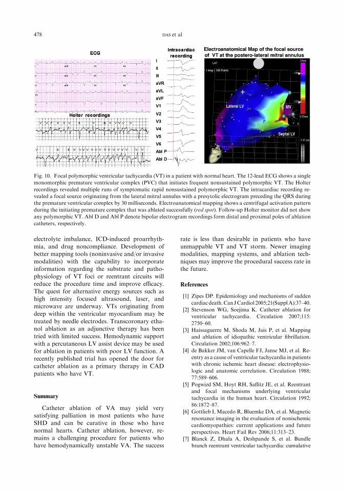
Fig. 10. Focal polymorphic ventricular tachycardia (VT) in a patient with normal heart. The 12-lead ECG shows a single
monomorphic premature ventricular complex (PVC) that initiates frequent nonsustained polymorphic VT. The Holter
recordings revealed multiple runs of symptomatic rapid nonsustained polymorphic VT. The intracardiac recording re-
vealed a focal source originating from the lateral mitral annulus with a presytolic electrogram preceding the QRS during
the premature ventricular complex by 30 milliseconds. Electroanatomical mapping shows a centrifugal activation pattern
during the initiating premature complex that was ablated successfully (red spot). Follow-up Holter monitor did not show
any polymorphic VT. Abl D and Abl P denote bipolar electrogram recordings form distal and proximal poles of ablation
catheters, respectively.
478 DAS et al
electrolyte imbalance, ICD-induced proarrhyth-
mia, and drug noncompliance. Development ofbetter mapping tools (noninvasive and/or invasivemodalities) with the capability to incorporateinformation regarding the substrate and patho-
physiology of VT foci or reentrant circuits willreduce the procedure time and improve efficacy.The quest for alternative energy sources such as
high intensity focused ultrasound, laser, andmicrowave are underway. VTs originating fromdeep within the ventricular myocardium may be
treated by needle electrodes. Transcoronary etha-nol ablation as an adjunctive therapy has beentried with limited success. Hemodynamic support
with a percutaneous LV assist device may be usedfor ablation in patients with poor LV function. Arecently published trial has opened the door forcatheter ablation as a primary therapy in CAD
patients who have VT.
Summary
Catheter ablation of VA may yield verysatisfying palliation in most patients who haveSHD and can be curative in those who have
normal hearts. Catheter ablation, however, re-mains a challenging procedure for patients whohave hemodynamically unstable VA. The success
rate is less than desirable in patients who have
unmappable VT and VT storm. Newer imagingmodalities, mapping systems, and ablation tech-niques may improve the procedural success rate inthe future.
References
[1] Zipes DP. Epidemiology and mechanisms of sudden
cardiac death.CanJCardiol 2005;21(SupplA):37–40.
[2] Stevenson WG, Soejima K. Catheter ablation for
ventricular tachycardia. Circulation 2007;115:
2750–60.
[3] Haissaguerre M, Shoda M, Jais P, et al. Mapping
and ablation of idiopathic ventricular fibrillation.
Circulation 2002;106:962–7.
[4] de Bakker JM, van Capelle FJ, Janse MJ, et al. Re-
entry as a cause of ventricular tachycardia in patients
with chronic ischemic heart disease: electrophysio-
logic and anatomic correlation. Circulation 1988;
77:589–606.
[5] Pogwizd SM, Hoyt RH, Saffitz JE, et al. Reentrant
and focal mechanisms underlying ventricular
tachycardia in the human heart. Circulation 1992;
86:1872–87.
[6] Gottlieb I, Macedo R, Bluemke DA, et al. Magnetic
resonance imaging in the evaluation of nonischemic
cardiomyopathies: current applications and future
perspectives. Heart Fail Rev 2006;11:313–23.
[7] Blanck Z, Dhala A, Deshpande S, et al. Bundle
branch reentrant ventricular tachycardia: cumulative
479ABLATION THERAPY IN VENTRICULAR ARRHYTHMIAS
experience in 48 patients. J Cardiovasc Electrophysiol
1993;4:253–62.
[8] Lopera G, Stevenson WG, Soejima K, et al. Identi-
fication and ablation of three types of ventricular
tachycardia involving the his-Purkinje system in
patients with heart disease. J Cardiovasc Electro-
physiol 2004;15:52–8.
[9] Miller JM, Marchlinski FE, Buxton AE, et al.
Relationship between the 12-lead electrocardiogram
during ventricular tachycardia and endocardial site
of origin in patients with coronary artery disease.
Circulation 1988;77:759–66.
[10] Badhwar N, ScheinmanMM. Idiopathic ventricular
tachycardia: diagnosis and management. Curr Probl
Cardiol 2007;32:7–43.
[11] Berruezo A, Mont L, Nava S, et al. Electrocar-
diographic recognition of the epicardial origin of
ventricular tachycardias. Circulation 2004;109:
1842–7.
[12] Rothman SA, Hsia HH, Cossu SF, et al. Radio-
frequency catheter ablation of postinfarction
ventricular tachycardia: long-term success and the
significance of inducible nonclinical arrhythmias.
Circulation 1997;96:3499–508.
[13] Bogun F, Good E, Reich S, et al. Isolated potentials
during sinus rhythm and pace-mapping within scars
as guides for ablation of postinfarction ventricular
tachycardia. J Am Coll Cardiol 2006;47:2013–9.
[14] Reddy VY, Reynolds MR, Neuzil P, et al. Prophy-
lactic catheter ablation for the prevention of defibril-
lator therapy. N Engl J Med 2007;357(26):2657–65.
[15] Josephson ME, Waxman HL, Cain ME, et al. Ven-
tricular activation during ventricular endocardial
pacing. II. Role of pace mapping to localize origin
of ventricular tachycardia. Am J Cardiol 1982;50:
11–22.
[16] Josephson ME, Horowitz LN, Spielman SR, et al.
Role of catheter mapping in the preoperative evalu-
ation of ventricular tachycardia. Am J Cardiol 1982;
49:207–20.
[17] Soejima K, StevensonWG, Sapp JL, et al. Endocar-
dial and epicardial radiofrequency ablation of
ventricular tachycardia associated with dilated car-
diomyopathy: the importance of low-voltage scars.
J Am Coll Cardiol 2004;43:1834–42.
[18] Sosa E, ScanavaccaM, d’Avila A, et al. Nonsurgical
transthoracic epicardial catheter ablation to treat
recurrent ventricular tachycardia occurring late after
myocardial infarction. J Am Coll Cardiol 2000;35:
1442–9.
[19] Harada T, Tomita Y, Nakagawa T, et al. Pace
mapping conduction delay at reentry circuit sites of
ventricular tachycardia after myocardial infarction.
Heart Vessels 1997;(Suppl 12):232–4.
[20] Delacretaz E, Stevenson WG, Ellison KE, et al.
Mapping and radiofrequency catheter ablation
of the three types of sustained monomorphic
ventricular tachycardia in nonischemic heart
disease. J Cardiovasc Electrophysiol 2000;11:11–7.
[21] Reithmann C, Hahnefeld A, Remp T, et al. Ventric-
ular tachycardia with participation of the left
bundle-Purkinje system in patients with structural
heart disease: identification of slow conduction
during sinus rhythm. J Cardiovasc Electrophysiol
2007;18:808–17.
[22] Mazur A, Kusniec J, Strasberg B. Bundle branch
reentrant ventricular tachycardia. Indian Pacing
Electrophysiol J 2005;5:86–95.
[23] Eckart RE, Hruczkowski TW, Tedrow UB, et al.
Sustained ventricular tachycardia associated with
corrective valve surgery. Circulation 2007;116:
2005–11.
[24] Morin DP, Lerman BB. Management of ventricular
tachycardia in the absence of structural heart dis-
ease. Curr Treat Options Cardiovasc Med 2007;9:
356–63.
[25] Carbucicchio C, Santamaria M, Trevisi N, et al.
Catheter ablation for the treatment of electrical
storm in patients with implantable cardioverter
defibrillators: short- and long-term outcomes in
a prospective single-center study. Circulation 2008;
117:462–9.
[26] Zipes DP, Camm AJ, Borggrefe M, et al. ACC/
AHA/ESC 2006 guidelines for management of
patients with ventricular arrhythmias and the pre-
vention of sudden cardiac death: a report of the
American College of Cardiology/American Heart
Association Task Force and the European Society
of Cardiology Committee for Practice Guidelines.
Circulation 2006;114:e385–484.