Robotically Assisted Prostate Therapies Under CT...
51
Copyright © CISST ERC, 2001 Engineering Research Center for Computer Integrated Surgical Systems and Technology Robotically Assisted Prostate Therapies Under CT Guidance Gabor Fichtinger, Ph.D. Director of Engineering, Clinical Thrust Leader, Engineering Research Center (ERC) for Computer Integrated Surgical Systems and Technology Johns Hopkins University email: [email protected] http: cisstweb.cs.jhu.edu
Transcript of Robotically Assisted Prostate Therapies Under CT...

Copyright © CISST ERC, 2001 Engineering Research Center for Computer Integrated Surgical Systems and Technology
Robotically Assisted Prostate Therapies Under CT Guidance
Gabor Fichtinger, Ph.D.Director of Engineering, Clinical Thrust Leader,
Engineering Research Center (ERC)for Computer Integrated Surgical Systems and Technology
Johns Hopkins University
email: [email protected]: cisstweb.cs.jhu.edu

Copyright © CISST ERC, 2001 Engineering Research Center for Computer Integrated Surgical Systems and Technology
Outline
• Vision & mission• Surgical CAD/CAM paradigm• Localized therapy• Prostate brachytherapy• Percutaneous robots for prostate • Experiments & results• Lessons learned• Future work

Copyright © CISST ERC, 2001 Engineering Research Center for Computer Integrated Surgical Systems and Technology
The glory goes to…• ERC: multi-institution & multi-disciplinary NSF
funded center– Johns Hopkins University + Medical Institutions– MIT AI Lab + Brigham & Women’s Hospital– CMU + Shadyside Hospital
• Johns Hopkins University– Dan Stoianovici, Louis Kavoussi, Ted
DeWeese, Russ Taylor, Jim Anderson, Ken Masamune (Japan)
• Students– Alex Patriciu,Robert Susil, Richard Wiard, Feras
Mousilli, Brian Handly, Dan Elgort, Andrew Bzostek, Rajesh Kumar
Copyright © CISST ERC, 2001 Engineering Research Center for Computer Integrated Surgical Systems and Technology
Surgical CAD/CAM ParadigmPre-op= CAD
MODELING&
PLANNING
UPDATE PLAN
Robot assistedplacement
Imaging &sensing
Intra-op = CAM
UPDATE
MODEL
Follow up
Post-op=TQM
-
Statistical AtlasAnatomy
DeformationPhysiologyPathologyDosimetry
Copyright © CISST ERC, 2001 Engineering Research Center for Computer Integrated Surgical Systems and Technology
Engineering goals
•Implement Surgical CAD-CAM paradigm in the context of percutaneous (needle-based) systems
•Create modular, factorable systems with a basic architecture that (to a reasonable extent) is invariant to imaging modality, target organ, and procedural details

Copyright © CISST ERC, 2001 Engineering Research Center for Computer Integrated Surgical Systems and Technology
Why Prostate?
We can make a huge differenceWe can make a huge difference– 200,000 new prostate cancer per year– 1,000,000 biopsies per year– BPH: 11,000,000 current cases– 25% of males has prostate condition
• PC, BPH, Prostatitis (chronic testicular and pelvic pain)
• Seems to be doable• Drives core research• Pre-existing experience• Strong clinical support • Industrial participation

Copyright © CISST ERC, 2001 Engineering Research Center for Computer Integrated Surgical Systems and Technology
Prostate diseases and treatments• Adenocarcinoma
– Prostatectomy - resection
– External beam – high energy X-ray
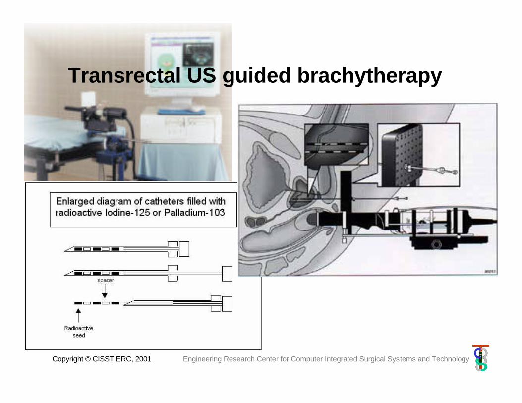
– Bracytherapy – implanted radioactive sources
– Experimental local therapies• Thermal (RF, Magnetic, HiFu, US, cryosurgery)• Gene therapy
• BPH– TURP– Thermal (RF, HiFu)
• Biopsy– Sextant + targeted
Copyright © CISST ERC, 2001 Engineering Research Center for Computer Integrated Surgical Systems and Technology
Target procedures
• Transperineal procedures under – US guidance w/ Burdette Medical Systems under
Phase I/II SBIR– CT guidance at JHU Urology– MRI at JHU Urology– Open MRI at BWH (in collaboration)
• Branch out to transrectal & transurethralprocedures

Copyright © CISST ERC, 2001 Engineering Research Center for Computer Integrated Surgical Systems and Technology
Gold standard
•TRUS•Template•Manual insertion
(Burdette’s Interplant®)
Copyright © CISST ERC, 2001 Engineering Research Center for Computer Integrated Surgical Systems and Technology
Transrectal US guided brachytherapy

Copyright © CISST ERC, 2001 Engineering Research Center for Computer Integrated Surgical Systems and Technology
Transrectal Ultrasound Probe,Template, and Stepper

Copyright © CISST ERC, 2001 Engineering Research Center for Computer Integrated Surgical Systems and Technology
In OR Patient Position and Setup
Copyright © CISST ERC, 2001 Engineering Research Center for Computer Integrated Surgical Systems and Technology
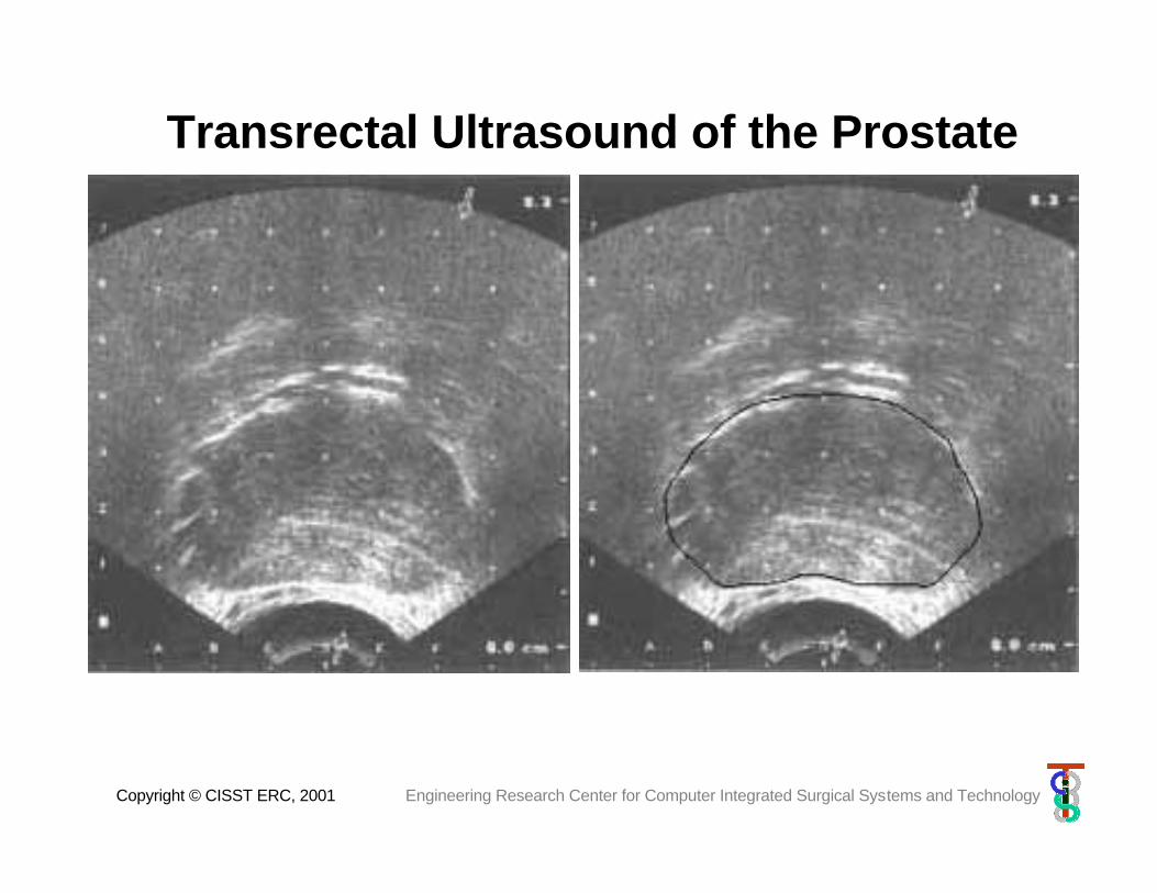
Transrectal Ultrasound of the Prostate
Copyright © CISST ERC, 2001 Engineering Research Center for Computer Integrated Surgical Systems and Technology
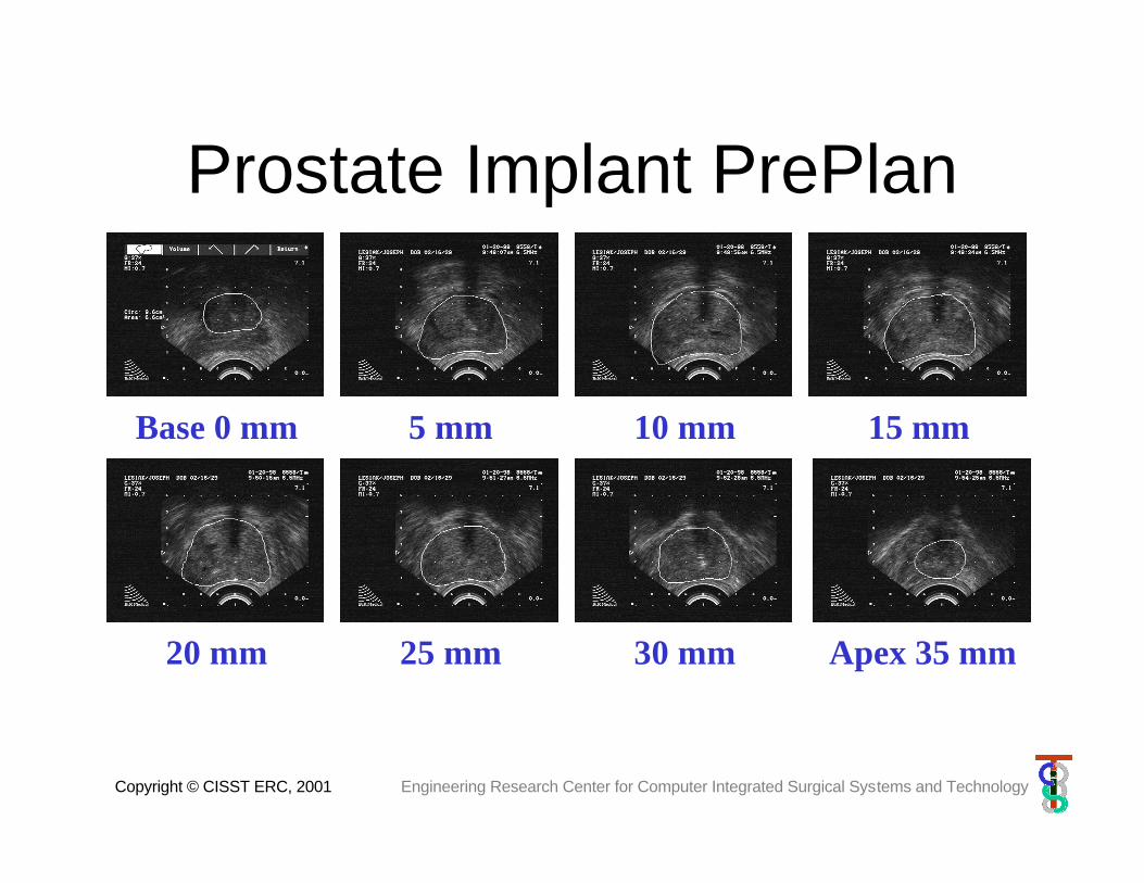
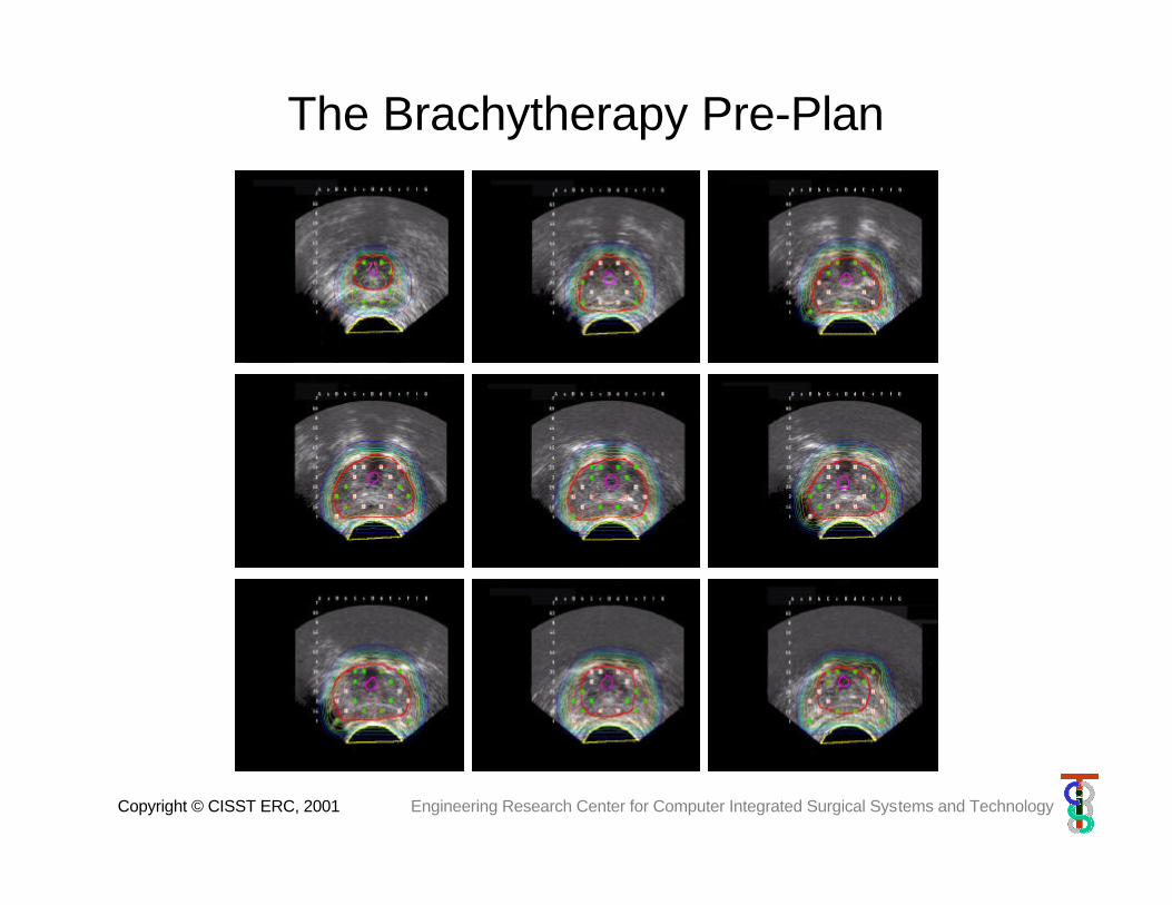
Prostate Implant PrePlanObtain axial ultrasound images of the prostate from
apex to base in 5 mm increments
Base 0 mm 5 mm 10 mm 15 mm
20 mm 25 mm 30 mm Apex 35 mm

Copyright © CISST ERC, 2001 Engineering Research Center for Computer Integrated Surgical Systems and Technology
Defining Regions of Interest
Copyright © CISST ERC, 2001 Engineering Research Center for Computer Integrated Surgical Systems and Technology
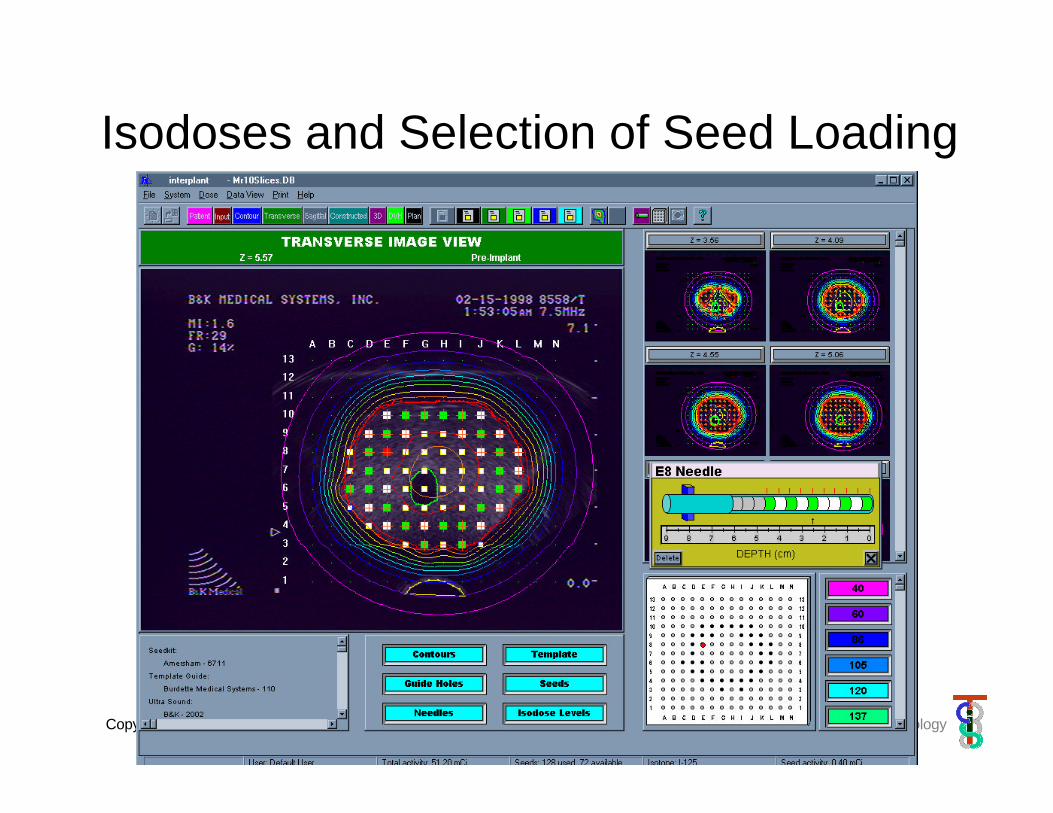
Isodoses and Selection of Seed Loading
Copyright © CISST ERC, 2001 Engineering Research Center for Computer Integrated Surgical Systems and Technology
The Brachytherapy Pre-Plan
Copyright © CISST ERC, 2001 Engineering Research Center for Computer Integrated Surgical Systems and Technology
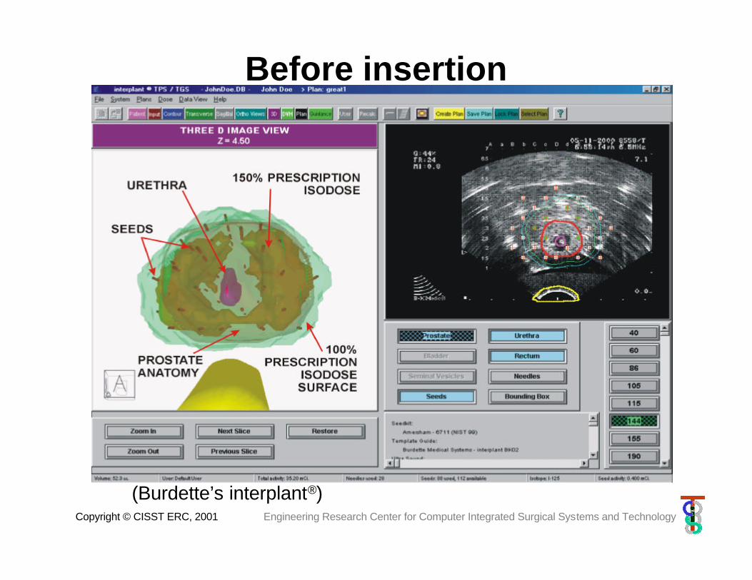
Before insertion
(Burdette’s interplant®)

Copyright © CISST ERC, 2001 Engineering Research Center for Computer Integrated Surgical Systems and Technology
Urethral Sparing

Copyright © CISST ERC, 2001 Engineering Research Center for Computer Integrated Surgical Systems and Technology
Prescription Dose Coverage
Copyright © CISST ERC, 2001 Engineering Research Center for Computer Integrated Surgical Systems and Technology
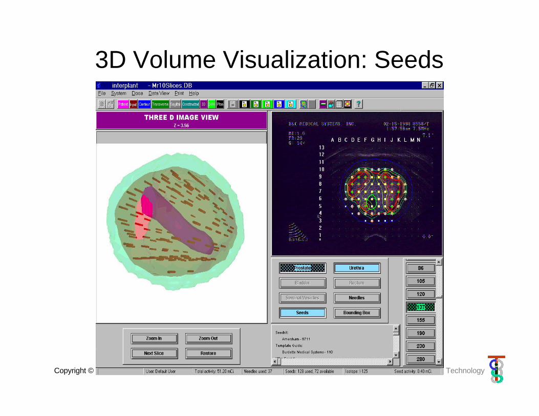
3D Volume Visualization: Seeds
Copyright © CISST ERC, 2001 Engineering Research Center for Computer Integrated Surgical Systems and Technology
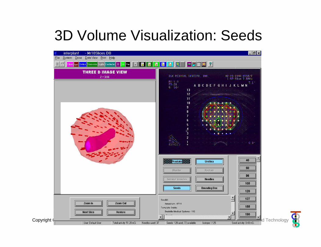
3D Volume Visualization: Seeds
Copyright © CISST ERC, 2001 Engineering Research Center for Computer Integrated Surgical Systems and Technology
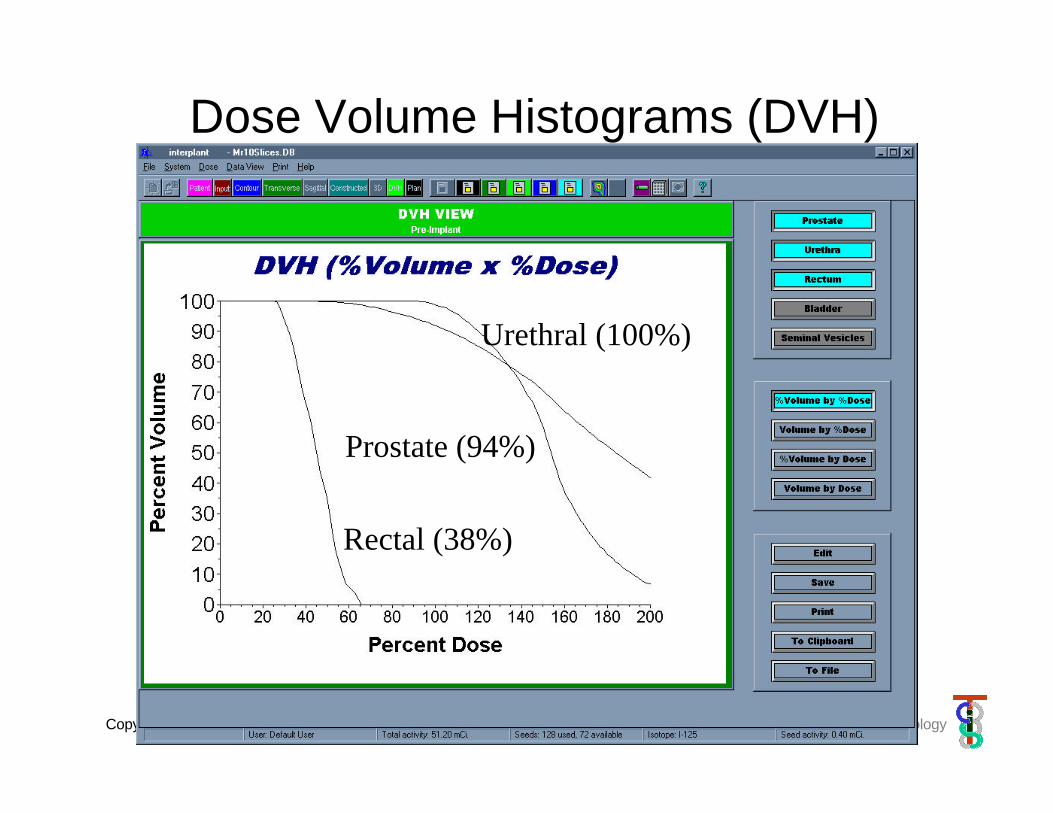
Dose Volume Histograms (DVH)
Urethral (100%)
Prostate (94%)
Rectal (38%)
Copyright © CISST ERC, 2001 Engineering Research Center for Computer Integrated Surgical Systems and Technology
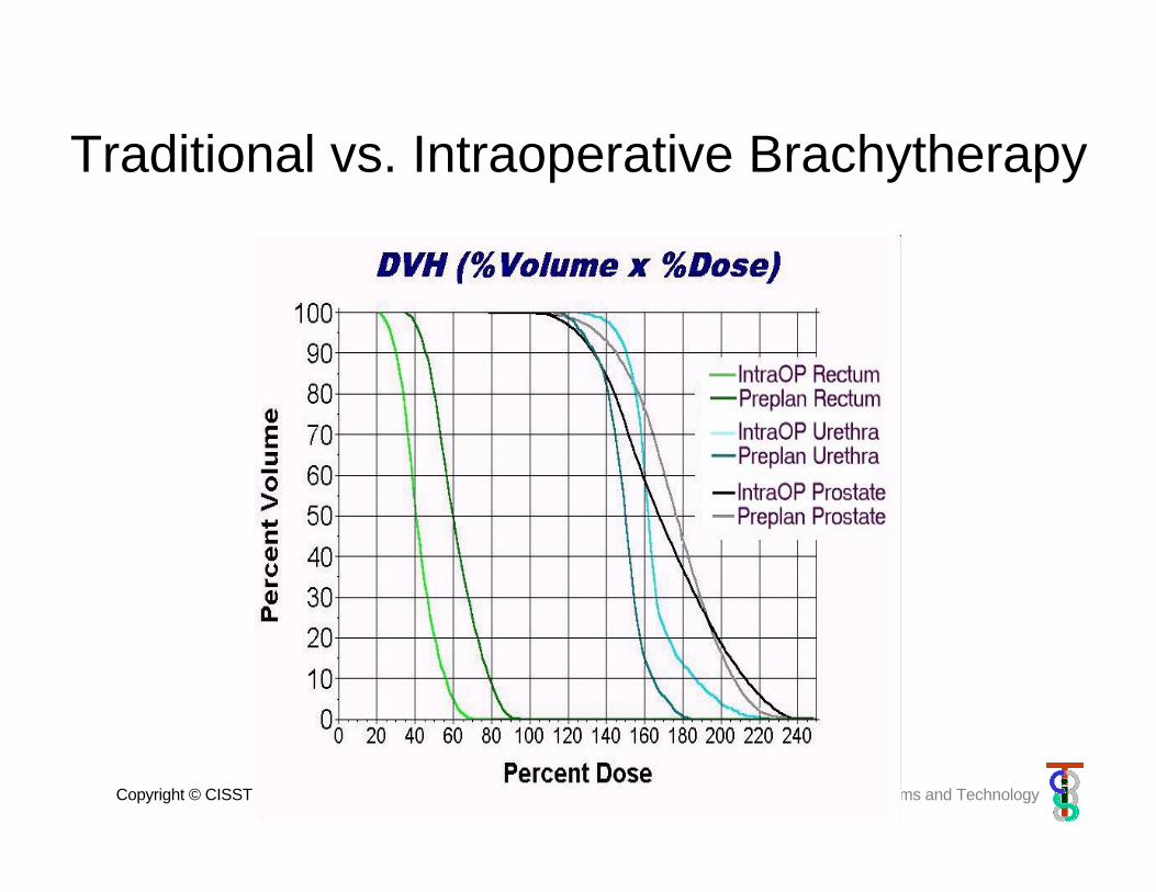
Traditional vs. Intraoperative Brachytherapy
Copyright © CISST ERC, 2001 Engineering Research Center for Computer Integrated Surgical Systems and Technology
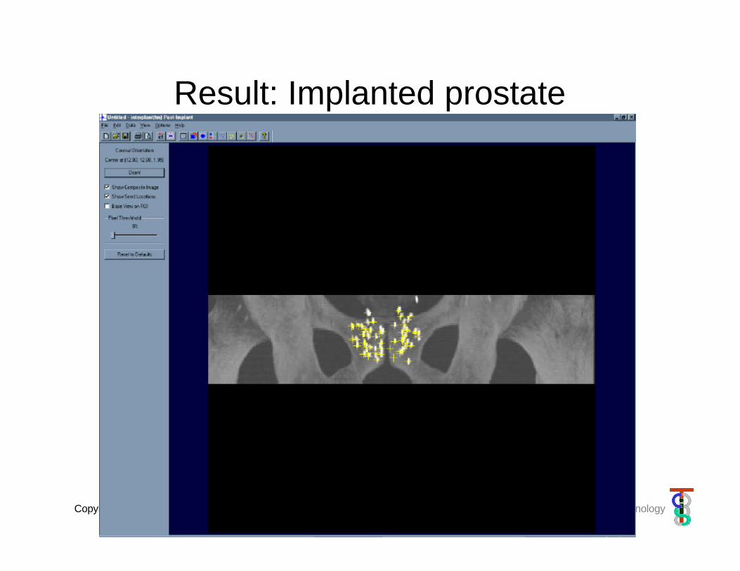
Result: Implanted prostate
Copyright © CISST ERC, 2001 Engineering Research Center for Computer Integrated Surgical Systems and Technology
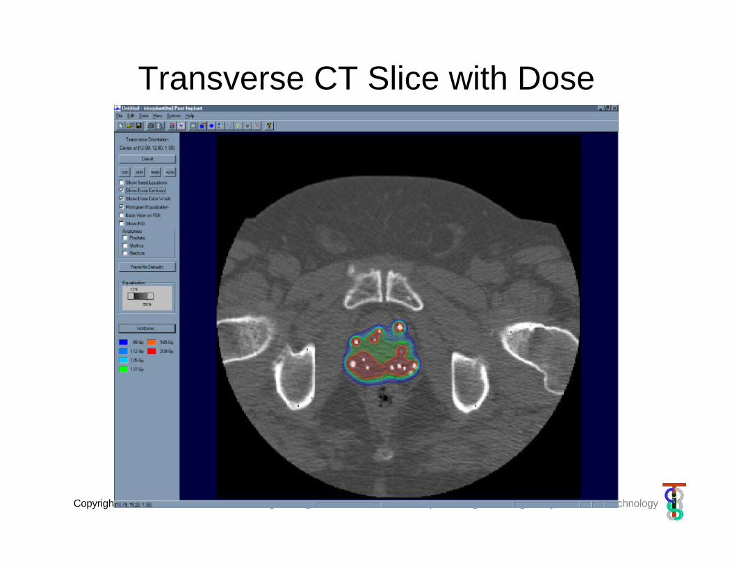
Transverse CT Slice with Dose
Copyright © CISST ERC, 2001 Engineering Research Center for Computer Integrated Surgical Systems and Technology
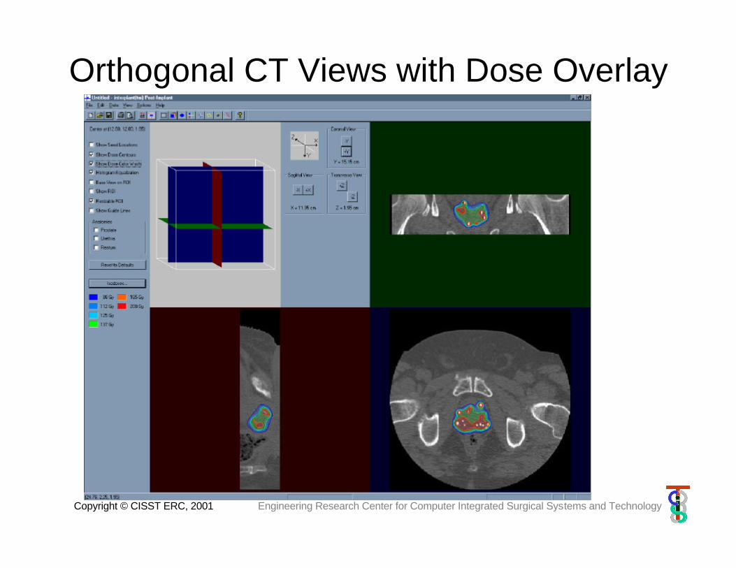
Orthogonal CT Views with Dose Overlay
Copyright © CISST ERC, 2001 Engineering Research Center for Computer Integrated Surgical Systems and Technology
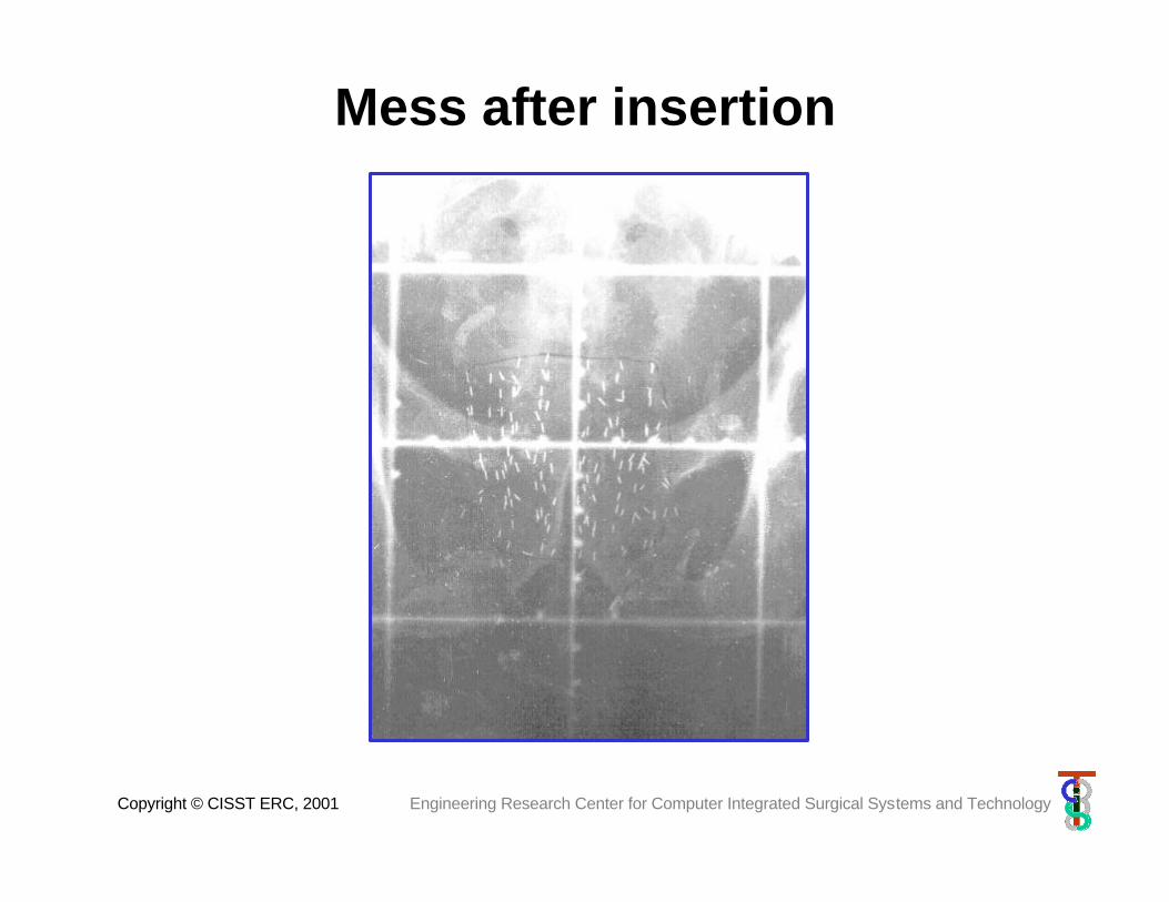
Mess after insertion

Copyright © CISST ERC, 2001 Engineering Research Center for Computer Integrated Surgical Systems and Technology
Why robotic assistance• No template
– Arbitrary entry & angle– No pubic arch interference (?)– Faster & easier setup/calibration
• Encoded robot– Track needles and seeds– Real-time optimization on per needle/seed basis– Full intraop dosimetry (calculation & measurement)– Less re-insertions (?)
• Less or no dose to crew• Electronic procedure logs
Copyright © CISST ERC, 2001 Engineering Research Center for Computer Integrated Surgical Systems and Technology
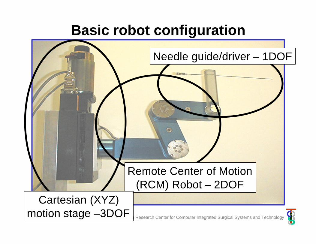
Basic robot configuration
Needle guide/driver – 1DOF
Remote Center of Motion(RCM) Robot – 2DOF
Cartesian (XYZ)motion stage –3DOF
Copyright © CISST ERC, 2001 Engineering Research Center for Computer Integrated Surgical Systems and Technology
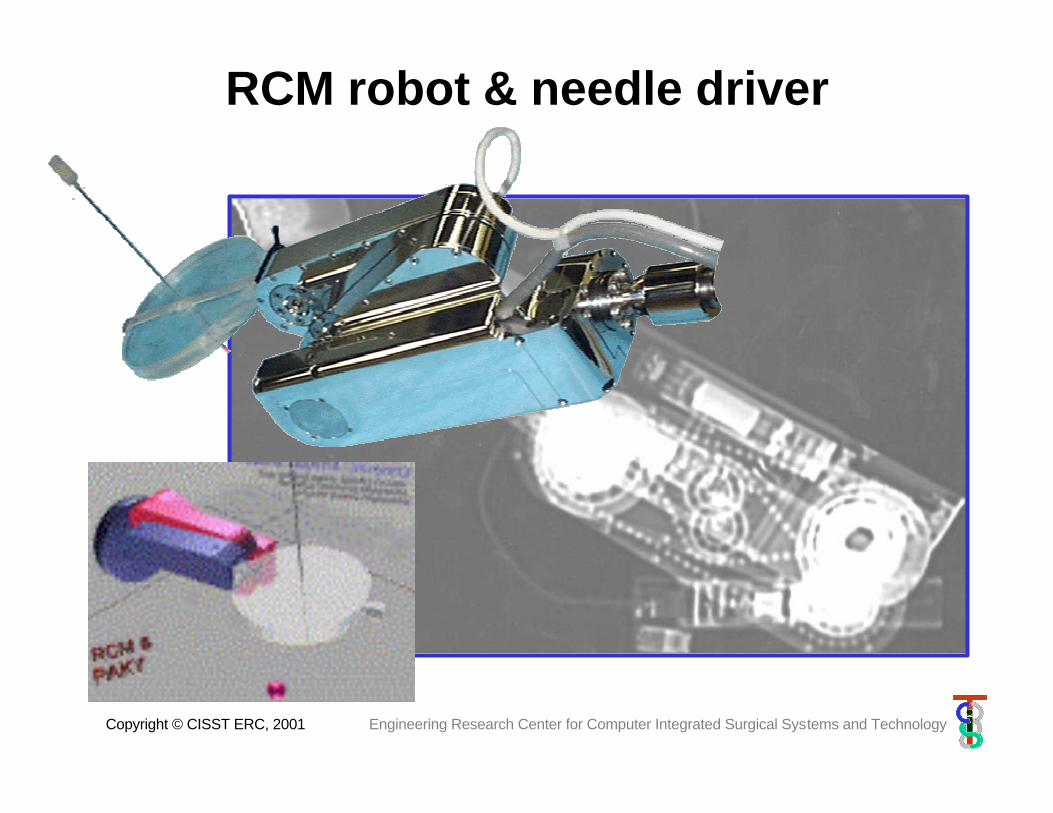
RCM robot & needle driver
Copyright © CISST ERC, 2001 Engineering Research Center for Computer Integrated Surgical Systems and Technology
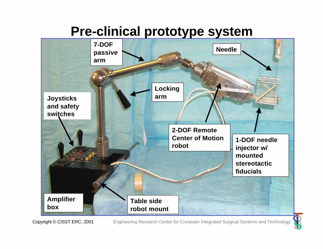
Pre-clinical prototype systemNeedle
1-DOF needle injector w/ mounted stereotactic fiducials
Joysticks and safety switches
Amplifier box
Table side robot mount
2-DOF Remote Center of Motionrobot
7-DOF passive arm
Locking arm

Copyright © CISST ERC, 2001 Engineering Research Center for Computer Integrated Surgical Systems and Technology
Schematic drawing of the prototype system
DICOM images
CT table
CT computer &DICOM server
Planning & control computer
RobotPatient
Surgeon
CT gantry

Copyright © CISST ERC, 2001 Engineering Research Center for Computer Integrated Surgical Systems and Technology
CT-guided biopsy
Copyright © CISST ERC, 2001 Engineering Research Center for Computer Integrated Surgical Systems and Technology
When the guinea pig chickens out…

Copyright © CISST ERC, 2001 Engineering Research Center for Computer Integrated Surgical Systems and Technology
Instrumented full-body phantom

Copyright © CISST ERC, 2001 Engineering Research Center for Computer Integrated Surgical Systems and Technology
Close-up view of needle insertion
Copyright © CISST ERC, 2001 Engineering Research Center for Computer Integrated Surgical Systems and Technology
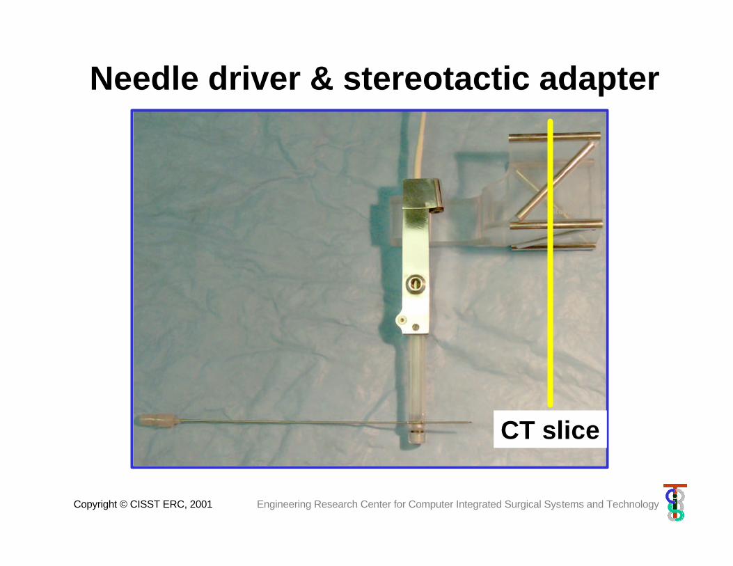
Needle driver & stereotactic adapter
CT slice
Copyright © CISST ERC, 2001 Engineering Research Center for Computer Integrated Surgical Systems and Technology
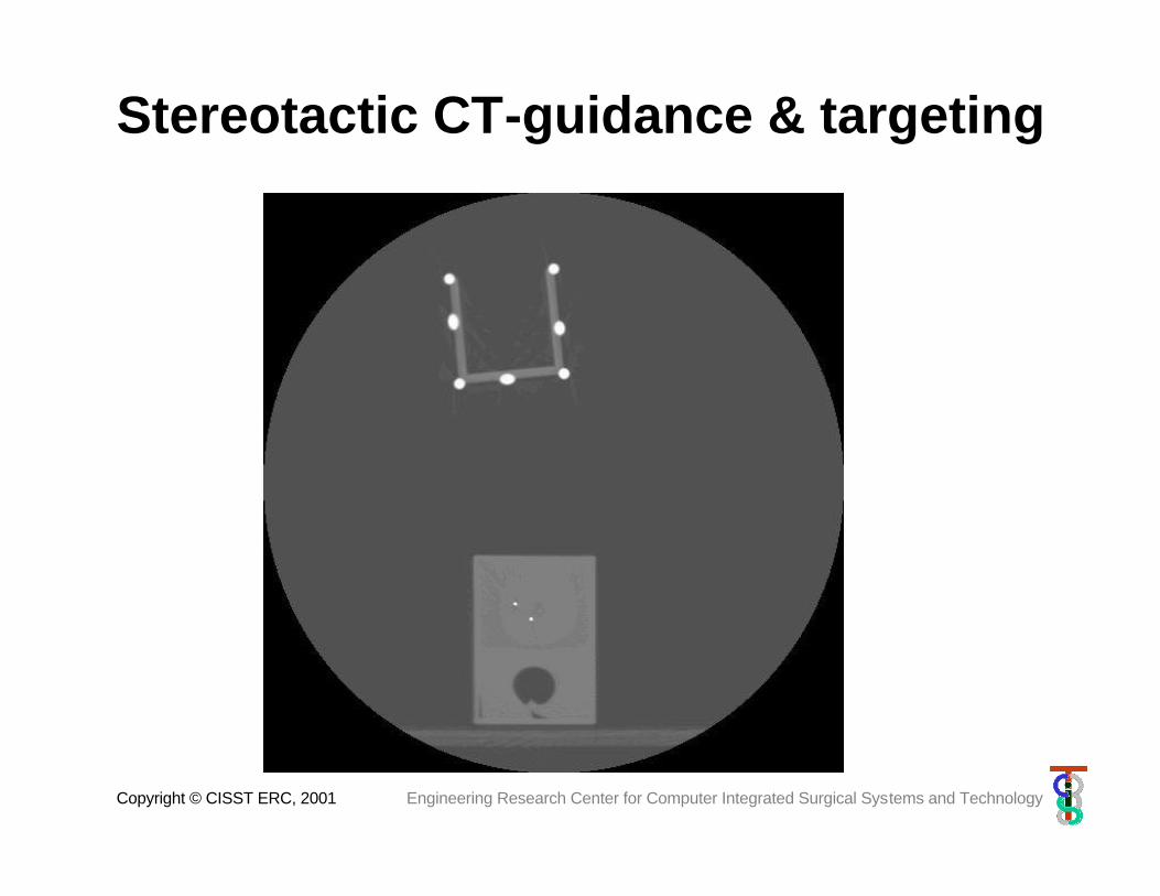
Stereotactic CT-guidance & targeting
Copyright © CISST ERC, 2001 Engineering Research Center for Computer Integrated Surgical Systems and Technology
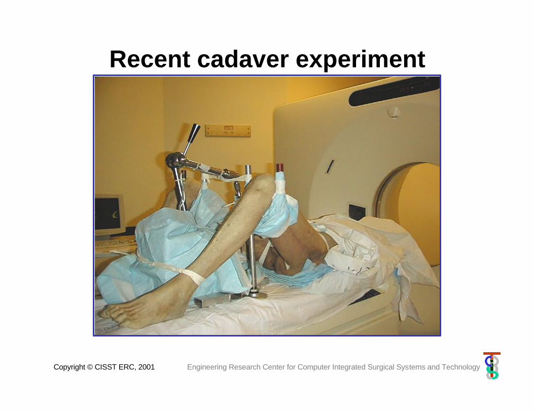
Recent cadaver experiment
Copyright © CISST ERC, 2001 Engineering Research Center for Computer Integrated Surgical Systems and Technology
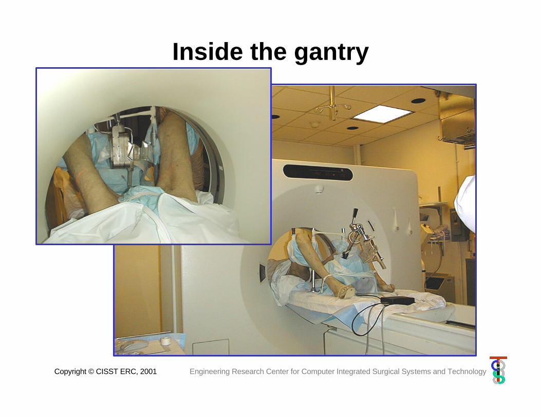
Inside the gantry

Copyright © CISST ERC, 2001 Engineering Research Center for Computer Integrated Surgical Systems and Technology
Targeting
Copyright © CISST ERC, 2001 Engineering Research Center for Computer Integrated Surgical Systems and Technology
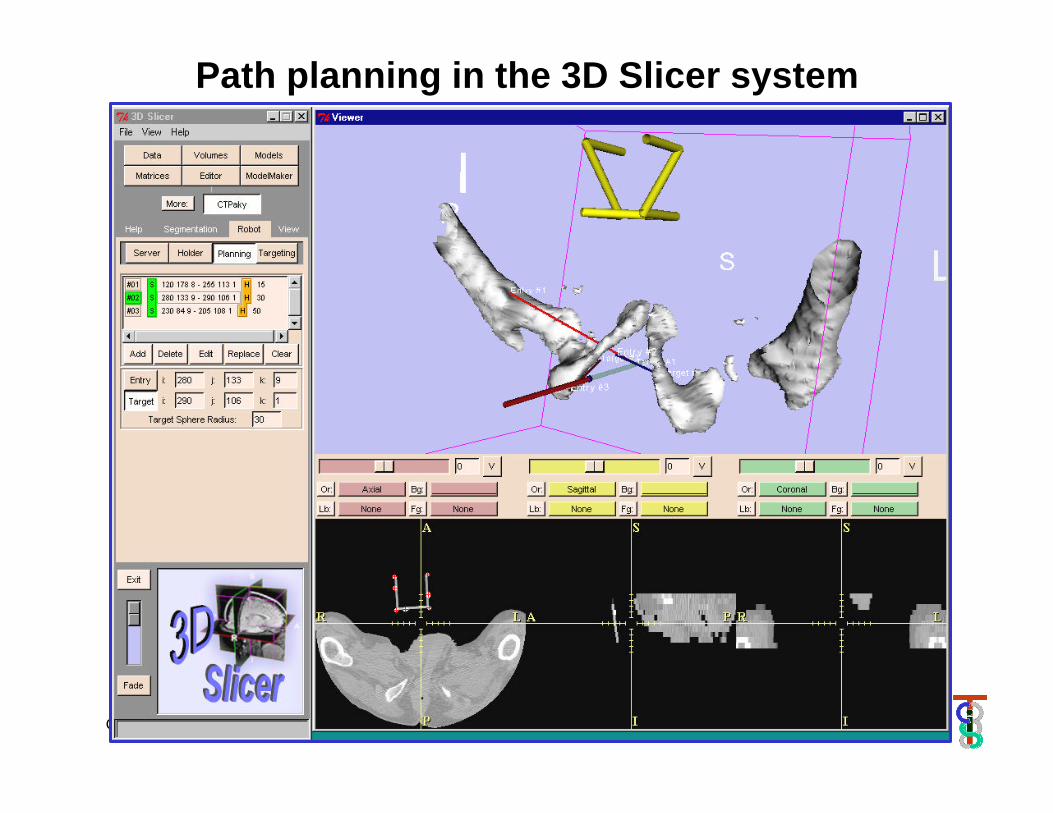
Path planning in the 3D Slicer system
Copyright © CISST ERC, 2001 Engineering Research Center for Computer Integrated Surgical Systems and Technology
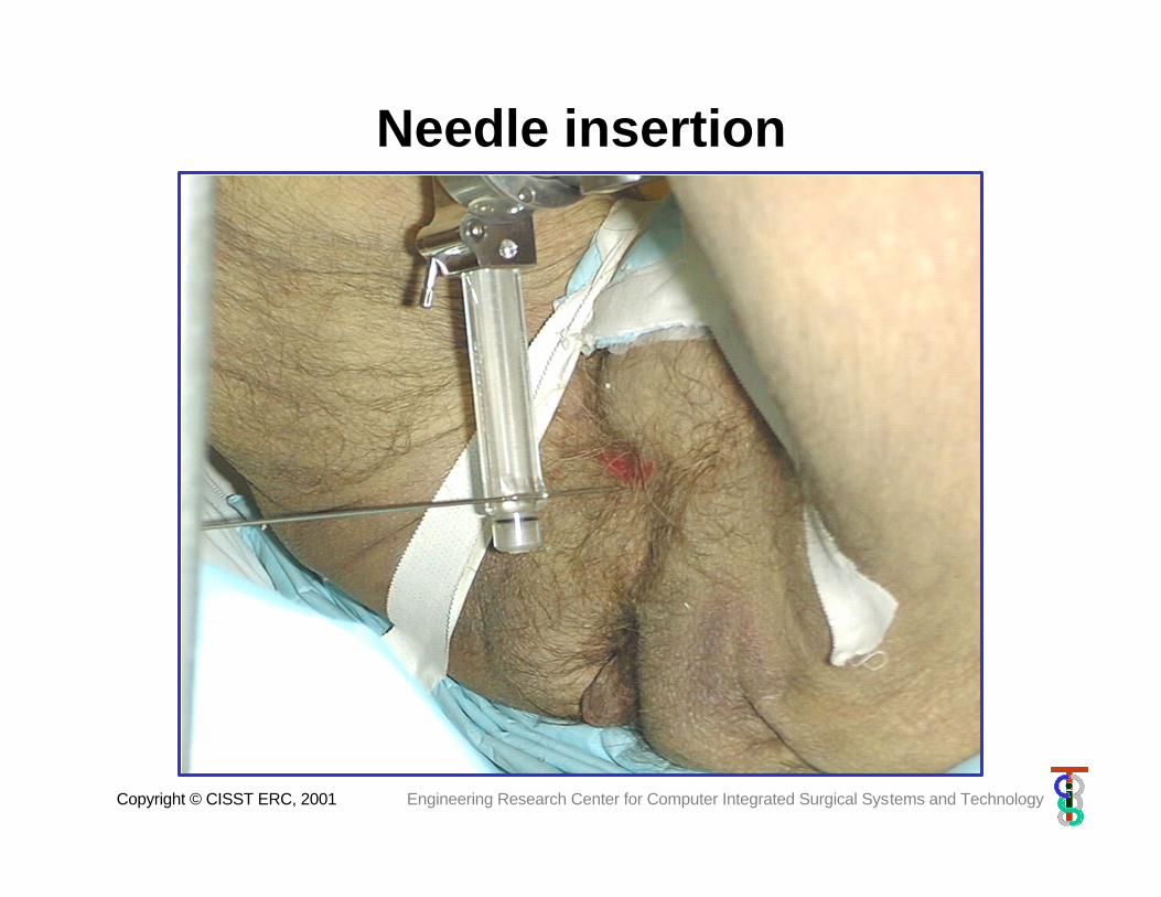
Needle insertion
Copyright © CISST ERC, 2001 Engineering Research Center for Computer Integrated Surgical Systems and Technology
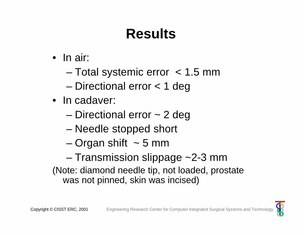
Results
• In air:– Total systemic error < 1.5 mm– Directional error < 1 deg
• In cadaver:– Directional error ~ 2 deg– Needle stopped short– Organ shift ~ 5 mm– Transmission slippage ~2-3 mm
(Note: diamond needle tip, not loaded, prostate was not pinned, skin was incised)
Copyright © CISST ERC, 2001 Engineering Research Center for Computer Integrated Surgical Systems and Technology
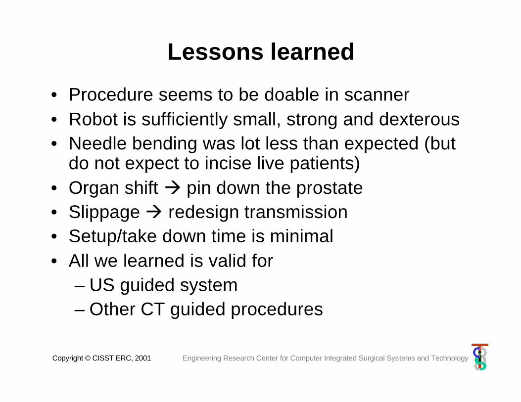
Lessons learned
• Procedure seems to be doable in scanner• Robot is sufficiently small, strong and dexterous• Needle bending was lot less than expected (but
do not expect to incise live patients)• Organ shift à pin down the prostate • Slippage à redesign transmission• Setup/take down time is minimal• All we learned is valid for
– US guided system– Other CT guided procedures
Copyright © CISST ERC, 2001 Engineering Research Center for Computer Integrated Surgical Systems and Technology
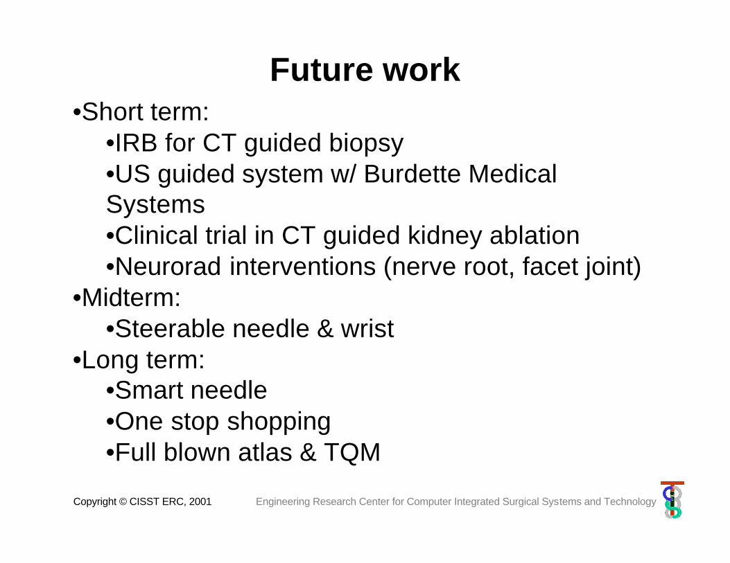
Future work•Short term:
•IRB for CT guided biopsy•US guided system w/ Burdette Medical Systems•Clinical trial in CT guided kidney ablation•Neurorad interventions (nerve root, facet joint)
•Midterm:•Steerable needle & wrist
•Long term:•Smart needle•One stop shopping•Full blown atlas & TQM
Copyright © CISST ERC, 2001 Engineering Research Center for Computer Integrated Surgical Systems and Technology
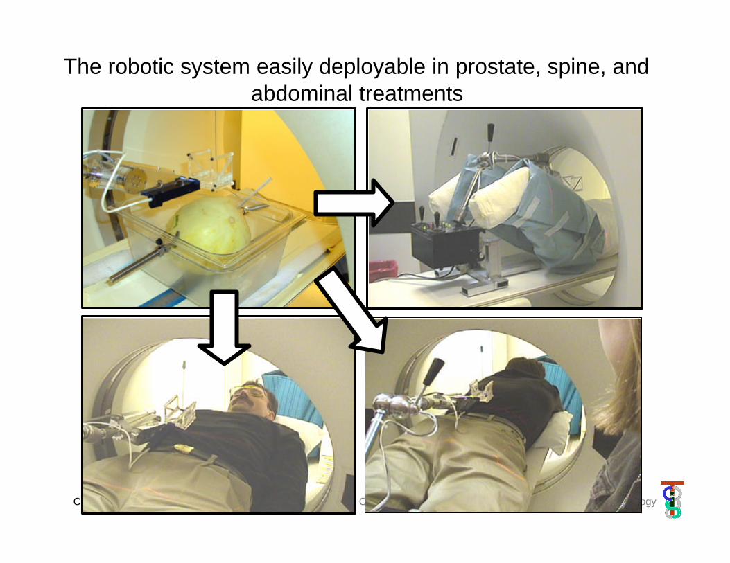
The robotic system easily deployable in prostate, spine, and abdominal treatments

Copyright © CISST ERC, 2001 Engineering Research Center for Computer Integrated Surgical Systems and Technology
Spinning needle driver (CAD drawing)
Remote Center Motion (RCM) robot to aim needle
Aluminum outer package
Radiolucent fixed arm
Radiolucent translating arm
Bi-directional translation motor
Bi-directional rotation motor
Non backlash transmission
Variable diameter, Independently controlled rotation and translation

Copyright © CISST ERC, 2001 Engineering Research Center for Computer Integrated Surgical Systems and Technology
Spinning needle driver prototype

Copyright © CISST ERC, 2001 Engineering Research Center for Computer Integrated Surgical Systems and Technology
Grateful acknowledgement
• National Science Foundation under grant #IIS9801684, the Engineering Research Center grant #EEC9731478
• Partially funded by AdMeTech, Faina Shtern, MD, President and CEO and sponsored by the Henry M. Jackson Foundation and the US Army Medical Research and Material Command
• Partial support by Johns Hopkins University internal funds
• Financial assistance of Whiting School of Engineering at the Johns Hopkins University
• Burdette Medical Systems & JCRT Harvard for images and background material