RNF4, a SUMO-targeted ubiquitin E3 ligase, …genesdev.cshlp.org/content/26/11/1179.full.pdfRNF4, a...
18
RNF4, a SUMO-targeted ubiquitin E3 ligase, promotes DNA double-strand break repair Yaron Galanty, Rimma Belotserkovskaya, Julia Coates, and Stephen P. Jackson 1 The Gurdon Institute, Department of Biochemistry, University of Cambridge, Cambridge CB2 1QN, United Kingdom Protein ubiquitylation and sumoylation play key roles in regulating cellular responses to DNA double-strand breaks (DSBs). Here, we show that human RNF4, a small ubiquitin-like modifier (SUMO)-targeted ubiquitin E3 ligase, is recruited to DSBs in a manner requiring its SUMO interaction motifs, the SUMO E3 ligases PIAS1 and PIAS4, and various DSB-responsive proteins. Furthermore, we reveal that RNF4 depletion impairs ubiquitin adduct formation at DSB sites and causes persistent histone H2AX phosphorylation (gH2AX) associated with defective DSB repair, hypersensitivity toward DSB-inducing agents, and delayed recovery from radiation-induced cell cycle arrest. We establish that RNF4 regulates turnover of the DSB-responsive factors MDC1 and replication protein A (RPA) at DNA damage sites and that RNF4-depleted cells fail to effectively replace RPA by the homologous recombination factors BRCA2 and RAD51 on resected DNA. Consistent with previous data showing that RNF4 targets proteins to the proteasome, we show that the proteasome component PSMD4 is recruited to DNA damage sites in a manner requiring its ubiquitin-interacting domains, RNF4 and RNF8. Finally, we establish that PSMD4 binds MDC1 and RPA1 in a DNA damage-induced, RNF4-dependent manner and that PSMD4 depletion cause MDC1 and gH2AX persistence in irradiated cells. RNF4 thus operates as a DSB response factor at the crossroads between the SUMO and ubiquitin systems. [Keywords: RNF4; SUMO; ubiquitin–proteasome system (UPS); DNA double-strand breaks (DSBs); RPA; RAD51] Supplemental material is available for this article. Received January 26, 2012; revised version accepted April 30, 2012. DNA double-strand breaks (DSBs) are generated by ion- izing radiation (IR) and various DNA-damaging chem- icals. If they are not repaired or are repaired incorrectly, DSBs result in cell death or genomic instability that can lead to immune deficiencies, neurodegeneration, prema- ture aging, and cancer (Jackson and Bartek 2009; Ciccia and Elledge 2010). DSB formation triggers activation of the DNA damage response (DDR) protein kinases ATM, ATR, and DNA-PK, which phosphorylate many protein targets, including the histone variant H2AX (H2AFX). Phosphorylated H2AX (gH2AX) then mediates the accu- mulation of DDR proteins such as MDC1 (NFBD1), 53BP1 (TP53BP1), and BRCA1 into IR-induced foci (IRIF) that are thought to amplify DSB signaling and promote DSB repair (Downs et al. 2007; Bekker-Jensen and Mailand 2010; Ciccia and Elledge 2010; Polo and Jackson 2011). DSBs are repaired by two principal pathways: non- homologous end-joining (NHEJ) that is active throughout the cell cycle, and homologous recombination (HR) that is normally restricted to S and G2 cells (Hartlerode and Scully 2009; Pardo et al. 2009). HR but not NHEJ relies on the presence of a sister chromatid and also requires cell cycle-regulated 59-to-39 exonucleolytic processing (resec- tion) of DNA ends that generates stretches of ssDNA. This ssDNA is bound by replication protein A (RPA), which is then replaced by RAD51 to produce a RAD51- ssDNA nucleoprotein filament, promoting DNA strand invasion and subsequent HR events. A key protein player in the essential step of replacing RPA with RAD51 is BRCA2, which directly binds RAD51 and promotes RAD51 loading onto RPA-coated ssDNA (Liu et al. 2010; Thorslund et al. 2010; Holloman 2011). Notably, RPA-coated ssDNA also leads to recruitment and activa- tion of the checkpoint kinase ATR, which phosphorylates various targets, including the downstream checkpoint kinase CHK1 (Cortez et al. 2001; Zou and Elledge 2003; Cimprich and Cortez 2008). Recent work has revealed that resection is promoted by various factors, including human CtIP (RBBP8) (Sartori et al. 2007), and has in- dicated how resection is subject to cell cycle control (Sartori et al. 2007; Gravel et al. 2008; Nimonkar et al. 2008, 2011; Huertas 2010). However, it remains to be determined specifically how the activities of the various 1 Corresponding author. Email [email protected]. Article is online at http://www.genesdev.org/cgi/doi/10.1101/gad. 188284.112. GENES & DEVELOPMENT 26:1179–1195 Ó 2012 by Cold Spring Harbor Laboratory Press ISSN 0890-9369/12; www.genesdev.org 1179 Cold Spring Harbor Laboratory Press on April 25, 2019 - Published by genesdev.cshlp.org Downloaded from
Transcript of RNF4, a SUMO-targeted ubiquitin E3 ligase, …genesdev.cshlp.org/content/26/11/1179.full.pdfRNF4, a...

RNF4, a SUMO-targeted ubiquitin E3ligase, promotes DNA double-strandbreak repair
Yaron Galanty, Rimma Belotserkovskaya, Julia Coates, and Stephen P. Jackson1
The Gurdon Institute, Department of Biochemistry, University of Cambridge, Cambridge CB2 1QN, United Kingdom
Protein ubiquitylation and sumoylation play key roles in regulating cellular responses to DNA double-strandbreaks (DSBs). Here, we show that human RNF4, a small ubiquitin-like modifier (SUMO)-targeted ubiquitin E3ligase, is recruited to DSBs in a manner requiring its SUMO interaction motifs, the SUMO E3 ligases PIAS1 andPIAS4, and various DSB-responsive proteins. Furthermore, we reveal that RNF4 depletion impairs ubiquitinadduct formation at DSB sites and causes persistent histone H2AX phosphorylation (gH2AX) associated withdefective DSB repair, hypersensitivity toward DSB-inducing agents, and delayed recovery from radiation-inducedcell cycle arrest. We establish that RNF4 regulates turnover of the DSB-responsive factors MDC1 and replicationprotein A (RPA) at DNA damage sites and that RNF4-depleted cells fail to effectively replace RPA by thehomologous recombination factors BRCA2 and RAD51 on resected DNA. Consistent with previous data showingthat RNF4 targets proteins to the proteasome, we show that the proteasome component PSMD4 is recruited toDNA damage sites in a manner requiring its ubiquitin-interacting domains, RNF4 and RNF8. Finally, we establishthat PSMD4 binds MDC1 and RPA1 in a DNA damage-induced, RNF4-dependent manner and that PSMD4depletion cause MDC1 and gH2AX persistence in irradiated cells. RNF4 thus operates as a DSB response factor atthe crossroads between the SUMO and ubiquitin systems.
[Keywords: RNF4; SUMO; ubiquitin–proteasome system (UPS); DNA double-strand breaks (DSBs); RPA; RAD51]
Supplemental material is available for this article.
Received January 26, 2012; revised version accepted April 30, 2012.
DNA double-strand breaks (DSBs) are generated by ion-izing radiation (IR) and various DNA-damaging chem-icals. If they are not repaired or are repaired incorrectly,DSBs result in cell death or genomic instability that canlead to immune deficiencies, neurodegeneration, prema-ture aging, and cancer (Jackson and Bartek 2009; Cicciaand Elledge 2010). DSB formation triggers activation ofthe DNA damage response (DDR) protein kinases ATM,ATR, and DNA-PK, which phosphorylate many proteintargets, including the histone variant H2AX (H2AFX).Phosphorylated H2AX (gH2AX) then mediates the accu-mulation of DDR proteins such as MDC1 (NFBD1), 53BP1(TP53BP1), and BRCA1 into IR-induced foci (IRIF) that arethought to amplify DSB signaling and promote DSB repair(Downs et al. 2007; Bekker-Jensen and Mailand 2010;Ciccia and Elledge 2010; Polo and Jackson 2011).
DSBs are repaired by two principal pathways: non-homologous end-joining (NHEJ) that is active throughoutthe cell cycle, and homologous recombination (HR) that
is normally restricted to S and G2 cells (Hartlerode andScully 2009; Pardo et al. 2009). HR but not NHEJ relies onthe presence of a sister chromatid and also requires cellcycle-regulated 59-to-39 exonucleolytic processing (resec-tion) of DNA ends that generates stretches of ssDNA.This ssDNA is bound by replication protein A (RPA),which is then replaced by RAD51 to produce a RAD51-ssDNA nucleoprotein filament, promoting DNA strandinvasion and subsequent HR events. A key protein playerin the essential step of replacing RPA with RAD51 isBRCA2, which directly binds RAD51 and promotesRAD51 loading onto RPA-coated ssDNA (Liu et al.2010; Thorslund et al. 2010; Holloman 2011). Notably,RPA-coated ssDNA also leads to recruitment and activa-tion of the checkpoint kinase ATR, which phosphorylatesvarious targets, including the downstream checkpointkinase CHK1 (Cortez et al. 2001; Zou and Elledge 2003;Cimprich and Cortez 2008). Recent work has revealedthat resection is promoted by various factors, includinghuman CtIP (RBBP8) (Sartori et al. 2007), and has in-dicated how resection is subject to cell cycle control(Sartori et al. 2007; Gravel et al. 2008; Nimonkar et al.2008, 2011; Huertas 2010). However, it remains to bedetermined specifically how the activities of the various
1Corresponding author.Email [email protected] is online at http://www.genesdev.org/cgi/doi/10.1101/gad.188284.112.
GENES & DEVELOPMENT 26:1179–1195 � 2012 by Cold Spring Harbor Laboratory Press ISSN 0890-9369/12; www.genesdev.org 1179
Cold Spring Harbor Laboratory Press on April 25, 2019 - Published by genesdev.cshlp.orgDownloaded from

HR-promoting factors are controlled and precisely howother HR events, such as the transition from RPA-ssDNAto RAD51-ssDNA, are mediated.
The reversible, covalent attachment of ubiquitin andsmall ubiquitin-like modifier (SUMO) proteins to DDRfactors is critical for effective DSB repair and signaling ineukaryotic cells (Bergink and Jentsch 2009; Al-Hakimet al. 2010; Bekker-Jensen and Mailand 2010; Morris2010a,b). For example, ubiquitin conjugates and ubiquitin-conjugating enzymes such as UBC13 (UBE2N), BRCA1,RNF8, HERC2, RNF168, RAD18, and the Fanconi ane-mia (FA) protein complex accumulate at DSB sites inmammalian cells, and defects in these factors result inimpaired DSB repair and signaling associated with hy-persensitivity toward DNA-damaging agents (Huen et al.2007; Kolas et al. 2007; Mailand et al. 2007; Wang andElledge 2007; Zhao et al. 2007; Bekker-Jensen et al. 2009;Doil et al. 2009; Huang et al. 2009; Stewart et al. 2009;Watanabe et al. 2009; Kee and D’Andrea 2010; Yang et al.2010). While the ubiquitin–proteasome system (UPS)was originally described as the main protein turnover/degradation machinery of eukaryotic cells, it is nowevident that it is also a major nondegradative regulatorof various cellular processes, including the DDR (Kirkinand Dikic 2007; Bergink and Jentsch 2009; Motegi et al.2009; Al-Hakim et al. 2010). Furthermore, it has beenshown that proteasomes accumulate on damaged chro-matin, suggesting that they may promote DDR signalingas well as DDR-dependent protein turnover and HR(Ustrell et al. 2002; Blickwedehl et al. 2007; Jacquemontand Taniguchi 2007; Murakawa et al. 2007; Shi et al.2008; Motegi et al. 2009; Al-Hakim et al. 2010; Ben-Aroya et al. 2010; Levy-Barda et al. 2011). The kineticsand mechanisms governing proteasome accumulationat DNA damage regions and the functions and targetsof proteasomes at such sites, however, remain largelyobscure.
It was recently established that modification of thelargest RPA subunit (RPA1) by SUMO2/3 promotes theinteraction of RPA with RAD51 and HR-mediated DSBrepair (Dou et al. 2010). Furthermore, we and othershave established that SUMO conjugates and the SUMO-conjugating enzymes UBC9 (UBE2I), PIAS1 (protein inhib-itor of activated STAT 1), and PIAS4 (PIASy) accumulateat DSB sites and are required for effective DSB signalingand repair (Galanty et al. 2009; Morris et al. 2009). Thiswork also revealed that PIAS1- and PIAS4-mediatedsumoylation promotes accumulation of 53BP1 and ef-fective RNF8-, RNF168-, and BRCA1-dependent accu-mulation of ubiquitin conjugates at DSB sites and thatPIAS1/4-mediated sumoylation directly promotes BRCA1E3 ubiquitin ligase activity (Galanty et al. 2009; Morriset al. 2009). Thus, rather than operating separately, DDR–protein sumoylation and ubiquitylation are intimatelyinterconnected.
An additional link between ubiquitylation and sumoy-lation has been established through work on the mam-malian RNF4 protein and its yeast counterparts (Slx5/Slx8 in Saccharomyces cerevisiae and Slx8/Rfp1 orRfp2 in Schizosaccharomyces pombe) (Prudden et al.
2007; Sun et al. 2007). These proteins act as SUMO-targeted ubiquitin E3 ligases (STUbLs), which recognizesumoylated or SUMO-like domain-containing proteintargets. For example, sumoylation of the acute promye-locytic leukemia (PML) protein triggers its recognitionby RNF4, which then mediates PML ubiquitylation andproteasome-dependent PML degradation (Lallemand-Breitenbach et al. 2008; Tatham et al. 2008; Weisshaaret al. 2008). Since various DDR components are sumoylatedand because RNF4 counterparts in yeast are implicatedin suppressing HR via an as-yet-undefined mechanism(Burgess et al. 2007; Kosoy et al. 2007; Prudden et al.2007), we investigated whether human RNF4 functionsin the DDR. Indeed, as described below, we show thatRNF4 accumulates at DSBs, regulates protein turnover/exchange at these sites, and is required for effective DSBrepair.
Results
SUMO-interacting motif (SIM)-dependentaccumulation of RNF4 at DSB sites
To investigate potential DDR functions for RNF4, wefirst determined whether it accumulates at DNA damagesites. Although we could not detect RNF4 recruitment toIRIF, by using both live-cell imaging and fixed-cell stain-ing, we found that endogenous RNF4 (Fig. 1A,B) andrecombinant RNF4 (Fig. 1C,D; Supplemental Fig. S1A,B)accumulated at DNA damage sites induced by lasermicro-irradiation. RNF4 accrual was weakly detectablewithin 15 min after irradiation (data not shown) butbecame readily apparent after 30 min (Fig. 1D) and thenpersisted for up to several hours (note that Fig. 1A–Cdisplays cells 2 h after laser micro-irradiation; Supple-mental Fig. S1B; data not shown). This persistence ofRNF4 recruitment within laser-micro-irradiated regionssuggested that RNF4 might function at DSBs, which inmany cases take several hours to repair. Consistent withthis idea, RNF4 depletion by either of two distinctsiRNAs enhanced the persistence of SUMO proteinsand the well-defined DSB marker gH2AX at micro-irradiated sites (Fig. 1A,B). Furthermore, effective RNF4accrual at DNA damage sites required the DSB-responsiveproteins MDC1, RNF8, 53BP1, and BRCA1 (Fig. 1C; Sup-plemental Fig. S1A, numbers represent the proportion ofcells displaying gH2AX recruitment that also displayedRNF4 recruitment), with RNF4 recruitment being mostmarkedly diminished when several of these factors werecodepleted (see Supplemental Fig. S1A for effects ofindividual factor depletions). These data therefore sug-gested that RNF4 might be targeted to DNA damagesites, most likely DSBs, by interactions with multipleDDR proteins.
Because RNF4 contains SIMs (Fig. 1E) and since theSUMO E3 ligases PIAS1 and PIAS4 mediate SUMOaccrual at DSBs (Galanty et al. 2009; Morris et al. 2009),we tested whether PIAS1/4 depletion affected RNF4recruitment to laser-induced DNA damage. Indeed,RNF4 accrual was essentially abolished by codepleting
Galanty et al.
1180 GENES & DEVELOPMENT
Cold Spring Harbor Laboratory Press on April 25, 2019 - Published by genesdev.cshlp.orgDownloaded from

PIAS1 and PIAS4 (Fig. 1C, bottom row; SupplementalFig. S1A). Notably, efficient accumulation of RNF4 alsorequired its four tandem SIMs but was not impaired bymutating its RING finger ubiquitin E3 ligase domain toyield an E3 ligase-dead (LD) protein (Fig. 1D; Supple-mental Fig. S1B). Collectively, these findings indicatedthat PIAS-mediated sumoylation of DDR proteins at DSBsites mediates SIM-dependent recruitment of RNF4 tothese regions.
RNF4 depletion impairs ubiquitin accrual but causesMDC1 and gH2AX persistence at DNA damage sites
Previous work has shown that RNF4 functions as a ubiq-uitin E3 ligase in a manner that requires its RING fingerdomain (Hakli et al. 2004). Consistent with RNF4 func-tioning in this way at DNA damage sites, RNF4 depletionsignificantly reduced the production of ubiquitin adductswithin IRIF, as detected by the FK2 antibody (Figs. 2A, 5E½below�). Moreover, complementation experiments withstable cell lines expressing siRNA-resistant RNF4 de-rivatives revealed that wild-type but not RING finger-mutated (LD) RNF4 corrected the impaired ubiquitinadduct staining caused by siRNA-mediated depletionof endogenous RNF4 (Fig. 2B; Supplemental Fig. S2A).In line with our data on laser-micro-irradiated cells
(Fig. 1A,B), RNF4 depletion also caused the persistenceof gH2AX staining in IRIF (Fig. 2A). Furthermore, RNF4depletion also markedly affected the gH2AX-bindingfactor MDC1 (Stucki et al. 2005), whose IRIF stainingwas substantially enhanced in RNF4-depleted cellscompared with control cells, particularly at later timepoints (Fig. 2A, numbers represent the proportion ofcells displaying detectable IRIF). As these effects of RNF4depletion on MDC1 and gH2AX were complemented bywild-type but not RING finger-mutated RNF4 (Fig. 2C;Supplemental Fig. S2B), these findings suggested thatimpaired RNF4-mediated ubiquitylation leads to gH2AXand MDC1 persistence at DSB sites, possibly reflectingdefective DSB repair.
RNF4 promotes DSB repair
In accord with a model in which RNF4 promotes DNArepair, we found that RNF4 depletion reduced rates ofchromosomal DSB repair, as measured by neutral cometassays (Fig. 3A; Supplemental Fig. S3). Furthermore, theseeffects of RNF4 depletion on DSB repair, as measured bycomet assay, were complemented by reintroducing wild-type but not RING finger-mutated or SIM-deleted RNF4into RNF4-depleted cells (Fig. 3B; Supplemental Fig. S4).To see which DSB repair pathways were affected by RNF4
Figure 1. RNF4 accumulation at DNA damage sitesrequires its SIM domains, PIAS1, PIAS4, and DDRproteins. (A) U2OS cells stably expressing RFP-SUMO1and transfected with the indicated siRNAs were laser-micro-irradiated, fixed after 2 h, and then analyzed byimmunofluorescence. (B) As in A, but with cells express-ing RFP-SUMO2. (C) Experiments with U2OS cellsstably expressing YFP-RNF4 were performed as in A.For quantifications, numbers represent proportions ofcells showing RNF4 accumulation out of gH2AX-positivecells 6SED (n > 100). Single depletions and their quan-tifications are presented in Supplemental Figure S1A.(D) U2OS cells stably expressing YFP-RNF4 wild-type(WT), LD, or SIM-deleted (DSIM) were micro-irradiatedand live-imaged after 30 min. Additional time pointsare presented in Supplemental Figure S1B. For siRNAdepletions, see Supplemental Figure S11, A and C. (E)Schematic of RNF4 structure. Bars: this and all otherfigures, 10 mm.
RNF4 promotes DSB repair
GENES & DEVELOPMENT 1181
Cold Spring Harbor Laboratory Press on April 25, 2019 - Published by genesdev.cshlp.orgDownloaded from

depletion, we assessed DSB repair by NHEJ or HR individ-ually. Thus, by using random plasmid integration intogenomic DNA as a measure of NHEJ (Stucki et al. 2005),we found that this pathway was markedly impaired byRNF4 depletion (Fig. 3C, note that similar effects wereobserved with two different siRNAs targeting RNF4 andthat depletion of the NHEJ component XRCC4 served asa positive control). Furthermore, RAD51-mediated repairof chromosomal DSBs by HR (Pierce et al. 2001) was alsoimpaired by RNF4 depletion (Fig. 3D, depletion of theHR-promoting factor CtIP served as a positive control).Importantly, these effects of RNF4 depletion on HR didnot reflect indirect effects on cell cycle progressionbecause flow cytometry analyses revealed that cellcycle profiles of RNF4-depleted cell populations wereessentially equivalent to those of control cells (Figs. 3F, 4B).Furthermore, RNF4-depleted cells progressed efficientlyinto and through S phase in the absence of DNA damage,as measured by quantifying incorporation of the thymi-dine analog ethynyl deoxyuridine (EdU) by pulse labeling(Fig. 3E).
Strikingly, while flow cytometry revealed that RNF4-depleted cells effectively mediated a G2–M cell cyclecheckpoint arrest after IR treatment (Fig. 3F), theyreproducibly displayed a pronounced delay in check-point recovery, as evidenced by hindered resumptionof cell cycle progression (cf. the red-boxed profiles inFig. 3F). These data, combined with the fact that RNF4depletion caused persistent MDC1 and gH2AX accu-mulation at DSB sites, suggested that defective DSBrepair in RNF4-depleted cells causes persistent DNAdamage checkpoint activation. In accord with theimpact of RNF4 on NHEJ and HR, clonogenic cellsurvival assays established that RNF4 depletion causedhypersensitivity to IR, which kills cells primarilythrough generating DSBs (Fig. 3G), and to chronichydroxyurea (HU) treatment, which causes replicationfork stalling and also yields some DSBs that are repairedby HR (Fig. 3H) (we note that the hypersensitivity ofRNF4-depleted cells to HU could also partially reflectroles for RNF4 in promoting responses to replicationfork stalling).
Figure 2. RNF4 depletion causes persistence of MDC1and gH2AX foci. (A) U2OS cells stably expressing GFP-MDC1 were transfected with the indicated siRNAs,exposed to 2 Gy of IR, fixed after the indicated times,and analyzed by immunofluorescence. Quantificationnumbers represent proportions of cells showing Ub/FK2, gH2AX, or MDC1 foci 6SED (n > 100). (B,C) Cellsstably expressing siRNA-resistant (siR) rat YFP-RNF4wild type (WT) or LD or vector only were transfectedwith RNF4 siRNAs, exposed to 2 Gy of IR, fixed 4 hlater, and processed as in A. MDC1 detection was withan anti-pSDTD-MDC1 antibody (Chapman and Jackson2008) that detects constitutively casein kinase 2 phos-phorylated MDC1. Quantifications for Ub/FK2 (B) andMDC1 and gH2AX (C) were done as in A. (Right panel)Corresponding samples were collected for immunoblot-ting. For additional time points, siRNA depletions, andresistant clones, see Supplemental Figures S2, A and B,and S11, A and B.
Galanty et al.
1182 GENES & DEVELOPMENT
Cold Spring Harbor Laboratory Press on April 25, 2019 - Published by genesdev.cshlp.orgDownloaded from

Figure 3. RNF4 promotes DSB repair, recovery from IR-induced G2 checkpoint arrest, and cell survival following genotoxic stress. (A)DSB repair is impaired in RNF4-depleted cells. U2OS cells were transfected with siRNAs and, 48 h following siRNA transfection,exposed to 10 Gy of IR, harvested at the indicated times, and subjected to neutral comet assays; tail moment quantifications arepresented in the histogram (n > 150 measurements accumulated over three independent experiments; error bars, 6SED), (NT) Cells nottreated with IR. Note that the total amount of DNA damage present in the cells immediately after irradiation was not significantlydifferent between control and RNF4-depeleted cells. See Supplemental Figure S3 for representative images. (B) U2OS cells stablyexpressing vector only or siRNA-resistant YFP-RNF4 wild type, LD, or DSIM were transfected with siRNAs and, 48 h later, exposed to10 Gy of IR, harvested at the indicated times, and subjected to neutral comet assays. Tail moment quantifications are presented. SeeSupplemental Figure S4 for representative images and expression levels of siRNA-resistant RNF4 derivatives. (C,D) Effects of RNF4depletion on NHEJ (C) or HR-mediated gene conversion (D); error bars, 6SED. (E) S-phase index of control and RNF4-depleted cells asmeasured by pulse EdU incorporation. (F) Cell cycle profiles of irradiated and nonirradiated control and RNF4-depleted cells. U2OScells were transfected with siRNAs and, 48 h following siRNA transfection, exposed to 2 Gy of IR, harvested at the indicated times, andsubjected to flow cytometry (16- and 24-h time points following irradiation are boxed in red). (G,H) Effects of RNF4 depletion on colonyformation following exposure to IR (G) or HU (H); error bars, 6SED; data represent four independent experiments. For siRNAdepletions, see Supplemental Figure S11, A and C.
RNF4 promotes DSB repair
GENES & DEVELOPMENT 1183
Cold Spring Harbor Laboratory Press on April 25, 2019 - Published by genesdev.cshlp.orgDownloaded from

RNF4 promotes RPA turnover and RAD51 loadingon ssDNA
As RNF4 was required for effective HR, we investigatedwhether RNF4 depletion influenced the accumulation ofHR components at DNA damage sites. To do this, weemployed laser micro-irradiation conditions in whichDNA damage-induced accumulation of HR-promotingfactors such as CtIP, RPA, BRCA2, and RAD51 onlyoccurred in cyclinA-positive cells (Fig. 4A, note thatCtIP, RPA2, and RAD51 accumulate at DNA damagesites in the S/G2 cell exhibiting pan-nuclear cyclinA½CycA� staining but not in the cell lacking detectablecyclinA, marked with an arrow). While RNF4 depletiondid not appreciably affect CtIP recruitment to DNA
damage sites in S/G2 cells (Fig. 4A, top right row), unlikethe situation in control cells, CtIP was also recruited toDNA damage sites in some RNF4-depleted cells thatdisplayed weak or no cyclinA staining (Fig. 4A, top rightrow). Similarly, accumulation of the second-largest sub-unit of RPA (RPA2) at sites of laser damage, reflectingthe resection of DSBs to produce ssDNA, took placeeffectively in RNF4-depleted cells, but unlike in controlcells, this occurred not only in cyclinA-positive cells,but also in a subset of cyclinA-negative cells (Fig. 4A,middle rows; Supplemental Fig. S5A). Importantly, onlysome (;20%) cyclinA-negative, RNF4-depleted cellsexhibited RPA2 or CtIP recruitment (see quantificationtables in Fig. 4A; also note that Fig. 4A and Supplemen-tal Fig. S5A only display examples of cyclinA-negative
Figure 4. RNF4 regulates RPA turnover andBRCA2-mediated RAD51 loading onto damagedDNA. (A) RNF4 is required for normal accumulationof CtIP and RPA2 and for efficient accumulation ofRAD51 at DNA damage sites. U2OS cells trans-fected with siRNAs were laser-micro-irradiated,fixed after 2 h, and then analyzed by immunoflu-orescence as indicated; arrows mark cyclinA(CycA)-negative cells. For additional images ofRPA staining and cyclinA immunoblotting incontrol and RNF4-depleted cells, see Supplemen-tal Figure S5A. Quantification numbers representproportions of cells showing CtIP or RPA2 accu-mulation out of gH2AX- or cyclinA-positive cells6SED (n > 100). (Note that the staining of RPA2and CtIP in cyclinA-negative cells is unlikely to beleft over from the previous G2, as these cellswould have to have been lasered/damaged inS/G2 and would then have had to progress throughmitosis with high amounts of damage even thoughthey possess an intact G2/M checkpoint ½see Fig. 3F�,all within the 2-h incubation step of this experi-ment.) (B) U2OS cells stably expressing GFP-RPA1(wild-type ½WT�) were transfected with siRNAsand laser-micro-irradiated 48 h later (to allowsubsequent quantification, the numbers of cellslasered were documented). GFP-RPA1 lines werelive-imaged and counted 40 min post-irradiation.Proportions of cells displaying GFP-RPA1 lines outof total cells irradiated are presented 6SED withthe corresponding flow cytometry profiles (n > 150cells per siRNA, accumulated over three indepen-dent experiments). (C) RNF4 is required of effi-cient accumulation of BRCA2 at DNA-damagedsites. U2OS cells stably expressing GFP-RPA1were treated as in A and stained with the indicatedantibodies. For additional images of RAD51 andBRCA2 accumulation in GFP-RPA1 control andRNF4-depleted cells, see Supplemental FigureS5, B and C. (D) Quantifications of RAD51-and BRCA2-positive cells in GFP-RPA1-posi-tive control and RNF4-deleted cells (n $ 100cells per siRNA, 6SED). (E,F) U2OS cells stablyexpressing GFP-RPA1 wild-type (WT) or GFP-
RPA1K449R,K577R (SM) were transfected with siRNAs and, 48 h later, laser-micro-irradiated and subjected to FRAP analysis 40 minpost-irradiation (n = 24 independent measurements; error bars, 6SED). For equations and calculations, see the Materials and Methods.For siRNA depletions, see Supplemental Figure S11A.
Galanty et al.
1184 GENES & DEVELOPMENT
Cold Spring Harbor Laboratory Press on April 25, 2019 - Published by genesdev.cshlp.orgDownloaded from
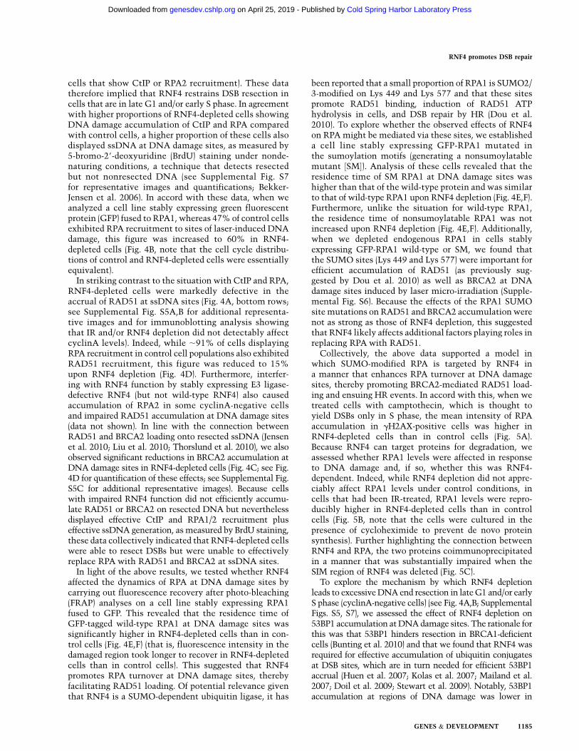
cells that show CtIP or RPA2 recruitment). These datatherefore implied that RNF4 restrains DSB resection incells that are in late G1 and/or early S phase. In agreementwith higher proportions of RNF4-depleted cells showingDNA damage accumulation of CtIP and RPA comparedwith control cells, a higher proportion of these cells alsodisplayed ssDNA at DNA damage sites, as measured by5-bromo-29-deoxyuridine (BrdU) staining under nonde-naturing conditions, a technique that detects resectedbut not nonresected DNA (see Supplemental Fig. S7for representative images and quantifications; Bekker-Jensen et al. 2006). In accord with these data, when weanalyzed a cell line stably expressing green fluorescentprotein (GFP) fused to RPA1, whereas 47% of control cellsexhibited RPA recruitment to sites of laser-induced DNAdamage, this figure was increased to 60% in RNF4-depleted cells (Fig. 4B, note that the cell cycle distribu-tions of control and RNF4-depleted cells were essentiallyequivalent).
In striking contrast to the situation with CtIP and RPA,RNF4-depleted cells were markedly defective in theaccrual of RAD51 at ssDNA sites (Fig. 4A, bottom rows;see Supplemental Fig. S5A,B for additional representa-tive images and for immunoblotting analysis showingthat IR and/or RNF4 depletion did not detectably affectcyclinA levels). Indeed, while ;91% of cells displayingRPA recruitment in control cell populations also exhibitedRAD51 recruitment, this figure was reduced to 15%upon RNF4 depletion (Fig. 4D). Furthermore, interfer-ing with RNF4 function by stably expressing E3 ligase-defective RNF4 (but not wild-type RNF4) also causedaccumulation of RPA2 in some cyclinA-negative cellsand impaired RAD51 accumulation at DNA damage sites(data not shown). In line with the connection betweenRAD51 and BRCA2 loading onto resected ssDNA (Jensenet al. 2010; Liu et al. 2010; Thorslund et al. 2010), we alsoobserved significant reductions in BRCA2 accumulation atDNA damage sites in RNF4-depleted cells (Fig. 4C; see Fig.4D for quantification of these effects; see Supplemental Fig.S5C for additional representative images). Because cellswith impaired RNF4 function did not efficiently accumu-late RAD51 or BRCA2 on resected DNA but neverthelessdisplayed effective CtIP and RPA1/2 recruitment pluseffective ssDNA generation, as measured by BrdU staining,these data collectively indicated that RNF4-depleted cellswere able to resect DSBs but were unable to effectivelyreplace RPA with RAD51 and BRCA2 at ssDNA sites.
In light of the above results, we tested whether RNF4affected the dynamics of RPA at DNA damage sites bycarrying out fluorescence recovery after photo-bleaching(FRAP) analyses on a cell line stably expressing RPA1fused to GFP. This revealed that the residence time ofGFP-tagged wild-type RPA1 at DNA damage sites wassignificantly higher in RNF4-depleted cells than in con-trol cells (Fig. 4E,F) (that is, fluorescence intensity in thedamaged region took longer to recover in RNF4-depletedcells than in control cells). This suggested that RNF4promotes RPA turnover at DNA damage sites, therebyfacilitating RAD51 loading. Of potential relevance giventhat RNF4 is a SUMO-dependent ubiquitin ligase, it has
been reported that a small proportion of RPA1 is SUMO2/3-modified on Lys 449 and Lys 577 and that these sitespromote RAD51 binding, induction of RAD51 ATPhydrolysis in cells, and DSB repair by HR (Dou et al.2010). To explore whether the observed effects of RNF4on RPA might be mediated via these sites, we establisheda cell line stably expressing GFP-RPA1 mutated inthe sumoylation motifs (generating a nonsumoylatablemutant ½SM�). Analysis of these cells revealed that theresidence time of SM RPA1 at DNA damage sites washigher than that of the wild-type protein and was similarto that of wild-type RPA1 upon RNF4 depletion (Fig. 4E,F).Furthermore, unlike the situation for wild-type RPA1,the residence time of nonsumoylatable RPA1 was notincreased upon RNF4 depletion (Fig. 4E,F). Additionally,when we depleted endogenous RPA1 in cells stablyexpressing GFP-RPA1 wild-type or SM, we found thatthe SUMO sites (Lys 449 and Lys 577) were important forefficient accumulation of RAD51 (as previously sug-gested by Dou et al. 2010) as well as BRCA2 at DNAdamage sites induced by laser micro-irradiation (Supple-mental Fig. S6). Because the effects of the RPA1 SUMOsite mutations on RAD51 and BRCA2 accumulation werenot as strong as those of RNF4 depletion, this suggestedthat RNF4 likely affects additional factors playing roles inreplacing RPA with RAD51.
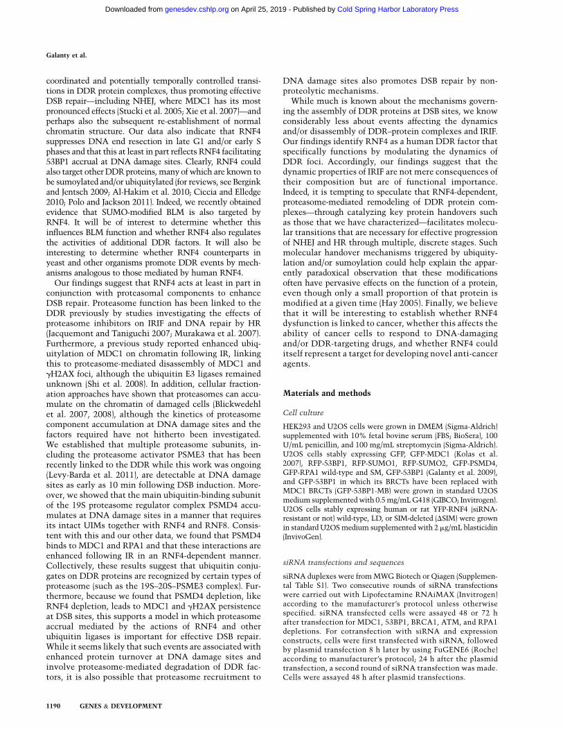
Collectively, the above data supported a model inwhich SUMO-modified RPA is targeted by RNF4 ina manner that enhances RPA turnover at DNA damagesites, thereby promoting BRCA2-mediated RAD51 load-ing and ensuing HR events. In accord with this, when wetreated cells with camptothecin, which is thought toyield DSBs only in S phase, the mean intensity of RPAaccumulation in gH2AX-positive cells was higher inRNF4-depleted cells than in control cells (Fig. 5A).Because RNF4 can target proteins for degradation, weassessed whether RPA1 levels were affected in responseto DNA damage and, if so, whether this was RNF4-dependent. Indeed, while RNF4 depletion did not appre-ciably affect RPA1 levels under control conditions, incells that had been IR-treated, RPA1 levels were repro-ducibly higher in RNF4-depleted cells than in controlcells (Fig. 5B, note that the cells were cultured in thepresence of cycloheximide to prevent de novo proteinsynthesis). Further highlighting the connection betweenRNF4 and RPA, the two proteins coimmunoprecipitatedin a manner that was substantially impaired when theSIM region of RNF4 was deleted (Fig. 5C).
To explore the mechanism by which RNF4 depletionleads to excessive DNA end resection in late G1 and/or earlyS phase (cyclinA-negative cells) (see Fig. 4A,B; SupplementalFigs. S5, S7), we assessed the effect of RNF4 depletion on53BP1 accumulation at DNA damage sites. The rationale forthis was that 53BP1 hinders resection in BRCA1-deficientcells (Bunting et al. 2010) and that we found that RNF4 wasrequired for effective accumulation of ubiquitin conjugatesat DSB sites, which are in turn needed for efficient 53BP1accrual (Huen et al. 2007; Kolas et al. 2007; Mailand et al.2007; Doil et al. 2009; Stewart et al. 2009). Notably, 53BP1accumulation at regions of DNA damage was lower in
RNF4 promotes DSB repair
GENES & DEVELOPMENT 1185
Cold Spring Harbor Laboratory Press on April 25, 2019 - Published by genesdev.cshlp.orgDownloaded from

RNF4-depleted cells than in control cells, as shown bymeasuring either 53BP1 intensity at damage sites or theproportion of cells displaying 53BP1 IRIF (Fig. 5D,E). To seewhether the reduced 53BP1 accrual upon RNF4 depletionmight be linked to excessive resection under these condi-tions, we established cell lines stably expressing GFP-53BP1or GFP-53BP1-MB, the latter of which comprises 53BP1with its BRCT domains replaced with the BRCT region ofMDC1, a region that is necessary and sufficient for MDC1recruitment to IRIF (Stucki et al. 2005). Strikingly, whileRNF4 depletion with two different siRNAs enhancedmarkers of resection in control cells (Figs. 4A,B, 5F; Supple-mental Figs. S5, S7) and in cells expressing GFP-53BP1
(Fig. 5F; Supplemental Fig. S8), this had little or no effecton resection in cells expressing GFP-53BP1-MB (Fig. 5F;Supplemental Fig. S9). These data therefore indicated thatforcing 53BP1 recruitment to DNA damage sites via theMDC1 BRCT region overcomes the effect of RNF4 onresection, thereby supporting a model in which the effectof RNF4 depletion on resection is mediated at least in partby reduced 53BP1 accumulation at DNA damage sites.
RNF4 regulates MDC1 turnover at DNA damage sites
Although the effects of RNF4 on RPA, BRCA2, and RAD51helped to explain the HR defects of RNF4-depleted cells,
Figure 5. RNF4 depletion causes hyperaccumula-tion of RPA and BrdU staining on damaged DNAand affects RPA levels following IR treatment. (A)U2OS cells stably expressing GFP-RPA1 (wild-type½WT�) were transfected with siRNAs and treatedwith 1 mM camptothecin for 1 h and then pre-extracted, fixed, and stained for gH2AX and DAPI(not shown); to the right, proportions of gH2AX-positive cells displaying GFP-RPA1 foci and themean GFP-RPA1 fluorescence intensities are pre-sented 6SED. Fluorescence intensity was calcu-lated with Volocity 6.0 software; n > 100 cells persiRNA, accumulated over two independent exper-iments. (B) RNF4 regulates RPA1 turnover follow-ing IR treatment. U2OS cells were transfected withsiRNAs. Forty-eight hours later, they were mock-treated or treated with 100 mg/mL cycloheximide(CHX) and/or 10 Gy of IR as indicated. Extractswere prepared 2 h following treatments, and sam-ples were analyzed by 4%–12% gradient SDS-PAGE and immunoblotted as indicated. Quantifi-cations of RPA1 were acquired with ImageJ soft-ware and normalized to tubulin. For each pair ofsamples (siCTRL and siRNF4), RPA1 levels in thesiCTRL sample was set to 1.0, and RPA1 levels inthe corresponding siRNF4 sample was normalizedto the siCTRL value. (C) U2OS cells stably express-ing GFP or siRNA-resistant (siR) YFP-RNF4 wild-type (WT) or SIM-deleted (DSIM) were transfectedwith siRNF4 (+). Forty-eight hours later, cells weremock-treated or treated with 10 Gy of IR and left torecover for 2 h. Samples were prepared with Ben-zonase (see the Materials and Methods), YFP-RNF4immunoprecipitations were with GFP-Trap-A beads,and samples were analyzed by 4%–12% gradientSDS-PAGE and immunoblotting as indicated. Im-munoprecipitation and detection of YFP-RNF4 de-rivatives were done with anti-GFP-Trap-A beads andGFP antibody, respectively. (D) Cells stably express-
ing GFP-53BP1 were transfected with RNF4 siRNA. Forty-eight hours later, they were laser-micro-irradiated and, 20 min after this, imaged,and 53BP1 accumulation was calculated (n = 24 independent measurements; error bars, 6SED). 53BP1 accumulation at DNA damage sitesreached a steady-state level 20 min after micro-irradiation. For equations and calculations, see the Materials and Methods. (E) U2OS cellswere transfected with siRNAs and treated with IR, fixed after the indicated time points, and then pre-extracted, fixed, and stained for 53BP1and Ub/FK2. Quantification numbers represent proportions of cells showing 53BP1 or Ub/FK2 foci 6SED (n > 100). (F) Control U2OS cellsand U2OS cells stably expressing GFP-53BP1 or GFP-53BP1 whose BRCT region was replaced by that of MDC1 (GFP-53BP1-MB) weretransfected with siRNAs, laser-micro-irradiated, fixed after 2 h, and then analyzed by immunofluorescence as indicated. Numberspresented are cells displaying BrdU staining at DNA damage sites under native conditions as a readout of resection as proportions of cellsdisplaying gH2AX or 53BP1 recruitment 6SED (n > 100); see Supplemental Figures S7–S9 for representative images and SupplementalFigure S11A for siRNA depletions.
Galanty et al.
1186 GENES & DEVELOPMENT
Cold Spring Harbor Laboratory Press on April 25, 2019 - Published by genesdev.cshlp.orgDownloaded from

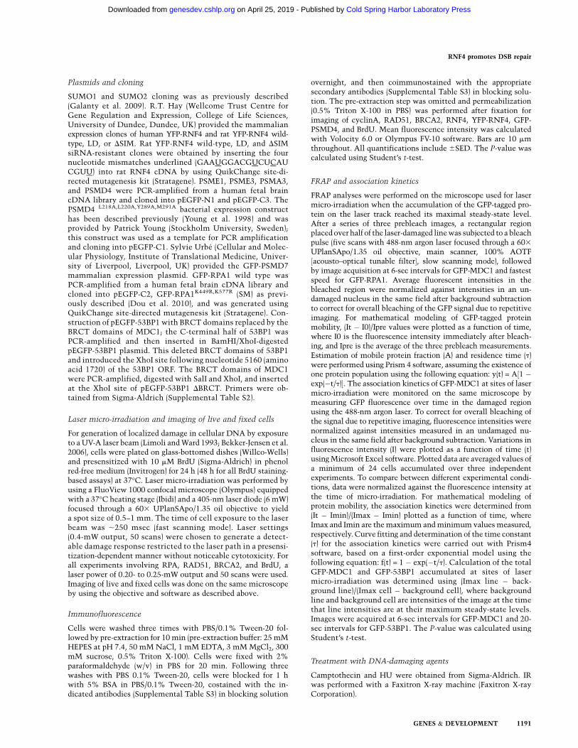
they could not easily explain the NHEJ defects of suchcells. We therefore used micro-irradiation and FRAPexperiments to see whether RNF4 affected the DSBrecruitment and/or dynamics of factors known to in-fluence NHEJ, including MDC1. By using a cell linestably expressing GFP-tagged MDC1 in live-cell imagingstudies coupled with laser micro-irradiation, we ob-served that the initial association kinetics of MDC1and the proportion of MDC1 accumulated at DNAdamage sites at early time points were not significantlyaffected by RNF4 depletion (Fig. 6A,B). In contrast,FRAP assays revealed that the mean residence time ofGFP-MDC1 at damaged sites was considerably higher inRNF4-depleted cells than in control cells (209 vs. 99 sec,respectively) (Fig. 6C). These data indicated that, as wehad observed for RPA, RNF4 promotes MDC1 turnoverand/or removal from DNA damage sites. Furthermore,because RNF4 recognizes sumoylated substrates (Sunet al. 2007; Lallemand-Breitenbach et al. 2008; Tathamet al. 2008), these findings suggested that MDC1 mightbe sumoylated. Indeed, by using a GFP-SUMO immu-noprecipitation strategy (employing high stringencyconditions) (see the Materials and Methods), we foundthat MDC1 was modified by both SUMO1 and SUMO2in a manner that was enhanced when cells were treatedwith IR or camptothecin (Fig. 6D, note that althoughRNF4 preferably binds poly-SUMO2/3 chains, it mightrecognize multiple adjacent SUMO1 sites or mixedchains containing SUMO1 as a chain terminator). Takentogether with our other data, these findings suggestedthat RNF4 likely impacts on HR and NHEJ by regulatingthe functions and turnover of multiple DDR compo-nents, including RPA and MDC1.
Because RNF4 can target proteins for degradation(Lallemand-Breitenbach et al. 2008; Tatham et al. 2008),we tested whether MDC1 protein levels were affected inresponse to DNA damage. Indeed, MDC1 levels wereslightly but reproducibly reduced in IR-treated cells,with this reduction being more evident when SUMO1or SUMO2 was stably overexpressed in cells (Fig. 6E).Moreover, through combining cycloheximide with IRtreatments in RNF4-depleted or control cells, we foundthat levels of full-length MDC1 were reduced in re-sponse to IR and that this was accompanied by anincrease in higher-molecular-weight forms of the pro-tein (Fig. 6F). Furthermore, and consistent with a modelin which RNF4 targets MDC1, these IR-induced changesin MDC1 were RNF4-dependent (Fig. 6F). Although wewere able to detect coimmunoprecipitation of MDC1with RNF4 in a SIM-dependent manner (data notshown), we could not reproducibly observe a clearincrease in this interaction following DNA damage,possibly because RNF4 binding leads to rapid MDC1degradation.
Links between RNF4 and proteasome functionsat DNA damage sites
Given that RNF4 can target proteins for proteasome-mediated degradation (Lallemand-Breitenbach et al. 2008;
Tatham et al. 2008), we speculated that the effects ofRNF4 on DSB repair might be associated with protea-some recruitment to DNA damage sites. In line with thisidea and published observations (Blickwedehl et al. 2007,2008; Ben-Aroya et al. 2010; Levy-Barda et al. 2011), weobserved that various GFP-tagged proteasome subunitswere recruited to laser-induced DNA damage (Fig. 7A).These included the proteasome activator PSME3, impli-cated in MDM2-mediated p53 degradation (Zhang andZhang 2008) and recently in the DDR (Levy-Barda et al.2011); PSMD7, a core component of the 19S proteasomeregulator (PA700); the peptidase PSMA3, a core componentof the proteolytic 20S proteasome; and PSMD4, anothercomponent of the 19S proteasome regulator (Fig. 7A)(PSME1, a component of the 11S immunoproteasomeactivator ½Stadtmueller and Hill 2011�, was not detectablyrecruited to DNA damage regions ½data not shown�).
To study proteasome recruitment to DNA damage sitesin more detail, we focused on PSMD4 because it is themain ubiquitin-binding component of the 19S protea-some regulator and is phosphorylated by the DDR proteinkinases ATM and/or ATR following DNA damage in-duction (Matsuoka et al. 2007). Notably, we found thatpoint mutations in the ubiquitin-interacting motifs(UIMs) of PSMD4 that abrogate their ability to bindubiquitin (Young et al. 1998) significantly reducedPSMD4 accumulation at DNA damage sites (Fig. 7A,bottom panels). Furthermore, by using a cell line stablyexpressing GFP-tagged PSMD4, we found that effectivePSMD4 accumulation at DNA damage sites requiredRNF4 and RNF8 (Fig. 7B) (RNF8 deletion had the stron-gest effect on PSMD4 accrual, probably because it pro-motes RNF4 recruitment to DNA damage ½see Figure 1C;Supplemental S1A� and also mediates DNA damage-induced recruitment of the RNF168 and BRCA1 ubiqui-tin E3 ligases ½Huen et al. 2007; Stewart et al. 2009�).Consistent with there being functional connections be-tween RNF4, PSMD4, and the proteasome, as was thecase for RNF4 depletion (Fig. 2A), PSMD4 depletion orproteasome inhibition with the compound MG132caused persistent MDC1 and gH2AX foci following IR(Fig. 7C; Supplemental Fig. S10). Moreover, in accordwith MDC1 and RPA1 being sumoylated and targeted byRNF4, we found that GFP-PSMD4 interacted with MDC1and RPA1 in a manner that was enhanced after IR and wasRNF4-dependent (Fig. 7D,E, note that GFP-PSMD4mainly bound the slower-migrating forms of MDC1).Unlike GFP-RPA1 binding, however, the GFP-PSMD4–MDC1 interaction was not clearly and reproduciblyobserved without proteasome inhibition, possibly reflect-ing faster proteasomal turnover of modified MDC1 thanof modified RPA1. Taken together with our other find-ings, these data supported a model in which RNF4accrual at DNA damage sites leads to PSMD4-targeted,proteasome-mediated MDC1 and RPA1 turnover.
Discussion
We showed that the STUbL RNF4 is rapidly recruited toDSB sites and promotes DSB repair. Consistent with its
RNF4 promotes DSB repair
GENES & DEVELOPMENT 1187
Cold Spring Harbor Laboratory Press on April 25, 2019 - Published by genesdev.cshlp.orgDownloaded from

known biochemical functions, RNF4 recruitment toDNA damage requires its tandem SIM region, DDRmediator proteins, and the SUMO E3 ligases PIAS1 andPIAS4. Furthermore, our data indicate that RNF4 andspecifically its ubiquitin E3 ligase activity and SIMs are
needed for effective DSB repair. Additionally, we showedthat RNF4 and its ubiquitin E3 ligase activity are alsoneeded for effective ubiquitin adduct formation atDNA damage regions. These findings therefore suggest amodel in which RNF4 promotes DSB repair by mediating
Figure 6. RNF4 regulates MDC1 turnover at DNA damage sites. (A,B) Cells stably expressing GFP-MDC1 were transfected withsiRNAs. Forty-eight hours later, cells were laser-micro-irradiated, and then MDC1 association kinetics (A) and its accumulatedamounts (B) were calculated (n = 24 independent measurements; error bars, 6SED). MDC1 accumulation reached a steady-state levelafter 10 min. For equations and calculations, see the Materials and Methods. (C) As in B, but cells were subjected to FRAP analysis (n =
10 independent measurements; error bars, 6SED). For equations and calculations, see the Materials and Methods. (D) HEK293 cellswere transfected with GFP-SUMO1, GFP-SUMO2, or GFP. Extracts were prepared 1 h after mock treatment or treatment with 10 Gy orIR or 1 mM camptothecin (CPT). GFP-SUMO immunoprecipitations were with GFP-Trap-A beads; samples were analyzed by 4%–15%gradient SDS-PAGE and immunoblotting as indicated. Arrows mark SUMO-modified MDC1, while the brackets mark the triplet ofMDC1 species. Quantifications of immunoblot signals of SUMO-modified MDC1 forms were with ImageJ software. (E) U2OS cellsstably expressing GFP, GFP-SUMO1, or GFP-SUMO2 were mock-treated (�) or treated with 10 Gy of IR. Whole-cell extracts wereprepared after the indicated times and samples analyzed as in D, but with 4%–20% gradient SDS-PAGE. (F) U2OS cells were transfectedwith siRNAs. Forty-eight hours later, they were mock-treated (�) or treated with 100 mg/mL cycloheximide (CHX) and 10 Gy of IR (top
panel) or with cycloheximide alone (CHX; bottom panel). Extracts were prepared after the indicated times, and samples were analyzedas in D. For siRNA depletions, see Supplemental Figure S11A.
Galanty et al.
1188 GENES & DEVELOPMENT
Cold Spring Harbor Laboratory Press on April 25, 2019 - Published by genesdev.cshlp.orgDownloaded from

RNF4-SIM-targeted ubiquitylation of sumoylated DDRcomponents at sites of DNA damage.
Crucially, we established that RNF4 depletion impairsboth NHEJ and HR and is associated with defectiveRAD51 and BRCA2 loading on ssDNA, with MDC1 andgH2AX persistence at DNA damage sites, and defectiverecovery from IR-induced cell cycle arrest. AlthoughRNF4 very likely operates at multiple levels in theDDR, our data suggest that one of its key functions is toaffect the dynamics of DDR–protein complexes. Thus,we found that it enhances RPA turnover at DNA damagesites, with this effect being abrogated when RPA1 ismutated on Lys 449 and Lys 557 so that it can no longerbe effectively sumoylated. Notably, previous work has
shown that cells expressing this nonsumoylatable RPA1derivative are defective in RAD51 loading and HR (Douet al. 2010), and in our experiments, we found that thismutated RPA1 derivative was defective in supportingefficient RAD51 and BRCA2 accrual at DNA damagesites. Because RNF4 depletion also causes such defects,this suggests that RNF4-mediated RPA turnover pro-motes the exchange of RPA with RAD51 and BRCA2 onssDNA so that HR can proceed effectively. We alsoestablished that RNF4 enhances the rate of turnover ofMDC1 at DNA damage sites. By analogy to its effectson RPA, RAD51, and BRCA2, we speculate that RNF4-mediated MDC1 turnover facilitates the access ofother DDR factors to damaged DNA. This could allow
Figure 7. Accumulation of the proteasome component PSMD4 at DNA damage sites requires its UIM domains, RNF4 and RNF8. (A)U2OS cells stably expressing RFP-53BP1 were transfected with GFP-PSME3, GFP-PSMD7, GFP-PSMA3, GFP-PSMD4, or GFP-PSMD4L218A,L220A,Y289A,M291A (UIM1/2 mut.). Forty-eight hours later, cells were laser-micro-irradiated, and live cells were imaged atthe indicated times. (B) U2OS cells stably expressing GFP-PSMD4 transfected with siRNAs were laser-micro-irradiated, fixed after 2 h,and then analyzed by immunofluorescence. Quantifications: Numbers represent proportion of cells showing GFP-PSMD4 accumu-lation out of gH2AX-positive, 6SED (n > 100). (C) PSMD4 depletion or proteasome inhibition causes persistence of MDC1 and gH2AXIRIF. U2OS cells were transfected with siRNAs or treated with MG132 or DMSO immediately after irradiation (2 Gy of IR), fixed after 4 h,and analyzed by immunofluorescence as indicated. Quantifications: Numbers represent proportion of cells showing gH2AX or MDC1foci, 6SED (n > 100). For time course and quantifications, see Supplemental Figure S10 (MDC1 detection was as in Fig. 2C). (D,E)PSMD4 interaction with MDC1 and RPA1 following IR is RNF4-dependent. (D) U2OS cells stably expressing GFP-PSMD4 weretransfected with siRNAs and HA-tagged ubiquitin. Forty-eight hours later, cells were exposed to 10 Gy of IR and MG132. Extracts wereprepared 4 h later and used for GFP-PSMD4 or GFP immunoprecipitations by GFP-Trap-A beads. Samples were analyzed by 4%–12%SDS-PAGE and immunoblotted as indicated. (E) As in D, but cells were not transfected with HA-Ub or treated with MG132. For siRNAdepletions, see Supplemental Figure S11, A and C.
RNF4 promotes DSB repair
GENES & DEVELOPMENT 1189
Cold Spring Harbor Laboratory Press on April 25, 2019 - Published by genesdev.cshlp.orgDownloaded from
coordinated and potentially temporally controlled transi-tions in DDR protein complexes, thus promoting effectiveDSB repair—including NHEJ, where MDC1 has its mostpronounced effects (Stucki et al. 2005; Xie et al. 2007)—andperhaps also the subsequent re-establishment of normalchromatin structure. Our data also indicate that RNF4suppresses DNA end resection in late G1 and/or early Sphases and that this at least in part reflects RNF4 facilitating53BP1 accrual at DNA damage sites. Clearly, RNF4 couldalso target other DDR proteins, many of which are known tobe sumoylated and/or ubiquitylated (for reviews, see Berginkand Jentsch 2009; Al-Hakim et al. 2010; Ciccia and Elledge2010; Polo and Jackson 2011). Indeed, we recently obtainedevidence that SUMO-modified BLM is also targeted byRNF4. It will be of interest to determine whether thisinfluences BLM function and whether RNF4 also regulatesthe activities of additional DDR factors. It will also beinteresting to determine whether RNF4 counterparts inyeast and other organisms promote DDR events by mech-anisms analogous to those mediated by human RNF4.
Our findings suggest that RNF4 acts at least in part inconjunction with proteasomal components to enhanceDSB repair. Proteasome function has been linked to theDDR previously by studies investigating the effects ofproteasome inhibitors on IRIF and DNA repair by HR(Jacquemont and Taniguchi 2007; Murakawa et al. 2007).Furthermore, a previous study reported enhanced ubiq-uitylation of MDC1 on chromatin following IR, linkingthis to proteasome-mediated disassembly of MDC1 andgH2AX foci, although the ubiquitin E3 ligases remainedunknown (Shi et al. 2008). In addition, cellular fraction-ation approaches have shown that proteasomes can accu-mulate on the chromatin of damaged cells (Blickwedehlet al. 2007, 2008), although the kinetics of proteasomecomponent accumulation at DNA damage sites and thefactors required have not hitherto been investigated.We established that multiple proteasome subunits, in-cluding the proteasome activator PSME3 that has beenrecently linked to the DDR while this work was ongoing(Levy-Barda et al. 2011), are detectable at DNA damagesites as early as 10 min following DSB induction. More-over, we showed that the main ubiquitin-binding subunitof the 19S proteasome regulator complex PSMD4 accu-mulates at DNA damage sites in a manner that requiresits intact UIMs together with RNF4 and RNF8. Consis-tent with this and our other data, we found that PSMD4binds to MDC1 and RPA1 and that these interactions areenhanced following IR in an RNF4-dependent manner.Collectively, these results suggest that ubiquitin conju-gates on DDR proteins are recognized by certain types ofproteasome (such as the 19S–20S–PSME3 complex). Fur-thermore, because we found that PSMD4 depletion, likeRNF4 depletion, leads to MDC1 and gH2AX persistenceat DSB sites, this supports a model in which proteasomeaccrual mediated by the actions of RNF4 and otherubiquitin ligases is important for effective DSB repair.While it seems likely that such events are associated withenhanced protein turnover at DNA damage sites andinvolve proteasome-mediated degradation of DDR fac-tors, it is also possible that proteasome recruitment to
DNA damage sites also promotes DSB repair by non-proteolytic mechanisms.
While much is known about the mechanisms govern-ing the assembly of DDR proteins at DSB sites, we knowconsiderably less about events affecting the dynamicsand/or disassembly of DDR–protein complexes and IRIF.Our findings identify RNF4 as a human DDR factor thatspecifically functions by modulating the dynamics ofDDR foci. Accordingly, our findings suggest that thedynamic properties of IRIF are not mere consequences oftheir composition but are of functional importance.Indeed, it is tempting to speculate that RNF4-dependent,proteasome-mediated remodeling of DDR protein com-plexes—through catalyzing key protein handovers suchas those that we have characterized—facilitates molecu-lar transitions that are necessary for effective progressionof NHEJ and HR through multiple, discrete stages. Suchmolecular handover mechanisms triggered by ubiquity-lation and/or sumoylation could help explain the appar-ently paradoxical observation that these modificationsoften have pervasive effects on the function of a protein,even though only a small proportion of that protein ismodified at a given time (Hay 2005). Finally, we believethat it will be interesting to establish whether RNF4dysfunction is linked to cancer, whether this affects theability of cancer cells to respond to DNA-damagingand/or DDR-targeting drugs, and whether RNF4 coulditself represent a target for developing novel anti-canceragents.
Materials and methods
Cell culture
HEK293 and U2OS cells were grown in DMEM (Sigma-Aldrich)supplemented with 10% fetal bovine serum (FBS; BioSera), 100U/mL penicillin, and 100 mg/mL streptomycin (Sigma-Aldrich).U2OS cells stably expressing GFP, GFP-MDC1 (Kolas et al.2007), RFP-53BP1, RFP-SUMO1, RFP-SUMO2, GFP-PSMD4,GFP-RPA1 wild-type and SM, GFP-53BP1 (Galanty et al. 2009),and GFP-53BP1 in which its BRCTs have been replaced withMDC1 BRCTs (GFP-53BP1-MB) were grown in standard U2OSmedium supplemented with 0.5 mg/mL G418 (GIBCO; Invitrogen).U2OS cells stably expressing human or rat YFP-RNF4 (siRNA-resistant or not) wild-type, LD, or SIM-deleted (DSIM) were grownin standard U2OS medium supplemented with 2 mg/mL blasticidin(InvivoGen).
siRNA transfections and sequences
siRNA duplexes were from MWG Biotech or Qiagen (Supplemen-tal Table S1). Two consecutive rounds of siRNA transfectionswere carried out with Lipofectamine RNAiMAX (Invitrogen)according to the manufacturer’s protocol unless otherwisespecified. siRNA transfected cells were assayed 48 or 72 hafter transfection for MDC1, 53BP1, BRCA1, ATM, and RPA1depletions. For cotransfection with siRNA and expressionconstructs, cells were first transfected with siRNA, followedby plasmid transfection 8 h later by using FuGENE6 (Roche)according to manufacturer’s protocol; 24 h after the plasmidtransfection, a second round of siRNA transfection was made.Cells were assayed 48 h after plasmid transfections.
Galanty et al.
1190 GENES & DEVELOPMENT
Cold Spring Harbor Laboratory Press on April 25, 2019 - Published by genesdev.cshlp.orgDownloaded from
Plasmids and cloning
SUMO1 and SUMO2 cloning was as previously described(Galanty et al. 2009). R.T. Hay (Wellcome Trust Centre forGene Regulation and Expression, College of Life Sciences,University of Dundee, Dundee, UK) provided the mammalianexpression clones of human YFP-RNF4 and rat YFP-RNF4 wild-type, LD, or DSIM. Rat YFP-RNF4 wild-type, LD, and DSIMsiRNA-resistant clones were obtained by inserting the fournucleotide mismatches underlined (GAAUGGACGUCUCAUCGUU) into rat RNF4 cDNA by using QuikChange site-di-rected mutagenesis kit (Stratagene). PSME1, PSME3, PSMA3,and PSMD4 were PCR-amplified from a human fetal braincDNA library and cloned into pEGFP-N1 and pEGFP-C3. ThePSMD4 L218A,L220A,Y289A,M291A bacterial expression constructhas been described previously (Young et al. 1998) and wasprovided by Patrick Young (Stockholm University, Sweden);this construct was used as a template for PCR amplificationand cloning into pEGFP-C1. Sylvie Urbe (Cellular and Molec-ular Physiology, Institute of Translational Medicine, Univer-sity of Liverpool, Liverpool, UK) provided the GFP-PSMD7mammalian expression plasmid. GFP-RPA1 wild type wasPCR-amplified from a human fetal brain cDNA library andcloned into pEGFP-C2, GFP-RPA1K449R,K577R (SM) as previ-ously described (Dou et al. 2010), and was generated usingQuikChange site-directed mutagenesis kit (Stratagene). Con-struction of pEGFP-53BP1 with BRCT domains replaced by theBRCT domains of MDC1; the C-terminal half of 53BP1 wasPCR-amplified and then inserted in BamHI/XhoI-digestedpEGFP-53BP1 plasmid. This deleted BRCT domains of 53BP1and introduced the XhoI site following nucleotide 5160 (aminoacid 1720) of the 53BP1 ORF. The BRCT domains of MDC1were PCR-amplified, digested with SalI and XhoI, and insertedat the XhoI site of pEGFP-53BP1 DBRCT. Primers were ob-tained from Sigma-Aldrich (Supplemental Table S2).
Laser micro-irradiation and imaging of live and fixed cells
For generation of localized damage in cellular DNA by exposureto a UV-A laser beam (Limoli and Ward 1993; Bekker-Jensen et al.2006), cells were plated on glass-bottomed dishes (Willco-Wells)and presensitized with 10 mM BrdU (Sigma-Aldrich) in phenolred-free medium (Invitrogen) for 24 h (48 h for all BrdU staining-based assays) at 37°C. Laser micro-irradiation was performed byusing a FluoView 1000 confocal microscope (Olympus) equippedwith a 37°C heating stage (Ibidi) and a 405-nm laser diode (6 mW)focused through a 603 UPlanSApo/1.35 oil objective to yielda spot size of 0.5–1 mm. The time of cell exposure to the laserbeam was ;250 msec (fast scanning mode). Laser settings(0.4-mW output, 50 scans) were chosen to generate a detect-able damage response restricted to the laser path in a presensi-tization-dependent manner without noticeable cytotoxicity. Forall experiments involving RPA, RAD51, BRCA2, and BrdU, alaser power of 0.20- to 0.25-mW output and 50 scans were used.Imaging of live and fixed cells was done on the same microscopeby using the objective and software as described above.
Immunofluorescence
Cells were washed three times with PBS/0.1% Tween-20 fol-lowed by pre-extraction for 10 min (pre-extraction buffer: 25 mMHEPES at pH 7.4, 50 mM NaCl, 1 mM EDTA, 3 mM MgCl2, 300mM sucrose, 0.5% Triton X-100). Cells were fixed with 2%paraformaldehyde (w/v) in PBS for 20 min. Following threewashes with PBS 0.1% Tween-20, cells were blocked for 1 hwith 5% BSA in PBS/0.1% Tween-20, costained with the in-dicated antibodies (Supplemental Table S3) in blocking solution
overnight, and then coimmunostained with the appropriatesecondary antibodies (Supplemental Table S3) in blocking solu-tion. The pre-extraction step was omitted and permeabilization(0.5% Triton X-100 in PBS) was performed after fixation forimaging of cyclinA, RAD51, BRCA2, RNF4, YFP-RNF4, GFP-PSMD4, and BrdU. Mean fluorescence intensity was calculatedwith Volocity 6.0 or Olympus FV-10 software. Bars are 10 mmthroughout. All quantifications include 6SED. The P-value wascalculated using Student’s t-test.
FRAP and association kinetics
FRAP analyses were performed on the microscope used for lasermicro-irradiation when the accumulation of the GFP-tagged pro-tein on the laser track reached its maximal steady-state level.After a series of three prebleach images, a rectangular regionplaced over half of the laser-damaged line was subjected to a bleachpulse (five scans with 488-nm argon laser focused through a 603
UPlanSApo/1.35 oil objective, main scanner, 100% AOTF½acousto–optical tunable filter�, slow scanning mode), followedby image acquisition at 6-sec intervals for GFP-MDC1 and fastestspeed for GFP-RPA1. Average fluorescent intensities in thebleached region were normalized against intensities in an un-damaged nucleus in the same field after background subtractionto correct for overall bleaching of the GFP signal due to repetitiveimaging. For mathematical modeling of GFP-tagged proteinmobility, (It � I0)/Ipre values were plotted as a function of time,where I0 is the fluorescence intensity immediately after bleach-ing, and Ipre is the average of the three prebleach measurements.Estimation of mobile protein fraction (A) and residence time (t)were performed using Prism 4 software, assuming the existence ofone protein population using the following equation: y(t) = A½1 �exp(�t/t)�. The association kinetics of GFP-MDC1 at sites of lasermicro-irradiation were monitored on the same microscope bymeasuring GFP fluorescence over time in the damaged regionusing the 488-nm argon laser. To correct for overall bleaching ofthe signal due to repetitive imaging, fluorescence intensities werenormalized against intensities measured in an undamaged nu-cleus in the same field after background subtraction. Variations influorescence intensity (I) were plotted as a function of time (t)using Microsoft Excel software. Plotted data are averaged values ofa minimum of 24 cells accumulated over three independentexperiments. To compare between different experimental condi-tions, data were normalized against the fluorescence intensity atthe time of micro-irradiation. For mathematical modeling ofprotein mobility, the association kinetics were determined from(It � Imin)/(Imax � Imin) plotted as a function of time, whereImax and Imin are the maximum and minimum values measured,respectively. Curve fitting and determination of the time constant(t) for the association kinetics were carried out with Prism4software, based on a first-order exponential model using thefollowing equation: f(t) = 1 � exp(�t/t). Calculation of the totalGFP-MDC1 and GFP-53BP1 accumulated at sites of lasermicro-irradiation was determined using (Imax line � back-ground line)/(Imax cell � background cell), where backgroundline and background cell are intensities of the image at the timethat line intensities are at their maximum steady-state levels.Images were acquired at 6-sec intervals for GFP-MDC1 and 20-sec intervals for GFP-53BP1. The P-value was calculated usingStudent’s t-test.
Treatment with DNA-damaging agents
Camptothecin and HU were obtained from Sigma-Aldrich. IRwas performed with a Faxitron X-ray machine (Faxitron X-rayCorporation).
RNF4 promotes DSB repair
GENES & DEVELOPMENT 1191
Cold Spring Harbor Laboratory Press on April 25, 2019 - Published by genesdev.cshlp.orgDownloaded from

Immunoprecipitation and immunoblotting
Cell extracts were prepared on plates using lysis buffercontaining 2% SDS, 50 mM Tris-HCl (pH 7.5), 10 mMN-ethylmaleimide (Sigma-Aldrich), and protease inhibitor cocktail(Roche), followed by sonication or passage of extracts througha 19-gauage needle-mounted syringe. For monitoring MDC1, cellextracts were prepared by using lysis buffer containing 20mM HEPES (pH 7.4), 500 mM NaCl, 1.5 mM MgCl2, 1 mMEGTA, 1% Tween20, 10% glycerol, serine/threonine phospha-tase inhibitor cocktail (Sigma-Aldrich), protease inhibitorcocktail (Roche), and 10 mM N-ethylmaleimide (Sigma-Aldrich). Subsequently, extracts were sonicated and diluted1:2 with the same buffer lacking NaCl. For GFP and YFP-RNF4immunoprecipitation using GFP-Trap-A beads (ChromoTekGmbH), cell extracts were prepared using lysis buffer contain-ing 20 mM HEPES (pH 7.4), 500 mM NaCl, 1.5 mM MgCl2, 1mM EGTA, 1% Tween20, 10% glycerol, serine/threoninephosphatase inhibitor cocktail (Sigma-Aldrich), protease in-hibitor cocktail (Roche), and 10 mM N-ethylmaleimide(Sigma-Aldrich). Extracts were then sonicated and diluted 1:2with the same buffer lacking NaCl. For all immunoprecipita-tions, extracts were supplemented with 200 mg/mL ethidiumbromide to prevent nonspecific binding via DNA and clearedusing centrifugation at 16,000g for 60 min at 4°C. Overnightincubation/binding with GFP-Trap-A beads at 4°C was fol-lowed by five washes, alternating three washes with immu-noprecipitation buffer (250 mM NaCl) and two washes withlysis buffer (500 mM NaCl), and 5 min boiling in 1.53 SDSsample buffer. Proteins were resolved by 4%–18% SDS-PAGE(unless otherwise specified) and transferred to PVDF mem-brane (GE Healthcare). Immunoblotting was performed withthe indicated antibodies (Supplemental Table S3). Immuno-blotting for BRCA1 was done using a 1:1 mix of the rabbitantibodies in Supplemental Table S3. For coimmunoprecipi-tation of YFP-RNF4 with RPA as well as for GFP-PSMD4 withMDC1 and RPA1, cells were lysed on plates in Benzonasenuclease buffer: 20 mM Tris-HCl, 40 mM NaCl, 2 mM MgCl2,10% glycerol, 0.5% NP-40, and EDTA free protease inhibitorcocktail (Roche) supplemented with 100 U/mL Benzonasenuclease (Novagen) and 10 mM N-ethylmaleimide. Extractswere then collected, and the NaCl concentration was in-creased to 450 mM followed by 20 min of incubation on ice.The extracts were then cleared using centrifugation at 16,000g
for 60 min at 4°C. The NaCl concentration was reduced to 225mM and supplemented with 10 mM N-ethylmaleimide(Sigma-Aldrich) and serine/threonine phosphatase inhibitorcocktail (Sigma-Aldrich). The resulting extracts were sub-jected to an overnight incubation with GFP-Trap-A beads at4°C followed by five washes with Benzonase nuclease bufferwith 225 mM NaCl. Samples were subsequently analyzed bySDS-PAGE as described above. Quantifications of immuno-blotting signals for SUMO-modified MDC1 and RPA1 levelswere normalized to tubulin signals and were acquired byImageJ software.
Flow cytometry and S-phase index measurements
To determine cell cycle distribution, cells were fixed with 70%ethanol, incubated for 30 min with RNase A (250 mg/mL) andpropidium iodide (10 mg/mL) at 37°C, and analyzed by flowcytometry. Data were analyzed using FlowJo software to revealthe percentage of cells in each cell cycle phase. The S-phaseindex was determined using Click-iT EdU Alexa Fluor 647flow cytometry assay kit (Invitrogen, A10202) according to themanufacturer’s protocol.
Random plasmid integration assay
Assays were performed as previously described (Galanty et al. 2009).Briefly, 1 d after transfection with siRNA, U2OS cells were trans-fected with BamHI–XhoI-linearized pEGFP-C1 (Clontech). Thefollowing day, cells were collected, counted, and plated on threeplates, one of which contained 0.5 mg/mL G418. One day afterplating, cells on the plate lacking G418 were fixed to assess trans-fection efficiency, and the other two plates were incubated for10–14 d at 37°C to allow colony formation. Colonies were stainedwith 0.5% crystal violet/20% ethanol and counted. Random plas-mid integration events were normalized to transfection and platingefficiencies. The P-value was calculated using Student’s t-test.
HR assay
A U2OS clone with the integrated HR reporter DR-GFP wasgenerated as described previously (Pierce et al. 2001; Sartori et al.2007). One day after transfection with siRNA, U2OS-DR-GFPcells were cotransfected with an I-SceI expression vector (pCBA-I-SceI) and a vector expressing monomeric RFP (pCS2-mRFP).The latter plasmid was added in a 1:10 ratio to mark the I-SceI-positive cells. Cells were harvested 1 d after I-SceI transfectionand subjected to flow cytometric analysis to examine recombi-nation induced by I-SceI digestion. Only RFP-positive cells wereanalyzed for HR efficiency to circumvent possible differencesin transfection efficiencies. FACS data were analyzed usingSummit version 4.3 software to reveal the percentage of GFP-positive cells relative to the number of transfected cells (RFP-positive). The data were normalized to a control siRNA treatmentin each individual experiment. The cut-off between GFP (HR)-positive and -negative cells was set to 0.5% background level ofGFP-positive cells in the internal control (RFP-positive, nottransfected with I-SceI). This gate was then applied to the RFP/I-SceI-positive samples to determine HR efficiency. Results arepresented as a percentage of control siRNA. The P-value wascalculated using Student’s t-test.
Comet assay
U2OS, U2OS stably expressing vector only, siRNA-resistantYFP-RNF4 wild-type, LD, or DSIM cells were transfected withsiRNA, exposed to 10 Gy IR, and harvested at the indicated timepoints. For the 0 time point, cells were irradiated on ice to slowdown repair and processed immediately following treatment.Neutral comet assays were performed using the comet assay kitfrom Trevigen (catalog no. 4250-050-K). Gelbond film (0.22 mmthick, 85 3 100 mm, LONZA catalog no. 53734, agarose gelsupport medium) was used instead of the standard glass slides.Images were collected using a 103 UPlanFLN objective mountedon an Olympus IX71 fluorescence microscope equipped witha CCD camera; comets were scored using Comet score software,and data were analyzed using Microsoft Excel.
Cell survival assay
U2OS cells were transfected with siRNAs and exposed to IR orHU at the indicated doses. Cells were incubated for 10–14 d at37°C to allow colony formation. Colonies were stained with0.5% crystal violet/20% ethanol and counted. Results werenormalized to plating efficiencies.
Acknowledgments
We thank S.P.J. laboratory members for support, in particularJ. Harrigan, J. Forment, and A. Kaidi, for helpful discussions;
Galanty et al.
1192 GENES & DEVELOPMENT
Cold Spring Harbor Laboratory Press on April 25, 2019 - Published by genesdev.cshlp.orgDownloaded from

J. Forment for determining the S-phase index of control andRNF4-depleted cells; S. Polo for setting up the laser system andhelping in the analysis of MDC1 FRAP and association kineticsexperiments; and K. Dry for help with the manuscript. We alsothank R.T Hay for YFP-tagged RNF4 constructs and siRNAsequences targeting RNF4, P. Young for PSMD4 UIM1/2 pointmutant, J. Palvimo for RNF4 antibody, T. Halazonetis for RNF8antibody, R. Baer for CtIP antibody, Y. Shiloh for ATM antibody,and R. Walker for help with FACS and cell sorting. Research inthe S.P.J. laboratory is supported by grants from Cancer ResearchUK (C6/A11226), the European Research Council, the EuropeanCommunity’s Seventh Framework Program (GENICA andDDResponse), and core infrastructure funding from Cancer Re-search UK and the Wellcome Trust. S.P.J. receives his salary fromthe University of Cambridge, supplemented by Cancer ResearchUK. R.B. cloned the RNF4 siRNA-resistant, proteasome compo-nent cDNAs, GFP-RPA1 (wild type and SM), and GFP-53BP1-MB;generated RFP-53BP1 and GFP-53BP1-MB stable cell lines;participated in testing siRNA efficiencies; provided help withthe processing of laser experiments and in the establishment andtesting of stable-cell-lines; and engaged in helpful discussions.J.C. extensively helped with cell survival and HR and NHEJexperiments and provided support with tissue culture mainte-nance and stable cell line generation. Y.G. initiated and led theproject and performed all other experiments. Y.G. and S.P.J.conceived the study and wrote the paper. All authors discussedand commented on the manuscript.
References
Al-Hakim A, Escribano-Diaz C, Landry MC, O’Donnell L,Panier S, Szilard RK, Durocher D. 2010. The ubiquitous roleof ubiquitin in the DNA damage response. DNA Repair
(Amst) 9: 1229–1240.Bekker-Jensen S, Mailand N. 2010. Assembly and function of
DNA double-strand break repair foci in mammalian cells.DNA Repair (Amst) 9: 1219–1228.
Bekker-Jensen S, Lukas C, Kitagawa R, Melander F, Kastan MB,Bartek J, Lukas J. 2006. Spatial organization of the mamma-lian genome surveillance machinery in response to DNAstrand breaks. J Cell Biol 173: 195–206.
Bekker-Jensen S, Rendtlew Danielsen J, Fugger K, Gromova I,Nerstedt A, Lukas C, Bartek J, Lukas J, Mailand N. 2009.HERC2 coordinates ubiquitin-dependent assembly of DNArepair factors on damaged chromosomes. Nat Cell Biol 12:80–86.
Ben-Aroya S, Agmon N, Yuen K, Kwok T, McManus K,Kupiec M, Hieter P. 2010. Proteasome nuclear activityaffects chromosome stability by controlling the turnoverof Mms22, a protein important for DNA repair. PLoS Genet
6: e1000852. doi: 10.1371/journal.pgen.1000852.Bergink S, Jentsch S. 2009. Principles of ubiquitin and SUMO
modifications in DNA repair. Nature 458: 461–467.Blickwedehl J, McEvoy S, Wong I, Kousis P, Clements J, Elliott R,
Cresswell P, Liang P, Bangia N. 2007. Proteasomes andproteasome activator 200 kDa (PA200) accumulate on chro-matin in response to ionizing radiation. Radiat Res 167: 663–674.
Blickwedehl J, Agarwal M, Seong C, Pandita RK, Melendy T,Sung P, Pandita TK, Bangia N. 2008. Role for proteasomeactivator PA200 and postglutamyl proteasome activity ingenomic stability. Proc Natl Acad Sci 105: 16165–16170.
Bunting SF, Callen E, Wong N, Chen HT, Polato F, Gunn A,Bothmer A, Feldhahn N, Fernandez-Capetillo O, Cao L, et al.2010. 53BP1 inhibits homologous recombination in Brca1-
deficient cells by blocking resection of DNA breaks. Cell
141: 243–254.Burgess RC, Rahman S, Lisby M, Rothstein R, Zhao X. 2007.
The Slx5-Slx8 complex affects sumoylation of DNA repairproteins and negatively regulates recombination. Mol CellBiol 27: 6153–6162.
Chapman JR, Jackson SP. 2008. Phospho-dependent interactionsbetween NBS1 and MDC1 mediate chromatin retention ofthe MRN complex at sites of DNA damage. EMBO Rep 9:795–801.
Ciccia A, Elledge SJ. 2010. The DNA damage response: Makingit safe to play with knives. Mol Cell 40: 179–204.
Cimprich KA, Cortez D. 2008. ATR: An essential regulator ofgenome integrity. Nat Rev Mol Cell Biol 9: 616–627.
Cortez D, Guntuku S, Qin J, Elledge SJ. 2001. ATR and ATRIP:Partners in checkpoint signaling. Science 294: 1713–1716.
Doil C, Mailand N, Bekker-Jensen S, Menard P, Larsen DH,Pepperkok R, Ellenberg J, Panier S, Durocher D, Bartek J,et al. 2009. RNF168 binds and amplifies ubiquitin conjugateson damaged chromosomes to allow accumulation of repairproteins. Cell 136: 435–446.
Dou H, Huang C, Singh M, Carpenter PB, Yeh ET. 2010.Regulation of DNA repair through deSUMOylation andSUMOylation of replication protein A complex. Mol Cell39: 333–345.
Downs JA, Nussenzweig MC, Nussenzweig A. 2007. Chromatindynamics and the preservation of genetic information.Nature 447: 951–958.
Galanty Y, Belotserkovskaya R, Coates J, Polo S, Miller KM,Jackson SP. 2009. Mammalian SUMO E3-ligases PIAS1 andPIAS4 promote responses to DNA double-strand breaks.Nature 462: 935–939.
Gravel S, Chapman JR, Magill C, Jackson SP. 2008. DNAhelicases Sgs1 and BLM promote DNA double-strand breakresection. Genes Dev 22: 2767–2772.
Hakli M, Lorick KL, Weissman AM, Janne OA, Palvimo JJ. 2004.Transcriptional coregulator SNURF (RNF4) possesses ubiq-uitin E3 ligase activity. FEBS Lett 560: 56–62.
Hartlerode AJ, Scully R. 2009. Mechanisms of double-strandbreak repair in somatic mammalian cells. Biochem J 423: 157–168.
Hay RT. 2005. SUMO: A history of modification. Mol Cell 18: 1–12.
Holloman WK. 2011. Unraveling the mechanism of BRCA2 inhomologous recombination. Nat Struct Mol Biol 18: 748–754.
Huang J, Huen MS, Kim H, Leung CC, Glover JN, Yu X, Chen J.2009. RAD18 transmits DNA damage signalling to elicithomologous recombination repair. Nat Cell Biol 11: 592–603.
Huen MS, Grant R, Manke I, Minn K, Yu X, Yaffe MB, Chen J.2007. RNF8 transduces the DNA-damage signal via histoneubiquitylation and checkpoint protein assembly. Cell 131:901–914.
Huertas P. 2010. DNA resection in eukaryotes: Deciding how tofix the break. Nat Struct Mol Biol 17: 11–16.
Jackson SP, Bartek J. 2009. The DNA-damage response inhuman biology and disease. Nature 461: 1071–1078.
Jacquemont C, Taniguchi T. 2007. Proteasome function is re-quired for DNA damage response and fanconi anemia path-way activation. Cancer Res 67: 7395–7405.
Jensen RB, Carreira A, Kowalczykowski SC. 2010. Purifiedhuman BRCA2 stimulates RAD51-mediated recombination.Nature 467: 678–683.
Kee Y, D’Andrea AD. 2010. Expanded roles of the Fanconianemia pathway in preserving genomic stability. Genes Dev24: 1680–1694.
RNF4 promotes DSB repair
GENES & DEVELOPMENT 1193
Cold Spring Harbor Laboratory Press on April 25, 2019 - Published by genesdev.cshlp.orgDownloaded from

Kirkin V, Dikic I. 2007. Role of ubiquitin- and Ubl-bindingproteins in cell signaling. Curr Opin Cell Biol 19: 199–205.
Kolas NK, Chapman JR, Nakada S, Ylanko J, Chahwan R,Sweeney FD, Panier S, Mendez M, Wildenhain J, ThomsonTM, et al. 2007. Orchestration of the DNA-damage responseby the RNF8 ubiquitin ligase. Science 318: 1637–1640.
Kosoy A, Calonge TM, Outwin EA, O’Connell MJ. 2007. Fissionyeast Rnf4 homologs are required for DNA repair. J BiolChem 282: 20388–20394.
Lallemand-Breitenbach V, Jeanne M, Benhenda S, Nasr R, Lei M,Peres L, Zhou J, Zhu J, Raught B, de The H. 2008. Arsenicdegrades PML or PML–RARa through a SUMO-triggeredRNF4/ubiquitin-mediated pathway. Nat Cell Biol 10: 547–555.
Levy-Barda A, Lerenthal Y, Davis AJ, Chung YM, Essers J, ShaoZ, van Vliet N, Chen DJ, Hu MC, Kanaar R, et al. 2011.Involvement of the nuclear proteasome activator PA28g inthe cellular response to DNA double-strand breaks. Cell
Cycle 10: 4300–4310Limoli CL, Ward JF. 1993. A new method for introducing
double-strand breaks into cellular DNA. Radiat Res 134:160–169.
Liu J, Doty T, Gibson B, Heyer WD. 2010. Human BRCA2protein promotes RAD51 filament formation on RPA-covered single-stranded DNA. Nat Struct Mol Biol 17:1260–1262.
Mailand N, Bekker-Jensen S, Faustrup H, Melander F, Bartek J,Lukas C, Lukas J. 2007. RNF8 ubiquitylates histones at DNAdouble-strand breaks and promotes assembly of repair pro-teins. Cell 131: 887–900.
Matsuoka S, Ballif BA, Smogorzewska A, McDonald ER 3rd,Hurov KE, Luo J, Bakalarski CE, Zhao Z, Solimini N,Lerenthal Y, et al. 2007. ATM and ATR substrate analysisreveals extensive protein networks responsive to DNA dam-age. Science 316: 1160–1166.
Morris JR. 2010a. More modifiers move on DNA damage. Cancer
Res 70: 3861–3863.Morris JR. 2010b. SUMO in the mammalian response to DNA
damage. Biochem Soc Trans 38: 92–97.Morris JR, Boutell C, Keppler M, Densham R, Weekes D,
Alamshah A, Butler L, Galanty Y, Pangon L, Kiuchi T, et al.2009. The SUMO modification pathway is involved in theBRCA1 response to genotoxic stress. Nature 462: 886–890.
Motegi A, Murakawa Y, Takeda S. 2009. The vital link betweenthe ubiquitin-proteasome pathway and DNA repair: Impacton cancer therapy. Cancer Lett 283: 1–9.
Murakawa Y, Sonoda E, Barber LJ, Zeng W, Yokomori K, KimuraH, Niimi A, Lehmann A, Zhao GY, Hochegger H, et al. 2007.Inhibitors of the proteasome suppress homologous DNArecombination in mammalian cells. Cancer Res 67: 8536–8543.
Nimonkar AV, Ozsoy AZ, Genschel J, Modrich P, KowalczykowskiSC. 2008. Human exonuclease 1 and BLM helicase interact toresect DNA and initiate DNA repair. Proc Natl Acad Sci 105:16906–16911.
Nimonkar AV, Genschel J, Kinoshita E, Polaczek P, Campbell JL,Wyman C, Modrich P, Kowalczykowski SC. 2011. BLM–DNA2–RPA–MRN and EXO1–BLM–RPA–MRN constitutetwo DNA end resection machineries for human DNA breakrepair. Genes Dev 25: 350–362.
Pardo B, Gomez-Gonzalez B, Aguilera A. 2009. DNA repair inmammalian cells: DNA double-strand break repair: Howto fix a broken relationship. Cell Mol Life Sci 66: 1039–1056.
Pierce AJ, Hu P, Han M, Ellis N, Jasin M. 2001. Ku DNA end-binding protein modulates homologous repair of double-strand breaks in mammalian cells. Genes Dev 15: 3237–3242.
Polo SE, Jackson SP. 2011. Dynamics of DNA damage responseproteins at DNA breaks: A focus on protein modifications.Genes Dev 25: 409–433.
Prudden J, Pebernard S, Raffa G, Slavin DA, Perry JJ, Tainer JA,McGowan CH, Boddy MN. 2007. SUMO-targeted ubiquitinligases in genome stability. EMBO J 26: 4089–4101.
Sartori AA, Lukas C, Coates J, Mistrik M, Fu S, Bartek J, Baer R,Lukas J, Jackson SP. 2007. Human CtIP promotes DNA endresection. Nature 450: 509–514.
Shi W, Ma Z, Willers H, Akhtar K, Scott SP, Zhang J, Powell S.2008. Disassembly of MDC1 foci is controlled by ubiquitin-proteasome-dependent degradation. J Biol Chem 283: 31608–31616.
Stadtmueller BM, Hill CP. 2011. Proteasome activators. Mol
Cell 41: 8–19.Stewart GS, Panier S, Townsend K, Al-Hakim AK, Kolas NK,
Miller ES, Nakada S, Ylanko J, Olivarius S, Mendez M, et al.2009. The RIDDLE syndrome protein mediates a ubiquitin-dependent signaling cascade at sites of DNA damage. Cell
136: 420–434.Stucki M, Clapperton JA, Mohammad D, Yaffe MB, Smerdon SJ,
Jackson SP. 2005. MDC1 directly binds phosphorylatedhistone H2AX to regulate cellular responses to DNA double-strand breaks. Cell 123: 1213–1226.
Sun H, Leverson JD, Hunter T. 2007. Conserved function ofRNF4 family proteins in eukaryotes: Targeting a ubiquitinligase to SUMOylated proteins. EMBO J 26: 4102–4112.
Tatham MH, Geoffroy MC, Shen L, Plechanovova A, HattersleyN, Jaffray EG, Palvimo JJ, Hay RT. 2008. RNF4 is a poly-SUMO-specific E3 ubiquitin ligase required for arsenic-induced PML degradation. Nat Cell Biol 10: 538–546.
Thorslund T, McIlwraith MJ, Compton SA, Lekomtsev S,Petronczki M, Griffith JD, West SC. 2010. The breast cancertumor suppressor BRCA2 promotes the specific targeting ofRAD51 to single-stranded DNA. Nat Struct Mol Biol 17:1263–1265.
Ustrell V, Hoffman L, Pratt G, Rechsteiner M. 2002. PA200,a nuclear proteasome activator involved in DNA repair.EMBO J 21: 3516–3525.
Wang B, Elledge SJ. 2007. Ubc13/Rnf8 ubiquitin ligases controlfoci formation of the Rap80/Abraxas/Brca1/Brcc36 complexin response to DNA damage. Proc Natl Acad Sci 104: 20759–20763.
Watanabe K, Iwabuchi K, Sun J, Tsuji Y, Tani T, Tokunaga K,Date T, Hashimoto M, Yamaizumi M, Tateishi S. 2009.RAD18 promotes DNA double-strand break repair duringG1 phase through chromatin retention of 53BP1. NucleicAcids Res 37: 2176–2193.
Weisshaar SR, Keusekotten K, Krause A, Horst C, Springer HM,Gottsche K, Dohmen RJ, Praefcke GJ. 2008. Arsenic tri-oxide stimulates SUMO-2/3 modification leading to RNF4-dependent proteolytic targeting of PML. FEBS Lett 582:3174–3178.
Xie A, Hartlerode A, Stucki M, Odate S, Puget N, Kwok A,Nagaraju G, Yan C, Alt FW, Chen J, et al. 2007. Distinctroles of chromatin-associated proteins MDC1 and 53BP1in mammalian double-strand break repair. Mol Cell 28:1045–1057.
Yang K, Moldovan GL, D’Andrea AD. 2010. RAD18-dependentrecruitment of SNM1A to DNA repair complexes by aubiquitin-binding zinc finger. J Biol Chem 285: 19085–19091.
Young P, Deveraux Q, Beal RE, Pickart CM, Rechsteiner M.1998. Characterization of two polyubiquitin binding sites
Galanty et al.
1194 GENES & DEVELOPMENT
Cold Spring Harbor Laboratory Press on April 25, 2019 - Published by genesdev.cshlp.orgDownloaded from
in the 26 S protease subunit 5a. J Biol Chem 273: 5461–5467.
Zhang Z, Zhang R. 2008. Proteasome activator PA28g regulatesp53 by enhancing its MDM2-mediated degradation. EMBO
J 27: 852–864.Zhao GY, Sonoda E, Barber LJ, Oka H, Murakawa Y, Yamada K,
Ikura T, Wang X, Kobayashi M, Yamamoto K, et al. 2007. Acritical role for the ubiquitin-conjugating enzyme Ubc13 ininitiating homologous recombination. Mol Cell 25: 663–675.
Zou L, Elledge SJ. 2003. Sensing DNA damage through ATRIPrecognition of RPA-ssDNA complexes. Science 300: 1542–1548.
RNF4 promotes DSB repair
GENES & DEVELOPMENT 1195
Cold Spring Harbor Laboratory Press on April 25, 2019 - Published by genesdev.cshlp.orgDownloaded from

10.1101/gad.188284.112Access the most recent version at doi: 26:2012, Genes Dev.
Yaron Galanty, Rimma Belotserkovskaya, Julia Coates, et al. double-strand break repairRNF4, a SUMO-targeted ubiquitin E3 ligase, promotes DNA
Material
Supplemental
http://genesdev.cshlp.org/content/suppl/2012/05/30/26.11.1179.DC1
Related Content
Genes Dev. June , 2012 26: 1196-1208
Yili Yin, Anne Seifert, Joy Shijia Chua, et al.cells to DNA damageSUMO-targeted ubiquitin E3 ligase RNF4 is required for the response of human
References
http://genesdev.cshlp.org/content/26/11/1179.full.html#related-urls
Articles cited in:
http://genesdev.cshlp.org/content/26/11/1179.full.html#ref-list-1This article cites 68 articles, 23 of which can be accessed free at:
License
ServiceEmail Alerting
click here.right corner of the article or
Receive free email alerts when new articles cite this article - sign up in the box at the top
Copyright © 2012 by Cold Spring Harbor Laboratory Press
Cold Spring Harbor Laboratory Press on April 25, 2019 - Published by genesdev.cshlp.orgDownloaded from