RNA isolation and quantitative PCR from HOPE and Formalin fixed
24
1 RNA isolation and quantitative PCR from HOPE and Formalin fixed bovine lymph node tissues Jaydene Witchell 1 , Dhaval Varshney 1 , Trusha Gajjar 1 , Arun Wangoo 2§ and Madhu Goyal 1* 1 School of Life Sciences, University of Hertfordshire, AL10 9AB 2 Department of Pathology, Veterinary Laboratories Agency, Weybridge, Addlestone, Surrey KT15 3NB 1* Correspondence author: School of Life Sciences, University of Hertfordshire, Hatfield, Herts AL10 9AB. Telephone: +44 1707284624. Fax: +44 1707285046. E-mail address: [email protected] 2§ Present address (A.Wangoo): Veterinary Medicines Directorate, Addlestone, Surrey, KT15 3LS Key words: Quantitative polymerase chain reaction; RNA extraction; HOPE fixative; formalin fixative; Mycobacterium bovis Abbreviations: RNA, ribonucleic acid; mRNA, messenger ribonucleic acid; RT-PCR, reverse transcriptase polymerase chain reaction; qRT-PCR, quantitative RT-PCR; HOPE, Hepes glutamic acid buffer mediated organic solvent protection effect; GAPDH, Glyceraldehyde-3-phosphate dehydrogenase; bp, base pairs; CP value, crossing point value; FFPE, formalin fixed paraffin embedded.
Transcript of RNA isolation and quantitative PCR from HOPE and Formalin fixed

1
RNA isolation and quantitative PCR from HOPE and Formalin fixed bovine
lymph node tissues
Jaydene Witchell1, Dhaval Varshney
1, Trusha Gajjar
1, Arun Wangoo
2§ and Madhu Goyal
1*
1School of Life Sciences, University of Hertfordshire, AL10 9AB
2 Department
of Pathology, Veterinary Laboratories Agency, Weybridge, Addlestone, Surrey KT15 3NB
1*Correspondence author: School of Life Sciences, University of Hertfordshire, Hatfield, Herts AL10 9AB.
Telephone: +44 1707284624. Fax: +44 1707285046. E-mail address: [email protected]
2§ Present address (A.Wangoo): Veterinary Medicines Directorate, Addlestone, Surrey, KT15 3LS
Key words: Quantitative polymerase chain reaction; RNA extraction; HOPE fixative; formalin fixative;
Mycobacterium bovis
Abbreviations: RNA, ribonucleic acid; mRNA, messenger ribonucleic acid; RT-PCR, reverse transcriptase
polymerase chain reaction; qRT-PCR, quantitative RT-PCR; HOPE, Hepes glutamic acid
buffer mediated organic solvent protection effect; GAPDH, Glyceraldehyde-3-phosphate dehydrogenase; bp, base
pairs; CP value, crossing point value; FFPE, formalin fixed paraffin embedded.

2
Abstract
The use of RNA extracted from HOPE fixed tissues in quantitative reverse transcriptase
polymerase chain reaction (qRT-PCR) is fairly novel. We compared qRT-PCR analysis of
formalin and HOPE fixed, paraffin embedded lymph node tissues from M.bovis infected cattle,
by extracting total RNA using a commercial kit (Ambion) and a trizol method. RNA extracted
from HOPE fixed tissues showed comparable quantities between the commercial kit (82.7-107.9
μg/ml total RNA) and the trizol method (87-161.1 μg/ml total RNA), displaying a high degree of
integrity when analysed by electrophoresis. RNA extracted from formalin fixed tissues using the
commercial kit produced similar concentrations (80.6-145.7 μg/ml total RNA) in comparison to
the HOPE tissue however the integrity was compromised. Extraction of RNA from the formalin
fixed tissues using trizol was unsuccessful.
Following qRT-PCR for GAPDH, total RNA from HOPE fixed tissues showed higher levels of
target mRNA (4.05x10⁻²
pg/100ng total RNA using the commercial kit and 6.45x10⁻²
pg/100ng
total RNA using trizol) in comparison to formalin fixed tissues (5.69x10⁻⁴
pg/100ng total RNA).
This could be attributed to RNA degradation by exposure to formalin fixation. In conclusion,
the HOPE fixative proved to be a better source for RNA extraction from cattle lymph nodes and
subsequent qRT-PCR.
Introduction
3
Molecular techniques such as quantitative reverse transcriptase polymerase chain reaction (qRT-
PCR) are rapidly becoming the preferred methods of disease prevention and diagnosis (14, 8,
17). The study of nucleic acids, in particular RNA, has provided vital information on the initial
infection and advancement (18) of disease within challenged hosts. The need for extracting
RNA of a high quality is integral in this process (3,4) and considerable effort has been focused
on developing methods that enable tissue fixation and archival storage without the associated
nucleic acid degradation (2,7). Archival storage of tissue samples has proved extremely useful in
disease pathology and molecular biology due to providing a vast array of material
morphologically conserved and with documented clinical backgrounds (6, 7). An example of
this is rabies infected brain tissue samples that were fixed in formalin in the late 1980’s and, 16
years post fixation, RT-PCR analysis of the disease was successfully performed (16).
One of the most widely used fixatives is neutral buffered formalin (13, 2) however much debate
has surrounded its practical use for RNA extraction (4). Variable results have been produced on
the efficacy of formalin fixation due to its ability to form methylene bridges between amino
acids within the RNA molecule, thus proving extraction methods less reliable (13, 7). As an
alternative to formalin, the Hepes glutamic acid buffer mediated organic solvent protection effect
(HOPE) fixative has been shown to conserve RNA integrity (10,19) by avoiding amino acid
cross linking. HOPE fixed paraffin embedded human tissues have been used as a source for
RNA extraction, producing transcript lengths of up to 2462 nucleotides and allowing the
amplification of RT-PCR products of 183 bp in length (19). More recently RNA has been
extracted from laser micro dissected human lung tissue and applied in real time RT-PCR (5).
However, as far as the authors are aware, HOPE fixation of animal tissues such as bovine
material and its use in quantitative real time PCR (providing actual data on bovine mRNA
4
concentration) is largely unknown.
In addition to a high quality starting material, the extraction method itself is important in
successfully procuring RNA (7). Studies on RNA extraction from HOPE fixed human tissues
using the RNeasy kit (Qiagen, Germany), RNAzol B (Campro Scientific, Netherlands) and the
GenoPrep mRNA kit on the GenoM- 48 Workstation (GenoVision, Norway) have provided
RNA transcripts of variable quality (19, 5). The same methods were applied to formalin fixed
tissues however no quantifiable RNA was produced (19). In this study we have compared two
RNA extraction methods, a commercial kit OptimumTM
FFPE kit (Ambion, UK) that is designed
for extraction from formalin fixed tissues (9) and a trizol (Invitrogen, UK) method which has had
much success in non-fixed material (1). Both of these methods are new in application to HOPE
fixed cattle lymph node tissues. These methods were also performed on formalin fixed cattle
lymph node tissues. The extracted transcript was then analysed by agarose gel electrophoresis
and qRT-PCR to determine the quantity of housekeeping gene Glyceraldehyde-3-phosphate
dehydrogenase (GAPDH) mRNA. The results will provide information on the reproducibility of
the fixative and extraction of RNA using different commercial sources.
Material and Methods
Sample preparation
Tissue samples of cervical and thoracic lymph nodes were taken at post mortem from
Mycobacterium bovis infected cattle at the Veterinary Laboratory Agency (VLA, Surrey). Cattle
lymph node tissue samples were either fixed in 10% neutral-buffered formalin for 7 days or were
incubated in aqueous protection solution HOPE for 14-36 hours (0-4oC) followed by incubation
in a pre-mixed ice-cold acetone solution (100 ml acetone and 100 μl HOPE II solution at 0-4oC)
5
for 2 hours and then dehydration with freshly prepared acetone (0-4oC) for 3x 2 hours (as per
manufacturers instructions, DCS Innovative, Germany). Both fixation methods were followed by
embedding of the sections in paraffin wax. The samples were cut into 10 μm sections and placed
into microcentrifuge tubes (Eppendorf, 3x 10 μm sections per tube) for subsequent RNA
extraction. All tissue fixation and paraffin embedding was performed at the VLA (Surrey). This
study details a representative number of samples performed for total RNA extraction using both
methods (3 samples of each fixative for each method) however the isolation procedure was
repeated for each fixative more then 10 times to ensure reproducibility of the results.
RNA extraction
OptimumTM
formalin fixed, paraffin embedded (FFPE) kit (Ambion) method:
Total RNA was extracted from both formalin and HOPE fixed, paraffin embedded sections
following the manufacturer’s instruction manual. Briefly, samples were deparaffinised using
xylene at room temperature and dissolved in Proteinase K (60 units/μl) solution (Ambion) at
37oC for 6 hours. RNA extraction buffer (Ambion, UK) was added to the supernatant and
vortexed vigorously for 10 seconds. The sample was passed through a micro filter cartridge by
centrifugation, followed by two washes and transferred to a micro elution tube into which 2x 10
μl volumes of pre-heated (70oC) RNA elution solution (Ambion, UK) were added. This was
then left at room temperature for 1 minute before centrifuging (16,500 g) for 1 minute to elute
the RNA.
Trizol extraction method:
Following deparaffinisation using xylene at 57oC, both formalin and HOPE fixed samples were

6
pulverised in liquid nitrogen and submerged in trizol (800 μl). The samples were left in a
sonicating bath for intervals of 1 minute (3 xs) before adding glycogen (1mg/ml) to aid in pellet
visualisation. The sample was then passed through a syringe and needle (24G) before the
addition of chloroform and the tube contents mixed by vortexing for 30 seconds. After
centrifuging for 10 minutes at 4oC, the supernatant was transferred into a new microcentrifuge
tube with ice cold isopropanol (500μl) and incubated at -20oC for 4 hours. The microcentrifuge
tube was then centrifuged for 15 minutes at 4oC and the superntant discarded. The resultant
pellet was washed in 70% ethanol and resuspended in RNase free water.
Spectrophotometry was performed to analyse the extracted total RNA for concentration and
purity. Each sample was also run on agarose gel (1%) electrophoresis (using Tris Acetate EDTA
(TAE) buffer) to determine the integrity of the RNA by visual analysis of the 28S and 18S rRNA
bands.
Quantitative RT-PCR
Quantitative RT-PCR was performed using a dual labelled probe and primer set (amplicon of 87
bp) for the housekeeping gene GAPDH and the QuantitectTM
Probe RT-PCR (Qiagen, UK) kit
according to the manufacturers' guidelines. Briefly, QuantiTect RT-PCR mastermix (1x),
forward primer (0.4μm), reverse primer (0.4μm), probe (0.2μm) and QuantiTect RT mix were
added to 100ng of total RNA to a final volume of 25ul. The RT-PCR reaction was carried out on
a Quantica thermal cycler (Techne, UK) applying the following conditions: 50oC for 30 minutes;
95oC for 15 minutes; 50 cycles at 94
oC for 15 seconds, 60
oC for 1 minute followed by a 4
oC
hold. The probe was dual labelled with a FAM fluorophore at the 5' end and a Black Hole
7
Quencher (BHQ-1) at the 3' end (Biomers, Germany). Each reaction was carried out in duplicate
and included two negative controls (one excluding reverse transcriptase and another excluding
the template). To produce a standard curve specific to GAPDH, an oligonucleotide (107 bp) was
designed and manufactured (Biomers) identical to the amplicon produced during PCR of the
GAPDH transcript. Known quantities of the designed oligonucleotide were then run alongside
the unknown samples to produce a standard curve of crossing point (CP) number (the point at
which the amplification curve crosses the threshold line) against concentration (picograms).
The results were expressed as picograms of GAPDH mRNA in 100 nanograms of total RNA
(pg/100ng)
Results
Total RNA extracted from formalin and HOPE fixed, paraffin embedded tissues using the
OptimumTM
FFPE kit (Ambion).
Total RNA extracted from both formalin and HOPE fixed tissues using the OptimumTM
FFPE kit
(Ambion) produced spectrophotometer results of comparative quantification (the 3 representative
formalin fixed tissue samples produced 80.6, 145.7 and 97.9 μg/ml total RNA and the HOPE
fixed tissue samples produced 107.9, 82.7 and 97.3 μg/ml total RNA, Table 1A). The purity of
each of these samples was also within range, between 1.7 and 1.9 (260/280 nm ratio, Table 1A).
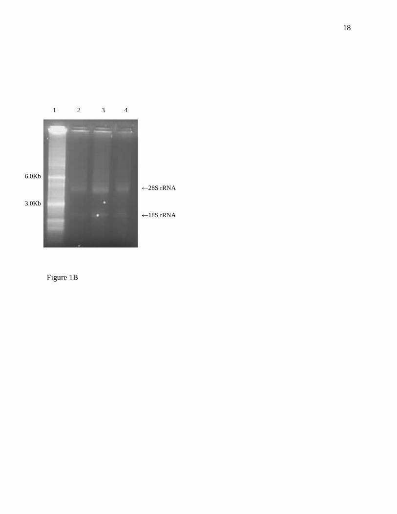
Agarose gel electrophoresis of the RNA extracted from formalin fixed tissues showed a visible
28S rRNA band (Figure 1A) and RNA extracted from HOPE fixed tissues using the same
method displayed both 28S and 18S rRNA bands (Figure 1B). Extracted transcripts for both
formalin and HOPE fixed tissues displayed successful mRNA expression of the housekeeping
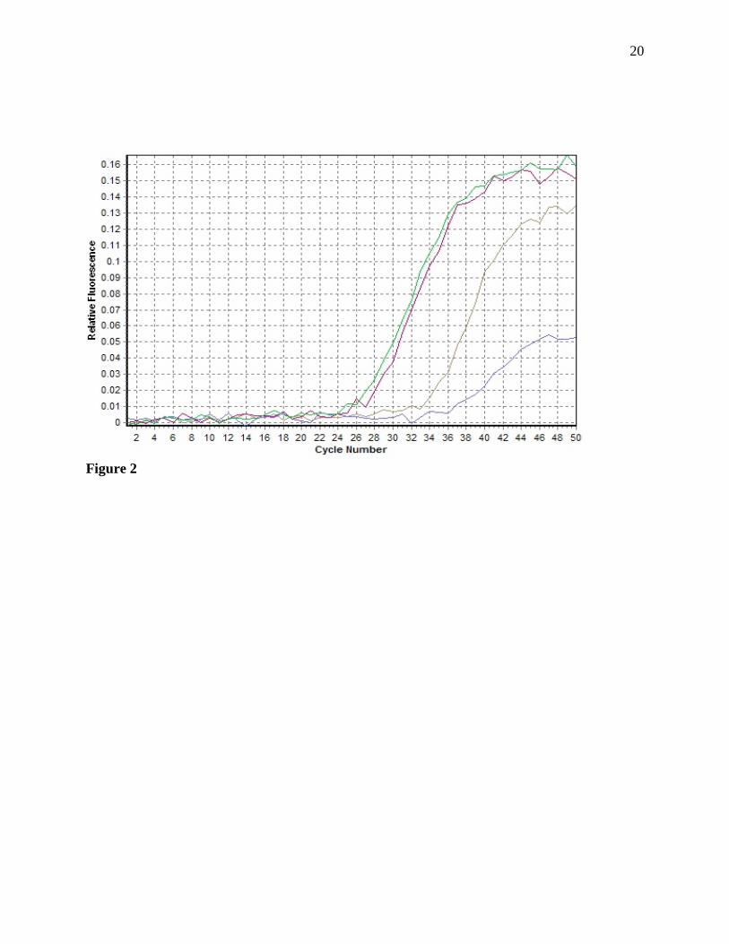
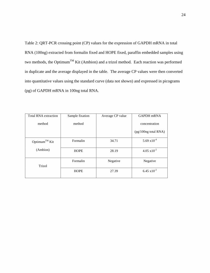
gene GAPDH after qRT-PCR (Figure 2) producing CP values of 34.71 and 28.19, respectively

8
(Table 2). This was converted to quantitative values using the standard curve (Figure 3) and it
was found that the total RNA extracted from HOPE fixed tissues had a higher level of GAPDH
mRNA at 4.05x10⁻²
pg/100ng of total RNA in comparison to total RNA extracted from formalin
fixed tissues at 5.69x10⁻⁴
pg/100ng of total RNA (Table 2).
Total RNA extracted from formalin and HOPE fixed, paraffin embedded tissues using the
trizol method.
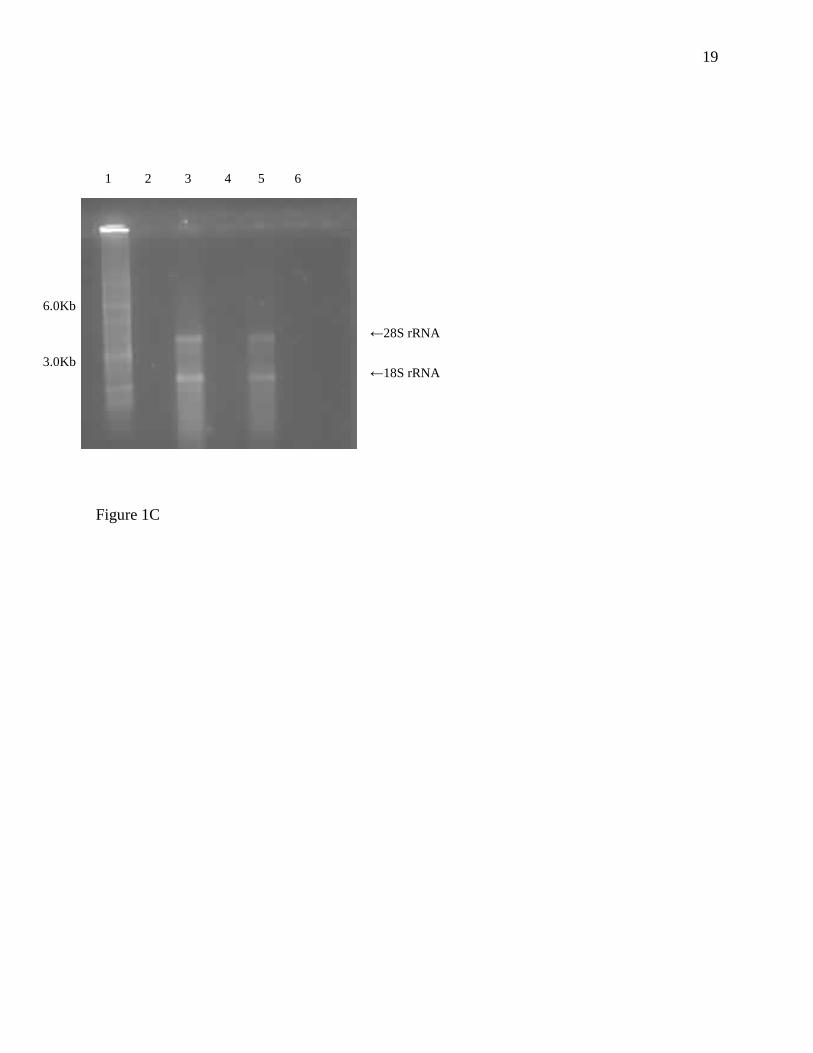
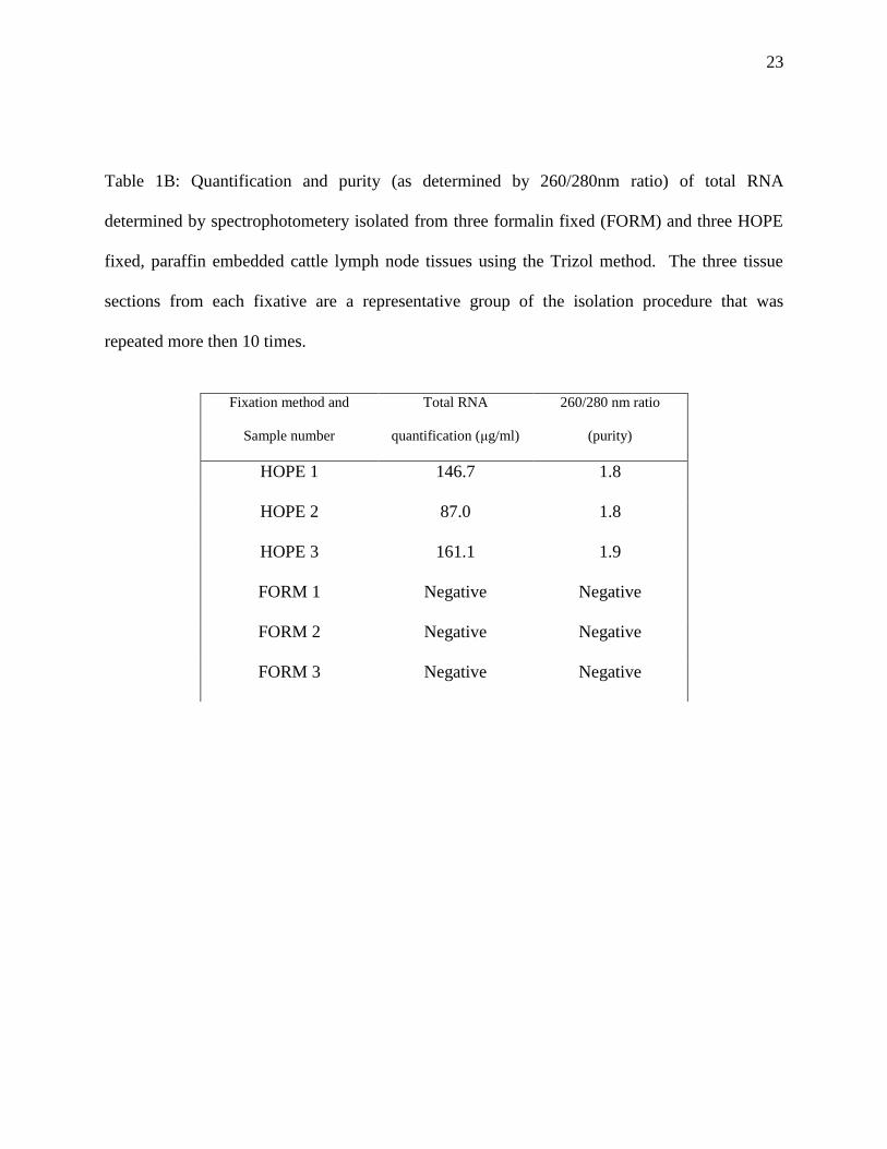
Total RNA extracted from formalin fixed tissues using the trizol method proved unsuccessful as
it could not be quantified using spectrophotometry (Table 1B) and the results were confirmed as
we did not get any rRNA bands on the agarose gel (Figure 1C). It subsequently did not produce
an amplifiable product above the set threshold in qRT-PCR (Table 2). The concentration of
RNA extracted from HOPE fixed tissues using spectrophotometry for the three representative
samples was 87, 146.7 and 161.1 μg/ml total RNA (Table 1B) and the purity was 1.8 and 1.9,
respectively (260/280 nm ratio, Table 1B). The total RNA from HOPE fixed tissues also
exhibited strong 28S and 18S rRNA bands when analysed by gel electrophoresis (Figure 1C).
The extracted transcript displayed successful mRNA expression of the housekeeping gene
GAPDH after qRT-PCR (Figure 2) producing a CP value of 27.39 (Table 2). This was
converted to quantitative values using the standard curve (Figure 3) and found to contain a
GAPDH mRNA concentration of 6.45x10⁻²
pg/100ng of total RNA (Table 2).
All controls used in qRT-PCR were negative and the standard curve produced an adequate line
coefficient of 0.98 and amplification efficiency of 1.9 (Figure 3).

9
Discussion
Studies on disease infection and progression are fast becoming reliant on modern molecular
techniques such as PCR (17). The need for intact RNA extraction methods is an extremely
important prelude to PCR (3, 4) and has seen vast improvements over the last decade (13). This
includes the use of different tissue fixation methods to secure the integrity of RNA during
archival storage. In this study we have compared two fixatives (HOPE and formalin) for their
ability to maintain RNA integrity. We have also compared two different RNA extraction
methods (a commercial kit (Ambion) and a trizol method) to determine which method produced
the highest quantity of total RNA from tissues fixed in both HOPE and formalin. Using the
commercial OptimumTM
FFPE kit (Ambion), total RNA extracted from HOPE fixed tissues
showed comparatively similar quantities and was of a similar purity to total RNA extracted from
formalin fixed tissues (statistically there was no significant difference between the concentration
of total RNA extracted from the two fixation methods, Mann-Whitney test p>0.05). However,
when using qRT-PCR to analyse the content of GAPDH mRNA, the mRNA concentration was
higher in HOPE fixed tissues (4.05x10⁻²
pg/100ng total RNA) compared to formalin fixed
tissues (5.69x10⁻⁴
pg/100ng total RNA) suggesting that HOPE fixation maintained the integrity
of the RNA to a much higher degree (19). This was supported by the agarose gel
electrophoresis, as the total RNA extracted from HOPE fixed tissues displayed both rRNA bands
(18S and 28S) in comparison to the one band (28S) shown in total RNA from formalin fixed
tissues. This work agrees with previously published data where it has been shown that total
RNA extracted from HOPE fixed tissues displayed a stronger integrity and therefore a much
higher level of detectable mRNA in RT-PCR (19, 5, 15). This ability of HOPE fixation could be
due to the increased rate of acetone tissue dehydration, enabled by the diffusion of a protective

10
solution of amino acids into the sample, thus conserving both its morphological and nucleic acid
composition (13).
Using the trizol method, the HOPE fixed tissues produced total RNA of a very similar quantity
and purity to the total RNA extracted from HOPE fixed tissues using the commercial kit
(statistically there was no significant difference between the concentration of total RNA using
the two extraction methods, Mann-Whitney test p>0.05). The resultant GAPDH mRNA quantity
calculated by qRT-PCR was also extremely similar, 4.05x10⁻²
pg/100ng total RNA extracted
using the commercial kit and 6.45x10⁻²
pg/100ng total RNA extracted using the trizol method.
As previously mentioned, the Ambion kit was successful in extracting total RNA from formalin
fixed tissues however we were unable to isolate quantifiable RNA from formalin fixed tissues
using the trizol method. In the Ambion OptimumTM
kit this is most likely attributed to the
proteinase K step in the protocol, which is much more efficient at separating the RNA from its
cross-linked matrix (7). However the period of sample incubation in proteinase K is extremely
long (in our experience it took 6 hours to solubilise only a fraction of the tissue present) and has
been reported to take up to 48 hours (11), leading to further RNA degradation. The cross-linked
matrix is believed to form due to the reaction of RNA with formaldehyde, producing methylene
bridges between the amino groups (13). Due to the absence of formaldehyde in the HOPE
fixative method, tissues fixed in HOPE do not possess cross linkage and therefore perform
equally well in RNA isolation methods with or without proteinase K solution. Similar results
have been reported by Weidorn et al (19).
In conclusion, this study supports a strong consensus that HOPE fixative is a more appropriate
fixative to use for subsequent RNA extraction and molecular techniques. This study is the first
11
to demonstrate the potential of HOPE fixation of M.bovis infected cattle lymph nodes with the
subsequent quantifying of gene specific mRNA within a known amount of total RNA using qRT-
PCR. This complements the work already shown on the benefits of HOPE fixative in human
tissue types such as lung (5), cancer cells (15), spleen and heart (19). Due to the vastly improved
nucleic acid conservation, HOPE fixative is being applied to tissues samples used in subsequent
disease diagnosis, such as the possible detection and even differentiation of the Mycobacterium
tuberculosis complex within archived infected mice tissues (12). In addition to this, this study
has highlighted the need to apply, not just the more efficient fixative technique but also the
appropriate extraction method specific to the fixative used, adding new methods to those
previously reported (19, 5). By considering these factors and exploring the most effective
combination of fixative and extraction method, the quality of molecular techniques such as PCR
and their practical use in exploring genetic expression of both host and pathogen can provide a
basis for future research into intervention and treatment.
Acknowledgements:
We would like to thank Linda Johnson (VLA), Julie Gough (VLA) and the Histopathology
department at the VLA (Surrey) for kindly supplying us with the formalin and HOPE fixed
M.bovis infected, cattle lymph node tissues.
12
References
1. D. Barbaric, L. Dalla-Pozza, J.A. Byrne, A reliable method for total RNA extraction from
frozen human bone marrow samples taken at diagnosis of acute leukaemia. J. Clin.
Pathol. 55 (2002) 865
2. B. Bhudevi and D. Weinstock, Detection of bovine viral diarrhoea virus in formalin fixed
paraffin embedded tissue sections by real time RT-PCR (Taqman). J. Virol. Meth. 109
(2003) 25
3. S.A. Bustin, Quantification of mRNA using real time reverse-transcription Polymerase
Chain Reaction. J. Biomol. Tech. 15 (2002) 155
4. S.A. Bustin and R. Mueller, Real-time reverse transcription PCR and the detection of
occult disease in colorectal cancer. Mol. Aspects Med. 27 (2006) 192
5. T. Goldmann, R. Burgemeister, U. Sauer, S. Loeschke, D.S. Lang, D. Branscheid, P.
Zabel, E. Vollmer, Enhanced molecular analyses by combination of the HOPE- technique
and laser microdissection. Diagn. Pathol. 1 (2006) 2
13
6. U. Lehmann, H. Kreipe, Real-time PCR analysis of DNA and RNA extracted from
formalin-fixed and paraffin-embedded biopsies. Methods. 25 (2006) 409
7. F. Lewis, N.J. Maughan, V. Smith, K. Hillan, P. Quirke, Unlocking the archive – gene
expression in paraffin-embedded tissue. J. Pathol. 195 (2001) 66
8. L. Louie, A.E. Simor, S. Chong, K. Luinstra, A. Petrich, J. Mahony, M. Smieja, G.
Johnson, F. Gharabaghi, R. Tellier, B.M. Willey, S. Poutanen, T. Mazzulli, G.
Broukhanski, F. Jamieson, M. Louie, S. Richardson, Detection of severe acute respiratory
syndrome coronavirus in stool specimens by commercially available real-time reverse
transcriptase PCR assays. J. Clin. Microbiol. 44 (2006) 4193-6
9. M. Macluskey, R. Baillie, H. Morrow, S.L. Schor, A.M. Schor, Extraction of RNA from
archival tissues and measurement of thrombospondin-1 mRNA in normal, dysplastic and
malignant oral tissues. Br. J. Oral. Maxillofac.Surg. 44 (2006) 116
10. J. Olert, K. Weidorn, T. Goldmann, H. Kühl, Y. Mehraein, H. Scherthan, F. Niketeghad,
E. Vollmer, A.M. Müller, J. Müller-Navia, HOPE Fixation: A novel fixing method and
paraffin-embedding technique for human soft tissues. Pathol. Res. Prac. 197 (2001) 823
11. A. Pollet, Y.C. Bedard, S.Q. Li, T. Rohan, R. Kandel, Correlation of p53 mutations in
ThinPrep-processed fine needle aspirates with surgically resected breast cancers. Mod.
pathol. 13 (2001) 1173
12. R. Sen Gupta, D. Hillemann, T. Kubica, G. Zissel, J. Muller-Quernheim, J. Galle, E.
Vollmer, T. Goldmann, HOPE fixation enables improved PCR-based detection and
differentitaiton of Mycobacterium tuberculosis complex in paraffin-embedded tissues.
Pathol. Res. Pract. 199 (2003) 619

14
13. M. Srinivasan, D. Sedmak, S. Jewell, Effect of fixatives and tissue processing on the
content and integrity of nucleic acids. Am. J. Pathol. 161 (2002) 1961
14. S.E. Stroup, S. Roy, J. McHele, V. Maro, S. Ntabaquzi, A. Siddique, G. Kang, R.L.
Guerrant, B.D. Kirkpatrick, R. Fayer, J. Herbein, H. Ward, R. Haque, E.R. Houpt, Real-
time PCR detection and speciation of Cryptosporidium infection using Scorpion probes.
J. Med. Microbiol. 55 (2006) 1217
15. E. Vollmer, J. Galle, D.S. Lang, S. Loeschke, H. Schultz, T. Goldmann, The HOPE
technique opens up a multitude of new possibilities in pathology. Rom. J. Morphol.
Embryol. 47 (2006) 15
16. S. Wacharapluesadee, P. Ruangvejvorachai, T. Hemachudha,
A simple method for
detection of rabies viral sequences in 16-year old archival brain specimens with one-week
fixation in formalin. J. Virol. Meth. 134 (2006) 267.
17. P.R. Wakeley, N. Johnson, L.M. McElhinney, D. Marston, J. Sawyer, A.R. Fooks,
Development of a Real-Time, TaqMan Reverse Transcription-PCR Assay for Detection
and Differentiation of Lyssavirus Genotypes 1, 5, and 6. J. Clin. Microbiol. 43 (2005),
2786
18. S. Widdison, L.J. Schreuder, B. Villarreal-Ramos, C.J. Howard, M. Watson, T.J. Coffey,
Cytokine expression profiles of bovine lymph nodes: effects of Mycobacterium bovis
infection and bacilli Calmette-Guérin vaccination. Clin. Exp. Immunol. 144 (2006) 281
19. K.H. Wiedorn, J. Olert, R.A. Stacey, T. Goldmann, H. Kühl, J. Matthus, E. Vollmer, A.
Bosse, HOPE-a new fixing technique enables preservation and extraction of high
molecular weight DNA and RNA of >20 Kb from paraffin-embedded tissues. Hepes-

15
Glutamic acid buffer mediated organic solvent protection effect. Pathol Res Pract. 198
(2002) 735
Figure legends:
Figure 1A: Agarose gel electrophoresis (ethidium bromide staining) of RNA samples
(1 μg) isolated with the Optimum TM
FFPE Kit (Ambion, UK) from formalin-fixed (lanes 3 and
4), paraffin embedded cattle lymph node tissue. Lane 1 is a 0.5-10 Kb RNA ladder. Lane 2 was
not used. Experiment was repeated three times.
Figure 1B: Agarose gel electrophoresis (ethidium bromide staining) of RNA samples
(1 μg) isolated with the Optimum TM
FFPE Kit (Ambion, UK) from HOPE fixed (lanes 2, 3 and
4) paraffin embedded cattle lymph node tissue. Lane 1 is a 0.5-10 Kb RNA ladder. Experiment
was repeated three times.
Figure 1C: Agarose gel electrophoresis (ethidium bromide staining) of RNA samples
(1 μg) isolated with a trizol method from HOPE fixed (lanes 3 and 5) and formalin-fixed (lanes 2
and 4), paraffin embedded cattle lymph node tissue. Lane 1 is a 0.5-10 Kb RNA ladder. Lane 6
was not used. Experiment was repeated three times.
Figure 2: Real time RT-PCR of total RNA extracted from both the formalin fixed and HOPE
fixed paraffin embedded tissues using the Optimum TM
FFPE kit (brown and red lines,
respectively) and the trizol method (blue and green lines, respectively). The target sequence was
16
an 87 bp fragment of the RNA of bovine Glyceraldehyde-3-phosphate dehydrogenase and each
reaction was performed in duplicate.
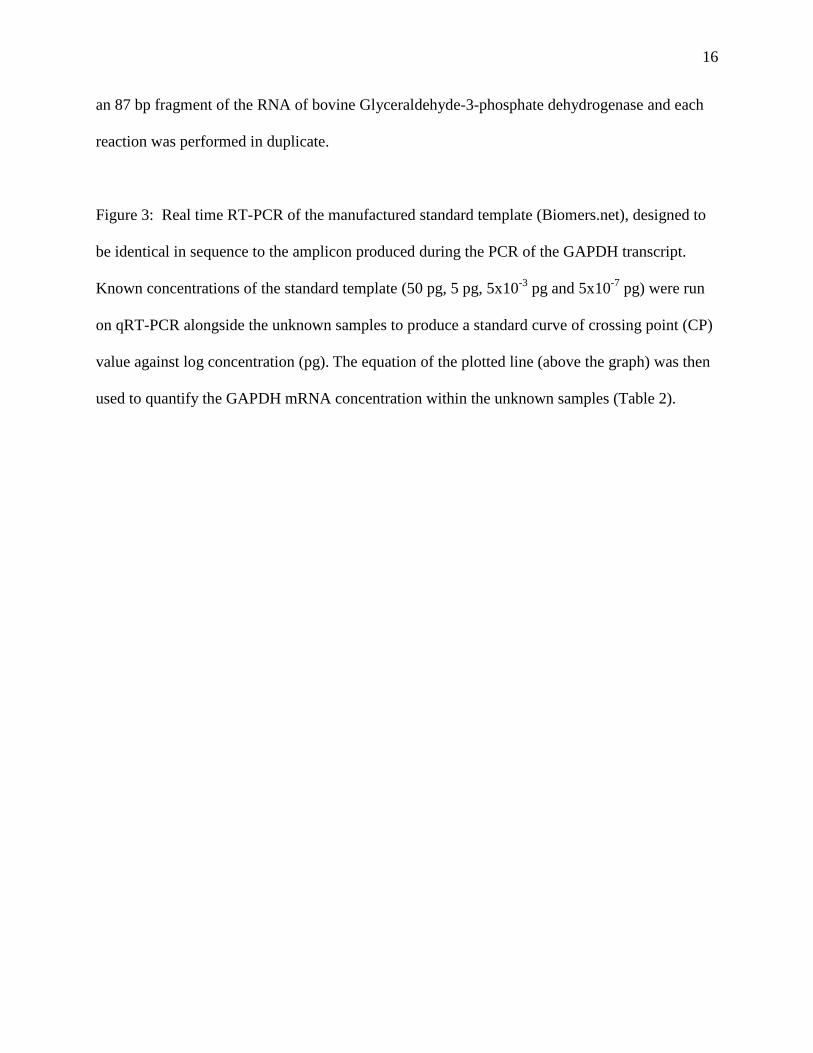
Figure 3: Real time RT-PCR of the manufactured standard template (Biomers.net), designed to
be identical in sequence to the amplicon produced during the PCR of the GAPDH transcript.
Known concentrations of the standard template (50 pg, 5 pg, 5x10⁻³
pg and 5x10⁻⁷
pg) were run
on qRT-PCR alongside the unknown samples to produce a standard curve of crossing point (CP)
value against log concentration (pg). The equation of the plotted line (above the graph) was then
used to quantify the GAPDH mRNA concentration within the unknown samples (Table 2).

17
Figure 1A
←28S rRNA
1 2 3 4
6.0Kb
3.0Kb
18
Figure 1B
←18S rRNA
←28S rRNA
1 2 3 4
6.0Kb
3.0Kb
19
Figure 1C
←28S rRNA
←18S rRNA
1 2 3 4 5 6
6.0Kb
3.0Kb
20
Figure 2

21
Figure 3

22
Table 1A: Quantification and purity (as determined by 260/280nm ratio) of total RNA
determined by spectrophotometery isolated from three formalin-fixed (FORM) and three HOPE
fixed, paraffin embedded cattle lymph node tissue sections using the OptimumTM
FFPE kit
(Ambion). The three tissue sections from each fixative are a representative group of the isolation
procedure that was repeated more then 10 times.
Fixation method and
sample number
Total RNA
quantification (μg/ml)
260/280 nm ratio
(purity)
HOPE 1 107.9 1.8
HOPE 2 82.7 1.9
HOPE 3 97.3 1.9
FORM 1 80.6 1.7
FORM 2 145.7 1.8
FORM 3 97.9 1.9
23
Table 1B: Quantification and purity (as determined by 260/280nm ratio) of total RNA
determined by spectrophotometery isolated from three formalin fixed (FORM) and three HOPE
fixed, paraffin embedded cattle lymph node tissues using the Trizol method. The three tissue
sections from each fixative are a representative group of the isolation procedure that was
repeated more then 10 times.
Fixation method and
Sample number
Total RNA
quantification (μg/ml)
260/280 nm ratio
(purity)
HOPE 1 146.7 1.8
HOPE 2 87.0 1.8
HOPE 3 161.1 1.9
FORM 1 Negative Negative
FORM 2 Negative Negative
FORM 3 Negative Negative
24
Table 2: QRT-PCR crossing point (CP) values for the expression of GAPDH mRNA in total
RNA (100ng) extracted from formalin fixed and HOPE fixed, paraffin embedded samples using
two methods, the OptimumTM
Kit (Ambion) and a trizol method. Each reaction was performed
in duplicate and the average displayed in the table. The average CP values were then converted
into quantitative values using the standard curve (data not shown) and expressed in picograms
(pg) of GAPDH mRNA in 100ng total RNA.
Total RNA extraction
method
Sample fixation
method
Average CP value GAPDH mRNA
concentration
(pg/100ng total RNA)
OptimumTM
Kit
(Ambion)
Formalin 34.71 5.69 x10⁻⁴
HOPE 28.19 4.05 x10⁻²
Trizol
Formalin Negative Negative
HOPE 27.39 6.45 x10⁻²