RK7802 A06 BTY 414
16
Transforming education transforming India Transforming education transforming India LOVELY PROFESSIONAL UNIVERSITY, PHAGWARA (Punjab) Term Paper (BTY-414) Cell Signaling TOPIC: Signaling by Nuclear Receptors TOPIC: Signaling by Nuclear Receptors SUBMITTED BY: - SUBMITTED TO:- Nitish Pathania Mr. Prashant Singh ROLL NO: - RK7802 A 06 REG. NO.: - 10803694
-
Upload
nitishpathania -
Category
Documents
-
view
224 -
download
0
Transcript of RK7802 A06 BTY 414

8/8/2019 RK7802 A06 BTY 414
http://slidepdf.com/reader/full/rk7802-a06-bty-414 1/16
Transforming education transforming IndiaTransforming education transforming India
LOVELY PROFESSIONAL UNIVERSITY,
PHAGWARA (Punjab)
Term Paper (BTY-414)
Cell Signaling
TOPIC: Signaling by Nuclear ReceptorsTOPIC: Signaling by Nuclear Receptors
SUBMITTED BY: - SUBMITTED TO:-
Nitish Pathania Mr. Prashant Singh
ROLL NO: - RK7802 A 06
REG. NO.: - 10803694

8/8/2019 RK7802 A06 BTY 414
http://slidepdf.com/reader/full/rk7802-a06-bty-414 2/16
ACKNOWLEDGEMENT:
I am extremely grateful and remain indebted to my friends and my guide Mr. Prashant
Singh for being a source of inspiration and for their constant support in the Design,
Implementation and Evaluation of this Term Paper. I am thankful to him for their constantconstructive criticism and invaluable suggestions, which benefited me a lot while developing this
paper on topic “Signalling by NUCLEAR Receptors”. Also they provide me a constant source
of inspiration and motivation for doing hard work while preparing this term paper. Through this
column, it would be my utmost pleasure to express my warm thanks to them for their
encouragement, co-operation and consent without which I mightn’t be able to accomplish this
work of Term Paper.
I also want to express my gratitude to my God and my parents those are a great source for me of
inspiration. I am again very thankful to Mr. Prashant Singh who gave me this chance to
express my thoughts with the help of this Term paper regarding the various pathways followed by
nuclear receptors for cellular or signal transduction and also classification of nuclear receptors.
Nitish Pathania
Contents:
1. Nuclear receptors: overview and classification.
Nuclear receptor: Introduction
Structure:
2. Signal transduction:
3. Mechanism of action:
4. nuclear receptors four mechanistic classes
5 . Nucl ear Hor mone Recept o rs :
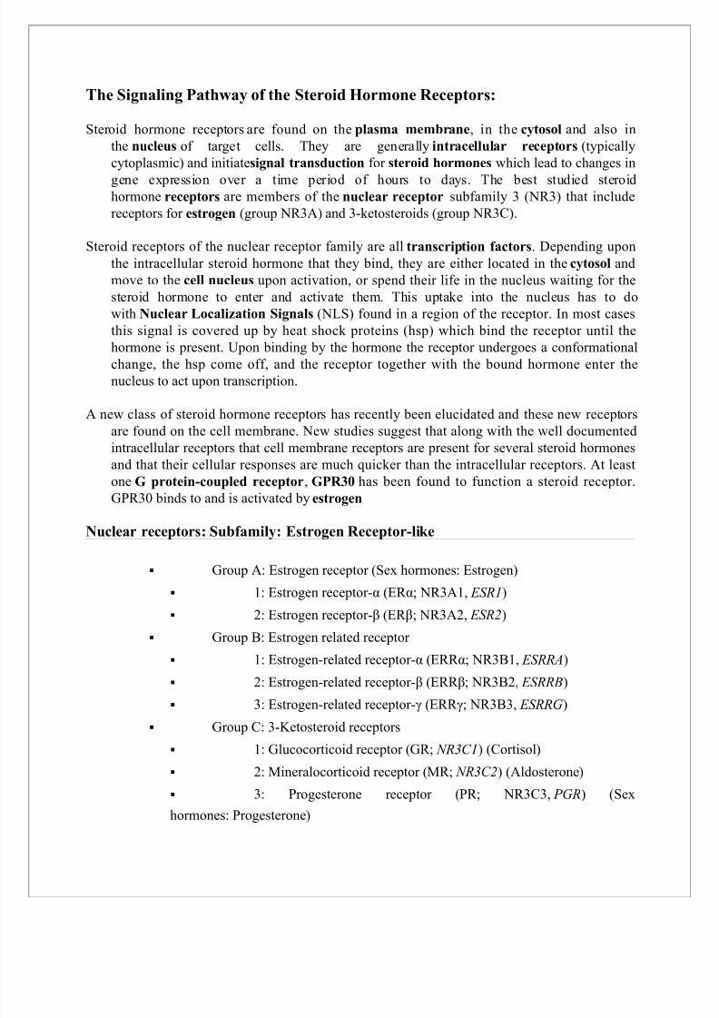
6. The Signaling Pathway of the Steroid Hormone Receptors:
Nuclear receptors: Subfamily: Estrogen Receptor-like
Nuclear receptors enhance our understanding of transcription regulation:
Nuclear receptor superfamily: Principles of signaling
Theraputic Obstacles and Opportunities
Conclusion
References:

8/8/2019 RK7802 A06 BTY 414
http://slidepdf.com/reader/full/rk7802-a06-bty-414 3/16
Nuclear receptors: overview and classification.
Abstract
The nuclear receptor super family comprises a large group of transcription factors that play a key
regulatory role in development and homeostasis of multicellular organisms. A special feature of nuclear receptors is their ability to bind to condensed chromatin templates, which makes them
important initiators of gene transcription. Moreover, the ability of nuclear receptors tosequentially recruit a variety of transcription factors and co regulators to target promoters and toorchestrate the whole process of gene transcription confirms their biological significance and
stimulates intensive research and a high level of scientific interest in this field. In this review, we
summarizes current knowledge regarding the structure and function of nuclear receptors as principal regulators of gene expression. Emphasis is given to the molecular mechanisms of
nuclear receptor-mediated transcriptional activation and repression including recent progress
made in this area.
Nuclear receptor: Introduction
In the field of molecular biology, nuclear receptors are a class of proteins found within cells thatare responsible for sensing steroid and thyroid hormones and certain other molecules. In
response, these receptors work with other proteins to regulate the expression of specific genes,
thereby controlling the development, homeostasis, and metabolism of the organism.
Nuclear receptors have the ability to directly bind to DNA and regulate the expression of adjacent
genes, hence these receptors are classified as transcription factors. The regulation of gene
expression by nuclear receptors happens only when a ligand — a molecule that affects the
receptor's behavior — is present. In more specific terms, ligand binding to a nuclear receptor results in a conformational change in the receptor, which, in turn, activates the receptor, resulting
in up-regulation of gene expression.
A unique property of nuclear receptors that differentiates them from other classes of receptors is
their ability to directly interact with and control the expression of genomic DNA. As aconsequence, nuclear receptors play key roles in both embryonic development and adult
homeostasis.
Intracellular Localization
Most NRs are constitutively localized in the nucleus, however, the major proportion of steroid
receptors and other a few other exceptional receptors may be located in the cytoplasm in the
absence of ligand. Nuclear localization of receptors is mainly regulated by protein-protein
interactions such as dimerization with RXRs or co-regulator proteins . In the cytoplasm, NRs are
bound to heat shock proteins and this association prevents receptor transportation through the
nuclear pores and thus sequesters NRs from binding to DNA . In the nucleus, ligand-mediated
activation of NRs causes redistribution of the receptor to chromatin. Recent evidence which will
be discussed in more detail has suggested that nuclear localization of some NRs is a cell
signaling- and phosphorylation-dependent event.

8/8/2019 RK7802 A06 BTY 414
http://slidepdf.com/reader/full/rk7802-a06-bty-414 4/16
Co-regulator Proteins:
The full activity of NRs depends on a large number of co-regulator proteins that do not bind to
DNA directly, but have a pronounced effect on the outcome of gene expression . In general, non-
liganded NRs form a complex with co-repressor proteins which inhibit transcriptional activity,
often through the recruitment of other cofactor proteins that contain histone deacetylase (HDAC)activity. HDACs alter chromatin structure by promoting chromatin compaction, thus rendering
enhancer regions of genes less accessible to the necessary basal transcriptional machinery.
Activation of NRs by ligand-binding or through phosphorylation induces a conformational
change which results in the dissociation of the co-repressor multiprotein complexes and
subsequent recruitment of co-activator protein complexes that enhance the rate of gene
transcription, often thought the recruitment multiprotein complexes containing histone
acetyltransferase (HAT) activity. Co-regulator proteins thus provide a second level of specificity
in the modulation of gene expression by NRs. Most NR-co-activator proteins identified to date
preferentially interact with NRs through the C-terminal AF-2 domain via an -LXXLL-motif,
which constitutes a prototypical NR-interaction motif. However, in contrast to most co-activator
proteins, the peroxisome proliferator activated receptor gamma co-activator 1 alpha
((PPARGC1a/PGC-1α) interacts not only with the AF-2 region of NRs, but also with the H
region of the selected liver-enriched NRs . In addition to NRs, it has also been shown that the
intrinsic and recruited enzymatic activities of several NR-associated co-factor proteins are
regulated by phosphorylation in a dynamic manner in response to specific signal transduction
pathways, and this will be discussed later in this review in more detail.
Structure:
Nuclear receptors are modular in structure and contain the following domains:
All nuclear receptors are composed of a variable N-terminal domain (NTD, A/B); a highly
conserved DNA Binding Domain (DBD, C); a flexible hinge region (D); and a C-terminal Ligand
Binding Domain (LBD, E). The estrogen receptor α is unique in that it contains an additional C-terminal (F) domain with unknown function.[12]
Nuclear receptors consist of six domains (A-F) based on regions of conserved sequence andfunction. The evolutionarily conserved regions are C and E, and the divergent regions A/B, D,
and F regions.

8/8/2019 RK7802 A06 BTY 414
http://slidepdf.com/reader/full/rk7802-a06-bty-414 5/16
Nuclear receptors contain a variable N-terminal amino acid sequence, (NTD, A/B), which
contains an autonomous transcriptional activation function known as AF-1. The AF-1 shows
weak conservation across the nuclear receptor superfamily and may mediate differential promoter regulation in vivo. The AF-1 sequence functions as a ligand-independent transcriptional activator,
but can also functionally synergize with AF-2. The NTD is unique to each SHR and has variable
sequence and length. The highly conserved C region harbors the DNA-binding domain thatconfers sequence-specific DNA recognition (DBD, C). Situated between the DBD and the LBDis a linker region, domain D. This region functions as a flexible hinge and contains the nuclear
localization signal (NLS). The LBD (E) is responsible for the binding of cognate ligand or
hormone. This domain also contains a ligand-dependent transcriptional activation function (AF-2) necessary for recruiting transcriptional coactivators, which interact with chromatin remodeling
proteins and the general transcriptional activation machinery. Nuclear receptors may or may not
contain a final domain in the C-terminus, the F region, whose sequence is extremely variable andwhose structure and function are unknown.
The domains starting from the N-terminus (left) to C-terminus (right). NTD = N-terminal
domain, DBD = DNA binding domain. LBD = ligand binding domain. AF = activation function.The steroid hormone receptor abbreviations are ER – estrogen receptor, GR – glucocorticoid
receptor, PR –progesterone receptor, AR – androgen receptor, and MR – mineralocorticoid
receptor. The numbers to the right are the lengths in amino acid residues.
A/B REGION (N-TERMINAL DOMAIN, NTD)
Thus far, there is no elucidation of a crystal structure of an A/B domain. The A/B region in thedifferent NR is highly variable, revealing a very weak evolutionary conservation. The N-terminal
region is the least conservedregion among NR, both in size and sequence. All the nuclear receptors have a unique N-terminal region (NTD) of variable length (100–500 amino acids)
whose 3D structure is unknown.
NR contains two transactivation functions. One maps to the structurally flexible N-terminal
domain (NTD) and is termed AF-1. The poorly defined N-terminal A/B region contains a

8/8/2019 RK7802 A06 BTY 414
http://slidepdf.com/reader/full/rk7802-a06-bty-414 6/16
transcriptional activation function, referred to as activation function 1 (AF-1) that can operate
autonomously. AF-1 can act in a ligand-independent manner when placed outside of the receptor.
Besides the one constitutionally active transactivation region (AF-1), NTD includes severalautonomous transactivation domains (AD). The activation domains (AD) contain transcriptional
activation functions that can activate transcription when fused to a heterologous DNA-binding
domain.
The main determinants for transactivation map to NTD of both the androgen (AR) andglucocorticoid (GR) receptors and while generally there is little sequence conservation between
the different NTD, in contrast to the DBD and LBD, short regions of similarity have been
observed for the AR and GR. The NTD is potentially involved in multiple protein-protein
interactions and the length of this domain has a positive correlation with the activity of AF-1 for different members of the nuclear receptor superfamily.
C REGION (DNA BINDING DOMAIN, DBD):
The DBD consists of a highly conserved residue core located between the N-terminal domain andthe C-terminal ligand-binding domain. The DNA binding domain lies toward the center of themolecule. The amino acid sequence of this domain is similar among different steroid receptors
(56–79% identity). The 3D structure of the DBD has been resolved for a number of nuclear
receptors. Nuclear magnetic resonance and crystallographic studies for different NR DBD in their DNA uncomplexed and complexed forms with the GR and ER homodimers on their cognate
DNA sequence were the first 3D crystal structure reported.
D REGION (HINGE REGION):
The D region, which is a poorly conserved domain, serves as a hinge between the DBD and the
LBD, allowing rotation of the DBD. The hinge region allows the DBD and LBD to adoptdifferent conformations without creating a steric hindrance. This domain also harbors a nuclear
localization signal (NLS) or at least some elements of a functional nuclear localization signal.
E REGION (LIGAND BINDING DOMAIN, LBD):
The largest domain is the moderately conserved ligand-binding domain (LBD, E region). Thehallmark of a nuclear receptor is its ligand-binding domain (LBD). This domain is highly
structured, and encodes a wealth of distinct functions most of which operate in a ligand-
dependent manner. The highly conserved region of the nuclear receptor proteins lies near thecarboxyl terminus. The AR C-terminal ligand-binding domain contains about 290 amino acids
and represents about 30% of the receptor. The ligand-binding domains of AR from humans, rats,
and mice are identical, and sequence homology with other steroid receptors ranges between 15%
and 54%.
Signal transduction:

8/8/2019 RK7802 A06 BTY 414
http://slidepdf.com/reader/full/rk7802-a06-bty-414 7/16
Overview of signal transduction pathways
• In biology, signal transduction is a mechanism that converts a
mechanical/chemical stimulus to acell into a specific cellular response. Signaltransduction starts with a signal to a receptor, and ends with a change in cell function.
• Transmembrane receptors span the cell membrane, with part of the receptor outside and
part inside the cell. The chemical signal binds to the outer portion of the receptor,
changing its shape and conveying another signal inside the cell. Some chemical
messengers, such as testosterone, can pass through the cell membrane, and bind directly
to receptors in the cytoplasm or nucleus.
• Sometimes there is a cascade of signals within the cell. With each step of the cascade, the
signal can be amplified, so a small signal can result in a large response. Eventually, the
signal creates a change in the cell, either in the expression of the DNA in the nucleus or in
the activity of enzymes in the cytoplasm.
• These processes can take milliseconds (for ion flux), minutes (for protein- and lipid-
mediated kinase cascades), hours, or days (for gene expression).

8/8/2019 RK7802 A06 BTY 414
http://slidepdf.com/reader/full/rk7802-a06-bty-414 8/16
Mechanism of action:
Mechanism nuclear receptor action. This figure depicts the mechanism of a class I nuclear
receptor (NR) that, in the absence of ligand, is located in the cytosol. Hormone binding to the NR
triggers dissociation of heat shock proteins (HSP), dimerization, and translocation to the nucleus,
where the NR binds to a specific sequence of DNA known as a hormone response element
(HRE). The nuclear receptor DNA complex in turn recruits other proteins that are responsible for
transcription of downstream DNA into mRNA, which is eventually translated into protein, which
results in a change in cell function.
Mechanism of nuclear receptor action. This figure depicts the mechanism of a class II nuclear

8/8/2019 RK7802 A06 BTY 414
http://slidepdf.com/reader/full/rk7802-a06-bty-414 9/16
receptor (NR), which, regardless of ligand-binding status, is located in the nucleus bound to
DNA. For the purpose of illustration, the nuclear receptor shown here is the thyroid hormone
receptor (TR ) heterodimerized to the RXR . In the absence of ligand, the TR is bound to
corepressor protein. Ligand binding to TR causes a dissociation of corepressor and recruitment of
coactivator protein, which, in turn, recruits additional proteins such as RNA polymerase that are
responsible for transcription of downstream DNA into RNA and eventually protein, which results
in a change in cell function.
Nuclear receptors (NRs) may be classified into two broad classes according to their mechanismof action and subcellular distribution in the absence of ligand.
Small lipophilic substances such as natural hormones diffuse past the cell membrane and bind to
nuclear receptors located in the cytosol (type I NR) or nucleus (type II NR) of the cell. This
causes a change in the conformation of the receptor, which, depending on the mechanistic class(type I or II), triggers a number of downstream events that eventually results in up or down
regulation of gene expression.
Accordingly, nuclear receptors may be subdivided into the following four mechanistic classes:
Type I
Ligand binding to type I nuclear receptors in the cytosol results in the dissociation of heat shock
proteins, homo-dimerization, translocation (i.e., active transport) from the cytoplasm into the cell
nucleus, and binding to specific sequences of DNA known as hormone response elements (HREs). Type I nuclear receptors bind to HREs consisting of two half-sites separated by a
variable length of DNA, and the second half-site has a sequence inverted from the first (inverted
repeat).
Type I nuclear receptors include members of subfamily 3, such as the androgen receptor, estrogen receptors, glucocorticoid receptor , and progesterone receptor .
The nuclear receptor/DNA complex then recruits other proteins that transcribe DNA downstream
from the HRE into messenger RNA and eventually protein, which causes a change in cellfunction.
Type II
Type II receptors, in contrast to type I, are retained in the nucleus regardless of the ligand binding
status and in addition bind as hetero-dimers (usually with RXR ) to DNA. In the absence of ligand, type II nuclear receptors are often complexed with corepressor proteins. Ligand binding to
the nuclear receptor causes dissociation of corepressor and recruitment of coactivator proteins.
Additional proteins including RNA polymerase are then recruited to the NR/DNA complex thattranscribe DNA into messenger RNA.

8/8/2019 RK7802 A06 BTY 414
http://slidepdf.com/reader/full/rk7802-a06-bty-414 10/16
Type II nuclear receptors include principally subfamily 1, for example the retinoic acid receptor,
retinoid X receptor and thyroid hormone receptor .
Type III
Type III nuclear receptors (principally NR subfamily 2) are similar to type I receptors in that bothclasses bind to DNA as homodimers. However, type III nuclear receptors, in contrast to type I,
bind to direct repeat instead of inverted repeat HREs.
Type III nuclear receptors are orphan receptors, with their endogenous ligands still unknown.
Type IV
Type IV nuclear receptors bind either as monomers or dimers, but only a single DNA binding
domain of the receptor binds to a single half site HRE. Examples of type IV receptors are foundin most of the NR subfamilies.
Nuclear Hormone Receptors:
Nuclear hormone receptor proteins form a class of ligand activated proteins that, when bound
to specific sequences of DNA serve as on-off switches for transcription within the cellnucleus. These switches control the development and differentiation of skin, bone and
behavioral centers in the brain, as well as the continual regulation of reproductive tissues.
Researchers at the Theoretical Biophysics Group study the interaction of some members of the nuclear hormone receptor with DNA as well as their interaction with hormones.
Nuclear hormone receptors are ligand-activated transcription factors that regulate geneexpression by interacting with specific DNA sequences upstream of their target genes. Asearly as 1968 a two-step mechanism of action was proposed for these receptors based upon
the observation of an inactive and an active state of the receptors. The first step involves
activation through binding of the hormone; the second step consists of receptor binding toDNA and regulation of transcription.
A hormone response element (HRE) is a specific DNA sequence that a receptor recognizeswith markedly increased affinity and typically contains two consensus hexameric half-sites.
The identity of a response element resides in three features: the sequence of the base pairs in
the half-site, the number of base pairs between the half-sites and the relative orientation of
the two half-sites. Thus each receptor protein dimer that binds the DNA has to recognize thesequence, spacing and orientation of the half-sites within their response element.
The nuclear hormone receptor proteins are composed of several domains which are
differentially conserved between the various receptors and have different roles: a variable N-terminal region, a conserved DNA binding domain (DBD), a variable hinge region, a
conserved ligand binding domain (LBD), and a variable C-terminal region.
8/8/2019 RK7802 A06 BTY 414
http://slidepdf.com/reader/full/rk7802-a06-bty-414 11/16

8/8/2019 RK7802 A06 BTY 414
http://slidepdf.com/reader/full/rk7802-a06-bty-414 12/16
4: Androgen receptor (AR ; NR3C4, AR) (Sex
hormones: Testosterone)
Structure:
Intracellular steroid hormone receptors share a common structure of four units that are
functionally homologous, so-called "domains":
1. Variable domain: It begins at the N-terminal and is the most variable domain
between the different receptors.
2. DNA binding domain: This centrally located highly conserved DNA binding
domain (DBD) consists of two non-repetitive globular motifs where zinc is coordinated
with four cysteine and nohistidine residues. Their secondary and tertiary structure is
distinct from that of classic zinc fingers. This region controls which gene will beactivated. On DNA it interacts with the hormone response element (HRE).
3. Hinge region: This area controls the movement of the receptor to the nucleus.
4. Hormone binding domain: The moderately conserved ligand-binding domain
(LBD) can include a nuclear localization signal, amino-acid sequences capable of binding
chaperones and parts of dimerization interfaces. Such receptors are closely related
to chaperones (namely heat shock proteins hsp90 and hsp56), which are required to
maintain their inactive (but receptive) cytoplasmicconformation. At the end of this
domain is the C-terminal. The terminal connects the molecule to its pair in the
homodimer or heterodimer. It may affect the magnitude of the response.
Depending on their mechanism of action and subcellular distribution, nuclear receptors may be
classified into at least two classes. Nuclear receptors that bind steroid hormones are all classified
as type I receptors. Only type I receptors have a heat shock protein (hsp) associated with the
inactive receptor that will be released when the receptor interacts with the ligand. Type I
receptors may be found in homodimer or heterodimer forms. Type II nuclear receptors have no
hsp, and in contrast to the classical type I receptor are located in the cell nucleus.
There is some evidence that certain steroid hormone receptors can extend through lipid bilayer
membranes at the surface of cells and might be able to interact with hormones that remain outside
of cells.
Steroid hormone receptors can also function outside of the nucleus and couple to cytoplasmic
signal transduction proteins such as PI3k and Akt kinase.

8/8/2019 RK7802 A06 BTY 414
http://slidepdf.com/reader/full/rk7802-a06-bty-414 13/16
Mechanism of action:
Free (that is, unbound) steroids enter the cell cytoplasm and interact with their receptor. In this
process heat shock protein is dissociated, and the activated receptor-ligand complex is
translocated into the nucleus.
After binding to the ligand (steroid hormone), steroid receptors often form dimers. In the nucleus,
the complex acts as a transcription factor , augmenting or
suppressing transcription particular genes by its action on DNA.
Type II receptors are located in the nucleus. Thus, their ligands pass through the cell wall and
cytoplasm and enter the nucleus where they activate the receptor without release of hsp. The
activated receptor interacts with the hormone response element and the transcription process is
initiated as with type I receptors.
The cell membrane aldosterone receptor has shown to increase the activity of the
basolateral Na/K ATPase, ENaC sodium channels and ROMK potassium channels of
the principal cell in the distal tubule and cortical collecting duct of nephrons (as well as in the
large bowel and possibly in sweat glands).
A key feature of steroid hormone receptors that is likely to provide a target for therapy confined
to one tissue is the capability of specific parts of the receptor to interact with proteins which have
important roles in regulation of cell physiology.
Nuclear receptors enhance our understanding of transcription regulation:
Receptors for retinoic acid, vitamin D3 and the steroid and thyroid hormones belong to a familyof ligand-activated enhancer-binding factors which are composed of a number of functional
domains required for ligand and DNA binding, nuclear translocation, dimerization and trans-
activation of transcription. Ligand binding, which may promote dissociation of the receptor froma heat shock protein, results in the binding of the ligand-receptor complex as a dimer to its
cognate palindromic responsive element. The mechanism by which the DNA-bound receptor
activates transcription is unknown but appears to involve more than one trans-activating domain.
Nuclear receptor superfamily: Principles of signaling
Nuclear receptors (NRs) comprise a family of 49 members that share a common structuralorganization and act as ligand-inducible transcription factors with major (patho)physiological
impact. For some NRs (“orphan receptors”), cognate ligands have not yet been identified or may
not exist. The principles of DNA recognition and ligand binding are well understood from both biochemical and crystal structure analyses. The 3D structures of several DNA-binding domains

8/8/2019 RK7802 A06 BTY 414
http://slidepdf.com/reader/full/rk7802-a06-bty-414 14/16

8/8/2019 RK7802 A06 BTY 414
http://slidepdf.com/reader/full/rk7802-a06-bty-414 15/16
Nuclear receptor subfamily 3, Estrogen Receptor-like contains Group A: Estrogen receptors
include members 1: Estrogen receptor-α (ERα; NR3A1, ESR1) and 2: Estrogen receptor-β
(ERβ; NR3A2, ESR2); Group B, the Estrogen related receptors include members 1: Estrogenrelated receptor-α (ERRα; NR3B1, ESRRA), 2: Estrogen related receptor-β (ERRβ; NR3B2,
ESRRB), and 3: Estrogen related receptor-γ (ERRγ; NR3B3, ESRRG); and Group C, the 3-
Ketosteroid receptors include members 1: Glucocorticoid receptor (GR; NR3C1) (Cortisol), 2:Mineralocorticoid receptor (MR; NR3C2) (Aldosterone), 3: Progesterone receptor (PR; NR3C3, PGR) (Sex hormones: Progesterone), and 4: Androgen receptor (AR; NR3C4, AR)
(Sex hormones: Testosterone).
Theraputic Obstacles and Opportunities
NRs control many aspects of biology including development, reproduction, and homeostasis
through target gene activation. The ability to modulate by their activity using fat-solublemolecules makes them extremely attractive drug targets. As our understanding of NR signaling
increases, so does our appreciation of the complexity of their regulation. It is possible that
management of diseases in the future will include therapies that not only target NRs, but also co-
regulator proteins and signaling pathways that are critical in the modulation of their function.
PPARs are the targets of some commonly used drugs in the treatment of hyperlipidemia and type-
2-diabetes. Activation of PPARα by fibrates causes the up-regulation of genes involved in the β-
oxidation of fatty acids.
However, recent evidence has indicated an increased risk of heart attacks with rosiglitazone(marketed as Avandia) and the FDA released a safety alert on the drug in May 2007. Further
research surrounding the signaling events and co-regulator proteins that affect PPARγ activity in
multiple tissues may be useful in separating the therapeutic effects from the toxic effects of drugs
like rosiglitazone.
One therapeutic challenge and opportunity in development of drugs that target NRs are selective
therapeutic modulators (SRMs). SRMs are NR ligands that exhibit agonistic or antagonistic
activity in a cell- or tissue-dependent manner. The classic SRM is tamoxifen, which can
selectively activate or inhibit estrogen receptor and is commonly used in the treatment of breast
cancer.
SRM-induced alterations in the conformation of NRs may affect the ability of the receptor to bind
to co-regulators or to be phosphorylated. The expression profile of specific co-activator proteins
and co-repressor proteins in a given cell type may affect the relative agonist –vs-antagonist
activity of SRMs.

8/8/2019 RK7802 A06 BTY 414
http://slidepdf.com/reader/full/rk7802-a06-bty-414 16/16
Conclusion
It is clear that multiple signaling pathways and phosphorylation events affect NR-mediated
signaling. They modulate protein-protein interactions, sub-cellular localization, DNA-binding,
protein stability, and transactivation capacity. The situation is further complicated by the fact that
many NR cofactor proteins are themselves modulated by signaling pathways and phosphorylation
events that affect their intrinsic and recruited enzymatic activities. Further investigation into the
role of cell signaling pathways in NR-mediated transcription, and into signaling pathway
crosstalk will be necessary to fully understand the functional implication of these signaling
events. In addition, further characterization of these processes will likely lead to the development
of novel and selective therapeutic molecules for a multitude of indications.
References:
1. http://en.wikipedia.org/wiki/Nuclear_receptor
2. http://www.nature.com/nsmb/journal/v5/n8/full/nsb0898_679.html
3. http://www.citeulike.org/user/ajaymalik/article/3213621
4. http://www.mesomorphosis.com/articles/scally/steroid-hormone-nuclear-
receptors.htm
5. http://www.ks.uiuc.edu/Research/pro_DNA/ster_horm_rec/
6. http://en.wikipedia.org/wiki/Steroid_hormone_receptor