Ribosome-Mediated Incorporation of Dipeptides and Dipeptide
4
Ribosome-Mediated Incorporation of Dipeptides and Dipeptide Analogues into Proteins in Vitro Rumit Maini, Larisa M. Dedkova, Rakesh Paul, Manikandadas M. Madathil, Sandipan Roy Chowdhury, Shengxi Chen, and Sidney M. Hecht* Center for BioEnergetics, Biodesign Institute, and Department of Chemistry & Biochemistry, Arizona State University, Tempe, Arizona 85287, United States * S Supporting Information ABSTRACT: Plasmids containing 23S rRNA randomized at positions 2057−2063 and 2502−2507 were introduced into Escherichia coli,affording a library of clones which produced modified ribosomes in addition to the pre- existing wild-type ribosomes. These clones were screened with a derivative of puromycin, a natural product which acts as an analogue of the 3′-end of aminoacyl-tRNA and terminates protein synthesis by accepting the growing polypeptide chain, thereby killing bacterial cells. The puromycin derivative in this study contained the dipeptide p-methoxyphenylalanylglycine, implying the ability of the modified ribosomes in clones sensitive to this puromycin analogue to recognize dipeptides. Several clones inhibited by the puromycin derivative were used to make S-30 preparations, and some of these were shown to support the incorporation of dipeptides into proteins. The four incorporated species included two dipeptides (Gly-Phe (2) and Phe-Gly (3)), as well as a thiolated dipeptide analogue (4) and a fluorescent oxazole (5) having amine and carboxyl groups approximately the same distance apart as in a normal dipeptide. A protein containing both thiolated dipeptide 4 and a 7-methoxycoumarin fluorophore was found to undergo fluorescence quenching. Introduction of the oxazole fluorophore 5 into dihydrofolate reductase or green fluorescent protein resulted in quite strong enhance- ment of its fluorescence emission, and the basis for this enhancement was studied. The aggregate results demon- strate the feasibility of incorporating dipeptides as a single ribosomal event, and illustrate the lack of recognition of the central peptide bond in the dipeptide, potentially enabling the incorporation of a broad variety of structural analogues. I n recent years, many laboratories have employed ribosome- mediated protein synthesis to produce proteins containing non-proteinogenic amino acids, enabling a more detailed study of protein structure, function, and dynamics. 1 While a broad range of amino acids can now be incorporated into proteins, the incorporation of α-amino acids containing large and negatively charged side chains is still challenging, 2 as is protein biosynthesis with non α-amino acids. 3 Recently, we have described the facilitated incorporation of non-proteinogenic amino acids by the use of modified ribosomes in which key regions of the Escherichia coli 23S rRNA have been altered. Initial efforts involved the successful incorporation of D-amino acids 4 and β-amino acids. 5 The strategy employed for the incorporation of β-amino acids was of special interest, involving the use of a puromycin analogue containing a β-amino acid to enable the selection of promising ribosomal architectures at the level of bacterial cells harboring the plasmids with the modified 23S rRNAs. 5a This study argued that it may well be possible to mediate the incorporation of other types of amino acids using ribosomes selected by the use of puromycin analogues containing the amino acid structures of interest. Presently, we employ a puromycin derivative (1) containing the dipeptide p-methoxyphenylalanylglycine, and demonstrate that the selected ribosomes successfully incorpo- Received: March 25, 2015 Published: August 24, 2015 Figure 1. Puromycin derivative 1 used for selection of modified ribosomes, and dipeptides (2 and 3) and dipeptide analogues (4 and 5) incorporated into proteins using the modified ribosomes. Communication pubs.acs.org/JACS © 2015 American Chemical Society 11206 DOI: 10.1021/jacs.5b03135 J. Am. Chem. Soc. 2015, 137, 11206−11209 Downloaded by NATL LBRY OF SERBIA on September 15, 2015 | http://pubs.acs.org Publication Date (Web): August 31, 2015 | doi: 10.1021/jacs.5b03135
-
Upload
romana-masnikosa -
Category
Documents
-
view
224 -
download
5
description
ribosome mediated
Transcript of Ribosome-Mediated Incorporation of Dipeptides and Dipeptide
Ribosome-Mediated Incorporation of Dipeptides and DipeptideAnalogues into Proteins in VitroRumit Maini, Larisa M. Dedkova, Rakesh Paul, Manikandadas M. Madathil, Sandipan Roy Chowdhury,Shengxi Chen, and Sidney M. Hecht*
Center for BioEnergetics, Biodesign Institute, and Department of Chemistry & Biochemistry, Arizona State University, Tempe,Arizona 85287, United States
*S Supporting Information
ABSTRACT: Plasmids containing 23S rRNA randomizedat positions 2057−2063 and 2502−2507 were introducedinto Escherichia coli, affording a library of clones whichproduced modified ribosomes in addition to the pre-existing wild-type ribosomes. These clones were screenedwith a derivative of puromycin, a natural product whichacts as an analogue of the 3′-end of aminoacyl-tRNA andterminates protein synthesis by accepting the growingpolypeptide chain, thereby killing bacterial cells. Thepuromycin derivative in this study contained the dipeptidep-methoxyphenylalanylglycine, implying the ability of themodified ribosomes in clones sensitive to this puromycinanalogue to recognize dipeptides. Several clones inhibitedby the puromycin derivative were used to make S-30preparations, and some of these were shown to supportthe incorporation of dipeptides into proteins. The fourincorporated species included two dipeptides (Gly-Phe (2)and Phe-Gly (3)), as well as a thiolated dipeptide analogue(4) and a fluorescent oxazole (5) having amine andcarboxyl groups approximately the same distance apart asin a normal dipeptide. A protein containing both thiolateddipeptide 4 and a 7-methoxycoumarin fluorophore wasfound to undergo fluorescence quenching. Introduction ofthe oxazole fluorophore 5 into dihydrofolate reductase orgreen fluorescent protein resulted in quite strong enhance-ment of its fluorescence emission, and the basis for thisenhancement was studied. The aggregate results demon-strate the feasibility of incorporating dipeptides as a singleribosomal event, and illustrate the lack of recognition ofthe central peptide bond in the dipeptide, potentiallyenabling the incorporation of a broad variety of structuralanalogues.
In recent years, many laboratories have employed ribosome-mediated protein synthesis to produce proteins containing
non-proteinogenic amino acids, enabling a more detailed studyof protein structure, function, and dynamics.1 While a broadrange of amino acids can now be incorporated into proteins,the incorporation of α-amino acids containing large andnegatively charged side chains is still challenging,2 as is proteinbiosynthesis with non α-amino acids.3
Recently, we have described the facilitated incorporation ofnon-proteinogenic amino acids by the use of modifiedribosomes in which key regions of the Escherichia coli 23S
rRNA have been altered. Initial efforts involved the successfulincorporation of D-amino acids4 and β-amino acids.5 Thestrategy employed for the incorporation of β-amino acids wasof special interest, involving the use of a puromycin analoguecontaining a β-amino acid to enable the selection of promisingribosomal architectures at the level of bacterial cells harboringthe plasmids with the modified 23S rRNAs.5a This study arguedthat it may well be possible to mediate the incorporation ofother types of amino acids using ribosomes selected by the useof puromycin analogues containing the amino acid structures ofinterest. Presently, we employ a puromycin derivative (1)containing the dipeptide p-methoxyphenylalanylglycine, anddemonstrate that the selected ribosomes successfully incorpo-
Received: March 25, 2015Published: August 24, 2015
Figure 1. Puromycin derivative 1 used for selection of modifiedribosomes, and dipeptides (2 and 3) and dipeptide analogues (4 and5) incorporated into proteins using the modified ribosomes.
Communication
pubs.acs.org/JACS
© 2015 American Chemical Society 11206 DOI: 10.1021/jacs.5b03135J. Am. Chem. Soc. 2015, 137, 11206−11209
Dow
nloa
ded
by N
AT
L L
BR
Y O
F SE
RB
IA o
n Se
ptem
ber
15, 2
015
| http
://pu
bs.a
cs.o
rg
Pub
licat
ion
Dat
e (W
eb):
Aug
ust 3
1, 2
015
| doi
: 10.
1021
/jacs
.5b0
3135

rated three dipeptides (2−4, Figure 1), including one dipeptidehaving a thioamide linkage (4), as well as a fluorescentdipeptidomimetic analogue (5, Scheme S1) reminiscent of thefluorophores in natural fluorescent proteins.6
The bacterial clones employed for screening were part of apreviously described library containing modifications in specificregions of 23S rRNA known to be involved in peptide bondformation.5a,7 These were screened for inhibition bydipeptidylpuromycin 1 (Figure 1), the preparation of whichis outlined in Scheme S2. A total of 419 clones were screened,and 13 were inhibited by at least 50% in the presence of 100μg/mL puromycin derivative 1. Nine of these clones,containing the sequence 2057UGCGUGG2063 in their 23SrRNA to confer erythromycin resistance, had also beenrandomized in the region 2502−2507, and are summarized inTable 1. As shown, four of these nine clones proved to be
identical, having the sequence 2502ACGAAG2507, andanother two shared the sequence 2502CUACAG2507. Clone010326R6 was chosen for further study based on its inhibitionby 1 and the favorable properties of the S-30 system preparedfrom this clone in cell-free protein synthesis experiments.As shown in Scheme S3, glycylphenylalanine (2) was
protected as its N-pentenoyl derivative and used to esterifythe dinucleotide pdCpA.8 Bacteriophage T4 RNA ligase wasthen employed to ligate the dinucleotide to an in vitro RNAtranscript, affording glycylphenylalanyl-tRNACUA. Phenylalanyl-glycine (3) was used to prepare phenylalanylglycyl-tRNACUAanalogously (not shown). When utilized in an S-30 systemprepared from clone 010326R6, and in the presence of E. colidihydrofolate reductase (DHFR) mRNA containing a UAGcodon corresponding to position 10 of DHFR, the dipeptidyl-tRNACUAs effected suppression of the UAG codon, producingDHFRs containing 2 (8% suppression) and 3 (14%suppression) at position 10 (Figure 2). Verification of the
incorporation of 2 was accomplished by MALDI-MS analysis ofa tryptic digest of the elaborated DHFR (Figure S2) whichcontained an ion at m/z 1377, corresponding to the peptidefragment MISLIAALAGFDR, while DHFR containing phenyl-alanine at position 10 had the analogous ion at m/z 1320,corresponding to MISLIAALAFDR. A complete analysis offragment ions is summarized in Table S1. It may be noted that,apart from clone 010326R6, the S-30 preparations derived froman additional three of the nine unique clones identified couldalso incorporate 2 and 3 into DHFR with comparablesuppression efficiency (not shown). Further, the DHFRprepared from clone 010326R6 containing 2 at position 10(Figure 2) was ∼84% as active as authentic wild-type DHFR,providing a measure of the fidelity with which this S-30 systemwas able to incorporate α-amino acids.9 The lower fidelity ofthe modified ribosomes is unsurprising, and will likely prove tobe general for such ribosomes.Thiopeptide moieties have been shown to be useful as
fluorescence quenchers in peptides and proteins, functioningboth by Forster resonance energy transfer (FRET)10 and byphotoinduced electron transfer (PET).11 This property hasbeen utilized very effectively by the Petersson laboratory forcharacterization of protein structure following introduction ofthe requisite thioamide by native chemical ligation of asynthetic peptide fragment containing the thioamide to theremainder of the ribosomally produced protein.10b,12,13 Sincethe modified ribosomes described here could plausibly providean alternative route to proteins containing thioamides atpredetermined positions, we incorporated a thiolated dipeptide(4) into position 16 of DHFR and the fluorescent probe L-(7-methoxycoumarinyl-4-yl)ethylglycine, which is known to besensitive to environment,14 into position 49.15
The requisite thiolated dipeptide was obtained as outlined inScheme S4. Fully protected phenylalanylglycine was convertedto the respective thiodipeptide (24) by treatment withLawesson’s reagent.16 Following conversion to the N-pentenoylprotected cyanomethyl ester (26), condensation with pdCpAafforded the thiolated dipeptidyl-pdCpA ester, the latter ofwhich was converted to thiophenylalanylglycyl-tRNACCCG viathe agency of T4 RNA ligase. In analogy with an earlier study,17
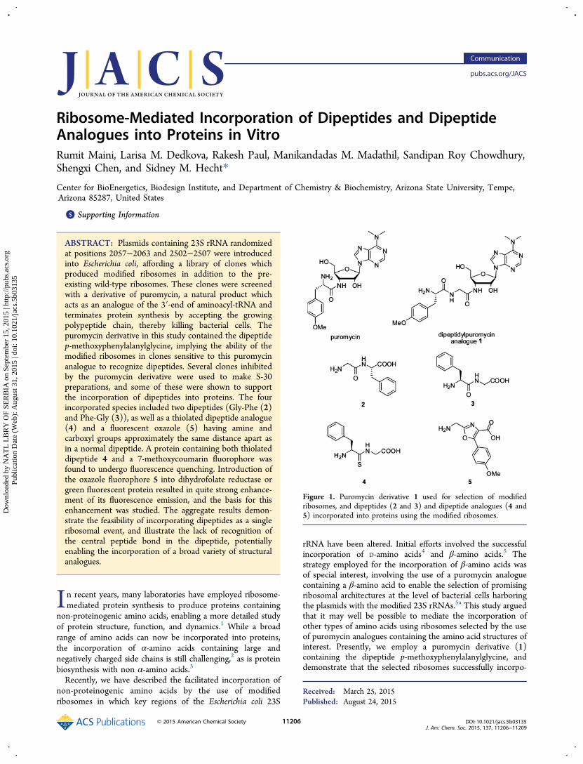
thiophenylalanylglycyl-tRNACCCG was used to introduce 4 intoposition 16 of DHFR, and (7-methoxycoumarin-4-yl)-ethylglycyl-tRNACUA was used to introduce the fluorophoreinto position 49. Another modified DHFR was preparedcontaining phenylalanylglycine at position 16 and the 7-methoxycoumarin fluorophore at position 49. As shown inFigure 3A, when equimolar amounts18 of the two samples wereexcited at 310 nm, only the DHFR containing thiophenyl-alanine at position 16 underwent fluorescence quenching.19
While the mechanism was not studied in detail, given thedistances involved (Figure 3B)15 it seems likely that thisoccurred by photoinduced electron transfer.In addition to the foregoing three dipeptides, dipeptido-
mimetic analogue 5 was also investigated. This compound lacksthe peptide bond which connects the amino acids in dipeptides2−4, but has a similar distance between the amine andcarboxylate groups which participate in incorporation of thedipeptide into the protein backbone. Oxazole 5 is fluorescent,having λex at 302 nm, and λem at 403 nm in water.20 It wasprepared and used to activate tRNACUA as outlined in SchemeS1. The incorporation of this oxazole within DHFR at position10 was verified by MALDI-MS of a tryptic digest (Figure S5).21
Incorporation by the S-30 preparations from two different
Table 1. Sequence in Region 2502−2507 of Clones Havingthe Same Sequeuce (UGCGUGG) in Region 2057−2063
Figure 2. Autoradiogram of a SDS−polyacrylamide gel showing thetranslation of DHFR from wild-type (lane 1) and modified (lanes 2−4) (UAG codon in position 10) mRNA in the presence of differentsuppressor tRNACUAs using an S-30 system prepared from clone010326R6. Lane 2, non-acylated tRNACUA; lane 3, glycylphenylalanyl-tRNACUA; lane 4, phenylalanylglycyl-tRNACUA. The suppressionefficiency relative to wild type is shown below each lane. Additionalincorporation data is provided in Figure S1.
Journal of the American Chemical Society Communication
DOI: 10.1021/jacs.5b03135J. Am. Chem. Soc. 2015, 137, 11206−11209
11207
Dow
nloa
ded
by N
AT
L L
BR
Y O
F SE
RB
IA o
n Se
ptem
ber
15, 2
015
| http
://pu
bs.a
cs.o
rg
Pub
licat
ion
Dat
e (W
eb):
Aug
ust 3
1, 2
015
| doi
: 10.
1021
/jacs
.5b0
3135
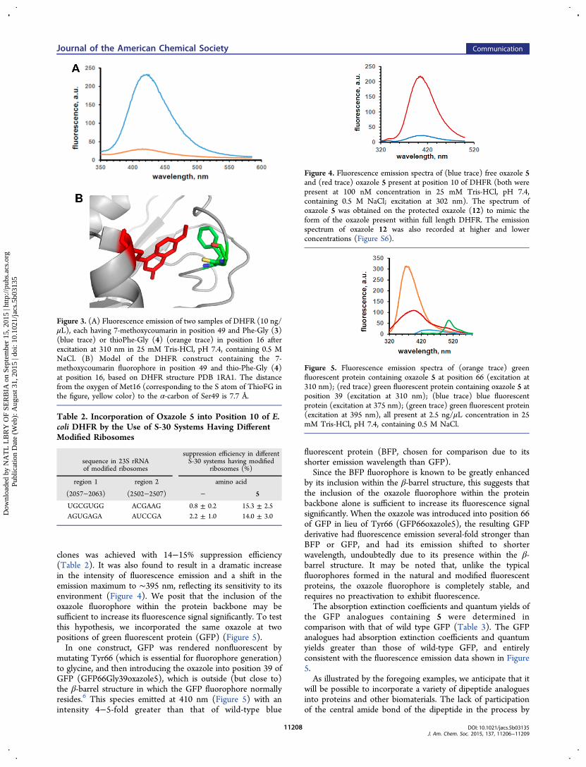
clones was achieved with 14−15% suppression efficiency(Table 2). It was also found to result in a dramatic increasein the intensity of fluorescence emission and a shift in theemission maximum to ∼395 nm, reflecting its sensitivity to itsenvironment (Figure 4). We posit that the inclusion of theoxazole fluorophore within the protein backbone may besufficient to increase its fluorescence signal significantly. To testthis hypothesis, we incorporated the same oxazole at twopositions of green fluorescent protein (GFP) (Figure 5).In one construct, GFP was rendered nonfluorescent by
mutating Tyr66 (which is essential for fluorophore generation)to glycine, and then introducing the oxazole into position 39 ofGFP (GFP66Gly39oxazole5), which is outside (but close to)the β-barrel structure in which the GFP fluorophore normallyresides.6 This species emitted at 410 nm (Figure 5) with anintensity 4−5-fold greater than that of wild-type blue
fluorescent protein (BFP, chosen for comparison due to itsshorter emission wavelength than GFP).Since the BFP fluorophore is known to be greatly enhanced
by its inclusion within the β-barrel structure, this suggests thatthe inclusion of the oxazole fluorophore within the proteinbackbone alone is sufficient to increase its fluorescence signalsignificantly. When the oxazole was introduced into position 66of GFP in lieu of Tyr66 (GFP66oxazole5), the resulting GFPderivative had fluorescence emission several-fold stronger thanBFP or GFP, and had its emission shifted to shorterwavelength, undoubtedly due to its presence within the β-barrel structure. It may be noted that, unlike the typicalfluorophores formed in the natural and modified fluorescentproteins, the oxazole fluorophore is completely stable, andrequires no preactivation to exhibit fluorescence.The absorption extinction coefficients and quantum yields of
the GFP analogues containing 5 were determined incomparison with that of wild type GFP (Table 3). The GFPanalogues had absorption extinction coefficients and quantumyields greater than those of wild-type GFP, and entirelyconsistent with the fluorescence emission data shown in Figure5.As illustrated by the foregoing examples, we anticipate that it
will be possible to incorporate a variety of dipeptide analoguesinto proteins and other biomaterials. The lack of participationof the central amide bond of the dipeptide in the process by
Figure 3. (A) Fluorescence emission of two samples of DHFR (10 ng/μL), each having 7-methoxycoumarin in position 49 and Phe-Gly (3)(blue trace) or thioPhe-Gly (4) (orange trace) in position 16 afterexcitation at 310 nm in 25 mM Tris-HCl, pH 7.4, containing 0.5 MNaCl. (B) Model of the DHFR construct containing the 7-methoxycoumarin fluorophore in position 49 and thio-Phe-Gly (4)at position 16, based on DHFR structure PDB 1RA1. The distancefrom the oxygen of Met16 (corresponding to the S atom of ThioFG inthe figure, yellow color) to the α-carbon of Ser49 is 7.7 Å.
Table 2. Incorporation of Oxazole 5 into Position 10 of E.coli DHFR by the Use of S-30 Systems Having DifferentModified Ribosomes
sequence in 23S rRNAof modified ribosomes
suppression efficiency in differentS-30 systems having modified
ribosomes (%)
region 1 region 2 amino acid
(2057−2063) (2502−2507) − 5
UGCGUGG ACGAAG 0.8 ± 0.2 15.3 ± 2.5AGUGAGA AUCCGA 2.2 ± 1.0 14.0 ± 3.0
Figure 4. Fluorescence emission spectra of (blue trace) free oxazole 5and (red trace) oxazole 5 present at position 10 of DHFR (both werepresent at 100 nM concentration in 25 mM Tris-HCl, pH 7.4,containing 0.5 M NaCl; excitation at 302 nm). The spectrum ofoxazole 5 was obtained on the protected oxazole (12) to mimic theform of the oxazole present within full length DHFR. The emissionspectrum of oxazole 12 was also recorded at higher and lowerconcentrations (Figure S6).
Figure 5. Fluorescence emission spectra of (orange trace) greenfluorescent protein containing oxazole 5 at position 66 (excitation at310 nm); (red trace) green fluorescent protein containing oxazole 5 atposition 39 (excitation at 310 nm); (blue trace) blue fluorescentprotein (excitation at 375 nm); (green trace) green fluorescent protein(excitation at 395 nm), all present at 2.5 ng/μL concentration in 25mM Tris-HCl, pH 7.4, containing 0.5 M NaCl.
Journal of the American Chemical Society Communication
DOI: 10.1021/jacs.5b03135J. Am. Chem. Soc. 2015, 137, 11206−11209
11208
Dow
nloa
ded
by N
AT
L L
BR
Y O
F SE
RB
IA o
n Se
ptem
ber
15, 2
015
| http
://pu
bs.a
cs.o
rg
Pub
licat
ion
Dat
e (W
eb):
Aug
ust 3
1, 2
015
| doi
: 10.
1021
/jacs
.5b0
3135

which the dipeptide is incorporated implies that it is essentiallyexpendable structurally, as illustrated by analogues 4 and 5.Thus, the incorporation of dipeptide mimetics can affordstructures not accessible by the successive incorporation of twoamino acids. Numerous applications, including the creation oflibraries of fluorescent proteins having a range of photophysicalproperties23 and the metabolic stabilization of proteins oftherapeutic interest,24 can be readily envisioned. Whilestructure 5, in particular, represents a fairly significant departurefrom any amino acid whose ribosomal incorporation intoprotein has been reported previously, it may ultimately provefeasible to select ribosomes capable of incorporating even morecomplex and potentially useful substrates.
■ ASSOCIATED CONTENT*S Supporting InformationThe Supporting Information is available free of charge on theACS Publications website at DOI: 10.1021/jacs.5b03135.
Synthetic methods, compound characterization forcompounds prepared, and additional characterization ofthe elaborated proteins (PDF)
■ AUTHOR INFORMATIONCorresponding Author*[email protected] authors declare no competing financial interest.
■ ACKNOWLEDGMENTSThis work was supported by National Institutes of HealthResearch Grant GM103861, awarded by the National Instituteof General Medical Sciences. We thank Dr. Sriloy Dey forassistance in preparing a synthetic intermediate.
■ REFERENCES(1) (a) Hendrickson, T. L.; de Crecy-Lagard, V.; Schimmel, P. Annu.Rev. Biochem. 2004, 73, 147−176. (b) Liu, C. C.; Schultz, P. G. Annu.Rev. Biochem. 2010, 79, 413−444.(2) (a) Karginov, V. A.; Mamaev, S. V.; An, H.; Van Cleve, M. D.;Hecht, S. M.; Komatsoulis, G. A.; Abelson, J. N. J. Am. Chem. Soc.1997, 119, 8166−8176. (b) Hohsaka, T.; Kajihara, D.; Ashizuka, Y.;Murakami, H.; Sisido, M. J. Am. Chem. Soc. 1999, 121, 34−40.(c) Rothman, D. M.; Petersson, E. J.; Vazquez, M. E.; Brandt, G. S.;Dougherty, D. A.; Imperiali, B. J. Am. Chem. Soc. 2005, 127, 848−847.(3) (a) Bain, J. D.; Diala, E. S.; Glabe, C. G.; Wacker, D. A.; Lyttle,M. H.; Dix, T. A.; Chamberlin, A. R. Biochemistry 1991, 30, 5411−5421. (b) Killian, J. A.; Van Cleve, M. D.; Shayo, Y. F.; Hecht, S. M. J.Am. Chem. Soc. 1998, 120, 3032−3042. (c) Eisenhauer, B. M.; Hecht,S. M. Biochemistry 2002, 41, 11472−11478.(4) (a) Dedkova, L. M.; Fahmi, N. E.; Golovine, S. Y.; Hecht, S. M. J.Am. Chem. Soc. 2003, 125, 6616−6617. (b) Dedkova, L. M.; Fahmi, N.E.; Golovine, S. Y.; Hecht, S. M. Biochemistry 2006, 45, 15541−15551.(5) (a) Dedkova, L. M.; Fahmi, N. E.; Paul, R.; del Rosario, M.;Zhang, L.; Chen, S.; Feder, G.; Hecht, S. M. Biochemistry 2012, 51,401−415. (b) Maini, R.; Nguyen, D.; Chen, S.; Dedkova, L. M.; Roy
Chowdhury, S.; Alcala-Torano, R.; Hecht, S. M. Bioorg. Med. Chem.2013, 21, 1088−1096. (c) Maini, R.; Roy Chowdhury, S.; Dedkova, L.M.; Roy, B.; Daskalova, S. M.; Paul, R.; Chen, S.; Hecht, S. M.Biochemistry 2015, 54, 3694−3706.(6) Tsien, R. Y. Annu. Rev. Biochem. 1998, 67, 509−544.(7) Modifications to the 23S rRNA also included one of eight newsequences for nucleotides 2057−2063, introduced to confer moderateresistance to erythromycin in support of the selection protocol.5a
(8) Robertson, S. A.; Noren, C. J.; Anthony-Cahill, S. J.; Griffith, M.C.; Schultz, P. G. Nucleic Acids Res. 1989, 17, 9649−9660.(9) While the S-30 preparation contained both modified and wild-type ribosomes, only the modified ribosomes can incorporate thedipeptide, and so must be the source of all DHFR polypeptide chainscontaining the dipeptide. Position 10 of DHFR has no knownfunction,2a so incorporation of Gly-Phe at position 10 per se would notbe expected to alter DHFR activity. See Table S2 for complete data.(10) (a) Goldberg, J. M.; Batjargal, S.; Petersson, E. J. J. Am. Chem.Soc. 2010, 132, 14718−14720. (b) Wissner, R. F.; Batjargal, S.; Fadzen,C. M.; Petersson, E. J. J. Am. Chem. Soc. 2013, 135, 6529−6540.(11) (a) Goldberg, J. M.; Speight, L. C.; Fegley, M. W.; Petersson, E.J. J. Am. Chem. Soc. 2012, 134, 6088−6091. (b) Goldberg, J. M.;Batjargal, S.; Chen, B. S.; Petersson, E. J. J. Am. Chem. Soc. 2013, 135,18651−18658.(12) Batjargal, S.; Wang, Y. J.; Goldberg, J. M.; Wissner, R. F.;Petersson, E. J. J. Am. Chem. Soc. 2012, 134, 9172−9182.(13) Muir, T. W. Annu. Rev. Biochem. 2003, 72, 249−289.(14) Murakami, H.; Hohsaka, T.; Ashizuka, Y.; Hashimoto, K.;Sisido, M. Biomacromolecules 2000, 1, 118−125. (Figures 7−9 in thisreport employ three amino acids containing the 7-methoxycoumarinfluorophore, each of which was attached to the backbone ofstreptavidin at position 120 via one of three different linkers. Theirfluorescence emission maxima differed by about 20 nm, establishingthe environmental sensitivity of this fluorophore.)(15) In an earlier study two pyrenylalanines incorporated into thesepositions were within several angstroms, and close enough to undergoexcimer formation. See: Chen, S.; Wang, L.; Fahmi, N. E.; Benkovic, S.J.; Hecht, S. M. J. Am. Chem. Soc. 2012, 134, 18883−18885 andreferences therein.(16) Thomsen, I.; Clausen, K.; Scheibye, S.; Lawesson, S.-O. Org.Synth. 1984, 62, 158−164.(17) Chen, S.; Fahmi, N. E.; Wang, L.; Bhattacharya, C.; Benkovic, S.J.; Hecht, S. M. J. Am. Chem. Soc. 2013, 135, 12924−12927.(18) Quantification of protein concentrations was carried out byCoomassie Brilliant Blue R-250 staining of bands of the proteinsfollowing separation by SDS−PAGE, in comparison with knownconcentrations of wild-type DHFR (Figure S3).(19) Also prepared in parallel was a modified DHFR containingthiodipeptide 4 at position 10. Analysis of a tryptic digest by MALDI-MS verified incorporation of the thiolated dipeptide, as well as theabsence of any detectable exchange of O in lieu of S (Figure S4).(20) For oxazole derivative 12, the molar absorptivity in MeOH was26 800 M−1 cm−1 and the quantum yield was 0.59.(21) To date, we have been unable to verify dipeptide incorporationby MS/MS analysis, relying instead on MS data derived fromproteolytic fragments. We note that these do not reflect any increase inmicroheterogeneity occasioned by the use of modified ribosomes.(22) Patterson, G. H.; Knobel, S. M.; Sharif, W. D.; Kain, S. R.;Piston, D. W. Biophys. J. 1997, 73, 2782−2790.(23) We have recently succeeded in incorporating a fluorescentthiazole amino acid into GFP. Roy Chowdhury et al., submitted.(24) Kamionka, M. Curr. Pharm. Biotechnol. 2011, 12, 268−274.
Table 3. Estimation of Quantum Yields of Modified GreenFluorescent Proteins in Comparison with GFPwt
proteinλex/abs,nm
abs extinction coeff,M−1 cm−1 quantum yield, λem
GFPwt 395 25,000 0.79 (509 nm)22
GFP66oxazole5 310 90,300 0.91 (378 nm)GFP66Gly39oxazole5 310 50,200 0.84 (407 nm)
Journal of the American Chemical Society Communication
DOI: 10.1021/jacs.5b03135J. Am. Chem. Soc. 2015, 137, 11206−11209
11209
Dow
nloa
ded
by N
AT
L L
BR
Y O
F SE
RB
IA o
n Se
ptem
ber
15, 2
015
| http
://pu
bs.a
cs.o
rg
Pub
licat
ion
Dat
e (W
eb):
Aug
ust 3
1, 2
015
| doi
: 10.
1021
/jacs
.5b0
3135


![AtPTR1 and AtPTR5 Transport Dipeptides in Planta1[OA]](https://static.fdocuments.in/doc/165x107/61fb4c152e268c58cd5c803d/atptr1-and-atptr5-transport-dipeptides-in-planta1oa.jpg)


![Ribosome Stoichiometry: From Form to Function · Ribosome abundance: A major model, also termed the ribosome concentration hypothesis [3], that explains how ribosomes could exert](https://static.fdocuments.in/doc/165x107/60de31e56d30fc4fb30719b8/ribosome-stoichiometry-from-form-to-function-ribosome-abundance-a-major-model.jpg)