Rhodamine B-Coated Gold Nanoparticles as Effective “Turn-on” Fluorescent Sensors for Detection...
23
This article was downloaded by: [The University Of Melbourne Libraries] On: 28 September 2013, At: 09:32 Publisher: Taylor & Francis Informa Ltd Registered in England and Wales Registered Number: 1072954 Registered office: Mortimer House, 37-41 Mortimer Street, London W1T 3JH, UK Spectroscopy Letters: An International Journal for Rapid Communication Publication details, including instructions for authors and subscription information: http://www.tandfonline.com/loi/lstl20 Rhodamine B-Coated Gold Nanoparticles as Effective “Turn-On” Fluorescent Sensors for Detection of Zinc II Ions in Water Daniela Simona Tira a , Monica Focsan a , Sorin Ulinici b , Dana Maniu a & Simion Astilean a a Nanobiophotonics Center, Institute of Interdisciplinary Research in Bio-NanoSciences and Faculty of Physics, Babes-Bolyai University, Cluj-Napoca, Romania b S.C. ICPE, Bistrita, Romania Accepted author version posted online: 10 Apr 2013. To cite this article: Spectroscopy Letters (2013): Rhodamine B-Coated Gold Nanoparticles as Effective “Turn-On” Fluorescent Sensors for Detection of Zinc II Ions in Water, Spectroscopy Letters: An International Journal for Rapid Communication, DOI: 10.1080/00387010.2013.782557 To link to this article: http://dx.doi.org/10.1080/00387010.2013.782557 Disclaimer: This is a version of an unedited manuscript that has been accepted for publication. As a service to authors and researchers we are providing this version of the accepted manuscript (AM). Copyediting, typesetting, and review of the resulting proof will be undertaken on this manuscript before final publication of the Version of Record (VoR). During production and pre-press, errors may be discovered which could affect the content, and all legal disclaimers that apply to the journal relate to this version also. PLEASE SCROLL DOWN FOR ARTICLE Taylor & Francis makes every effort to ensure the accuracy of all the information (the “Content”) contained in the publications on our platform. However, Taylor & Francis, our agents, and our licensors make no representations or warranties whatsoever as to the accuracy, completeness, or suitability for any purpose of the Content. Any opinions and views expressed in this publication are the opinions and views of the authors, and are not the views of or endorsed by Taylor & Francis. The accuracy of the Content should not be relied upon and should be independently verified with primary sources of information. Taylor and Francis shall not be liable for any losses, actions, claims, proceedings, demands, costs, expenses, damages, and other liabilities whatsoever or howsoever caused arising directly or indirectly in connection with, in relation to or arising out of the use of the Content. This article may be used for research, teaching, and private study purposes. Any substantial or systematic reproduction, redistribution, reselling, loan, sub-licensing, systematic supply, or distribution in any form to anyone is expressly forbidden. Terms & Conditions of access and use can be found at http:// www.tandfonline.com/page/terms-and-conditions
Transcript of Rhodamine B-Coated Gold Nanoparticles as Effective “Turn-on” Fluorescent Sensors for Detection...
This article was downloaded by: [The University Of Melbourne Libraries]On: 28 September 2013, At: 09:32Publisher: Taylor & FrancisInforma Ltd Registered in England and Wales Registered Number: 1072954 Registered office: Mortimer House,37-41 Mortimer Street, London W1T 3JH, UK
Spectroscopy Letters: An International Journal forRapid CommunicationPublication details, including instructions for authors and subscription information:http://www.tandfonline.com/loi/lstl20
Rhodamine B-Coated Gold Nanoparticles as Effective“Turn-On” Fluorescent Sensors for Detection of Zinc IIIons in WaterDaniela Simona Tira a , Monica Focsan a , Sorin Ulinici b , Dana Maniu a & Simion Astilean aa Nanobiophotonics Center, Institute of Interdisciplinary Research in Bio-NanoSciences andFaculty of Physics, Babes-Bolyai University, Cluj-Napoca, Romaniab S.C. ICPE, Bistrita, RomaniaAccepted author version posted online: 10 Apr 2013.
To cite this article: Spectroscopy Letters (2013): Rhodamine B-Coated Gold Nanoparticles as Effective “Turn-On” FluorescentSensors for Detection of Zinc II Ions in Water, Spectroscopy Letters: An International Journal for Rapid Communication, DOI:10.1080/00387010.2013.782557
To link to this article: http://dx.doi.org/10.1080/00387010.2013.782557
Disclaimer: This is a version of an unedited manuscript that has been accepted for publication. As a serviceto authors and researchers we are providing this version of the accepted manuscript (AM). Copyediting,typesetting, and review of the resulting proof will be undertaken on this manuscript before final publication ofthe Version of Record (VoR). During production and pre-press, errors may be discovered which could affect thecontent, and all legal disclaimers that apply to the journal relate to this version also.
PLEASE SCROLL DOWN FOR ARTICLE
Taylor & Francis makes every effort to ensure the accuracy of all the information (the “Content”) containedin the publications on our platform. However, Taylor & Francis, our agents, and our licensors make norepresentations or warranties whatsoever as to the accuracy, completeness, or suitability for any purpose of theContent. Any opinions and views expressed in this publication are the opinions and views of the authors, andare not the views of or endorsed by Taylor & Francis. The accuracy of the Content should not be relied upon andshould be independently verified with primary sources of information. Taylor and Francis shall not be liable forany losses, actions, claims, proceedings, demands, costs, expenses, damages, and other liabilities whatsoeveror howsoever caused arising directly or indirectly in connection with, in relation to or arising out of the use ofthe Content.
This article may be used for research, teaching, and private study purposes. Any substantial or systematicreproduction, redistribution, reselling, loan, sub-licensing, systematic supply, or distribution in anyform to anyone is expressly forbidden. Terms & Conditions of access and use can be found at http://www.tandfonline.com/page/terms-and-conditions

1
Rhodamine B-coated Gold Nanoparticles as Effective “Turn-on” Fluorescent
Sensors for Detection of Zinc II Ions in Water
Daniela Simona Tira1, Monica Focsan1, Sorin Ulinici2, Dana Maniu1, Simion Astilean1,
1Nanobiophotonics Center, Institute of Interdisciplinary Research in Bio-NanoSciences and Faculty of Physics, Babes-Bolyai University, Cluj-Napoca, Romania 2S.C. ICPE,
Bistrita, Romania
Prof. dr. Simion Astilean, Babes-Bolyai University, Institute of Interdisciplinary Research in BioNanoSciences and Faculty of Physics, Nanobiophotonics Center,
Treboniu Laurian 42, 400271, Cluj-Napoca, Romania Tel.: +40 264 454554/+40 264 405300; Fax: +40 (264) 591906; E-mail: [email protected]
Abstract We design a Fluorescence Resonance Energy Transfer system consisting of fluorophore Rhodamine B and gold nanoparticles for sensing of zinc ions in aqueous solution. The electrostatic attraction between positively charged N-atoms in Rhodamine molecules and negatively charged citrate corona of gold nanoparticles leads to substantial fluorescence quenching. However, the quenching is switched off in the presence of zinc ions ions and therefore the system can be used as effective “turn-on” fluorescence sensor. UV-Vis absorption, fluorescence spectroscopy and transmission electron microscopy were used for sensor evaluation. The approach of “turn-on” fluorescence proves a real potential for sensing metallic ions in water. KEYWORDS: Gold nanoparticles, Zn2+ ions, fluorescence, FRET
1. INTRODUCTION
The detection of metallic ions in aquatic ecosystems, drinking waters and biological
systems is a problem of major interest due to the serious health problems that such
species can cause to living organisms. The fluorescent methods implemented for
detection of metal ions require a combination between an organic molecule whose
mission is to attach to the target compound (metal ion) and a fluorescence probe which
provides the fluorescence signal. The recognition event is translated into a change in
intensity of fluorescence signal, fluorescence lifetime, or a shift of fluorescence
Dow
nloa
ded
by [
The
Uni
vers
ity O
f M
elbo
urne
Lib
rari
es]
at 0
9:32
28
Sept
embe
r 20
13
2
wavelength. In principle, fluorescent chemosensors satisfy these requirements and could
be particularly appropriate for detection of various metallic ions [1,2]. However the
approach of fluorescent chemosensors based exclusively on organic compounds has
several limits, including complicated synthetic procedures, poor water solubility and loss
of activity caused by fluorophore photobleaching. Gold nanoparticles (GNPs) could
represent promising alternatives to various organic molecules used as scaffold units in
fabrication of fluorescent chemosensors. This is because GNPs exhibit localized surface
plasmon resonance (LSPR) of large extinction coefficient, high stability under
illumination and “super quenching” properties against nearby fluorophores - which allow
their integration in so-called “turn-on” fluorescence sensors [3]. So far several
spectroscopic techniques based on GNPs have been demonstrated for detection of metal
ions, ranging from the exploitation of LSPR [4–7] and nonlinear optical properties [7] to
surface-enhanced Raman scattering (SERS) [8,9] and fluorescence spectroscopy [10,11]. For
example, Lin et al. reported several functional GNPs based sensors that allow the
sensitive and selective detection of metal ions through colorimetric and fluorescence
changes [12]. Wang et al. reported a sensitive GNPs-amplified SPR sensor for the
detection of mercury ions in aqueous media [13]. Xu et al. revealed that the association of
dye molecules onto the surface of metallic nanoparticles leads to formation of organic-
inorganic hybrid moieties which have numerous applications in light energy conversion,
cell imaging or detection of molecules of different substances [14]. Makarova et al. [15] and
Templeton et al. [16] modified the surface of GNPs using fluorescein isothiocyanate,
demonstrating that dye molecules chemisorbed on the gold nanoparticles surface do not
induce any aggregation.
Dow
nloa
ded
by [
The
Uni
vers
ity O
f M
elbo
urne
Lib
rari
es]
at 0
9:32
28
Sept
embe
r 20
13

3
In this work, we report a sensitive GNPs-based nanosensor for the detection of zinc
(Zn2+) ions in water. The concept is based on the substantial fluorescence quenching of
Rhodamine B (RhB) molecules after dye adsorption onto GNPs surface. If target are Zn2+
ions in solution, as a result of interaction between GNPs and Zn2+ ions, the RhB
molecules are desorbed from the metal surface and the fluorescence recovered. The
analysis of fluorescence intensity changes provides valuable information on the
interactions between GNP, fluorescent probe and target compound. Finally we
demonstrate that the system RhB-coated GNPs can be used as effective “turn-on”
fluorescence sensor for precise dosing of Zn2+ ions in water.
2. EXPERIMENTAL SECTION
2.1. Chemicals
Tetrachloroauric acid (HAuCl4 · 4H2O, 99.99%), sodium citrate (C6H5Na3O7), and zinc
acetate ((C2H3O2)2Zn) were obtained from Aldrich. Rhodamine B (C28H31CIN2O3) was
purchased from Sigma. All reagents used through the experiments were of analytical
grade and used without any purification. All glassware and magnetic stir bars were
cleaned in aqua regia solution (HCl:NO3 3:1) and then rinsed with deionized water.
Ultrapure water (resistivity of 18 MΩ cm) was used for preparation of colloids and all the
other samples.
2.2. Synthesis Of Gold Nanoparticles
Dow
nloa
ded
by [
The
Uni
vers
ity O
f M
elbo
urne
Lib
rari
es]
at 0
9:32
28
Sept
embe
r 20
13

4
Spherical GNPs were synthesized according to Turkevich-Frens method by
aqueous reduction of HAuCl4 with sodium citrate where the citrate ions act as both
reducing agent and a capping agent [17,18]. Briefly, 500 ml of 10-3 M HAuCl4 was brought
to boil on a heat plate, with vigorous stirring. 10 ml of 38.8 mM sodium citrate was added
all at once and the stirring and boiling continued for 15 min. The solution was then
removed from heat and kept stirring another 15 min. After the colloid got cold the
volume was adjusted to 500 ml by adding deionized water. Before being mixed with RhB
solution, as prepared colloidal GNPs were separated from the reactive solution by
centrifugation (6000 rpm, 10 minutes) and resuspended in ultra pure water. The molar
concentration of GNPs was determined from measured optical absorbance by the means
of Beer-Lambert’s law. The transmission electron microscopy (TEM) examination was
performed by drop-drying the colloidal solution on carbon-coated copper grids at room
temperature.
2.3. Samples Preparation
Stock solutions of RhB (5·10-4 M) and zinc acetate (25·10-3 M) were prepared in ultrapure
water of 18 MΩ cm resistivity. To analyze the fluorescence quenching of RhB as
function of GNPs concentration, various volumes (0.2, 0.4, 0.6, 0.8, 1 ml) of GNPs stock
solution (0.3 10-8 M) were added to RhB solution to a final concentration of 1.87·10-6 M.
A similar procedure was used to analyze the fluorescence recovery of RhB as function of
increasing concentrations of zinc acetate in RhB-coated GNPs solution.
2.4. Instruments
Dow
nloa
ded
by [
The
Uni
vers
ity O
f M
elbo
urne
Lib
rari
es]
at 0
9:32
28
Sept
embe
r 20
13

5
UV-Vis absorption measurements were carried out using a Jasco V-670
spectrophotometer. Fluorescence spectra were collected using a Jasco LP-6500
spectrofluorimeter. All fluorescence measurements were recorded in the right angle cell
geometry (10 mm cell), under the same excitation conditions, specifically bandwidths of
1 nm in excitation and 3 nm in emission and excitation wavelength set at 532 nm. TEM
images were obtained with a JOEL model JEM 1010 microscope.
3. RESULTS AND DISCUSSION
3.1. Spectroscopic Characterization Of The Interaction Between Rhb Molecules And
Gold Nanoparticles
Rhodamine B was chosen here as fluorescencent dye due to its suitable photophysical
properties in water, including a high fluorescence quantum yield (0.71), high extinction
coefficient (>75,000 cm−1 M−1) and good photo-stability [19,20]. In neutral solution RhB is
a zwitterion due to the deprotonation of the carboxylic group at the benzene ring [21] and
therefore would tend to interact with negatively charged GNPs. The UV-Vis absorption
spectrum of RhB molecules in solution (see Fig. 1A, curve a) presents a strong absorption
band at 556 nm, characteristic to the π-π* electronic transition of monomers, and a weak
band at 520 nm assigned to the dimeric states of RhB molecules. It is known that in
aqueous solutions, the monomers are in equilibrium with dimers and the molar fraction of
the dimers varies with RhB concentration [21,22]. In our case for a concentration of about
10-6 M, the feature of absorption spectrum suggests that most of RhB molecules should
exist as monomers.
Dow
nloa
ded
by [
The
Uni
vers
ity O
f M
elbo
urne
Lib
rari
es]
at 0
9:32
28
Sept
embe
r 20
13
6
The colloidal solution of citrate-capped GNPs exhibits a well-resolved extinction band at
521 nm (Fig. 1A curve b) assignable to LSPR of individual spherical nanoparticles of
18±2 nm diameter, as given by TEM analysis (see representative TEM pictures in Fig.
1B). The molar concentration of GNPs in solution (cGNP= 0.3 10-8 M) was calculated by
using Beer-Lambert’s law from measured absorbance of 0.8 and molar extinction
coefficient of ε521 = 2.6 108 cm-1 M-1 given in literature [22]. It is well known that the
spectral position and shape of LSPR band of GNPs is strongly dependent on
nanoparticles size and shape as well as local surrounding medium and inter-particles
distance [23]. For instance, upon adding 1 ml RhB solution of 10-5 M concentration to
same volume of colloidal solution (1ml), a new plasmonic absorption band at about 639
nm is observed (Fig. 1A curve c). This new emerging band together with the drastic color
change of the mixture solution – from original red to violet blue (see insert in Fig. 1) -
represents the hallmark of electromagnetic coupling of nanoparticles with formation of
small aggregates (Fig. 1A, curve c). The main plasmonic band decreases and shifts
slightly to red as result of dielectric changes around nanoparticles [24]. The induced
aggregation of citrate-capped GNPs is further confirmed by TEM analysis. The TEM
pictures recorded before (Fig. 1B) and after (Fig. 1C) addition of RhB molecules clearly
prove the partial aggregation of GNPs in the form of linear and branched chains, as result
of dye adsorption. We can admit that RhB molecules are adsorbed onto the GNPs surface
due to the electrostatic attraction between positively charged N-atoms in RhB molecules
and negatively charged citrate corona of GNPs. More specifically, it was established that
RhB and citrate molecules of the gold NPs shell can form a hydrogen bond between
carboxyphenyl group of RhB and one of the carboxyl groups of citric acid, while another
Dow
nloa
ded
by [
The
Uni
vers
ity O
f M
elbo
urne
Lib
rari
es]
at 0
9:32
28
Sept
embe
r 20
13
7
carboxylate group of citric acid interacts with the N+ center of the pyronine group of
RhB, forming a scissors-like structure [25]. A spherical GNP of 18 nm in diameter
exhibiting a surface area of approx. 1017 nm2 can accommodate at least 1000 molecules
of RhB on its surface, considering an uniform distribution of molecules with a molecular
surface “print” at about 1 nm2. Within this assumption, the minimum number of RhB
molecules necessary to coat all GNPs in a specified sample can be estimated. As for
example for coating a number of Nnanoparticles = NAvogadro·c =6.023·1023·10-8= 6·1015
nanoparticles/L a number of about nRhB= 6·1018 RhB molecules /L are necessary, which
is consistent with the molar concentrations used in this investigation (cRhB = nRhB/NAvogadro
= 6·1018/6.023·1023 = 0.996·10-5≈ 1·10-5 M).
The likely reason for the formation of nanoparticle chains should be the nonuniform
distribution of RhB molecules on the surface of gold nanoparticles, which makes that
bare area on the surface of nanoparticles can be attracted against area covered by the RhB
from other nanoparticles. Previous studies have demonstrated the possibility of GNPs
coverage by a dye shell as result of increasing number of RhB molecules in solution [25].
On the contrary to the process of aggregation observed above, large GNPs aggregates
with no particular form were previously observed with eosin molecules [26].
Previous studies demonstrated also that electroactive molecules within dye solutions of
high concentrations can associate onto the solid substrate and their relative orientation
determine two different types of dimeric structures. Parallel arrangements determine the
appearance of H-aggregates in the form of vertical stacks. This type of aggregates yields
Dow
nloa
ded
by [
The
Uni
vers
ity O
f M
elbo
urne
Lib
rari
es]
at 0
9:32
28
Sept
embe
r 20
13
8
a blue shift of the absorption band of the dye. The other type of dye association, named J-
aggregates, have a “head-to-tail” arrangement and exhibit a red shift of the absorption
band relative to the monomer band [27]. Comparing the extinction spectrum (spectrum c in
Fig 1) of the mixed solutions with the extinction spectrum of free RhB (spectrum a) the
absorption band of the dye is blue-shifted by 3-4 nm. The adsorption of RhB molecules
from solution leads to a concentration of molecules onto the surfaces of GNPs and this
local concentration can exceed the equivalent solution concentration for formation of
dimers and aggregates. Therefore, although RhB concentration in solution is relatively
small, we can not exclude the formation of some patches of H-aggregates onto the
surface of GNPs.
Next we study the fluorescence of RhB molecules in the presence of GNPs. The
fluorescence intensity of RhB is strongly reduced upon the addition of GNPs, as seen in
Fig. 2. The electrostatic interaction between RhB and the citric acid corona of
nanoparticles as well as the formation of hydrogen bonds does not suppose an important
change in the electronic transition moment of xanthene group of RhB molecules.
Therefore we exclude any important effect in the reduction of fluorescence quantum yield
as direct result of molecule-molecule interaction (RhB - citric acid corona). On the other
hand, although RhB concentration in solution is relatively small, the accumulation of dye
on the surface of GNPs with formation of RhB dimers and aggregates can not be totally
excluded. Therefore it is conceivable that molecules in dimers and aggregates could
contribute to partial decrease of fluorescence signal. However, the main effect on
quantum yield decrease should be correlated to Fluorescence Resonance Energy Transfer
Dow
nloa
ded
by [
The
Uni
vers
ity O
f M
elbo
urne
Lib
rari
es]
at 0
9:32
28
Sept
embe
r 20
13
9
(FRET). This is because for a constant concentration of RhB molecules in solution the
quenching increases drastically with the amount of GNPs, as illustrated in Fig.2.
Additionally, it is important to note that RhB molecules show a strong fluorescence
emission band centered at 577 nm which exhibit good overlapping with the absorbance
band of aggregated GNPs – a pre-requisite condition for an efficient resonance energy
transfer. Indeed, it is expected that the excited-state energy from the fluorophores to be
transferred to the nanoparticle through a non-radiative process, named resonance energy
transfer (RET) or FRET. The FRET can occur only when there is a sufficient overlap
between the absorption band of acceptor (here GNPs) and the fluorescence band of donor
(here RhB molecules) and the molecule are in close vicinity, as the efficiency of FRET
depends on the inverse sixth power of the distance of separation between the donor and
acceptor. This FRET occurs via an induced-dipole-dipole interaction when the dye
molecule exchanges energy with the GNP of similar resonance wavelength without
requiring of photon emission [28]. By plotting the ration (F0/F) vs. GNPs concentration,
where F0 and F represent the fluorescence intensity of RhB solution in the absence and
presence of GNPs, respectively, a sloping curve was obtained (see inset in Fig. 3),
achieving thus some information about the quenching mechanism of RhB fluorescence.
The obtained plot in our case is not linear, indicating that the RhB molecules exhibit a
faster quenching than resulted from a pure static mechanism [28,29]. We should exclude the
possibility of dynamic collisional quenching mechanism because of permanent and strong
interaction existing between molecules and nanoparticles.
Dow
nloa
ded
by [
The
Uni
vers
ity O
f M
elbo
urne
Lib
rari
es]
at 0
9:32
28
Sept
embe
r 20
13

10
The deviation from linearity can be modeled by the extended model of classical Stern-
Volmer equation presented bellow [30], where K represents the binding equilibrium
constant, Qtot is the total concentration of active sites on the nanoparticle quenchers and
β is the dye fluorescence intensity factor (β = F/cRhB):
+
+=
βKF
KQFF tot
110
The upward deviation from the standard linear behavior can be explained by the
contributions of several factors, among them the fact that a fraction of RhB molecules are
captured in between nanoparticles as result of aggregation. Therefore, it is conceivable
that the rate of nonradiative decay of molecules located at the interstice between
nanoparticles should be higher that of molecules adsorbed directly onto the surface of
individual nanoparticles. Moreover, the spectra of RhB and GNPs aggregates overlap
completely which in turn increases the energy transfer efficiency in the system. This
could explain the accelerated static quenching as function of GNPs concentration and the
existence of non-Forster resonance energy transfer mechanism from RhB molecules to
GNPs. We admit also that the direct energy transfer (without emitted photons) can occur
simultaneously with effects like inner filter effect and reabsorbing effect. However FRET
is the only effect which involves a true quenching mechanism by increasing the non-
radiative rate of de – excitation and induce modification of fluorescence lifetime without
emission of photons. The re-absorption of emitted photons does not impact on the excited
state of emitter molecules (i.e. fluorescence lifetime) but on the intensity of collected
signal. It is conceivable that a number of molecules can accumulation on the surface of
Dow
nloa
ded
by [
The
Uni
vers
ity O
f M
elbo
urne
Lib
rari
es]
at 0
9:32
28
Sept
embe
r 20
13
11
GNPs with formation of dimers and aggregates which could contribute to a decrease of
fluorescence signal (chemical quenching). To correct for the formation of dimers in
solution, although RhB concentration is relatively small, all spectra presented in Figs 2
and 3 were corrected by normalization to the spectrum of free RhB solution (reference
spectrum).
Assuming the value of quantum efficiency of fluorescence for free RhB in water (i.e
φ0=0.71) [19,20], we also calculate the quantum efficiency of RhB fluorescence in the
presence of GNPs as follows below, where F0 and Fq are the fluorescence intensities of
RhB molecules in the absence and in the presence of GNPs respectively, k is the
correction factor for chemical quenching (formation of dimers) on the surface of GNPs
due to high local concentration of the dye:
( )q q 0 0φ F / F k φ=
The calculated values for φq using different concentrations of RhB were between 0.011
and 0.045, that confirms the high quenching effectiveness of the fluorescence of RhB by
GNPs.
3.2. Turn-On Fluorescence For Sensing Zn2+ Ions Based On Rhb-Coated Gnps System
Next, we evaluate the sensitivity in detection of Zn2+ by “turning-on” the fluorescence
emission of RhB molecules in RhB-coated GNPs hybrid system.
Dow
nloa
ded
by [
The
Uni
vers
ity O
f M
elbo
urne
Lib
rari
es]
at 0
9:32
28
Sept
embe
r 20
13
12
As mentioned above, while the free RhB molecules are highly fluorescent in solution, the
fluorescence emission of RhB adsorbed onto GNPs surface is quenched via energy
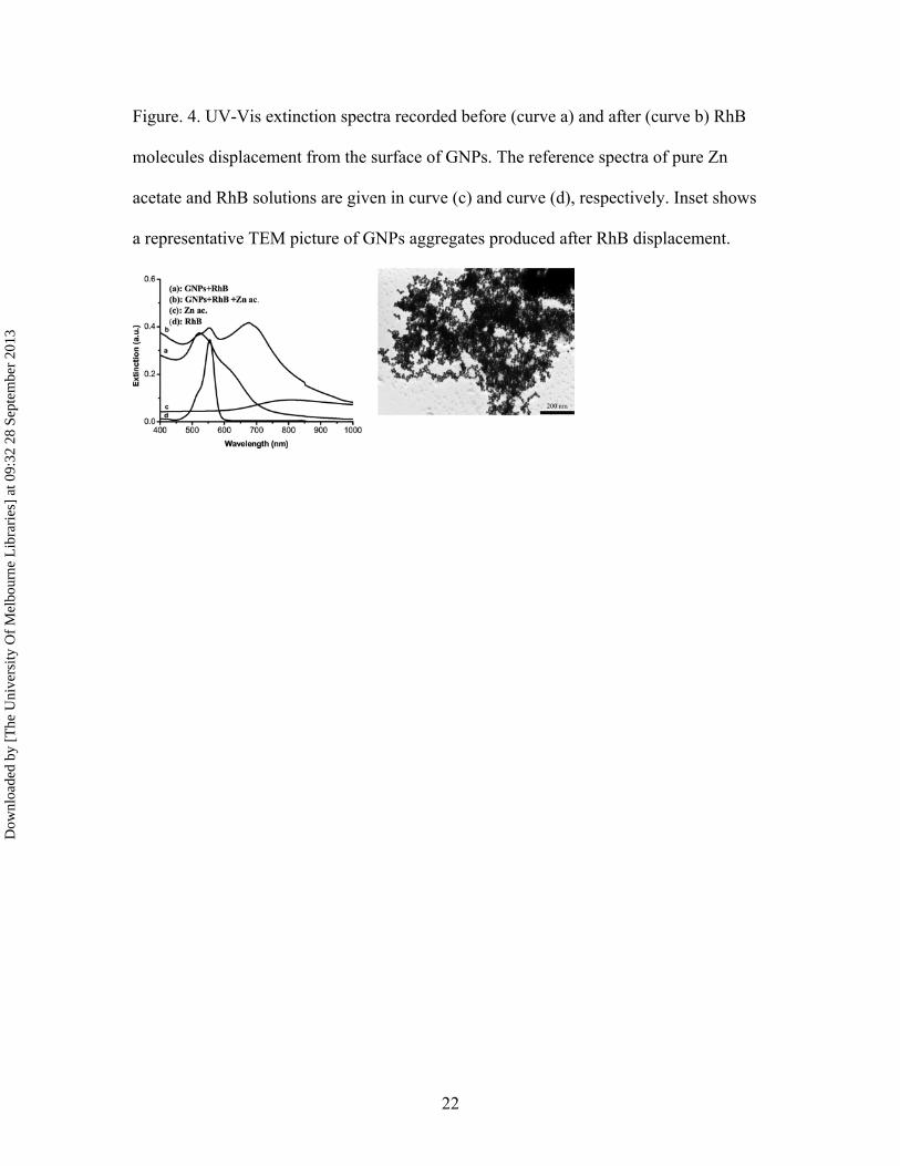
transfer. However, as we can see in Fig. 4, in the presence of Zn2+ ions, the fluorescence
intensity of RhB molecules recovers gradually with the increase of added Zn2+ ions. This
means that RhB molecules are released from the GNPs’ surface, restoring their
fluorescence and proving the successful demonstration of a “turn-on” fluorescence sensor
for the detection of Zn2+ ions. The detachment of RhB molecules from the surface of gold
nanoparticles is triggered by a stronger affinity of Zn2+ cations toward gold surface and
citrate corona, although the mechanism of detachment is not clear.
The release of RhB molecules from the surface of GNPs after mixing RhB-coated GNPs
with zinc acetate was studied by UV-vis spectroscopy and TEM analysis. Firstly, the
extinction spectrum indicates a better spectral separation of the “fingerprint” band of free
RhB molecules at 556 nm from the red-shifted plasmonic band corresponding to GNPs
aggregate (see Fig 4, spectrum b). With increasing concentration of zinc ions added to
solution, the LSPR bands increases also in intensity and features larger and lager
aggregates.
The TEM image in Fig 4 shows GNPs aggregates which exhibit size and form
particularly different relative to aggregates existing in solution before adding Zn+2 ions.
In fact the addition of metallic ions modifies the ionic strength of colloidal solution and
this induces accelerated aggregation as result of dramatic change in charge surface of
GNPs after chemical displacement of RhB.
Dow
nloa
ded
by [
The
Uni
vers
ity O
f M
elbo
urne
Lib
rari
es]
at 0
9:32
28
Sept
embe
r 20
13
13
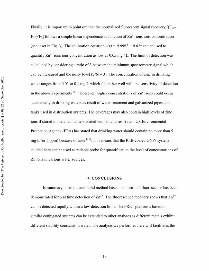
Finally, it is important to point out that the normalized fluorescent signal recovery [(Fon-
Foff)/F0] follows a simple linear dependence as function of Zn2+ ions ions concentration
(see inset in Fig. 3). The calibration equation y(x) = 0.0097 + 0.02x can be used to
quantify Zn2+ ions ions concentration as low as 0.05 mg / L. The limit of detection was
calculated by considering a ratio of 3 between the minimum spectrometer signal which
can be measured and the noisy level (S/N = 3). The concentration of zinc in drinking
water ranges from 0.01 to 0.1 mg/L which fits rather well with the sensitivity of detection
in the above experiments [31]. However, higher concentrations of Zn2+ ions could occur
accidentally in drinking waters as result of water treatment and galvanized pipes and
tanks used in distribution systems. The beverages may also contain high levels of zinc
ions if stored in metal containers coated with zinc to resist rust. US Environmental
Protection Agency (EPA) has stated that drinking water should contain no more than 5
mg/L (or 5 ppm) because of taste [31]. This means that the RhB-coated GNPs system
studied here can be used as reliable probe for quantification the level of concentrations of
Zn ions in various water sources.
4. CONCLUSIONS
In summary, a simple and rapid method based on “turn-on” fluorescence has been
demonstrated for real time detection of Zn2+. The fluorescence recovery shows that Zn2+
can be detected rapidly within a low detection limit. The FRET platforms based on
similar conjugated systems can be extended to other analytes as different metals exhibit
different stability constants in water. The analysis we performed here will facilitates the
Dow
nloa
ded
by [
The
Uni
vers
ity O
f M
elbo
urne
Lib
rari
es]
at 0
9:32
28
Sept
embe
r 20
13

14
development of similar detection systems toward discrimination between metallic ions
and their detection in real samples collected from rivers or tap water.
ACKNOWLEDGEMENTS
This work was supported by CNCSIS–UEFISCSU, project number
PNII_ID_PCCE_312/2008
REFERENCES
[1] Czarnik, A. W. (Editor), Fluorescent Chemosensors for Ion and Molecule
Recognition. ACS Symposium Series 1993, 538, 238.
[2] Nolan, E. M.; Lippard, S. J. Small-Molecule Fluorescent Sensors for Investigating
Zinc Metalloneurochemistry. Accounts of Chemical Research 2009, 42, 193–203.
[3] Wang, H.; Wang, Y.; Jin, J.; Yang, R. Gold Nanoparticle-Based Colorimetric and
“Turn-On” Fluorescent Probe for Mercury (II) Ions in Aqueous solution. Analytical
Chemistry 2008, 80(23), 9021-9028
[4] Sugunan, A.; Thanachayanont, C.; Dutta, J.; Hilborn, J. G. Heavy-metal Ion Sensors
Using Chitosan-Capped Gold Nanoparticles. Science and Technology of Advanced
Materials 2005, 6, 335-340.
[5] Haes, A. J.; Van Duyne, R. P. Nanoscale Optical Biosensors Based on Localized
Surface Plasmon Resonance Spectroscopy. SPIE 2003, 5221, 47-58.
[6] Kim, Y.; Johnson, R. C.; Hupp, J. T. Gold Nanooparticle-Based Sensing of
“Spectroscopically Silent” Heavy Metal Ions. Nano Letters 2001, 1, 165-167.
Dow
nloa
ded
by [
The
Uni
vers
ity O
f M
elbo
urne
Lib
rari
es]
at 0
9:32
28
Sept
embe
r 20
13
15
[7] Darbha, G. K.; Singh, A. K.; Rai, U. S.; Yu, E.; Yu, H.; Ray, P. C. Selective
Detection of Mercury (II) Ion Using Nonlinear Optical Properties of Gold Nanoparticles.
Journal of American Chemical Society 2008, 130, 8038-8043.
[8] Alvarez-Puebla, R. A.; dos Santos, D. S. Jr.; Aroca, R. F. SERS Detection of
Environmental Pollutants in Humic Acid-gold Nanoparticle Composite Materials. The
Analyst 2007, 132, 1210-1214
[9] Yuan, Y.-X.; Ling, L.; Wang, X.-Y.; Wang, M.; Gu, R.-A.; Yao, J.-L. Surface
Enhanced Raman Spectroscopic Readout on Heavy Metal Ions Based on Surface Self –
Assembly. Journal of Raman Spectroscopy 2007, 38, 1280–1287.
[10] Darbha, G. K.; Ray, A.; Ray, P. C. Gold Nanoparticle-Based Miniaturized
Nanomaterial Surface Energy Transfer Probe for Rapid and Ultrasensitive Detection of
Mercury in Soil, Water and Fish. ACS NANO 2007, 1(3), 208-214.
[11] Ray, P. C.; Darbha, G. K.; Ray, A.; Hardy, W.; Walker, J. A Gold-Nanoparticle-
Based Fluorescence Resonance Energy Transfer Probe for Multiplexed Hybridization
Detection: Accurate Identification of Bio-agents DNA. Nanotechnology 2008, 18,
375504 (6pp)
[12] Lin, Y.-W.; Huangb, C.-C.; Chang, H.-T. Gold Nanoparticle Probes for the
Detection of Mercury. Lead and Copper ions, Analyst. 2011, 136, 863-871.
[13] Wang, L.; Li, T.; Du, Y.; Chen, C.; Li, B.; Zhou, M.; Dong, S. Au NPs-Enhanced
Surface Plasmon Resonance for Sensitive Detection of Mercury (II) Ions. Biosensors and
Bioelectronics 2010, 25, 2622–2626.
[14] Xu, P.; Yanagi, H. Fluorescence patterning in Dye-Doped Sol-Gel Films by
Generation of Gold Nanoparticles. Chemistry of Materials 1999, 11, 2626-2628.
Dow
nloa
ded
by [
The
Uni
vers
ity O
f M
elbo
urne
Lib
rari
es]
at 0
9:32
28
Sept
embe
r 20
13

16
[15] Makarova, O. V.; Ostafin, A. E.; Miyoshi, H.; Norris, J. R.; Miesel, D. Adsorption
and Encapsulation of Fluorescent Probes in Nanoparticles. The Journal of Physical
Chemistry 1999, 103, 9080-9084.
[16] Templeton, A. C.; Wuelfing, M. P.; Murray, R. W. Monolayer Protected Cluster
Molecules. Accounts of Chemical Research 2000, 33, 27-36.
[17] Frens, G. Controlled Nucleation for the Regulation of the Particle Size in
Monodisperse Gold Suspensions. Nature Physical Science 1973, 241, 20-22.
[18] Baia, M.; Toderaş, F.; Baia, L.; Popp, J.; Aştilean, S. Probing the enhancement
mechanisms of SERS with p-Aminothiophenol Molecules Adsorbed on Self-assembled
Gold Colloidal Nanoparticles. Chemical Physics Letters 2006, 422, 127-132.
[19] Naga Srinivas, N. K. M.; Venugopal, R. S.; Narayana, R. D. Saturable and Reverse
Saturable Absorption of Rhodamine B in Methanol and Water. Journal of Optical Society
of America B 2003, 20(12), 2470-2479.
[20] Huang, C. C.; Chang, H. T. Selective Gold-Nanoparticle-Based “Turn-On”
Fluorescent Sensors for Detection of Mercury(II) in Aqueous Solution. Analytical
Chemistry 2006, 78, 8332-8338.
[21] Arbeloa, I. L.; Ojeda, P. R. Dimeric States of Rhodamine B. Chemical Physics
Letters 1982, 87(6), 556-560.
[22] Schafer, F. P. (Editor), Dye Lasers 1973, Springer-Verlag, Berlin-Heidelberg-New
York, p21.
[22] Saha, K.; Agasti, S. S.; Kim, C.; Li, X.; Rotello, M. V. Gold Nanoparticles in
Chemical and Biological Sensing. Chemical Reviews 2012, 112, 2739−2779.
Dow
nloa
ded
by [
The
Uni
vers
ity O
f M
elbo
urne
Lib
rari
es]
at 0
9:32
28
Sept
embe
r 20
13

17
[23] Lee, K. S.; El-Sayed, M. A. Gold and Silver Nanoparticles in Sensing and Imaging:
Sensitivity of Plasmon Response to Size, Shape, and Metal Composition. Journal of
Physical Chemistry B 2006, 110(39), 19220–19225.
[24] Hutter, E.; Fendler, J. H. Exploitation of Localized Surface Plasmon Resonance.
Advanced Materials 2004, 16, 1685-1706.
[25] Zhu, J.; Zhu, K.; Huang, Li-q. Using Gold Colloid Nanoparticles to Modulate the
Surface Enhanced Fluorescence of Rhodamine B. Physics Letters A 2008, 372(18), 3283-
3288.
[26] Ghosh, S. K.; Pal, A.; Nath, S.; Kundu, S.; Panigrahi, S.; Pal, T. Dimerization of
Eosin on Nanostructured Gold Surfaces: Size Regime Dependence of the Small Metallic
Particles. Chemical Physics Letters 2005, 412, 5-11
[27] D’ Ilario, L.; Martinelli, A. Toluidine Blue: Aggregation Properties and Structural
Aspects. Modelling and Simulation in Materials Science and Engineering 2006, 14, 581-
595.
[28] Lakowicz, J. R. Principles of Fluorescence Spectroscopy, Kluwer Academic /
Plenum Publishers 1999, New York, U.S.A., 698 pp.
[29] Madge, D.; Rojas, G. E.; Seybold, P. Solvent Dependence of the Fluorescence
Lifetimes of Xanthene Dyes. Photochemistry & Photobiology 1999, 70, 737-744.
[30] Stobiecka, M.; Hepel, M. Multimodal Coupling of Optical Transitions and
Plasmonic Oscillations in Rhodamine B Modified Gold Nanoparticles. Physical
Chemistry Chemical Physics 2011, 13, 1131-1139.
Dow
nloa
ded
by [
The
Uni
vers
ity O
f M
elbo
urne
Lib
rari
es]
at 0
9:32
28
Sept
embe
r 20
13

18
[31] http://www.atsdr.cdc.gov/, Toxicological Profile for Zinc, Toxic Substances Portal,
Agency for Toxic Substances and Disease Registry (ATSDR), 1600 Clifton Road NE,
Atlanta, USA.
Dow
nloa
ded
by [
The
Uni
vers
ity O
f M
elbo
urne
Lib
rari
es]
at 0
9:32
28
Sept
embe
r 20
13

19
Figure. 1 (A). UV-Vis absorption spectra of RhB solution (a), GNPs solution (b), and 1:1
mixture between RhB and GNPs solutions (c). Inset shows the photographic images of
RhB solution before and after mixing with GNPs. (B). TEM image of GNPs; (C). TEM
image of RhB-coated GNPs.
Dow
nloa
ded
by [
The
Uni
vers
ity O
f M
elbo
urne
Lib
rari
es]
at 0
9:32
28
Sept
embe
r 20
13
20
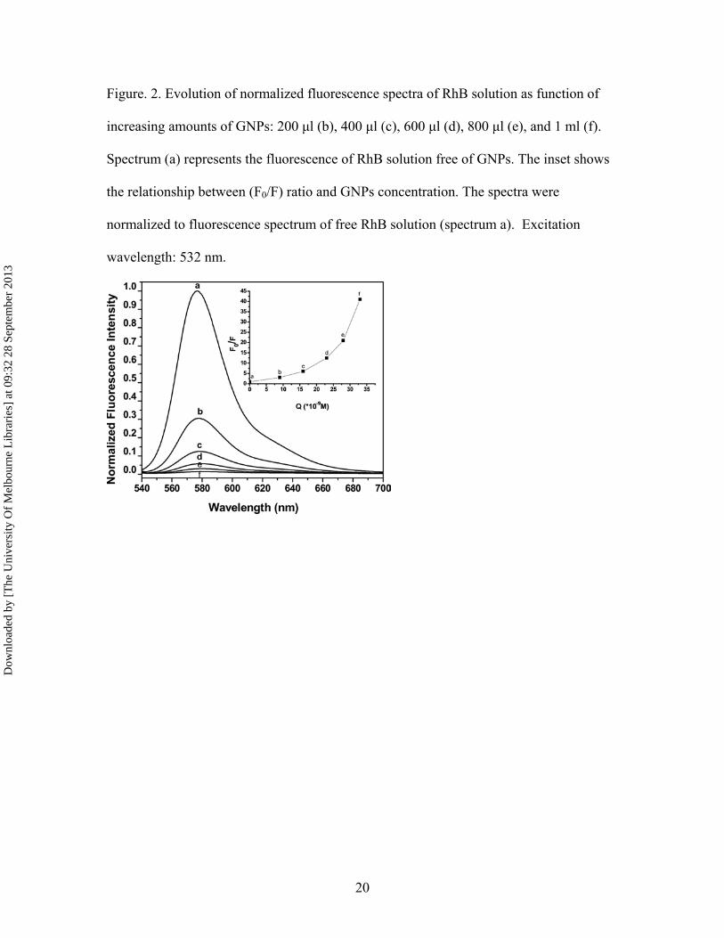
Figure. 2. Evolution of normalized fluorescence spectra of RhB solution as function of
increasing amounts of GNPs: 200 µl (b), 400 µl (c), 600 µl (d), 800 µl (e), and 1 ml (f).
Spectrum (a) represents the fluorescence of RhB solution free of GNPs. The inset shows
the relationship between (F0/F) ratio and GNPs concentration. The spectra were
normalized to fluorescence spectrum of free RhB solution (spectrum a). Excitation
wavelength: 532 nm.
Dow
nloa
ded
by [
The
Uni
vers
ity O
f M
elbo
urne
Lib
rari
es]
at 0
9:32
28
Sept
embe
r 20
13
21
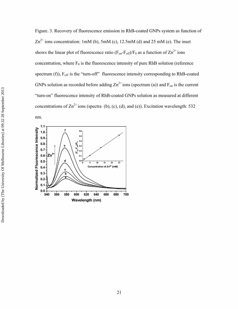
Figure. 3. Recovery of fluorescence emission in RhB-coated GNPs system as function of
Zn2+ ions concentration: 1mM (b), 5mM (c), 12.5mM (d) and 25 mM (e). The inset
shows the linear plot of fluorescence ratio (Fon-Foff)/F0 as a function of Zn2+ ions
concentration, where F0 is the fluorescence intensity of pure RhB solution (reference
spectrum (f)), Foff is the “turn-off” fluorescence intensity corresponding to RhB-coated
GNPs solution as recorded before adding Zn2+ ions (spectrum (a)) and Fon is the current
“turn-on” fluorescence intensity of RhB-coated GNPs solution as measured at different
concentrations of Zn2+ ions (spectra (b), (c), (d), and (e)). Excitation wavelength: 532
nm.
Dow
nloa
ded
by [
The
Uni
vers
ity O
f M
elbo
urne
Lib
rari
es]
at 0
9:32
28
Sept
embe
r 20
13
22
Figure. 4. UV-Vis extinction spectra recorded before (curve a) and after (curve b) RhB
molecules displacement from the surface of GNPs. The reference spectra of pure Zn
acetate and RhB solutions are given in curve (c) and curve (d), respectively. Inset shows
a representative TEM picture of GNPs aggregates produced after RhB displacement.
Dow
nloa
ded
by [
The
Uni
vers
ity O
f M
elbo
urne
Lib
rari
es]
at 0
9:32
28
Sept
embe
r 20
13