RhoandRasGTPasesinAxonGrowth,Guidance, and...
19
Rho and Ras GTPases in Axon Growth, Guidance, and Branching Alan Hall 1 and Giovanna Lalli 2 1 Memorial Sloan-Kettering Cancer Center, Cell Biology Program, New York, New York 10065 2 Wolfson Centre for Age-related Diseases, King’s College London, London SE1 1UL, United Kingdom Correspondence: [email protected] The establishment of precise neuronal cell morphology provides the foundation for all aspects of neurobiology. During development, axons emerge from cell bodies after an initial polarization stage, elongate, and navigate towards target regions guided by a range of environmental cues. The Rho and Ras families of small GTPases have emerged as critical players at all stages of axonogenesis. Theirability to coordinately direct multiple signal trans- duction pathways with precise spatial control drives many of the activities that underlie this morphogenetic program: the dynamic assembly, disassembly, and reorganization of the actin and microtubule cytoskeletons, the interaction of the growing axon with other cells and extracellular matrix, the delivery of lipids and proteins to the axon through the exocytic machinery, and the internalization of membrane and proteins at the leading edge of the growth cone through endocytosis. This article highlights the contribution of Rho and Ras GTPases to axonogenesis. T he Ras superfamily of small GTPases, con- sisting of almost 200 proteins, can be sub- classified into six families: Rho, Ras, Rab, Arf, Sar, and Ran (Colicelli 2004). These proteins act as molecular switches, cycling between an inactive, GDP-bound state and an active, GTP-bound state (Fig. 1). The activated confor- mation interacts with specific effectors to propagate downstream signaling events that influence many aspects of cell biology. Guanine nucleotide exchange factors (GEFs) activate the switch by catalyzing the exchange of GDP for GTP, whereas GTPase-activating proteins (GAPs) increase the intrinsic GTPase activity and inactivate the switch (Fig. 1) (Jaffe and Hall 2005). Dominant – negative (DN) and con- stitutively active (CA) versions of small GTPases (created through specific amino acid substitu- tions) have been used extensively to dissect the individual roles of these proteins. Although these have been incredibly informative, they do have potential drawbacks: dominant–negative constructs, which act by sequestering GEFs, may interfere with closely related family mem- bers, whereas constitutively activated GTPases interact indiscriminately with all their potential targets, something that does not happen under normal conditions. RNAi and gene knockout Editors: Marc Tessier-Lavigne and AlexL. Kolodkin Additional Perspectives on Neuronal Guidance available at www.cshperspectives.org Copyright # 2010 Cold Spring Harbor Laboratory Press; all rights reserved; doi: 10.1101/cshperspect.a001818 Cite this article as Cold Spring Harb Perspect Biol 2010;2:a001818 1 on May 10, 2018 - Published by Cold Spring Harbor Laboratory Press http://cshperspectives.cshlp.org/ Downloaded from
Transcript of RhoandRasGTPasesinAxonGrowth,Guidance, and...
Rho and Ras GTPases in Axon Growth, Guidance,and Branching
Alan Hall1 and Giovanna Lalli2
1Memorial Sloan-Kettering Cancer Center, Cell Biology Program, New York, New York 100652Wolfson Centre for Age-related Diseases, King’s College London, London SE1 1UL, United Kingdom
Correspondence: [email protected]
The establishment of precise neuronal cell morphology provides the foundation for allaspects of neurobiology. During development, axons emerge from cell bodies after aninitial polarization stage, elongate, and navigate towards target regions guided by a rangeof environmental cues. The Rho and Ras families of small GTPases have emerged as criticalplayers at all stages of axonogenesis. Their ability to coordinately direct multiple signal trans-duction pathways with precise spatial control drives many of the activities that underlie thismorphogenetic program: the dynamic assembly, disassembly, and reorganization of the actinand microtubule cytoskeletons, the interaction of the growing axon with other cells andextracellular matrix, the delivery of lipids and proteins to the axon through the exocyticmachinery, and the internalization of membrane and proteins at the leading edge of thegrowth cone through endocytosis. This article highlights the contribution of Rho and RasGTPases to axonogenesis.
The Ras superfamily of small GTPases, con-sisting of almost 200 proteins, can be sub-
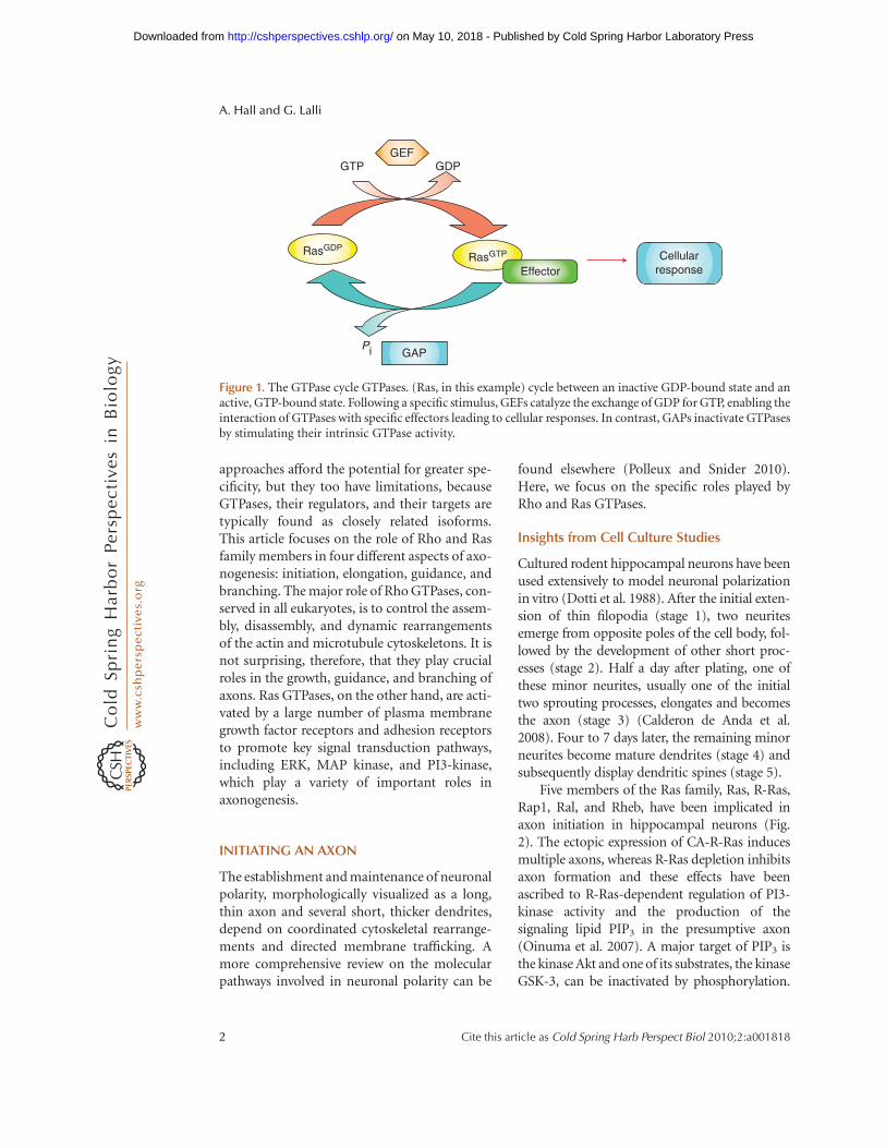
classified into six families: Rho, Ras, Rab, Arf,Sar, and Ran (Colicelli 2004). These proteinsact as molecular switches, cycling betweenan inactive, GDP-bound state and an active,GTP-bound state (Fig. 1). The activated confor-mation interacts with specific effectors topropagate downstream signaling events thatinfluence many aspects of cell biology. Guaninenucleotide exchange factors (GEFs) activate theswitch by catalyzing the exchange of GDP forGTP, whereas GTPase-activating proteins(GAPs) increase the intrinsic GTPase activity
and inactivate the switch (Fig. 1) (Jaffe andHall 2005). Dominant–negative (DN) and con-stitutively active (CA) versions of small GTPases(created through specific amino acid substitu-tions) have been used extensively to dissect theindividual roles of these proteins. Althoughthese have been incredibly informative, they dohave potential drawbacks: dominant–negativeconstructs, which act by sequestering GEFs,may interfere with closely related family mem-bers, whereas constitutively activated GTPasesinteract indiscriminately with all their potentialtargets, something that does not happen undernormal conditions. RNAi and gene knockout
Editors: Marc Tessier-Lavigne and Alex L. Kolodkin
Additional Perspectives on Neuronal Guidance available at www.cshperspectives.org
Copyright # 2010 Cold Spring Harbor Laboratory Press; all rights reserved; doi: 10.1101/cshperspect.a001818
Cite this article as Cold Spring Harb Perspect Biol 2010;2:a001818
1
on May 10, 2018 - Published by Cold Spring Harbor Laboratory Press http://cshperspectives.cshlp.org/Downloaded from
approaches afford the potential for greater spe-cificity, but they too have limitations, becauseGTPases, their regulators, and their targets aretypically found as closely related isoforms.This article focuses on the role of Rho and Rasfamily members in four different aspects of axo-nogenesis: initiation, elongation, guidance, andbranching. The major role of Rho GTPases, con-served in all eukaryotes, is to control the assem-bly, disassembly, and dynamic rearrangementsof the actin and microtubule cytoskeletons. It isnot surprising, therefore, that they play crucialroles in the growth, guidance, and branching ofaxons. Ras GTPases, on the other hand, are acti-vated by a large number of plasma membranegrowth factor receptors and adhesion receptorsto promote key signal transduction pathways,including ERK, MAP kinase, and PI3-kinase,which play a variety of important roles inaxonogenesis.
INITIATING AN AXON
The establishment and maintenance of neuronalpolarity, morphologically visualized as a long,thin axon and several short, thicker dendrites,depend on coordinated cytoskeletal rearrange-ments and directed membrane trafficking. Amore comprehensive review on the molecularpathways involved in neuronal polarity can be
found elsewhere (Polleux and Snider 2010).Here, we focus on the specific roles played byRho and Ras GTPases.
Insights from Cell Culture Studies
Cultured rodent hippocampal neurons have beenused extensively to model neuronal polarizationin vitro (Dotti et al. 1988). After the initial exten-sion of thin filopodia (stage 1), two neuritesemerge from opposite poles of the cell body, fol-lowed by the development of other short proc-esses (stage 2). Half a day after plating, one ofthese minor neurites, usually one of the initialtwo sprouting processes, elongates and becomesthe axon (stage 3) (Calderon de Anda et al.2008). Four to 7 days later, the remaining minorneurites become mature dendrites (stage 4) andsubsequently display dendritic spines (stage 5).
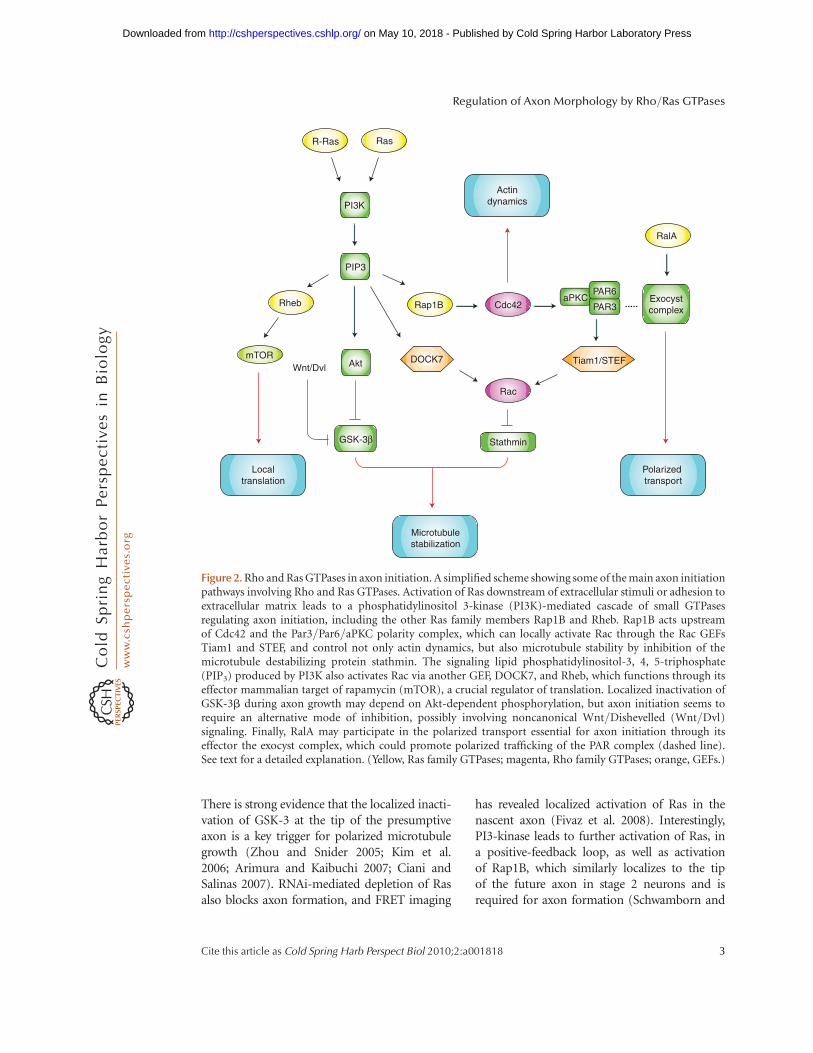
Five members of the Ras family, Ras, R-Ras,Rap1, Ral, and Rheb, have been implicated inaxon initiation in hippocampal neurons (Fig.2). The ectopic expression of CA-R-Ras inducesmultiple axons, whereas R-Ras depletion inhibitsaxon formation and these effects have beenascribed to R-Ras-dependent regulation of PI3-kinase activity and the production of thesignaling lipid PIP3 in the presumptive axon(Oinuma et al. 2007). A major target of PIP3 isthe kinase Akt and one of its substrates, the kinaseGSK-3, can be inactivated by phosphorylation.
GAP
Cellularresponse
GTP GDP
Pi
GEF
RasGDPRasGTP
Effector
Figure 1. The GTPase cycle GTPases. (Ras, in this example) cycle between an inactive GDP-bound state and anactive, GTP-bound state. Following a specific stimulus, GEFs catalyze the exchange of GDP for GTP, enabling theinteraction of GTPases with specific effectors leading to cellular responses. In contrast, GAPs inactivate GTPasesby stimulating their intrinsic GTPase activity.
A. Hall and G. Lalli
2 Cite this article as Cold Spring Harb Perspect Biol 2010;2:a001818
on May 10, 2018 - Published by Cold Spring Harbor Laboratory Press http://cshperspectives.cshlp.org/Downloaded from
There is strong evidence that the localized inacti-vation of GSK-3 at the tip of the presumptiveaxon is a key trigger for polarized microtubulegrowth (Zhou and Snider 2005; Kim et al.2006; Arimura and Kaibuchi 2007; Ciani andSalinas 2007). RNAi-mediated depletion of Rasalso blocks axon formation, and FRET imaging
has revealed localized activation of Ras in thenascent axon (Fivaz et al. 2008). Interestingly,PI3-kinase leads to further activation of Ras, ina positive-feedback loop, as well as activationof Rap1B, which similarly localizes to the tipof the future axon in stage 2 neurons and isrequired for axon formation (Schwamborn and
Polarized transport
Microtubulestabilization
Actindynamics
Localtranslation
Ras
Rheb Rap1B
PIP3
PI3K
Akt
GSK-3β
mTOR
Cdc42
RalA
Exocystcomplex
DOCK7 Tiam1/STEF
Rac
Stathmin
aPKCPAR3
PAR6
Wnt/Dvl
R-Ras
Figure 2. Rho and Ras GTPases in axon initiation. A simplified scheme showing some of the main axon initiationpathways involving Rho and Ras GTPases. Activation of Ras downstream of extracellular stimuli or adhesion toextracellular matrix leads to a phosphatidylinositol 3-kinase (PI3K)-mediated cascade of small GTPasesregulating axon initiation, including the other Ras family members Rap1B and Rheb. Rap1B acts upstreamof Cdc42 and the Par3/Par6/aPKC polarity complex, which can locally activate Rac through the Rac GEFsTiam1 and STEF, and control not only actin dynamics, but also microtubule stability by inhibition of themicrotubule destabilizing protein stathmin. The signaling lipid phosphatidylinositol-3, 4, 5-triphosphate(PIP3) produced by PI3K also activates Rac via another GEF, DOCK7, and Rheb, which functions through itseffector mammalian target of rapamycin (mTOR), a crucial regulator of translation. Localized inactivation ofGSK-3b during axon growth may depend on Akt-dependent phosphorylation, but axon initiation seems torequire an alternative mode of inhibition, possibly involving noncanonical Wnt/Dishevelled (Wnt/Dvl)signaling. Finally, RalA may participate in the polarized transport essential for axon initiation through itseffector the exocyst complex, which could promote polarized trafficking of the PAR complex (dashed line).See text for a detailed explanation. (Yellow, Ras family GTPases; magenta, Rho family GTPases; orange, GEFs.)
Regulation of Axon Morphology by Rho/Ras GTPases
Cite this article as Cold Spring Harb Perspect Biol 2010;2:a001818 3
on May 10, 2018 - Published by Cold Spring Harbor Laboratory Press http://cshperspectives.cshlp.org/Downloaded from
Puschel 2004). This same study reported thatRap1B acts upstream of Cdc42 (a Rho familymember), suggesting a cascade of small GTPasesacting at the tip of the presumptive axon. Yetanother potential role for PI3-kinase is in theRheb/mTOR pathway, a central regulator of cellgrowth that also seems to influence neuronal po-larization (Li et al. 2008b). Finally, the asymmet-ric transport of proteins to the axon and to thedendrites is essential for the establishment andmaintenance of neuronal polarity. RalA interactswith the exocyst, an octameric protein complexpromoting membrane delivery. The depletionof RalA, or of components of the exocyst complexin neurons causes axon loss, suggesting an impor-tant role for this GTPase in polarized traffickingto the axon (Lalli 2009).
One theme to emerge from these studies isthe importance of the localized phosphoryla-tion and inhibition of GSK-3 by Akt. However,neurons from a double knockin mouse in whichthe Akt-phosphorylation sites present on thetwo GSK-3 isoforms (Ser9 in GSK-3b- andSer21 in GSK-3a) have been replaced by analanine residue and show no defects in neuronalpolarization (Gartner et al. 2006). This suggeststhat GSK-3 inhibition occurs through an alter-native mechanism and there is evidence thatthis may involve noncanonical Wnt/Dishev-elled signaling (Zhang et al. 2007).
A close interplay between the actin andmicrotubule cytoskeletons occurs at the tip of thepresumptive axon, often visualized as growth-cone enlargement, shortening of actin ribs,and subsequent penetration of microtubulesinto the central growth-cone region at the stage2-3 transition. Indeed, localized microtubulestabilization (by taxol) or localized depolymeri-zation of actin filaments (by cytochalasin) issufficient to initiate axonal extension in aneurite (Witte and Bradke 2008). Rho familyGTPases control the actin and microtubulecytoskeletons through specific effectors andplay important roles in regulating neuronalpolarization. A tight regulation of Cdc42 activityis required for axon specification, becauseneurons expressing CA-Cdc42 do not extendneurites, whereas cells transfected with a “fastcycling” Cdc42 mutant extend multiple axons
(Schwamborn and Puschel 2004). An importantCdc42 effector is the Par6/aPKC complex, whichcontrols polarity in a variety of contexts, includ-ing morphogenesis, migration, and asymmetriccell division (Goldstein and Macara 2007).aPKC, in turn, regulates APC (the adenomatouspolyposis coli tumor suppressor protein), whichstabilizes microtubule plus ends at the tip of thepresumptive axon (Shi et al. 2004). Cdc42 mayalso locally activate Rac through an interactionbetween Par3 (complexed with Par6) and twoRac GEFs, Tiam1 or Tiam2/STEF, both of whichare reported to be essential for neuronal polariza-tion (Nishimura et al. 2005). Because Rac canstimulate PI3-kinase, this may establish apositive-feedback loop between Cdc42 and Racto reinforce neuronal polarization.
Rac regulates a parallel pathway that triggersmicrotubule growth in the nascent axon. Acti-vation of the Rac GEF DOCK7 leads to Rac-mediated phosphorylation and inactivation ofthe microtubule destabilizing protein stath-min/Op18 in the nascent axon (Watabe-Uchidaet al. 2006). One candidate for triggeringlocalized activation of DOCK7 at the presu-mptive axon tip is the extracellular matrix pro-tein laminin, which acts through the lamininreceptor and PIP3 to recruit DOCK7 to theplasma membrane. The kinase responsible forstathmin phosphorylation is likely to be theRac effector PAK (Daub et al. 2001). In hippo-campal neurons, PAK is present in all neurites,but the activated (phosphorylated) form is spa-tially restricted to the growing axon (Jacobset al. 2007).
Rho appears to act antagonistically toCdc42/Rac and negatively regulates neuriteextension through ROCK, a target kinase (DaSilva et al. 2003). DN-Rho does not alter axonnumber, but CA-Rho inhibits axon formationand induces round cells (Schwamborn andPuschel 2004). Inactivation of Rho/ROCK in-creases the amount of dephosphorylated profi-lin IIa, resulting in F-actin destabilization andsprout formation (Da Silva et al. 2003). Interest-ingly, the Rho/ROCK/PIIa cascade can be in-activated by plasma membrane gangliosidesialidase (PMGS), an enzymatic activity thatlocalizes at the tip of the future axon in stage 2
A. Hall and G. Lalli
4 Cite this article as Cold Spring Harb Perspect Biol 2010;2:a001818
on May 10, 2018 - Published by Cold Spring Harbor Laboratory Press http://cshperspectives.cshlp.org/Downloaded from
neurons and induces axon specification byenhancing TrkA activity (Da Silva et al. 2005).
Insights from In Vivo Studies
Loss-of-function studies of members of theDrosophila Rac family (DRac1, DRac2, andMtl) in both the visual system and mushroom-body neurons have revealed specific roles in axongrowth, guidance, and branching (Hakeda-Suzuki et al. 2002; Ng et al. 2002). Expressionof CA-DRac1 or DN-DRac1 causes failure inaxon, but not dendrite outgrowth, whereasmutants of DCdc42 affect general aspects ofmorphogenesis, including cell migration anddendrite/axonal outgrowth. A RhoA null muta-tion in mushroom-body neurons does notaffect axon specification and growth, but causesdendrite overextension, whereas activated RhoAreduces dendritic complexity. These data sup-port an important role for RhoA in dendritic,but not axonal morphogenesis (Lee et al.2000). There are three Rac-like proteins inC. elegans: CED-10, Rac2/3, and MIG-2. Theyappear to have overlapping functions in severalaspects of neuronal development. Interestingly,Rac double mutants display ectopic axons andbranches, suggesting that Rac is involved in thesuppression of superfluous axon and branches(Lundquist 2003). The analysis of a Cdc42 con-ditional knockout mouse, in which Cdc42 isdepleted in late gestation in the cortex andhippocampus, revealed normal initial neuritesprouting, but a strong inhibition of axonformation (Garvalov et al. 2007). Further ex-periments using cultured Cdc42-null neuronssuggested that this GTPase acts upstream of alocal actin depolymerizing activity (perhapscofilin), which is required for initial axonformation.
GROWING AN AXON
After the establishment of neuronal polarity, theaxon may extend considerably depending onthe type of neuron. Rho and Ras GTPasescontinue to play important roles during axongrowth (Fig. 3).
Insights from Cell Culture Studies
In addition to their role in neuron polarization,the Rac GEFs Tiam1, and Tiam2/STEF are im-portant players in axon extension (de Curtis2008). The Rac/Cdc42 effector IQGAP3 isrequired for axon growth in hippocampal neu-rons—it localizes asymmetrically at the distalend of the axon, where it likely modulatesboth the actin and the microtubule cytoskele-tons (Wang et al. 2007). In addition to IQGAP3,N-WASP may be an important Cdc42 effectorduring neurite extension. It promotes actin pol-ymerization through the Arp2/3 complex and adominant–negative version of N-WASP wasreported to abolish neurite extension in hippo-campal neurons (Banzai et al. 2000). However,in another report, a different dominant–negative version of N-WASP led to enhancedaxon elongation in hippocampal neurons,suggesting that Arp2/3 antagonizes neuriteextension (Strasser et al. 2004).
Rho and its downstream effector ROCKnegatively regulate the early steps of axon out-growth in cultured neurons (Govek et al.2005). However, axon elongation in cerebellargranule neurons promoted by the neural che-mokine stromal cell-derived factor (SDF)-1ais mediated by Rho, though through a distinctsignaling pathway involving another down-stream effector, mDia (Arakawa et al. 2003).These experiments show the potential for con-fusion using a dominant–negative GTPase;thus, although a variety of upstream signalsconverge on Rho, they may lead to the activa-tion of different signaling pathways.
Other members of the Rho family have alsobeen linked to axon growth. The first GEF do-main of Trio, which activates RhoG, is requiredfor NGF-mediated neurite outgrowth in PC12cells (Estrach et al. 2002). RhoG binds toElmo, a scaffold for the Rac GEF Dock180 andthus may mediate Rac activation (Katoh andNegishi 2003). TC10, a close relative of Cdc42,was initially identified in neuronal cells as agene that was dramatically re-expressed afteraxotomy of motor neurons in the hypoglossalnuclei (Tanabe et al. 2000). Low levels ofTC10 are found in developing and adult brain,
Regulation of Axon Morphology by Rho/Ras GTPases
Cite this article as Cold Spring Harb Perspect Biol 2010;2:a001818 5
on May 10, 2018 - Published by Cold Spring Harbor Laboratory Press http://cshperspectives.cshlp.org/Downloaded from
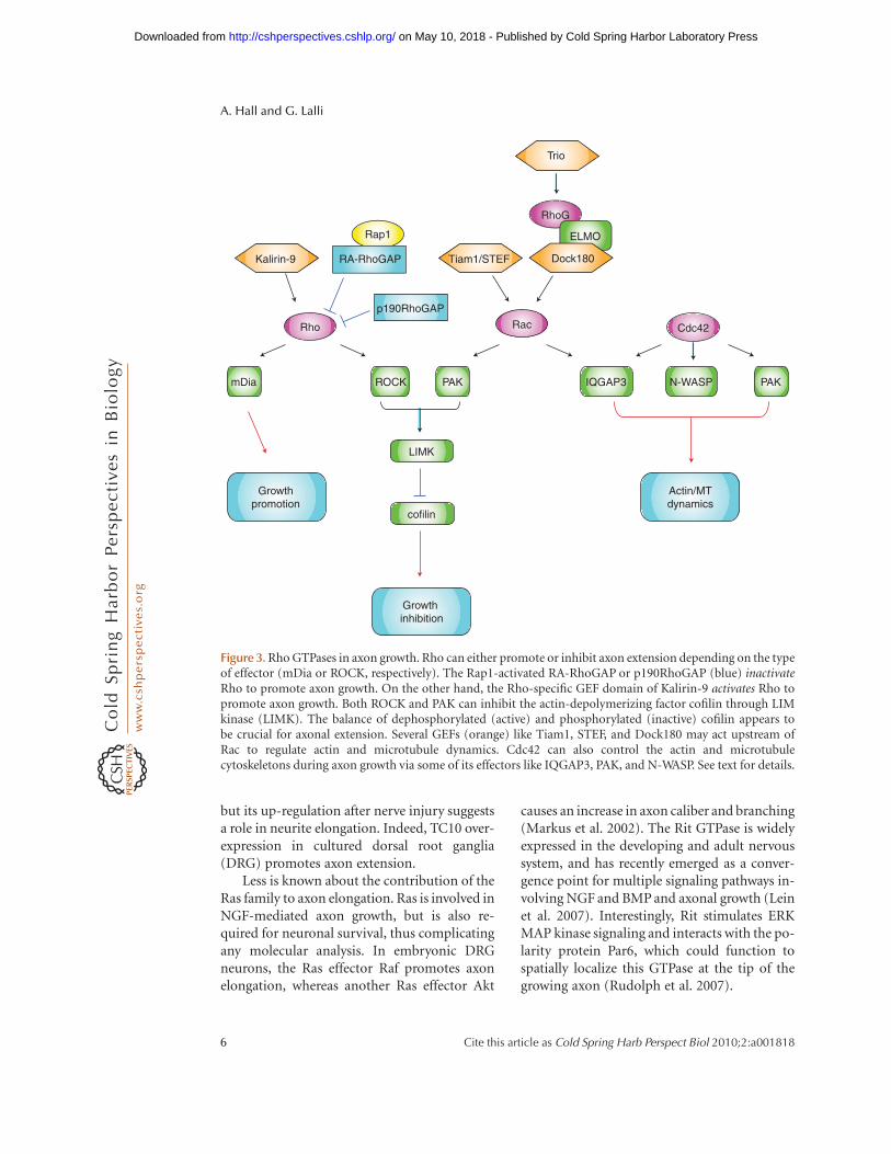
but its up-regulation after nerve injury suggestsa role in neurite elongation. Indeed, TC10 over-expression in cultured dorsal root ganglia(DRG) promotes axon extension.
Less is known about the contribution of theRas family to axon elongation. Ras is involved inNGF-mediated axon growth, but is also re-quired for neuronal survival, thus complicatingany molecular analysis. In embryonic DRGneurons, the Ras effector Raf promotes axonelongation, whereas another Ras effector Akt
causes an increase in axon caliber and branching(Markus et al. 2002). The Rit GTPase is widelyexpressed in the developing and adult nervoussystem, and has recently emerged as a conver-gence point for multiple signaling pathways in-volving NGF and BMP and axonal growth (Leinet al. 2007). Interestingly, Rit stimulates ERKMAP kinase signaling and interacts with the po-larity protein Par6, which could function tospatially localize this GTPase at the tip of thegrowing axon (Rudolph et al. 2007).
RhoG
ELMO
Rho Rac Cdc42
Growthpromotion
Growth inhibition
PAK
LIMK
cofilin
mDia ROCK IQGAP3 PAKN-WASP
Rap1
Kalirin-9
Actin/MTdynamics
p190RhoGAP
RA-RhoGAP Tiam1/STEF Dock180
Trio
Figure 3. Rho GTPases in axon growth. Rho can either promote or inhibit axon extension depending on the typeof effector (mDia or ROCK, respectively). The Rap1-activated RA-RhoGAP or p190RhoGAP (blue) inactivateRho to promote axon growth. On the other hand, the Rho-specific GEF domain of Kalirin-9 activates Rho topromote axon growth. Both ROCK and PAK can inhibit the actin-depolymerizing factor cofilin through LIMkinase (LIMK). The balance of dephosphorylated (active) and phosphorylated (inactive) cofilin appears tobe crucial for axonal extension. Several GEFs (orange) like Tiam1, STEF, and Dock180 may act upstream ofRac to regulate actin and microtubule dynamics. Cdc42 can also control the actin and microtubulecytoskeletons during axon growth via some of its effectors like IQGAP3, PAK, and N-WASP. See text for details.
A. Hall and G. Lalli
6 Cite this article as Cold Spring Harb Perspect Biol 2010;2:a001818
on May 10, 2018 - Published by Cold Spring Harbor Laboratory Press http://cshperspectives.cshlp.org/Downloaded from

Insights from In Vivo Studies
In Drosophila sensory neurons, expression ofeither CA-DRac or DN-DRac causes axonaloutgrowth arrest without affecting dendrites,whereas in motor neurons, an inactive DRac1mutation disrupts axon trajectory, withoutinhibiting their extension (Luo et al. 1994;Kaufmann et al. 1998). In giant fibers, over-expression of wild-type DRac1 affects axonalmorphology, CA-DRac1 inhibits neurite out-growth, and DN-DRac1 causes axonal ectopicbranching (Allen et al. 2000). It is not so clearhow to interpret these results; notwithstandingthe potential problems associated with express-ing constitutively activated GTPases, it doesseem that the role of Rac differs in different neu-ronal types. Partially redundant functions ofDRac1, DRac2, and Mtl in axon outgrowthhave been shown in two distinct contexts: thevisual system and mushroom-body neurons.Furthermore, varying the level of Rac activationresults in different outcomes, such that progres-sive loss of activity leads first to defects inbranching, then guidance, and finally out-growth (Ng et al. 2002). This suggests that Racmay activate different downstream pathwaysunder these different conditions. Interestingly,CRIB motif-containing effectors (such asPAK) seem not to be required for axon out-growth (Kim et al. 2003). To complicate mattersfurther, Rac may have a dual role: inhibitingaxon growth through Trio/Rac/PAK/LIMKand cofilin phosphorylation, but promotinggrowth in a PAK-independent fashion down-stream of another Rac GEF, still life (STEF)(Ng and Luo 2004). Activation of RhoA throughthe GEF Pebble results in axon growth inhibitionthrough ROCK, which inactivates cofilin viaLIMK-mediated phosphorylation. Cofilin phos-phorylation and dephosphorylation thereforerepresent important regulatory steps in axonalgrowth.
Drosophila RacGAP50C (Tum) is importantduring cytokinesis, but in the post-mitotic neu-rons of the mushroom body, RNAi-mediateddepletion leads to axon overextension (Gold-stein et al. 2005). Its axon growth limiting func-tion is lost if the GAP domain, which likely acts
on Rac, is mutated (Canman et al. 2008).Through an interaction with the kinesin Pava-rotti (pav), Tum may help spatially regulateRac activity during axon growth. Mosaic analy-sis in the Drosophila mushroom body revealed asurprisingly selective function for RhoA indendritic, but not axonal morphogenesis,and neurons lacking RhoA overextend theirdendrites, but display normal axon projections(Lee et al. 2000). In C. elegans, Rac mutantsdisplay premature axon growth arrest (Lund-quist 2003). RhoA RNAi causes embryonic le-thality; however, studies with CA-Rho andDN-Rho suggest an inhibitory role in neuriteoutgrowth of chemosensory neurons (Zallenet al. 2000).
The expression of CA-Rac1 in mouse Pur-kinje neurons inhibits axonal outgrowth and al-ters the number and morphology of dendriticspines, without blocking dendritic growth orbranching (Luo et al. 1996). The negative regu-lator of Rho, p190 RhoGAP, is a major Src sub-strate in the brain and is involved in axongrowth, guidance, and defasciculation. Extra-cellular matrix components, such as laminin,enhance p190-mediated neurite outgrowth,whereas mice lacking functional p190 RhoGAPhave substantial defects in axon extension(Brouns et al. 2001). In contrast to the Droso-phila studies, some of the CRIB motif-containingeffectors appear to be involved in axon growth inmice. Indeed, knockout of the Cdc42 effectorPAK4 impairs axon extension, besides causing avariety of other neuronal development defects(Qu et al. 2003). A tight regulation of PAK3expression levels by the homeobox transcriptionfactors Dlx1/2 seems to be crucial in restrainingneurite growth and promoting tangential migra-tion (Cobos et al. 2007).
GUIDING THE AXON
Axon guidance is directed by numerous soluble,matrix and cell-bound ligands acting throughdistinct receptors at the tip of the axon—thegrowth cone. Localized changes in filamentousactin in the growth cone direct microtubule dy-namics and thereby the direction of axonalelongation. Given the importance of Rho
Regulation of Axon Morphology by Rho/Ras GTPases
Cite this article as Cold Spring Harb Perspect Biol 2010;2:a001818 7
on May 10, 2018 - Published by Cold Spring Harbor Laboratory Press http://cshperspectives.cshlp.org/Downloaded from
GTPases in regulating the actin cytoskeleton,they have been a major focus in the analysis ofsignaling by guidance cues. Rho and ROCKoften appear to work antagonistically to Rac/Cdc42; the former associated with repulsivecues and growth-cone collapse and the latterwith attractive cues and forward protrusion.However, closer examination of the signalingpathways reveals a far more complex story. Wealso refer the reader to the article by Bashawand Klein for more extensive discussion onsignaling downstream of guidance receptors(Bashaw and Klein 2010).
In Drosophila, gain-of-function and loss-of-function Rac mutations have distinct effectson motor axon pathways, pointing to a role forRac in executing specific guidance decisions(Luo et al. 1994). Similarly, Rac activation is re-quired during neuromuscular development fordirectional specificity and target recognition,whereas Cdc42 is necessary for motor-axonextension (Kaufmann et al. 1998). A targetedknockout approach for Rac1 in mouse ventraltelencephalic neurons revealed a requirementin the formation of midline commissures, aswell as proper fasciculation and targeting of tha-lamocortical and corticothalamic axons (Chenet al. 2007). The ability of Rac to affect specificguidance decisions is further shown by the factthat commissural axons of cortex-restrictedRac1 knockout mice still display potent exten-sion, yet fail to cross the midline (Kassai et al.2008). Misregulation of Rho activity has a sub-stantial impact on axonal guidance. p190Rho-GAP knockout mice, for example, display clearguidance defects in the axonal projections ofthe posterior limb of the anterior commissure(Brouns et al. 2001). The abundance and broaddistribution of p190RhoGAP in the nervoussystem makes this GAP a crucial mediator link-ing diverse extracellular guidance cues withRho-dependent cytoskeletal rearrangements.
Some of the upstream activators and down-stream effectors that participate in Rac pathwayshave been identified. Single mutants of any ofthe three Rac-like genes in C. elegans (ced-10,mig-2, and rac-2) lead to subtle phenotypes,but double mutants cause severe defects inaxon pathfinding, as well as outgrowth. The
Rac GEF UNC-73 (Trio) acts in all Rac-depend-ent pathfinding pathways, whereas anotherGEF, CED-5 (DOCK180), acts with MIG-2,but not with CED-10 (Lundquist et al. 2001).Trio was initially characterized in Drosophilaas an essential regulator of PAK during photo-receptor axon guidance (Bateman et al. 2000).The axon patterning defects observed in Triomosaics are remarkably similar to those seenin both PAK and Dock mutants, suggestingthat Trio/Dock/PAK act together in a singlesignaling pathway regulating axon guidance(Newsome et al. 2000).
Semaphorins
Semaphorins (collapsins) are a family of se-creted or membrane-bound proteins that pro-mote either attractive or repulsive guidancethrough their interaction with the plexin/neu-ropilin receptor family (Kruger et al. 2005).Growth-cone collapse induced by Sema3A,which acts through the neuropilin1/plexinA1receptor complex, depends on Rac and Rho ac-tivity (Jin and Strittmatter 1997; Kuhn et al.1999; Vastrik et al. 1999; Turner et al. 2004)(Fig. 4A). Rac.GTP interacts directly with plex-inA and plexinB receptors, a highly unusualsituation for a small GTPase and a trans-membrane receptor. Some have suggested thatthis represents sequestration (essentially inacti-vation) of Rac (Hu et al. 2001; Vikis et al.2002). However, collapse induced by a ligand-independent version of plexinA1 does not re-quire Rac, which would be consistent withthe receptor being a downstream effector ofRac (Turner et al. 2004). One idea is that Racinduces a conformational change in the cyto-plasmic tail of the receptor (i.e., in a similarway to other targets of Rho GTPases), thoughan alternative possibility is that Rac promotesreceptor endocytosis, which is required forSema3A-induced growth-cone collapse (Jurneyet al. 2002).
Sema3A leads to the activation of Rhoand ROCK, and attenuates actin polymeriza-tion, while promoting intra-axonal F-actinbundling and myosin II-mediated force genera-tion (Gallo 2006). In hippocampal neurons,
A. Hall and G. Lalli
8 Cite this article as Cold Spring Harb Perspect Biol 2010;2:a001818
on May 10, 2018 - Published by Cold Spring Harbor Laboratory Press http://cshperspectives.cshlp.org/Downloaded from

R-Ras
FARP2
Rho
RacRhoD
Rac
Adhesion
Receptor activation/endocytosis
ROCK
Actincontractility
PI3K
Akt
GSK-3β
Microtubulestabilization
Rnd1
B
A PlexinA1
PlexinB1
R-RasPDZ-RhoGEFLARG
Rho
Rac
Rho
Rnd1
Transient
p190RhoGAP
Microtubulestabilization
Adhesion
ActinContractility
Adhesion
Figure 4. Rho and Ras GTPases in Semaphorin signaling. (A) Signaling downstream of plexinA1 receptorsinvolves up-regulation of Rho activity, leading to an increase in ROCK-mediated actin contractility. Inaddition, direct binding of Rac.GTP to the GTPase-binding region of plexin (pink rhombus) may induce aconformational change in the plexin cytoplasmic tail and enhance receptor endocytosis. Sema3A-induceddissociation of the Rac GEF FARP2 from the plexinA1/neuropilin complex promotes the recruitment ofRnd1 to plexinA1. The Rnd1/plexinA1 interaction opens the two R-Ras GAP domains of plexinA1 (gray),thus leading to R-Ras inactivation. This event may facilitate growth-cone collapse by inhibitingintegrin-mediated adhesion and promoting microtubule destabilization (through a decrease in PI3K/Aktand a subsequent increase in GSK-3 activities). Only the intracellular domain of the plexin receptor is shownand interactions with co-receptors are not shown. (B) Rho activity downstream of plexinB1 undergoestransient down-regulation via p190RhoGAP, possibly to mediate inhibition of integrin function. The PDZdomain-binding motif exclusively present in the plexinB receptor subfamily (orange triangle) interacts withthe Rho GEFs PDZ-RhoGEF and LARG in a Sema-dependent fashion, thereby causing Rho activation andgrowth-cone collapse. Similar to plexinA1, the association with Rnd1 is required to enable the R-Ras GAPactivity of plexinB1. Only the intracellular domain of the plexin receptor is shown and interactions withco-receptors are not shown.
Regulation of Axon Morphology by Rho/Ras GTPases
Cite this article as Cold Spring Harb Perspect Biol 2010;2:a001818 9
on May 10, 2018 - Published by Cold Spring Harbor Laboratory Press http://cshperspectives.cshlp.org/Downloaded from
Sema4D/plexin-B1 activates Rho through theGEF, LARG (Swiercz et al. 2002) (Fig. 4B).The relationship between plexin signaling andRho may be more complex, because a transientdown-regulation of Rho by p190RhoGAP ap-pears to be necessary for growth-cone collapse(Barberis et al. 2005). A further twist to this storystems from the observation that Sema3A inducesintra-axonal translation of RhoA mRNA, which,through an axonal 30 UTR targeting element, islocalized in developing axons and growth cones(Wu et al. 2005). This local translation appearsto be necessary and sufficient for Sema3A-in-duced growth-cone collapse.
The atypical Rho family members, Rnd1/2/3, have also been linked to plexin signaling.Rnd1 promotes an R-Ras GAP activity encodedwithin the cytoplasmic tail of plexinB1 (Oinu-ma et al. 2004a; Oinuma et al. 2004b). The inter-action of Rnd1 with plexinA1 is required forSema3A/plexinA1-mediated repulsion, althoughin this context, yet another small GTPase, RhoD,has been reported to antagonize Rnd1 signalingand prevent Sema3A-induced repulsion (Zanataet al. 2002). The R-Ras GAP-related domain iswell conserved among different plexin subfami-lies and is likely to be a significant feature of allsemaphorin signaling. PlexinC1 and plexinD1have R-Ras GAP activity, though in the formercase this appears to be constitutive, whereas inthe latter, it requires Rnd2 (Uesugi et al. 2009).The role of R-Ras in growth-cone guidance, how-ever, is not clear. It regulates integrin-mediatedcell adhesion, and so down-regulation could re-duce cell adhesiveness, but in addition can regu-late PI3-kinase and therefore potentially modifymicrotubule dynamics via Akt/GSK-3 (Ito et al.2006). Interestingly, Sema3A promotes the disso-ciation of the Rac GEF FARP2 from plexinA1/neuropilin-1, which leads not only to Rac activa-tion, but also to the recruitment of Rnd1 to plex-inA1, thus triggering its R-Ras GAP activity(Toyofuku et al. 2005).
Ephrins
Ephrins are transmembrane or glycosylphos-phatidylinositol-anchored ligands recognizedby the Eph family of receptor tyrosine kinases.
They act as repulsive cues through activationof Rho and ROCK in the growth cone (Wahlet al. 2000; Reber et al. 2007) (Fig. 5A). TheGEF ephexin has been reported to be involvedin Rho activation downstream of EphA4 (Sha-mah et al. 2001). However, ephexin-1 knockoutmice are apparently normal, raising the possi-bility of redundancy within the very large family(82) of mammalian Rho family GEFs (Schmidtand Hall 2002; Sahin et al. 2005). An alternativemechanism to regulate Rho GTPases is throughGAPs. EphrinB3 acting through EphA4 con-tributes to the formation of two major motorcircuits: the corticospinal tract (CST) and thespinal neuronal circuit controlling locomotion(central pattern generator). The signaling path-way involves activation of the Rac GAP,a-chimerin, and the loss of a-chimerin inmice (leading presumably to hyperactivationof Rac) causes a failure in CSTaxons from stop-ping at the midline (Iwasato et al. 2007). Simi-lar conclusions were reached using culturedhippocampal neurons, where EphrinA-inducedgrowth-cone collapse was associated with tyro-sine phosphorylation of a2-chimerin and in-hibition of Rac/PAK signaling (Shi et al. 2007).
On the face of it, inhibition of Rac makessense in the context of repulsive cues. Sur-prisingly, however, one study reported thatalthough Rac activity decreases transiently inneurons treated with ephrin-A2, growth-conecollapse correlates with the resumption of Racactivity (Jurney et al. 2002). The authors con-cluded that this is due to Rac-mediated endocy-tosis of the growth-cone plasma membrane.Indeed, Eph triggered Vav2 (a Cdc42/RacGEF) activation leads to endocytosis of theligand/receptor complex and contributes to arepulsive phenotype (Cowan et al. 2005).
Eph receptors activate Ras family GTPases,but their contributions are less clear. EphB2 in-teracts with a GEF, SHEP1, which can activateboth R-Ras and Rap1A, whereas EphA4 inter-acts with a Rap1 GAP, SPAR (Dodelet et al.1999; Richter et al. 2007). SPAR-mediated inac-tivation of Rap1 is necessary for ephrinA-dependent growth-cone collapse in hippocam-pal neurons. Both R-Ras and Rap1 have beenshown to affect integrin activity, but whether
A. Hall and G. Lalli
10 Cite this article as Cold Spring Harb Perspect Biol 2010;2:a001818
on May 10, 2018 - Published by Cold Spring Harbor Laboratory Press http://cshperspectives.cshlp.org/Downloaded from

A
B Robo
DCCC
Growthcone
collapse
SPAR
Rap1
p120RasGAP
R-Ras
PAK
α-chimerin
Rac
Ephexin
ROCK
Rho
SHEP1
R-Ras Rap1A
Vav2
Rac
Endocytosis Adhesion ?
EphB2EphA4
PAK
Sos
Rac
Repulsion
CrGAP/Vilse Dock/Nck
?
Rho
Rac
Rho
RacNck1
Dock180
Trio
PAK1
Cdc42N-WASP
Attraction
Figure 5. Rho and Ras GTPases in Ephrin/Slit/Netrin signaling. (A) Schematic of the GEFs and GAPs involved inEphA4 (left) or EphB2 (right) signaling. Inhibition of Rac/PAK cascade (by the Rac GAP a–chimerin) mayfunction together with ephexin-mediated Rho activation in controlling the cytoskeletal rearrangementsleading to growth-cone collapse. However, Rac activation (by Vav2) can also contribute to collapse bystimulating endocytosis of the Ephrin/Eph complex. A tight regulation of R-Ras and Rap1 through GAPs(SPAR, p120RasGAP) and GEFs (like SHEP1) seems to be needed downstream of Eph signaling, possibly forthe control of matrix adhesion. (B) Rac activity appears to be tightly regulated downstream of Slit through theGEF Sos and the GAP CrGAP/Vilse. Slit stimulation recruits the adaptor protein Dreadlocks (DrosophilaDock/vertebrate Nck) and subsequently PAK to Robo conserved cytoplasmic (CC) sequences (orange). TheRobo/Dock complex interacts with Sos, mediating Slit-dependent Rac activation. The role of Rho in Slit/Robo signaling remains unclear and may depend on the neuronal context. (C) DCC homodimers promotegrowth-cone attraction through Rac, Cdc42, and PAK activation. On Netrin-1 binding, the adaptor Nck(which constitutively interacts with DCC), active Rac, Cdc42, Pak1, and N-WASP are recruited into a complexwith the intracellular domain of DCC, triggering reorganization of the growth-cone actin cytoskeleton. TheGEFs DOCK180 and Trio appear to be involved in netrin-1-dependent Rac activation. DCC may alsodown-regulate Rho and ROCK; however, the signaling mechanisms leading to the modulation of Rho activitydownstream of netrin are still unclear, and they are likely to include cross talk with other GTPases. Only theintracellular domains of Robo and the DCC homodimer are shown in (B) and (C), respectively.
Regulation of Axon Morphology by Rho/Ras GTPases
Cite this article as Cold Spring Harb Perspect Biol 2010;2:a001818 11
on May 10, 2018 - Published by Cold Spring Harbor Laboratory Press http://cshperspectives.cshlp.org/Downloaded from
their role in growth-cone guidance relates tomatrix adhesion is not known.
Other Guidance Cues
The Slit family of repulsive guidance factorsare present at the midline of the nervous systemand act through Roundabout (Robo) receptors(Guan and Rao 2003). Slit stimulation in Droso-phila leads to Rac activation and recruitment ofthe Nck adaptor protein and PAK to the Roboreceptor (Fig. 5B) (Fan et al. 2003). Dose-sensitive and loss-of-function genetic analysesreveal that limiting Nck, PAK, or Rac activitiesdisrupts Robo repulsion. Subsequent studieshave suggested that the dual specificity Ras/Rac GEF, Sos, is responsible for Rac activationdownstream of Slit (Yang and Bashaw 2006).These findings show yet again a role for Rac inmediating repulsive cues. Robo also interactswith the Rac/Cdc42 GAP, CrGAP/Vilse, andboth RNAi and gain-of-function experimentsindicate that regulation of Rac activity by bothGEFs and GAPs is necessary for proper Robosignaling (Hu et al. 2005). Consistent with anactive role for Rho in Slit-dependent repulsion,DN-DRho enhances and CA-DRho suppressmidline-crossing defects caused by homozy-gous loss of Sos in Drosophila CNS (Fritz andVanBerkum 2002). In contrast to this, overex-pression of the Rho GEF GEF64C results intoo many axons crossing the midline, similarto the Robo loss-of-function phenotype. Inthis context, however, it appears that Rho (atleast when activated by this specific GEF) inter-feres with the repulsive cues downstream ofRobo (Bashaw et al. 2001).
Netrins, acting through the DCC family of re-ceptors, promote growth-cone attraction throughDCC homodimers or repulsion through DCC/UNC-5 heterodimers (Moore et al. 2007).Netrin-1-induced neurite outgrowth in embry-onic rat spinal commissural axons requires bothRac and Cdc42 (Shekarabi and Kennedy 2002).Down-regulation of Rho or ROCK, on the otherhand, stimulates the DCC-induced neuriteoutgrowth (Li et al. 2002b). Nck constitutivelyinteracts with DCC in embryonic spinal com-missural neurons through its two SH3 domains
and, in the presence of netrin-1, mediates Racactivation (Li et al. 2002a) (Fig. 5C). Netrin-1also promotes activation of Cdc42 and PAK,which together with N-WASP are recruitedinto a complex with the intracellular domainof DCC (Shekarabi et al. 2005). Recent studieshave identified two distinct Rac GEFs, Trioand Dock180, in the netrin-1 attractive pathway.Netrin-1- and DCC-dependent neuronal pro-jections in the developing spinal cord and inthe brain (such as the anterior commissure,internal capsule, and the corpus callosum) areimpaired in Trio-deficient mouse embryos(Briancon-Marjollet et al. 2008). In addition,Dock180 depletion attenuates netrin-stimu-lated axon growth in vitro as well as projectionsof commissural axons to the floor plate in chickembryos (Li et al. 2008a).
Inhibitory molecules associated with myelinand oligodendrocytes include myelin-associatedglycoprotein (MAG), Nogo-A, chondroitin sul-fate proteoglycans (CSPGs), and oligodendro-cyte myelin glycoprotein (OMgp) (Govek et al.2005). Although traditionally studied for theirpresence in the glial scar, their expression inthe CNS and PNS during neuronal develop-ment indicates a likely role in axon guidance.The Nogo receptor (NgR), a glycosyl-phospha-tidyl-inositol (GPI)-anchored protein mediatesthe inhibitory effects of Nogo-A, MAG, andOMgp (Schwab 2004). Active domains of Nogoplus the extracellular domain of MAG activateRho and decrease Rac activity, and accordinglytheir inhibitory effect on neurite outgrowthcan be abolished by the Rho inhibitor C3 exoen-zyme, or by the ROCK inhibitor Y-27632(Govek et al. 2005). The low-affinity neurotro-phin receptor p75NTR acts as an NgR co-recep-tor (Wang et al. 2002) and leads to Rhoactivation by promoting its dissociation froma RhoGDI complex (Yamashita and Tohyama2003). The GEF Trio can mediate Rho activa-tion in response to MAG, triggering a Rho/ROCK/LIMK and cofilin phosphorylationpathway to inhibit neurite outgrowth (Harring-ton et al. 2008). ROCK also phosphorylates andinactivates CRMP-2 (a promoter of microtu-bule assembly) in postnatal cerebellar neuronsand overexpression of a nonphosphorylatable
A. Hall and G. Lalli
12 Cite this article as Cold Spring Harb Perspect Biol 2010;2:a001818
on May 10, 2018 - Published by Cold Spring Harbor Laboratory Press http://cshperspectives.cshlp.org/Downloaded from

form of CRMP-2 prevents the MAG inhibitoryeffect (Mimura et al. 2006).
Overall, it is hard to extract clear conclu-sions concerning the relative contributions ofRho GTPases to axon guidance. Several explan-ations may account for this, including the use ofconstitutively active and dominant–negativeGTPases, the use of growth-cone collapse assaysas a surrogate for guidance, and the potentialvariations between different neuronal types.Furthermore, because each of the Rho GTPaseshas maybe ten to twenty distinct downstreamtarget proteins, this too is likely to account forsome of the apparent confusion.
BRANCHING THE AXON
Axon branching provides a single neuron withthe ability to establish synaptic contacts withmultiple targets and is crucial for the assemblyof highly interconnected networks. There areseveral branching modes: terminal axon branch-ing through growth-cone bifurcation, delayedbranching where a branch springs from anunstable membrane region left behind by a pre-vious growth cone, and interstitial branchingwhere a branch emerges from the axon shaft atsites distant from the growth cone, usually inan orthogonal orientation (Acebes and Ferrus2000). Axonal branching is characterized bybundled microtubules splaying apart and “in-vading” local actin-rich filopodial-like struc-tures on the axon shaft (Kalil et al. 2000).
Insights from Cell Culture Studies
The Rac isoform Rac3, but not Rac1, promotesneuritogenesis and branching when overex-pressed in cultured chick retinal neurons(Albertinazzi et al. 1998; Albertinazzi et al.2003). Notably, this activity maps to thecarboxy-terminus of Rac3, the only region ofthe protein that differs significantly fromRac1. However, Rac3 null mice develop nor-mally, suggesting potential redundancy invivo (Corbetta et al. 2005). Because branchingrelies on filopodial extension, Cdc42-mediatedactin polymerization through N-WASP/Arp2/3might be expected to be important. However,
depletion of N-WASP or Arp2/3 activity inrodent hippocampal neurons enhances axonbranching (Strasser et al. 2004; Kakimoto et al.2006). Perhaps mDia, which promotes actinpolymerization and affects microtubule dy-namics, or IRSp53, which has been linked to fi-lopodia formation and spine morphogenesis,might be better candidates for Cdc42 targets in-volved in branching (Fukata et al. 2003; Scitaet al. 2008). The role of Rho in branching iscomplex. Rho facilitates branching in culturedhippocampal neuron axons and activity-de-pendent branching of upper cortical layer axonsin slice cultures (Ahnert-Hilger et al. 2004;Ohnami et al. 2008). On the other hand, Rhois required for TNF-induced inhibition ofbranching in hippocampal neurons (Neumannet al. 2002). An explanation for these apparentdiscrepancies might lie in the nature of the Rho-dependent signals elicited in the two contexts.
RalA and RalB promote neurite branchingin cortical and SCG neurons through distinct ef-fectors, the exocyst complex and phospholipaseD, respectively. Moreover, they both enhancephosphorylation of GAP-43, a neuronal proteinwith sprout-promoting activity involved in ter-minal arborization at innervation sites (Lalliand Hall 2005). In neurons, Ral proteins are ac-tivated by laminin, suggesting a role for theseGTPases in mediating branching downstreamof integrin signaling. The in vivo function ofRal in axon branching and the upstream activa-tors involved remain to be investigated.
Insights from In Vivo Studies
In Drosophila mushroom-body neurons, loss ofone allele of Rac1 causes defects in branching,but does not affect axon growth or guidance.Moreover, Rac mutants unable to interact withCRIB motif-containing effectors can rescueguidance (seen after inactivation of both allelesencoding Rac1) and growth (seen after inactiva-tion of all three genes encoding Rac-likeGTPases), but not branching, suggesting thatthese play an active role in this process (Nget al. 2002). The importance of Rac regulationin axon branching is further supported bystudies in C. elegans, where neuron-specific
Regulation of Axon Morphology by Rho/Ras GTPases
Cite this article as Cold Spring Harb Perspect Biol 2010;2:a001818 13
on May 10, 2018 - Published by Cold Spring Harbor Laboratory Press http://cshperspectives.cshlp.org/Downloaded from

expression of a CA-Rac results in extensive axonbranching and formation of ectopic lamellipo-dia and filopodia (Lundquist 2003). In the Dro-sophila giant fiber system, on the other hand,DN-Rac increases axon branching, suggestinga negative role for Rac (Allen et al. 2000). Thesecontrary indications linking Rac to branchingare not easy to reconcile, other than by invokingdifferences in neuron type. Inhibiting either Racor Cdc42 in chick RGC axons impaired thebirth and growth of collateral branches of theretinotectal map. The interstitial axonal branch-ing observed in this system is tightly controlledby repellent tectal cues activating Rho/ROCKand promoting branch retraction (Thies andDavenport 2003). p190RhoGAP was identifiedas a key modulator of axonal branching in theDrosophila mushroom-body system and lossof this protein leads to axon branch retraction;a phenotype that can be mimicked by activatedRho or ROCK and involves activation of myosinII (Billuart et al. 2001). Interestingly, loss ofRho in mushroom-body neurons did not causedetectable phenotypes.
The deletion of the RasGAP neurofibromin(NF1) in adult mouse DRG proprioceptivesensory neurons results not only in enhancedintrinsic axonal outgrowth and branching, butalso enhanced capacity for collateral sproutingin response to spinal cord injury (Romeroet al. 2007). Sensory neurons respond to neuro-trophins, such as NGF, primarily through acti-vation of Raf/MEK/ERK and PI3-kinase/Akt/GSK-3b. Although the former is generallyassociated with survival, the latter seems to belinked to neurite growth (Zhou et al. 2004). Insensory neurons, activated Raf led to axonextension similar to NGF, whereas active Aktspecifically increased axon branching and cali-ber (Markus et al. 2002). How Akt acts is notknown, but PAK or GSK-3b could be involved.Local inactivation of GSK-3b at the distal axonleads to accumulation of dephosphorylatedAPC at microtubule plus ends and promotionof microtubule assembly, which is necessary forNGF-induced axon growth. In contrast, a moreglobal inactivation of GSK-3b causes increasedAPC accumulation along the axon shaft,promoting the appearance of microtubule
protrusions and branches along the axon(Zhou et al. 2004). Inactivation of GSK-3b couldalso decrease phosphorylation of CRMP-2, thusenhancing its ability to bind tubulin and pro-mote microtubule assembly, and overexpressionof CRMP-2 has been shown to promote axongrowth and branching (Fukata et al. 2002).
CONCLUDING REMARKS
Awide range of studies carried out in tissue cul-ture and in animals has identified Rho and RasGTPases as critical regulators of axon morpho-genesis. Early indications that axon growth andguidance could simply be explained by antago-nistic effects of Rac and Rho on the actin cyto-skeleton have given way to a more complexscenario, in which multiple small GTPaseseach act locally to promote discrete down-stream signaling events, sometimes antagonis-tic, sometimes synergistic. Deciphering theindividual biochemical processes that operatein highly localized regions of the developingaxon represents a considerable future challenge,even in cell culture, and will likely depend on thedevelopment of sophisticated imaging tools ca-pable of monitoring signal transduction path-ways in living cells.
ACKNOWLEDGMENTS
We apologize to those authors whose workcould not be cited due to space limitation.G.L. is funded by a King’s College New Investi-gator Fellowship.
REFERENCES
Acebes A, Ferrus A. 2000. Cellular and molecular featuresof axon collaterals and dendrites. Trends Neurosci 23:557–565.
Ahnert-Hilger G, Holtje M, Grosse G, Pickert G, Mucke C,Nixdorf-Bergweiler B, Boquet P, Hofmann F, Just I. 2004.Differential effects of Rho GTPases on axonal and den-dritic development in hippocampal neurones. J Neuro-chem 90: 9–18.
Albertinazzi C, Gilardelli D, Paris S, Longhi R, de Curtis I.1998. Overexpression of a neural-specific rho familyGTPase, cRac1B, selectively induces enhanced neurito-genesis and neurite branching in primary neurons. JCell Biol 142: 815–825.
A. Hall and G. Lalli
14 Cite this article as Cold Spring Harb Perspect Biol 2010;2:a001818
on May 10, 2018 - Published by Cold Spring Harbor Laboratory Press http://cshperspectives.cshlp.org/Downloaded from

Albertinazzi C, Za L, Paris S, de Curtis I. 2003. ADP-ribosy-lation factor 6 and a functional PIX/p95-APP1 complexare required for Rac1B-mediated neurite outgrowth. MolBiol Cell 14: 1295–1307.
Allen MJ, Shan X, Murphey RK. 2000. A role for DrosophilaDrac1 in neurite outgrowth and synaptogenesis in thegiant fiber system. Mol Cell Neurosci 16: 754–765.
Arakawa Y, Bito H, Furuyashiki T, Tsuji T, Takemoto-KimuraS, Kimura K, Nozaki K, Hashimoto N, Narumiya S. 2003.Control of axon elongation via an SDF-1a/Rho/mDiapathway in cultured cerebellar granule neurons. J CellBiol 161: 381–391.
Arimura N, Kaibuchi K. 2007. Neuronal polarity: fromextracellular signals to intracellular mechanisms. NatRev Neurosci 8: 194–205.
Banzai Y, Miki H, Yamaguchi H, Takenawa T. 2000. Essentialrole of neural Wiskott-Aldrich syndrome protein in neu-rite extension in PC12 cells and rat hippocampal primaryculture cells. J Biol Chem 275: 11987–11992.
Barberis D, Casazza A, Sordella R, Corso S, Artigiani S, Set-tleman J, Comoglio PM, Tamagnone L. 2005. p190Rho-GTPase activating protein associates with plexinsand it is required for semaphorin signalling. J Cell Sci118: 4689–4700.
Bashaw GJ, Klein R. 2010. Signaling from axon guidance re-ceptors. Cold Spring Harb Perspect Biol 2: a001941.
Bashaw GJ, Hu H, Nobes CD, Goodman CS. 2001. A novelDbl family RhoGEF promotes Rho-dependent axon at-traction to the central nervous system midline in Droso-phila and overcomes Robo repulsion. J Cell Biol 155:1117–1122.
Bateman J, Shu H, Van Vactor D. 2000. The guanine nucleo-tide exchange factor trio mediates axonal development inthe Drosophila embryo. Neuron 26: 93–106.
Billuart P, Winter CG, Maresh A, Zhao X, Luo L. 2001. Reg-ulating axon branch stability: The role of p190 RhoGAPin repressing a retraction signaling pathway. Cell 107:195–207.
Briancon-Marjollet A, Ghogha A, Nawabi H, Triki I, AuziolC, Fromont S, Piche C, Enslen H, Chebli K, Cloutier JF, etal. 2008. Trio mediates netrin-1-induced Rac1 activationin axon outgrowth and guidance. Mol Cell Biol 28:2314–2323.
Brouns MR, Matheson SF, Settleman J. 2001. p190 RhoGAPis the principal Src substrate in brain and regulates axonoutgrowth, guidance and fasciculation. Nat Cell Biol 3:361–367.
Calderon de Anda F, Gartner A, Tsai LH, Dotti CG. 2008.Pyramidal neuron polarity axis is defined at the bipolarstage. J Cell Sci 121: 178–185.
Canman JC, Lewellyn L, Laband K, Smerdon SJ, Desai A,Bowerman B, Oegema K. 2008. Inhibition of Rac by theGAP activity of centralspindlin is essential for cytokine-sis. Science 322: 1543–1546.
Chalasani SH, Sabelko KA, Sunshine MJ, Littman DR,Raper JA. 2003. A chemokine, SDF-1, reduces the effec-tiveness of multiple axonal repellents and is requiredfor normal axon pathfinding. J Neurosci 23: 1360–1371.
Chalasani SH, Sabol A, Xu H, Gyda MA, Rasband K, Gran-ato M, Chien CB, Raper JA. 2007. Stromal cell-derived
factor-1 antagonizes slit/robo signaling in vivo. J Neuro-sci 27: 973–980.
Chen L, Liao G, Waclaw RR, Burns KA, Linquist D, Camp-bell K, Zheng Y, Kuan CY. 2007. Rac1 controls the forma-tion of midline commissures and the competency oftangential migration in ventral telencephalic neurons. JNeurosci 27: 3884–3893.
Ciani L, Salinas PC. 2007. c-Jun N-terminal kinase (JNK)cooperates with Gsk3b to regulate Dishevelled-mediatedmicrotubule stability. BMC Cell Biol 8: 27.
Cobos I, Borello U, Rubenstein JL. 2007. Dlx transcriptionfactors promote migration through repression of axonand dendrite growth. Neuron 54: 873–888.
Colicelli J. 2004. Human RAS superfamily proteins andrelated GTPases. Sci STKE 2004: RE13.
Corbetta S, Gualdoni S, Albertinazzi C, Paris S, Croci L,Consalez GG, de Curtis I. 2005. Generation and charac-terization of Rac3 knockout mice. Mol Cell Biol 25:5763–5776.
Cowan CW, Shao YR, Sahin M, Shamah SM, Lin MZ, GreerPL, Gao S, Griffith EC, Brugge JS, Greenberg ME. 2005.Vav family GEFs link activated Ephs to endocytosis andaxon guidance. Neuron 46: 205–217.
Da Silva JS, Hasegawa T, Miyagi T, Dotti CG, Abad-Rodri-guez J. 2005. Asymmetric membrane ganglioside siali-dase activity specifies axonal fate. Nat Neurosci 8:606–615.
Da Silva JS, Medina M, Zuliani C, Di Nardo A, Witke W,Dotti CG. 2003. RhoA/ROCK regulation of neuritogen-esis via profilin IIa-mediated control of actin stability. JCell Biol 162: 1267–1279.
Daub H, Gevaert K, Vandekerckhove J, Sobel A, Hall A.2001. Rac/Cdc42 and p65PAK regulate the microtubule-destabilizing protein stathmin through phosphorylationat serine 16. J Biol Chem 276: 1677–1680.
de Curtis I. 2008. Functions of Rac GTPases during neuronaldevelopment. Dev Neurosci 30: 47–58.
Dodelet VC, Pazzagli C, Zisch AH, Hauser CA, Pasquale EB.1999. A novel signaling intermediate, SHEP1, directlycouples Eph receptors to R-Ras and Rap1A. J BiolChem 274: 31941–31946.
Dotti CG, Sullivan CA, Banker GA. 1988. The establishmentof polarity by hippocampal neurons in culture. J Neurosci8: 1454–1468.
Estrach S, Schmidt S, Diriong S, Penna A, Blangy A, Fort P,Debant A. 2002. The Human Rho-GEF trio and its targetGTPase RhoG are involved in the NGF pathway, leadingto neurite outgrowth. Curr Biol 12: 307–312.
Fan X, Labrador JP, Hing H, Bashaw GJ. 2003. Slit stimula-tion recruits Dock and Pak to the roundabout receptorand increases Rac activity to regulate axon repulsion atthe CNS midline. Neuron 40: 113–127.
Fivaz M, Bandara S, Inoue T, Meyer T. 2008. Robust neuro-nal symmetry breaking by Ras-triggered local positivefeedback. Curr Biol 18: 44–50.
Fritz JL, VanBerkum MF. 2002. Regulation of rho familyGTPases is required to prevent axons from crossing themidline. Dev Biol 252: 46–58.
Fukata M, Nakagawa M, Kaibuchi K. 2003. Roles of Rho-family GTPases in cell polarisation and directionalmigration. Curr Opin Cell Biol 15: 590–597.
Regulation of Axon Morphology by Rho/Ras GTPases
Cite this article as Cold Spring Harb Perspect Biol 2010;2:a001818 15
on May 10, 2018 - Published by Cold Spring Harbor Laboratory Press http://cshperspectives.cshlp.org/Downloaded from
Fukata Y, Itoh TJ, Kimura T, Menager C, Nishimura T, Shir-omizu T, Watanabe H, Inagaki N, Iwamatsu A, et al..2002. CRMP-2 binds to tubulin heterodimers to promotemicrotubule assembly. Nat Cell Biol 4: 583–591.
Gallo G. 2006. RhoA-kinase coordinates F-actin organiza-tion and myosin II activity during semaphorin-3A-induced axon retraction. J Cell Sci 119: 3413–3423.
Gartner A, Huang X, Hall A. 2006. Neuronal polarity isregulated by glycogen synthase kinase-3 (GSK-3b) inde-pendently of Akt/PKB serine phosphorylation. J Cell Sci119: 3927–3934.
Garvalov BK, Flynn KC, Neukirchen D, Meyn L, Teusch N,Wu X, Brakebusch C, Bamburg JR, Bradke F. 2007.Cdc42 regulates cofilin during the establishment of neu-ronal polarity. J Neurosci 27: 13117–13129.
Goldstein B, Macara IG. 2007. The PAR proteins: Funda-mental players in animal cell polarization. Dev Cell 13:609–622.
Goldstein AY, Jan YN, Luo L. 2005. Function and regulationof Tumbleweed (RacGAP50C) in neuroblast proliferationand neuronal morphogenesis. Proc Natl Acad Sci 102:3834–3839.
Govek EE, Newey SE, Van Aelst L. 2005. The role of the RhoGTPases in neuronal development. Genes Dev 19: 1–49.
Guan KL, Rao Y. 2003. Signalling mechanisms mediatingneuronal responses to guidance cues. Nat Rev Neurosci4: 941–956.
Hakeda-Suzuki S, Ng J, Tzu J, Dietzl G, Sun Y, Harms M,Nardine T, Luo L, Dickson BJ. 2002. Rac function andregulation during Drosophila development. Nature 416:438–442.
Harrington AW, Li QM, Tep C, Park JB, He Z, Yoon SO.2008. The role of Kalirin9 in p75/nogo receptor-mediated RhoA activation in cerebellar granule neurons.J Biol Chem 283: 24690–24697.
Hu H, Marton TF, Goodman CS. 2001. Plexin B mediatesaxon guidance in Drosophila by simultaneously inhibit-ing active Rac and enhancing RhoA signaling. Neuron32: 39–51.
Hu H, Li M, Labrador JP, McEwen J, Lai EC, Goodman CS,Bashaw GJ. 2005. Cross GTPase-activating protein(CrossGAP)/Vilse links the Roundabout receptor toRac to regulate midline repulsion. Proc Natl Acad Sci102: 4613–4618.
Ito Y, Oinuma I, Katoh H, Kaibuchi K, Negishi M. 2006.Sema4D/plexin-B1 activates GSK-3bthrough R-RasGAP activity, inducing growth cone collapse. EMBORep 7: 704–709.
Iwasato T, Katoh H, Nishimaru H, Ishikawa Y, Inoue H, Sai-to YM, Ando R, Iwama M, Takahashi R, Negishi M, et al.2007. Rac-GAP alpha-chimerin regulates motor-circuitformation as a key mediator of EphrinB3/EphA4 forwardsignaling. Cell 130: 742–753.
Jacobs T, Causeret F, Nishimura YV, Terao M, Norman A,Hoshino M, Nikolic M. 2007. Localized activation ofp21-activated kinase controls neuronal polarity and mor-phology. J Neurosci 27: 8604–8615.
Jaffe AB, Hall A. 2005. Rho GTPases: Biochemistry and bi-ology. Annu Rev Cell Dev Biol 21: 247–269.
Jin Z, Strittmatter SM. 1997. Rac1 mediates collapsin-1-induced growth cone collapse. J Neurosci 17: 6256–6263.
Jurney WM, Gallo G, Letourneau PC, McLoon SC. 2002.Rac1-mediated endocytosis during ephrin-A2- and sem-aphorin 3A-induced growth cone collapse. J Neurosci 22:6019–6028.
Kakimoto T, Katoh H, Negishi M. 2006. Regulation of neu-ronal morphology by Toca-1, an F-BAR/EFC proteinthat induces plasma membrane invagination. J BiolChem 281: 29042–29053.
Kalil K, Szebenyi G, Dent EW. 2000. Common mechanismsunderlying growth cone guidance and axon branching. JNeurobiol 44: 145–158.
Kassai H, Terashima T, Fukaya M, Nakao K, Sakahara M,Watanabe M, Aiba A. 2008. Rac1 in cortical projectionneurons is selectively required for midline crossingof commissural axonal formation. Eur J Neurosci 28:257–267.
Katoh H, Negishi M. 2003. RhoG activates Rac1 by directinteraction with the Dock180-binding protein Elmo.Nature 424: 461–464.
Kaufmann N, Wills ZP, Van Vactor D. 1998. DrosophilaRac1 controls motor axon guidance. Development 125:453–461.
Kim MD, Kamiyama D, Kolodziej P, Hing H, Chiba A. 2003.Isolation of Rho GTPase effector pathways during axondevelopment. Dev Biol 262: 282–293.
Kim WY, Zhou FQ, Zhou J, Yokota Y, Wang YM, YoshimuraT, Kaibuchi K, Woodgett JR, Anton ES, Snider WD. 2006.Essential roles for GSK-3s and GSK-3-primed substratesin neurotrophin-induced and hippocampal axon growth.Neuron 52: 981–996.
Kruger RP, Aurandt J, Guan KL. 2005. Semaphorins com-mand cells to move. Nat Rev 6: 789–800.
Kuhn TB, Brown MD, Wilcox CL, Raper JA, Bamburg JR.1999. Myelin and collapsin-1 induce motor neurongrowth cone collapse through different pathways: Inhib-ition of collapse by opposing mutants of rac1. J Neurosci19: 1965–1975.
Lalli G. 2009. RalA and the exocyst complex influence neu-ronal polarity through PAR-3 and aPKC. J Cell Sci 122:1499–1506.
Lalli G, Hall A. 2005. Ral GTPases regulate neurite branch-ing through GAP-43 and the exocyst complex. J Cell Biol171: 857–869.
Lee T, Winter C, Marticke SS, Lee A, Luo L. 2000. Essentialroles of Drosophila RhoA in the regulation of neuroblastproliferation and dendritic but not axonal morphogene-sis. Neuron 25: 307–316.
Lein PJ, Guo X, Shi GX, Moholt-Siebert M, Bruun D,Andres DA. 2007. The novel GTPase Rit differentiallyregulates axonal and dendritic growth. J Neurosci 27:4725–4736.
Li YH, Werner H, Puschel AW. 2008b. Rheb and mTOR reg-ulate neuronal polarity through Rap1B. J Biol Chem 283:33784–33792.
Li X, Gao X, Liu G, Xiong W, Wu J, Rao Y. 2008a. Netrin sig-nal transduction and the guanine nucleotide exchangefactor DOCK180 in attractive signaling. Nat Neurosci11: 28–35.
Li X, Meriane M, Triki I, Shekarabi M, Kennedy TE, LaroseL, Lamarche-Vane N. 2002a. The adaptor protein Nck-1couples the netrin-1 receptor DCC (deleted in colorectal
A. Hall and G. Lalli
16 Cite this article as Cold Spring Harb Perspect Biol 2010;2:a001818
on May 10, 2018 - Published by Cold Spring Harbor Laboratory Press http://cshperspectives.cshlp.org/Downloaded from

cancer) to the activation of the small GTPase Rac1through an atypical mechanism. J Biol Chem 277:37788–37797.
Li X, Saint-Cyr-Proulx E, Aktories K, Lamarche-Vane N.2002b. Rac1 and Cdc42 but not RhoA or Rho kinaseactivities are required for neurite outgrowth inducedby the Netrin-1 receptor DCC (deleted in colorectalcancer) in N1E-115 neuroblastoma cells. J Biol Chem277: 15207–15214.
Lundquist EA. 2003. Rac proteins and the control of axondevelopment. Curr Opin Neurobiol 13: 384–390.
Lundquist EA, Reddien PW, Hartwieg E, Horvitz HR, Barg-mann CI. 2001. Three C. elegans Rac proteins and severalalternative Rac regulators control axon guidance, cell mi-gration and apoptotic cell phagocytosis. Development128: 4475–4488.
Luo L, Hensch TK, Ackerman L, Barbel S, Jan LY, Jan YN.1996. Differential effects of the Rac GTPase on Purkinjecell axons and dendritic trunks and spines. Nature 379:837–840.
Luo L, Liao YJ, Jan LY, Jan YN. 1994. Distinct morphogeneticfunctions of similar small GTPases: Drosophila Drac1 isinvolved in axonal outgrowth and myoblast fusion. GenesDev 8: 1787–1802.
Markus A, Zhong J, Snider WD. 2002. Raf and akt mediatedistinct aspects of sensory axon growth. Neuron 35:65–76.
Mimura F, Yamagishi S, Arimura N, Fujitani M, Kubo T,Kaibuchi K, Yamashita T. 2006. Myelin-associated glyco-protein inhibits microtubule assembly by a Rho-kinase-dependent mechanism. J Biol Chem 281: 15970–15979.
Moore SW, Tessier-Lavigne M, Kennedy TE. 2007. Netrinsand their receptors. Adv Exp Med Biol 621: 17–31.
Neumann H, Schweigreiter R, Yamashita T, Rosenkranz K,Wekerle H, Barde YA. 2002. Tumor necrosis factor inhib-its neurite outgrowth and branching of hippocampalneurons by a rho-dependent mechanism. J Neurosci 22:854–862.
Newsome TP, Schmidt S, Dietzl G, Keleman K, Asling B, De-bant A, Dickson BJ. 2000. Trio combines with dock toregulate Pak activity during photoreceptor axon path-finding in Drosophila. Cell 101: 283–294.
Ng J, Luo L. 2004. Rho GTPases regulate axon growththrough convergent and divergent signaling pathways.Neuron 44: 779–793.
Ng J, Nardine T, Harms M, Tzu J, Goldstein A, Sun Y, DietzlG, Dickson BJ, Luo L. 2002. Rac GTPases control axongrowth, guidance and branching. Nature 416: 442–447.
Nishimura T, Yamaguchi T, Kato K, Yoshizawa M, Nabe-shima Y, Ohno S, Hoshino M, Kaibuchi K. 2005.PAR-6-PAR-3 mediates Cdc42-induced Rac activationthrough the Rac GEFs STEF/Tiam1. Nat Cell Biol 7:270–277.
Ohnami S, Endo M, Hirai S, Uesaka N, Hatanaka Y, Yama-shita T, Yamamoto N. 2008. Role of RhoA in activity-dependent cortical axon branching. J Neurosci 28:9117–9121.
Oinuma I, Katoh H, Negishi M. 2007. R-Ras controls axonspecification upstream of glycogen synthase kinase-3b through integrin-linked kinase. J Biol Chem 282:303–318.
Oinuma I, Ishikawa Y, Katoh H, Negishi M. 2004a. The Sem-aphorin 4D receptor Plexin-B1 is a GTPase activatingprotein for R-Ras. Science 305: 862–865.
Oinuma I, Katoh H, Negishi M. 2004b. Molecular dissectionof the semaphorin 4D receptor plexin-B1-stimulatedR-Ras GTPase-activating protein activity and neuriteremodeling in hippocampal neurons. J Neurosci 24:11473–11480.
Polleux F, Snider W. 2010. Initiating and growing an axon.Cold Spring Harb Perspect Biol 2: a001925.
Qu J, Li X, Novitch BG, Zheng Y, Kohn M, Xie JM, Kozinn S,Bronson R, Beg AA, Minden A. 2003. PAK4 kinase is es-sential for embryonic viability and for proper neuronaldevelopment. Mol Cell Biol 23: 7122–7133.
Reber M, Hindges R, Lemke G. 2007. Eph receptors andephrin ligands in axon guidance. Adv Exp Med Biol621: 32–49.
Richter M, Murai KK, Bourgin C, Pak DT, Pasquale EB.2007. The EphA4 receptor regulates neuronal morp-hology through SPAR-mediated inactivation of RapGTPases. J Neurosci 27: 14205–14215.
Romero MI, Lin L, Lush ME, Lei L, Parada LF, Zhu Y. 2007.Deletion of Nf1 in neurons induces increased axon collat-eral branching after dorsal root injury. J Neurosci 27:2124–2134.
Rudolph JL, Shi GX, Erdogan E, Fields AP, Andres DA. 2007.Rit mutants confirm role of MEK/ERK signaling in neu-ronal differentiation and reveal novel Par6 interaction.Biochim Biophys Acta 1773: 1793–1800.
Sahin M, Greer PL, Lin MZ, Poucher H, Eberhart J, SchmidtS, Wright TM, Shamah SM, O’Connell S, Cowan CW, etal. 2005. Eph-dependent tyrosine phosphorylation ofephexin1 modulates growth cone collapse. Neuron 46:191–204.
Schmidt A, Hall A. 2002. Guanine nucleotide exchange fac-tors for Rho GTPases: Turning on the switch. Genes Dev16: 1587–1609.
Schwab ME. 2004. Nogo and axon regeneration. Curr OpinNeurobiol 14: 118–124.
Schwamborn JC, Puschel AW. 2004. The sequential activityof the GTPases Rap1B and Cdc42 determines neuronalpolarity. Nat Neurosci 7: 923–929.
Scita G, Confalonieri S, Lappalainen P, Suetsugu S. 2008.IRSp53: Crossing the road of membrane and actin dy-namics in the formation of membrane protrusions.Trends Cell Biol 18: 52–60.
Shamah SM, Lin MZ, Goldberg JL, Estrach S, Sahin M, HuL, Bazalakova M, Neve RL, Corfas G, Debant A, et al.2001. EphA receptors regulate growth cone dynamicsthrough the novel guanine nucleotide exchange factorephexin. Cell 105: 233–244.
Shekarabi M, Kennedy TE. 2002. The netrin-1 receptorDCC promotes filopodia formation and cell spreadingby activating Cdc42 and Rac1. Mol Cell Neurosci 19:1–17.
Shekarabi M, Moore SW, Tritsch NX, Morris SJ, BouchardJF, Kennedy TE. 2005. Deleted in colorectal cancer bind-ing netrin-1 mediates cell substrate adhesion and recruitsCdc42, Rac1, Pak1, and N-WASP into an intracellular sig-naling complex that promotes growth cone expansion. JNeurosci 25: 3132–3141.
Regulation of Axon Morphology by Rho/Ras GTPases
Cite this article as Cold Spring Harb Perspect Biol 2010;2:a001818 17
on May 10, 2018 - Published by Cold Spring Harbor Laboratory Press http://cshperspectives.cshlp.org/Downloaded from
Shi L, Fu WY, Hung KW, Porchetta C, Hall C, Fu AK, Ip NY.2007. aa-chimaerin interacts with EphA4 and regulatesEphA4-dependent growth cone collapse. Proc Natl AcadSci 104: 16347–16352.
Shi SH, Cheng T, Jan LY, Jan YN. 2004. APC and GSK-3bare involved in mPar3 targeting to the nascent axonand establishment of neuronal polarity. Curr Biol 14:2025–2032.
Strasser GA, Rahim NA, VanderWaal KE, Gertler FB, LanierLM. 2004. Arp2/3 is a negative regulator of growth conetranslocation. Neuron 43: 81–94.
Swiercz JM, Kuner R, Behrens J, Offermanns S. 2002.Plexin-B1 directly interacts with PDZ-RhoGEF/LARGto regulate RhoA and growth cone morphology. Neuron35: 51–63.
Tanabe K, Tachibana T, Yamashita T, Che YH, Yoneda Y,Ochi T, Tohyama M, Yoshikawa H, Kiyama H. 2000.The small GTP-binding protein TC10 promotes nerveelongation in neuronal cells, and its expression is inducedduring nerve regeneration in rats. J Neurosci 20:4138–4144.
Thies E, Davenport RW. 2003. Independent roles ofRho-GTPases in growth cone and axonal behavior. J Neu-robiol 54: 358–369.
Toyofuku T, Yoshida J, Sugimoto T, Zhang H, KumanogohA, Hori M, Kikutani H. 2005. FARP2 triggers signalsfor Sema3A-mediated axonal repulsion. Nat Neurosci 8:1712–1719.
Turner LJ, Nicholls S, Hall A. 2004. The activity of theplexin-A1 receptor is regulated by Rac. J Biol Chem 279:33199–33205.
Uesugi K, Oinuma I, Katoh H, Negishi M. 2009. Differentrequirement for Rnd GTPases of R-Ras GAP activity ofPlexin-C1 and Plexin-D1. J Biol Chem 284: 6743–6751.
Vastrik I, Eickholt BJ, Walsh FS, Ridley A, Doherty P. 1999.Sema3A-induced growth-cone collapse is mediated byRac1 amino acids 17-32. Curr Biol 9: 991–998.
Vikis HG, Li W, Guan KL. 2002. The plexin-B1/Rac interac-tion inhibits PAK activation and enhances Sema4Dligand binding. Genes Dev 16: 836–845.
Wahl S, Barth H, Ciossek T, Aktories K, Mueller BK. 2000.Ephrin-A5 induces collapse of growth cones by activatingRho and Rho kinase. J Cell Biol 149: 263–270.
Wang KC, Kim JA, Sivasankaran R, Segal R, He Z. 2002. P75interacts with the Nogo receptor as a co-receptor forNogo, MAG and OMgp. Nature 420: 74–78.
Wang S, Watanabe T, Noritake J, Fukata M, Yoshimura T,Itoh N, Harada T, Nakagawa M, Matsuura Y, Arimura N,et al. 2007. IQGAP3, a novel effector of Rac1 and Cdc42,regulates neurite outgrowth. J Cell Sci 120: 567–577.
Watabe-Uchida M, John KA, Janas JA, Newey SE, Van AelstL. 2006. The Rac activator DOCK7 regulates neuronalpolarity through local phosphorylation of stathmin/Op18. Neuron 51: 727–739.
Witte H, Bradke F. 2008. The role of the cytoskeleton duringneuronal polarization. Curr Opin Neurobiol 18: 479–487.
Wu KY, Hengst U, Cox LJ, Macosko EZ, Jeromin A, Urqu-hart ER, Jaffrey SR. 2005. Local translation of RhoA reg-ulates growth cone collapse. Nature 436: 1020–1024.
Yamashita T, Tohyama M. 2003. The p75 receptor acts as adisplacement factor that releases Rho from Rho-GDI.Nat Neurosci 6: 461–467.
Yang L, Bashaw GJ. 2006. Son of sevenless directly links theRobo receptor to rac activation to control axon repulsionat the midline. Neuron 52: 595–607.
Zallen JA, Peckol EL, Tobin DM, Bargmann CI. 2000.Neuronal cell shape and neurite initiation are regulatedby the Ndr kinase SAX-1, a member of the Orb6/COT-1/warts serine/threonine kinase family. Mol BiolCell 11: 3177–3190.
Zanata SM, Hovatta I, Rohm B, Puschel AW. 2002. Antago-nistic effects of Rnd1 and RhoD GTPases regulate recep-tor activity in Semaphorin 3A-induced cytoskeletalcollapse. J Neurosci 22: 471–477.
Zhang X, Zhu J, Yang GY, Wang QJ, Qian L, Chen YM, ChenF, Tao Y, Hu HS, Wang T, et al. 2007. Dishevelled pro-motes axon differentiation by regulating atypical proteinkinase C. Nat Cell Biol 9: 743–754.
Zhou FQ, Snider WD. 2005. Cell biology. GSK-3beta andmicrotubule assembly in axons. Science 308: 211–214.
Zhou FQ, Zhou J, Dedhar S, Wu YH, Snider WD. 2004.NGF-induced axon growth is mediated by localizedinactivation of GSK-3beta and functions of the micro-tubule plus end binding protein APC. Neuron 42:897–912.
A. Hall and G. Lalli
18 Cite this article as Cold Spring Harb Perspect Biol 2010;2:a001818
on May 10, 2018 - Published by Cold Spring Harbor Laboratory Press http://cshperspectives.cshlp.org/Downloaded from

January 27, 20102010; doi: 10.1101/cshperspect.a001818 originally published onlineCold Spring Harb Perspect Biol
Alan Hall and Giovanna Lalli Rho and Ras GTPases in Axon Growth, Guidance, and Branching
Subject Collection
http://cshperspectives.cshlp.org/cgi/collection/ For additional articles in this collection, see
Copyright © 2010 Cold Spring Harbor Laboratory Press; all rights reserved
on May 10, 2018 - Published by Cold Spring Harbor Laboratory Press http://cshperspectives.cshlp.org/Downloaded from