Rheumatology Summer Board Review Session 1 Mashkur Husain.
92
-
Upload
tobias-randall -
Category
Documents
-
view
221 -
download
1
Transcript of Rheumatology Summer Board Review Session 1 Mashkur Husain.


Question 1Question 1A 47-year-old man is evaluated in the emergency department for a 5-day history of acute swelling and pain of the right knee. He has a 15-year history of gout, with multiple attacks annually; he also has diabetes mellitus and chronic kidney disease. Medications are enalapril, glipizide, and allopurinol.
On physical examination, temperature is 38.2 °C (100.8 °F), blood pressure is 146/88 mm Hg, pulse rate is 96/min, and respiration rate is 15/min. BMI is 27. Several nodules are noted on the metacarpophalangeal and proximal interphalangeal joints and within the olecranon bursa. The right knee is swollen, erythematous, warm, tender, and fluctuant.

Question 1Question 1Laboratory studies:Hemoglobin 10.1 g/dL (101 g/L)Leukocyte count 13,000/µL ([13 × 109/L], 85% neutrophils)Serum creatinine 2.8 mg/dL (247.5 µmol/L)Serum uric acid 9.2 mg/dL (0.54 mmol/L)Radiographs of the knee reveal soft-tissue swelling.
Aspiration drainage of the right knee is performed. Synovial fluid leukocyte count is 110,000/µL ([110 × 109/L], 88% neutrophils). Polarized light microscopy of the fluid demonstrates extracellular and intracellular negatively birefringent crystals. Gram stain is negative for bacteria. Culture results are pending.

Answer ChoiceAnswer ChoiceWhich of the following is the most appropriate initial treatment?A Intra-articular methylprednisoloneB prednisoneC Surgical debridement and drainageD Vancomycin plus piperacillin-tazobactam

Answer ChoiceAnswer ChoiceWhich of the following is the most appropriate initial treatment?A Intra-articular methylprednisoloneB prednisoneC Surgical debridement and drainageD Vancomycin plus piperacillin-tazobactam

Explanation Explanation This patient requires empiric therapy with vancomycin plus piperacillin-
tazobactam, pending the results of synovial fluid cultures. Based on his history of gout as well as the presence of tophi and intracellular and extracellular negatively birefringent (urate) crystals, the patient is currently having a gout attack. However, an excessively high synovial fluid leukocyte count of the joint (>50,000/µL [50 × 109/L]) requires that the acute joint process be presumed infectious until proved otherwise. In this setting, a negative Gram stain is of insufficient sensitivity to rule out infection. Patients with chronic joint damage such as that seen in gout and other arthritides are at greater risk for joint infection. This patient also has diabetes mellitus and is presumed to be immunocompromised and susceptible not only to gram-positive, but also to gram-negative and anaerobic, organisms. Therefore, empiric combination therapy with vancomycin and piperacillin-tazobactam is an appropriate approach.
Although intra-articular methylprednisolone is an appropriate approach to treat an acute gout attack while minimizing systemic corticosteroid effects, corticosteroids should never be injected into potentially infected joints.

Explanation Explanation
Prednisone is also an effective treatment for acute gout, particularly if polyarticular; however, use in this patient with diabetes and a potential joint infection would not be justifiable unless and until infection were ruled out.
In this patient, infection is empirically assumed but not proved, and the joint has been adequately drained percutaneously for the time being. Surgical debridement and drainage can be considered for a definitively infected joint, particularly if the percutaneous approach is inadequate to fully drain the entire joint, but is premature at this time.

Key PointsKey Points Manage infectious arthritis in a patient with concurrent gout. Bacterial infectious arthritis and gout can occur concomitantly in
the same joint and should be suspected when there is a very high (>50,000/µL [50 × 109/L]) synovial fluid leukocyte count.

Septic ArthritisSeptic ArthritisDiagnosis
Septic arthritis should be considered in any patient who presents with:sudden onset of monoarthritisacute worsening of chronic joint diseasepreviously painless joint prosthesis that is now painfulradiographic loosening or migration of a cemented prosthetic deviceThe risk for infection is increased in persons with previously damaged joints (e.g., patients with rheumatoid arthritis), in older adults, and in immunosuppressed patients. In patients with underlying rheumatologic disorders, a sudden joint flare that is not accompanied by other features of the preexisting disorder and is unresponsive to usual therapy suggests a diagnosis of infectious arthritis.

Septic ArthritisSeptic Arthritis The hallmark of a septic joint is pain on passive range
of motion in the absence of trauma, and an infected joint typically appears swollen and warm with overlying erythema.
Gonococcal arthritis is the most common form of bacterial arthritis in young sexually active persons in the United States. This condition manifests as either a purulent arthritis or a syndrome of disseminated gonococcemia. The arthritis usually involves one or two joints sequentially, most commonly the knees, wrists, ankles, or elbows. Disseminated gonococcemia is characterized by a prodrome of tenosynovitis, polyarthralgia, and cutaneous lesions that progress from papules or macules to pustules and usually are sterile on culture. Fever and rigors are common

Septic ArthritisSeptic ArthritisMost patients with purulent gonococcal arthritis do not have systemic features or cutaneous involvement; therefore, gonococcal arthritis should be considered in all sexually active patients. Blood cultures for Neisseria gonorrhoeae are positive in 50% of infected patients. Obtaining culture specimens from the pharynx, genitals, and rectum in addition to synovial fluid cultures increases the diagnostic yield.Other less common causes of septic arthritis:Gram-negative infections are more common in older, immunosuppressed, and postoperative patients and those with IV catheters.Tuberculous arthritis typically is indolent, does not cause systemic features, and is not associated with positive TST; synovial fluid is usually inflammatory with a predominance of polymorphonuclear cells and a negative Gram stain.Fungal arthritis typically manifests as subacute monoarthritis in patients with a systemic fungal infection.


Question 2Question 2 A 36-year-old man is evaluated for a 5-month history of left
knee pain and swelling. He is a gardener and frequently scrapes his knees while working in the soil. He has mild but chronic discomfort when walking and at rest. The patient reports no diarrhea or urethral discharge and has been sexually inactive for 2 years. He has a 10-year history of type 2 diabetes mellitus that is managed with insulin.
On physical examination, temperature is 38.0 °C (100.4 °F), blood pressure is 135/77 mm Hg, pulse rate is 78/min, and respiration rate is 12/min. BMI is 20. The left knee is warm and swollen with a palpable effusion. The knee has decreased flexion, and increasing discomfort is noted at the limits of range of motion.
Laboratory studies reveal a leukocyte count of 11,000/µL ([11 × 109/L], 35% lymphocytes) and an erythrocyte sedimentation rate of 48 mm/h.

Question 2Question 2 Radiographs of the left knee reveal soft-tissue
swelling and diffuse joint-space narrowing, with periarticular osteopenia. Aspiration of the knee is performed. Synovial fluid leukocyte count is 6500/µL ([6.5 × 10⁹/L], 65% lymphocytes). Polarized light microscopy reveals no crystals. Gram stain is negative.
Subsequent bacterial cultures, Lyme disease titers, rheumatoid factor, and anti–cyclic citrullinated peptide antibody titers are negative. Tuberculin skin test results are negative.

Answer ChoiceAnswer ChoiceWhich of the following is the most appropriate diagnostic test to perform next?A Alizarin red staining of synovial fluidB Anti–streptolysin O antibody titersC MRI of the kneeD Synovial biopsy

Answer ChoiceAnswer ChoiceWhich of the following is the most appropriate diagnostic test to perform next?A Alizarin red staining of synovial fluidB Anti–streptolysin O antibody titersC MRI of the kneeD Synovial biopsy

Explanation Explanation Synovial biopsy is indicated for this patient with probable fungal
arthritis. Fungal arthritis is rare, typically occurs in patients who are immunocompromised, and manifests as subacute monoarthritis. This patient has long-standing, indolent, chronic monoarticular arthritis; a history of diabetes mellitus; and recurrent skin breaks with likely soil exposure. In this setting, infection with a fungus, particularly Sporothrix schenckii, is the likely cause. S. schenckii is associated with plant litter and other organic materials. S. schenckii arthritis usually manifests as progressive joint pain, swelling, and loss of range of motion. The diagnosis of fungal arthritis requires a high degree of suspicion and is most commonly made by synovial biopsy and/or culture of joint fluid. Because joint fluid culture may take weeks, obtaining a synovial biopsy is appropriate at this time.
Alizarin red staining of synovial fluid is not done routinely but is theoretically helpful for identifying basic calcium phosphate (BCP) crystals, which are invisible under polarized light microscopy. However, the chronic nature of the patient's condition, along with his relatively young age and an absence of calcification seen on radiographs, makes a diagnosis of BCP arthritis unlikely.

Explanation Explanation
Obtaining anti–streptolysin O antibody titers aids in the diagnosis of rheumatic fever; however, this patient lacks the systemic signs (such as cardiac and/or neurologic involvement) that warrant consideration of rheumatic fever.
MRI of the knee would help delineate the extent of the joint damage but would not provide insight into the nature of the infectious process.
Key Point Fungal arthritis is rare, typically occurs in patients who are
immunocompromised, and manifests as subacute monoarthritis

Question 3Question 3 A 52-year-old man is evaluated for a 5-year history of
gradually progressive left knee pain. He has 20 minutes of morning stiffness, which returns after prolonged inactivity. He has minimal to no pain at rest. He reports no clicking or locking of the knee. Over the past several months, the pain has limited his ambulation to no more than a few blocks.
On physical examination, vital signs are normal. BMI is 25. The left knee has a small effusion and some fullness at the back of the knee; the knee is not erythematous or warm. Range of motion of the knee elicits crepitus. There is medial joint line tenderness to palpation, bony hypertrophy, and a moderate varus deformity. There is no evidence of joint instability on stress testing.
Radiographs of the knee reveal bone-on-bone joint-space loss and numerous osteophytes

Answer ChoiceAnswer ChoiceWhich of the following is the most appropriate next diagnostic step for this patient?A CT of the kneeB Joint aspirationC MRI of the kneeD No diagnostic testing

Answer ChoiceAnswer ChoiceWhich of the following is the most appropriate next diagnostic step for this patient?A CT of the kneeB Joint aspirationC MRI of the kneeD No diagnostic testing

Explanation Explanation No additional diagnostic testing is indicated for this patient who has
osteoarthritis, which is a clinical diagnosis. According to the American College of Rheumatology's clinical criteria, knee osteoarthritis can be diagnosed if knee pain is accompanied by at least three of the following features: age greater than 50 years, stiffness lasting less than 30 minutes, crepitus, bony tenderness, bony enlargement, and no palpable warmth. These criteria are 95% sensitive and 69% specific but have not been validated for clinical practice. Additional diagnostic testing is not appropriate, because it has no impact on the management of advanced disease.
CT of the knee is very sensitive for pathologic findings in bone and can be used to look for evidence of an occult fracture, osteomyelitis, or bone erosions. However, none of these are suspected in this patient.
Small- to moderate-sized effusions can occur in patients with osteoarthritis, and the fluid is typically noninflammatory. Joint aspiration in this patient without evidence of joint inflammation and evident osteoarthritis is not useful diagnostically but is often done in the context of intra-articular corticosteroid injection or viscosupplementation.

Explanation Explanation
MRI is useful to evaluate soft-tissue structures in the knee such as meniscal tears. Patients with meniscal tears may report a clicking or locking of the knee secondary to loose cartilage but often have pain only on walking, particularly going up or down stairs. Patients with degenerative arthritis often have MRI findings that indicate meniscus tears. These tears are part of the degenerative process but do not impact management; arthroscopic knee surgery for patients with osteoarthritis provides no clinical benefit. The one exception may be in patients with meniscal tears that result in a free flap or loose body, producing painful locking of the joint. These symptoms are not present in this patient.
Key Point Osteoarthritis is diagnosed clinically and does not require advanced
imaging to establish the diagnosis.

Question 4Question 4 A 76-year-old woman is evaluated for a 3-month
history of left knee pain of moderate intensity that worsens with ambulation. She reports minimal pain at rest and no nocturnal pain. There are no clicking or locking symptoms. She has tried naproxen and ibuprofen but developed dyspepsia; acetaminophen provides mild to moderate relief. The patient has hypertension, hypercholesterolemia, and chronic stable angina. Medications are lisinopril, metoprolol, simvastatin, low-dose aspirin, and nitroglycerin as needed.
On physical examination, vital signs are normal. BMI is 32. Range of motion of the left knee elicits crepitus. There is a small effusion without redness or warmth and tenderness to palpation along the medial joint line. Testing for meniscal or ligamentous injury is negative.

Question 4Question 4 Laboratory studies, including complete blood count
and erythrocyte sedimentation rate, are normal. Radiographs of the knee reveal medial tibiofemoral
compartment joint-space narrowing and sclerosis; small medial osteophytes are present.

Answer ChoiceAnswer ChoiceWhich of the following is the next best step in management?A Add celecoxibB Add glucosamine sulfateC MRI of the kneeD Weight loss and exercise

Answer ChoiceAnswer ChoiceWhich of the following is the next best step in management?A Add celecoxibB Add glucosamine sulfateC MRI of the kneeD Weight loss and exercise

Explanation Explanation Weight loss and exercise are indicated for this patient with knee
osteoarthritis. Her knee pain, which is worse with weight bearing, is suggestive of tibiofemoral knee osteoarthritis, a diagnosis supported by the presence of medial joint line tenderness and radiographic findings of medial tibiofemoral compartment joint-space narrowing. The strongest risk factors for osteoarthritis are advancing age, obesity, female gender, joint injury (caused by occupation, repetitive use, or actual trauma), and genetic factors. Obesity, in particular, is the most important modifiable risk factor for knee osteoarthritis. Several trials have demonstrated that weight loss and/or exercise programs can offer relief of pain and improved function comparable to the benefits of NSAID use. In long-term studies, sustained weight loss of approximately 6.8 kg (15 lb) has resulted in symptomatic relief.
Celecoxib carries an increased myocardial risk and is therefore not appropriate for this patient who has coronary artery disease. Although celecoxib has a lower risk of gastrointestinal ulcers than other NSAIDs, it can still cause dyspepsia, which occurred in this patient after taking naproxen and ibuprofen.

Explanation Explanation
There have been several contradictory studies regarding glucosamine sulfate in the management of osteoarthritis. After several favorable smaller studies, a trial sponsored by the National Institutes of Health showed no effectiveness in reducing pain. A recently conducted meta-analysis also found negative results for the use of glucosamine sulfate.
MRI of the knee would be indicated to evaluate for meniscal or other ligamentous injuries, none of which is suggested by this patient's history (the knee locking or giving way) or examination findings (negative examination for tendinous or ligamentous injury).
Key Point Obesity is the most important modifiable risk factor for knee
osteoarthritis, and weight loss and exercise are recommended to reduce pain and improve function

OsteoarthritisOsteoarthritisDiagnosisAge is the most important risk factor for developing primary OA in women and men. Additional risk factors include genetics, obesity, and trauma-induced mechanical joint instability. OA most often affects the lower cervical and lumbar spine; hips; knees; DIP, PIP, and first carpometacarpal joints.
Characteristic findings include:morning joint stiffness lasting <30 minutesgelling (brief stiffness after inactivity)crepitustenderness along the joint linereduced joint motionbony enlargement (including Heberden and Bouchard nodes)involvement of the first carpometacarpal joint results in “squaring” at the base of the thumb

OsteoarthritisOsteoarthritis Two important variants are erosive OA of the hand and DISH. Erosive inflammatory OA is characterized by pain and
palpable swelling of the soft tissue in the PIP and DIP joints. This condition also may be associated with disease flares during which these joints become more swollen and painful.
DISH is an often asymptomatic form of OA that causes significant radiographic changes similar to those associated with degenerative spondylosis or ankylosing spondylitis. X-rays of the spine in patients with DISH reveal flowing ossification that develops along the anterolateral aspect of the vertebral bodies, particularly the anterior longitudinal ligament. However, neither disk-space narrowing nor syndesmophytes are visible in this setting, as they are in lumbar spondylosis or ankylosing spondylitis, respectively.

OsteoarthritisOsteoarthritis Secondary OA results from previous joint injury or
metabolic diseases such as hemochromatosis. Consider metabolic causes when OA develops in atypical joints (e.g., MCP joints, shoulder, wrist).
Be alert for an acutely painful calf mimicking a DVT, which represents a ruptured Baker cyst (herniation of fluid-filled synovium of the posterior knee) or ruptured gastrocnemius muscle.
No pathognomonic laboratory tests are available for OA. An x-ray is not helpful in the diagnosis of symptomatic hand OA (clinical examination is more specific) but is the “gold standard” for hip and knee OA. X-rays show joint-space narrowing, subchondral sclerosis, and osteophytes. Synovial fluid is usually noninflammatory, with a leukocyte count <2000/microliter. Ultrasonography is useful in the diagnosis of Baker cyst.

Bony enlargement of the DIP joints and squaring of the first Bony enlargement of the DIP joints and squaring of the first carpometacarpal joint characteristic of osteoarthritis.carpometacarpal joint characteristic of osteoarthritis.

Don't Be TrickedDon't Be TrickedTypical OA radiographic changes do not exclude other diagnoses. Be alert for:septic arthritis superimposed on OAtrochanteric and anserine bursitis causing hip and knee painde Quervain tenosynovitis mimicking carpometacarpal OAhemochromatosis, particularly if involving the second and third metacarpophalangeal jointsgout or CPPD deposition disease

TherapyTherapyMedical therapy includes:acetaminophen as first-line therapy for hip and knee OANSAIDs in patients who do not respond to acetaminophen or as initial therapy for severe paintramadol if NSAIDs are contraindicated or ineffectiveintra-articular corticosteroids for acute exacerbations of knee OAintra-articular hyaluronan injection, which has comparable efficacy to NSAID therapy for knee OAglucosamine sulfate, although data for its use are conflictingPatients with hip and knee OA benefit from weight loss; patients with knee OA benefit from quadriceps-strengthening exercises. Joint arthroplasty of the hip or knee is indicated for pain that does not respond to nonsurgical treatment, especially when lifestyle or activities of daily living are affected.

Don't Be TrickedDon't Be TrickedPatients with signs of
inflammation should not undergo intra-articular corticosteroid therapy until synovial fluid analysis excludes infection.
Do not select arthroscopic lavage, debridement, or closed lavage for knee OA.

Medial compartment joint space-narrowing and Medial compartment joint space-narrowing and subchondral sclerosis consistent with osteoarthritis are subchondral sclerosis consistent with osteoarthritis are shown.shown.

Question 5Question 5 A 52-year-old man is evaluated for an 8-week history of
pain and 2 hours of morning stiffness of the hands that improves with activity. The patient has no pertinent personal or family medical history. He takes no medications.
On physical examination, vital signs are normal. Synovitis is noted at the metacarpophalangeal joints of the second through fifth digits bilaterally with swelling, tenderness, and pain on range of motion. The remainder of the examination is normal.
Laboratory studies, including complete blood count, chemistries, liver chemistry tests, thyroid-stimulating hormone, C-reactive protein, and urinalysis, are normal; erythrocyte sedimentation rate is 13 mm/h, and rheumatoid factor is negative. Parvovirus serology results are negative.
Radiographs of the hands are normal

Answer ChoiceAnswer ChoiceWhich of the following antibody assays is most helpful in establishing this patient's diagnosis?A Anti–cyclic citrullinated peptide antibodiesB Antimitochondrial antibodiesC Antineutrophil cytoplasmic antibodiesD Antinuclear antibodies

Answer ChoiceAnswer ChoiceWhich of the following antibody assays is most helpful in establishing this patient's diagnosis?A Anti–cyclic citrullinated peptide antibodiesB Antimitochondrial antibodiesC Antineutrophil cytoplasmic antibodiesD Antinuclear antibodies

Explanation Explanation An anti–cyclic citrullinated peptide (CCP) antibody assay is
warranted for this patient in whom rheumatoid arthritis is suspected. Anti-CCP antibodies are present in approximately 40% to 60% of patients with early rheumatoid arthritis, including some patients with a negative rheumatoid factor. These antibodies are 95% specific for rheumatoid arthritis. The presence of higher titers of either rheumatoid factor or anti-CCP antibodies or the presence of both increases the likelihood of disease. Although this patient's rheumatoid factor is negative and his acute phase reactants are normal, rheumatoid arthritis remains a significant concern because he has synovitis of eight small joints and morning stiffness lasting more than 1 hour, common symptoms of rheumatoid arthritis. An anti-CCP antibody assay is therefore appropriate to determine whether this patient's symptoms are caused by rheumatoid arthritis.
Antimitochondrial antibodies are present in patients with autoimmune hepatitis. Patients with this disease can develop arthralgia and arthritis similar to this patient; however, he does not have liver chemistry test abnormalities that are characteristic of autoimmune hepatitis.
Explanation Explanation
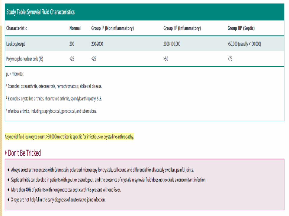
Antineutrophil cytoplasmic antibodies are typically associated with vasculitis such as granulomatosis with polyangiitis (also known as Wegener granulomatosis), microscopic polyangiitis, Churg-Strauss syndrome, anti–glomerular basement membrane antibody disease, and drug-induced vasculitis. Arthritis and arthralgia can be associated with these syndromes; however, the presence of these vascular inflammatory disorders would be unusual in the absence of other system involvement.
Antinuclear antibodies (ANA) can be clinically useful when there is clinical suspicion for autoimmune conditions associated with these antibodies such as systemic lupus erythematosus (SLE). SLE may present with arthritis but, in this case, SLE is less likely than rheumatoid arthritis. SLE typically occurs in women of childbearing age, with additional clinical and/or laboratory abnormalities rather than isolated arthritis. ANA are present in some patients with rheumatoid arthritis but are not specific for this disorder.
Key Point Anti–cyclic citrullinated peptide antibodies are a highly specific marker
for rheumatoid arthritis.

Question 6Question 6 A 36-year-old woman is evaluated for a 5-week
history of pain and swelling of the fingers accompanied by morning stiffness lasting more than 1 hour. Her only medication is ibuprofen, which provides minimal relief.
On physical examination, vital signs are normal. Musculoskeletal examination reveals tenderness and swelling of the right second, third, and fourth metacarpophalangeal joints and the left third, fourth, and fifth metacarpophalangeal joints. There is no bony enlargement, ulnar deviation, or other abnormalities.
Radiographs of the hands and wrists are normal.
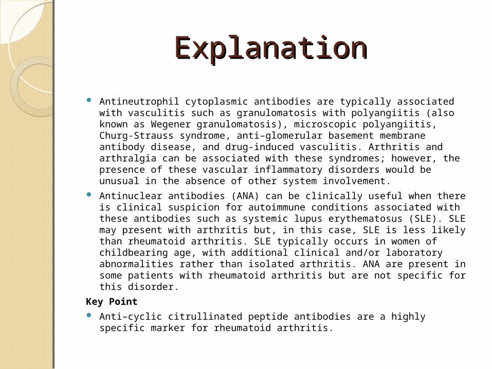
LabsLabs
Erythrocyte sedimentation rate
40 mm/h
Rheumatoid factor 43 units/mL (43 kU/L)
Antinuclear antibodies Negative
Anti–cyclic citrullinated peptide antibodies
Positive
IgM antibodies against parvovirus B19
Negative
Hepatitis B surface antigen Negative
Hepatitis B surface antibodies Positive
Hepatitis C virus antibodies Negative

Answer ChoiceAnswer ChoiceWhich of the following is the most appropriate next step in management?A EtanerceptB HydroxychloroquineC MethotrexateD Reevaluate in 6 weeks

Answer ChoiceAnswer ChoiceWhich of the following is the most appropriate next step in management?A EtanerceptB HydroxychloroquineC MethotrexateD Reevaluate in 6 weeks

Explanation Explanation Methotrexate is indicated for this patient with early rheumatoid arthritis.
Experts recommend that patients begin disease-modifying antirheumatic drug (DMARD) therapy within 3 months of onset. The sooner DMARDs are instituted, the more likely that damage will be limited. Methotrexate is the gold standard DMARD therapy for rheumatoid arthritis and is central to most treatments for the disease. This agent can be effective as initial monotherapy for patients with rheumatoid arthritis of any duration or degree of activity. This patient has synovitis of six metacarpophalangeal joints with a symmetric distribution not involving the distal interphalangeal joints, which is consistent with rheumatoid arthritis. She has swelling, prolonged morning stiffness, an elevated erythrocyte sedimentation rate (ESR), and positive rheumatoid factor, which further support the diagnosis of rheumatoid arthritis, and initial treatment with methotrexate is warranted at this time.
Etanercept is a tumor necrosis factor α inhibitor used for initial therapy in some patients with high disease activity and poor prognostic features. This agent may be necessary for this patient if her disease does not respond to methotrexate.
Hydroxychloroquine as monotherapy may be effective only in mild cases early in the disease course for patients without poor prognostic features. This patient has evidence of moderate disease activity, given the extent of her synovitis and elevated ESR; therefore, hydroxychloroquine as a single agent is unlikely to control this degree of inflammation and is more beneficial as an adjunctive agent.
Explanation Explanation
Reevaluation in 6 weeks is not indicated for this patient whose laboratory studies reveal no evidence of acute parvovirus or hepatitis B infection. Such viral infections can cause an acute polyarthritis syndrome that mimics rheumatoid arthritis. The diagnosis of rheumatoid arthritis previously was predicated on symptoms lasting more than 6 weeks to exclude many self-limiting viral syndromes. However, classification criteria no longer require symptoms to occur for 6 weeks to avoid delays in treatment. The likelihood of rheumatoid arthritis is now calculated on the distribution of joints involved, rheumatoid factor, anti–citrullinated peptide antibodies, acute phase reactants, and duration of symptoms.
Key Point Methotrexate is the gold standard disease-modifying antirheumatic
drug therapy for rheumatoid arthritis and is central to most treatments for the disease

Rheumatoid ArthritisRheumatoid ArthritisDiagnosisRA is a symmetric inflammatory polyarthritis that primarily involves the small joints of the hands and feet. Characteristic findings include:morning stiffness lasting >1 hourseven classic sites of symmetric joint pain (PIP, MCP, wrist, elbow, knee, ankle, and MTP joints)synovitis characterized by soft-tissue swelling or effusionsubcutaneous nodules over bony prominences or extensor surfaces

Laboratory findings include:positive rheumatoid factor (sensitivity 80%;
specificity 87%)elevated ESR or CRP levelnormocytic anemiapositive anti-CCP antibody assay (sensitivity
76%; specificity 96%)An x-ray can reveal periarticular osteopenia,
erosions, and symmetric joint-space narrowing. MRI and ultrasonography are more sensitive for detecting early RA.

Don't Be TrickedNegative rheumatoid factor does
not exclude RA; anti-CCP antibody assay may be positive.
A positive rheumatoid factor alone is not diagnostic of RA.
Not all symmetric arthritis is RA.


RA extra-articular manifestations:arm paresthesias and hyperreflexia → C1-C2 subluxation (increased risk of cord compression with tracheal intubation)cough, fever, pulmonary infiltrates → BOOPfoot drop or wrist drop → mononeuritis multiplex (vasculitis)hoarseness → cricoarytenoid involvementmultiple basilar pulmonary nodules → Caplan syndromedry eyes and/or mouth → Sjögren syndromepleural effusion with low plasma glucose (<30 mg/dL) → rheumatoid pleuritispulmonary fibrosis → rheumatoid interstitial lung diseaseskin ulcers, peripheral neuropathy → rheumatoid vasculitissplenomegaly and granulocytopenia → Felty syndromered, painful eye → scleritisHF → rheumatoid disease or anti-TNF therapyOther complications include increased risk of CAD and osteoporosis.

Carpal, metacarpal, and PIP joints show periarticular Carpal, metacarpal, and PIP joints show periarticular osteopenia, joint-space narrowing, and marginal osteopenia, joint-space narrowing, and marginal erosions, all characteristic of rheumatoid arthritis.erosions, all characteristic of rheumatoid arthritis.

TherapyTherapy Early treatment with one or more DMARDs is essential. Choose NSAIDs
and low-dose oral and intra-articular corticosteroids for quick symptomatic relief, but recognize these agents do not alter the course of the disease.
Monotherapy with hydroxychloroquine or sulfasalazine or combination therapy with these agents is indicated to manage early, mild, and nonerosive disease.
In the absence of contraindications, methotrexate with or without the addition of another DMARD should be instituted immediately in patients with erosive disease.
In some patients, combination therapy with hydroxychloroquine, sulfasalazine, and methotrexate has been shown to be more effective than monotherapy with methotrexate or sulfasalazine plus hydroxychloroquine.
Initiate biologic therapy when adequate disease control is not achieved with oral DMARDs. The initial biologic therapy should be a TNF-α inhibitor:
add a TNF-α inhibitor to baseline methotrexate therapy screen for TB before starting therapy treat for latent TB if TST is positive before beginning any biologic therapy

TherapyTherapy perform periodic TST screening while the patient continues to
receive biologic therapy Common toxicities of TNF-α inhibitor therapy include pancytopenia,
positive ANA formation associated with lupus-like syndromes, and demyelinating disorders. Combination therapy with multiple biologic therapies is not recommended.
Indications for surgical intervention include intractable pain or severe functional disability from joint destruction. Patients may also require surgical repair of ruptured tendons.
All patients taking corticosteroids require osteoporosis screening and serum calcium and vitamin D level measurement. If osteopenia or osteoporosis is diagnosed, prescribe a bisphosphonate. Annual influenza vaccination is indicated for all patients using immunosuppressants, and pneumococcal vaccination is indicated before beginning treatment with methotrexate, leflunomide, or a biologic agent.

TherapyTherapy The most common cause of death in patients with RA is CAD. Begin
aggressive coronary risk factor reduction in all patients. Also, begin adjuvant physical and occupational therapy.
Don't Be Tricked Hydroxychloroquine and sulfasalazine can be used during
pregnancy. Test Yourself A 46-year-old man has a 3-month history of swelling of the PIP and
MCP joints and 90 minutes of morning stiffness. Rheumatoid factor is negative.
ANSWER: The probable diagnosis is RA. Select anti-CCP antibody assay.

Question 7Question 7 A 56-year-old woman is evaluated during a follow-up visit
for a 6-year history of Sjögren syndrome treated with low-dose hydroxychloroquine and cyclosporine eyedrops. She has had two episodes of cutaneous vasculitis, which resolved with corticosteroids.
On physical examination, temperature is 36.4 °C (97.6 °F), blood pressure is 116/64 mm Hg, pulse rate is 72/min, and respiration rate is 18/min. Oral mucous membranes are dry. There is a new firm, left parotid gland enlargement without tenderness or warmth, reported by the patient to be progressive over several months, with asymmetry of the parotid glands.
Laboratory studies at the time of diagnosis revealed elevated serum immunoglobulin levels; positive mixed monoclonal cryoglobulin levels; and positive rheumatoid factor, antinuclear antibodies, and anti-Ro/SSA antibodies.

LabsLabs
Complete blood count Normal
Alkaline phosphatase Normal
Calcium Normal
Rheumatoid factor Negative
C3 Normal
C4 Decreased
Antinuclear antibodies Positive
Anti-Ro/SSA antibodies Positive

Answer ChoiceAnswer ChoiceWhich of the following is the most appropriate management?A Add pilocarpineB Add prednisoneC Bone marrow biopsyD Increase hydroxychloroquineE Parotid gland biopsy

Answer ChoiceAnswer ChoiceWhich of the following is the most appropriate management?A Add pilocarpineB Add prednisoneC Bone marrow biopsyD Increase hydroxychloroquineE Parotid gland biopsy
Explanation Explanation Parotid gland biopsy is indicated for this patient who has Sjögren syndrome
and progressive parotid swelling suggesting possible non-Hodgkin lymphoma. Patients with Sjögren syndrome have up to a 44-fold increased incidence of lymphoma, which may be confined to glandular tissue. Risk factors for the development of lymphoma include disappearance of rheumatoid factor, the presence of mixed monoclonal cryoglobulinemia, cutaneous vasculitis, and low C4 levels, all of which are seen in this patient. Although benign parotid gland swelling can occur and be unilateral or bilateral in patients with Sjögren syndrome, this patient's high-risk profile and new asymmetric parotid enlargement should prompt a biopsy to evaluate for extranodal lymphoma in the parotid gland. Extranodal marginal zone B-cell lymphomas of the mucosa-associated lymphoid tissue (MALT) are the most common lymphomas in patients with Sjögren syndrome, and salivary glands are the most common location; other extranodal sites include the stomach, nasopharynx, skin, liver, and lungs. The risk of nodal lymphoma is also increased in Sjögren syndrome. Although benign lymphadenopathy is a common disease manifestation, the presence of new or rapidly enlarging lymph nodes may indicate development of nodal lymphoma and should prompt biopsy.
Pilocarpine is effective for reducing dry mouth symptoms but is not used to treat parotid enlargement.

Explanation Explanation
Prednisone is generally used to treat inflammatory symptoms of Sjögren syndrome, including arthritis, vasculitis, and cytopenias, but does not reduce parotid swelling or treat symptoms of keratoconjunctivitis sicca and xerostomia (dry eyes and dry mouth).
Patients with Sjögren syndrome often have elevated immunoglobulin levels with monoclonal gammopathy; stability of this during the patient's disease course, as well as normal hemoglobin, calcium, and alkaline phosphatase levels, suggests that bone marrow biopsy to evaluate for myeloma is not warranted. Extranodal lymphoma in Sjögren syndrome involves the bone marrow in less than 10% of patients.
Hydroxychloroquine is used to treat arthritis associated with Sjögren syndrome; however, it is unclear if this agent has efficacy in reducing sicca symptoms or parotid swelling.
Key Point Patients with Sjögren syndrome have up to a 44-fold increased
incidence of lymphoma, which may be confined to glandular tissue

Sjögren SyndromeSjögren SyndromeDiagnosisSjögren syndrome is an autoimmune disease characterized by keratoconjunctivitis sicca and xerostomia. Salivary gland enlargement occurs in nearly half of patients during the course of the disease and is most obvious in the parotid glands. This condition may occur as a primary disease process or may be associated with another autoimmune disease, most commonly rheumatoid arthritis. A cardinal feature of Sjögren syndrome is the presence of antibodies to Ro/SSA and La/SSB. A positive ANA, RF, and hypergammaglobulinemia are also frequently found. Diagnosis is established by biopsy of a labial minor salivary gland. Patients with Sjögren syndrome are up to 44 times more likely than the general population to have a B-cell lymphoma. Careful follow-up is therefore required.

Bilateral parotid gland enlargement in a patient Bilateral parotid gland enlargement in a patient with Sjögren syndrome.with Sjögren syndrome.

TherapyTherapy
Treatment is symptomatic. Choose artificial tear replacement and artificial saliva and mouth lubricants. Systemic immunosuppressive therapy is indicated only in patients with severe systemic manifestations.
SpondyloarthritisSpondyloarthritisKey Considerations
Spondyloarthritis comprises several systemic inflammatory joint disorders that share distinct clinical, radiographic, and genetic features. These disorders are:psoriatic arthritisreactive arthritis (formerly Reiter syndrome)ankylosing spondylitisIBD-associated arthritis
Characteristics include:inflammatory spine and sacroiliac diseaseasymmetric inflammation in four or fewer peripheral joints (typically large joints)inflammation at the sites of ligament and tendon insertion (enthesitis)the presence of HLA-B27extra-articular conditions, such as uveitis, colitis, urethritis, aortitis, and psoriasis

Question 8Question 8A 42-year-old man is evaluated for a 1-month history
of a painful, swollen right finger and a swollen left toe.
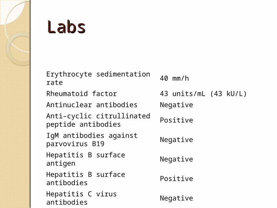
On physical examination, vital signs are normal. The right third distal interphalangeal joint is swollen, with localized tenderness to palpation and pain with active and passive range of motion. The appearance of the nails
is shown The left second toe is remarkable for fusiform
swelling and mild diffuse tenderness, with decreased active and passive range of motion. There is onycholysis of several toenails, including the left second toenail. The remainder of the physical examination is normal.


Answer ChoiceAnswer ChoiceWhich of the following is the most likely diagnosis?A Lyme arthritisB OsteoarthritisC Psoriatic arthritisD Rheumatoid arthritis

Answer ChoiceAnswer ChoiceWhich of the following is the most likely diagnosis?A Lyme arthritisB OsteoarthritisC Psoriatic arthritisD Rheumatoid arthritis

Explanation Explanation This patient has psoriatic arthritis, a systemic chronic inflammatory arthritis
associated with numerous clinical manifestations. Typically, psoriasis predates the arthritis by years, whereas arthritis develops before skin disease in 15% of patients. Although there is a poor correlation between the severity of skin and joint disease, there is a good correlation between the severity of nail disease and the severity of both skin and joint disease. Psoriatic findings may also be limited to nail pitting and onycholysis. There are five patterns of joint involvement in psoriatic arthritis: involvement of the distal interphalangeal joints; asymmetric oligoarthritis; symmetric polyarthritis (similar to that of rheumatoid arthritis); arthritis mutilans (extensive osteolysis of the digits with striking deformity); and spondylitis. Characteristic features of psoriatic arthritis include enthesitis, dactylitis, and tenosynovitis. This patient has findings characteristic of psoriatic arthritis, including inflammation of a distal interphalangeal joint and dactylitis of a toe. He also has nail changes, including pitting and onycholysis.
Lyme arthritis typically involves medium- or large-sized joints rather than distal interphalangeal joints and does not typically cause tenosynovitis. Furthermore, this disorder does not cause nail changes, as seen in this patient.
Osteoarthritis can involve the distal interphalangeal joints but does not cause dactylitis or nail changes.

Explanation Explanation
Rheumatoid arthritis can initially present with an asymmetric pattern, although it classically takes on a symmetric distribution with time. In contrast to this case, rheumatoid arthritis typically spares the distal interphalangeal joints, involving the proximal interphalangeal joints and metacarpophalangeal joints preferentially. Finally, this condition does not cause nail changes
Key Point Psoriatic arthritis is associated with various patterns of joint
involvement, most notably distal interphalangeal joint involvement, and is characterized by enthesitis, dactylitis, tenosynovitis, and cutaneous involvement such as nail pitting

Psoriatic ArthritisPsoriatic Arthritis Characteristic findings are classic psoriasis (thick silvery scale on a well-
demarcated red patch) and nail pitting in a patient with joint pain and stiffness. Skin involvement commonly precedes joint inflammation, although 15% of patients first develop joint inflammation.
Patterns of joint involvement are various. Approximately 40% of patients present with symmetric oligoarthritis of the large joints of the lower extremities, and 25% of these patients develop small-joint polyarthritis, similar to RA. Spinal involvement occurs in almost 50% of patients with psoriatic arthritis. A sausage-shaped finger or toe (dactylitis) may be found and the DIP joints are often involved, which helps distinguishes psoriatic arthritis from RA.
Patients with psoriatic arthritis tend to be seronegative for rheumatoid factor, but at least 15% are seropositive, as are a similar percentage of patients with uncomplicated psoriasis. Serum uric acid levels may be elevated in patients with psoriatic arthritis because of rapid turnover of skin cells. X-rays may show a “pencil-in-the-cup” appearance of one or more involved joints. Other radiographic findings include syndesmophytes and sacroiliitis of the axial skeleton.

Don't Be Tricked No relationship exists between the extent of skin and
joint disease in patients with psoriatic arthritis. Do not make a diagnosis of gout based solely on joint
pain and elevated serum uric acid levels. Use NSAIDs for mild joint inflammation and minimal
skin involvement; prescribe methotrexate for severe skin and erosive peripheral joint disease. A TNF-α inhibitor is indicated for methotrexate-resistant peripheral disease and may be indicated as first-line treatment for patients with axial involvement and for those with dactylitis or enthesitis that does not respond to NSAIDs and locally injected corticosteroids. NSAIDs, antimalarial drugs, and (withdrawal from) oral corticosteroids may exacerbate psorias

Diffuse swelling of the left third and fourth toes and right fourth Diffuse swelling of the left third and fourth toes and right fourth toe characteristic of dactylitis.toe characteristic of dactylitis.

Tiny pits scattered over the nail plate resulting from psoriatic Tiny pits scattered over the nail plate resulting from psoriatic involvement of the nail matrix.involvement of the nail matrix.

Question 9Question 9 A 24-year-old woman is evaluated for a 3-week history of pain and
swelling of the right knee and left ankle. She also has urinary frequency and urgency. The patient has no history of tick exposure, skin rash, diarrhea, or abdominal pain. She has not been sexually active in the past month. She takes no medications.
On physical examination, vital signs are normal. Musculoskeletal examination reveals swelling, tenderness, warmth, pain on active and passive range of motion, and a palpable effusion of the right knee; the left ankle is swollen and tender, with pain at the extremes of active range of motion and no significant pain with passive range of motion.
Serologic test results for Borrelia burgdorferi are negative. Urinalysis reveals 2+ leukocyte esterase, 18 leukocytes/hpf, and no protein, bacteria, squamous epithelial cells, or erythrocytes.
Aspiration of the right knee is performed; synovial fluid analysis reveals an erythrocyte count of 150/µL and a leukocyte count of 7500/µL (7.5 × 10⁹/L). Gram stain is negative. Synovial fluid culture results are pending.

Answer ChoiceAnswer ChoiceWhich of the following is the most appropriate next step in management?A Antinuclear antibody testingB Rheumatoid factor testingC Synovial fluid polymerase chain reaction testing for Borrelia burgdorferiD Urine nucleic acid amplification testing for Chlamydia trachomatis and Neisseria gonorrhoeae

Answer ChoiceAnswer ChoiceWhich of the following is the most appropriate next step in management?A Antinuclear antibody testingB Rheumatoid factor testingC Synovial fluid polymerase chain reaction testing for Borrelia burgdorferiD Urine nucleic acid amplification testing for Chlamydia trachomatis and Neisseria gonorrhoeae

Explanation Explanation This patient requires urine nucleic acid amplification testing for Chlamydia
trachomatis and Neisseria gonorrhoeae. She has acute arthritis of the right knee, enthesitis of the left ankle, and urethritis, all of which can be seen with disseminated gonorrheal infection; however, in the absence of any recent history of sexual activity, these findings are more suggestive of reactive arthritis as can be seen after C. trachomatis infection. Reactive arthritis occurs in both men and women, and enthesitis and oligoarthritis are common. The classic triad of arthritis, urethritis, and conjunctivitis occurs in only one third of patients. Symptoms typically develop 2 to 4 weeks after an infection.
Classic pathogens associated with reactive arthritis include C. trachomatis as well as several enteric pathogens. C. trachomatis infection may be asymptomatic. Urine sample evaluation with ligase reaction can establish a diagnosis of C. trachomatis infection; at that time, antibiotic treatment (azithromycin or levofloxacin) is warranted to prevent potential sequelae of untreated disease. Sexual partners should also be counseled and treated.
Antinuclear antibody testing can be helpful in the diagnosis of systemic lupus erythematosus (SLE). Although arthritis and pyuria can be seen in SLE, the pyuria typically results from glomerulonephritis and is therefore not associated with the lower urinary tract symptoms of frequency and urgency. The patient does not have any other symptoms or signs of SLE.

Explanation Explanation Rheumatoid factor is present in approximately 70% of patients with
rheumatoid arthritis. This disorder typically presents with a symmetric, small joint polyarthritis and does not explain this patient's urinary symptoms and pyuria.
This patient's negative Lyme disease serology results indicate that she does not have Lyme arthritis, and testing the synovial fluid for Borrelia burgdorferi infection is not needed. In patients with Lyme arthritis, testing by polymerase chain reaction (PCR) can detect B. burgdorferi DNA in synovial fluid. Unlike B. burgdorferi serology, synovial fluid PCR testing becomes negative after successful antibiotic treatment. However, synovial fluid PCR testing has not been validated for wide use.
Key Point Detection of pathogens such as Chlamydia trachomatis in patients
with arthritis, urethritis, conjunctivitis, and/or enthesitis supports a diagnosis of reactive arthritis.

Reactive ArthritisReactive Arthritis Reactive arthritis is an acute aseptic inflammatory
arthritis that occurs 1 to 3 weeks after an infectious event originating in the GU or GI tract. A high prevalence of HIV infection is found in patients with symptoms of reactive arthritis.
Characteristic findings include: acute asymmetric oligoarthritis (usually in weight-
bearing joints) dactylitis mouth ulcers inflammatory eye conditions Patients may also have keratoderma blennorrhagicum
(a psoriasis-like skin lesion on the palms and soles) or circinate balanitis (shallow, moist, serpiginous ulcers with raised borders on the glans penis).

Diagnostic studies include throat culture for Streptococcus, urogenital culture for Chlamydia, and serologic studies for Salmonella, Yersinia, Campylobacter, Neisseria, and HIV. Obtain urinalysis for protein, blood, and leukocytes.
If the causative organism can be isolated (β-hemolytic streptococci, N. gonorrhoeae, Chlamydia), begin specific antibiotics. If infection is ruled out, treat with an intra-articular corticosteroid injection in the acutely inflamed joint. If arthritis persists for longer than 3 to 5 months, begin DMARDs. Sulfasalazine is a common first-line choice.
Don't Be Tricked The classic triad of arthritis, conjunctivitis, and urethritis (or
cervicitis) is found in only one third of patients with reactive arthritis.
Do not prescribe chronic antibiotic therapy for patients with reactive arthritis.

A papular and pustular rash of the palms and soles is A papular and pustular rash of the palms and soles is associated with reactive arthritis.associated with reactive arthritis.

Ankylosing SpondylitisAnkylosing Spondylitis Ankylosing spondylitis primarily affects the spine and sacroiliac
joints. It also may involve the shoulders and hips, although the small peripheral joints are not affected. Ankylosing spondylitis occurs most often in patients <40 years of age and presents as chronic low back pain. Characteristic findings are pain and stiffness that worsen at night and are relieved with physical activity or heat.
Physical examination findings include: decreased hyperextension, forward flexion, lateral flexion, and axial
rotation diminished chest expansion asymmetric peripheral arthritis involving the large joints painful heels (enthesitis) X-rays show subchondral bony sclerosis, vertebral body squaring,
and bony ankylosis (“bamboo spine”). When radiographic findings are equivocal or absent, MRI can detect the early changes of sacroiliitis. HLA-B27 testing is neither sensitive nor specific for ankylosing spondylitis and does not distinguish ankylosing spondylitis from other spondyloarthropathies.

Extra-articular manifestations include acute anterior uveitis (most common), aortic valvular regurgitation and cardiac conduction defects, apical pulmonary fibrosis and cavitation, and cauda equina syndrome.A patient with ankylosing spondylitis with increased pain and mobility of the neck following a minor accident may have a fracture and requires an urgent CT of the cervical spine.
Don't Be TrickedAnkylosing spondylitis occurs in both men and women.
NSAIDs, not aspirin, are the mainstay of management. Use corticosteroid injections for recalcitrant enthesitis and persistent synovitis. Prescribe sulfasalazine for patients with primarily peripheral arthritis and a TNF-α inhibitor for primarily axial disease. Calcium and vitamin D supplements and exercise to retain good posture, spinal mobility, and chest expansion are beneficial. Begin a bisphosphonate if osteopenia or osteoporosis is present.
Don't Be Tricked
Do not prescribe traditional DMARDs for patients with axial disease because they are ineffective. Select a TNF-α inhibitor.

Sclerosis and erosions of sacroiliac joints and Sclerosis and erosions of sacroiliac joints and bridging of the intervertebral disks by bridging of the intervertebral disks by syndesmophytes are characteristic of ankylosing syndesmophytes are characteristic of ankylosing spondylitis.spondylitis.

IBD-Associated ArthritisIBD-Associated Arthritis Inflammatory arthritis can complicate Crohn disease and
ulcerative colitis. Up to 20% of patients with IBD develop a peripheral arthritis, which manifests as either a polyarticular arthritis resembling RA or an asymmetric oligoarthritis predominantly of the lower extremities resembling reactive arthritis. The course of arthritis often fluctuates with the activity of the underlying bowel involvement. Another 20% of patients have spinal involvement ranging from asymptomatic sacroiliac disease to a clinical presentation identical to that of ankylosing spondylitis. Unlike peripheral arthritis, the progression of spinal involvement is independent of the course of the bowel disease.
The therapies that benefit intestinal disease also have efficacy in the treatment of the associated peripheral joint and extra-articular manifestations. These therapies include corticosteroids, sulfasalazine, azathioprine, methotrexate, infliximab, and adalimumab.
Don't Be Tricked Etanercept has not shown efficacy in treating IBD.