Rheumatoid Forefoot Reconstruction. A Long-Term Follow- up Study* by MICHAEL J. COUGHLIN J Bone...
16
Rheumatoid Forefoot Reconstruction. A Long-Term Follow-up Study* by MICHAEL J. COUGHLIN J Bone Joint Surg Am Volume 82(3):322-41 March 1, 2000 ©2000 by The Journal of Bone and Joint Surgery, Inc.
-
Upload
lenard-gregory -
Category
Documents
-
view
213 -
download
0
Transcript of Rheumatoid Forefoot Reconstruction. A Long-Term Follow- up Study* by MICHAEL J. COUGHLIN J Bone...

Rheumatoid Forefoot Reconstruction. A Long-Term Follow-up Study*
by MICHAEL J. COUGHLIN
J Bone Joint Surg AmVolume 82(3):322-41
March 1, 2000
©2000 by The Journal of Bone and Joint Surgery, Inc.

Fig. 1 Diagram demonstrating the hallux valgus angle, the first-second intermetatarsal angle, and the MTP-2 angle.
MICHAEL J. COUGHLIN J Bone Joint Surg Am 2000;82:322-41
©2000 by The Journal of Bone and Joint Surgery, Inc.

Fig. 2 Diagram demonstrating the dorsiflexion angle of fusion.
MICHAEL J. COUGHLIN J Bone Joint Surg Am 2000;82:322-41
©2000 by The Journal of Bone and Joint Surgery, Inc.

Fig. 3-A Diagram and radiograph demonstrating the medial-lateral position of the lesser metatarsophalangeal joints.
MICHAEL J. COUGHLIN J Bone Joint Surg Am 2000;82:322-41
©2000 by The Journal of Bone and Joint Surgery, Inc.

Fig. 3-B Diagram and radiograph demonstrating the medial-lateral position of the lesser metatarsophalangeal joints.
MICHAEL J. COUGHLIN J Bone Joint Surg Am 2000;82:322-41
©2000 by The Journal of Bone and Joint Surgery, Inc.
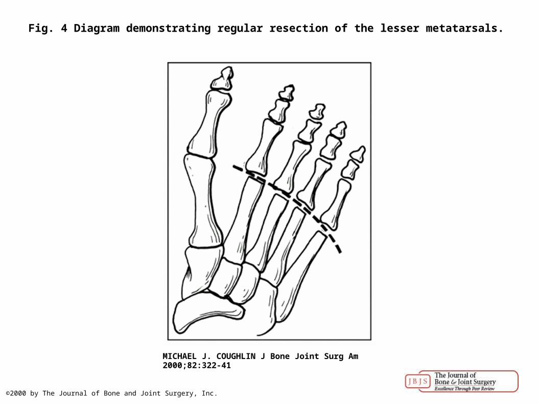
Fig. 4 Diagram demonstrating regular resection of the lesser metatarsals.
MICHAEL J. COUGHLIN J Bone Joint Surg Am 2000;82:322-41
©2000 by The Journal of Bone and Joint Surgery, Inc.

Fig. 5-A Preoperative radiograph demonstrating hallux valgus deformity and dislocation of the second, third, and fourth metatarsophalangeal joints.
MICHAEL J. COUGHLIN J Bone Joint Surg Am 2000;82:322-41
©2000 by The Journal of Bone and Joint Surgery, Inc.

Fig. 5-B Intraoperative photograph showing the dorsal longitudinal incisions and the dorsal plate used for internal fixation at the site of the arthrodesis of the first metatarsophalangeal joint.
MICHAEL J. COUGHLIN J Bone Joint Surg Am 2000;82:322-41
©2000 by The Journal of Bone and Joint Surgery, Inc.

Fig. 5-C: Postoperative radiograph demonstrating arthrodesis of the first metatarsophalangeal joint, resection arthroplasty of the lesser metatarsophalangeal joints, hammer-toe repairs of the
lesser toes, and intramedullary Kirschner-wire fixation.
MICHAEL J. COUGHLIN J Bone Joint Surg Am 2000;82:322-41
©2000 by The Journal of Bone and Joint Surgery, Inc.

Fig. 5-D: Radiograph, made at the time of the three-year follow-up, demonstrating anatomical alignment of the lesser metatarsophalangeal joints.
MICHAEL J. COUGHLIN J Bone Joint Surg Am 2000;82:322-41
©2000 by The Journal of Bone and Joint Surgery, Inc.

Fig. 5-E Lateral radiograph demonstrating fusion of the first metatarsophalangeal joint.
MICHAEL J. COUGHLIN J Bone Joint Surg Am 2000;82:322-41
©2000 by The Journal of Bone and Joint Surgery, Inc.

Fig. 6-A: Radiograph, made five years after an unsuccessful Keller resection arthroplasty, demonstrating fixed dislocation of the lesser metatarsophalangeal joints with hammer-toe
deformities.
MICHAEL J. COUGHLIN J Bone Joint Surg Am 2000;82:322-41
©2000 by The Journal of Bone and Joint Surgery, Inc.

Fig. 6-B: Radiograph, made five years and eight months after arthrodeses of the metatarsophalangeal joint and the interphalangeal joint, demonstrating alignment of the lesser
metatarsophalangeal joints.
MICHAEL J. COUGHLIN J Bone Joint Surg Am 2000;82:322-41
©2000 by The Journal of Bone and Joint Surgery, Inc.

Fig. 7 Radiograph, made approximately eight years postoperatively, demonstrating successful arthrodesis of the first metatarsophalangeal joint and severe degenerative changes with joint
destruction and subchondral cyst formation (grade-4 arthritis) of the i...
MICHAEL J. COUGHLIN J Bone Joint Surg Am 2000;82:322-41
©2000 by The Journal of Bone and Joint Surgery, Inc.

Fig. 8-A: Radiograph, made two years preoperatively, demonstrating severe hallux valgus with dislocation of the first and second metatarsophalangeal joints.
MICHAEL J. COUGHLIN J Bone Joint Surg Am 2000;82:322-41
©2000 by The Journal of Bone and Joint Surgery, Inc.

Fig. 8-B: Following arthrodesis, the first metatarsophalangeal joint is realigned.
MICHAEL J. COUGHLIN J Bone Joint Surg Am 2000;82:322-41
©2000 by The Journal of Bone and Joint Surgery, Inc.