Rheumatic heart disease heart disease Eloi Marijon*, Mariana Mirabel*, David S Celermajer, Xavier...
12
Seminar www.thelancet.com Vol 379 March 10, 2012 953 Lancet 2012; 379: 953–64 *Authors contributed equally Paris Cardiovascular Research Centre, INSERM U970 (E Marijon MD, M Mirabel MD, Prof X Jouven PhD), and Department of Cardiology (E Marijon, Prof X Jouven), European Georges Pompidou Hospital, Paris, France; Paris Descartes University, Paris, France (E Marijon, M Mirabel, Prof X Jouven); Maputo Heart Institute (ICOR), Maputo, Mozambique (E Marijon, Prof X Jouven); University College London, London, UK (M Mirabel); and Sydney Medical School, University of Sydney, NSW, Australia (Prof D S Celermajer PhD) Correspondence to: Dr Eloi Marijon, Paris Cardiovascular Research Centre, INSERM U970, Hôpital Européen Georges Pompidou, 75737 Paris, CEDEX 15, France [email protected] Rheumatic heart disease Eloi Marijon*, Mariana Mirabel*, David S Celermajer, Xavier Jouven Rheumatic heart disease, often neglected by media and policy makers, is a major burden in developing countries where it causes most of the cardiovascular morbidity and mortality in young people, leading to about 250 000 deaths per year worldwide. The disease results from an abnormal autoimmune response to a group A streptococcal infection in a genetically susceptible host. Acute rheumatic fever—the precursor to rheumatic heart disease—can affect different organs and lead to irreversible valve damage and heart failure. Although penicillin is effective in the prevention of the disease, treatment of advanced stages uses up a vast amount of resources, which makes disease management especially challenging in emerging nations. Guidelines have therefore emphasised antibiotic prophylaxis against recurrent episodes of acute rheumatic fever, which seems feasible and cost effective. Early detection and targeted treatment might be possible if populations at risk for rheumatic heart disease in endemic areas are screened. In this setting, active surveillance with echocardiography-based screening might become very important. Introduction Rheumatic heart disease is the result of valvular damage caused by an abnormal immune response to group A streptococcal infection, usually during childhood. 1 Although this disease—associated with poverty—has almost disappeared from wealthy countries, its burden remains a major challenge in developing nations. 2,3 Preventive measures, based mainly on penicillin use and associated with economic and social development, are very efficient and have nearly eradicated rheumatic heart disease in developed countries. However, according to the 2008 Population Reference Bureau, about 80–85% of children younger than 15 years (around 2 billion) live in areas where rheumatic heart disease is endemic. 4 Worldwide, this disease is the leading cause of heart failure in children and young adults, resulting in disability and premature death and severely affecting the workforce in emerging nations. 3 Demographic trends in the developing world, including poor access to birth control and rural exodus, will probably contribute to the substantial rise in the number of people at risk for rheumatic heart disease in the next 20 years. 4 The disease receives little attention from the medical community, as shown by the low number of publications and congress presentations on this subject, and consequently is poorly covered by the media. Acute rheumatic fever usually occurs 3 weeks after group A streptococcal pharyngitis and can affect the joints, skin, brain, and heart. 5 Around half of patients with acute rheumatic fever present with cardiac inflam- mation mainly involving the valvular endocardium. 6–11 Although the initial attack can lead to severe valvular disease, rheumatic heart disease most often results from cumulative valve damage due to recurrent paucisymp- tomatic episodes of acute rheumatic fever, which suggests that it might be insidious at onset. 3,5,12 Because secondary prevention can prevent adverse outcomes, early echo- cardiography-based identification of silent rheumatic heart disease (showing no clinical signs) with minimal valve lesions by active surveillance programmes might be of major importance. 13–15 In this Seminar we discuss epidemiology, patho- physiology, available preventive strategies, the rationale for and first experiences with echocardiography-based screening, and treatments for rheumatic heart disease. Epidemiology Improved living conditions, nutrition, access to medical care, and penicillin use have substantially changed the epidemiology of acute rheumatic fever and rheumatic heart disease. 3 Nevertheless, both prevail in developing nations and some underprivileged, mainly indigenous, populations in affluent countries. 16,17 Carapetis and colleagues 3,18 have reviewed the worldwide burden of these diseases, but prevalence is difficult to estimate, mainly because of the scarcity of comprehensive disease registries, the use of passive survey systems, and under- reporting of acute and chronic cases. 16,19 Rheumatic heart disease causes at least 200 000–250 000 premature deaths every year, 3 and is the major cause of cardiovascular death in children and young adults in developing countries. 3,20 Epidemiological data for Africa are scarce, despite local efforts to raise awareness and to launch prevention programmes such as those outlined in the Drakensberg declaration. 21,22 In areas of poor or no medical attention, the natural course of the disease prevails because patients have no access to treatment. Mortality Search strategy and selection criteria We searched PubMed for publications in English with the terms “rheumatic heart disease and epidemiology”, “rheumatic heart disease and pathophysiology”, “rheumatic heart disease and diagnosis”, “rheumatic heart disease and screening”, “rheumatic heart disease and echocardiography”, “rheumatic heart disease and therapy”, “rheumatic heart disease and prevention”, and “rheumatic fever”. We focused on, but did not restrict the search to, publications from the past 5 years. We selected relevant articles published in any language and have referenced several review articles and book chapters, particularly on pathophysiology, because they provided comprehensive overviews that are beyond the scope of this Seminar. We also searched the Cochrane database with the term “rheumatic heart disease”, and our own database of references and those of linked articles in the searched journals. When more than one article referred to the same point, the most representative article was chosen. Regarding the acute phase of rheumatic heart disease (acute rheumatic fever), we describe only the most classic forms of presentation.
Transcript of Rheumatic heart disease heart disease Eloi Marijon*, Mariana Mirabel*, David S Celermajer, Xavier...
Seminar
www.thelancet.com Vol 379 March 10, 2012 953
Lancet 2012; 379: 953–64
*Authors contributed equally
Paris Cardiovascular Research Centre, INSERM U970 (E Marijon MD, M Mirabel MD, Prof X Jouven PhD), and Department of Cardiology (E Marijon, Prof X Jouven), European Georges Pompidou Hospital, Paris, France; Paris Descartes University, Paris, France (E Marijon, M Mirabel, Prof X Jouven); Maputo Heart Institute (ICOR), Maputo, Mozambique (E Marijon, Prof X Jouven); University College London, London, UK (M Mirabel); and Sydney Medical School, University of Sydney, NSW, Australia (Prof D S Celermajer PhD)
Correspondence to:Dr Eloi Marijon, Paris Cardiovascular Research Centre, INSERM U970, Hôpital Européen Georges Pompidou, 75737 Paris, CEDEX 15, [email protected]
Rheumatic heart diseaseEloi Marijon*, Mariana Mirabel*, David S Celermajer, Xavier Jouven
Rheumatic heart disease, often neglected by media and policy makers, is a major burden in developing countries where it causes most of the cardiovascular morbidity and mortality in young people, leading to about 250 000 deaths per year worldwide. The disease results from an abnormal autoimmune response to a group A streptococcal infection in a genetically susceptible host. Acute rheumatic fever—the precursor to rheumatic heart disease—can aff ect diff erent organs and lead to irreversible valve damage and heart failure. Although penicillin is eff ective in the prevention of the disease, treatment of advanced stages uses up a vast amount of resources, which makes disease management especially challenging in emerging nations. Guidelines have therefore emphasised antibiotic prophylaxis against recurrent episodes of acute rheumatic fever, which seems feasible and cost eff ective. Early detection and targeted treatment might be possible if populations at risk for rheumatic heart disease in endemic areas are screened. In this setting, active surveillance with echocardiography-based screening might become very important.
IntroductionRheumatic heart disease is the result of valvular damage caused by an abnormal immune response to group A streptococcal infection, usually during childhood.1 Although this disease—associated with poverty—has almost disappeared from wealthy countries, its burden remains a major challenge in developing nations.2,3
Preventive measures, based mainly on penicillin use and associated with economic and social development, are very effi cient and have nearly eradicated rheumatic heart disease in developed countries. However, according to the 2008 Population Reference Bureau, about 80–85% of children younger than 15 years (around 2 billion) live in areas where rheumatic heart disease is endemic.4 Worldwide, this disease is the leading cause of heart failure in children and young adults, resulting in disability and premature death and severely aff ecting the workforce in emerging nations.3 Demographic trends in the developing world, including poor access to birth control and rural exodus, will probably contribute to the substantial rise in the number of people at risk for rheumatic heart disease in the next 20 years.4 The disease receives little attention from the medical community, as shown by the low number of publications and congress presentations on this subject, and consequently is poorly covered by the media.
Acute rheumatic fever usually occurs 3 weeks after group A streptococcal pharyngitis and can aff ect the joints, skin, brain, and heart.5 Around half of patients with acute rheumatic fever present with cardiac infl am-mation mainly involving the valvular endo cardium.6–11 Although the initial attack can lead to severe valvular disease, rheumatic heart disease most often results from cumulative valve damage due to recurrent paucisymp-tomatic episodes of acute rheumatic fever, which suggests that it might be insidious at onset.3,5,12 Because secondary prevention can prevent adverse outcomes, early echo-cardiography-based identifi cation of silent rheumatic heart disease (showing no clinical signs) with minimal valve lesions by active surveillance programmes might be of major importance.13–15
In this Seminar we discuss epidemiology, patho-physiology, available preventive strategies, the rationale
for and fi rst experiences with echocardiography-based screening, and treatments for rheumatic heart disease.
EpidemiologyImproved living conditions, nutrition, access to medical care, and penicillin use have substantially changed the epidemiology of acute rheumatic fever and rheumatic heart disease.3 Nevertheless, both prevail in developing nations and some underprivileged, mainly indigenous, populations in affl uent countries.16,17 Carapetis and colleagues3,18 have reviewed the worldwide burden of these diseases, but prevalence is diffi cult to estimate, mainly because of the scarcity of comprehensive disease registries, the use of passive survey systems, and under-reporting of acute and chronic cases.16,19
Rheumatic heart disease causes at least 200 000–250 000 premature deaths every year,3 and is the major cause of cardiovascular death in children and young adults in developing countries.3,20 Epidemiological data for Africa are scarce, despite local eff orts to raise awareness and to launch prevention programmes such as those outlined in the Drakensberg declaration.21,22 In areas of poor or no medical attention, the natural course of the disease prevails because patients have no access to treatment. Mortality
Search strategy and selection criteria
We searched PubMed for publications in English with the terms “rheumatic heart disease and epidemiology”, “rheumatic heart disease and pathophysiology”, “rheumatic heart disease and diagnosis”, “rheumatic heart disease and screening”, “rheumatic heart disease and echocardiography”, “rheumatic heart disease and therapy”, “rheumatic heart disease and prevention”, and “rheumatic fever”. We focused on, but did not restrict the search to, publications from the past 5 years. We selected relevant articles published in any language and have referenced several review articles and book chapters, particularly on pathophysiology, because they provided comprehensive overviews that are beyond the scope of this Seminar. We also searched the Cochrane database with the term “rheumatic heart disease”, and our own database of references and those of linked articles in the searched journals. When more than one article referred to the same point, the most representative article was chosen. Regarding the acute phase of rheumatic heart disease (acute rheumatic fever), we describe only the most classic forms of presentation.

Seminar
954 www.thelancet.com Vol 379 March 10, 2012
rates in these areas can be as high as 20% at 6-year follow-up according to a Nigerian paediatric cohort study,23 or 12·5% every year, as documented in rural Ethiopia.24
Additionally, rheumatic heart disease causes sub stan-tial morbidity in children25 and adults, and can aff ect quality of life26 and economic growth. According to a 2004 WHO report, the number of disability-adjusted life-years lost to the disease was as high as 5·2 million per year, worldwide.20
The global incidence of acute rheumatic fever in children aged 5–14 years is roughly 300 000–350 000 per year, although incidence varies substantially by region.3,5,27 The yearly incidence of a fi rst attack of acute rheumatic fever ranges from 5 to 51 per 100 000 population in Indigenous New Zealand communities, and can reach 80–254 per 100 000 in Indigenous Australian communities.28,29 Identifi ed modi fi able risk factors for acute rheumatic fever include poverty, overcrowding, malnutrition, and maternal edu-cational level and employment.6,30–33 Virulence of strepto-coccal strains and genetic susceptibility might partly account for the reported variations in acute rheumatic fever incidences worldwide.34,35
According to traditional diagnostic criteria, 15·6–19·6 million people worldwide have rheumatic heart disease.3 These data mainly originate from surveys of school children in whom diagnosis is made by clinical assessment.20,32,36,37 Prevalence is highest in adults aged 20–50 years.3,5,12 Distribution of rheumatic heart disease varies between continents, and sub-Saharan Africans and Indigenous Australians seem to have the highest prevalence.3,27,38,39 In Pacifi c Islanders and Indigenous Australians, the prevalence is 5–10 per 1000 school children, and roughly 30 per 1000 adults aged 35–44 years.17,37 In Asia, rheumatic heart disease preva-lence varies,18—eg, in rural Pakistan it has a prevalence in the community as high as 12 per 1000 people.40 In South and Central America, rheumatic heart disease has a lower reported prevalence (1·3 per 1000 school children).3
The recent use of echocardiography-based screening and the subsequent detection of silent cases is challenging these traditional epidemiological data.13,14,41,42
PathophysiologyThe pathogenesis of rheumatic heart disease results from an immune response consisting of humoral and cellular components after exposure to Streptococcus pyogenes (classifi ed as a group A streptococcus by the Lancefi eld system), usually after a throat infection. The precise pathophysiology is obscure but several advances have now been reviewed.34,43 Antigenic mimicry in association with an abnormal host immune response is the cornerstone of pathophysiology, based on the triad of rheumatogenic group A streptococcal strain, genetically susceptible host, and aberrant host immune response.44,45
Some strains are more likely to cause acute rheumatic fever than are others.5 S pyogenes contains M, T,
and R surface proteins, which are all associated with bacterial adherence to throat epithelial cells. The rheumato-genicity of some streptococcus families has trad itionally been considered a feature of strains belonging to specifi c M serotypes. However, data show that rheumato genic M serotypes were infrequently identifi ed in commun ities with high burdens of acute rheumatic fever and rheumatic heart disease. These results question the poten tial importance of other disease-causing serotypes, especially those that cause streptococcal skin infections, which might be implicated in cases of acute rheumatic fever.46–48
In 1889, Cheadle noted that the chance of an individual with a family history of acute rheumatic fever acquiring the disease is “nearly fi ve times as great as that of an individual who has no such hereditary taint”.49 Generally, HLA class II molecules (which participate in antigen presentation to T-cell receptors) seem to be more closely associated with an increased risk of acute rheumatic fever or rheumatic heart disease than are class I molecules, although no single HLA haplotype or combination has been consistently associated with disease susceptibility.34 The exact molecular mechanism by which HLA class II molecules confer susceptibility to autoimmune diseases is unknown.
The role of autoimmune reactions in the pathogenesis of acute rheumatic fever was substantiated when antibodies against group A streptococcus reacted with human heart preparations.50,51 After binding to the antigenic peptide, the particular HLA complexes can initiate inappropriate T-cell activation.52 Molecular mimicry takes place between strepto-coccal M protein and several cardiac proteins (cardiac myosin, tropomyosin, keratin, laminin, and vimentin), and diff erent patterns of T-cell antigen cross-recognition have been identifi ed.53,54 Mannose binding lectin (MBL) is an acute-phase infl ammatory protein that functions as a soluble pathogen recognition receptor. MBL binds to a wide range of sugars on the surface of pathogens and plays a major part in innate immunity because of its ability to opsonise pathogens, enhancing their phagocytosis and activating the complement cascade via the lectin pathway.55 One study reported that genotypes that correlated with high concentrations of MBL were associated with rheumatic heart disease.56 Cytokines (interleukins 1 and 6, and tumour necrosis factor α [TNFα]) are thought to play a part in acute rheumatic fever, and the TNFα gene maps close to the MHC region; however, whether this association is related to other possible risk-associated genes is unclear.57
Case-control association studies using a fi ne-resolution genome-wide approach should help to identify genetic variants aff ecting individual susceptibility to rheumatic heart disease.35
Natural history and presentationAcute rheumatic feverThe disorder manifests as a combination of fever, poly-arthritis, carditis, chorea, erythema marginatum, and subcutaneous nodules in patients about 3 weeks after
Seminar
www.thelancet.com Vol 379 March 10, 2012 955
they have had pharyngitis (most often paucisymptomatic or asymptomatic) caused by a group A streptococcal infection (diagnosed by a positive throat swab culture or a high or rising streptococcal antibody titre), and most often aff ects children, adolescents, and young adults.5,58 The clinical presentation of acute rheumatic fever varies and can be aff ected by delayed consultation or the use of over-the-counter treatments such as anti-infl am matory drugs. In 1944, Jones described the main clinical features of the disease, which have since been modifi ed and revised to become more stringent.59 Other criteria have been put forward to increase sensitivity and encourage investigators to standardise patients’ charac teristics under the auspices of WHO and the National Heart Foundation of Australia and the Cardiac Society of Australia and New Zealand.5,60,61
The peak incidence of acute rheumatic fever is in children aged 5–14 years. Arthritis is usually the earliest feature of the disease, present in 60–80% of patients, and is often very painful and migratory, aff ecting medium and large joints.5 Sydenham’s chorea presents later, usually between 1 and 6 months after the initial exposure to group A streptococcus, and manifests as involuntary, irregular movements, including fi brillatory tongue movements, and spooning with external rotation of the hands. The proportion of patients with chorea varies considerably, from 7% to 28% in diff erent settings.58,62 Cutaneous manifestations are rare and sometimes diffi cult to diagnose.
Carditis occurs a few weeks after the initial infection in about 50% of patients with acute rheumatic fever, and presents as valvulitis, sometimes combined with peri-carditis or (more contentiously) myocarditis.8,63 Patients are examined for various hallmarks of acute carditis, such as sinus tachycardia (particularly its persistence at night) and a diminished fi rst heart sound caused by a frequent, extended PR interval, verifi ed by electrocardiography.59,64 A soft, blowing, pansystolic murmur is characteristic of mitral regurgitation and strongly suggests rheumatic valvulitis. Pericarditis is common in acute rheumatic fever, and is characterised by chest pain and a transient pericardial friction rub accompanied by a small pericardial eff usion on echocardiogram. A very large eff usion causing cardiac tamponade is rare. Signs of poorly tolerated valvular regurgitation include a prominent left ventricu-lar impulse due to dilatation, and signs of left or right heart failure. A chest radiograph might show cardiac enlarge ment or signs of congestive heart failure.
Minich and colleagues65 were among the fi rst to describe subclinical carditis in children. In their cohort, several patients with no murmur had echocardiographic fi ndings consistent with pathological mitral regurgi-tation,65 a result supported by many others.7,11
Rheumatic heart diseasePatients might be diagnosed with rheumatic heart disease after a known acute rheumatic fever attack; however, the
disease is often diagnosed in patients who were previously asymptomatic or who do not recall acute rheumatic fever symptoms or episodes. Most patients present after the onset of shortness of breath at ages 20–50 years.12 Although controversy exists about the female predominance of acute rheumatic fever,29 women of childbearing age do have a higher prevalence of established rheumatic heart disease than do men.12,40 Researchers have not fully addressed the reasons for this female predominance, but some have proposed that social factors (such as child rearing, which might result in repeated exposure to group A strepto-coccus), access to health care (especially preventive medi-cine), and genetically-mediated immuno logical factors that predispose women to autoimmune diseases might be associated.
Clinical diagnosis is based on pathological valvular heart murmur detected during auscultation. Mitral valve incom petence is the most common valvular lesion in patients with rheumatic heart disease, particularly in the early stages.8,66 Mitral stenosis usually develops later as a result of persistent or recurrent valvulitis with bicom-missural fusion,67 although mitral stenosis has been described in adolescents.66,68 Patients with mitral incompetence can remain asympto matic for up to 10 years as a result of compensatory left atrial and left ventricular dilatation before the onset of left ventricular systolic dysfunction. Aortic regurgitation is most often associated with some degree of mitral regurgitation, but can be isolated and severe. Tricuspid regurgitation is often functional, mainly caused by mitral stenosis with high pulmonary pressures and consequent right ventricular dilatation.69 Isolated pulmonary or tricuspid regurgitation are not classic features of rheumatic heart disease. The disease might also present after a complication such as atrial arrhythmia, an embolic event, acute heart failure, or infective endocarditis.
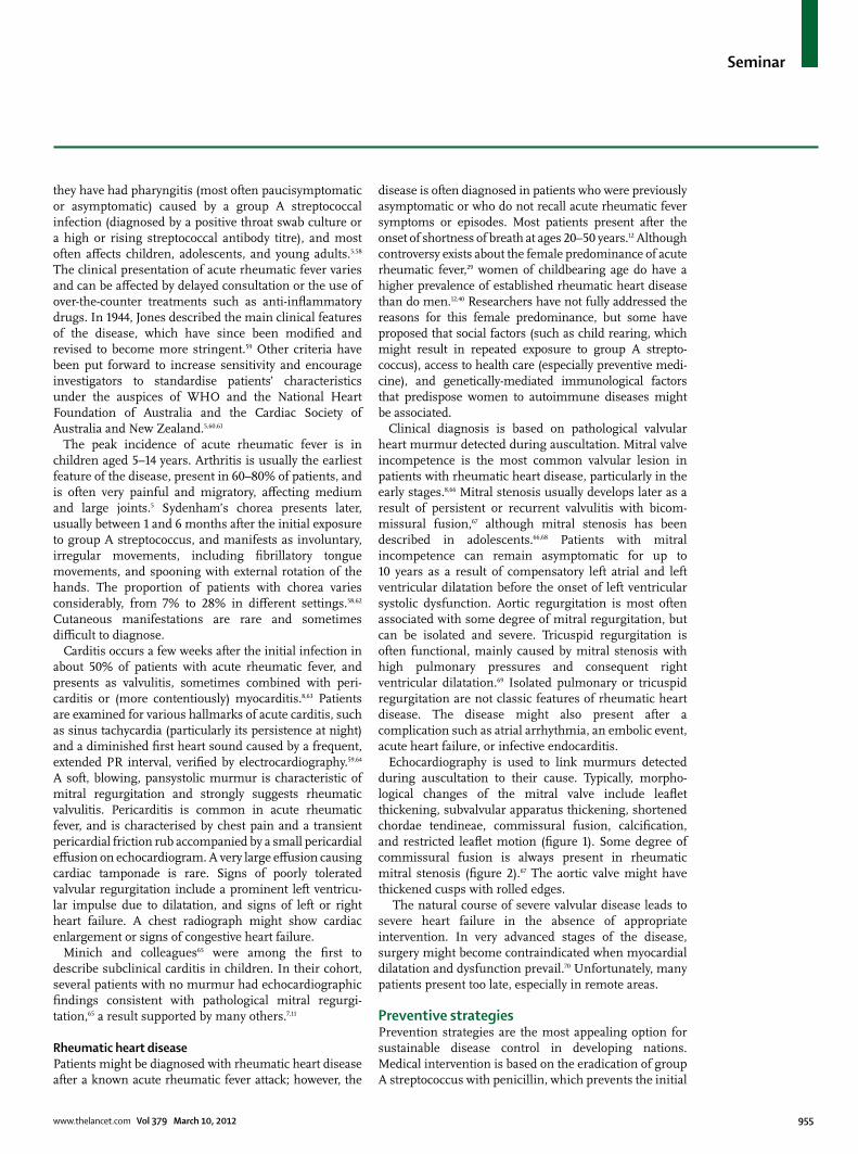
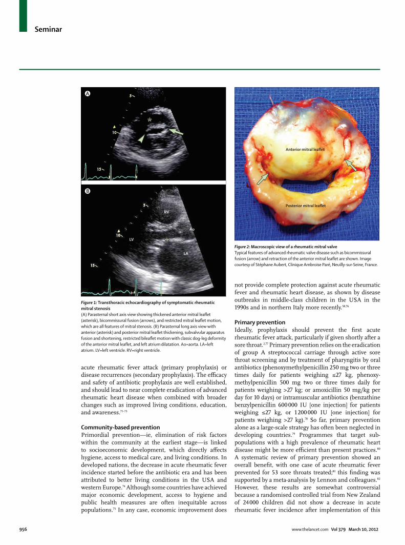
Echocardiography is used to link murmurs detected during auscultation to their cause. Typically, morpho-logical changes of the mitral valve include leafl et thickening, subvalvular apparatus thickening, shortened chordae tendineae, commissural fusion, calcifi cation, and restricted leafl et motion (fi gure 1). Some degree of commissural fusion is always present in rheumatic mitral stenosis (fi gure 2).67 The aortic valve might have thickened cusps with rolled edges.
The natural course of severe valvular disease leads to severe heart failure in the absence of appropriate intervention. In very advanced stages of the disease, surgery might become contraindicated when myocardial dilatation and dysfunction prevail.70 Unfortunately, many patients present too late, especially in remote areas.
Preventive strategiesPrevention strategies are the most appealing option for sustainable disease control in developing nations. Medical intervention is based on the eradication of group A streptococcus with penicillin, which prevents the initial
Seminar
956 www.thelancet.com Vol 379 March 10, 2012
acute rheumatic fever attack (primary prophylaxis) or disease recurrences (secondary prophylaxis). The effi cacy and safety of antibiotic prophylaxis are well established, and should lead to near complete eradication of advanced rheumatic heart disease when combined with broader changes such as improved living conditions, education, and awareness.71–73
Community-based preventionPrimordial prevention—ie, elimination of risk factors within the community at the earliest stage—is linked to socioeconomic development, which directly aff ects hygiene, access to medical care, and living conditions. In developed nations, the decrease in acute rheumatic fever incidence started before the antibiotic era and has been attributed to better living conditions in the USA and western Europe.74 Although some countries have achieved major economic development, access to hygiene and public health measures are often inequitable across populations.75 In any case, economic improvement does
not provide complete protection against acute rheumatic fever and rheumatic heart disease, as shown by disease outbreaks in middle-class children in the USA in the 1990s and in northern Italy more recently.58,76
Primary preventionIdeally, prophylaxis should prevent the fi rst acute rheumatic fever attack, particularly if given shortly after a sore throat.2,77 Primary prevention relies on the eradication of group A streptococcal carriage through active sore throat screening and by treatment of pharyngitis by oral antibiotics (phenoxymethylpenicillin 250 mg two or three times daily for patients weighing ≤27 kg, phenoxy-methylpenicillin 500 mg two or three times daily for patients weighing >27 kg; or amoxicillin 50 mg/kg per day for 10 days) or intramuscular antibiotics (benzathine benzylpenicillin 600 000 IU [one injection] for patients weighing ≤27 kg, or 1 200 000 IU [one injection] for patients weighing >27 kg).78 So far, primary prevention alone as a large-scale strategy has often been neglected in developing countries.79 Programmes that target sub-populations with a high prevalence of rheumatic heart disease might be more effi cient than present practices.80 A systematic review of primary prevention showed an overall benefi t, with one case of acute rheumatic fever prevented for 53 sore throats treated;81 this fi nding was supported by a meta-analysis by Lennon and colleagues.82 However, these results are somewhat controversial because a randomised controlled trial from New Zealand of 24 000 children did not show a decrease in acute rheumatic fever incidence after implementation of this
Figure 1: Transthoracic echocardiography of symptomatic rheumatic mitral stenosis (A) Parasternal short axis view showing thickened anterior mitral leafl et (asterisk), bicommissural fusion (arrows), and restricted mitral leafl et motion, which are all features of mitral stenosis. (B) Parasternal long axis view with anterior (asterisk) and posterior mitral leafl et thickening, subvalvular apparatus fusion and shortening, restricted bileafl et motion with classic dog-leg deformity of the anterior mitral leafl et, and left atrium dilatation. Ao=aorta. LA=left atrium. LV=left ventricle. RV=right ventricle.
RV
LV
Ao
LA
A
B
Figure 2: Macroscopic view of a rheumatic mitral valve Typical features of advanced rheumatic valve disease such as bicommissural fusion (arrow) and retraction of the anterior mitral leafl et are shown. Image courtesy of Stéphane Aubert, Clinique Ambroise Paré, Neuilly-sur-Seine, France.
Posterior mitral leaflet
Anterior mitral leaflet
Seminar
www.thelancet.com Vol 379 March 10, 2012 957
strategy.83 The diagnosis of group A streptococcal pharyngitis is diffi cult on clinical grounds alone and needs microbiological confi rmation.78 However, labora-tory analysis is rarely available in developing countries. Two other fundamental limitations of primary prevention strategies are the existence of asymptomatic throat infection complicated by an infl am matory response, and the possibility of other sites of pathogenic infection (such as skin).84,85
Another possibility for primary prevention is vaccine development. Research initially focused on targeting the variable region of the M protein.86 Investigators have completed phase 2 trials of a multivalent M-type-specifi c vaccine in adults, and have reported evidence of safety and immunogenicity.87 However, most vaccine develop-ments have targeted strains prevalent in low-risk areas such as North America. Ubiquitous vaccines using highly conserved antigens would be the ideal solution. Although research remains active, vaccines are not scheduled to be introduced to the market in the foreseeable future.88
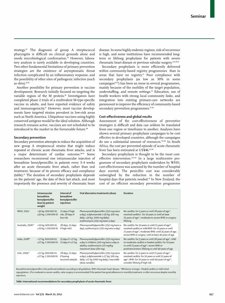
Secondary preventionSecondary prevention attempts to reduce the acquisition of new group A streptococcal strains that might induce repeated or chronic acute rheumatic fever attacks, and is a major determinant of cardiac outcome.89,90 Some researchers recommend one intramuscular injection of benzathine benzylpenicillin in patients every 3–4 weeks after an acute rheumatic fever attack, rather than oral treatment, because of its proven effi cacy and compliance (table).92 The duration of secondary prophylaxis depends on the patients’ age, the date of their last attack, and most importantly the presence and severity of rheumatic heart
disease. In some highly endemic regions, risk of recurrence is high, and some institutions have recom mended long-term or lifelong prophylaxis for patients with severe rheumatic heart disease or previous valvular surgery.2,60,78,91
Secondary prophylaxis is more effi ciently delivered within community-based registry programmes than in areas that have no registry.93 Poor compliance with secondary prophylaxis (as low as 50% in some campaigns94,95) has been an issue in several programmes, mainly because of the mobility of the target population, understaffi ng, and remote settings.96 Education, use of health workers with strong local community links, and integration into existing primary-care networks are paramount to improve the effi ciency of community-based secondary prevention programmes.97,98
Cost-eff ectiveness and global resultsAssessment of the cost-eff ectiveness of preventive strategies is diffi cult and data can seldom be translated from one region or timeframe to another. Analyses have shown several primary prophylaxis campaigns to be cost eff ective in developed countries, although the campaigns do use a substantial amount of resources.99,100 In South Africa, the cost per prevented episode of acute rheumatic fever has been estimated at US$46.81,101
Secondary prophylaxis is thought to be the most cost-eff ective intervention.102,103 In a large multicentre pro-gramme of secondary prophylaxis undertaken by WHO, cost-eff ectiveness was assessed by the number of hospital days averted. The penicillin cost was considerably outweighed by the reduction in the number of hospital days that patients needed.94 In New Zealand, the cost of an effi cient secondary prevention pro gramme
Intramuscular benzathine benzylpenicillin dose by patient weight
Interval of benzathine benzylpenicillin injections
Oral alternative treatments (dose) Duration
WHO, 20012 <30 kg: 600 000 IU; ≥30 kg: 1 200 000 IU
21 days if high risk; 28 days if low risk
Phenoxymethylpenicillin (250 mg twice a day); sulphonamide (<30 kg: 500 mg daily; ≥30 kg: 1000 mg daily); erythromycin (250 mg twice a day)
No carditis: for 5 years or until 18 years of age*; resolved carditis†: for 10 years or until at least 25 years of age*; moderate to severe RHD or surgery: lifelong
Australia, 200660 <20 kg: 600 000 IU; ≥20 kg: 1 200 000 IU
28 days; 21 days if high risk‡
Phenoxymethylpenicillin (250 mg twice a day); erythromycin (250 mg twice a day)
No carditis: for 10 years or until 21 years of age*; resolved carditis or mild RHD: for 10 years or until 21 years of age*; moderate RHD: until 35 years of age; severe RHD or surgery: until at least 40 years of age
India, 200891 <27 kg: 600 000 IU; ≥27 kg: 1 200 000 IU
15 days if <27 kg; 21 days if ≥27 kg
Phenoxymethylpenicillin (250 mg twice a day in children; 500 mg twice a day in adults); erythromycin (20 mg/kg; maximum dose 500 mg)
No carditis: for 5 years or until 18 years of age*; mild to moderate carditis or healed carditis: for 10 years or until 25 years of age*; severe RHD or postintervention: lifelong or until 40 years of age
USA, 200978 <27 kg: 600 000 IU; ≥27 kg: 1 200 000 IU
28 days; 21 days if having recurrent attacks
Phenoxymethylpenicillin (250 mg twice a day); sulphonamide (<27 kg: 500 mg daily; ≥27 kg 1000 mg daily); macrolide (dose variable)
No carditis: for 5 years or until 21 years of age*; resolved carditis: for 10 years or until 21 years of age*; RHD: for 10 years or until 40 years of age*; consider lifelong if high risk
Benzathine benzylpenicillin is the preferred antibiotic according to all guidelines. RHD=rheumatic heart disease. *Whichever is longer. †Healed carditis or mild mitral regurgitation. ‡For moderate to severe carditis, valve surgery is recommended if the patient has good adherence to monthly treatment, or after recurrence despite monthly injections.
Table: International recommendations for secondary prophylaxis of acute rheumatic fever
Seminar
958 www.thelancet.com Vol 379 March 10, 2012
accounted for only 13% of the total budget allocated to acute rheumatic fever.104
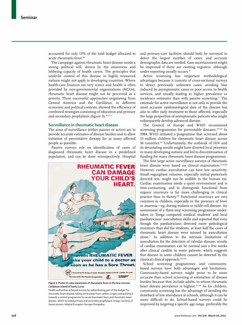
The campaign against rheumatic heart disease needs a strong political will, driven by the awareness and lobbying capacity of health carers. The principles that underlie control of this disease in highly resourced nations might not apply to developing countries. Where health-care fi nances are very scarce and health is often provided by non-governmental organisations (NGOs), rheumatic heart disease might not be perceived as a priority. Three successful approaches originating from Central America and the Caribbean, in diff erent economic and political contexts, showed the effi ciency of combined strategies consisting of education and primary and secondary prophylaxis (fi gure 3).30,72,73
Surveillance in rheumatic heart diseaseThe aims of surveillance (either passive or active) are to provide accurate estimates of disease burden and to allow initiation of preventative therapy for as many aff ected people as possible.
Passive surveys rely on identifi cation of cases of diagnosed rheumatic heart disease in a predefi ned population, and can be done retrospectively. Hospital
and primary-care facilities should both be surveyed to detect the largest number of cases, and accurate demographic data are needed. Case ascertainment might be improved if there are existing registers, although under-reporting usually occurs.19
Active screening has important methodological advantages because it consists of cross-sectional surveys to detect previously unknown cases, avoiding bias induced by asymptomatic cases or poor access to health services, and usually leading to higher prevalence or incidence estimates than with passive screening.27 The rationale for active surveillance is not only to provide the most accurate epidemiological data of the disease but also to off er early treatment to those aff ected, especially the large proportion of asymptomatic patients who might subsequently develop advanced disease.
The Council of Europe and WHO recommend screening programmes for preventable diseases.105,106 In 1984, WHO initiated a programme that screened about 15 million children for rheumatic heart disease across 16 countries.107 Unfortunately, the outbreak of HIV and its devastating results might have diverted local priorities in many developing nations and led to discontinuation of funding for many rheumatic heart disease programmes.
The fi rst large active surveillance surveys of rheumatic heart disease were based on clinical examination.20,94,108 However, cardiac auscultation can have low sensitivity. Small regurgitant volumes, especially mitral posteriorly directed jets, might not be audible to the human ear. Cardiac examination needs a quiet environment and is time-consuming, and to distinguish functional from organic murmurs is far more challenging in clinical practice than in theory.109 Functional murmurs are very common in children, especially in the presence of fever or anaemia—eg, during malaria or sickle-cell disease. An assessment of a three-step screening programme under-taken in Tonga compared medical students’ and local paediatricians’ auscultation skills and reported that even though the paediatricians detected more pathological murmurs than did the students, at least half the cases of rheumatic heart disease were missed by auscultation alone.14 In addition to the intrinsic limitations of auscultation for the detection of valvular disease, results of cardiac examination can be normal just a few weeks after clinical carditis in some patients, which suggests that disease in some children cannot be detected by the classical clinical approach.6,66,110
School screening programmes and community-based surveys have both advantages and limitations. Community-based surveys might prove to be more accurate than school screening at estimation of disease burden because they include adults, in whom rheumatic heart disease prevalence is highest.3,12,40 As for children, community screening has the advantage of avoiding the drawback of low attendance at schools, although it can be more diffi cult to do. School-based surveys could be improved by targeting a specifi c age range, preferably the
Figure 3: Poster to raise awareness of rheumatic fever in the low-income Caribbean island of Santa Lucia Health authorities achieved success by redistributing part of the budget for rheumatic heart disease, taking some away from cardiac surgery and putting it towards a control programme for acute rheumatic fever and rheumatic heart disease, which included primary and secondary prophylaxis. Image courtesy of Xavier Jouven, HÔpital Européen Georges Pompidou.
Seminar
www.thelancet.com Vol 379 March 10, 2012 959
earliest age when the prevalence starts to peak in pilot studies (around 12 years of age in some regions).13,14
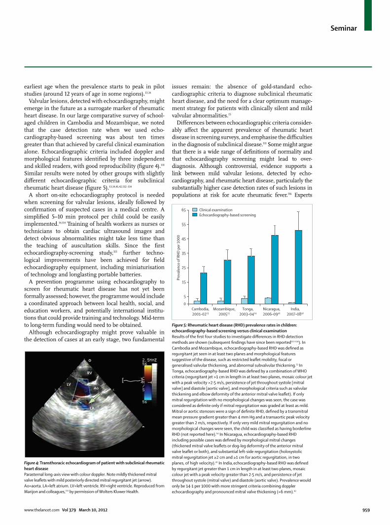
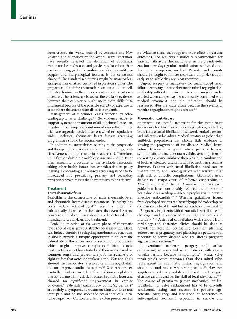
Valvular lesions, detected with echocardiography, might emerge in the future as a surrogate marker of rheumatic heart disease. In our large comparative survey of school-aged children in Cambodia and Mozambique, we noted that the case detection rate when we used echo-cardiography-based screening was about ten times greater than that achieved by careful clinical examination alone. Echocardiographic criteria included doppler and morpho logical features identifi ed by three independent and skilled readers, with good reproducibility (fi gure 4).111 Similar results were noted by other groups with slightly diff erent echocardiographic criteria for subclinical rheumatic heart disease (fi gure 5).13,14,41,42,112–114
A short on-site echocardiography protocol is needed when screening for valvular lesions, ideally followed by confi rmation of suspected cases in a medical centre. A simplifi ed 5–10 min protocol per child could be easily implemented.14,114 Training of health workers as nurses or technicians to obtain cardiac ultrasound images and detect obvious abnormalities might take less time than the teaching of auscultation skills. Since the fi rst echocardiography-screening study,115 further techno-logical improvements have been achieved for fi eld echocardiography equipment, including miniaturisation of technology and longlasting portable batteries.
A prevention programme using echocardiography to screen for rheumatic heart disease has not yet been formally assessed; however, the programme would include a coordinated approach between local health, social, and education workers, and potentially inter national institu-tions that could provide training and technology. Mid-term to long-term funding would need to be obtained.
Although echocardiography might prove valuable in the detection of cases at an early stage, two fundamental
issues remain: the absence of gold-standard echo-cardiographic criteria to diagnose subclinical rheumatic heart disease, and the need for a clear optimum manage-ment strategy for patients with clinically silent and mild valvular abnormalities.15
Diff erences between echocardiographic criteria consider-ably aff ect the apparent prevalence of rheumatic heart disease in screening surveys, and emphasise the diffi culties in the diagnosis of subclinical disease.111 Some might argue that there is a wide range of defi nitions of normality and that echocardiography screening might lead to over-diagnosis. Although controversial, evidence supports a link between mild valvular lesions, detected by echo-cardiography, and rheumatic heart disease, particularly the substantially higher case detection rates of such lesions in populations at risk for acute rheumatic fever.116 Experts
Figure 4: Transthoracic echocardiogram of patient with subclinical rheumatic heart disease Parasternal long-axis view with colour doppler. Note mildly thickened mitral valve leafl ets with mild posteriorly directed mitral regurgitant jet (arrow). Ao=aorta. LA=left atrium. LV=left ventricle. RV=right ventricle. Reproduced from Marijon and colleagues,111 by permission of Wolters Kluwer Health.
RV
LVAo
LA
Figure 5: Rheumatic heart disease (RHD) prevalence rates in children: echocardiography-based screening versus clinical examinationResults of the fi rst four studies to investigate diff erences in RHD detection methods are shown (subsequent fi ndings have since been reported112–114). In Cambodia and Mozambique, echocardiography-based RHD was defi ned as regurgitant jet seen in at least two planes and morphological features suggestive of the disease, such as restricted leafl et mobility, focal or generalised valvular thickening, and abnormal subvalvular thickening.13 In Tonga, echocardiography-based RHD was defi ned by a combination of WHO criteria (regurgitant jet >1 cm in length in at least two planes, mosaic colour jet with a peak velocity >2·5 m/s, persistence of jet throughout systole [mitral valve] and diastole [aortic valve], and morphological criteria such as valvular thickening and elbow deformity of the anterior mitral valve leafl et). If only mitral regurgitation with no morphological changes was seen, the case was considered as defi nite only if mitral regurgitation was graded at least as mild. Mitral or aortic stenoses were a sign of defi nite RHD, defi ned by a transmitral mean pressure gradient greater than 4 mm Hg and a transaortic peak velocity greater than 2 m/s, respectively. If only very mild mitral regurgitation and no morphological changes were seen, the child was classifi ed as having borderline RHD (not reported here).14 In Nicaragua, echocardiography-based RHD including possible cases was defi ned by morphological mitral changes (thickened mitral valve leafl ets or dog-leg deformity of the anterior mitral valve leafl et or both), and substantial left-side regurgitation (holosystolic mitral regurgitation jet ≥2 cm and ≥1 cm for aortic regurgitation, in two planes, of high velocity).41 In India, echocardiography-based RHD was defi ned by regurgitant jet greater than 1 cm in length in at least two planes, mosaic colour jet with a peak velocity greater than 2·5 m/s, and persistence of jet throughout systole (mitral valve) and diastole (aortic valve). Prevalence would only be 14·1 per 1000 with more stringent criteria combining doppler echocardiography and pronounced mitral valve thickening (>6 mm).42
Prev
alen
ce o
f RH
D pe
r 100
0
45
0Cambodia,2001–0213
Mozambique,200513
Tonga,2003–0414
Nicaragua,2006–0941
India,2007–0842
5
15
25
35
65
55
Clinical examinationEchocardiography-based screening
Seminar
960 www.thelancet.com Vol 379 March 10, 2012
from around the world, chaired by Australia and New Zealand and supported by the World Heart Federation, have recently revisited the defi nition of subclinical rheumatic heart disease, and guidelines based on their conclusions suggest that a combination of semiquantitative doppler and morphological features is the consensus choice.117 The standardised criteria might be more or less stringent than what has been used in previous studies. The proportion of defi nite rheumatic heart disease cases will probably diminish as the proportion of borderline patterns increases. The criteria are based on the available evidence; however, their complexity might make them diffi cult to implement because of the possible scarcity of expertise in areas where rheumatic heart disease is endemic.
Management of subclinical cases detected by echo-cardiography is a challenge.118 No evidence exists to support systematic treatment of all subclinical cases, so long-term follow-up and randomised controlled clinical trials are urgently needed to assess whether population-wide subclinical rheumatic heart disease screening programmes should be recommended.
In addition to uncertainties relating to the prognostic and therapeutic implications of abnormal fi ndings, cost-eff ectiveness is another issue to be addressed. Therefore, until further data are available, clinicians should tailor their screening procedure to the available resources, taking other health issues into consideration in policy making. Echocardiography-based screening needs to be introduced into pre-existing primary and secondary prevention programmes that have proven to be eff ective.
TreatmentAcute rheumatic feverPenicillin is the cornerstone of acute rheumatic fever and rheumatic heart disease treatment. Its safety has been widely acknowledged71,73 and its price has substantially decreased to the extent that even the most poorly resourced countries should not be deterred from introducing prophylaxis and treatment.
Penicillin injection at the acute phase of rheumatic fever should clear group A streptococcal infection which can induce chronic or relapsing autoimmune reactions. It should provide a unique opportunity to educate the patient about the importance of secondary prophylaxis, which might improve compliance.119 Most classic treatments have not been tested and their use is based on common sense and proven safety. A meta-analysis of eight studies that were undertaken in the 1950s and 1960s showed that salicylates, steroids, or immunoglobulins did not improve cardiac outcomes.120 One randomised controlled trial assessed the effi cacy of immunoglobulin therapy during a fi rst attack of acute rheumatic fever and showed no signifi cant improvement in cardiac outcomes.121 Salicylates (aspirin 80–100 mg/kg per day)60 are mainly a symptomatic treatment aimed at fever and joint pain and do not aff ect the prevalence of clinical valve sequelae.122 Corticosteroids are often prescribed but
no evidence exists that supports their eff ect on cardiac outcomes. Bed rest was historically recommended for patients with acute rheumatic fever in the preantibiotic era, but nowadays gradual mobilisation is advised once the initial symptoms resolve.5 Patients and parents should be taught to initiate secondary prophylaxis at an early stage, while they are most receptive.
Urgent surgery is mandatory for uncontrolled heart failure secondary to acute rheumatic mitral regurgitation, preferably with valve repair.123,124 However, surgery can be avoided when congestive signs are easily controlled with medical treatment, and the indication should be reassessed after the acute phase because the severity of valvular regurgitation might decrease.7,10
Rheumatic heart diseaseAt present, no specifi c treatment for rheumatic heart disease exists other than for its complications, including heart failure, atrial fi brillation, ischaemic embolic events, and infective endocarditis. Medical treatment (other than antibiotic prophylaxis) has shown little evidence of slowing the progression of the disease. Medical heart-failure treatment is given when patients become symptomatic, and includes mainly β blockers, angiotensin-converting-enzyme inhibitor therapies, or a combination of both, as tolerated, and symptomatic treatments such as diuretics. Patients with atrial fi brillation need rate or rhythm control and anticoagulation with warfarin if at high risk of embolic complications. Rheumatic heart disease is a major cause of infective endocarditis in African countries.125 North American and European guidelines have considerably reduced the number of heart disorders needing antibiotic prophylaxis to prevent infective endocarditis.126,127 Whether guidelines issued from developed regions can be safely applied to developing countries is debatable, and further studies are warranted.
Pregnancy in patients with rheumatic heart disease is a challenge, and is associated with high morbidity and mortality.128,129 Antenatal consultation with support from cardiology and obstetrics clinics should be done to provide contraception, counselling, treatment planning before start of pregnancy, and planning for patients with moderate to severe disease who are already pregnant (eg, caesarean section).130
Interventional treatment (surgery and cardiac catheter ism) is warranted when patients with severe valvular lesions become symptomatic.131 Mitral valve repair yields better outcomes than does mitral valve replacement in rheumatic mitral regurgitation and should be undertaken whenever possible.132 However, long-term results vary and depend mainly on the degree of active carditis and on the skill of local physicians.124,133 The choice of prosthesis (either mechanical or bio-prosthetic) for valve replacement has to be carefully considered, taking into account the patient’s age, potential pregnancy, and likelihood of adherence to anticoagulant treatment, especially in remote and
Seminar
www.thelancet.com Vol 379 March 10, 2012 961
socially underprivileged areas.134 Investigators with detailed experience of rheumatic heart disease in remote areas have recommended tissue valves in Indigenous Australian and New Zealand populations because of poor anticoagulation control,135 which diff ers from clinical practice in more affl uent settings.136,137 In very advanced stages of the disease, surgery might be contraindicated when myocardial dilatation and dys-function coexist.
In cases of substantial mitral stenosis, percutaneous mitral balloon commissurotomy has replaced surgical commissurotomy and yields excellent early outcomes, with a 50–60% event-free outcome at 10-year follow-up.138 Patient selection through predictive score might ensure the intervention is successful and avoid acute severe mitral regurgitation.139 Clinical presentation of mitral stenosis varies with time and region, with younger patients in Africa having more severe mitral stenosis and raised pulmonary artery pressures than do patients from developed countries.140,141 However, these diff erences do not seem to aff ect immediate and mid-term results of percutaneous mitral balloon commissurotomy.141
In low-income countries, most invasive procedures are either done abroad, at great expense for the individual, or locally by visiting NGOs, which have focused on the initiation of programmes and the training of local staff to ensure continuity. Unfortunately, medical and surgical care for people with severe rheumatic heart disease is the least cost-eff ective intervention and consumes almost all funds available for this disease.
ContributorsAll authors contributed to the concept, reference search, and writing of
this Seminar under the coordination of the corresponding author. Figure
design was managed by EM and MM.
Confl icts of interestWe declare that we have no confl icts of interest.
AcknowledgmentsWe thank Said El-Haou for his technical assistance and Alexandre Loupy
for his helpful advice.
References1 Kaplan MH, Bolande R, Rakita L, Blair J. Presence of bound
immunoglobulins and complement in the myocardium in acute rheumatic fever—association with cardiac failure. N Engl J Med 1964; 271: 637–45.
2 WHO Technical Report Series 923. Rheumatic fever and rheumatic heart disease—Report of a WHO expert consultation, Geneva, Oct 29–Nov 1, 2001. Geneva: World Health Organization, 2004. http://www.who.int/cardiovascular_diseases/resources/en/cvd_trs923.pdf (accessed Oct 27, 2011).
3 Carapetis JR, Steer AC, Mulholland EK, Weber M. The global burden of group A streptococcal diseases. Lancet Infect Dis 2005; 5: 685–94.
4 Population Reference Bureau. 2008 world population data sheet. http://www.prb.org/Publications/Datasheets/2008/2008wpds.aspx (accessed Oct 27, 2011).
5 Carapetis JR, McDonald M, Wilson NJ. Acute rheumatic fever. Lancet 2005; 366: 155–68.
6 Meira ZM, Goulart EM, Colosimo EA, Mota CC. Long term follow up of rheumatic fever and predictors of severe rheumatic valvar disease in Brazilian children and adolescents. Heart 2005; 91: 1019–22.
7 Caldas AM, Terreri MT, Moises VA, et al. What is the true frequency of carditis in acute rheumatic fever? A prospective clinical and Doppler blind study of 56 children with up to 60 months of follow-up evaluation. Pediatr Cardiol 2008; 29: 1048–53.
8 Sanyal SK, Thapar MK, Ahmed SH, Hooja V, Tewari P. The initial attack of acute rheumatic fever during childhood in North India; a prospective study of the clinical profi le. Circulation 1974; 49: 7–12.
9 Vardi P, Markiewicz W, Weiss Y, Levi J, Benderly A. Clinical-echocardiographic correlations in acute rheumatic fever. Pediatrics 1983; 71: 830–34.
10 Vasan RS, Shrivastava S, Vijayakumar M, Narang R, Lister BC, Narula J. Echocardiographic evaluation of patients with acute rheumatic fever and rheumatic carditis. Circulation 1996; 94: 73–82.
11 Figueroa FE, Fernandez MS, Valdes P, et al. Prospective comparison of clinical and echocardiographic diagnosis of rheumatic carditis: long term follow up of patients with subclinical disease. Heart 2001; 85: 407–10.
12 Sliwa K, Carrington M, Mayosi BM, Zigiriadis E, Mvungi R, Stewart S. Incidence and characteristics of newly diagnosed rheumatic heart disease in urban African adults: insights from the heart of Soweto study. Eur Heart J 2010; 31: 719–27.
13 Marijon E, Ou P, Celermajer DS, et al. Prevalence of rheumatic heart disease detected by echocardiographic screening. N Engl J Med 2007; 357: 470–76.
14 Carapetis JR, Hardy M, Fakakovikaetau T, et al. Evaluation of a screening protocol using auscultation and portable echocardiography to detect asymptomatic rheumatic heart disease in Tongan schoolchildren. Nat Clin Pract Cardiovasc Med 2008; 5: 411–17.
15 Marijon E, Ou P, Celermajer DS, et al. Echocardiographic screening for rheumatic heart disease. Bull World Health Organ 2008; 86: 84.
16 Wilson N. Rheumatic heart disease in indigenous populations—New Zealand experience. Heart Lung Circ 2010; 19: 282–88.
17 Parnaby MG, Carapetis JR. Rheumatic fever in indigenous Australian children. J Paediatr Child Health 2010; 46: 527–33.
18 Carapetis JR. Rheumatic heart disease in Asia. Circulation 2008; 118: 2748–53.
19 Nkgudi B, Robertson KA, Volmink J, Mayosi BM. Notifi cation of rheumatic fever in South Africa—evidence for underreporting by health care professionals and administrators. S Afr Med J 2006; 96: 206–08.
20 WHO. The global burden of disease. 2004 update. http://www.who.int/healthinfo/global_burden_disease/GBD_report_2004update_full.pdf (accessed Oct 27, 2011).
21 Mayosi B, Robertson K, Volmink J, et al. The Drakensberg declaration on the control of rheumatic fever and rheumatic heart disease in Africa. S Afr Med J 2006; 96: 246.
22 Robertson KA, Volmink JA, Mayosi BM. Towards a uniform plan for the control of rheumatic fever and rheumatic heart disease in Africa—the Awareness Surveillance Advocacy Prevention (A.S.A.P.) programme. S Afr Med J 2006; 96: 241.
23 Jaiyesimi F, Antia AU. Prognostic factors in childhood rheumatic disease. Trop Geogr Med 1981; 33: 14–38.
24 Günther G, Asmera J, Parry E. Death from rheumatic heart disease in rural Ethiopia. Lancet 2006; 367: 391.
25 Terreri MT, Ferraz MB, Goldenberg J, Len C, Hilario MO. Resource utilization and cost of rheumatic fever. J Rheumatol 2001; 28: 1394–97.
26 Essawy MA, Bahgat ZS, Kassem HA. Health-related quality of life of school-age children with rheumatic fever. J Egypt Public Health Assoc 2010; 85: 205–22.
27 Tibazarwa KB, Volmink JA, Mayosi BM. Incidence of acute rheumatic fever in the world: a systematic review of population-based studies. Heart 2008; 94: 1534–40.
28 Talbot RG. Rheumatic fever and rheumatic heart disease in the Hamilton health district: I. An epidemiological survey. N Z Med J 1984; 97: 630–34.
29 Carapetis JR, Wolff DR, Currie BJ. Acute rheumatic fever and rheumatic heart disease in the top end of Australia’s Northern Territory. Med J Aust 1996; 164: 146–49.
30 Bach JF, Chalons S, Mosser A, et al. 10-year educational programme aimed at rheumatic fever in two French Caribbean islands. Lancet 1996; 347: 644–48.
31 Longo-Mbenza B, Bayekula M, Ngiyulu R, et al. Survey of rheumatic heart disease in school children of Kinshasa town. Int J Cardiol 1998; 63: 287–94.
32 Sadiq M, Islam K, Abid R, et al. Prevalence of rheumatic heart disease in school children of urban Lahore. Heart 2009; 95: 353–57.

Seminar
962 www.thelancet.com Vol 379 March 10, 2012
57 Ramasawmy R, Fae KC, Spina G, et al. Association of polymorphisms within the promoter region of the tumor necrosis factor-alpha with clinical outcomes of rheumatic fever. Mol Immunol 2007; 44: 1873–78.
58 Veasy LG, Wiedmeier SE, Orsmond GS, et al. Resurgence of acute rheumatic fever in the intermountain area of the United States. N Engl J Med 1987; 316: 421–27.
59 Special Writing Group of the Committee on Rheumatic Fever, Endocarditis, and Kawasaki Disease of the Council on Cardiovascular Disease in the Young of the American Heart Association. Guidelines for the diagnosis of rheumatic fever. Jones criteria, 1992 update. JAMA 1992; 268: 2069–73.
60 National Heart Foundation of Australia and the Cardiac Society of Australia and New Zealand. Diagnosis and management of acute rheumatic fever and rheumatic heart disease in Australia: an evidence-based review, 2006. http://www.racgp.org.au/Content/NavigationMenu/ClinicalResources/RACGPGuidelines/Diagnosisandmanagementofacuterheumaticfeverandrheumatic heartdiseaseinAustralia/NHFA-CSANZ_ARF_RHD_2006.pdf (accessed Oct 27, 2011).
61 Carapetis JR, Paar J, Cherian T. Standardization of epidemiologic protocols for surveillance of post-streptococcal sequelae: acute rheumatic fever, rheumatic heart disease and acute post-streptococcal glomerulonephritis, 2006. http://www.niaid.nih.gov/topics/strepThroat/Documents/groupasequelae.pdf (accessed Oct 27, 2011).
62 Carapetis JR, Currie BJ. Rheumatic chorea in northern Australia: a clinical and epidemiological study. Arch Dis Child 1999; 80: 353–58.
63 Kamblock J, Payot L, Iung B, et al. Does rheumatic myocarditis really exists? Systematic study with echocardiography and cardiac troponin I blood levels. Eur Heart J 2003; 24: 855–62.
64 Chesler E, ed. Clinical cardiology, 5th edn. New York: Springer-Verlag, 1983.
65 Minich LL, Tani LY, Pagotto LT, Shaddy RE, Veasy LG. Doppler echocardiography distinguishes between physiologic and pathologic “silent” mitral regurgitation in patients with rheumatic fever. Clin Cardiol 1997; 20: 924–26.
66 Bland EF, Jones TD. Rheumatic fever and rheumatic heart disease; a twenty year report on 1000 patients followed since childhood. Circulation 1951; 4: 836–43.
67 Marcus RH, Sareli P, Pocock WA, Barlow JB. The spectrum of severe rheumatic mitral valve disease in a developing country. Correlations among clinical presentation, surgical pathologic fi ndings, and hemodynamic sequelae. Ann Intern Med 1994; 120: 177–83.
68 Roy SB, Bhatia ML, Lazaro EJ, Ramalingaswami V. Juvenile mitral stenosis in India. Lancet 1963; 282: 1193–96.
69 Marijon E, Jani D, Garbarz E. P-wave dispersion and percutaneous mitral valvuloplasty. Cardiol Rev 2007; 15: 42–45.
70 Wisenbaugh T, Skudicky D, Sareli P. Prediction of outcome after valve replacement for rheumatic mitral regurgitation in the era of chordal preservation. Circulation 1994; 89: 191–97.
71 International Rheumatic Fever Study Group. Allergic reactions to long-term benzathine penicillin prophylaxis for rheumatic fever. Lancet 1991; 337: 1308–10.
72 Nordet P, Lopez R, Duñeas A, Sarmiento L. Prevention and control of rheumatic fever and rheumatic heart disease: the Cuban experience (1986–1996–2002). Cardiovasc J Afr 2008; 19: 135–40.
73 Arguedas A, Mohs E. Prevention of rheumatic fever in Costa Rica. J Pediatr 1992; 121: 569–72.
74 Kaplan EL. T Duckett Jones Memorial Lecture. Global assessment of rheumatic fever and rheumatic heart disease at the close of the century. Infl uences and dynamics of populations and pathogens: a failure to realize prevention? Circulation 1993; 88: 1964–72.
75 Carapetis JR, Currie BJ. Mortality due to acute rheumatic fever and rheumatic heart disease in the Northern Territory: a preventable cause of death in aboriginal people. Aust N Z J Public Health 1999; 23: 159–63.
76 Pastore S, De Cunto A, Benettoni A, Berton E, Taddio A, Lepore L. The resurgence of rheumatic fever in a developed country area: the role of echocardiography. Rheumatology (Oxford) 2011; 50: 396–400.
77 Bisno AL, Gerber MA, Gwaltney JM Jr, Kaplan EL, Schwartz RH, for the Infectious Diseases Society of America. Practice guidelines for the diagnosis and management of group A streptococcal pharyngitis. Clin Infect Dis 2002; 35: 113–25.
33 Dobson J, Steer AC, Colquhoun S, Kado J. Environmental factors and rheumatic heart disease in Fiji. Pediatr Cardiol 2011; published online Nov 6. DOI:10.1007/s00246-011-0139-x.
34 Bryant PA, Robins-Browne R, Carapetis JR, Curtis N. Some of the people, some of the time: susceptibility to acute rheumatic fever. Circulation 2009; 119: 742–53.
35 Engel ME, Stander R, Vogel J, Adeyemo AA, Mayosi BM. Genetic susceptibility to acute rheumatic fever: a systematic review and meta-analysis of twin studies. PLoS One 2011; 6: e25326.
36 Periwal KL, Gupta BK, Panwar RB, Khatri PC, Raja S, Gupta R. Prevalence of rheumatic heart disease in school children in Bikaner: an echocardiographic study. J Assoc Physicians India 2006; 54: 279–82.
37 Steer AC, Kado J, Wilson N, et al. High prevalence of rheumatic heart disease by clinical and echocardiographic screening among children in Fiji. J Heart Valve Dis 2009; 18: 327–35.
38 Nkomo VT. Epidemiology and prevention of valvular heart diseases and infective endocarditis in Africa. Heart 2007; 93: 1510–19.
39 Longo-Mbenza B, Bayekula M, Ngiyulu R, et al. Survey of rheumatic heart disease in school children of Kinshasa town. Int J Cardiol 1998; 63: 287–94.
40 Rizvi SF, Khan MA, Kundi A, Marsh DR, Samad A, Pasha O. Status of rheumatic heart disease in rural Pakistan. Heart 2004; 90: 394–99.
41 Paar JA, Berrios NM, Rose JD, et al. Prevalence of rheumatic heart disease in children and young adults in Nicaragua. Am J Cardiol 2010; 105: 1809–14.
42 Bhaya M, Panwar S, Beniwal R, Panwar RB. High prevalence of rheumatic heart disease detected by echocardiography in school children. Echocardiography 2010; 27: 448–53.
43 Guilherme L, Kalil J. Rheumatic fever: from innate to acquired immune response. Ann N Y Acad Sci 2007; 1107: 426–33.
44 Bisno AL, Brito MO, Collins CM. Molecular basis of group A streptococcal virulence. Lancet Infect Dis 2003; 3: 191–200.
45 Guilherme L, Kalil J. Rheumatic fever and rheumatic heart disease: cellular mechanisms leading autoimmune reactivity and disease. J Clin Immunol 2010; 30: 17–23.
46 Martin DR, Voss LM, Walker SJ, Lennon D. Acute rheumatic fever in Auckland, New Zealand: spectrum of associated group A streptococci diff erent from expected. Pediatr Infect Dis J 1994; 13: 264–69.
47 Pruksakorn S, Sittisombut N, Phornphutkul C, Pruksachatkunakorn C, Good MF, Brandt E. Epidemiological analysis of non-M-typeable group A streptococcus isolates from a Thai population in northern Thailand. J Clin Microbiol 2000; 38: 1250–54.
48 Carapetis JR, Currie BJ. Group A streptococcus, pyoderma, and rheumatic fever. Lancet 1996; 347: 1271–72.
49 Cheadle W. Barbeian lectures on the various manifestations of the rheumatic state as exemplifi ed in childhood and early life. Lancet 1889; 133: 821–27.
50 Kaplan MH, Suchy ML. Immunologic relation of streptococcal and tissue antigens. II. Cross-reaction of antisera to mammalian heart tissue with a cell wall constituent of certain strains of group A streptococci. J Exp Med 1964; 119: 643–50.
51 Goldstein I, Rebeyrotte P, Parlebas J, Halpern B. Isolation from heart valves of glycopeptides which share immunological properties with Streptococcus haemolyticus group A polysaccharides. Nature 1968; 219: 866–68.
52 Fae KC, Oshiro SE, Toubert A, Charron D, Kalil J, Guilherme L. How an autoimmune reaction triggered by molecular mimicry between streptococcal M protein and cardiac tissue proteins leads to heart lesions in rheumatic heart disease. J Autoimmun 2005; 24: 101–09.
53 Krisher K, Cunningham MW. Myosin: a link between streptococci and heart. Science 1985; 227: 413–15.
54 Cunningham MW. Pathogenesis of group A streptococcal infections. Clin Microbiol Rev 2000; 13: 470–511.
55 Jack DL, Klein NJ, Turner MW. Mannose-binding lectin: targeting the microbial world for complement attack and opsonophagocytosis. Immunol Rev 2001; 180: 86–99.
56 Messias-Reason IJ, Schafranski MD, Jensenius JC, Steff ensen R. The association between mannose-binding lectin gene polymorphism and rheumatic heart disease. Hum Immunol 2006; 67: 991–98.
Seminar
www.thelancet.com Vol 379 March 10, 2012 963
78 Gerber MA, Baltimore RS, Eaton CB, et al. Prevention of rheumatic fever and diagnosis and treatment of acute streptococcal pharyngitis. A scientifi c statement from the American Heart Association Rheumatic Fever, Endocarditis, and Kawasaki Disease Committee of the Council on Cardiovascular Disease in the Young, the Interdisciplinary Council on Functional Genomics and Translational Biology, and the Interdisciplinary Council on Quality of Care and Outcomes Research: endorsed by the American Academy of Pediatrics. Circulation 2009; 119: 1541–51.
79 Karthikeyan G, Mayosi BM. Is primary prevention of rheumatic fever the missing link in the control of rheumatic heart disease in Africa? Circulation 2009; 120: 709–13.
80 Gordis L. Eff ectiveness of comprehensive-care programs in preventing rheumatic fever. N Engl J Med 1973; 289: 331–35.
81 Robertson KA, Volmink JA, Mayosi BM. Antibiotics for the primary prevention of acute rheumatic fever: a meta-analysis. BMC Cardiovasc Disord 2005; 5: 11.
82 Lennon D, Kerdemelidis M, Arroll B. Meta-analysis of trials of streptococcal throat treatment programs to prevent rheumatic fever. Pediatr Infect Dis J 2009; 28: e259–64.
83 Lennon D, Stewart J, Farrell E, Palmer A, Mason H. School-based prevention of acute rheumatic fever: a group randomized trial in New Zealand. Pediatr Infect Dis J 2009; 28: 787–94.
84 McDonald MI, Towers RJ, Andrews RM, Benger N, Currie BJ, Carapetis JR. Low rates of streptococcal pharyngitis and high rates of pyoderma in Australian aboriginal communities where acute rheumatic fever is hyperendemic. Clin Infect Dis 2006; 43: 683–89.
85 McDonald M, Currie BJ, Carapetis JR. Acute rheumatic fever: a chink in the chain that links the heart to the throat? Lancet Infect Dis 2004; 4: 240–45.
86 Bisno AL, Rubin FA, Cleary PP, Dale JB. Prospects for a group A streptococcal vaccine: rationale, feasibility, and obstacles—report of a National Institute of Allergy and Infectious Diseases workshop. Clin Infect Dis 2005; 41: 1150–56.
87 McNeil SA, Halperin SA, Langley JM, et al. Safety and immunogenicity of 26-valent group A streptococcus vaccine in healthy adult volunteers. Clin Infect Dis 2005; 41: 1114–22.
88 Steer AC, Law I, Matatolu L, Beall BW, Carapetis JR. Global emm type distribution of group A streptococci: systematic review and implications for vaccine development. Lancet Infect Dis 2009; 9: 611–16.
89 Sanyal SK, Berry AM, Duggal S, Hooja V, Ghosh S. Sequelae of the initial attack of acute rheumatic fever in children from north India. A prospective 5-year follow-up study. Circulation 1982; 65: 375–79.
90 Majeed HA, Yousof AM, Khuff ash FA, Yusuf AR, Farwana S, Khan N. The natural history of acute rheumatic fever in Kuwait: a prospective six year follow-up report. J Chronic Dis 1986; 39: 361–69.
91 Saxena A, Kumar RK, Gera RP, Radhakrishnan S, Mishra S, Ahmed Z. Consensus guidelines on pediatric acute rheumatic fever and rheumatic heart disease. Indian Pediatr 2008; 45: 565–73.
92 Manyemba J, Mayosi BM. Penicillin for secondary prevention of rheumatic fever. Cochrane Database Syst Rev 2002; 3: CD002227.
93 McDonald M, Brown A, Noonan S, Carapetis JR. Preventing recurrent rheumatic fever: the role of register based programmes. Heart 2005; 91: 1131–33.
94 Strasser T, Dondog N, El Kholy A, et al. The community control of rheumatic fever and rheumatic heart disease: report of a WHO international cooperative project. Bull World Health Organ 1981; 59: 285–94.
95 Stewart T, McDonald R, Currie B. Acute rheumatic fever: adherence to secondary prophylaxis and follow up of Indigenous patients in the Katherine region of the Northern Territory. Aust J Rural Health 2007; 15: 234–40.
96 Eissa S, Lee R, Binns P, Garstone G, McDonald M. Assessment of a register-based rheumatic heart disease secondary prevention program in an Australian Aboriginal community. Aust N Z J Public Health 2005; 29: 521–25.
97 Kearns TM, Schultz R, McDonald V, Andrews RM. Prophylactic penicillin by the full moon: a novel approach in Central Australia that may help to reduce the risk of rheumatic heart disease. Rural Remote Health 2010; 10: 1464.
98 Grayson S, Horsburgh M, Lennon D. An Auckland regional audit of the nurse-led rheumatic fever secondary prophylaxis programme. N Z Med J 2006; 119: U2255.
99 Tompkins RK, Burnes DC, Cable WE. An analysis of the cost-eff ectiveness of pharyngitis management and acute rheumatic fever prevention. Ann Intern Med 1977; 86: 481–92.
100 Strasser T. Cost-eff ective control of rheumatic fever in the community. Health Policy 1985; 5: 159–64.
101 Monya-Tambi I, Robertson KR, Volmink JA, Mayosi BM. Acute rheumatic fever. Lancet 2005; 366: 1355.
102 Steer AC, Carapetis JR. Prevention and treatment of rheumatic heart disease in the developing world. Nat Rev Cardiol 2009; 6: 689–98.
103 Michaud C, Rammohan R, Narula J. Cost-eff ectiveness analysis of intervention strategies for reduction of the burden of rheumatic heart disease. In: Narula J, Virmani R, Reddy KS, Tandon R, eds. Rheumatic fever. Washington, DC: American Registry of Pathology, 1999: 485–97.
104 North DA, Heynes RA, Lennon DR, Neutze J. Analysis of costs of acute rheumatic fever and rheumatic heart disease in Auckland. N Z Med J 1993; 106: 400–03.
105 WHO Cardiovascular Diseases Unit and principal investigators. WHO programme for the prevention of rheumatic fever/rheumatic heart disease in 16 developing countries: report from Phase I (1986–90). Bull World Health Organ 1992; 70: 213–18.
106 Council of Europe, Committee of Ministers, Recommendation No. R (94) 11 on screening as a tool of preventive medicine, Oct 10, 1994. http://www1.umn.edu/humanrts/instree/coerecr94-11.html (accessed Oct 27, 2011).
107 The WHO global programme for the prevention of rheumatic fever and rheumatic heart disease. Report of a consultation to review progress and develop future activities. Geneva; Nov 29–Dec 1, 1999. http://whqlibdoc.who.int/hq/2000/WHO_CVD_00.1.pdf (accessed Oct 27, 2011).
108 Mathur KS, Wahal PK. Epidemiology of rheumatic heart disease— a study of 29,922 school children. Indian Heart J 1982; 34: 367–71.
109 Biancaniello T. Innocent murmurs. Circulation 2005; 111: e20–22.
110 Marijon E, Taffl et M, Jouven X. Time to use ultrasound and not stethoscopes for rheumatic heart disease screening. Nat Clin Pract Cardiovasc Med 2008; 5: E1–3.
111 Marijon E, Celermajer DS, Taffl et M, et al. Rheumatic heart disease screening by echocardiography: the inadequacy of World Health Organization criteria for optimizing the diagnosis of subclinical disease. Circulation 2009; 120: 663–68.
112 Reeves BM, Kado J, Brook M. High prevalence of rheumatic heart disease in Fiji detected by echocardiography screening. J Paediatr Child Health 2011; 47: 473–78.
113 Saxena A, Ramakrishnan S, Roy A, et al. Prevalence and outcome of subclinical rheumatic heart disease in India: The RHEUMATIC (Rheumatic Heart Echo Utilisation and Monitoring Actuarial Trends in Indian Children) study. Heart 2011; 97: 2018–22.
114 Webb RH, Wilson NJ, Lennon DR, et al. Optimising echocardiographic screening for rheumatic heart disease in New Zealand: not all valve disease is rheumatic. Cardiol Young 2011; 21: 436–43.
115 Anabwani GM, Bonhoeff er P. Prevalence of heart disease in school children in rural Kenya using colour-fl ow echocardiography. East Afr Med J 1996; 73: 215–17.
116 Webb R, Gentles T, Stirling J, et al. Echocardiographic fi ndings in a low risk population for rheumatic heart disease: implications for RHD screening. XVIII Lancefi eld International Symposium; Palermo, Italy; Sept 4–8, 2011.
117 Reményi B, Wilson N, Steer A, et al. World Heart Federation criteria for echocardiographic diagnosis of rheumatic heart disease—an evidence-based guideline. Nat Rev Cardiol 2012; published online Feb 28. DOI:10.1038/nrcardio.2012.7.
118 Marijon E, Celermajer DS, Jouven X. Management of patients with subclinical rheumatic heart disease. Int J Cardiol 2009; 134: 295–96.
119 Mortimer EA Jr, Vaisman S, Vignau A, et al. The eff ect of penicillin on acute rheumatic fever and valvular heart disease. N Engl J Med 1959; 260: 101–12.
120 Cilliers AM, Manyemba J, Saloojee H. Anti-infl ammatory treatment for carditis in acute rheumatic fever. Cochrane Database Syst Rev 2003; 2: CD003176.
121 Voss LM, Wilson NJ, Neutze JM, et al. Intravenous immunoglobulin in acute rheumatic fever: a randomized controlled trial. Circulation 2001; 103: 401–06.

Seminar
964 www.thelancet.com Vol 379 March 10, 2012
122 Dorfman A, Gross JI, Lorincz AE. The treatment of acute rheumatic fever. Pediatrics 1961; 27: 692–706.
123 Barlow JB, Marcus RH, Pocock WA, Barlow CW, Essop R, Sareli P. Mechanisms and management of heart failure in active rheumatic carditis. S Afr Med J 1990; 78: 181–86.
124 Skoularigis J, Sinovich V, Joubert G, Sareli P. Evaluation of the long-term results of mitral valve repair in 254 young patients with rheumatic mitral regurgitation. Circulation 1994; 90: II167–74.
125 Essop MR, Nkomo VT. Rheumatic and nonrheumatic valvular heart disease: epidemiology, management, and prevention in Africa. Circulation 2005; 112: 3584–91.
126 Wilson W, Taubert KA, Gewitz M, et al. Prevention of infective endocarditis: guidelines from the American Heart Association: a guideline from the American Heart Association Rheumatic Fever, Endocarditis, and Kawasaki Disease Committee, Council on Cardiovascular Disease in the Young, and the Council on Clinical Cardiology, Council on Cardiovascular Surgery and Anesthesia, and the Quality of Care and Outcomes Research Interdisciplinary Working Group. Circulation 2007; 116: 1736–54.
127 Habib G, Hoen B, Tornos P, et al. Guidelines on the prevention, diagnosis, and treatment of infective endocarditis (new version 2009): the Task Force on the Prevention, Diagnosis, and Treatment of Infective Endocarditis of the European Society of Cardiology (ESC). Endorsed by the European Society of Clinical Microbiology and Infectious Diseases (ESCMID) and the International Society of Chemotherapy (ISC) for Infection and Cancer. Eur Heart J 2009; 30: 2369–413.
128 Soma-Pillay P, MacDonald AP, Mathivha TM, Bakker JL, Mackintosh MO. Cardiac disease in pregnancy: a 4-year audit at Pretoria Academic Hospital. S Afr Med J 2008; 98: 553–56.
129 Diao M, Kane A, Ndiaye MB, et al. Pregnancy in women with heart disease in sub-Saharan Africa. Arch Cardiovasc Dis 2011; 104: 370–74.
130 Iung B. Pregnancy-related cardiac complications: a consequence of the burden of rheumatic heart disease in sub-Saharan Africa. Arch Cardiovasc Dis 2011; 104: 367–69.
131 Bonow RO, Carabello BA, Chatterjee K, et al. 2008 focused update incorporated into the ACC/AHA 2006 guidelines for the management of patients with valvular heart disease: a report of the American College of Cardiology/American Heart Association Task Force on Practice Guidelines (Writing Committee to Revise the 1998 Guidelines for the Management of Patients With Valvular Heart Disease): endorsed by the Society of Cardiovascular Anesthesiologists, Society for Cardiovascular Angiography and Interventions, and Society of Thoracic Surgeons. Circulation 2008; 118: e523–661.
132 Enriquez-Sarano M, Akins CW, Vahanian A. Mitral regurgitation. Lancet 2009; 373: 1382–94.
133 Chauvaud S, Fuzellier JF, Berrebi A, Deloche A, Fabiani JN, Carpentier A. Long-term (29 years) results of reconstructive surgery in rheumatic mitral valve insuffi ciency. Circulation 2001; 104 (suppl 1): I12–15.
134 North RA, Sadler L, Stewart AW, McCowan LM, Kerr AR, White HD. Long-term survival and valve-related complications in young women with cardiac valve replacements. Circulation 1999; 99: 2669–76.
135 White H, Walsh W, Brown A, et al. Rheumatic heart disease in indigenous populations. Heart Lung Circ 2010; 19: 273–81.
136 Iung B, Baron G, Butchart EG, et al. A prospective survey of patients with valvular heart disease in Europe: The Euro Heart Survey on Valvular Heart Disease. Eur Heart J 2003; 24: 1231–43.
137 Rahimtoola SH. Choice of prosthetic heart valve in adults an update. J Am Coll Cardiol 2010; 55: 2413–26.
138 Iung B, Garbarz E, Michaud P, et al. Late results of percutaneous mitral commissurotomy in a series of 1024 patients. Analysis of late clinical deterioration: frequency, anatomic fi ndings, and predictive factors. Circulation 1999; 99: 3272–78.
139 Essop MR, Wisenbaugh T, Skoularigis J, Middlemost S, Sareli P. Mitral regurgitation following mitral balloon valvotomy. Diff ering mechanisms for severe versus mild-to-moderate lesions. Circulation 1991; 84: 1669–79.
140 Iung B, Nicoud-Houel A, Fondard O, et al. Temporal trends in percutaneous mitral commissurotomy over a 15-year period. Eur Heart J 2004; 25: 701–07.
141 Marijon E, Iung B, Mocumbi AO, et al. What are the diff erences in presentation of candidates for percutaneous mitral commissurotomy across the world and do they infl uence the results of the procedure? Arch Cardiovasc Dis 2008; 101: 611–17.