Revista Mexicana de Neurociencia - Medigraphic · Revista Mexicana de Neurociencia ... vessels,...
19
Revista Mexicana de Neurociencia Órgano Oficial de Difusión de la AMN Academia Mexicana de Neurología, A.C. Publicación oficial de la Academia Mexicana de Neurología A.C. Enero-Febrero Volumen 16, Año 2015 Número 1 www.revmexneuroci.com / ISSN 1665-5044 Revista Mexicana de Neurociencia 2015; 16(1): 73-89
Transcript of Revista Mexicana de Neurociencia - Medigraphic · Revista Mexicana de Neurociencia ... vessels,...

Revista Mexicana de
Neurociencia
Órgano Oficial de Difusión de la AMN
AcademiaMexicana deNeurología, A.C.
Publicación oficial de la Academia Mexicana de Neurología A.C.
Enero-FebreroVolumen 16, Año 2015 Número 1
www.revmexneuroci.com / ISSN 1665-5044
Rev
ista
Mex
ican
a d
e N
euro
cien
cia
20
15
; 16
(1):
73
-89
Revista Mexicana de Neurociencias Enero-Febrero, 2015; 16(1):73-89
73RevisiónJugular glomus
Review
Glomus yugular: Revisión de la literatura y técnica quirúrgica
Jugular glomus: literature review and surgical technique
Zottis-Grapiglia Cassio,¹ Diaz-Castillejos Ali,¹ Rehder Roberta,1 Borba Luis AB³
1Department of Neurosurgery. Evangelic Universitary Hospital, Curitiba, Paraná, Brazil² Brain and Heart Institute, Curitiba, Paraná, Brazil.3 “Hospital de Clinicas”, Curitiba, Paraná, Brazil.4 Hospital Beneficencia Portuguesa, São Paulo, Brazil.
Abstract
Glomus jugulare tumors consist in rare, vascularized masses, belonging to the paraganglia tumors, considered a challenging pathology to treat. They are usually benign, slow-growing and locally invasive lesions. The diagnosis is performed with clinical examination and CT, MRI, and specially, arteriography. Multimodal treatment can be performed to manage these tumors. We reviewed the literature of glomus tumors including the experience of the senior author with his series. Intraoperative pictures and the correlation with cadaveric preparations were used to show important relationships between osseous, dural, vascular and neural elements. Glomus jugulare tumors involve a complex anatomical region with important relations among vessels, nerves, bones and dural structures. Anatomical knowledge is essential to achieve good surgical results. The surgical treatment is an excellent option to treat these lesions, considering the immediate cure and low morbidity range
KeywordsGlomus jugulare, paraganglia tumors, jugular foramen, surgical treatment
Corresponding address: [email protected]ículo recibido: octubre 27, 2013Artículo aceptado: enero 04, 2014

Revista Mexicana de Neurociencias Enero-Febrero, 2015; 16(1):73-89
74 RevisiónJugular glomus
Introduction
Glomus jugulare tumors consist in rare, vascularized masses, belonging to the paraganglia tumors, considered a challenging pathology to treat. Approximately 80% of the paraganglia tissue is located in the suprarenal glands. The remaining 20% of the respective tissue has close association with autonomic and some cranial nerves.1 Although these tumors have shown in histopathological studies neurosecretory granules, they are usually non-functional.2 Paraglangliomas are classified in four kinds of tumors according to their origin, such as glomus caroticum, arising from carotid artery baroreceptors, glomus jugulare, from internal jugular vein, glomus vagale, from along the vagal nerve, and glomus tympanicum, from the tympanic cavity (Figure 1). The first three kinds of paragangliomas are clinically recognized as neck tumors. Believed to arise from the glomus body, in the region of the jugular bulb, they may be found along the proper adventitia of the jugular bulb dome (50% of all bodies) and in the course of parasympathetic nerves, such as IX and X cranial nerves (Arnold and Jacobson nerves, tympanic branches).
Due to the variety of tumor development, its location may vary according to its growth, as middle ear, jugular bulb, and cervical region. Sometimes clinical presentation might be similar, despite to the initial location of the tumor3.
Table 1. Relation between different glomus and their origin.
Glomus Caroticum: Carotid ArteryGlomus Jugulare: Internal Jugular VeinGlomus Tympanicum: Arnold´s or Jacobson´s Nerves Glomus Vagale: Vagus Nerve
In 1840, Valentine described for the first time the glomus tissue, referring to a ganglion as a small cellular formation close to the origin of the tympanic nerve. Guild coined the term “glomus jugularis” or “jugular body” to describe a paraganglionic tissue composed of capillary or precapillary vessels interspersed with numerous epithelyoid cells4. Indistinguishable from carotid bodies, the respective relationship with glomus tissue was first described by Krause in 18785.
The exact function of the glomus body remains unknown and it similarities with carotid body has suggested a chemoreceptor role related to hypoxia, hypercarbia, and acidosis. Speculations about its function consist in the regulation of the microcirculatory blood flow or blood gas homeostasis, and pressure in the middle ear.6
Epidemiology Age presentation of the glomus tumors ranges from the second to the ninth decade, although most tumors manifest during the middle age, affecting six times more woman than man. Familial glomus has been described in literature, varying from a minority of cases report to 50% in some series description.
Figure 1. Angiography showing concomitant occurrence - Glomus Jugulare and Caroticum.
Revista Mexicana de Neurociencias Enero-Febrero, 2015; 16(1):73-89
75RevisiónJugular glomus
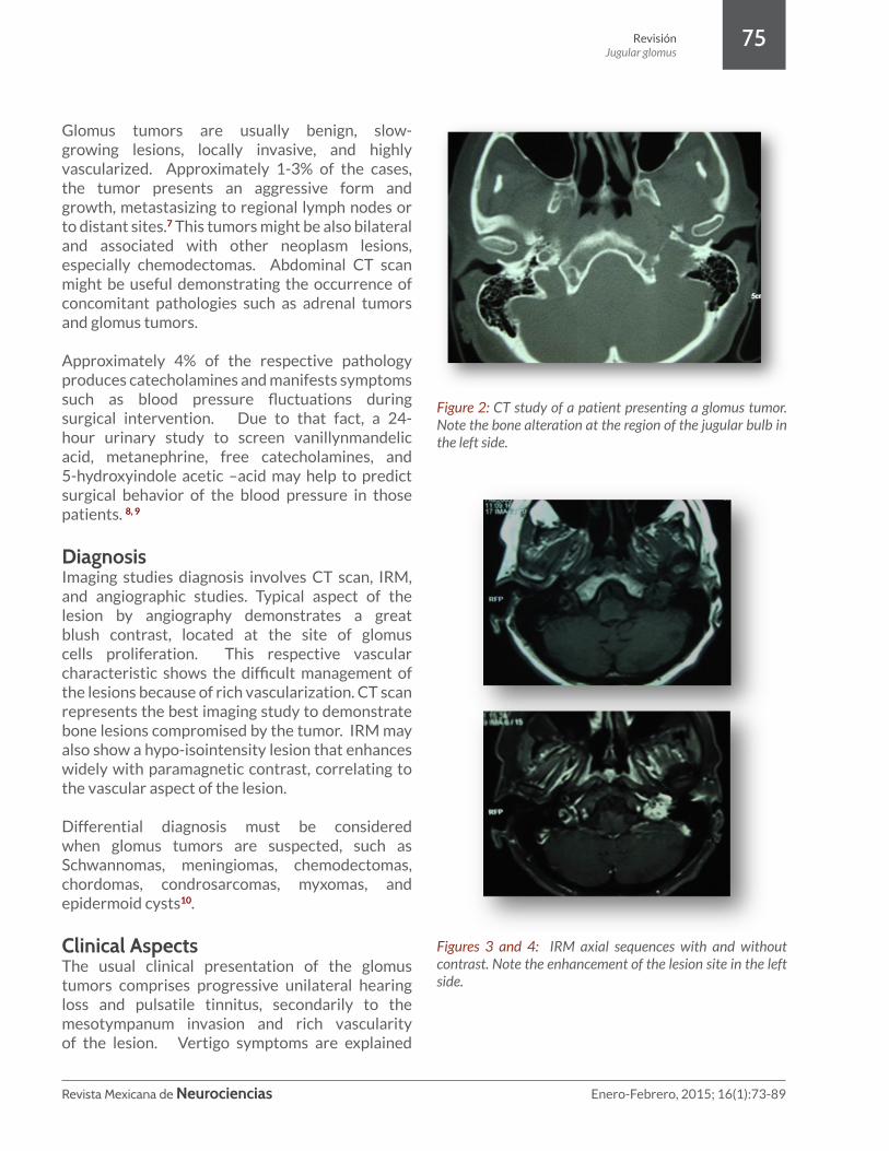
Glomus tumors are usually benign, slow-growing lesions, locally invasive, and highly vascularized. Approximately 1-3% of the cases, the tumor presents an aggressive form and growth, metastasizing to regional lymph nodes or to distant sites.7 This tumors might be also bilateral and associated with other neoplasm lesions, especially chemodectomas. Abdominal CT scan might be useful demonstrating the occurrence of concomitant pathologies such as adrenal tumors and glomus tumors.
Approximately 4% of the respective pathology produces catecholamines and manifests symptoms such as blood pressure fluctuations during surgical intervention. Due to that fact, a 24-hour urinary study to screen vanillynmandelic acid, metanephrine, free catecholamines, and 5-hydroxyindole acetic –acid may help to predict surgical behavior of the blood pressure in those patients. 8, 9
DiagnosisImaging studies diagnosis involves CT scan, IRM, and angiographic studies. Typical aspect of the lesion by angiography demonstrates a great blush contrast, located at the site of glomus cells proliferation. This respective vascular characteristic shows the difficult management of the lesions because of rich vascularization. CT scan represents the best imaging study to demonstrate bone lesions compromised by the tumor. IRM may also show a hypo-isointensity lesion that enhances widely with paramagnetic contrast, correlating to the vascular aspect of the lesion.
Differential diagnosis must be considered when glomus tumors are suspected, such as Schwannomas, meningiomas, chemodectomas, chordomas, condrosarcomas, myxomas, and epidermoid cysts10.
Clinical AspectsThe usual clinical presentation of the glomus tumors comprises progressive unilateral hearing loss and pulsatile tinnitus, secondarily to the mesotympanum invasion and rich vascularity of the lesion. Vertigo symptoms are explained
Figure 2: CT study of a patient presenting a glomus tumor. Note the bone alteration at the region of the jugular bulb in the left side.
Figures 3 and 4: IRM axial sequences with and without contrast. Note the enhancement of the lesion site in the left side.

Revista Mexicana de Neurociencias Enero-Febrero, 2015; 16(1):73-89
76
once the labyrinth is invaded by the tumor. Other manifestations depend upon the tumor rate of growth and extensiveness of the lesion, knowing the possibility to compromise cranial nerves dysfunction (VII, VIII, IX, X, XI, and XII), Horner´s syndrome, petrous bone posterior syndrome (V and VI), cavernous sinus invasion, brainstem compression, and hydrocephalus. The Senior author reported in his series hearing loss as the most common symptom, compromising about 88% of the cases, followed by dysphagia and dysphonia in about 50% of the patients.
Recently Cheesman at cols presented a protocol management that has been made based on a 30-year experience of 134 glomus jugulare tumors11. The current protocol involves a thorough preoperative assessment of swallowing, considering that low cranial nerves palsies have great morbidity, it is considered to be important to know ahead the management of such deficits. The author describe that after jugular foramen surgical procedure, patients undergo further examination using fiber optic endoscopic evaluation of swallowing (FEES), video fluoroscopy, and manometry studies. Those patients presented prolonged or poorly compensated dysphasia are offered rehabilitation surgery.11 Traqueostomy and gastrostomy must also be considered in cases presenting low cranial nerves deficits and prolonged intubation. Sometimes the aggressive management represents the best option to avoid futures and possible complications.
Surgical AnatomyComprising a complex region, the jugular foramen involves nerves, sinuses cavities, dural folds, and osseous structures. Not only this respective complex region, but also the difficult approach to this area converts the foramen’s pathologies in a real challenge for the neurosurgeon.
The jugular foramen varies in size and shape from each person, and in the same person it can varies according to the left or right side. It is surrounded by important structures: carotid artery (anteriorly), facial nerve (laterally), hypoglossal nerve (medially), and vertebral artery (inferiorly).
The foramen is located between the margins of the temporal bone (petrosal part) and the occipital bone (condylar part) and it comprises three portions: 2 venous and 1 neural compartment 12. The venous compartments consist of a larger posterolateral venous channel, known as sigmoid portion, and a smaller anteromedial venous channel, known as petrosal portion. The sigmoid portion receives blood flow from the sigmoid sinus and the petrosal portions from the inferior petrosal sinus. The petrosal portion empties into the sigmoid part through an opening at the medial wall of the jugular bulb, located between the IX cranial nerve anteriorly and the X-XI cranial nerves posteriorly.
Surrounding the dome of the jugular bulb in the temporal bone are numerous structures, including the posterior semicircular canal, the middle ear, the medial portion of the external auditory canal, and the VII cranial nerve. The neural structures comprise the IX, X, and XI cranial nerves, located between the 2 venous compartments. The IX cranial nerve travels alone, medially and slightly superior to the X and XI cranial nerves, intimately associated with each other. The respective nerves penetrate the dura in the anteromedial margin of the intrajugular process, a fibrous or osseous bridge located between the temporal and occipital bones. Other structures crossing the jugular foramen consist in meningeal branches of the ascending pharyngeal and occipital arteries, cochlear aqueduct, Jacobson´s, and Arnold´s nerves3. The glossopharyngeal nerve exits medially and superiorly of the jugular foramen, passing lateral to the internal carotid artery (ICA). The spinal accessory nerve leaves the jugular foramen medially to the jugular vein, usually passing anteriorly to the jugular vein extracranially before going under the sternocleidomastoid muscle. Traveling between the internal carotid artery and the jugular vein, the vagus nerve composes with the respective vascular structures the cervical vasculonervous complex. The hypoglossal nerve leaves the hypoglossal canal, passing behind the vagus nerve before coursing anteriorly the internal carotid artery (ICA) towards the tongue.
RevisiónJugular glomus

Revista Mexicana de Neurociencias Enero-Febrero, 2015; 16(1):73-89
77
Glasscock-Jackson and Fish classifications (tables 1 and 2) have been used to classify the extension of glomus tumors. According to its extension and tumor´s, different approaches have been performed. The Senior author consider the combined craniocervical approach to be the best option to achieve cranial sinus, allowing good structures visualization and cervical vascular control, although variations must be performed in according to each tumor peculiarities.
Tumors involving the jugular foramen are considered a challenging for the neurosurgeon due to the variability relationship among the neurovascular structures (jugular vein, carotid artery and cranial nerves) crossing the foramens through the skull base. The surgeon must be familiar to the origin site, growth pattern, and geometry of each lesion affecting this respective region. It is a real advantage when undertaking a function-sparing procedure.
Tumors that arise within or penetrate in the jugular foramen lateral to the neural plane displace the nerves medially, a position favorable for their preservation during tumor extirpation. By contrast, medially positioned tumors displace the cranial nerves to the lateral tumor surface, presenting to the surgeon an unfavorable tumor location 13.
Glomus tumors consistently arise at the lateral aspect of the jugular foramen, displacing the lower cranial nerves medially. This position accounts for the high rate of neural preservation in small and medium-size glomus tumors that have not invaded the foramen’s central partition. Meningiomas arising laterally to the foramen (e.g., the posterior petrous surface, sigmoid sinus) favorably displace the lower cranial nerves medially. By contrast, tumors originate medially to the jugular foramen (e.g., clivus, foramen magnum) are unfavorable, displacing laterally the multiple small rootlets that coalesce the IX – XI cranial nerves into a vulnerable location. Schwannomas arise within the neural plane and have a variable geometry that depends partially on the nerve of origin. Theoretically, tumors that arise from the IX cranial nerve, located at the lateral surface of the neural plane, should be
more favorable than those originating from the X or XI cranial nerves, located on its deep surface13.
Figure 5. Intracranial view of the left jugular foramen. A stillet is inserted into the inferior petrosal sinus (IPS - petrosal compartment) and goes to the jugular bulb. The medial compartment has the dural attachment preserved. The IX, X and XI cranial nerves are seen in this compartment. The left window, in the left side of the neural compartment, corresponds to the sigmoidal compartment. Note the entry of the Sigmoid Sinus (SS) into the Jugular Bulb (JB). The Internal Auditory Canal is also showed (IAC)
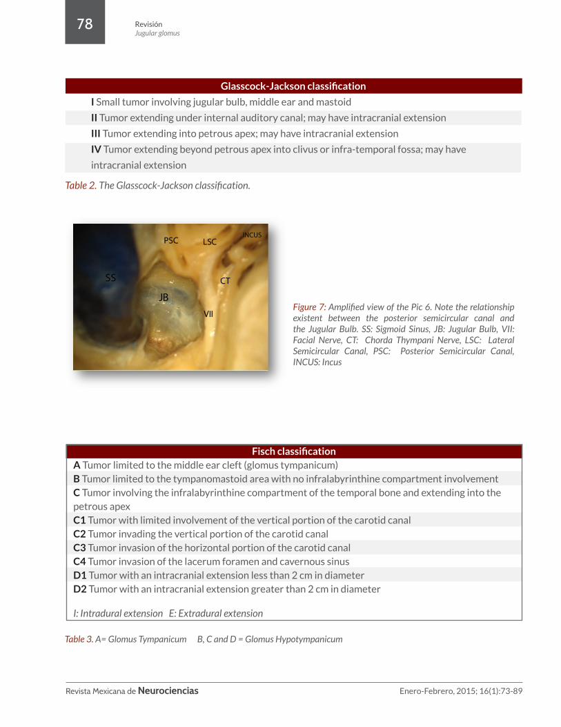
Figure 6: This picture shows the mastoid´s right side relationship among vascular, osseous and neural elements. The anatomical knowledge is crucial to procedure surgical intervention in this area. SS: Sigmoid Sinus, SPS: Superior Petrosal Sinus, JB: Jugular Bulb, VII: Facial Nerve, CT: Chorda Thympani Nerve, EAC: External Auditory Canal, LSC: Lateral Semicircular Canal, PSC: Posterior Semicircular Canal, SSC: Superior Semicircular Canal
RevisiónJugular glomus
Revista Mexicana de Neurociencias Enero-Febrero, 2015; 16(1):73-89
78
Glasscock-Jackson classification
I Small tumor involving jugular bulb, middle ear and mastoid
II Tumor extending under internal auditory canal; may have intracranial extension
III Tumor extending into petrous apex; may have intracranial extension
IV Tumor extending beyond petrous apex into clivus or infra-temporal fossa; may have
intracranial extension
Table 2. The Glasscock-Jackson classification.
Figure 7: Amplified view of the Pic 6. Note the relationship existent between the posterior semicircular canal and the Jugular Bulb. SS: Sigmoid Sinus, JB: Jugular Bulb, VII: Facial Nerve, CT: Chorda Thympani Nerve, LSC: Lateral Semicircular Canal, PSC: Posterior Semicircular Canal, INCUS: Incus
Table 3. A= Glomus Tympanicum B, C and D = Glomus Hypotympanicum
Fisch classificationA Tumor limited to the middle ear cleft (glomus tympanicum)B Tumor limited to the tympanomastoid area with no infralabyrinthine compartment involvementC Tumor involving the infralabyrinthine compartment of the temporal bone and extending into the petrous apexC1 Tumor with limited involvement of the vertical portion of the carotid canalC2 Tumor invading the vertical portion of the carotid canalC3 Tumor invasion of the horizontal portion of the carotid canalC4 Tumor invasion of the lacerum foramen and cavernous sinus D1 Tumor with an intracranial extension less than 2 cm in diameterD2 Tumor with an intracranial extension greater than 2 cm in diameter
I: Intradural extension E: Extradural extension
RevisiónJugular glomus

Revista Mexicana de Neurociencias Enero-Febrero, 2015; 16(1):73-89
79
Management of Glomus TumorsA complete examination of the cranial nerves is important to evaluate previous deficits and to define surgical routes. Audiometric studies are important to evaluate middle and internal ear function and should be performed in all cases. TC images are important for determining the extent of osseous involvement. MRI with and without gadolinium enhancement provides an excellent definition of the tumor and adjacent structures. Cerebral angiography consists in a useful tool for evaluating tumor´s blood supply and to define doubtable images causes by flow in MRI studies. Cerebral angiography also may provide information about the sinuses anatomy, involvement of the internal carotid artery (ACI), association between glomus and carotid body tumors, and torcular left-right drainage communication. Balloon occlusion test must be performed in some cases when ACI sacrifice is a possibility.
EmbolizationThe use of preoperative embolization has been described as great advance in the successful surgical management of glomus tumors (3). Embolization may reduce blood loss, the need of blood transfusion, and may also shorten operative time. Despite of these descriptions, the senior author do not use this method in all cases.
The preoperative arterial embolization is rarely complete in paraganglia tumors of the petrous bone. Intraoperative fluorescence angiography with ICG (indocyanine green) is a reliable procedure to evaluate the efficiency of preoperative embolization and may help the surgeon to estimate intra-operative bleeding14. The identification of tumor blood supply branches represents a very important step in order to determine surgical strategy intervention. Ascending pharyngeal and stylomastoid arteries send branches to the respective tumor, consisting the two most important vessels to be considered. The identification of such arteries and the transitory intra-surgical closing of the external carotid artery correspond to the gold surgical management. Such a method avoids the high cost of the embolization. The Senior author consider that one may reserve
the pre-operative embolization for D2 or D3 tumors of the Fish classification, or in those cases presenting lower cranial nerves deficits. The embolization should be performed in a period not superior to 72 hours previously surgical treatment, because new afferent vessels might have blood flow, mainly by tympanic branches of the internal carotid artery. The senior author found that embolization may cause nerve dysfunction due to the density increasing of the glomus tumors, making the resection an arduous process. Sometimes the partially embolization may be present worse results than not having embolization at all, once the tumor is modified and still highly vascularized.
RadiationThere are controversies about the role of the radiation therapy in the management of the glomus tumors. No controlled trials have been performed comparing surgery and radiation therapy. Some authors recommend its application on ancient patients or in those middle age patients presenting important comorbidities15. Some formal indications have been on residual or recidivating tumors. Glomus tumor rarely decreases in size following radiation therapy and the lack of growth is considered to indicate success. Radiosurgery and stereotactic radiosurgery consist of recently methods and may avoid complications whenever compared to conventional radiation therapy16,17. Stereotactic radiosurgery consists is an effective and safe treatment modality in the management of glomus jugulare tumors, particularly for residual or previously untreated small tumors17. Studies show that radiosurgery results are similar glomus jugulare tumor control and a superior morbidity profile compared to surgical treatment alone. In addition, patients treated with radiosurgery usually remain clinically stable or presents improvements. Given the indolent nature of these tumors, radiosurgery would be considered the first option to treat this type of tumors. Embolization followed by radio surgery may be an effective way for controlling pulsatile tinnitus produced by glomus tumors, probably because of the double effect of such therapy in the tumor blood supply. However, histological studies based on previously irradiated glomus tumors conclude that the tumor cells
RevisiónJugular glomus

Revista Mexicana de Neurociencias Enero-Febrero, 2015; 16(1):73-89
80
appear unaffected by irradiation. Catecholamine’s secretion has not been affected by radiation therapy. A recent publication has described a case presenting secreting-rate reduction in irradiated glomus tumors18. This would be a significant founding. However, the patient may not be cured by this method. The resection-recurrence rate is affected in the surgical procedure once the morbidity of the surgery increases due to the irradiated field. Besides those arguments, radiation therapy has not been show to increase cranial injury and CSF leaking, presenting a great argument against surgery. Broader studies and follow-up must be developed in order to ensure the real benefits and complications related to the technique.
Surgery The role of surgical procedures treating glomus tumors has proved its excellence through the decades. However, the surgical management of this challenging pathology is associated with severe injuries. Surgery has been related with variables morbidity rates, but it is an effective modality treatment and the unique method that may eradicate immediately the tumor19. The proportion of the resections has been increased along of years. Microsurgical techniques,
advances in anatomy of the neural regions, the better understanding of adjacent structures, and the performance of a good neuro-anesthesia have contributed widely to achieve complete resections with low-rate complications20-22. Cranial nerves palsies consist in possible complication and some authors have associated the invasiveness and clinical presentation comparing to post-operative results23,24,25. Complete excision rates are variable and directly related to tumor size and its extension.20-22, 26-30. In the Senior author series, complete tumor extirpation was achieved in 91% of the cases. Between the period from December 1997 through December 2007, thirty-four patients (22 females and 12 males) with glomus jugulare tumors were treated. The mean age was 48 years old and the mean follow-up was 52.5 months. Clinical findings included hearing loss in 88%, swallowing disturbance in 50%, and facial nerve palsy in 41% cases. Magnetic resonance imaging demonstrated a mass in the jugular foramen in 100% of the cases, a mass in the middle ear in 97%, a cervical mass in 85%, and an intradural mass in 41% of the cases. The tumor was supplied by the external carotid artery in all cases, by the internal carotid artery in 44%, and by the vertebral artery in 32% of the cases. Preoperative embolization was performed in 15 cases (44%).
Type A – Infra-labyrinthine retro-facial
Type B – Infra-labyrinthine pre and retro-facial
Type C – Infra-labyrinthine pre and retro-facial with occlusion
of the external acoustic meatus
Type D – Infra-labyrinthine approach with transposition of the
Facial Nerve
Table 4 and Figure 8: Types of approaches to glomus surgery and skull-cervical anatomy to understand glomus surgery
RevisiónJugular glomus
Revista Mexicana de Neurociencias Enero-Febrero, 2015; 16(1):73-89
81
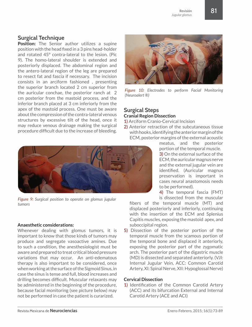
Surgical Technique Position: The Senior author utilizes a supine position with the head fixed in a 3 pins head-holder and rotated 45° contra-lateral to the lesion. (Pic 9). The homo-lateral shoulder is extended and posteriorly displaced. The abdominal region and the antero-lateral region of the leg are prepared to resect fat and fascia if necessary. The incision consists in an arciform fashioned , presenting the superior branch located 2 cm superior from the auricular conchae, the posterior ranch at 2 cm posterior from the mastoid process, and the inferior branch placed at 3 cm inferiorly from the apex of the mastoid process. One must be aware about the compression of the contra-lateral venous structures by excessive tilt of the head, once it may reduce venous drainage making the surgical procedure difficult due to the increase of bleeding.
Anaesthetic considerations: Whenever dealing with glomus tumors, it is important to know that those kinds of tumors may produce and segregate vasoactive amines. Due to such a condition, the anesthesiologist must be aware and prepared to treat critical blood pressure variations that may occur. An anti-edematous therapy is also important to be considered, once when working at the surface of the Sigmoid Sinus, in case the sinus is tense and full, blood increases and drilling becomes difficult. Muscular relaxants may be administered in the beginning of the procedure, because facial monitoring (see picture below) may not be performed in case the patient is curarized.
Surgical StepsCranial Region Dissection1) Arciform Cranio-Cervical Incision2) Anterior retraction of the subcutaneous tissue
with hooks, identifying the anterior margin of the ECM, posterior margins of the external acoustic
meatus, and the posterior portion of the temporal muscle.3) On the external surface of the ECM, the auricular magnus nerve and the external jugular vein are identified. (Auricular magnus preservation is important in cases neural anastomosis needs to be performed). 4) The temporal fascia (FMT) is dissected from the muscular
fibers of the temporal muscle (MT) and displaced posteriorly and inferiorly, continuing with the insertion of the ECM and Splenius Capitis muscles, exposing the mastoid apex, and suboccipital region.
5) Dissection of the posterior portion of the temporal muscle from the scamous portion of the temporal bone and displaced it anteriorly, exposing the posterior part of the zygomatic arch. The posterior part of the digastric muscle (MD) is dissected and separated anteriorly. (VJI: Internal Jugular Vein, ACC: Common Carotid Artery, XI: Spinal Nerve, XII: Hypoglossal Nerve)
Cervical Dissection 1) Identification of the Common Carotid Artery
(ACC) and its bifurcation External and Internal Carotid Artery (ACE and ACI)
Figure 10: Electrodes to perform Facial Monitoring (Neuroalert ®)
Figure 9: Surgical position to operate on glomus jugular tumors
RevisiónJugular glomus

Revista Mexicana de Neurociencias Enero-Febrero, 2015; 16(1):73-89
82
2) Identification of the Internal Jugular vein (VJI)3) Cranial Nerves X, XI and XII are identified and
preserves4) External Carotid Artery, as well as occipital and
ascendant pharyngeal arteries are coagulated, ligated or sectioned.
5) The stilomastoid process is maintained intact to protect the extra-temporal segment of the facial nerve
6) Mastoidectomy, with visualization of the semicircular channels, sigmoid sinus , sinodural angle and middle fossa base. Abundant and permanent irrigation consist in an important procedure in this step. The protection of the facial nerve and dissection or the sigmoid sinus depend on this step.
Figure 11: A and B – Left surgical approach to operate on glomus. The Temporalis Muscle Fascia (FMT) is identify and the Auricular Magnus Nerve (NAM) is preserved.
Figure 12: Laboratory view of the right mastoid region. Three lines have been drawn: The first line(I) follows the posterior projection of the zygomatic arch, the second line (II), pass in the level of the posterior wall of the external acoustic meatus, and the third line (III) crosses the supero-posterior part of the mastoid directing trough the mastoid apex
Figure 13: View of the left cranio-cervical laboratory dissection. A mastoidectomy was done to identify the sigmoid sinus, the jugular bulb, facial nerve and semicircular canals.
RevisiónJugular glomus

Revista Mexicana de Neurociencias Enero-Febrero, 2015; 16(1):73-89
83
Five crucial points in the management of glomus tumors:1) Early vascular control (Internal and External
Carotid Arteries)2) Identification and coagulation\ligation of the
afferent vessels3) Proximal control of the Sigmoid Sinus and
Internal Jugular Vein4) Identification and preservation of the Lower
Cranial Nerves and Facial Nerve5) Obliteration and reconstruction of the surgical
cavity
In the last decades, many approaches have been developed to access glomus tumor of the jugular foramen. Basically they vary in the amount of the temporal bone resection and dissection of the soft tissues. Independently of the approach discussed, the management of the facial nerve is considered the most important point during procedure. In 1952, Capps described the anterior facial transposition technique, until Fish e Pillsbury (1979) ruled the technique of the definitive anterior transposition, describing the necessity of the fixation of the nerve in the parotid gland. When approaching glomus tumors, the decision about the anterior transposition of the facial nerve preconized by Fish and Pillsbury31 ends up on discussion. A posterior transposition technique also has been described by House and Hitselberger (1976)32, but the devascularization of the facial nerve may result in ischemia and a poor post-operative result. The respective technique was practically abandoned because the facial is devascularized and the amount of the resection that can be achieved is not as good as with anterior transposition. Farrior (1984)33, Manigila et al (1992)34 and Martin and Prades (1992)35have described the preservation of the facial in loci in the origin, inside the Falopian Canal. Pensak and Jackler (1997), developed the technique called “Falopian Bridge” that maintain the facial nerve in its canal and proceed the resection of the tumor in an anteriorly, inferiorly, and posteriorly way.36
Facial nerve manipulation has been associated with severe conmorbidity. Advising about it, the senior author suggests working pre-facial and
retro-facial, plus infra-facial zones, depending on the tumor´s size and localization, avoiding though the facial mobilization whenever possible.13 When facial infiltration or lesion, anastomosis may be performed using graft or XII cranial nerve branch. Despite of its reconstruction, facial nerve function recovery is not as expected in most of cases.
The relationship between the facial management and the extension of tumor resection has been the main point to justify the anterior transposition of the nerve. In 1982, Fisch performed facial nerve anterior transposition in all cases of the glomus jugular tumors treated, presenting its functional recovery grades 1 or 2 (House and Brackman) in 87% and total tumor resection in 82% of all cases. Senior author found in 91% of total resection without necessity of facial transposition and described different approaches to manage glomus tumors. The type A consists in the infra-labyrinthine retrofacial approach. Similar technique has been utilized by Maniglia and Martin and Prades while dealing with small or non-glomus tumors. The tumor in this zone is removed by aspiration, coagulation, and impaction of the venous canals that entry the jugular bulb, located in the inferior petrosal sinus and the internal condylar vein. In the author´s series, this respective approach has been performed in 11 patients, presenting one recidivating, although post-operative RMN shown total resection. This approach is indicated in cases the tumor is not vascularized by the tympanic branches of the ICA or the anterior extension of the tumor does not reach the ICA. The technique has the advantage of the preservation of the facial nerve anatomy and function associated to the integrity of the vestibulo-cochlear structures and external acoustic meatus permeability. The facial preservation was achieved in all cases of the series, presenting one patient coursing with a transitory palsy and posterior total recovery.
The Falopian Bridge was utilized by the senior author in 21 patients, middle ear structures were preserved in 7 patients and in 14 of them the respective structures were removed and the external acoustic meatus occluded. The risk of preserving the middle ear structures relates to the
RevisiónJugular glomus

Revista Mexicana de Neurociencias Enero-Febrero, 2015; 16(1):73-89
84
CSF leakage possibility by the meatus or Eustachian tube. The disadvantage of this occlusion consists in the high rate of cholesteatomas formation and infection. In these cases, it is fundamental to complete drilling the mastoid cells, to resection the tympanic membrane, and mucosa in the promontorium area. Senior author advices the only situation that indicates meatus occlusion and middle ear structures removing when tumor grows cranially to the petrous portion of the ICA. The Senior author performed facial anastomosis in 2 cases. The technique chosen was initially
preconized by Hammerschlag (1999)37, consisting on nervous graft termino-lateral suture to the Hypoglossal nerve and termino-terminal in the Facial nerve. Functional recovery was grade 4 of House and Brackman classification38 with light hemi tongue atrophy. Senior author suggests the management of the facial nerve based on the following criteria:1) The blood supply of the tumor2) Functional state of the facial nerve3) Extension of the tumor
Figure 14: Step by step dissection of the left jugular bulb. In the first picture, note the relationship among the posterior semicircular canal, jugular bulb and facial nerve. The next picture the sigmoid sinus and the bulb have been ressected partialy. In the last picture the jugular foramen and its content is showed
1950´s - Radiation Therapy or Radiation Therapy + Limited Surgery 1960´s - Increased Surgical Ressection (Advances in Neuroimaging) New techniques - Rerouting of the Facial Nerve 1970´s - Improved Surgical Results (Advances in Anatomy, operating microscope and neuroanesthesia)
Table 4. Evolution of the management of the glomus tumors.
RevisiónJugular glomus

Revista Mexicana de Neurociencias Enero-Febrero, 2015; 16(1):73-89
85
The approach may be patterned as follows: A) Infra-labyrinthine: Indicated to resection of tumors receiving the mainly blood supply the ECA, without anterior extension or around the ICA in its ascending segment.
B) Falopian Bridge: Tumor visualization anteriorly and posteriorly to the facial nerve, without occlusion of the external acoustic meatus or resection of middle ear structures. It is indicated when tumor involves the ICA in the ascending way and tympanic branches of the Internal Carotid Artery blood supply contribution.
Infra-labyrinthine: Indicated to resection of tumors receiving the mainly blood supply the ECA, without anterior extension or around the ICA in its ascending segment
Figure 15: 1 e 2- IRM sagittal and axial contrast-enhancing sequences showing a right glomus tumor approach by Infra-labyrinthine retro-facial (Type A Approach) 3 - Angiography showing the vascularization of the tumor from branches of the External Carotid Artery. 4- Post-Operative IRM study in axial sequences that show complete resection of the lesion (TU: Tumor)
Figure 16: 1- IRM axial sequence with contrast enhancement showing a left jugular glomus tumor. Type B approach has been performed. 3- Angiography with blush in the region of the tumor. 4- Coronal IRM sequences that show complete resection of the tumor (TU: Tumor)
Figure 17: 1- IRM axial sequences with contrast enhanced. 2- Angiography that show a large left glomus tumor. 3- Post- operative IRM. The tumor has been totally removed. (TU: Tumor)
C) Similar of B, with occlusion of the external acoustic meatus and resection of middle ear structures. It is reserved to those tumors presenting cranial extension, following the ascending pathway of the ICA through the cavernous sinus
RevisiónJugular glomus

Revista Mexicana de Neurociencias Enero-Febrero, 2015; 16(1):73-89
86
D) Anterior extension Reserved to those tumors presenting wide anterior extension involving the ICA in its complete petrous segment, with extension to cavernous sinus, and also when the patient presents clinical symptoms and signs of hearing and facial dysfunction
As discussed previously, cranial nerves preservation consists in another important aspect of the surgical management, knowing the pathway of them by anteromedial wall of the jugular bulb when descending to the jugular vein and maintaining this position related to this vessel. Called by Al-Mefty and Teixeira in 2002 as “Intrabulbar resection”, when operating glomus tumors, it is important to preserve a portion of the jugular bulb and the wall of the jugular vein in order to avoid damaging nerves27. According to Lustin and Jackler (1996)39, CT and IRM are very helpful whenever showing a regular middle bone surface as a great chance than nerves may not be invaded by the tumor. In
Figure 18: 1- A very large tumor demonstrated by this IRM contrast enhanced in axial sequences. 2- Angiography that shows the extended tumor highly vascularized, with participation of the Internal and External Carotid Artery branches. 3- Post-operative IRM axial contrast-enhanced. The tumor has been totally removed. (TU: Tumor)
contrast, intracranial extension has been related with post-operative lesion of nerves and non-complete tumor resection. (Jackson et al 1991)40
Types A, B, C, and D approaches: The approach was tailored to each patient and classified in 4 types of them. 14, 15 The infra-labyrinthine retro-facial approach (Type A) was used in 32.5%; infra-labyrinthine pre- and retro-facial approach without occlusion of the external acoustic meatus (Type B) in 20.5%; infra-labyrinthine pre- and retro-facial approach with occlusion of the external acoustic meatus (Type C) in 41%; and the infra-labyrinthine approach with transposition of the facial nerve and removal of the middle ear structures (Type D) in 6% of the patients. Radical removal was achieved in 91% of the cases and partial removal in 9%. Among 20 patients without preoperative facial nerve dysfunction, the nerve kept its anatomical position in 19 (95%), and facial nerve function was normal during the immediate postoperative period in 17 (85%). Six patients (17.6%) had a new lower cranial nerve deficit; recovering of swallowing function was adequate in all cases. Voice disturbance remained in all 6 cases. Cerebrospinal fluid leakage occurred in 6 patients (17.6%), without the need of re-intervention. One patient died in the postoperative period due to pulmonary complications. The global recovery, based on the Karnofsky Performance Scale (KPS), was 100% in 15% of the patients, 90% in 45%, 80% in 33%, and 70% in 6%. Considering the experience of the senior author, radical removal of glomus jugulare tumor may be achieved without anterior transposition of the facial nerve. However, the extension of dissection should be tailored to each case based on tumor blood supply, preoperative symptoms, and tumor extension.
RevisiónJugular glomus

Revista Mexicana de Neurociencias Enero-Febrero, 2015; 16(1):73-89
87
Glomus tumors are challenging cranio-cervical lesions. Surgical treatment is the only that can cure immediately the patient. Anatomical knowledge and practice are crucial items to minimize surgical complications The operative field provided by the retro-facial infra-labyrinthine approach, or the pre- and retro-facial approaches, with or without closure of the external acoustic meatus, allows a wide exposure of the jugular foramen area 23,24,25. Global functional recovery based on the KPS was acceptable in 94% of the patients. Despite of surgical management described by the Senior author, one should be aware of the variety of management options and its results. Treatment should be based on the age, clinical condition, tumor localization, and its extension.
Glomus Jugular Tips: 1) Glomus tumors are rare tumors and consist
in the most frequent tumors located in the jugular region
2) Females may be six times more affected than males
3) Glomus are highly vascularized tumors and the correct management must consider the vascular blood supply
4) A complete examination of facial and lower cranial nerves must be performed. This aspect may help to define the best way to approach the lesion
5) The most common clinical presentation consists in hearing disturbance, swallowing deficit and facial nerve palsy
6) Facial mobilization should be avoided in most of cases. Working in infra-labyrinthine, retro and pre-facial areas, we can achieve complete tumor ressections
7) Surgical treatment is possible in most of the cases, presenting low morbidity and excellent functional results
Conclusion
8) The anatomical complexity of the area, the high vascularization of the lesion and the inadequate planning are the main causes of treatment fail
9) Radiation therapy is effective and considering the benignity of the lesion, this modality must also be considered
10) Tumor embolization must be considered in some cases
11) CSF leakage consists in the most frequent complication of the surgical procedure , followed by lower cranial nerves dysfunction
12) Like other cranial base approaches, you must program the close-reconstruction before your opening-approache
13) Fat, fascia and pediculated muscle rotation, reinforced by biological glues are the most important way to prevent fistula
RevisiónJugular glomus
Revista Mexicana de Neurociencias Enero-Febrero, 2015; 16(1):73-89
88
1. Zak FG, Lawson W: Glomus jugulare tumors. The Paraganglionic Chemoreceptor System.New York: Springuer-Verlag, 1982, pp339-391
2. Szyfter W; Kopec T; Kawczynski M Glomus caroticum, jugulare and vagale-problems in diagnosis and treatment. Otolaryngol Pol; 60(3): 305-12,2006
3. 3) Heilman, Cal and cols. Surgical Management of Glomus Jugulare Tumors: 4. In: Schmidek & Sweet – Operative Neurosurgical Techniques – Pg1014-1027; Vol I - Fifth Edition –
Saunders Elsevier – 2006 Guild SR: A hitherto unrecognized structure: The glomus jugularis in man. Anat Rec 79(Suppl 2):28-107, 1941
5. Krause W: Die Glandula tympanica des Menschen. Zentralbl Med Wiss 16:737-739, 18786. Rockley TJ, Hawk M: Glomus bodies in the temporal bone. J Otolaryngol 19:51-56, 19907. Bhansali SA, Bojrab DI, Zarbo RJ: Malignant paragangliomas of the head and neck. Clinical and
immunohistochemical characterization. Otolaryngol Head Neck Surg 104:261-264, 19918. Blumenfeld JD, Cohen N, Laragh JH, Ruggiero DA: Hypertension and catecholamine biosynthesis
associated with a glomus jugulare tumor. N Engl J Med 327:894, 19929. Schwaber MK, Glasscock ME, Nissen AJ, et al: Diagnosis and management of catecholamine
secreting glomus tumors. Laryngoscope 94:1008-1015, 198410. Chung-Cheng W, Liu A and Chung-Jiang Y: Surgical Management of Non Glomus Tumors of the
Jugular Foramen. In Schmidek & Sweet– Operative Neurosurgical Techniques – Pg1028-1038; Vol I - Fifth Edition – Saunders Elsevier – 2006
11. Cheesman AD; Kelly AM. Rehabilitation after treatment for jugular foramen lesions. Skull Base; 19(1): 99-108, 2009 Jan.
12. Rhoton: Jugular Foramen. Cranial Anatomy and Surgical Approaches – Chapter 9:699-71713. 13) Lustig LR, Jackler RK: The variable relationship between the lower cranial nerves and jugular
foramen tumors: implications for neural preservation. Am J Otol. 1996 Jul;17(4):658-6814. Siedek V; Waggershauser T; Berghaus A; Matthias C: Intraoperative monitoring of intraarterial
paraganglioma embolization by indocyanin green fluorescence angiography. Eur Arch Otorhinolaryngol; 266(9): 1449-54, 2009 Sep.
15. Cosetti M; Linstrom C; Alexiades G; Tessema B; Parisier S: Glomus tumors in patients of advanced age: a conservative approach. Laryngoscope; 118(2): 270-4, 2008 Feb.
16. Guss ZD, Batra S, Li G, Chang SD, Parsa AT, Rigamonti D, Kleinberg L, Lim M: Radiosurgery for glomus jugulare: history and recent progress. Neurosurg Focus. 2009 Dec;27(6):E5.
17. Genç A, Bicer A, Abacioglu U, Peker S, Pamir MN, Kilic T: Gamma knife radiosurgery for the treatment of glomus jugulare tumors. J Neurooncol. 2010 Mar;97(1):101-8. Epub 2009 Aug 26.
18. Castrucci WA, Chiang VL, Hulinsky I, Knisely JP: Biochemical and clinical responses after treatment of a catecholamine-secreting glomus jugulare tumor with gamma knife radiosurgery.Head Neck. 2009 Sep 29. [Epub ahead of print]
19. Gottfried ON, Liu JK, Couldwell WT: Comparison of radiosurgery and conventional surgery for the treatment of glomus jugulare tumors. Neurosurg Focus. 2004 Aug 15;17(2):E4.
20. Sanna M; De Donato G; Piazza P; Falcioni M: Revision glomus tumor surgery. Otolaryngol Clin North Am; 39(4): 763-82, vii, 2006 Aug.
21. Ramina R, Maniglia JJ, Fernandes YB, Paschoal JR, Pfeilsticker LN, Neto MC, Borges G: Jugular foramen tumors: diagnosis and treatment. Neurosurg Focus. 2004 Aug 15;17(2):E5.
22. De la Cruz A; Teufert KB; Santa Cruz S: Surgical treatment of temporal, tympanic and jugular paragangliomas. Indications and surgical technique. Acta Otorrinolaringol Esp; 60 Suppl 1: 106-18, 2009 Feb.
23. Leonetti JP; Anderson DE; Marzo SJ; Origitano TC; Vandevender D; Quinonez R: Facial paralysis associated with glomus jugulare tumor. Otol Neurotol; 28(1): 104-6, 2007 Jan.
24. Parhizkar N; Hiltzik DH; Selesnick SH: Facial nerve rerouting in skull base surgery. Otolaryngol Clin North Am; 38(4): 685-710, ix, 2005 Aug.
25. Al Mefty, O Intraoperative electrophysiological monitoring. In Al Mefty, O (ed) Surgery of the skull base. Boston: Kluwer Academic, 1989 p 335-340
26. Al Mefty, O; Fox, J.L; Rifai, A; Smith, R.R. A combined infratemporal and posterior fossa approach for the removal of giant glomus tumors and chondrosarcomas. Sur. Neur. V.28, N.6, p 423-431, Dec 1987
References
RevisiónJugular glomus

Revista Mexicana de Neurociencias Enero-Febrero, 2015; 16(1):73-89
89
27. Al Mefty, O; Teixeira, A. Complex tumors of the glomus jugulare: criteria, treatment and outcome. J. Neurosurgery – V.97, N.6 p 1356-1366, Dec 2002
28. Borba LA; Ale-Bark S; London C: Surgical treatment of glomus jugulare tumors without rerouting of the facial nerve: an infralabyrinthine approach. Neurosurg Focus; 17(2): E8, 2004 Aug 15.
29. Borba LA, Araújo JC, de Oliveira JG, Filho MG, Moro MS, Tirapelli LF, Colli BO: Surgical management of glomus jugulare tumors: a proposal for approach selection based on tumor relationships with the facial nerve. J Neurosurg. 2010 Jan;112(1):88-98.
30. Borba LA: Surgical treatment of Glomic Tumors of the Jugular Foramen: Proposition of sistematization considering de approaching in relationship with facial nerve – Tesis – Ribeirão Preto – São Paulo - 2008
31. Fish, U; Pillsbury, H.C. Infratemporal fossa approach to lesions in the temporal bone and base of the skull. Ach. Otolaryngol,V: 105, N:2, p99-107, Feb – 1979
32. House, W.F. Histselberguer,W.E; The transcoclear approach to the skull base. Arch Otolaryngol.,V.102,N.6, p:334-342, Jun 1976
33. Farrior, J.B; Anterior hypotympanic approach for glomus tumor of the infratemporal fossa. Laryngoscope, V.94, N.8, 1016-1021, Aug. 1984
34. Maniglia A.J; Sprecher, R.C; Megerian,C.A; Lanzieri, C. Inferior mastoidectomy –hypothympanic approach for surgical removal of glomus jugulare tumors.an anatomical and radiologic study emphasizing distances between critical structures. Laryngoscope,V.102,N.4, p407-414.Apr. 1992
35. Martin, C; Prades, J.M; Removal of selected infralabyrinthine lesions without facial nerve mobilization. Skull Base Surg. V.2, N.4, p 220-226, 1992
36. Pensak, M.L; Jackler, R,K; Removal of jugular foramen tumors: The fallopian bridge technique. Otolaryngol. Head and Neck Surg. V.117, N.6, p. 586-591, Dec 1997
37. Hammerschlag, P.E; Facial reanimation with jump interpositional graft hypoglossal-facial anastomosis and hypoglossal-facial anastomosis: evolution in management of facial paralysis, Laryngoscope,V.109, N2, pt2, p1-23, Supplement 90, 1999
38. House, J.W.; Brackmann, D.E; Facial nerve grading system. Otolaryngl. Head and Neck Surg, V.93, N.2, p.146-147, Apr. 1985
39. Lusting, L; Jackler, R. K. The variable relationship between the lower cranial nerves and jugular foramen tumors: implications for neural preservation. Am.J.Otol, V, .17, N.4, p 658-668, Jul 1996
40. Jackson, C.G; Cueva, R.A; Thedinger, B.A; Glasscock, M.E, Cranial nerve preservation in lesions of the jugular fossa. Otolaryngol. Head Neck Surg. V105, N5, p. 687-693, Nov 1991
RevisiónJugular glomus
www.revmexneuroci.comRevista Mexicana de Neurociencia, 2015; 16(1): 73-89