Revisiting Neuroimaging of Abusive Head Trauma in Infants ... · Revisiting Neuroimaging of Abusive...
9
944 AJR:204, May 2015 Revisiting Neuroimaging of Abusive Head Trauma in Infants and Young Children Kevin Li-Chun Hsieh 1,2 Robert A. Zimmerman 3 Hung Wen Kao 4 Cheng-Yu Chen 1, 2, 5 Hsieh KLC, Zimmerman RA, Kao HW, Chen CY 1 Department of Medical Imaging, Taipei Medical University Hospital, No. 252, Wu Hsing St, Taipei City 110, Taiwan, ROC. Address correspondence to C. Y. Chen ([email protected]). 2 Imaging Research Center, College of Medicine, Taipei Medical University, Taipei City, Taiwan, ROC. 3 Department of Radiology, The Children Hospital of Philadelphia, 34th Street and Civic Center Blvd, Philadelphia, PA. 4 Department of Radiology, Tri-Service General Hospital, Taipei City, Taiwan, ROC . 5 School of Medicine, National Defense Medical Center, Taipei City, Taiwan, ROC. Pediatric Imaging • Review This article is available for credit. AJR 2015; 204:944–952 0361–803X/15/2045–944 © American Roentgen Ray Society Keywords: abusive head trauma, children, infants, neuroimaging DOI:10.2214/AJR.14.13228 Received May 30, 2014; accepted after revision September 1, 2014. level of consciousness without external evi- dence of head injury [7]. These findings are nonspecific and can be misleading. Investi- gating them is time-consuming, resulting in delays in diagnosis and treatment. Although abused children may be reported as having accidental falls at home, these falls are rarely associated with severe intracranial injury [8– 10]. Having no history of trauma reported to treating physicians is the most predictive his- torical feature that a child with head trauma has been abused [11]. Therefore, radiologists are frequently called to court to testify about imaging findings [12]. The general mortality rate of AHT is greater than 15% [4]. Only one third of abused children completely recover, and as many as one half of survivors have cognitive or other neurologic deficits [13]. A number of psychiatric and physical disorders are associ- ated with child abuse [14–18] and have been related to alteration of epigenetic regulation of several disease-related genes [19, 20]. The protective mechanisms of the brain that guard against injury mature with age. Infants are particularly vulnerable to nonac- cidental trauma, such as shaking, because of the size of the head in relation to the rest of the body (almost one tenth of the body mass), the sizable subarachnoid spaces, and ineffec- tive head support from weak neck muscles [21]. The infant brain is soft owing to imma- ture myelination and small axonal size [13]. Furthermore, the infant skull is thin, soft, and easily deformed and readily transmits impact to the deeper brain structures. T he prevalence of abusive head trauma remains high with near- ly 690,000 substantiated reports each year [1]. Because of the morbidity and mortality due to injuries to the CNS, the focus has continued to be on the heads of abused children. In the United States each year, approximately 1640 chil- dren die of nonaccidental injury, and at least 10% of cases of mental retardation and ce- rebral palsy result from child abuse [1, 2]. As of 2000, child abuse was the most com- mon cause of serious head injury among children younger than 1 year [3]. The risk factors for inflicted traumatic brain injury include age younger than 1 year and mother young and unmarried with a low education- al level, low socioeconomic status, disabili- ty, behavior or emotional problems, and multiple births [1, 4]. The mother is the most common perpetrator, followed by par- ent (i.e., the perpetrator was identified as the parent, but the sex was not specified), father, and male relatives [1]. Abusive head trauma (AHT) involves dif- ferent mechanisms of forces, including shak- ing and impact injuries, direct blows to the head, compression, strangulation, penetrat- ing injuries, smothering, and suffocating [5, 6]. There is often a combination of cranial and skeletal injuries to a young child without a history of overt trauma or to a child with a history of trivial trauma inconsistent with the degree of the CNS injuries. The most common presentations are seizures, enceph- alopathy, retinal hemorrhages, and altered OBJECTIVE. The purpose of this article is to use a mechanism-based approach to review the neuroimaging findings of abusive head trauma to infants. Advanced neuroimaging pro- vides insights into not only the underlying mechanisms of craniocerebral injuries but also the long-term prognosis of brain injury for children on whom these injuries have been inflicted. CONCLUSION. Knowledge of the traumatic mechanisms, the key neuroimaging find- ings, and the implications of functional imaging findings should help radiologists character- ize the underlying causes of the injuries inflicted, thereby facilitating effective treatment. Hsieh et al. Neuroimaging of Abusive Head Trauma in Infants Pediatric Imaging Review FOCUS ON: Downloaded from www.ajronline.org by American Roentgen Ray Society on 04/30/15 from IP address 205.201.248.242. Copyright ARRS. For personal use only; all rights reserved
Transcript of Revisiting Neuroimaging of Abusive Head Trauma in Infants ... · Revisiting Neuroimaging of Abusive...
944 AJR:204, May 2015
Revisiting Neuroimaging of Abusive Head Trauma in Infants and Young Children
Kevin Li-Chun Hsieh1,2 Robert A. Zimmerman3 Hung Wen Kao4 Cheng-Yu Chen1, 2, 5
Hsieh KLC, Zimmerman RA, Kao HW, Chen CY
1Department of Medical Imaging, Taipei Medical University Hospital, No. 252, Wu Hsing St, Taipei City 110, Taiwan, ROC. Address correspondence to C. Y. Chen ([email protected]).
2Imaging Research Center, College of Medicine, Taipei Medical University, Taipei City, Taiwan, ROC.
3Department of Radiology, The Children Hospital of Philadelphia, 34th Street and Civic Center Blvd, Philadelphia, PA.
4Department of Radiology, Tri-Service General Hospital, Taipei City, Taiwan, ROC .
5School of Medicine, National Defense Medical Center, Taipei City, Taiwan, ROC.
Pediatr ic Imaging • Review
This article is available for credit.
AJR 2015; 204:944–952
0361–803X/15/2045–944
© American Roentgen Ray Society
Keywords: abusive head trauma, children, infants, neuroimaging
DOI:10.2214/AJR.14.13228
Received May 30, 2014; accepted after revision September 1, 2014.
level of consciousness without external evi-dence of head injury [7]. These findings are nonspecific and can be misleading. Investi-gating them is time-consuming, resulting in delays in diagnosis and treatment. Although abused children may be reported as having accidental falls at home, these falls are rarely associated with severe intracranial injury [8–10]. Having no history of trauma reported to treating physicians is the most predictive his-torical feature that a child with head trauma has been abused [11]. Therefore, radiologists are frequently called to court to testify about imaging findings [12].
The general mortality rate of AHT is greater than 15% [4]. Only one third of abused children completely recover, and as many as one half of survivors have cognitive or other neurologic deficits [13]. A number of psychiatric and physical disorders are associ-ated with child abuse [14–18] and have been related to alteration of epigenetic regulation of several disease-related genes [19, 20].
The protective mechanisms of the brain that guard against injury mature with age. Infants are particularly vulnerable to nonac-cidental trauma, such as shaking, because of the size of the head in relation to the rest of the body (almost one tenth of the body mass), the sizable subarachnoid spaces, and ineffec-tive head support from weak neck muscles [21]. The infant brain is soft owing to imma-ture myelination and small axonal size [13]. Furthermore, the infant skull is thin, soft, and easily deformed and readily transmits impact to the deeper brain structures.
The prevalence of abusive head trauma remains high with near-ly 690,000 substantiated reports each year [1]. Because of the
morbidity and mortality due to injuries to the CNS, the focus has continued to be on the heads of abused children. In the United States each year, approximately 1640 chil-dren die of nonaccidental injury, and at least 10% of cases of mental retardation and ce-rebral palsy result from child abuse [1, 2]. As of 2000, child abuse was the most com-mon cause of serious head injury among children younger than 1 year [3]. The risk factors for inflicted traumatic brain injury include age younger than 1 year and mother young and unmarried with a low education-al level, low socioeconomic status, disabili-ty, behavior or emotional problems, and multiple births [1, 4]. The mother is the most common perpetrator, followed by par-ent (i.e., the perpetrator was identified as the parent, but the sex was not specified), father, and male relatives [1].
Abusive head trauma (AHT) involves dif-ferent mechanisms of forces, including shak-ing and impact injuries, direct blows to the head, compression, strangulation, penetrat-ing injuries, smothering, and suffocating [5, 6]. There is often a combination of cranial and skeletal injuries to a young child without a history of overt trauma or to a child with a history of trivial trauma inconsistent with the degree of the CNS injuries. The most common presentations are seizures, enceph-alopathy, retinal hemorrhages, and altered
OBJECTIVE. The purpose of this article is to use a mechanism-based approach to review the neuroimaging findings of abusive head trauma to infants. Advanced neuroimaging pro-vides insights into not only the underlying mechanisms of craniocerebral injuries but also the long-term prognosis of brain injury for children on whom these injuries have been inflicted.
CONCLUSION. Knowledge of the traumatic mechanisms, the key neuroimaging find-ings, and the implications of functional imaging findings should help radiologists character-ize the underlying causes of the injuries inflicted, thereby facilitating effective treatment.
Hsieh et al.Neuroimaging of Abusive Head Trauma in Infants
Pediatric ImagingReview
FOCU
S O
N:
Dow
nloa
ded
from
ww
w.a
jron
line.
org
by A
mer
ican
Roe
ntge
n R
ay S
ocie
ty o
n 04
/30/
15 f
rom
IP
addr
ess
205.
201.
248.
242.
Cop
yrig
ht A
RR
S. F
or p
erso
nal u
se o
nly;
all
righ
ts r
eser
ved
AJR:204, May 2015 945
Neuroimaging of Abusive Head Trauma in Infants
Neuroimaging Features That Differentiate Abusive From Nonabusive Head Trauma
Although AHT and nonabusive head trauma have many common neuroradiolog-ic findings, some features are helpful in dif-ferentiating one from the other. In a meta-analysis of 21 studies [22], hemorrhages that were subdural, multiple, in the convexity, in-terhemispheric, or in the posterior fossa were significantly associated with AHT. In addi-tion, hypoxic-ischemic injury and cerebral edema were significantly associated with AHT, but focal parenchymal injury and sub-arachnoid hemorrhage (SAH) were not dis-criminatory features. In a systematic review of 24 studies [23], subdural hematoma, ce-rebral ischemia, retinal hemorrhage, skull fracture, and intracranial injury were found to be significantly associated with AHT. In contrast, epidural hematoma, scalp swell-ing, and isolated skull fracture were signifi-cantly associated with nonabusive head trau-ma. In a pooled analysis, Maguire et al. [24] found that retinal hemorrhages and rib frac-tures were the most discriminating findings of AHT in children younger than 3 years with intracranial injuries and any of the oth-er clinical features, including apnea, seizure, and head or neck bruising. A positive corre-lation was found between the predictive val-ue of AHT and number of clinical features; the finding implied a combination of multi-ple mechanisms involved in AHT.
The radiologic findings of AHT may vary in different types of injury, such as blunt im-pact, shaking with or without impact, stran-gling, stabbing, and poisoning. We review the common and uncommon neuroimaging pat-terns of CNS injuries in the categories of dif-ferent traumatic mechanisms of child abuse.
Injury Mechanisms and Related Neuroimaging FeaturesBlunt Impact
Blunt impact is a dynamic force delivered at a point. It varies by type and degree of as-sault, such as being hit by a hard object, be-ing thrown out of a window and hitting the ground, or being struck against a wall [25]. This type of injury usually leads to an imme-diate impact effect that includes subgaleal he-matoma, skull fracture, brain contusion, and epidural or subdural hemorrhages (Fig. 1A).
Skull fractures are found in as many as one third of children with AHT [26]. Non-accidental skull fractures may be linear or depressed, mainly in the parietal or occipi-
tal bones. The fractures can be stellate (egg-shell fractures), bilateral, and multiple and can cross the sutures. Growing fractures are frequent in infants [27] (Fig. 1B and 1C). In children with inflicted injuries, fractures of the long bones and ribs are more frequent than skull fractures [25]. Therefore, careful examination of the long bones and ribs with a high-quality radiologic skeletal series sur-vey is mandatory to search for recent or old fracture in suspicious cases [28, 29].
The force resulting in skull fracture tends to cause focal underlying brain injuries, such as hemorrhagic contusion and subdural he-matoma. The subdural hematoma is along the interhemispheric fissure and over the con-vexities. Extradural and intracerebral hemor-rhages are relatively uncommon and may be overlooked when the hematomas are small. Susceptibility-weighted imaging has shown promise compared with CT and convention-al MRI, depicting additional hemorrhagic lesions 30% of the time in cases of pediat-ric traumatic brain injury [30] (Fig. 2B). In AHT, the number and volume of hemorrhag-ic lesions on susceptibility-weighted images have been proved to correlate with Glasgow coma scale score, need for surgical interven-tion, length of hospital stay, length of intuba-tion, and intellectual function [31]. The imag-ing findings in the initial stages of AHT may be important for treatment and prognosis.
Intracranial hemorrhages due to blunt im-pact are often accompanied by brain edema and white matter laceration, which are im-portant causes of increased intracranial pres-sure in the acute stage. Late sequelae of con-tusion injury include gray and white matter necrosis, encephalomalacia, ventricular dila-tion, shrinkage of the cerebral hemispheres, and subdural collections. In children with mild traumatic brain injury, trivial chang-es in regional brain volume can be detected with whole-brain parcellation analysis [32].
Shaking InjuryShaking injury is caused by forceful shak-
ing of an infant’s head by grasping the shoul-ders or chest in an impulse that changes the momentum of the force by acceleration or deceleration [33]. The asynchronous to-and-fro movements of both cerebral hemi-spheres at different speeds along the sides of the falx cerebri are further aggravated by the large size of the head and the weakness of the neck muscles. The shearing force on the retina and cerebral veins and the high intracranial venous pressure resulting from
chest wall compression are thought to lead to retinal hemorrhage and bleeding into the potential subdural space. In addition to the linear force, the angular or rotational shear-ing force, especially when prolonged shears are applied, is likely to cause diffuse axo-nal injury (DAI), which frequently involves the parasagittal white matter [34] (Fig. 3A). Moreover, shaking without impact may not generate enough force to cause the typical brain damage of shaken baby syndrome [21]. Classic shaking-impact trauma to the head encompasses a scalp bruise, depressed skull fracture, and brain contusions (Fig. 2). Shak-ing-impact injury may accompany fractures of the humeral shafts and the ribs at the cos-tovertebral junction owing to grasping by the shoulders, upper arms, or rib cage [21].
After traumatic injury, cerebral autoreg-ulation and ionic homeostasis can be lost, and leakage of fluid from damaged blood vessels and increased cerebral blood flow from direct disturbance of vasomotor con-trol may result in subsequent brain edema. In an animal model of head impact injury, raised intracranial pressure caused by exten-sive breakdown of the blood-brain barrier and albumin extravasation plays an impor-tant role in cerebral edema [35]. The dysreg-ulated cerebral region is also at high risk of ischemic or hyperemic injury. The immature infant brain is more vulnerable to brain ede-ma, though the underlying mechanism is not fully understood [36, 37].
Acute cerebral edema can sometimes be recognized at CT within the first 12 hours af-ter injury but is less well visualized at MRI owing to the high water content of the unmy-elinated white matter during infancy. At CT, the major findings are decreased attenuation of cerebral gray and white matter, resulting in low attenuation of the involved brain re-gion with loss of gray-white matter differen-tiation. The thalamus, brainstem, and cere-bellum may be selectively spared during the initial phase of ischemia, probably because the posterior circulation is preserved by au-toregulation. The low-attenuation cortical mantle and relatively dense thalamus and cerebellum seen at CT are known as the re-versal sign [38].
Infants sustaining shaking injury typically have retinal hemorrhages and subdural and diffuse cerebral edema [24]. This type of in-jury can present without external evidence of injury, such as a skull fracture, scalp bruise, or soft-tissue swelling, making a clinical di-agnosis challenging.
Dow
nloa
ded
from
ww
w.a
jron
line.
org
by A
mer
ican
Roe
ntge
n R
ay S
ocie
ty o
n 04
/30/
15 f
rom
IP
addr
ess
205.
201.
248.
242.
Cop
yrig
ht A
RR
S. F
or p
erso
nal u
se o
nly;
all
righ
ts r
eser
ved
946 AJR:204, May 2015
Hsieh et al.
Subdural hematomas are uncommon as a result of nonabusive head trauma in the first 2 years of life but are the most frequent im-aging findings in infants with AHT [9]. The hematomas occur most often in the posteri-or interhemispheric region and tend to ex-tend over or under the cerebral hemispheres [39] (Fig. 3B). CT shows thin layers of sub-dural hematoma, which may be obscured by the beam-hardening effect, commonly in the posterior cranial fossa. In addition, small subacute and chronic subdural hematomas are difficult to detect owing to similar atten-uation between the hematomas and the CSF or the brain. Unlike CT, MRI is sensitive to the degraded RBCs and can be used to esti-mate the age of a subdural hematoma [40]. In general, the evolution of subdural hematoma is similar to that of intracerebral hematoma; acute hematomas are mildly hypointense on T1-weighted images and hypointense on T2-weighted images. Early subacute hematomas are hyperintense on T1-weighted images and hypointense on T2-weighted images. In late subacute subdural hematomas, the T2 relax-ation time is prolonged owing to RBC lysis; the result is high signal intensity on both T1- and T2-weighted images (Fig. 4A). In the chronic stage, the signal intensity of a he-matoma is less than in the acute phase but remains higher than that of CSF on FLAIR images because its protein content is greater than that of CSF (Fig. 4B).
The signal-intensity changes of subdural hematomas can be different from those of intracerebral hematomas. For example, the hemosiderin deposition in chronic paren-chymal hematomas may not be observed in chronic subdural hematomas because of the paucity of phagocytosis in the subdural space [41]. Subdural hematomas may re-solve slower than parenchymal hematomas because brain tissue contains high concen-trations of tissue thromboplastin, which ac-celerates the resolution of a hematoma [42]. In addition, the signal intensity of subdural hematoma also varies with the size and lo-cation of the hematoma, rate of hemoglo-bin degradation, and mixed components of hematoma and CSF [43, 44]. Owing to the complex signal characteristics of subdural hematoma, particularly in patients with re-peated trauma, caution should be exercised when timing the traumatic event solely on the basis of MRI findings.
In a 2012 meta-analysis [23] AHT was found to be not significantly associated with SAH and DAI [23]. A 2011 systematic review
[22], however, had shown a significant asso-ciation between AHT and SAH. The meta-analysis was limited by the considerable het-erogeneity of the studies included, and the systematic review did not include DAI in the analysis. In the neuroimaging literature, DAI and SAH are frequently reported to be relat-ed to AHT [45–48]. In shaking injury, SAH commonly results from tears of the small ves-sels in the pia and arachnoid mater and ap-pears in the interhemispheric fissure or high convexity. CT and T2-weighted FLAIR MRI both are sensitive for SAH [49].
DAI is an important sequela of severe ac-celeration-deceleration brain shear injuries. In a classic study, Duhaime et al. [50] found DAI in pathologic examinations of both in-fants who died and a shaking-impact mod-el in subhuman primates. The findings high-lighted the importance of impact as part of the pathogenesis of DAI in shaking injuries. Shear injury often involves the subcortical white matter, corpus callosum, brainstem, and internal capsule. Gradient-echo se-quences and susceptibility-weighted imag-ing are the techniques of choice for detecting small white matter hemorrhages. Diffusion-weighted MRI (DWI) together with apparent diffusion coefficient mapping can be used to detect nonhemorrhagic axonal injury [51, 52] (Fig. 5A). DWI is sensitive in the detec-tion of early DAI lesions and for showing a greater extent of injury to the cerebral white matter than does conventional T2-weighted imaging [51, 53, 54]. The pathogenesis of re-stricted water diffusion in DAI is not entire-ly understood. Factors such as ischemia and propagated injury from oxidants or free rad-icals to the differentiating oligodendrocytes in cerebral white matter have been suggested as possible causes of DAI [55, 56].
Retinal hemorrhage has been strongly as-sociated with AHT [22, 23]. It is believed to be associated with the firm vitreous in in-fants and is caused by a sudden increase in intracranial venous pressure due to violent chest compression. Hemorrhage may also oc-cur in the subretinal, preretinal, and intraret-inal spaces and sometimes fills the vitreous. On MR images, susceptibility-sensitive im-aging techniques, such as gradient-recalled echo and susceptibility-weighted imaging, can delineate the nodular foci of low signal intensity in the posterior aspect of the globe along the retina with relatively high sensitivi-ty [57, 58]. Although retinal hemorrhage was considered one of the classic presentations of AHT with high prevalence (54–100%) [59],
the finding is controversial and can be asso-ciated with various other diseases [60].
Diffusion-tensor imaging can delineate subtle white matter alterations in nonabusive head trauma patients with DAI [61]. Arfanakis et al. [62] reported reduced fractional anisot-ropy values in the corpus callosum and inter-nal capsule in mild brain injury. Bazarian et al. [63] also found changes in fractional anisotro-py and mean diffusivity in patients subjected to multiple subconcussive blows to the head. The value of diffusion-tensor imaging in AHT must be further investigated. DAI may also disrupt brain networks by damaging white matter tracts, a finding that can be revealed with resting-state functional MRI [64, 65].
MR spectroscopy can reveal metabolite derangement in injured brain tissue that is not seen on conventional MR images. In pa-tients with shaking injury, low ratios of N-acetyl aspartate (NAA) to creatine and of NAA to choline and the presence of lactate peaks in the gray matter may appear with-out structural signal-intensity abnormalities. These findings are associated with poor out-come in the care of neonates with brain inju-ry [66] (Fig. 5B).
Whiplash-Shaking Injury at Craniovertebral Junction
In addition to damaging the cerebrum, whiplash-shaking injury can damage the lower brainstem and upper cervical cord [36], con-tributing to apnea and hypoxia. Violent shak-ing may lead to substantial cervical spinal in-jury with subdural and epidural hematomas and contusions of the spinal cord at the cervi-comedullary junction [67]. Ligamentous rup-tures at the craniovertebral junction are not rare in patients with this type of injury (Fig. 6).
Strangulation InjuryManual strangulation can lead to two
types of craniocerebral trauma. One is hy-poxic-ischemic encephalopathy (HIE) sec-ondary to compromise of the blood flow from the common carotid artery. The other is subdural hematoma secondary to shearing of bridging veins while the infant is being grasped by the neck and shaken. HIE is usu-ally confined to the territories of the inter-nal carotid artery. DW images show largely unilateral white matter injury after extensive cortical infarction [47]. This is caused by the inherent anatomic vulnerability of the ca-rotid arteries to extrinsic compression where the arteries lie between the transverse proc-esses of the cervical vertebrae and the ster-
Dow
nloa
ded
from
ww
w.a
jron
line.
org
by A
mer
ican
Roe
ntge
n R
ay S
ocie
ty o
n 04
/30/
15 f
rom
IP
addr
ess
205.
201.
248.
242.
Cop
yrig
ht A
RR
S. F
or p
erso
nal u
se o
nly;
all
righ
ts r
eser
ved
AJR:204, May 2015 947
Neuroimaging of Abusive Head Trauma in Infants
nocleidomastoid muscles. Bimanual assault can produce bilateral injuries (Fig. 7), a first sign of strangling.
Although not pathognomonic, AHT with strangulation should be suspected when a young child presents with HIE [68]. Care-ful inspection of CT or MR images for traces of intracranial hemorrhage, such as subdural hematoma, is important for correct diagno-sis (Fig. 8). In addition to vascular compres-sion, reactive vasospasm adjacent to hemor-rhagic lesions, cervicomedullary injuries, and apnea can also lead to HIE in strangulation injury [69–71]. Because cerebral hypoperfu-sion caused by strangling is transient, the in-farction is usually not associated with arteri-al stenosis or occlusion. Hemorrhagic laminar necrosis may occur in strangulation survivors and usually appears 7–10 days after the initial hypoxic injury [45] (Figs. 7B and 7C).
Whole-brain arterial spin-labeled per-fusion MRI has been successfully used to evaluate neonatal brains [72, 73]. Because it does not carry the risk of contrast adminis-tration, this noninvasive technique may play a role in the assessment of ischemic injury inflicted on a young brain.
Stabbing InjuryStabbing injury to the brain is a neurosur-
gical emergency with a high risk of death from intracranial bleeding and subsequent infection [74, 75]. Potential complications in-clude intracerebral hematoma, posttraumatic aneurysm, carotid-cavernous fistula, arterial occlusion, venous thrombosis, and CSF leak-age [76]. The severity and extent of the inju-ry should be confirmed with neuroimaging. Because MRI is contradicted in the presence of a metallic foreign body, CT is the modal-ity of choice in this circumstance (Fig. 9). Most patients need a craniotomy because di-rect extraction carries risk of bleeding [75].
Fetal AbuseFetal abuse includes physical assaults on an
unborn fetus and neglect or failure to protect the fetus from toxic substances introduced by the maternal route, such as alcohol, nicotine, and drugs [77]. Direct fetal injury involves a blow to the abdomen of a pregnant woman. This type of assault can be partially cush-ioned by the maternal tissue and amniotic flu-id, and thus the fetus is protected. When fetal injury occurs, the most frequent finding is in-tracranial hemorrhage [78, 79] (Fig. 10).
Fetal alcohol syndrome, the most serious adverse consequence of prenatal alcohol expo-
sure, is fairly frequent with an estimated prev-alence rate ranging from 0.5 to 7.0 cases per 1000 births in the United States [80]. Alco-hol disrupts developmental processes through multiple sites of action, harming the develop-ing embryo and fetus [81]. Presentations of fe-tal alcohol syndrome include characteristic fa-cial abnormalities, such as smooth philtrum, thin vermillion border, and short palpebral fis-sures; prenatal and postnatal growth deficits; and CNS abnormalities, such as neurologic, behavioral, and structural anomalies [81].
Proposed Imaging ProtocolsJaspan et al. [82] proposed a neuroimaging
protocol for cases of high clinical suspicion of AHT. Cranial CT should be performed on the day of presentation. On days 1–2, skull ra-diography and cranial ultrasound are suggest-ed. On days 3–4, MRI should be performed if the child has neurologic symptoms in the presence of initially normal or equivocal first CT findings. In addition to conventional T1- and T2-weighted and FLAIR sequences, the MRI protocol should include T2*-weighted gradient-echo or susceptibility-weighted im-aging to detect hemorrhage. DWI is suggest-ed for evaluating HIE. Major arterial trauma is rarely encountered and can be evaluated with MR angiography. Other advanced im-aging techniques may be considered optional and be used on a case-by-case basis.
ConclusionWith knowledge of the mechanisms of
AHT and the related neuroimaging findings in young children, radiologists continue to play a pivotal role not only in diagnosis but also in facilitating effective treatments and improving patient outcome.
References 1. U.S. Department of Health and Human Services,
Administration for Children and Families, Admin-
istration on Children, Youth and Families, Chil-
dren’s Bureau. Child maltreatment 2012. www.
acf.hhs.gov/sites/default/files/cb/cm2012.pdf. Ac-
cessed January 8, 2015
2. Minns RA, Brown JK, eds. Shaking and other
non-accidental head injuries in children. Lon-
don, UK: Mac Keith Press, 2005:154–184
3. Barlow KM, Minns RA. Annual incidence of
shaken impact syndrome in young children. Lan-
cet 2000; 356:1571–1572
4. Keenan HT, Runyan DK, Marshall SW, Nocera
MA, Merten DF, Sinal SH. A population-based
study of inflicted traumatic brain injury in young
children. JAMA 2003; 290:621–626
5. Merten DF, Osborne DR, Radkowski MA, Leoni-
das JC. Craniocerebral trauma in the child abuse
syndrome: radiological observations. Pediatr Ra-
diol 1984; 14:272–277
6. Cohen RA, Kaufman RA, Myers PA, Towbin RB.
Cranial computed tomography in the abused child
with head injury. AJR 1986; 146:97–102
7. Alexander R, Sato Y, Smith W, Bennett T. Incidence
of impact trauma with cranial injuries ascribed to
shaking. Am J Dis Child 1990; 144:724–726
8. Williams RA. Injuries in infants and small chil-
dren resulting from witnessed and corroborated
free falls. J Trauma 1991; 31:1350–1352
9. Duhaime AC, Alario AJ, Lewander WJ, et al.
Head injury in very young children: mechanisms,
injury types, and ophthalmologic findings in 100
hospitalized patients younger than 2 years of age.
Pediatrics 1992; 90:179–185
10. Thomas AG, Hegde SV, Dineen RA, Jaspan T.
Patterns of accidental craniocerebral injury oc-
curring in early childhood. Arch Dis Child 2013;
98:787–792
11. Hettler J, Greenes DS. Can the initial history pre-
dict whether a child with a head injury has been
abused? Pediatrics 2003; 111:602–607
12. Royal College of Radiology and Paediatrics and
Child Health. Standards for radiological investi-
gations of suspected non-accidental injury. Lon-
don, UK: Royal College of Radiology and Paedi-
atrics and Child Health, 2008
13. Case ME, Graham MA, Handy TC, Jentzen JM,
Monteleone JA. Position paper on fatal abusive
head injuries in infants and young children. Am J
Forensic Med Pathol 2001; 22:112–122
14. Felitti VJ, Anda RF, Nordenberg D, et al. Rela-
tionship of childhood abuse and household dys-
function to many of the leading causes of death in
adults: the Adverse Childhood Experiences
(ACE) study. Am J Prev Med 1998; 14:245–258
15. Dong M, Giles WH, Felitti VJ, et al. Insights into
causal pathways for ischemic heart disease: ad-
verse childhood experiences study. Circulation
2004; 110:1761–1766
16. Anda RF, Brown DW, Dube SR, Bremner JD, Fe-
litti VJ, Giles WH. Adverse childhood experienc-
es and chronic obstructive pulmonary disease in
adults. Am J Prev Med 2008; 34:396–403
17. Dube SR, Fairweather D, Pearson WS, Felitti VJ,
Anda RF, Croft JB. Cumulative childhood stress
and autoimmune diseases in adults. Psychosom
Med 2009; 71:243–250
18. Brown DW, Anda RF, Felitti VJ, et al. Adverse
childhood experiences are associated with the
risk of lung cancer: a prospective cohort study.
BMC Public Health 2010; 10:20
19. Yang BZ, Zhang H, Ge W, et al. Child abuse and
epigenetic mechanisms of disease risk. Am J Prev
Med 2013; 44:101–107
Dow
nloa
ded
from
ww
w.a
jron
line.
org
by A
mer
ican
Roe
ntge
n R
ay S
ocie
ty o
n 04
/30/
15 f
rom
IP
addr
ess
205.
201.
248.
242.
Cop
yrig
ht A
RR
S. F
or p
erso
nal u
se o
nly;
all
righ
ts r
eser
ved

948 AJR:204, May 2015
Hsieh et al.
20. Beach SR, Brody GH, Todorov AA, Gunter TD,
Philibert RA. Methylation at SLC6A4 is linked to
family history of child abuse: an examination of
the Iowa adoptee sample. Am J Med Genet B Neu-
ropsychiatr Genet 2010; 153B:710-713
21. Bandak FA. Shaken baby syndrome: a biome-
chanics analysis of injury mechanisms. Forensic
Sci Int 2005; 151:71–79
22. Kemp AM, Jaspan T, Griffiths J, et al. Neuroimag-
ing: what neuroradiological features distinguish
abusive from non-abusive head trauma? A system-
atic review. Arch Dis Child 2011; 96:1103–1112
23. Piteau SJ, Ward MG, Barrowman NJ, Plint AC.
Clinical and radiographic characteristics associ-
ated with abusive and nonabusive head trauma: a
systematic review. Pediatrics 2012; 130:315–323
24. Maguire SA, Kemp AM, Lumb RC, Farewell DM.
Estimating the probability of abusive head trauma:
a pooled analysis. Pediatrics 2011; 128:e550–e564
25. Kraus JF, Fife D, Cox P, Ramstein K, Conroy C.
Incidence, severity, and external causes of pediatric
brain injury. Am J Dis Child 1986; 140:687–693
26. Arnholz D, Hymel KP, Hay TC, Jenny C. Bilateral
pediatric skull fractures: accident or abuse? J
Trauma 1998; 45:172–174
27. Rinehart GC, Pittman T. Growing skull fractures:
strategies for repair and reconstruction. J Cranio-
fac Surg 1998; 9:65–72
28. Hornor G. Physical abuse: recognition and report-
ing. J Pediatr Health Care 2005; 19:4–11
29. Nimkin K, Kleinman PK. Imaging of child abuse.
Radiol Clin North Am 2001; 39:843–864
30. Beauchamp MH, Ditchfield M, Babl FE, et al. De-
tecting traumatic brain lesions in children: CT
versus MRI versus susceptibility weighted imag-
ing (SWI). J Neurotrauma 2011; 28:915–927
31. Beauchamp MH, Beare R, Ditchfield M, et al.
Susceptibility weighted imaging and its relation-
ship to outcome after pediatric traumatic brain
injury. Cortex 2013; 49:591–598
32. Zhou Y, Kierans A, Kenul D, et al. Mild traumatic
brain injury: longitudinal regional brain volume
changes. Radiology 2013; 267:880–890
33. Gerber P, Coffman K. Nonaccidental head trauma
in infants. Childs Nerv Syst 2007; 23:499–507
34. Hymel KP, Partington MD, Winston KR. Abusive
head trauma? A biomechanics-based approach.
Child Maltreat 1998; 3:116–128
35. Byard RW, Bhatia KD, Reilly PL, Vink R. How
rapidly does cerebral swelling follow trauma? Ob-
servations using an animal model and possible
implications in infancy. Leg Med (Tokyo) 2009;
11(suppl 1):S128–S131
36. Hadley MN, Sonntag VK, Rekate HL, Murphy A.
The infant whiplash-shake injury syndrome: a
clinical and pathological study. Neurosurgery
1989; 24:536–540
37. Kinney HC, Armstrong DD. Perinatal neuropa-
thology. In: Graham DI, Lantos PL, eds. Green-
field’s neuropathology 7th ed. London, UK: Ar-
nold, 2002:519–606
38. Han BK, Towbin RB, De Courten-Myers G,
McLaurin RL, Ball WS Jr. Reversal sign on CT:
effect of anoxic/ischemic cerebral injury in chil-
dren. AJR 1990; 154:361–368
39. Ewing-Cobbs L, Prasad M, Kramer L, et al. Acute
neuroradiologic findings in young children with
inflicted or noninflicted traumatic brain injury.
Childs Nerv Syst 2000; 16:25–33; discussion, 34
40. Datta S, Stoodley N, Jayawant S, Renowden S,
Kemp A. Neuroradiological aspects of subdural
haemorrhages. Arch Dis Child 2005; 90:947–951
41. Chen CY, Zimmerman RA, Rorke LB. Neuroim-
aging in child abuse: a mechanism-based ap-
proach. Neuroradiology 1999; 41:711–722
42. Vezina G. Assessment of the nature and age of sub-
dural collections in nonaccidental head injury with
CT and MRI. Pediatr Radiol 2009; 39:586–590
43. Zouros A, Bhargava R, Hoskinson M, Aronyk
KE. Further characterization of traumatic sub-
dural collections of infancy: report of five cases. J
Neurosurg 2004; 100:512–518
44. Duhem R, Vinchon M, Tonnelle V, Soto-Ares G,
Leclerc X. Main temporal aspects of the MRI signal
of subdural hematomas and practical contribution to
dating head injury. Neurochirurgie 2006; 52:93–104
45. Reece RM, Sege R. Childhood head injuries: ac-
cidental or inflicted? Arch Pediatr Adolesc Med
2000; 154:11–15
46. Rajaram S, Batty R, Rittey CD, Griffiths PD,
Connolly DJ. Neuroimaging in non-accidental
head injury in children: an important element of
assessment. Postgrad Med J 2011; 87:355–361
47. Ashwal S, Wycliffe ND, Holshouser BA. Ad-
vanced neuroimaging in children with nonacci-
dental trauma. Dev Neurosci 2010; 32:343–360
48. Matschke J, Herrmann B, Sperhake J, Korber F,
Bajanowski T, Glatzel M. Shaken baby syndrome:
a common variant of non-accidental head injury
in infants. Dtsch Arztebl Int 2009; 106:211–217
49. Noguchi K, Ogawa T, Inugami A, et al. Acute sub-
arachnoid hemorrhage: MR imaging with fluid-
attenuated inversion recovery pulse sequences.
Radiology 1995; 196:773–777
50. Duhaime AC, Gennarelli TA, Thibault LE, Bruce
DA, Margulies SS, Wiser R. The shaken baby
syndrome: a clinical, pathological, and biome-
chanical study. J Neurosurg 1987; 66:409–415
51. Chan YL, Chu WC, Wong GW, Yeung DK. Diffu-
sion-weighted MRI in shaken baby syndrome.
Pediatr Radiol 2003; 33:574–577
52. Liu AY, Maldjian JA, Bagley LJ, Sinson GP, Gross-
man RI. Traumatic brain injury: diffusion-weighted
MR imaging findings. AJNR 1999; 20:1636–1641
53. Biousse V, Suh DY, Newman NJ, Davis PC, Map-
stone T, Lambert SR. Diffusion-weighted mag-
netic resonance imaging in shaken baby syn-
drome. Am J Ophthalmol 2002; 133:249–255
54. Dan B, Damry N, Fonteyne C, Jissendi P, Zierei-
sen F, Christophe C. Repeated diffusion-weighted
magnetic resonance imaging in infantile non-
haemorrhagic, non-accidental brain injury. Dev
Med Child Neurol 2008; 50:78–80
55. Shannon P, Smith CR, Deck J, Ang LC, Ho M,
Becker L. Axonal injury and the neuropathology
of shaken baby syndrome. Acta Neuropathol
1998; 95:625–631
56. Volpe JJ. Brain injury in the premature infant:
neuropathology, clinical aspects, pathogenesis,
and prevention. Clin Perinatol 1997; 24:567–587
57. Altinok D, Saleem S, Zhang Z, Markman L,
Smith W. MR imaging findings of retinal hemor-
rhage in a case of nonaccidental trauma. Pediatr
Radiol 2009; 39:290–292
58. Zuccoli G, Panigrahy A, Haldipur A, et al. Sus-
ceptibility weighted imaging depicts retinal hem-
orrhages in abusive head trauma. Neuroradiology
2013; 55:889–893
59. Aryan HE, Ghosheh FR, Jandial R, Levy ML.
Retinal hemorrhage and pediatric brain injury:
etiology and review of the literature. J Clin Neu-
rosci 2005; 12:624–631
60. Graham DL, Gennarelli TA, McIntosh TK. Trauma.
In: Graham DI, Lantos PL, eds. Greenfield’s neuropa-
thology, 7th ed. London, UK: Arnold, 2002:823–898
61. Shenton ME, Hamoda HM, Schneiderman JS, et al.
A review of magnetic resonance imaging and diffu-
sion tensor imaging findings in mild traumatic brain
injury. Brain Imaging Behav 2012; 6:137–192
62. Arfanakis K, Haughton VM, Carew JD, Rogers
BP, Dempsey RJ, Meyerand ME. Diffusion tensor
MR imaging in diffuse axonal injury. AJNR 2002;
23:794–802
63. Bazarian JJ, Zhu T, Blyth B, Borrino A, Zhong J.
Subject-specific changes in brain white matter on
diffusion tensor imaging after sports-related con-
cussion. Magn Reson Imaging 2012; 30:171–180
64. Pandit AS, Expert P, Lambiotte R, et al. Traumat-
ic brain injury impairs small-world topology.
Neurology 2013; 80:1826–1833
65. Palacios EM, Sala-Llonch R, Junque C, et al.
Resting-state functional magnetic resonance im-
aging activity and connectivity and cognitive out-
come in traumatic brain injury. JAMA Neurol
2013; 70:845–851
66. Ashwal S, Holshouser BA, Shu SK, et al. Predic-
tive value of proton magnetic resonance spectros-
copy in pediatric closed head injury. Pediatr Neu-
rol 2000; 23:114–125
67. Geddes JF, Vowles GH, Hackshaw AK, Nickols
CD, Scott IS, Whitwell HL. Neuropathology of
inflicted head injury in children. Part II. Micro-
scopic brain injury in infants. Brain 2001;
124:1299–1306
Dow
nloa
ded
from
ww
w.a
jron
line.
org
by A
mer
ican
Roe
ntge
n R
ay S
ocie
ty o
n 04
/30/
15 f
rom
IP
addr
ess
205.
201.
248.
242.
Cop
yrig
ht A
RR
S. F
or p
erso
nal u
se o
nly;
all
righ
ts r
eser
ved
AJR:204, May 2015 949
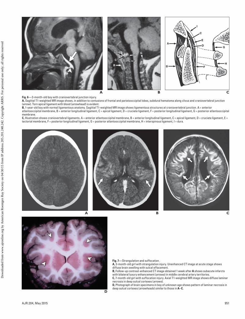
Neuroimaging of Abusive Head Trauma in Infants 68. Grant PE, Yu D. Acute injury to the immature
brain with hypoxia with or without hypoperfu-
sion. Radiol Clin North Am 2006; 44:63–77
69. Kemp AM, Stoodley N, Cobley C, Coles L, Kemp
KW. Apnoea and brain swelling in non-accidental
head injury. Arch Dis Child 2003; 88:472–476,
discussion, 472–476
70. Pierce MC, Bertocci GE, Berger R, Vogeley E.
Injury biomechanics for aiding in the diagnosis of
abusive head trauma. Neurosurg Clin N Am 2002;
13:155–168
71. Zimmerman RA, Bilaniuk LT, Farina L. Non-ac-
cidental brain trauma in infants: diffusion imag-
ing, contributions to understanding the injury
process. J Neuroradiol 2007; 34:109–114
72. Miranda MJ, Olofsson K, Sidaros K. Noninvasive
measurements of regional cerebral perfusion in pre-
term and term neonates by magnetic resonance ar-
terial spin labeling. Pediatr Res 2006; 60:359–363
73. Wang Z, Fernandez-Seara M, Alsop DC, et al.
Assessment of functional development in normal
infant brain using arterial spin labeled perfusion
MRI. Neuroimage 2008; 39:973–978
74. James G, Blakeley CJ, Hashemi K, Channing K,
Duff M. A case of self-inflicted craniocerebral
penetrating injury. Emerg Med J 2006; 23:e32
75. Large M, Babidge N, Nielssen O. Intracranial self-
stabbing. Am J Forensic Med Pathol 2012; 33:13–18
76. du Trevou MD, van Dellen JR Penetrating stab
wounds to the brain: the timing of angiography in pa-
tients presenting with the weapon already removed.
Neurosurgery 1992; 31:905–911; discussion, 911–912
77. Condon JT. The spectrum of fetal abuse in preg-
nant women. J Nerv Ment Dis 1986; 174:509–516
78. Lane PL. Traumatic fetal deaths. J Emerg Med
1989; 7:433–435
79. Pearlman MD, Tintinalli JE, Lorenz RP. Blunt
trauma during pregnancy. N Engl J Med 1990;
323:1609–1613
80. May PA, Gossage JP, Kalberg WO, et al. Preva-
lence and epidemiologic characteristics of FASD
from various research methods with an emphasis
on recent in-school studies. Dev Disabil Res Rev
2009; 15:176–192
81. Warren KR, Hewitt BG, Thomas JD. Fetal alco-
hol spectrum disorders: research challenges and
opportunities. Alcohol Res Health 2011; 34:4–14
82. Jaspan T, Griffiths PD, McConachie NS, Punt JA.
Neuroimaging for non-accidental head injury in
childhood: a proposed protocol. Clin Radiol
2003; 58:44–53
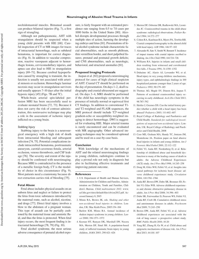
AFig. 1—Impact type of nonaccidental trauma.A, 6-month-old boy. Axial CT scan shows right frontal depressed skull fracture (straight arrow) along with frontoparietal lobe contusion (arrowheads). Subgaleal hematoma is also present (curved arrow).B, 5-month-old girl subjected to blunt impact over right frontal region. Axial CT image shows scalp swelling and cortical contusion hemorrhages (arrows). Linear diastatic fracture in right coronal suture was identified at bone window setting (not shown).C, Same infant as in B. Axial CT image 3 months after initial injury shows fracture (arrows) has grown due to expansion of leptomeningeal cyst through fracture gap into subgaleal region.
CB
AFig. 2—11-month-old boy with shaking-impact injury.A, Axial T2-weighted MR image shows edema and intracranial hemorrhages of both temporooccipital lobes.B, Susceptibility-weighted MR image shows additional bilateral hemorrhagic foci (arrows) in frontal lobes; finding is consistent with diffuse axonal injury.C, Coronal T2-weighted MR image shows cortical edema (arrow) over contralateral high convexity of frontal lobe.
CB
Dow
nloa
ded
from
ww
w.a
jron
line.
org
by A
mer
ican
Roe
ntge
n R
ay S
ocie
ty o
n 04
/30/
15 f
rom
IP
addr
ess
205.
201.
248.
242.
Cop
yrig
ht A
RR
S. F
or p
erso
nal u
se o
nly;
all
righ
ts r
eser
ved
950 AJR:204, May 2015
Hsieh et al.
A
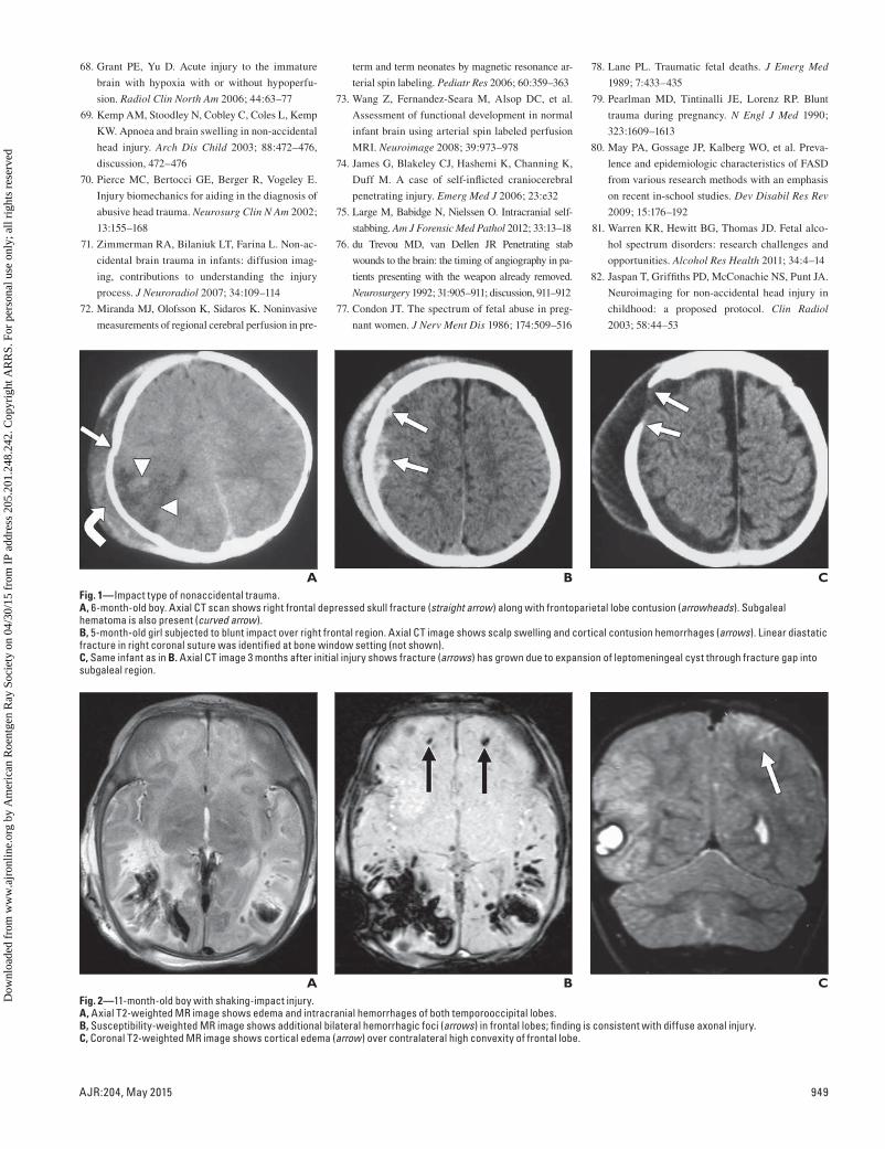
Fig. 3—CT of shaking injury.A, 4-month-old girl subjected to violent shaking insult. CT scan shows typical parasagittal hemorrhages (arrows) in both superior medial frontal lobes as result of shearing injury.B, 11-month-old boy. CT scan shows acute subdural hematomas, mainly over frontoparietal convexities and along interhemispheric fissure.
B
A
Fig. 4—MRI of shaking injury.A, 4-month-old boy with subdural hematoma due to shaking injury. Coronal T1-weighted MR image shows bilateral subdural hematomas (arrows) in cerebral convexities in different stages. Right subdural hematoma is approximately in late subacute stage, and left is in chronic stage.B, 4-month-old girl. Axial FLAIR image shows intermediate-signal-intensity chronic subdural hematoma (arrows) over high convexity of left hemisphere. Hyperintense right subdural hematoma is in subacute stage.
B
A
Fig. 5—Shaking-impact and diffuse axonal injury.A, 8-month-old boy. Axial apparent diffusion coefficient image shows one spot of diffusion restriction (arrow), which appeared otherwise normal in other sequences (not shown).B, 9-month-old girl with shaking injury. Voxel spectrum from multivoxel MR spectroscopic image (TE, 270 ms) of left frontal cortical region shows decrease in N-acetyl aspartate (NAA)-to-choline ratio compared with normal right frontal lobe spectrum (not shown) and presence of lactate peak (arrow) in frontal gray matter without abnormal signal intensity. Chronic subdural hematomas (arrowheads) are present in left cerebral convexity and along falx cerebri. Cr = creatine.
BDow
nloa
ded
from
ww
w.a
jron
line.
org
by A
mer
ican
Roe
ntge
n R
ay S
ocie
ty o
n 04
/30/
15 f
rom
IP
addr
ess
205.
201.
248.
242.
Cop
yrig
ht A
RR
S. F
or p
erso
nal u
se o
nly;
all
righ
ts r
eser
ved
AJR:204, May 2015 951
Neuroimaging of Abusive Head Trauma in Infants
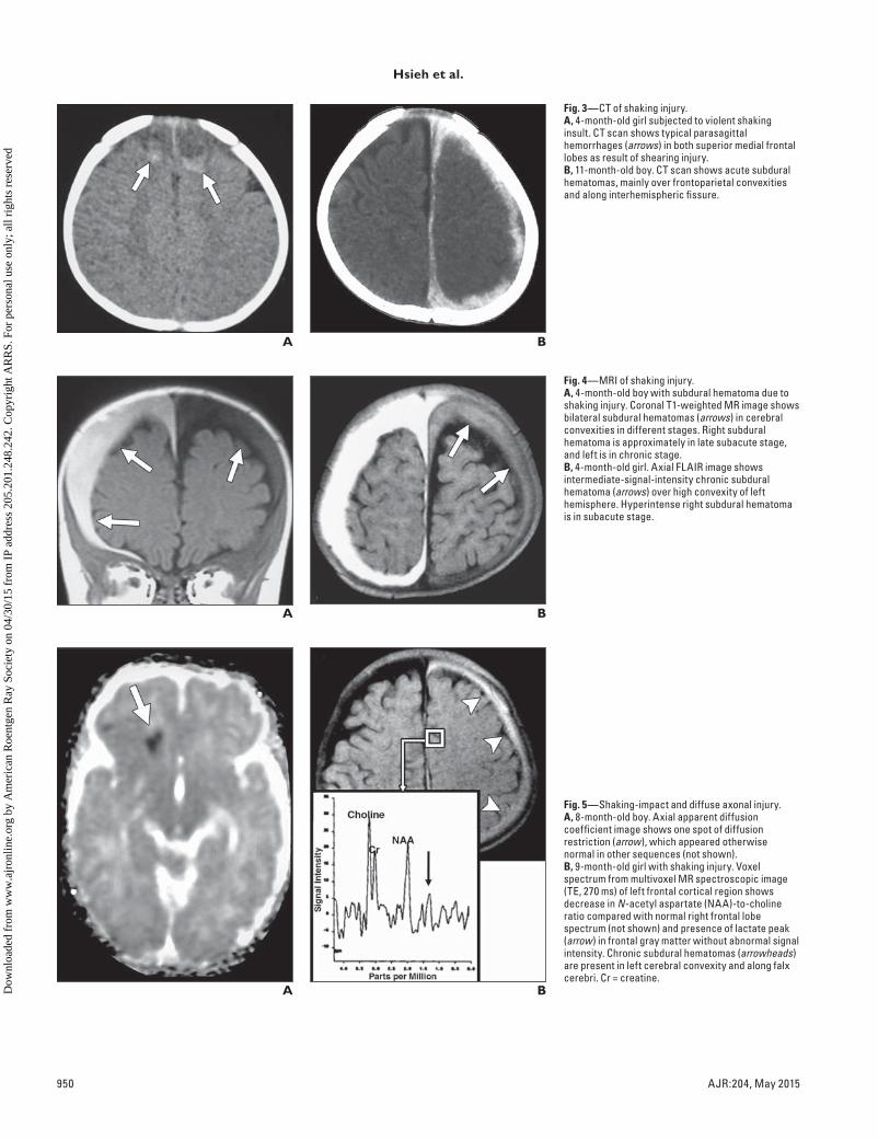
AFig. 6—3-month-old boy with craniovertebral junction injury.A, Sagittal T1-weighted MR image shows, in addition to contusions of frontal and parietooccipital lobes, subdural hematoma along clivus and craniovertebral junction (arrow). Torn apical ligament with blood (arrowhead) is evident.B, 1-year-old boy with normal ligamentous anatomy. Sagittal T1-weighted MR image shows ligamentous structures at craniovertebral junction. A = anterior atlantooccipital membrane, B = anterior longitudinal ligament, C = apical ligament, D = cruciate ligament, F = posterior longitudinal ligament, G = posterior atlantooccipital membrane.C, Illustration shows craniovertebral ligaments. A = anterior atlantooccipital membrane, B = anterior longitudinal ligament, C = apical ligament, D = cruciate ligament, E = tectorial membrane, F = posterior longitudinal ligament, G = posterior atlantooccipital membrane, H = interspinous ligament, I = dura.
CB
A
Fig. 7—Strangulation and suffocation.A, 3-month-old girl with strangulation injury. Unenhanced CT image at acute stage shows diffuse brain swelling with sulcal effacement.B, Follow-up contrast-enhanced CT image obtained 1 week after A shows subacute infarcts with bilateral luxury enhancement (arrows) in middle cerebral artery territories.C, 7-month-old girl with suffocation injury. Axial T1-weighted MR image shows diffuse laminar necrosis in deep sulcal cortexes (arrows).D, Photograph of brain specimens in boy of unknown age shows pattern of laminar necrosis in deep sulcal cortexes (arrowheads) similar to those in A–C.
CB
D
Dow
nloa
ded
from
ww
w.a
jron
line.
org
by A
mer
ican
Roe
ntge
n R
ay S
ocie
ty o
n 04
/30/
15 f
rom
IP
addr
ess
205.
201.
248.
242.
Cop
yrig
ht A
RR
S. F
or p
erso
nal u
se o
nly;
all
righ
ts r
eser
ved

952 AJR:204, May 2015
Hsieh et al.
AFig. 8—2-month-old boy with shaking and strangulation injuries.A, Axial T2-weighted MR image shows poorly defined injuries in unmyelinated brain.B, Apparent diffusion coefficient map shows bilateral cytotoxic edema over posterior medial temporal and occipital lobes (arrows).C, Susceptibility-weighted image shows subdural hematoma (arrows) and contusion hemorrhage (arrowhead).
CB
Fig. 9—6-month-old boy with stabbing injury through anterior fontanel. Axial CT scan shows retained paper clip in superior frontal interhemispheric cistern. Kidney-ureter-bladder radiograph (not shown) of mother, who had pica, showed numerous paper clips in stomach and intestines. (Reprinted with permission from Wang HS. A needle in an infant’s brain. Eur J Neurol 1998; 5:517–518. Copyright 2003 by John Wiley and Sons)
Fig. 10—12-day-old boy battered as fetus. Coronal contrast-enhanced T1-weighted MR image shows chronic bilateral subdural hematomas (arrows) in cerebral convexities.
F O R Y O U R I N F O R M A T I O N
This article is available for CME and Self-Assessment (SA-CME) credit that satisfies Part II requirements for maintenance of certification (MOC). To access the examination for this article, follow the prompts associated with the online version of the article.
Dow
nloa
ded
from
ww
w.a
jron
line.
org
by A
mer
ican
Roe
ntge
n R
ay S
ocie
ty o
n 04
/30/
15 f
rom
IP
addr
ess
205.
201.
248.
242.
Cop
yrig
ht A
RR
S. F
or p
erso
nal u
se o
nly;
all
righ
ts r
eser
ved