Hepatoxylon trichiuri (Cestoda: Trypanorhyncha) plerocercoids in ...

491
THE RAFFLES BULLETIN OF ZOOLOGY 2013
REVISION OF ANTEROPORA (CESTODA: LECANICEPHALIDEA) AND DESCRIPTIONS OF FIVE NEW SPECIES FROM STINGRAYS
(MYLIOBATIFORMES: DASYATIDAE) IN BORNEO
Kendra R. Mojica Department of Ecology and Evolutionary Biology and the Biodiversity Institute, University of Kansas
1200 Sunnyside Ave., Lawrence, Kansas, 66045, USADepartment of Ecology and Evolutionary Biology, University of Connecticut
75 N. Eagleville Road, Storrs, Connecticut, 06269–3043, USA
Kirsten Jensen Department of Ecology and Evolutionary Biology and the Biodiversity Institute, University of Kansas
1200 Sunnyside Ave., Lawrence, Kansas, 66045, USAEmail: [email protected] (Corresponding author)
Janine N. CairaDepartment of Ecology and Evolutionary Biology, University of Connecticut
75 N. Eagleville Road, Storrs, Connecticut, 06269–3043, USA
ABSTRACT. — The discovery of fi ve new species of Anteropora from dasyatid stingrays in Malaysian and Indonesian Borneo requires expansion of the concepts of the genus and family to accommodate these euapolytic (rather than hyperapolytic) forms. The fi ve species are as follows: Anteropora joannae, new species, and A. patulobothridium, new species, both from Taeniura lymma 1, A. cuba, new species, from Himantura cf. gerrardi 1, as well as A. glandapiculis, new species, and A. pumilionis, new species, both from Himantura pastinacoides 1. Unlike the apical organs of A. patulobothridium, new species, and A. pumilionis, new species, the apical organs of A. joannae, new species, A. glandapiculis, new species, and A. cuba, new species, are primarily glandular, rather than muscular. The latter is the largest of the fi ve species and possesses a spherical rather than dorso-ventrally fl attened scolex. Anteropora pumilionis, new species, is unique in its possession of lateral (as well as posterior) bothridial notches and also in possessing fewer proglottids than its four euapolytic congeners. Among the euapolytic species, A. joannae, new species, and A. glandapiculis, new species, are most similar, but differ in genital pore position. A key to the nine species of Anteropora is presented. This is the fi rst report of lecanicephalidean cestodes from the Himantura pastinacoides and Himantura gerrardi species complexes.
KEY WORDS. — Cestoda, Lecanicephalidea, Anteropora, Taeniura, Himantura, Borneo
THE RAFFLES BULLETIN OF ZOOLOGY 2013 61(2): 491–506 Date of Publication: 30 Aug.2013 © National University of Singapore
INTRODUCTION
Both genera in the lecanicephalidean family Anteroporidae Euzet, 1994, Anteropora Subhapradha, 1955 and Sesquipedalapex Jensen, Nikolov & Caira, 2011, are diagnosed as hyperapolytic; thus all fi ve species in these genera (A. indica Subhapradha, 1955, A. japonica [Yamaguti, 1934] Euzet, 1994, A. klosmamorphis Jensen, Nikolov & Caira, 2011, A. leelongi Jensen, 2005, and S. comicus Jensen, Nikolov & Caira, 2011) exhibit strobila bearing immature proglottids only. Jensen et al. (2011) distinguished the two genera primarily on the basis of the remarkably elongate scolex seen in Sesquipedalapex. Collectively, species of both genera parasitise electric rays (Torpediniformes) in the families Narcinidae and Narkidae, with only A. leelongi
described from the epaulette shark, Hemiscyllium ocellatum (Bonnaterre) (Orectolobiformes: Hemiscylliidae).
During a survey of the metazoan parasites of elasmobranchs of Borneo, fi ve lecanicephalidean species consistent in scolex morphology with Anteropora were collected from dasyatid stingrays (Myliobatiformes). Unlike the four described species of Anteropora, all fi ve of these new species are euapolytic (i.e., retain mature, but not gravid proglottids on their strobila). Two species were found parasitising Taeniura lymma 1 (sensu Naylor et al., 2012a), a host also known to be parasitised by the lecanicephalidean Aberrapex manjajiae Jensen, 2006 (see Jensen, 2006; as Taeniura lymma). The remaining three species parasitise dasyatids not reported to host lecanicephalideans. The fi ve species are described

492
Mojica et al.: Five new species of Anteropora
here as new species and the diagnoses of Anteroporidae and Anteropora are modifi ed to accommodate these euapolytic forms. In addition, a key to the nine species of Anteropora is presented.
MATERIAL AND METHODS
Naylor et al. (2012a) found substantial undescribed novelty within each of the nominal species Taeniura lymma (Forsskål), Himantura pastinacoides (Bleeker), and Himantura gerrardi (Gray). For precision in host identifi cations, we have followed their taxon designations for stingrays from which the cestodes described here were collected. Eleven specimens of Taeniura lymma 1, nine of Himantura pastinacoides 1, and one of Himantura cf. gerrardi 1 were examined and found to be parasitised by new species of Anteropora (detailed host and collection data are provided in Table 1). These stingrays were collected either using hand spears or by local fi sherman using gill nets or small trawls. Images of the stingrays are available via the Global Cestode Database (http://elasmobranchs.tapewormdb.uconn.edu) by searching the host specimen numbers provided in Table 1.
In the fi eld, the body cavity of each host specimen was opened by a ventral longitudinal incision from the anus to the pericardial chamber, and the spiral intestine removed and opened with a longitudinal incision. Cestodes encountered were immediately removed and fixed in 10% formalin buffered with seawater and later transferred to 70% ethanol for storage. In each case, the spiral intestine and its contents were fi xed in 10% formalin buffered with seawater and subsequently transferred to 70% ethanol for storage. A small piece of liver tissue from each stingray specimen was preserved in 95% ethanol for later molecular identity confi rmation (see Naylor et al., 2012a).
At the University of Kansas, the spiral intestines were examined for cestodes using a dissecting microscope. All cestode specimens were sorted to order and lecanicephalideans further sorted to genus. Specimens of Anteropora were prepared as whole mounts for examination with light microscopy as follows. They were hydrated in distilled water, stained with Delafi eld’s hematoxylin, differentiated in tap water, destained in 70% acid ethanol, alkalinised in 70% basic ethanol, dehydrated in a graded ethanol series, then cleared in methyl salicylate and mounted in Canada balsam on glass slides.
Whole worms and scoleces prepared for scanning electron microscopy (SEM) were hydrated in a graded ethanol series, post-fi xed in 1% osmium tetroxide overnight, washed in distilled water, dehydrated in a graded ethanol series, transferred to hexamethyldisilizane (HMDS) for 20 minutes, air-dried and mounted on aluminum stubs on double-sided adhesive carbon tape. Specimens were sputter coated with c. 35 nm of gold and examined with a Zeiss LEO 1550 fi eld emission scanning electron microscope at the Microscopy and Analytical Imaging Laboratory, University of Kansas, Lawrence, Kansas, USA.
For histological sections, selected proglottids were dehydrated in a graded ethanol series, cleared in xylene and embedded in paraffi n according to conventional techniques. Serial sections were cut at 7-μm intervals using a TBS OLYMPUS CUT 4060 microtome, attached to glass slides by fl oating sections on 3% sodium silicate solution and air-drying. Sections were subsequently stained with Delafi eld’s hematoxylin, counterstained with eosin, differentiated in Scott’s solution, dehydrated in a graded ethanol series, cleared in xylene and mounted in Canada balsam.
Line drawings were made using a drawing tube attached to a Zeiss Axioskop 2 plus. Light microscope images of whole mounts and histological sections were taken using a Leica FireCam DFC 320 or DFC 480, scale bars were added using ImageJ 1.36b. Measurements were taken with a Leica Firecam DFC 320 digital camera mounted on a Zeiss Axioscop 2 Plus and the image analysis program Openlab Demo Version 4.0.4. Reproductive organs were measured in terminal mature proglottids only. Measurements are given in micrometers (μm) unless otherwise indicated, and are provided as the range followed in parentheses by the mean, standard deviation, number of worms examined, and the total number of measurements if more than one measurement was taken per worm. Microthrix terminology follows Chervy (2009). Elasmobranch classifi cation follows Naylor et al. (2012b).
Museum abbreviations used are as follows: LRP, Lawrence R. Penner Parasitology Collection, Department of Ecology and Evolutionary Biology, University of Connecticut, Storrs, Connecticut, USA; MZUM(P), Parasite Collection, Muzium Zoologi, Universiti Malaya, Kuala Lumpur, Malaysia; MZB, Museum Zoologicum Bogoriense, Center for Biology, Indonesian Institute of Science, Cibinong, Jakarta-Bogor, Java, Indonesia; SBC, Sarawak Biodiversity Center, Kuching, Sarawak, Malaysia; USNPC, United States National Parasite Collection, Beltsville, Maryland, USA; ZRC, Zoological Reference Collection, Raffles Museum of Biodiversity Research, National University of Singapore, Singapore.
TAXONOMY
Anteropora joannae, new species(Figs. 1A–C, 2A–E, 8A, C, D)
Type and only known host. — Taeniura lymma 1 (sensu Naylor et al., 2012a) (Myliobatiformes: Dasyatidae)
Site of infection. — Spiral intestine
Holotype. — MZUM(P) 2013.7(H) ex Taeniura lymma 1 (sensu Naylor et al., 2012a) (host no. BO-127), MALAYSIA: Pulau [=Island] Mabul (04°14'N, 118°38'E), Sabah, Celebes Sea, 5 May 2003, coll. J. N. Caira & K. Jensen.
Paratypes. — Ex Taeniura lymma 1 (sensu Naylor et al., 2012a), MALAYSIA: Semporna (04°28'N, 118°37'E), Sabah, Celebes Sea, 27 Jun.2002 (host no. BO-86) and Pulau

493
THE RAFFLES BULLETIN OF ZOOLOGY 2013
Table 1. Host specimen collection data. Host specimen numbers in bold indicate specimens included in Naylor et al. (2012a).
Host Sex DW Host specimen Collection Localityspecies (in cm) no.* date Taeniura lymma 1 1 female 21 BO-86 27 Jun.2002 Semporna (04°28'N, 118°37'E), Sabah, Celebes Sea, Malaysia 4 females, 2 males 19–24 BO-122, BO-125, 5 May 2003 Pulau [=Island] Mabul (04°14'N, 118°38'E), BO-127, BO-128, Sabah, Celebes Sea, Malaysia BO-130 BO-131 1 male 20 KA-417 24 Jul.2008 Tanjung Batu (02°16'N, 118°06'E), East Kalimantan, Sulawesi Sea, Indonesia 1 female, 27–30 KA-418, KA-419, 25 Jul.2008 Pulau [=Island] Rabu Rabu (02°19'N, 118°07'E), 2 males KA-420 East Kalimantan, Sulawesi Sea, Indonesia
Himantura pastinacoides 1 2 females 20–42 BO-12, BO-168 1 Jun.2002 – Sematan (01°48'N, 109°46'E), Sarawak, 14 May 2003 South China Sea, Malaysia 1 male 46 BO-61 12 Jun.2002 Mukah (02°54'N, 112°05'E), Sarawak, South China Sea, Malaysia 3 females, 51–59 BO-76, BO-98, 21 Jun.2002 – Kampung [=Village] Tetabuan (06°01'N, 117°42'E), 1 male BO-100, BO-116 3 May 2003 Sabah, Sulu Sea, Malaysia
1 female 23 KA-105 4 Dec.2006 Kalapseban (03°14'S, 112°55'E), Central Kalimantan, Java Sea, Indonesia 1 female 57 KA-421 29 Jul.2008 Manggar (01°13'S, 116°58'E), East Kalimantan, Makassar Strait, Indonesia
Himantura cf. gerrardi 1 1 male 50 BO-400 19 Apr.2004 Kuching (02°00'N, 110°38'E), Sarawak, South China Sea, Malaysia
*See http://elasmobranchs.tapewormdb.uconn.edu for host specimen details.
[=Island] Mabul (04°14'N, 118°38'E), Sabah, Celebes Sea, 5 May 2003 (host nos. BO-122, BO-125, BO-127, BO-130, BO-131), INDONESIA: Tanjung Batu (02°16'N, 118°06'E), East Kalimantan, Sulawesi Sea, Pulau [=Island] Rabu-Rabu (02°19'N, 118°07'E), East Kalimantan, Celebes Sea, 25 Jul.2008 (host nos. KA-418, KA-419, KA-420), coll. J. N. Caira & K. Jensen. MZUM(P) 2013.8(P)–11(P) (4 whole mounts) (host nos. BO-86, BO-122, BO-127); MZBCa 178, 179 (2 whole mounts) (host no. KA-418); ZRC.PAR. 25, 26 (2 whole mounts) (host nos. BO-130, KA-419); USNPC 10625–10630 (6 whole mounts and proglottid cross-section series) (host nos. BO-86, BO-127, BO-131, KA-418, KA-419, KA-420); LRP 7974–7981 (5 whole mounts, and proglottid cross-section series and voucher) (host nos. BO-125, BO-127, BO-130). Three whole worms (host nos. BO-125, BO-131) prepared for SEM retained by K. Jensen at the University of Kansas.
Etymology. — This species is named in honor of Joanna Cielocha for her support of the senior author throughout this project.
Description. — Based on 25 specimens: 20 whole mounts of mature worms, two cross-section series of mature proglottids, and three whole worms prepared for SEM.
Worms 986–2,657 (1,541 ± 391; 20) long; maximum width at scolex, mid-strobila, or terminal proglottid, euapolytic; proglottids 10–25 (18 ± 6; 20) in number. Scolex 142–217 (177 ± 23; 20) long by 123–190 (150 ± 20; 15) wide, consisting of four acetabula, apical modifi cation of scolex proper and apical organ; cephalic peduncle absent. Acetabula bothridiate in form, elongate oval in shape, with posterior notch at midline, 105–178 (143 ± 20; 19; 37) long by 57–110 (80 ± 14; 16; 30) wide. Apical modifi cation of scolex proper conspicuously dome-shaped, with aperture at center, housing apical organ. Apical organ (Fig. 8A) primarily glandular, weakly muscular, conical in form, 38–58 (49 ± 5; 20) long by 33–63 (47 ± 6; 20) wide, non-protrusible.
Apical modifi cation of scolex proper covered with hastate spinitriches and acicular fi litriches (Fig. 2B); scolex proper at base of apical modifi cation with acicular fi litriches only (Fig. 2C). Distal (Fig. 2D) and proximal (Fig. 2E) bothridial surfaces covered with trullate spinitriches and acicular fi litriches. Proglottids covered with capilliform fi litriches throughout, also with small hastate spinitriches on anterior margins and with small scolopate spinitriches along posterior proglottid margins (Fig. 2F).
Proglottids craspedote, non-laciniate. Immature proglottids 9–23 (16 ± 5; 20) in number, initially wider than long,
494
Mojica et al.: Five new species of Anteropora
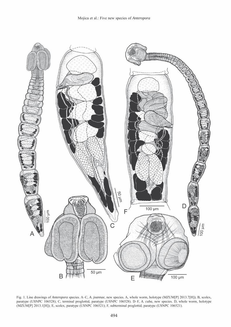
Fig. 1. Line drawings of Anteropora species. A–C, A. joannae, new species. A, whole worm, holotype (MZUM[P] 2013.7[H]); B, scolex, paratype (USNPC 106528); C, terminal proglottid, paratype (USNPC 106528). D–F, A. cuba, new species. D, whole worm, holotype (MZUM[P] 2013.1[H]); E, scolex, paratype (USNPC 106521); F, subterminal proglottid, paratype (USNPC 106521).

495
THE RAFFLES BULLETIN OF ZOOLOGY 2013
Fig. 2. Scanning electron micrographs of Anteropora species. A–F, A. joannae, new species. A, scolex (small letters indicate location of details in B–F); B, surface of apical modifi cation of scolex proper; C, surface of scolex proper at base of apical modifi cation; D, distal bothridial surface; E, proximal bothridial surface; F, surface of posterior margin of proglottid. G–K, A. cuba, new species. G, scolex (small letters indicate location of details in H–K); H, surfaces of apical modifi cation of scolex proper; I, distal bothridial surface; J, proximal bothridial surface; K, surface of posterior margin of proglottid.
becoming longer than wide with maturity. Mature proglottids 1–3 in number; subterminal proglottid 162–454 (243 ± 71; 20) long by 60–181 (127 ± 32; 20) wide; terminal proglottid 293–543 (395 ± 69; 20) long by 105–170 (135 ± 21; 20) wide. Testes invariably four in number, arranged in single column, 21–62 (40 ± 9; 20; 60) long by 49–120 (78 ± 18; 20; 60) wide, extending from anterior margin of proglottid to slightly overlap anterior margin of ovary. Vasa efferentia not observed. Vas deferens in fully mature proglottids enlarged
to form external seminal vesicle, extending along lateral margin of proglottid from ootype region to anterior margin of cirrus-sac. Internal seminal vesicle not observed. Cirrus-sac pyriform, positioned between anterior-most two testes, slightly angled anteriorly, 64–117 (93 ± 15; 20) long by 19–40 (29 ± 6; 19) wide, containing coiled cirrus. Cirrus armed with spinitriches. Ovary smooth, H-shaped in frontal view, tetralobed in cross-section (Fig. 8D), symmetrical, 68–134 (95 ± 19; 20) long by 57–191 (89 ± 28; 19) wide; ovarian

496
Mojica et al.: Five new species of Anteropora
bridge at middle of ovary. Mehlis’ gland posterior to ovarian bridge. Vagina extending along lateral margin of proglottid from ootype region to genital atrium, opening into genital atrium posterior to cirrus-sac. Genital pores lateral, irregularly alternating, 71–81% (77 ± 3; 20) of proglottid length from posterior end. Uterus saccate, extending essentially along midline of proglottid from level of ovarian bridge to posterior margin of anterior-most testis. Vitellarium follicular; vitelline follicles 13–32 (23 ± 5; 19; 57) long by 13–39 (24 ± 7; 19; 57) wide, in two lateral fi elds; each fi eld consisting of two columns (Fig. 8C), extending from posterior margin of anterior-most testis on aporal side and from posterior margin of cirrus-sac on poral side to posterior margin of proglottid, interrupted by ovary. Two pairs of excretory vessels. Eggs not observed.
Remarks. — This species is unlike A. indica, A. japonica, A. klosmamorphis and A. leelongi in that it is euapolytic rather than hyperapolytic. Anteropora joannae, new species, differs further from A. indica in that its ovary is tetralobed in cross-section, rather than consisting of two to three lobes on each side (see Subhapradha, 1955, fi g. 5). Whereas the apical organ of the new species is primarily glandular, that of A. japonica is muscular. Furthermore, whereas both, A. japonica and A. leelongi possess six testes and bothridia that are essentially round with intact margins, A. joannae, new species, has four testes and bothridia that are elongate-oval with a posterior notch at the midline. The scolex of A. joannae, new species, is most similar to that of A. klosmamorphis, however the strobila of the new species has many fewer proglottids (10–25 vs 87–274).
This new species appears to be more variable in total length, number of proglottids and width of the subterminal proglottids, than seen in any of its congeners. However, this is considered to represent intraspecifi c variation at this time.
Anteropora cuba, new species(Figs. 1D–F, 2G–K)
Type and only host. — Himantura cf. gerrardi 1 (sensu Naylor et al., 2012a) (Myliobatiformes: Dasyatidae)
Site of infection. — Spiral intestine
Holotype. — MZUM(P) 2013.1(H) ex Himantura cf. gerrardi 1 (sensu Naylor et al., 2012a) (host no. BO-400), MALAYSIA: ~32 km off Kuching (02°00'N, 110°38'E), Sarawak, South China Sea, 9 Apr.2004, coll. J. N. Caira & K. Jensen.
Paratypes. — Ex Himantura cf. gerrardi 1 (sensu Naylor et al., 2012a) (host no. BO-400), same as holotype. MZUM(P) 2013.2(P), 3(P) (2 whole mounts); SBC-P-00060 (1 whole mount); ZRC.PAR. 22 (1 whole mount); USNPC 106521 (4 whole mounts) and LRP 7968, 7969 (2 whole mounts). Two scoleces prepared for SEM retained by K. Jensen at the University of Kansas.
Etymology. — Derived from cubus (L.), referring to the shape of the scolex.
Description. — Based on 13 specimens: 11 whole mounts of mature worms and two scoleces prepared for SEM.
Worms 2,925–6,195 (4,139 ± 1,108; 11) long; maximum width at scolex, euapolytic; proglottids 43–82 (60 ± 11; 11) in number. Scolex 205–295 (250 ± 30; 11) long by 270–392 (327 ± 37; 10) wide, more or less spherical, consisting of four acetabula, apical modifi cation of scolex proper and apical organ; cephalic peduncle absent. Acetabula bothridiate in form, oval to rectangular in shape, with slight posterior indentation at midline, 150–231 (184 ± 21; 11; 22) long by 153–217 (176 ± 18; 10; 20) wide. Apical modifi cation of scolex proper dome-shaped, with aperture at center, housing apical organ. Apical organ primarily glandular, weakly muscular, spherical to conical in form, 88–146 (110 ± 15; 11) long by 82–144 (100 ± 17; 11) wide, non-protrusible.
Apical modifi cation of scolex proper covered with hastate spinitriches and acicular fi litriches (Fig. 2H). Distal (Fig. 2I) and proximal (Fig. 2J) surfaces of bothridia covered with trullate spinitriches and acicular fi litriches. Proglottids with capilliform fi litriches throughout (Fig. 2K), also with small scolopate spinitriches along posterior proglottid margins.
Proglottids craspedote, non-laciniate. Immature proglottids 38–76 (54 ± 10; 11) in number, initially wider than long, becoming longer than wide with maturity. Mature proglottids 3–8 in number; subterminal proglottid 326–534 (433 ± 70; 11) long by 122–189 (163 ± 25; 11) wide; terminal proglottid 468–720 (584 ± 75; 11) long by 125–197 (164 ± 26; 11) wide. Testes invariably four in number, arranged in single column, 31–66 (46 ± 8; 11; 33) long by 50–99 (73 ± 14; 11; 33) wide, extending from anterior margin of proglottid to slightly overlap anterior margin of ovary. Vasa efferentia not observed. Vas deferens in fully mature proglottids enlarged to form extensive external seminal vesicle, extending from ootype region to posterior margin of anterior-most testis. Internal seminal vesicle not observed. Cirrus-sac pyriform, lateral to second testis, slightly angled anteriorly, 44–67 (57 ± 7; 11) long by 68–140 (107 ± 22; 11) wide, containing coiled cirrus. Cirrus armed with spinitriches. Ovary smooth, H-shaped in frontal view, tetralobed in cross-section, symmetrical, 108–180 (143 ± 23; 11) long by 74–144 (110 ± 21; 11) wide; ovarian bridge at middle of ovary. Mehlis’ gland posterior to ovarian bridge. Vagina extending along lateral margin of proglottid from ootype region to genital atrium, opening into genital atrium posterior to cirrus-sac. Genital pores lateral, irregularly alternating, 64–73% (68 ± 3; 11) of proglottid length from posterior end. Uterus saccate, extending along midline of proglottid from ovarian bridge to posterior margin of anterior-most testis. Vitellarium follicular; vitelline follicles 22–66 (41 ± 13; 11; 33) long by 23–64 (37 ± 10; 11; 33) wide, in two lateral fi elds; each fi eld consisting of two columns, extending from posterior margin of anterior-most testis on aporal side and from posterior margin of cirrus-sac on poral side to posterior margin of proglottid, partially interrupted by ovary. Two pairs of excretory vessels. Eggs not observed.

497
THE RAFFLES BULLETIN OF ZOOLOGY 2013
Remarks. — Unlike A. indica, A. japonica, A. klosmamorphis and A. leelongi, A. cuba, new species, is euapolytic rather than hyperapolytic. In addition, A. cuba, new species, has fewer testes than A. japonica and A. leelongi (four vs six), and a larger glandular apical organ than A. klosmamorphis (88–146 long by 82–144 wide vs 53–67 long by 51–68 wide). Furthermore, A. cuba, new species, possesses a vas deferens that is expanded to form an extensive external seminal vesicle while the vas deferens of A. indica is minimal. Like Anteropora joannae, A. cuba, new species, is euapolytic and possesses a glandular apical organ, but it differs conspicuously from A. joannae in its greater total length (2,925–6,195 vs 986–2,657), greater number of proglottids (43–82 vs 10–25) and unlike A. joannae, the vas deferens of A. cuba, new species, is expanded to form an extensive external seminal vesicle (Fig. 1F). Furthermore, the scolex of A. cuba, new species, is spherical and its bothridia are oval to rectangular in shape with only a slight posterior indentation at midline, rather than oval and clearly posteriorly notched.
Anteropora glandapiculis, new species(Figs. 3A–C, 4)
Type and only host. — Himantura pastinacoides 1 (sensu Naylor et al., 2012a) (Myliobatiformes: Dasyatidae)
Site of infection. — Spiral intestine
Holotype . — MZUM(P) 2013.4(H) ex Himantura pastinacoides 1 (sensu Naylor et al., 2012a) (host no. BO-98), MALAYSIA: off Kampung [=Village] Tetabuan (06°01'N, 117°42'E), Sabah, Sulu Sea, 28 Apr.2003, coll. J. N. Caira & K. Jensen.
Paratypes. — Ex Himantura pastinacoides 1 (sensu Naylor et al., 2012a), MALAYSIA: Sematan (01°48'N, 109°46'E), Sarawak, South China Sea, 1 Jun.2002 (host no. BO-12) and 14 May 2003 (host no. BO-168), and Kampung [=Village] Tetabuan (06°01'N, 117°42'E), Sabah, Sulu Sea, 21 Jun.2002 (host nos. BO-76) and 3 May 2003 (host no. BO-116), INDONESIA: Kalapseban (03°14'S, 112°55'E), Central Kalimantan, Java Sea, 4 Dec.2006 (host no. KA-105) and Manggar (01°13'S, 116°58'E), East Kalimantan, Makassar Strait, 29 Jul.2008 (host no. KA-421), coll. J. N. Caira & K. Jensen. MZUM(P) 2013.5(P), 6(P) (2 whole mounts) (host nos. BO-116, KA-105); SBC-P-00061, 00062 (2 whole mounts) (host no. BO-168); MZBCa 176, 177 (2 whole mounts) (host no. KA-105); ZRC.PAR. 23, 24 (2 whole mounts) (host nos. BO-12, KA-421); USNPC 106522–106524 (5 whole mounts) (host nos. BO-12, KA-105, KA-421); LRP 7970–7973 (4 whole mounts) (host nos. BO-12, BO-76, BO-168). Four specimens (host no. BO-168) prepared for SEM retained by K. Jensen at the University of Kansas.
Etymology. — Derived from glans (L., acorn-shaped) and apiculus (L. diminutive, point) referring to the glandular nature of the prominent apical organ.
Description. — Based on 22 specimens: 18 whole mounts of mature worms, four specimens prepared for SEM.
Worms 553–1,180 (830 ± 170; 18) long; maximum width at scolex, euapolytic; proglottids 7–14 (10 ± 2; 18) in number. Scolex 123–187 (152 ± 17; 18) long by 164–237 (189 ± 23; 9) wide, consisting of four acetabula, apical modifi caton of scolex proper and apical organ; cephalic peduncle absent. Acetabula bothridiate in form, elongate oval in shape, with posterior notch at midline, 114–206 (158 ± 22; 18; 34) long by 74–118 (93 ± 12; 14; 24) wide. Apical modifi cation of scolex proper dome-shaped, with aperture at center, housing apical organ. Apical organ primarily glandular, weakly muscular, spherical to conical in form, 28–73 (54 ± 10; 18) long by 47–82 (56 ± 9; 18) wide, non-protrusible.
Apical modifi cation of scolex proper (Fig. 4C) and scolex proper at base of apical modifi cation (Fig. 4D) covered with hastate spinitriches and acicular fi litriches. Distal (Fig. 4E) and proximal (Fig. 4F) surfaces of bothridia covered with trullate spinitriches and acicular fi litriches. Proglottids with capilliform fi litriches throughout, also with small hastate spinitriches along anterior margins and with small scolopate spinitriches along posterior proglottid margins (Fig. 4G).
Proglottids craspedote, non-laciniate. Immature proglottids 6–12 (8 ± 2; 18) in number, initially wider than long, becoming longer than wide with maturity. Mature proglottids 1–2 in number; subterminal proglottid 84–201 (141 ± 40; 18) long by 83–150 (111 ± 19; 18) wide; terminal proglottid 235–456 (349 ± 56; 18) long by 107–165 (128 ± 14; 18) wide. Testes invariably four in number, arranged in single column, 32–62 (44 ± 6; 20; 53) long by 62–104 (83 ± 10; 20; 52) wide, extending from anterior margin of proglottid to slightly overlap anterior margin of ovary. Vasa efferentia not observed. Vas deferens in fully mature proglottids enlarged to form external seminal vesicle, extending along lateral margin of proglottid from ootype region to anterior margin of cirrus-sac. Internal seminal vesicle not observed. Cirrus-sac pyriform, at level of second testis, slightly angled anteriorly, 54–96 (79 ± 14; 13) long by 36–67 (48 ± 8; 18) wide, containing coiled cirrus. Cirrus armed with spinitriches. Ovary smooth, H-shaped in frontal view, tetralobed in cross-section, symmetrical, 36–99 (70 ± 17; 18) long by 63–110 (82 ± 12; 17) wide; ovarian bridge at middle of ovary. Mehlis’ gland posterior to ovarian bridge. Vagina extending along lateral margin of proglottid from ootype region to genital atrium, opening into genital atrium posterior to cirrus-sac. Genital pores lateral, irregularly alternating, 58–70% (65 ± 3; 16) of proglottid length from posterior end. Uterus saccate, extending essentially along midline of proglottid from ovarian bridge to posterior margin of anterior-most testis. Vitellarium follicular; vitelline follicles 7–33 (17 ± 5; 15; 42) long by 12–43 (27 ± 8; 14; 39) wide, in two lateral fi elds; each fi eld consisting of two columns, extending from posterior margin of anterior-most testis on aporal side and from posterior margin of cirrus-sac on poral side to posterior margin of proglottid, partially interrupted by ovary. Two pairs of excretory vessels. Eggs not observed.

498
Mojica et al.: Five new species of Anteropora
Fig. 3. Line drawings of Anteropora species. A–C, A. glandapiculis, new species. A, whole worm, paratype (USNPC 106524); B, scolex, paratype (USNPC 106522); C, terminal proglottid, holotype (MZUM[P] 2013.4[H]). D–F, A. patulobothridium, new species. D, whole worm, holotype (MZBCa 181); E, scolex, paratype (USNPC 106531); F, terminal proglottid, paratype (USNPC 106532).

499
THE RAFFLES BULLETIN OF ZOOLOGY 2013
Remarks. — The euapolytic nature of A. glandapiculis, new species, clearly distinguishes it from A. indica, A. japonica, A. klosmamorphis and A. leelongi. In addition, Anteropora glandapiculis, new species, has fewer testes than A. japonica and A. leelongi (four vs six), and many fewer proglottids than A. klosmamorphis (7–14 vs 87–274). It further differs from A. indica in that its genital pore is positioned between the second and third testis from the anterior end of the proglottid rather than between the fi rst and second testis. With respect to its euapolytic congeners, A. glandapiculis, new species, is a shorter worm (553–1,180 vs 2,925–6,195), possesses many fewer proglottids overall (7–14 vs 43–82) and also fewer mature proglottids (1–2 vs 3–8) than A. cuba. With respect to A. joannae, A. glandapiculis, new species, possesses a more posterior genital pore (58–70% vs 71–81% of proglottid length from posterior end) and the cirrus-sac is positioned at the level of the second testis, rather than between the fi rst and second testis as seen in A. joannae.
Anteropora patulobothridium, new species(Figs. 3D–F, 5)
Type and only known . — Taeniura lymma 1 (sensu Naylor et al., 2012a) (Myliobatiformes: Dasyatidae)
Site of infection. — Spiral intestine
Fig. 4. Scanning electron micrographs of Anteropora glandapiculis, new species. A, whole worm; B, scolex (small letters indicate location of details in C–G); C, surface of apical modifi cation of scolex proper; D, surface of scolex proper at base of apical modifi cation; E, distal bothridial surface; F, proximal bothridial surface; G, surfaces at boundary between adjacent proglottids.
Holotype. — MZBCa 181 ex Taeniura lymma 1 (sensu Naylor et al., 2012a) (host no. KA-419), INDONESIA: off Pulau [=Island] Rabu-Rabu (02°19'N, 118°07'E), East Kalimantan, Celebes Sea, 25 Jul.2008, coll. J. N. Caira & K. Jensen.
Paratypes. — Ex Taeniura lymma 1 (sensu Naylor et al., 2012a), MALAYSIA: Pulau [=Island] Mabul (04°14'N, 118°38'E), Sabah, Celebes Sea, 5 May 2003 (host nos. BO-128, BO-131), INDONESIA: Tanjung Batu (02°16'N, 118°06'E), East Kalimantan, Celebes Sea, 24 Jul.2008 (host no. KA-417) and Pulau [=Island] Rabu-Rabu (02°19'N, 118°07'E), East Kalimantan, Celebes Sea, 25 Jul.2008 (host nos. KA-418, KA-419, KA-420), coll. J. N. Caira & K. Jensen. MZBCa 180, 182 (2 whole mounts) (host no. KA-419); MZUM(P) 2013.12(P), 13(P) (2 whole mounts) (host nos. KA-418, KA-420); ZRC.PAR. 27, 28 (2 whole mounts) (host nos. KA-417, KA-419); USNPC 106531, 106532 (6 whole mounts) (host nos. KA-418, KA-419); LRP 7982–7986 (5 whole mounts) (host nos. KA-417, KA-418, KA-419). Two whole worms (host nos. BO-128, BO-131) prepared for SEM retained by K. Jensen at the University of Kansas.
Etymology. — Derived from patulus (L., open, spread out, broad) referring to the ability of this species to extend its scolex laterally.
Description. — Based on 20 specimens: 18 whole mounts of mature worms and two whole worms prepared for SEM.

500
Mojica et al.: Five new species of Anteropora
Fig. 5. Scanning electron micrographs of Anteropora patulobothridium, new species. A, scolex (small letters indicate location of details in E, F, H); B, scolex with scolex proper laterally extended; C, apical modifi cation of scolex proper (small letters indicate location of details in D, G); D, surface of raised rim of apical modifi cation of scolex proper; E, distal bothridial surface; F, proximal bothridial surface; G, surface of apical modifi cation of scolex proper; H, surface of scolex proper posterior to bothridia; I, surface of posterior margin of proglottid.
Worms 583–1,129 (859 ± 138; 18) long; maximum width at scolex, euapolytic; proglottids 8–11 (10 ± 1; 18) in number. Scolex 84–187 (152 ± 22; 18) long by 119–317 (190 ± 59; 16) wide, consisting of four acetabula, apical modifi cation of scolex proper, apical organ, and conspicuous region of scolex proper posterior to acetabula; cephalic peduncle absent. Acetabula bothridiate in form, triangular to rectangular in shape, lacking posterior notch at midline in some, 65–125 (99 ± 17; 16; 32) long by 58–102 (83 ± 11; 15; 30) wide. Apical modifi cation of scolex proper variable, dome-shaped to conical, with raised rim, apparently lacking aperture at center, housing apical organ. Apical organ primarily muscular, with gland cells at base, inverted campanulate in form, 19–33 (27 ± 3; 18) long by 22–33 (27 ± 3; 18) wide, non-protrusible.
Apical modifi cation of scolex proper (Fig. 5C) covered with hastate spinitriches and capilliform fi litriches (Fig. 5G), with conspicuous apical rim bearing hamulate spinitriches and capilliform fi litriches (Fig. 5D). Distal (Fig. 5E) and proximal (Fig. 5F) surfaces of bothridia covered with gladiate spinitriches and acicular fi litriches. Scolex proper posterior to bothridia covered with hastate spinitriches and acicular fi litriches (Fig. 5H). Proglottids covered with capilliform fi litriches throughout, also with small scolopate spinitriches along posterior proglottid margins (Fig. 5I).
Proglottids craspedote, non-laciniate. Immature proglottids 6–10 (8 ± 1; 18) in number, initially wider than long, becoming longer than wide with maturity. Mature proglottids

501
THE RAFFLES BULLETIN OF ZOOLOGY 2013
1–2 in number; subterminal proglottid 95–232 (162 ± 42; 18) long by 62–105 (87 ± 13; 18) wide; terminal proglottid 253–470 (341 ± 50; 18) long by 80–142 (101 ± 14; 18) wide. Testes invariably four in number, arranged in single column, 28–55 (38 ± 6; 18; 54) long by 45–84 (65 ± 9; 18; 54) wide, extending from anterior margin of proglottid to slightly overlap anterior margin of ovary. Vasa efferentia not observed. Vas deferens in fully mature proglottids enlarged to form external seminal vesicle, extending along lateral margin of proglottid from ootype region to anterior margin of cirrus-sac. Internal seminal vesicle not observed. Cirrus-sac pyriform, extending between fi rst and second testis, slightly angled anteriorly, 41–70 (57 ± 8; 18) long by 16–34 (24 ± 4; 18) wide, containing coiled cirrus. Cirrus armed with spinitriches. Ovary smooth, H-shaped in frontal view, tetralobed in cross-section, symmetrical, 46–105 (83 ± 14; 18) long by 48–89 (68 ± 10; 18) wide; ovarian bridge at middle of ovary. Mehlis’ gland present posterior to ovarian bridge. Vagina extending along lateral margin of proglottid from ootype region to genital atrium, opening into genital atrium posterior to cirrus-sac. Genital pores lateral, irregularly alternating, 69–79% (76 ± 3; 18) of proglottid length from posterior end. Uterus saccate, extending essentially along midline of proglottid from ovarian bridge to posterior margin of anterior-most testis. Vitellarium follicular; vitelline follicles 11–40 (20 ± 7; 18; 54) long by 9–36 (18 ± 5; 18; 54) wide, in two lateral fi elds; each fi eld consisting of two columns, extending from posterior margin of anterior-most testis on aporal side and from posterior margin of cirrus-sac on poral side to posterior margin of proglottid, interrupted by ovary. Two pairs of excretory vessels. Eggs not observed.
Remarks. — This species differs from A. indica, A. japonica, A. klosmamorphis and A. leelongi in that it is euapolytic rather than hyperapolytic. Moreover, it differs from A. klosmamorphis and A. leelongi in that its apical organ is primarily muscular, rather than glandular. It differs further from A. japonica in its possession of four rather than six testes and from A. indica in its possession of an ovary that is tetralobed in cross-section, rather than consisting of two to three lobes on each side. With respect to its euapolytic congeners, A. patulobothridium, new species, conspicuously differs from A. joannae, A. cuba and A. glandapiculis in that its apical organ is primarily muscular and associated with an apical modifi cation of the scolex proper with a raised rim that bears hamulate spinitriches, rather than an apical organ that is primarily glandular and lacking such an apical modifi cation of the scolex proper. In addition, unlike its euapolytic congeners, A. patulobothridium, new species, possesses an elongated region of the scolex proper posterior to the bothridia that bears hastate spinitriches. In addition, A. patulobothridium, new species, possesses fewer proglottids than A. joannae (8–11 vs 10–25) and a narrower cirrus-sac (16–34 vs 36–67) than A. glandapiculis; it is shorter (583–1,129 vs 2,925–6,195 in total length), possesses conspicuously fewer proglottids overall (8–11 vs 43–82) and also fewer mature proglottids (1–2 vs 3–8) than A. cuba.
The scolex of A. patulobothridium, new species, varied substantially in form depending on its degree of contraction.
In some specimens, the scolex was extremely wide and the bothridia in a pair were separated from one another by a distance of greater than the width of a bothridium (e.g., Figs. 3E and 5B); in other specimens, the bothridia in a pair were adjacent to one another (e.g., Figs. 3D and 5A). Presumably, this morphological fl exibility refl ects this worm’s ability to alter the form of its scolex in order to lodge its bothridia between the rows of adjacent villi of the spiral intestine of its host.
Anteropora pumilionis, new species(Figs. 6, 7, 8B)
Type and only host. — Himantura pastinacoides 1 (sensu Naylor et al., 2012a) (Myliobatiformes: Dasyatidae)
Site of infection. — Spiral intestine
Holotype. — MZUM(P) 2013.14(H) ex Himantura pastinacoides 1 (sensu Naylor et al., 2012a) (host no. BO-61), MALAYSIA: Mukah (02°54'N, 112°05'E), Sarawak, South China Sea, 12 Jun.2002, coll. J. N. Caira & K. Jensen.
Paratypes. — Ex Himantura pastinacoides 1 (sensu Naylor et al., 2012a), MALAYSIA: Mukah (02°54'N, 112°05'E), Sarawak, South China Sea, 12 Jun.2002 (host nos. BO-61, BO-168), and Kampung [=Village] Tetabuan (06°01'N, 117°42'E), Sabah, Sulu Sea, 21 Jun.2002 (host no. BO-76), 28 Apr.2003 (host no. BO-100), and 3 May 2003 (host no. BO-116), coll. J. N. Caira & K. Jensen. MZUM(P) 2013.15(P)–17(P) (3 whole mounts) (host nos. BO-61, BO-100); SBC-P-00063, 00064 (2 whole mounts) (host no. BO-61); ZRC.PAR. 29, 30 (2 whole mounts) (host nos. BO-61, BO-76); USNPC 106533–106535 (7 whole mounts) (host nos. BO-61, BO-100, BO-116); LRP 7987–7991 (5 whole mounts) (host no. BO-61). Two specimens (host no. BO-61) prepared for SEM retained by K. Jensen at the University of Kansas.
Etymology. — Derived from pumilio (L., dwarf) in reference to the small size of this species.
Description. — Based on 22 specimens: 20 whole mounts of mature worms and two whole worms prepared for SEM.
Worms 567–814 (697 ± 71; 20) long; maximum width at scolex, euapolytic; proglottids 6–7 (6 ± 1; 20) in number. Scolex 128–171 (144 ± 11; 20) long by 125–172 (145 ± 13; 15) wide, consisting of four acetabula, apical modifi caton of scolex proper, apical organ, and short region of scolex proper posterior to acetabula; cephalic peduncle absent. Acetabula bothridiate in form, oval in shape; each bothridium with medial bilateral notches and posterior notch at midline, 97–127 (110 ± 7; 20; 40) long by 61–87 (73 ± 6; 16; 32) wide. Apical modifi cation of scolex proper dome-shaped, with raised rim, apparently lacking aperture at center, housing apical organ. Apical organ inverted campanulate in form, primarily muscular, with few gland cells at base, 19–27 (23 ± 2; 20) long by 23–43 (37 ± 5; 20) wide, non-protrusible.

502
Mojica et al.: Five new species of Anteropora
Fig. 6. Line drawings of Anteropora pumilionis, new species. A, whole worm, holotype (MZUM[P] 2013.14[H]); B, scolex, holotype (MZUM[P] 2013.14[H]); C, terminal proglottid, paratype (USNPC 106534).
Apical modifi cation of scolex proper covered with hastate spinitriches and acicular fi litriches (Fig. 7D), with conspicuous apical rim bearing hamulate spinitriches and capilliform fi litriches (Fig. 7C). Distal (Fig. 7E) and proximal (Fig. 7F) surfaces of bothridia covered with gladiate spinitriches and acicular fi litriches. Scolex proper posterior to bothridia with hastate spinitriches and acicular fi litriches (Fig. 7G). Proglottids covered with capilliform fi litriches throughout, also with small hastate spinitriches on anterior margins and with small scolopate spinitriches along posterior proglottid margins (Fig. 7H).
Proglottids craspedote, non-laciniate. Immature proglottids 5–6 (5 ± 1; 20) in number, initially wider than long, becoming longer than wide with maturity. Mature proglottid one in number; subterminal proglottid 77–214 (126 ± 35; 20) long by 50–86 (73 ± 8; 20) wide; terminal proglottid 285–432 (359 ± 43; 20) long by 89–134 (115 ± 14; 20) wide. Testes invariably four in number, arranged in single column, 25–43 (32 ± 4; 14; 36) long by 50–84 (69 ± 8; 14; 39) wide, extending from anterior margin of proglottid to slightly overlap anterior margin of ovary. Vasa efferentia not observed. Vas deferens in fully mature proglottids enlarged to form external seminal vesicle, extending from ootype region to anterior margin of cirrus-sac. Internal seminal vesicle not observed. Cirrus-sac pyriform, extending between fi rst and second testis, slightly angled anteriorly, 59–87 (74 ± 10; 19) long by 23–44 (32 ± 5; 17) wide, containing coiled cirrus. Cirrus armed with spinitriches. Ovary smooth, H-shaped in frontal view, tetralobed in cross-section, symmetrical, 60–121 (88 ± 17; 19) long by 55–98 (83 ± 13; 20) wide; ovarian bridge at middle of ovary. Mehlis’ gland posterior to ovarian bridge. Vagina extending along lateral margin of proglottid from ootype region to genital atrium, opening into genital atrium posterior to cirrus-sac. Genital pores lateral, irregularly alternating, 69–80% (74 ± 3; 20) of proglottid length from posterior end. Uterus saccate, extending essentially along midline of proglottid from ovarian bridge to posterior margin of anterior-most testis. Vitellarium follicular; vitelline follicles 11–29 (19 ± 5; 19; 57) long by 14–39 (26 ± 7; 19; 57) wide, in two lateral fi elds; each fi eld consisting of two columns, extending from posterior margin of anterior-most testis on aporal side and from posterior margin of cirrus-sac on poral side to posterior margin of proglottid, interrupted by ovary. Two pairs of excretory vessels. Eggs not observed.
Remarks. — Unlike A. indica, A. japonica, A. klosmamorphis and A. leelongi, A. pumilionis, new species, is euapolytic, rather than hyperapolytic. Unlike all of its congeners except A. patulobothridium and A. japonica (and A. indica in which the scolex is not known), this new species exhibits an apical organ that is primarily muscular rather than glandular. It has fewer testes than A. japonica (four vs six) and possesses a vas deferens expanded to form an external seminal vesicle while the vas deferens in A. indica is minimal. In addition, it is a much shorter worm (total length 567–814 vs 2,925–6,195 and 986–2,657, respectively) with many fewer total proglottids (6–7 vs 43–82 and 10–25, respectively) than A. cuba and A. joannae. Like A. patulobothridium, A. pumilionis, new species, possesses an apical modification of the scolex

503
THE RAFFLES BULLETIN OF ZOOLOGY 2013
Fig. 7. Scanning electron micrographs of Anteropora pumilionis, new species. A, whole worm; B, scolex (small letters indicate location of details in C–H); C, surface of raised rim of apical modifi cation of scolex proper; D, surface of apical modifi cation of scolex proper; E, distal bothridial surface; F, proximal bothridial surface; G, surface of scolex proper posterior to bothridia; H, surfaces at boundary between adjacent proglottids.
proper with a raised rim bearing hamulate spinitriches and a region of the scolex proper posterior to the bothridia that bears hastate spinitriches. It is unique among its congeners in that its bothridia are notched on the lateral margins (as well as on the posterior margin) and can be further distinguished from A. patulobothridium in its possession of fewer total proglottids (6–7 vs 8–11).
The diagnoses of family and genus are emended below to accommodate the inclusion of these fi ve new species.
Anteroporidae Euzet, 1994
Type genus. — Anteropora Subhapradha, 1955
Familial diagnosis. — (Modifi ed from Jensen et al., 2011). Scolex with short or extremely long apical modifi cation of scolex proper, apex expanded to accommodate muscular and/or glandular apical organ, with four acetabula in the form of bothridia or suckers. Strobila craspedote, euapolytic or hyperapolytic. Genital pores lateral or sublateral, irregularly
alternating. Testes few (usually four or six) aligned in single median column anterior to ovary. Ovary posterior, H-shaped or irregular in frontal view, tetralobed or irregular in cross-section. Vagina opening into genital atrium posterior to, or at same level as, cirrus-sac. Vitellarium follicular; vitelline follicles arranged in two lateral fi elds, each fi eld consisting of one to two columns. Uterus saccate. Eggs with or without bipolar fi laments; surface of eggs corrugated or with papilliform protuberances. Type genus: Anteropora Subhapradha, 1955. Other genus: Sesquipedalapex Jensen, Nikolov & Caira, 2011. Parasites of sleeper rays (Narkidae), numbfi shes (Narcinidae), the epaulette shark Hemiscyllium ocellatum (Bonnaterre) (Hemiscylliidae) and stingrays (Dasyatidae).
Anteropora Subhapradha, 1955
Type species. — Anteropora indica Subhapradha, 1955
Other species. — Anteropora cuba, new species, A. glandapiculis, new species, A. japonica (Yamaguti, 1934)

504
Mojica et al.: Five new species of Anteropora
Fig. 8. Light micrographs of Anteropora species. A, primarily glandular apical organ of A. joannae, new species, holotype (MZUM[P] 2013.7[H]); B, primarily muscular apical organ of A. pumilionis, new species, paratype (USNPC 106535); C, cross-section through proglottid of A. joannae, new species anterior to cirrus-sac, paratype (USNPC 106526); D, cross-section through proglottid of A. joannae, new species at level of ovary, paratype (USNPC 106526). (Abbreviations: ESV, external seminal vesicle; MG, Mehlis’ gland; O, ovary; T, testis; U, uterus; VG, vagina; VT, vitelline follicle.)
Euzet, 1994, A. joannae, new species, A. klosmamorphis Jensen, Nikolov & Caira, 2011, A. leelongi Jensen, 2005, A. patulobothridium, new species, A. pumilionis, new species.
Generic diagnosis. — (Modifi ed from Jensen, 2005). Worms hyperapolytic or euapolytic. Scolex with four acetabula, apical modifi cation of scolex proper, and apical organ; with or without region of scolex proper posterior to acetabula; cephalic peduncle absent. Acetabula bothridiate in form, facially unmodifi ed, oval to triangular or rectangular in shape, with or without posterior notch at midline and/or bilateral notches. Apical modifi cation of scolex proper variable, dome-shaped to conical in form, with or without raised rim covered with hastate spinitriches, housing apical organ.
Apical organ primarily muscular or glandular, spherical or conical to inverted campanulate in form, non-protrusible. Proglottids craspedote, non-laciniate, anterior region surface covered with gladiate or hastate spinitriches; detached proglottids of hyperapolytic species with conspicuous anterior, vacuous spherical region. Testes 4–6 (rarely three) in number, in single median column anterior to ovary. Vas deferens extending from near ootype to cirrus-sac, may be expanded to form external seminal vesicle. Internal seminal vesicle absent. Cirrus-sac elliptical or elongate oval to pyriform; cirrus armed with spinitriches. Ovary essentially H-shaped in frontal view, irregular or tetralobed in cross-section. Vagina lateral or medial in proglottid, entering genital atrium posterior to or at same level as cirrus-sac. Genital pores lateral to sublateral, irregularly alternating. Uterus saccate, extending along or near midline of proglottid to posterior margin of anterior-most testis. Vitellarium follicular; vitelline follicles arranged in two lateral fi elds; each fi eld consisting of one to two columns, extending from near genital pore on poral side, and near anterior-most testis or genital pore on aporal side to posterior margin of proglottid, partially or entirely interrupted by ovary in some. Excretory ducts four, arranged in one dorsal and one ventral pair. Eggs single, with bipolar fi laments. Parasites of sleeper rays (Narkidae), numbfi shes (Narcinidae), the epaulette shark Hemiscyllium ocellatum (Bonnaterre) (Hemiscylliidae) and stingrays (Dasyatidae) in the Central Indo-Pacifi c, including off India and Japan. Anteroporidae, Lecanicephalidea.
DISCUSSION
The four hyperapolytic species of Anteropora (i.e., Anteropora indica, A. japonica, A. leelongi, and A. klosmamorphis) recognised prior to this study produce free mature proglottids that are very distinctive in bearing a conspicuous anterior spherical region, the surface of which is covered by gladiate spinitriches (e.g., see Jensen et al., 2011, fi gs. 5D, 6H). Gravid proglottids were not seen in any of the fi ve euapolytic species described here for comparison. However, a number of scolex and other proglottid characteristics support their placement in Anteropora. Consistent with the generic diagnosis presented by Jensen (2005) and revised by Jensen et al. (2011), these are as follows: a scolex with bothridiate acetabula, an apical modifi cation of the scolex proper covered with spinitriches,

505
THE RAFFLES BULLETIN OF ZOOLOGY 2013
an apical organ that is either primarily muscular or glandular, few testes (six or less) arranged in a single column, generally no vitelline follicles anterior to the cirrus-sac on the poral side, and spinitriches on the anterior regions of the proglottids. An additional feature uniting at least a subset of species of Anteropora is the presence of a region of the scolex proper posterior to the bothridia that is conspicuously armed with large, hastate spinitriches. In addition to A. patulobothridium and A. pumilionis, this feature is seen in A. leelongi and A. klosmamorphis. Expanding the concepts of the family and genus to accommodate euapolytic species would seem to be the most appropriate taxonomic action to take at this time.
Discovery of these fi ve new species greatly expands the known host associations of Anteropora. Members of the genus had been previously reported from two families of electric rays (Torpediniformes: Narkidae and Narcinidae) and one of carpet sharks (Orectolobiformes: Hemiscylliidae). The species described herein require that stingrays of the family Dasyatidae (Myliobatiformes), specifi cally the genera Himantura Müller & Henle and Taeniura Müller & Henle, be added to the list of known hosts. Moreover, although not formally described here, our work from Borneo has yielded evidence of additional novel species of Anteropora parasitising other members of these two stingray genera as well as other dasyatid genera beyond those reported here, specifi cally Pastinachus Rüppell, Neotrygon Castelnau, and Dasyatis Rafi nesque. The pattern seen here of more than a single species of Anteropora parasitising the same host species has also been observed in these other dasyatid taxa. As a consequence, the genus is likely to be much more speciose than formal reports would suggest. It is of note that in the instances of congeners parasitising the same host species (A. joannae and A. patulobothridium in Taeniura lymma 1; A. glandapiculis and A. pumilionis in Himantura pastinacoides 1), one species in each pair exhibits an apical organ that is primarily muscular with an apical modifi cation of the scolex proper with a raised rim bearing hamulate spinitriches and a region of the scolex proper posterior to the bothridia that bears hastate spinitriches, whereas the other species in the pair bears an apical organ that is primarily glandular, and lacks the specialized raised rim and scolex proper.
This study serves to emphasize the importance of the Dasyatidae, and specifi cally members of one of its most speciose genera, Himantura, as hosts of lecanicephalidean cestodes. These are the fi rst lecanicephalideans reported from the Himantura pastinacoides and H. gerrardi species complexes. Tens of species of Himantura remain to be examined for cestodes, not only in Borneo, but also in other regions of the Indo-Pacifi c. Such work is likely to be particularly rewarding, but care must be taken in the determination of host identities, because the work of Naylor et al. (2012a) revealed over ten potentially novel species of Himantura from among their Indo-Pacifi c samples.
This study more than doubled the number of Anteropora species described to date. Given this is a relatively morphologically heterogeneous assemblage of species (e.g., with primarily glandular or muscular apical organ,
hyperapolytic or euapolytic, with or without region of scolex proper posterior to the acetabula), a phylogenetic analysis based on molecular sequence data of its members is needed to confi rm generic monophyly. In the meantime, a key to the nine species of Anteropora is presented to facilitate identifi cation.
Key to species of Anteropora
1. Hyperapolytic ..........................................................................2– Euapolytic ................................................................................52. Four (rarely three) testes .........................................................3– Six testes ..................................................................................43. Egg diameter 12–14 μm. Spinithrix length on anterior region
of proglottid 5–6 μm ....................................A. klosmamorphis– Egg diameter 15 μm. Spinithrix length on anterior region of
proglottid 15 μm ......................................................... A. indica4. Primarily glandular apical organ. Pyriform cirrus-sac .............
................................................................................. A. leelongi– Primarily muscular apical organ. Oblong cirrus-sac ................
................................................................................ A. japonica5. Primarily glandular apical organ .............................................6– Primarily muscular apical organ .............................................76. Fewer than 30 proglottids. Scolex dorso-ventrally fl attened.
Bothridia with conspicuous posterior notch at midline ..........8– Greater than 30 proglottids. Scolex approx. spherical. Bothridia
without or with inconspicuous posterior notch at midline ....... ...................................................................................... A. cuba
7. Cirrus-sac and genital pore positioned between two anterior-most testes ............................................................... A. joannae
– Cirrus-sac and genital pore positioned between second and third testis from anterior margin of proglottid ....... A. glandapiculis
8. Fewer than eight proglottids. Oval bothridia ...... A. pumilionis– Eight or more proglottids. Triangular to rectangular bothridia
.................................................................. A. patulobothridium
ACKNOWLEDGEMENTS
We thank Loren Caira for collecting many of the specimens of Taeniura lymma 1. We are grateful to two anonymous reviewers for their thoughtful comments on an earlier version of this manuscript. This project was supported in part with funds from NSF BS&I 0103640, NSF BS&I 0542846 and 0542941, and NSF PBI 0818696 and 0818823.
LITERATURE CITED
Chervy, L., 2009. Unifi ed terminology for cestode microtriches: a proposal from the International Workshops on Cestode Systematics in 2002–2008. Folia Parasitologica, 56: 199–230.
Euzet, L., 1994. Order Lecanicephalidea Wardle & McLeod, 1952. In: Khalil, L. F., A. Jones & R. A. Bray (eds.), Keys to the Cestode Parasites of Vertebrates. CAB International, Wallingford. Pp. 195–204.
Jensen, K., 2005. Tapeworms of Elasmobranchs (Part 1). A monograph on the Lecanicephalidea (Platyhelminthes, Cestoda). Bulletin of the University of Nebraska State Museum, 18: 1–241.
Jensen, K., 2006. A new species of Aberrapex Jensen, 2001 (Cestoda: Lecanicephalidea) from Taeniura lymma (Forsskål) (Myliobatiformes: Dasyatidae) from off Sabah, Malaysia. Systematic Parasitology, 64: 117–123.

506
Mojica et al.: Five new species of Anteropora
Jensen, K., P. Nikolov & J. N. Caira, 2011. A new genus and two new species of Anteroporidae (Cestoda: Lecanicephalidea) from the darkspotted numbfi sh, Narcine maculata (Torpediniformes: Narcinidae), off Malaysian Borneo. Folia Parasitologica, 58: 95–107.
Naylor, G. J. P., J. N. Caira, K. Jensen, K. A. M. Rosana, W. T. White & P. R. Last, 2012a. A DNA sequence based approach to the identifi cation of shark and ray species and its implications for global elasmobranch diversity and parasitology. American Museum of Natural History Bulletin, 367: 1–262.
Naylor, G. J. P., J. N. Caira, K. Jensen, K. A. M. Rosana, N. Straube & C. Lakner, 2012b. Elasmobranch phylogeny: A mitochondrial estimate based on 595 species. In: Carrier, J. C., J. A. Musick & M. R. Heithaus (eds.), Biology of Sharks and Rays and their Relatives. CRC Press, Boca Raton, Florida. Pp. 31–57.
Subhapradha, C. K., 1955. Cestode parasite of fi shes of Madras Coast. Indian Journal of Helminthology, 7: 41–132.
Yamaguti, S., 1934. Studies on the helminth fauna of Japan. Part 4. Cestodes of fi shes. Japanese Journal of Zoology, 6: 1–112.