
REVIEW Two Phylogenetic Specializations in the Human...
12
Volume 8, Number 4, 2002 THE NEUROSCIENTIST 335 Copyright © 2002 Sage Publications ISSN 1073-8584 Recently, our colleagues have identified a class of neu- rons that are unique to humans and our closest relatives, the great apes (bonobos, common chimpanzees, gorillas, and orangutans) (Nimchinsky and others 1999). These neurons are large, spindle-shaped cells located in layer 5 of the anterior cingulate cortex, which is labeled in orange on the brain map in Figure 1. The spindle cells are characterized by their bipolar shape resulting from the large apical dendrite extending toward the pial sur- face of the cortex and the single large basal dendrite extending toward the underlying white matter (see Fig. 2). Apart from these two very large dendrites, there are typically no other dendrites branching from the vicinity of the cell body. The volume of the average spindle cell is four times greater than that of the average layer 5 pyramidal neuron (Nimchinsky and others 1999). The spindle cells are largest and most abundant in humans and decline in density in the following progression: bonobos > common chimpanzees > gorillas > orang- utans (Nimchinsky and others 1999). They are not pres- ent in lesser apes (gibbons). Thus, within the hominoids, the group comprising humans and apes, the density of spindle cells declines with approximately the phyloge- netic distance from humans. Spindle cells are not pres- ent in the 22 species of monkeys and prosimians exam- ined or in 30 nonprimate species (Nimchinsky and oth- ers 1999). The spindle cells likely arose in the common ancestor of the great apes and humans, a dryopithecine ape probably living in East Africa about 15 million years ago (Szalay and Delson 1979). Very recently, Hof and his colleagues (2001) have reported another specialized population of anterior cingulate neurons present only in apes and humans. These neurons are also located in layer 5 but are pyramidal cells containing the calcium-binding protein calretinin. Like the spindle cells, these special- ized neurons are most abundant in humans. The anterior cingulate cortex, as a whole, is a specialized region of neocortex, characterized by a reduced or absent layer 4 and a well-developed layer 5, which are features of motor areas. Anterior cingulate cortex appears to be present in all mammals but is larger and anatomically differentiated in primates (Brodmann 1909; Allman and others 2001). Frontal polar cortex, area 10 of Brodmann (1909), which is labeled in blue in Figure 1, is a similar phylo- genetic specialization in hominoids. Area 10 possesses a well-differentiated layer 4 and can be easily identified in histological sections. Area 10 is large, both absolutely and relatively, in humans; it is smaller but well devel- Two Phylogenetic Specializations in the Human Brain JOHN ALLMAN, ATIYA HAKEEM, and KARLI WATSON Division of Biology California Institute of Technology Pasadena, California In this study, two anatomical specializations of the brain in apes and humans are considered. One of these is a whole cortical area located in the frontal polar cortex (Brodmann’s area 10), and the other is a mor- phologically distinctive cell type, the spindle neuron of the anterior cingulate cortex. The authors suggest that the spindle cells may relay to other parts of the brain—especially to area 10, the outcome of process- ing within the anterior cingulate cortex. This relay conveys the motivation to act. It particularly concerns the recognition of having committed an error that leads to the initiation of adaptive responses to these adverse events so as to reduce error commission. This capacity is related to the development of self-control as an individual matures and gains social insight. Although the anterior cingulate deals with the individual’s immediate response to changing conditions, area 10 is involved in the retrieval of memories from the indi- vidual’s past experience and the capacity to plan adaptive responses. The authors suggest that these neu- robehavioral specializations are crucial aspects of intelligence as defined as the capacity to make adaptive responses to changing conditions. The authors further hypothesize that these specializations facilitated the evolution of the unique capacity for the intergenerational transfer of the food and information characteris- tic of human extended families. NEUROSCIENTIST 8(4):335–346, 2002 KEY WORDS Anterior cingulate cortex, Area 10 of Brodmann, Spindle cells We thank Archibald Fobbs, curator of the brain collections at the National Museum of Health and Science, for his support and assis- tance in using this resource; Andrea Vasconcellos for staining the non- phosphorlyated neurofilament-labeled cells depicted in Figure 2; Stephen Shepherd for preparing Figure 7; David Grether for Figure 9D; and Eliot Bush and Terry Sejnowski for their comments. This work was supported by the Mettler Fund for Autism Research and the Frank P. Hixon Fund. Address correspondence to: John Allman, Division of Biology, California Institute of Technology, Pasadena, CA 91125 (e-mail: [email protected]). REVIEW ■
Transcript of REVIEW Two Phylogenetic Specializations in the Human...

Volume 8, Number 4, 2002 THE NEUROSCIENTIST 335Copyright © 2002 Sage PublicationsISSN 1073-8584
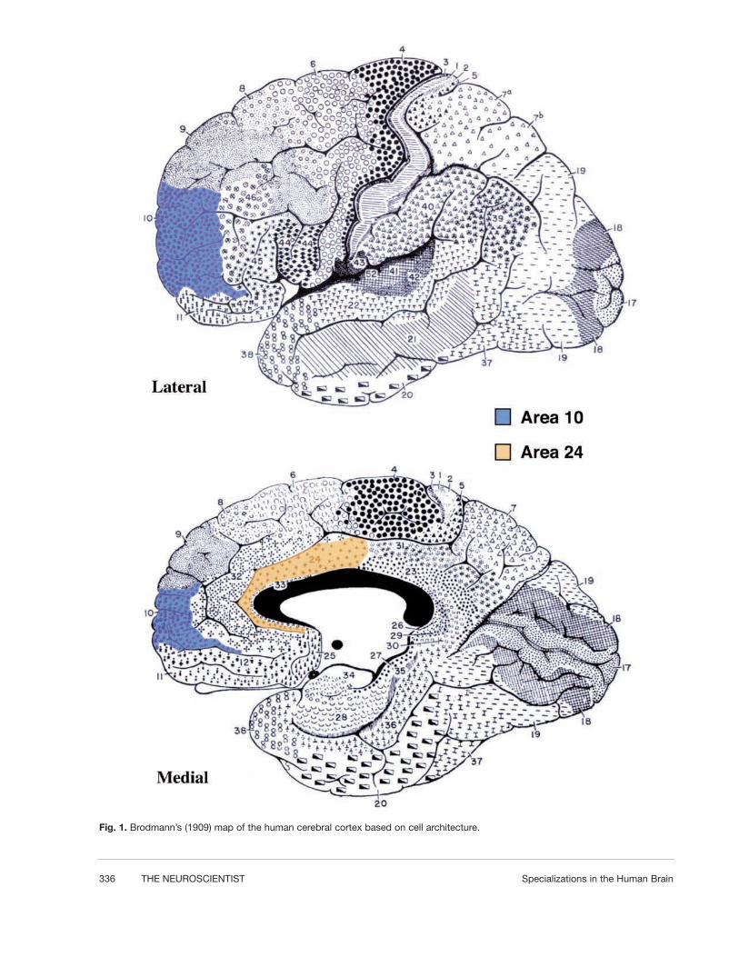
Recently, our colleagues have identified a class of neu-rons that are unique to humans and our closest relatives,the great apes (bonobos, common chimpanzees, gorillas,and orangutans) (Nimchinsky and others 1999). Theseneurons are large, spindle-shaped cells located in layer 5of the anterior cingulate cortex, which is labeled inorange on the brain map in Figure 1. The spindle cellsare characterized by their bipolar shape resulting fromthe large apical dendrite extending toward the pial sur-face of the cortex and the single large basal dendriteextending toward the underlying white matter (see Fig.2). Apart from these two very large dendrites, there aretypically no other dendrites branching from the vicinityof the cell body. The volume of the average spindle cellis four times greater than that of the average layer 5pyramidal neuron (Nimchinsky and others 1999). Thespindle cells are largest and most abundant in humansand decline in density in the following progression:bonobos > common chimpanzees > gorillas > orang-
utans (Nimchinsky and others 1999). They are not pres-ent in lesser apes (gibbons). Thus, within the hominoids,the group comprising humans and apes, the density ofspindle cells declines with approximately the phyloge-netic distance from humans. Spindle cells are not pres-ent in the 22 species of monkeys and prosimians exam-ined or in 30 nonprimate species (Nimchinsky and oth-ers 1999). The spindle cells likely arose in the commonancestor of the great apes and humans, a dryopithecineape probably living in East Africa about 15 million yearsago (Szalay and Delson 1979). Very recently, Hof andhis colleagues (2001) have reported another specializedpopulation of anterior cingulate neurons present only inapes and humans. These neurons are also located in layer5 but are pyramidal cells containing the calcium-bindingprotein calretinin. Like the spindle cells, these special-ized neurons are most abundant in humans. The anteriorcingulate cortex, as a whole, is a specialized region ofneocortex, characterized by a reduced or absent layer 4and a well-developed layer 5, which are features ofmotor areas. Anterior cingulate cortex appears to bepresent in all mammals but is larger and anatomicallydifferentiated in primates (Brodmann 1909; Allman andothers 2001).
Frontal polar cortex, area 10 of Brodmann (1909),which is labeled in blue in Figure 1, is a similar phylo-genetic specialization in hominoids. Area 10 possesses awell-differentiated layer 4 and can be easily identified inhistological sections. Area 10 is large, both absolutelyand relatively, in humans; it is smaller but well devel-
Two Phylogenetic Specializationsin the Human BrainJOHN ALLMAN, ATIYA HAKEEM, and KARLI WATSONDivision of BiologyCalifornia Institute of TechnologyPasadena, California
In this study, two anatomical specializations of the brain in apes and humans are considered. One of theseis a whole cortical area located in the frontal polar cortex (Brodmann’s area 10), and the other is a mor-phologically distinctive cell type, the spindle neuron of the anterior cingulate cortex. The authors suggestthat the spindle cells may relay to other parts of the brain—especially to area 10, the outcome of process-ing within the anterior cingulate cortex. This relay conveys the motivation to act. It particularly concerns therecognition of having committed an error that leads to the initiation of adaptive responses to these adverseevents so as to reduce error commission. This capacity is related to the development of self-control as anindividual matures and gains social insight. Although the anterior cingulate deals with the individual’simmediate response to changing conditions, area 10 is involved in the retrieval of memories from the indi-vidual’s past experience and the capacity to plan adaptive responses. The authors suggest that these neu-robehavioral specializations are crucial aspects of intelligence as defined as the capacity to make adaptiveresponses to changing conditions. The authors further hypothesize that these specializations facilitated theevolution of the unique capacity for the intergenerational transfer of the food and information characteris-tic of human extended families. NEUROSCIENTIST 8(4):335–346, 2002
KEY WORDS Anterior cingulate cortex, Area 10 of Brodmann, Spindle cells
We thank Archibald Fobbs, curator of the brain collections at theNational Museum of Health and Science, for his support and assis-tance in using this resource; Andrea Vasconcellos for staining the non-phosphorlyated neurofilament-labeled cells depicted in Figure 2;Stephen Shepherd for preparing Figure 7; David Grether for Figure9D; and Eliot Bush and Terry Sejnowski for their comments. Thiswork was supported by the Mettler Fund for Autism Research and theFrank P. Hixon Fund.
Address correspondence to: John Allman, Division of Biology,California Institute of Technology, Pasadena, CA 91125 (e-mail:[email protected]).
REVIEW �
336 THE NEUROSCIENTIST Specializations in the Human Brain
Fig. 1. Brodmann’s (1909) map of the human cerebral cortex based on cell architecture.

Volume 8, Number 4, 2002 THE NEUROSCIENTIST 337
oped in the great apes and much smaller in gibbons andmonkeys (see Fig. 3) (Brodmann 1909; Semendeferi andothers 2001). Within the hominoid species, area 10declines in size in the same order as the decline in den-sity of the spindle cells. However, unlike the spindlecells, area 10 is not a unique specialization of humansand great apes but is much larger in these than in otherprimates (Brodmann 1909; Semendeferi and others2001). In this review, we will explore the evidence forfunctional specializations related to the spindle cells of
the anterior cingulate cortex and to the frontal polar cor-tex and how they may be linked to each other and to evo-lution of behavioral specializations in humans.
Functional Studies of Anterior Cingulate Cortex
A clue to the possible functions of the spindle cellscomes from another distinctive set of large neurons, thegigantopyramidal cells in layer 5 of the mid-cingulate
Fig. 2. Nonphosphorlyated neurofilament staining of a spindle cell and a pyramidal cell in layer 5 of the anterior cingulate cortex.

338 THE NEUROSCIENTIST Specializations in the Human Brain
cortex (Braak 1976). The gigantopyramidal cells arelocated just posterior to the spindle cell field, buried inthe cingulate sulcus. These cells are motor neurons thatcontrol the muscles. In imaging experiments, the mid-cingulate motor area is strongly activated when the sub-ject performs the precision grip in which the thumb andindex finger grasp an object (Ehrsson and others 2000).Only humans and some monkeys and apes can performthe precision grip, which is necessary for the fine manip-ulation of objects. The precision grip produces strongeractivation of the mid-cingulate motor cortex than thepower grip in which all the fingers wrap around theobject to be manipulated. The power grip producesstronger activity in the primary motor cortex. Similarly,the mid-cingulate cortex is more strongly activated whenthe subject makes small, precisely controlled move-ments, whereas in the primary motor cortex, activityincreases with the force exerted by the subject (see Fig.4) (Ehrsson and others 2001a). The mental imagery ofhand movements also selectively activates the cingulatemotor area (Ehrsson and others 2001b). These findingsindicate that the mid-cingulate motor cortex containsphylogenetic specialized circuitry for executing the pre-cise manipulation of objects.
At present, we have little direct knowledge of thefunctions of the spindle cells, but we can make infer-ences from functional studies of the anterior cingulatecortex and from the morphology of the spindle cells.Anterior cingulate cortex participates in functions thatare commonly associated both with emotional states andwith cognition. It controls autonomic functions such asheart rate and blood pressure, the generation of vocal-izations (Pool and Ransohoff 1949; Jurgens 1998), andthe production and recognition of facial expressions(Smith 1945; George and others 1993). The experienceof virtually any intense emotion, whether it be anger,love, or lust, is associated with the activation of the ante-rior cingulate cortex (Dougherty and others 1999;Bartels and Zeki 2000; Bush and others 2000; Redouté
and others 2000). States of intense behavioral drive suchas pain, hunger, thirst, and breathlessness are also relat-ed to strong activity in the anterior cingulate cortex(Liotti and others 2001). However, the dorsal part of theanterior cingulate cortex is powerfully activated duringthe performance of cognitively demanding tasks. In ananalysis of more than 70 PET and functional magneticresonance imaging (fMRI) studies involving attention-demanding cognitive tasks, Bush and his colleagues(2000) found the centers of activation to be consistentlylocated in the dorsal part of the anterior cingulate cortex(see Fig. 5). This activation increases with task difficul-ty (Paus and others 1998). Interestingly, the dorsal partof the anterior cingulate is also activated in recent fMRIstudies in which the subjects are experiencing intensedrive states such as love and lust (Bartels and Zeki 2000;Redouté and others 2000). The commonality betweenelicitation of activity in the dorsal part of the anteriorcingulate cortex in cognitive functioning and drive statesis that both involve intense mental focus.
In electro-encephalographic (EEG) studies, a 4 to 7Hz signal (the frontal midline theta rhythm) arises fromthe dorsal part of the anterior cingulate cortex when thesubject is engaged in problem solving, and the amplitudeof this signal increases with task difficulty (Gevins andothers 1997). This signal is attenuated when the subjectis anxious and restored when the anxiety is relieved withdrugs (Suetsugi and others 2000). Thus, there is adimension to anterior cingulate cortex activity thatranges from restless anxiety, poor problem solving, andan attenuated EEG signal to focused problem solving,superior performance, and an increased frontal midlinetheta rhythm. All of us have probably experienced anxi-ety-related interference with concentration or, converse-ly, the relief from anxiety associated with intense andsuccessful problem solving. These experiences probablyreflect this dimension of anterior cingulate functioning.When the subject makes an error, there is a change in theEEG activity arising from the anterior cingulate cortex,which is termed error-related negativity (Deheaene andothers 1994; Bush and others 2000). Direct electrophys-iological recordings from the anterior cingulate cortex inneurosurgical patients confirm the relationship witherror commission, task difficulty, and the source of thisEEG signal (Wang and others 2001).
Although the relationship between error recognitionand anterior cingulate function has been most extensive-ly investigated in human subjects, it was originally dis-covered in electrophysiological recordings from mon-keys (Niki and Watanabe 1979; Brooks 1986). Shidaraand Richmond (2001) have shown that activation of neu-rons in the anterior cingulate cortex is related to theexpectation of reward in behaving monkeys. These stud-ies support the concept that the anterior cingulate cortexis continuously monitoring changes in feedback from theindividual’s interaction with his or her environment thataffect survival and reproduction and initiate behavioralresponses to maintain or improve these conditions.These studies also indicate that the error recognition andcorrecting function of the anterior cingulate cortex
Fig. 3. The relative and absolute size of area 10 in humans andapes, modified after Semendeferi and others (2001).

Volume 8, Number 4, 2002 THE NEUROSCIENTIST 339
Fig. 4. Activation of the mid-cingulate cortex when performing the precision grip relative to the power grip (A) and when using the min-imum force necessary relative to stronger force (B) (Ehrsson and others 2000, 2001a).
Fig. 5. Centers of activation in brain imaging studies of the anterior cingulate cortex, modified after Bush and others (2000). Each dotor square indicates a separate study. Note that the separation between sites activated by emotional states and cognitive tasks is notperfect, and in each case the zone of activation extends over larger regions of the cingulate cortex. The cortex containing spindle cellsis indicated by the purple shading, as based on Nimchinsky and others (1995). The gradient in shading indicates the anterior to pos-terior gradient in the density of spindle cells.

340 THE NEUROSCIENTIST Specializations in the Human Brain
evolved before the spindle cells appeared in an ancestralhominoid about 15 million years ago.
The spindle cells may serve to augment and relay theerror-correcting information to other parts of the brain.The spindle cells are located in layer 5, which typicallyrelays the output of cortical processing to other corticalareas and subcortical structures. The axons of the spin-dle cells are known to project into the underlying whitematter (Nimchinsky and others 1995), but the sites oftermination of these axons have not yet been determined.The cell body and dendrites of the spindle cells containa rich concentration of nonphosphorlyated neurofila-ments, which are a characteristic feature of neurons withlarge axons (see Fig. 2) (Hoffman and others 1987;Nimchinsky and others 1995). The average volume ofthe cell bodies of spindle cells varies as a function of rel-ative brain size (encephalization) across humans, bono-bos, chimpanzees, gorillas, and orangutans (see Fig. 6).This relationship is not a general feature of layer 5 neu-rons because it does not hold for pyramidal cells in theanterior cingulate cortex. Because cell body size proba-bly scales with the size of the axonal arborization, thearborizations of spindle cells may scale with encephal-ization. These observations suggest that the spindle cellsmay have widespread connections with other parts of thebrain and that they may participate in the broadcast toerror recognition and correcting information to manyeffector systems within the brain. The morphology of thespindle cells also suggests another aspect of their func-tioning. Mainen and Sejnowski (1996) have noted thatpyramidal neurons with a prominent apical dendrite tendto fire in regular bursts with intervals of several hundredmilliseconds between bursts. They propose that theaction potentials invade the large apical dendrite, whichafter a brief delay can reexcite the cell body and give riseto a rapid burst of spikes. The spindle cell is morpholog-ically like a super pyramidal cell with two large den-drites. This morphology could result in an even morepronounced cycle of bursts and intervals, which couldcontribute to the prominent EEG signals originatingfrom the anterior cingulate cortex.
Abnormalities in the physiological activity and anato-my of the anterior cingulate cortex are present in most ofthe major neuropsychiatric disorders. The spindle cellsare reduced in number in Alzheimer’s disease(Nimchinsky and others 1995). Reduced size of the ante-rior cingulate cortex, as revealed in structural MRIs, is amajor risk factor to the subsequent development ofdementia in elderly subjects (Killiany and others 2000).The anterior cingulate cortex is reduced in both size andmetabolic activity in autistic patients versus control sub-jects, as revealed in structural MRI and PET studies(Haznedar and others 2000). Both the size and activity ofthe ventral part of the anterior cingulate cortex arereduced in depressed patients (Drevets and others 1997).There is a substantial reduction in the density of layer 2nonpyramidal neurons in the anterior cingulate cortex ofpatients who suffer from bipolar depression and schizo-phrenia (Benes and others 2001). The activity in the ven-tral part of the anterior cingulate cortex is increased in
patients with obsessive-compulsive, phobic, and anxietydisorders when subjects are presented with stimuli thataggravate their symptoms (see Fig. 5) (Rauch and others1994, 1996; Bush and others 2000). These symptomprovocation studies suggest that the hyperactivation orpossibly the breakdown in the coherence of the errormessage originating from the ventral anterior cingulatecortex is responsible for the obsessive, phobic, and anx-ious thoughts in these patients. As mentioned earlier, thefrontal midline theta rhythm is attenuated in anxioussubjects (Suetsugi and others 2000).
Large strokes involving the anterior cingulate cortexproduce akinetic mutes, patients who lie in their hospitalbeds and say or do little. However, with strong arousal,these patients can move or speak a few words, revealingthat they are not paralyzed. If these patients recover, theyreport that they felt “empty” and had “no will” to say ordo anything during the acute period following theirstrokes (Damasio and Van Hoesen 1983). Small lesionsin the anterior cingulate cortex reduce the anxiety pro-duced by chronic pain but also reduce the patient’s
Fig. 6. The average volumes of the cell bodies of spindle cellsand pyramidal cells from layer 5 of the anterior cingulate cortexin humans and great apes plotted against relative brain (brainvolume residuals). The spindle cell volumes are correlated withrelative brain size (r2 = 0.99; P = 0.001). The pyramidal cells arenot significantly correlated with relative brain size (r2 = 0.49; P =0.196).
Volume 8, Number 4, 2002 THE NEUROSCIENTIST 341
capacity to generate responses to novel stimuli (Cohenand others 1999).
The cingulate cortex contains two phylogenetic spe-cializations characteristic of higher primates. The mid-cingulate motor cortex controls precise, volitional handmovements. The anterior cingulate cortex controlsthought and adaptive behavior. The functions of the ante-rior cingulate cortex are the analog of precise manipula-tion in the realm of thought processes. The anterior cin-gulate cortex is implicated in volition, the experience ofintense drive states, self-awareness and control, the dis-crimination of information from conflicting cues,focused problem solving, and error recognition. Thespindle cells are a phylogenetically recent specializationwithin hominoids that may relay information concerningthese functions to other parts of the brain, especially toanother phylogenetic specialization in hominoids, thefrontal polar cortex (area 10).
The Development of Spindle Cells
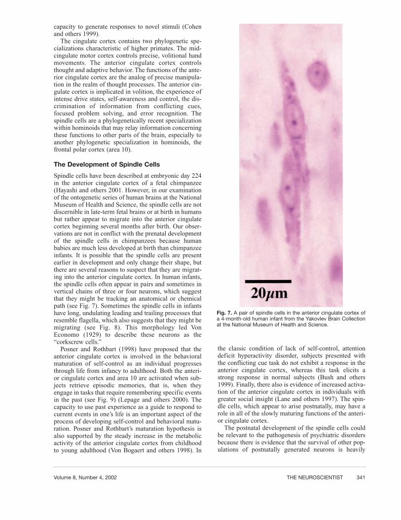
Spindle cells have been described at embryonic day 224in the anterior cingulate cortex of a fetal chimpanzee(Hayashi and others 2001. However, in our examinationof the ontogenetic series of human brains at the NationalMuseum of Health and Science, the spindle cells are notdiscernible in late-term fetal brains or at birth in humansbut rather appear to migrate into the anterior cingulatecortex beginning several months after birth. Our obser-vations are not in conflict with the prenatal developmentof the spindle cells in chimpanzees because humanbabies are much less developed at birth than chimpanzeeinfants. It is possible that the spindle cells are presentearlier in development and only change their shape, butthere are several reasons to suspect that they are migrat-ing into the anterior cingulate cortex. In human infants,the spindle cells often appear in pairs and sometimes invertical chains of three or four neurons, which suggestthat they might be tracking an anatomical or chemicalpath (see Fig. 7). Sometimes the spindle cells in infantshave long, undulating leading and trailing processes thatresemble flagella, which also suggests that they might bemigrating (see Fig. 8). This morphology led VonEconomo (1929) to describe these neurons as the“corkscrew cells.”
Posner and Rothbart (1998) have proposed that theanterior cingulate cortex is involved in the behavioralmaturation of self-control as an individual progressesthrough life from infancy to adulthood. Both the anteri-or cingulate cortex and area 10 are activated when sub-jects retrieve episodic memories, that is, when theyengage in tasks that require remembering specific eventsin the past (see Fig. 9) (Lepage and others 2000). Thecapacity to use past experience as a guide to respond tocurrent events in one’s life is an important aspect of theprocess of developing self-control and behavioral matu-ration. Posner and Rothbart’s maturation hypothesis isalso supported by the steady increase in the metabolicactivity of the anterior cingulate cortex from childhoodto young adulthood (Von Bogaert and others 1998). In
the classic condition of lack of self-control, attentiondeficit hyperactivity disorder, subjects presented withthe conflicting cue task do not exhibit a response in theanterior cingulate cortex, whereas this task elicits astrong response in normal subjects (Bush and others1999). Finally, there also is evidence of increased activa-tion of the anterior cingulate cortex in individuals withgreater social insight (Lane and others 1997). The spin-dle cells, which appear to arise postnatally, may have arole in all of the slowly maturing functions of the anteri-or cingulate cortex.
The postnatal development of the spindle cells couldbe relevant to the pathogenesis of psychiatric disordersbecause there is evidence that the survival of other pop-ulations of postnatally generated neurons is heavily
Fig. 7. A pair of spindle cells in the anterior cingulate cortex ofa 4-month-old human infant from the Yakovlev Brain Collectionat the National Museum of Health and Science.

342 THE NEUROSCIENTIST Specializations in the Human Brain
influenced by environmental factors. For example, thepostnatally generated neurons in the dentate gyrus of thehippocampus are vulnerable to many stress-related
events, and their survival can be enhanced by enrichedenvironments, physical activity, and serotonin-mediatedmechanisms (Gould and others 1997; Jacobs and others
Fig. 8. A cluster of three possible migratory spindle cells in the anterior cingulate cortex of a 4-month-old human infant from theYakovlev Brain Collection at the National Museum of Health and Science. Note the flagella-like undulations in the apical and basaldendrites.

Volume 8, Number 4, 2002 THE NEUROSCIENTIST 343
2000). If the survival of the spindle cells is similarlyinfluenced by environmental conditions during infancy,it is conceivable that positive environments could favorspindle cell survival and enhance emotional stability,self-control, and cognitive functioning later in life andthat unfavorable environments could lead to the death ofspindle cells and increased vulnerability to psychiatricor learning disorders. Because neuron death is also nec-essary for the normal development of the brain (Kuidaand others 1998), it is also possible that in some instances,the excessive survival of spindle cells might contributeto the genesis of psychiatric disorders associated withhyperactivity of the anterior cingulate cortex, such asobsessive-compulsive disorder, which is characterizedby excessively active vigilance and error-correctingbehavior.
Frontal Polar Cortex: Area 10
Much less is known about the functions of area 10 thanfor the anterior cingulate cortex. The best functional datacome from PET and fMRI studies that indicate that thelateral part of area 10 is involved in episodic as opposed
to semantic memory (Buckner 1996; Lepage and others2000). Episodic memory is related to specific events inone’s past as opposed to general (semantic) knowledge.The lateral part of area 10 is consistently activated byboth verbal and nonverbal episodic memory in a large num-ber of studies (see Fig. 10A) (Buckner 1996). Just belowthe part of area 10 activated in the studies of episodicmemory, there is another part that is activated when sub-jects choose between small, likely rewards and large, unlike-ly rewards (see Fig. 10B) (Rogers and others 1999). Theanterior cingulate cortex is also activated in makingthese reward-related decisions, which provides furtherfunctional evidence linking this structure with area 10.
Recently, the medial part of area 10 has been activat-ed in a study in which subjects were presented with emo-tionally charged moral dilemmas requiring them tochoose a course of action affecting the lives of others(see Fig. 10C) (Greene and others 2001). The medial andanterior parts of area 10 are activated when subjectsdevelop a successful decision-making strategy in a sim-ulated auction involving real monetary rewards (see Fig.10D). This activation may be related to the recollectionof the outcome of recent bids (episodic memory), theassessment of reward probability, and the choice of astrategy for the next bid. The anterior cingulate was alsoactivated in this bidding task. Taken together, these find-ings suggest a functional linkage between the anteriorcingulate cortex and area 10, in which the anterior cin-gulate monitors the current state of reward and punish-ment and signals the need for behavioral adaptation,whereas area 10 compares the current state with pastexperience and on this basis makes choices governingfuture behavior. The subject is likely to be consciouslyaware of many of these processes, and consciousnessmay be an important and perhaps crucial aspect of theindividual’s behavioral adaptation to changing rewardsand penalties. This behavioral adaptation also extends toconsiderations of the well-being of other individuals.The size of the dendritic arborizations and the number ofsynapses are greater in area 10 pyramidal neurons thanin any other cortical area, which suggests the integrativerole of this area (Jacobs and others 2001). In summary,the activity of the anterior cingulate cortex brings aboutthe awareness of discrepancies between the current stateand desired states for the individual and initiates behav-ior to improve his or her state. Area 10 compares the cur-rent state with past experience, calculates reward proba-bilities, formulates strategies, and makes choices basedon these calculations.
Brain Specializations and the Economy ofHuman Extended Families
One of the key behavioral specializations in humans isthe extended family (Allman 2000). Human infantsdevelop very slowly and depend for a very long time onthe support of their families. We propose that theenlargement of area 10 and the increased size and densi-ty of spindle cells in the anterior cingulate cortex inhumans were part of a suite of adaptations related to the
Fig. 9. Coactivation of the anterior cingulate cortex and area 10in the retrieval of episodic memory in a PET imaging study byLepage and others (2000). The area 10 activation was obtainedin a slice 20 mm from the midline.

344 THE NEUROSCIENTIST Specializations in the Human Brain
special economic needs of human extended families.Quantitative studies of foraging production and foodconsumption in chimpanzee and human hunter-gathererpopulations indicate that young apes are able to providenearly all their sustenance through their own foragingactivities; however, young humans do not (see Fig. 11)(Kaplan and others 2000). Human males do not acquirethrough their own efforts as many calories as they con-sume until about age 17 in hunter-gatherer populations.Human females, because they are engaged in childbear-ing and rearing, do not acquire through their own forag-
ing as many calories as they consume until about age 45.The economy of the human extended family is based ontransfers of food resources from fathers, grandfathers,and grandmothers to their children and grandchildren.Much of the food acquired by men is obtained by hunt-ing; much of the food acquired by women is obtainedthrough the extractive foraging of resources that are dif-ficult to harvest; both activities require many years ofhard-won expertise to be performed efficiently (Kaplanand others 2000). Effective food acquisition requiresintense focus and monitoring of the outcome in terms of
Fig. 10. (A) Sites of activation in area 10 produced by verbal and nonverbal episodic memory tasks from 10 separate brain imagingstudies reviewed by Buckner (1996). (B) Activation in the ventral part of area 10 by choosing between small, likely rewards and large,unlikely rewards in a PET imaging study by Rogers and others (1999). (C) Activation in medial area 10 by emotionally charged moraljudgments from Greene and others (2001). (D) Activation of anterior and medial area 10 during the formulation of a successful biddingstrategy in an auction from an ongoing fMRI study by Daniel Rowe, David Grether, Charles Plott, and John Allman. The map was gen-erated by subtracting the block of trials before the subject arrived at the optimal strategy from the block of trails during which shearrived at the optimal strategy. The scale is in differences in t values between the first block and the successful block of trials. Theauction involved actual monetary rewards for the subject.

Volume 8, Number 4, 2002 THE NEUROSCIENTIST 345
size and quality of nutritional reward. The anterior cin-gulate is clearly involved in these functions, and we sug-gest that the spindle cells may participate in the relay ofthese assessments to other parts of the brain. Effectivefood acquisition also requires changing to more reward-ing activities in the face of decreasing reward, which alsoactivates neurons in the anterior cingulate cortex (Shimaand Tanji 1998). Effective hunting and foraging requiresknowledge and appropriate application of specializedtechniques, which require the retrieval of episodic mem-ory and probability assessment that activate both theanterior cingulate cortex and area 10. For example, itmight involve childhood memories of how a parent orgrandparent captured or foraged for a particular type offood. It also requires choices among different possibleforaging strategies in terms of their likely payoff andlinks these choices to concerns for the well-being ofdependent family members. These last two functions arerelated to the activation of area 10 in formulating anoptimal economic strategy and decision making in theface of emotionally charged moral judgments, both ofwhich activate area 10.
References
Allman JM. 2000. Evolving brains. New York: Freeman.Allman JM, Hakeem A, Erwin JM, Nimchinsky E, Hof P. 2001. The
anterior cingulate cortex: the evolution of an interface betweenemotion and cognition. Ann N Y Acad Sci 935:107–17.
Bartels A, Zeki S. 2000. The neural basis of romatic love. Neuroreport11:3829–34.
Benes FM, Vincent SL, Todtenkopf M. 2001. The density of pyramidaland nonpyramidal neurons in anterior cingulate cortex of schizo-phrenic and bipolar subjects. Biol Psychiatry 50:395–406.
Braak H. 1976. A primitive gigantopyramidal field buried in the depthof the cingulate sulcus of the human brain. Brain Res 109:219–33.
Brodmann K. 1909. Vergleichende Lokalisationslehre derGroshirnrinde. Leipzig: Barth.
Brooks VA. 1986. How does the limbic system assist motor learning?A limbic comparator hypothesis. Brain Behav Evol 29:29–53.
Buckner R. 1996. Beyond HERA: contributions of specific prefrontalbrain areas to long-term memory retrieval. Psychonomic Bulletinand Review 3:149–58.
Bush G, Frazier JA, Rauch SL, Seidman LJ, Whalen PJ, Lenike MA,Rosen BR, Biederman J. 1999. Anterior cingulate cortex dysfunc-tion in attention-deficit/hyperactivity disorder revealed by fMRIand the counting stroop. Biol Psychiatry 45:1542–52.
Bush G, Luu P, Posner M. 2000. Cognitive and emotional influences inanterior cingulate cortex. Trends in Cognitive Science 4:215–22.
Cohen RA, Kaplan RF, Zuffante P, Moser DJ, Jenkins MA, Salloway S,Wilkinson H. 1999. Alteration of intention and self-initiated actionassociated with bilateral anterior cingulotomy. J Neuropsych ClinNeurosci 11:444–53.
Damasio A, Van Hoesen GW. 1983. Emotional disturbances associat-ed with focal lesions of the limbic frontal lobe. In: Heilman KM,Satz P, editors. Neuropsychology of human emotion. New York:Guilford. p 85–110.
Deheaene S, Posner M, Tucker DM. 1994. Localization of a neural sys-tem for error detection and compensation. Physiol Sci 5:303–5.
Dougherty D, Shin LM, Alpert NM, Pitman RK, Orr SP, Lasko M,Macklin ML, Fischman AJ, Rauch SL. 1999. Anger in healthymen: a PET study using script-driven imagery. Biol Psychiatry46:466–72.
Drevets WC, Price JL, Thompson JR, Todd RD, Reich, Vannier TM,Raichle ME. 1997. Subgenual prefrontal cortex abnormalities inmood disorders. Nature 386:824–7.
Ehrsson H, Fagergren A, Forssberg H. 2001a. Differential fronto-pari-etal activation depending of force used in a precision grip task: afMRI study. J Neurophysiol 85:2613–23.
Ehrsson H, Fagergren A, Jonsson T, Westling G, Johansson R,Forssberg H. 2000. Cortical activity in precision-versus power-griptasks: an fMRI study. J Neurophysiol 83:528–36.
Ehrsson H, Naito E, Roland P. 2001b. Activation of human motor cor-tices during mental motor imagery of hand, foot and tongue move-ments. Neuroimage 13:S1158.
George MS, Ketter TS, Gill DS, Haxby JV, Ungerleider LG,Hersovitch P, Post RM. 1993. Brain regions involved in recogniz-ing facial emotion or identity: an oxygen-15 PET study. JNeuropsych Clin Neurosci 5:384–94.
Gevins A, Smith ME, McEnvoy L, Yu D. 1997. High-resolution EEGmapping of cortical activation related to working memory: diffi-culty, types of processing, and practice. Cerebral Cortex 7:374–85.
Gould E, McEwen BS, Tanapat P, Galea LA, Fuchs E. 1997.Neurogenesis in the dentate gyrus of the adult tree shrew is regu-lated by psychosocial stress and NMDA receptor activation. JNeurosci 17:2492–8.
Greene JD, Sommerville RB, Nystrom LE, Darley JM, Cohen JD.2001. An fMRI investigation of emotional engagement in moraljudgement. Science 293:2105–8.
Hayashi M, Ito M, Shimuzu K. 2001. The spindle cells are present inthe cingulate cortex of chimpanzee fetus. Neurosci Lett 309:97-100.
Haznedar M, Buchsbaum M, Wei T-C, Hof P, Cartwright C, BienstockC, Hollander E. 2000. Limbic circuitry in patients with autismspectrum disorders studied with positron emission tomography andmagnetic resonance imaging. Amer J Psychiatry 157:1994–2001.
Fig. 11. The mean daily energy production and consumption byindividuals of each sex as a function of age in chimpanzees andhumans from several hunter-gatherer populations (Kaplan andothers 2000).

346 THE NEUROSCIENTIST Specializations in the Human Brain
Hof PR, Nimchinsky EA, Perl DP, Erwin JM. 2001. An unusual popu-lation of pyramidal neurons in the anterior cingulate cortex ofhominids contains the calcium-binding protein calretinin. NeurosciLett 307:139–42.
Hoffman PN, Cleveland DW, Griffin JW, Landes PW, Cowan NJ, PriceDL. 1987. Neurofilament gene expression: a major determinant ofaxonal caliber. Proc Nat Acad Sci 84:3472–6.
Jacobs B, Schall M, Prather M, Kapler E, Driscoll L, Baca S, Jacobs J,Wainwright M, Treml M. 2001. Regional dendritic and spine vari-ation in human cerebral cortex: a quantitative Golgi study. CerebralCortex 11:558–71.
Jacobs B, van Praag H, Gage F. 2000. Adult brain neurogenesis andpsychiatry: a novel theory of depression. Molecular Psychiatry5:262–9.
Jurgens U. 1998. Neuronal control of mammalian vocalization with spe-cial reference to the squirrel monkey. Naturwissenschaften 85:376–88.
Kaplan H, Hill K, Lancaster J, Hurtado M. 2000. A theory of humanlife history evolution: diet, intelligence, and longevity.Evolutionary Anthropology 9:156–85.
Killiany RJ, Gomez-Isla T, Moss M, Kikinis R, Sandor T, Jolesz F,Tanzi R, Jones K, Hyman BT, Albert MS. 2000. Use of structuralmagnetic resonance imaging to predict who will get Alzheimer’sdisease. Ann Neurol 47:419–20.
Kuida K, Haydar T, Kuan C-Y, Gu Y, Taya C, Karasuyama H, Su M,Rakic P, Flavell R. 1998. Reduced apoptosis and cytochrome-C-mediated caspase activation in mice lacking Caspase-9. Cell94:325–37.
Lane RD, Reiman EM, Axelrod B, Yun LS, Holmes A, Schwartz GE.1997. Neural correlates of levels of emotional awareness: evidenceof an interaction between emotion and attention in the anterior cin-gulate cortex. J Cogn Neurosci 10:525–35.
Lepage M, Ghaffar O, Nyberg L, Tulving E. 2000. Prefrontal cortex andepisodic memory retrieval mode. Proc Nat Acad Sci 97:506–11.
Liotti M, Brannan S, Egan G, Shade R, Madden L, Abplanalp B,Robillard R, Lancaster J, Zamarripa F, Fox P, Denton D. 2001.Brain responses associated with consciousness of breathlessness(air hunger). Proc Nat Acad Sci 98:2035–40.
Mainen Z, Sejnowski T. 1996. Influence of dendritic structure on fir-ing pattern model neocortical neurons. Nature 382:363-6.
Niki H, Watanabe M. 1979. Prefrontal and cingulate unit activity dur-ing timing behavior in the monkey. Brain Res 171:213–24.
Nimchinsky E, Gilissen E, Allman JM, Perl DP, Erwin JM, Hof PR.1999. A neuronal morphologic type unique to humans and greatapes. Proc Nat Acad Sci 96:5268–73.
Nimchinsky E, Vogt BA, Morrison J, Hof PR. 1995. Spindle neuronsof the human anterior cingulate cortex. J Comp Neurol 355:27–37.
Paus T, Koski L, Caramanos Z, Westbury C. 1998. Regional differ-ences in the effects of task difficulty and motor output on blood
flow response in the human anterior cingulate cortex: a review of107 PET activation studies. Neuroreport 9:37–47.
Pool JL, Ransohoff J. 1949. Autonomic effects on stimulating rostralportion of cingulate gyri in man. J Neurophysiol 12:385–92.
Posner M, Rothbart MK. 1998. Attention, self-regulation and con-sciousness. Phil Trans Roy Soc B 353:1915–27.
Rauch SL, Jenike MA, Alpert, NM, Baer L, Breiter, HC, Savage CR,Fischman AJ. 1994. Regional cerebral blood flow measured duringsymptom provocation in obsessive-compulsive disorder using oxy-gen 15-labeled carbon dioxide and positron emission tomography.Arch Gen Psychiatry 51:62–70.
Rauch SL, Van der Kolk BA, Fisler RE, Alpert NM, Orr SP, SavageCR, Fischman AJ, Jenike MA, Pitman RK. 1996. A symptomprovocation study of posttraumatic stress disorder using positronemission tomography and script-driven imagery. Arch GenPsychiatry 53:380–7.
Redouté J, Stoleru S, Gregoire M-C, Costes N, Cinotti L, Lavenne F,Le Bars D, Forest M, Pujol J-F. 2000. Brain processing of visualsexual stimuli in human males. Human Brain Mapping 11:162–77.
Rogers RD, Owen AM, Middleton HC, Williams EJ, Pickard JD,Sahakian BJ, Robbins TW. 1999. Choosing between small, likelyrewards and large, unlikely rewards activates inferior and orbitalprefrontal cortex. J Neurosci 20:9029–38.
Semendeferi K, Armstrong E, Schleichter A, Zilles K, Van Hoesen G.2001. Prefrontal cortex in humans and apes: a comparative study ofarea 10. Amer J Physical Anthropology 114:224–41.
Shidara M, Richmond BJ. 2001. Single neuronal signals in the anteri-or cingulate related to degree of reward expectancy. Soc NeurosciAbst 27.
Shima K, Tanji J. 1998. Role for cingulate motor cells in voluntarymovement selection based on reward. Science 282:1335–38.
Smith W. 1945. The functional significance of the rostral cingular cor-tex as revealed by its responses to electrical stimulation. JNeurophysiol 8:241–55.
Suetsugi M, Mizuki Y, Ushijima, Kobahashi IT, Tsuchiya K, Aoki T,Watanabe Y. 2000. Appearance of frontal midline theta activity inpatients with generalized anxiety disorder. Neurospychobiology41:108–12.
Szalay FS, Delson E. 1979. Evolutionary history of the primates. NewYork: Academic Press.
Van Bogaert P, Wikler D, Damhaut P, Szliwowski H, Goldman S. 1998.Regional changes in glucose metabolism during brain develop-ment. Neuroimage 8:62–8.
Von Economo C. 1929. The cytoarchitectonics of the human cerebralcortex. Oxford, UK: Oxford University Press.
Wang C, Ulbert I, Ives JR, Blume H, Marinkovic K, Heit G, SchomerDL, Halgren E. 2001. Synaptic and unit activity in the human ante-rior cingulate gyrus to errors and difficulty. Soc Neurosci Abst 27.


![Al-Hakeem Thesis 1991[1]](https://static.fdocuments.in/doc/165x107/547035c3b4af9f8d6c8b45ef/al-hakeem-thesis-19911.jpg)


![C. ABDUL HAKEEM COLLEGE [AUTONOMOUS]MELVISHARAM …](https://static.fdocuments.in/doc/165x107/61bf0c15a6790e3c2519ad0d/c-abdul-hakeem-college-autonomousmelvisharam-.jpg)

