Review The primate basal ganglia: parallel and integrative ...coaxlab/documents/Haber_2003.pdf ·...
14
Journal of Chemical Neuroanatomy 26 (2003) 317–330 Review The primate basal ganglia: parallel and integrative networks Suzanne N. Haber ∗ Department of Pharmacology and Physiology, University of Rochester School of Medicine, 601 Elmwood Avenue, Rochester, NY 14642, USA Received 3 April 2003; received in revised form 20 August 2003; accepted 5 October 2003 Abstract The basal ganglia and frontal cortex operate together to execute goal directed behaviors. This requires not only the execution of motor plans, but also the behaviors that lead to this execution, including emotions and motivation that drive behaviors, cognition that organizes and plans the general strategy, motor planning, and finally, the execution of that plan. The components of the frontal cortex that mediate these behaviors, are reflected in the organization, physiology, and connections between areas of frontal cortex and in their projections through basal ganglia circuits. This comprises a series of parallel pathways. However, this model does not address how information flows between circuits thereby developing new learned behaviors (or actions) from a combination of inputs from emotional, cognitive, and motor cortical areas. Recent anatomical evidence from primates demonstrates that the neuro-networks within basal ganglia pathways are in a position to move information across functional circuits. Two networks are: the striato-nigral-striatal network and the thalamo-cortical-thalamic network. Within each of these sets of connected structures, there are both reciprocal connections linking up regions associated with similar functions and non-reciprocal connections linking up regions that are associated with different cortical basal ganglia circuits. Each component of information (from limbic to motor outcome) sends both feedback connection, and also a feedforward connection, allowing the transfer of information. Information is channeled from limbic, to cognitive, to motor circuits. Action decision-making processes are thus influenced by motivation and cognitive inputs, allowing the animal to respond appropriate to environmental cues. © 2003 Elsevier B.V. All rights reserved. Keywords: Striatum; Pallidum; Thalamus; Substantia nigra; Dopamine 1. Introduction The basal ganglia (BG) work in concert with cortex to or- chestrate and execute planned, motivated behaviors requir- ing motor, cognitive, and limbic circuits. While best known for their motor functions, the BG are involved in several aspects of goal-directed behaviors, including not only its expression through the control of movement, but also the processes that lead to movement, including the elements that drive actions, such as emotions, motivation, and cognition. Indeed, regions within each of the BG nuclei are anatom- ically and physiologically associated with each of these functional circuits. Ventral regions of the basal ganglia play a key role in reward and reinforcement and are important in the development of addictive behaviors and habit forma- tion (Schultz, 1997; Wise, 1998; Koob, 1999; Rolls, 2000; Everitt et al., 2001). More central basal ganglia areas are ∗ Tel.: +1-585-275-4538; fax: +1-585-273-2652. E-mail address: suzanne [email protected] (S.N. Haber). involved in cognitive functions such as procedural learning and working memory tasks (Mishkin et al., 1984; Phillips and Carr, 1987; Jueptner et al., 1997; Levy et al., 1997; Jog et al., 1999). Finally, the dorsolateral portion of the striatum, caudal to the anterior commissure is associated with the control of movement. Consistent with this topography, dis- eases affecting mental health, including schizophrenia, drug addiction, and obsessive compulsive disorder, are all linked to pathology in the basal ganglia, as are diseases affecting motor control (Stevens, 1973; Kalivas et al., 1993; McGuire et al., 1994; Breiter et al., 1996; Koob and Nestler, 1997; Pantelis et al., 1997; Kegeles et al., 2000; Menon et al., 2001; Rauch et al., 2001). The association of the basal gan- glia with frontal cortical function along with its relationship to multiple neurological and psychiatric diseases empha- sizes the importance of understanding the basal ganglia with respect to cortical function. Differentiation of frontal cortex and basal ganglia structures as they relate to human function and disease are best modeled from a combination of physi- ological, anatomical, and imaging studies in primates (both human and non-human). Thus, the present review describes 0891-0618/$ – see front matter © 2003 Elsevier B.V. All rights reserved. doi:10.1016/j.jchemneu.2003.10.003
Transcript of Review The primate basal ganglia: parallel and integrative ...coaxlab/documents/Haber_2003.pdf ·...

Journal of Chemical Neuroanatomy 26 (2003) 317–330
Review
The primate basal ganglia: parallel and integrative networks
Suzanne N. Haber∗
Department of Pharmacology and Physiology, University of Rochester School of Medicine, 601 Elmwood Avenue, Rochester, NY 14642, USA
Received 3 April 2003; received in revised form 20 August 2003; accepted 5 October 2003
Abstract
The basal ganglia and frontal cortex operate together to execute goal directed behaviors. This requires not only the execution of motorplans, but also the behaviors that lead to this execution, including emotions and motivation that drive behaviors, cognition that organizes andplans the general strategy, motor planning, and finally, the execution of that plan. The components of the frontal cortex that mediate thesebehaviors, are reflected in the organization, physiology, and connections between areas of frontal cortex and in their projections throughbasal ganglia circuits. This comprises a series of parallel pathways. However, this model does not address how information flows betweencircuits thereby developing new learned behaviors (or actions) from a combination of inputs from emotional, cognitive, and motor corticalareas. Recent anatomical evidence from primates demonstrates that the neuro-networks within basal ganglia pathways are in a position tomove information across functional circuits. Two networks are: the striato-nigral-striatal network and the thalamo-cortical-thalamic network.Within each of these sets of connected structures, there are both reciprocal connections linking up regions associated with similar functions andnon-reciprocal connections linking up regions that are associated with different cortical basal ganglia circuits. Each component of information(from limbic to motor outcome) sends both feedback connection, and also a feedforward connection, allowing the transfer of information.Information is channeled from limbic, to cognitive, to motor circuits. Action decision-making processes are thus influenced by motivationand cognitive inputs, allowing the animal to respond appropriate to environmental cues.© 2003 Elsevier B.V. All rights reserved.
Keywords: Striatum; Pallidum; Thalamus; Substantia nigra; Dopamine
1. Introduction
The basal ganglia (BG) work in concert with cortex to or-chestrate and execute planned, motivated behaviors requir-ing motor, cognitive, and limbic circuits. While best knownfor their motor functions, the BG are involved in severalaspects of goal-directed behaviors, including not only itsexpression through the control of movement, but also theprocesses that lead to movement, including the elements thatdrive actions, such as emotions, motivation, and cognition.Indeed, regions within each of the BG nuclei are anatom-ically and physiologically associated with each of thesefunctional circuits. Ventral regions of the basal ganglia playa key role in reward and reinforcement and are importantin the development of addictive behaviors and habit forma-tion (Schultz, 1997; Wise, 1998; Koob, 1999; Rolls, 2000;Everitt et al., 2001). More central basal ganglia areas are
∗ Tel.: +1-585-275-4538; fax:+1-585-273-2652.E-mail address: [email protected] (S.N. Haber).
involved in cognitive functions such as procedural learningand working memory tasks (Mishkin et al., 1984; Phillipsand Carr, 1987; Jueptner et al., 1997; Levy et al., 1997; Joget al., 1999). Finally, the dorsolateral portion of the striatum,caudal to the anterior commissure is associated with thecontrol of movement. Consistent with this topography, dis-eases affecting mental health, including schizophrenia, drugaddiction, and obsessive compulsive disorder, are all linkedto pathology in the basal ganglia, as are diseases affectingmotor control (Stevens, 1973; Kalivas et al., 1993; McGuireet al., 1994; Breiter et al., 1996; Koob and Nestler, 1997;Pantelis et al., 1997; Kegeles et al., 2000; Menon et al.,2001; Rauch et al., 2001). The association of the basal gan-glia with frontal cortical function along with its relationshipto multiple neurological and psychiatric diseases empha-sizes the importance of understanding the basal ganglia withrespect to cortical function. Differentiation of frontal cortexand basal ganglia structures as they relate to human functionand disease are best modeled from a combination of physi-ological, anatomical, and imaging studies in primates (bothhuman and non-human). Thus, the present review describes
0891-0618/$ – see front matter © 2003 Elsevier B.V. All rights reserved.doi:10.1016/j.jchemneu.2003.10.003

318 S.N. Haber / Journal of Chemical Neuroanatomy 26 (2003) 317–330
the organization of the primate basal ganglia from the per-spective of cortical function. In some situations, when nec-essary and indicated, data are presented from rodent work.
The BG includes the caudate n., putamen, and the globuspallidus and three closely related structures, the substantianigra (SN), the ventral tegmental area (VTA), and the subtha-lamic nucleus (STh). Based on connectivity, histology, andfunctional considerations, the concept of the ventral striatumwas introduced as the ventral extension of the striatum thatincludes the N. accumbens, the medial and ventral portionsof the caudate n. and putamen, and the striatal cells of theolfactory tubercle (Heimer, 1978; Heimer et al., 1994). Theventral striatum contains a subregion, the shell. This region,which was first demonstrated in rodents (Zaborszky et al.,1985), is best distinguished by its lack of calbindin-positivestaining (Martin et al., 1991; Meredith et al., 1996; Haberand McFarland, 1999). While the ventral and medial bor-ders of the ventral striatum are relatively are clear, thedorsal and lateral boundaries of the ventral striatum mergeimperceptibly with the dorsal striatum (Fig. 1) (Haber andMcFarland, 1999). The striatum is the main input structureto the basal ganglia. Its afferent projections are derived fromthree major sources: (1) cerebral cortex; (2) thalamus; and(3) brainstem. The striatum projects to the pallidal complexand to the substantia nigra, pars reticulata (SNr). The palli-dal complex includes the external (GPe) and internal. seg-ments (GPi) of the globus pallidus and the ventral pallidum(VP), the pallidal segment connected to the ventral striatum.The substantia nigra (and VTA) contains the dopaminergiccells of the pars compacta (Snc/VTA), and the pars retic-ulata (SNr) (Fig. 2A). The outputs from the GPi and SNr
Fig. 1. Photomicrographs of the striatum at the level of the shell and core, immunostained for various transmitter-related molecules. AchE, acetyl-cholinesterase; CaBP, calbindin-28; ENK, enkephalin; GluR1,GluR1 AMPA receptor subunit; m-R, m-opiate receptor; 5-HT, serotonin.
are to the thalamus, which then projects back to the cortex,completing what is referred to as the ‘direct’ cortico-basalganglia pathway. The GPe is reciprocally connected to theSTh, which in turn projects to the GPi. This is referred toas the ‘indirect’ cortico-basal ganglia pathway. For a com-prehensive review of basal ganglia pathways (seePercheronet al., 1994; Graybiel, 1995; Parent and Hazrati, 1995;Parent et al., 2000; Middleton and Strick, 2002; Haber,2003).
2. Functional organization of the basal-gangliapathways
Frontal cortex is the main driving force of the BG as in-dicated by its massive topographically and functionally or-ganized pathways. Together they control the ability to carryall aspects of goal directed behaviors including the moti-vation and cognition that drives and organizes them, alongwith their execution. Frontal cortex in primates can be di-vided into several functional regions: the orbital and medialprefrontal cortex (OMPFC), involved in emotions and moti-vation; the dorsolateral prefrontal cortex (DLPFC), involvedin higher cognitive processes or ‘executive functions’; thepremotor and motor areas, involved in motor planning andthe execution of those plans (see the section below). Further-more, prefrontal, premotor, and motor cortices are thoughtto be organized in a hierarchical manner, with motor cor-tex being the final step to action (Fuster, 2001). Function-ally defined regions of frontal cortex project topographicallythrough the basal ganglia, to thalamus, and back to cortex.
S.N. Haber / Journal of Chemical Neuroanatomy 26 (2003) 317–330 319
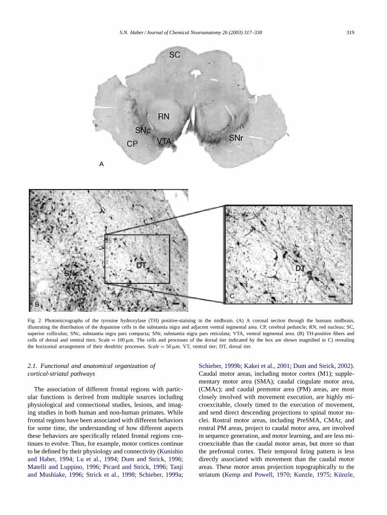
Fig. 2. Photomicrographs of the tyrosine hydroxylase (TH) positive-staining in the midbrain. (A) A coronal section through the humans midbrain,illustrating the distribution of the dopamine cells in the substantia nigra and adjacent ventral tegmental area. CP, cerebral peduncle; RN, red nucleus; SC,superior colliculus; SNc, substantia nigra pars compacta; SNr, substantia nigra pars reticulata; VTA, ventral tegmental area. (B) TH-positive fibers andcells of dorsal and ventral tiers. Scale= 100�m. The cells and processes of the dorsal tier indicated by the box are shown magnified in C) revealingthe horizontal arrangement of their dendritic processes.Scale = 50�m. VT, ventral tier; DT, dorsal tier.
2.1. Functional and anatomical organization ofcortical-striatal pathways
The association of different frontal regions with partic-ular functions is derived from multiple sources includingphysiological and connectional studies, lesions, and imag-ing studies in both human and non-human primates. Whilefrontal regions have been associated with different behaviorsfor some time, the understanding of how different aspectsthese behaviors are specifically related frontal regions con-tinues to evolve. Thus, for example, motor cortices continueto be defined by their physiology and connectivity (Kunishioand Haber, 1994; Lu et al., 1994; Dum and Strick, 1996;Matelli and Luppino, 1996; Picard and Strick, 1996; Tanjiand Mushiake, 1996; Strick et al., 1998; Schieber, 1999a;
Schieber, 1999b; Kakei et al., 2001; Dum and Strick, 2002).Caudal motor areas, including motor cortex (M1); supple-mentary motor area (SMA); caudal cingulate motor area,(CMAc); and caudal premotor area (PM) areas, are mostclosely involved with movement execution, are highly mi-croexcitable, closely timed to the execution of movement,and send direct descending projections to spinal motor nu-clei. Rostral motor areas, including PreSMA, CMAr, androstral PM areas, project to caudal motor area, are involvedin sequence generation, and motor learning, and are less mi-croexcitable than the caudal motor areas, but more so thanthe prefrontal cortex. Their temporal firing pattern is lessdirectly associated with movement than the caudal motorareas. These motor areas projection topographically to thestriatum (Kemp and Powell, 1970; Kunzle, 1975; Künzle,

320 S.N. Haber / Journal of Chemical Neuroanatomy 26 (2003) 317–330
1977; Kunzle, 1978; Aosaki et al., 1994b; Flaherty andGraybiel, 1994; McFarland and Haber, 2000). Projectionsfrom M1 terminate almost entirely in the dorsolateral puta-men, caudal to the anterior commissure. There are few termi-nals rostral to the anterior commissure. The caudal premotorarea projects to a striatal region that is just adjacent to M1projections, extending only slightly into the caudate n. Therostral premotor areas terminate in both the caudate n. andputamen, bridging the two with a continuous projection. Thisprojection extends more rostrally than those from the mo-tor cortex, although not into the rostral pole of the striatum.Thus, both caudal and rostral areas occupy much of the puta-men caudal to the anterior commissure. This area receivesoverlapping projections from parietal areas associated withsomatosensory function, resulting in a sensory-motor areathat is somatotopically organized. Physiological and imag-ing studies support the involvement of this striatal region insensorimotor control and motor plans (Aldridge et al., 1980;DeLong and Georgopoulos, 1981; Alexander and DeLong,1985a,b; Alexander and DeLong, 1986; Kimura, 1986, 1990;Kermadi and Joseph, 1995; Hikosaka et al., 1996; Boeckeret al., 1998). Thus, the premotor and motor areas (and thefrontal eye fields) mediate different aspects of motor behav-ior, including planning, learning, and execution, which arein turn reflected both anatomically and physiologically inthe central and lateral caudate n. and in the central, dorsal,and lateral putamen, respectively.
Prefrontal cortical regions (DLPFC and OMPFC), are theleast microexcitable of frontal cortex (Fuster, 1997). TheDLPFC is involved in working memory, set shifting, andstrategic planning, often referred to as ‘executive functions’(Passingham, 1995; Goldman-Rakic, 1996; Smith andJonides, 1997; Fuster, 2000, 2001). The DLPFC projectsmost densely to the rostral striatum including both the cau-date n. and the putamen, rostral to the anterior commissure.The rostrocaudal extent of this projection is large and topo-graphically organized. While there are few terminals in thecentral and caudal putamen posterior to the anterior com-missure, the caudate n. does remain innervated (Selemonand Goldman-Rakic, 1985; Arikuni and Kubota, 1986).Physiological, imaging and lesion studies support the ideathat the head of the caudate n. is instrumental in delayedtasks, particularly in specific working memory tasks (Battiget al., 1960; Butters and Rosvold, 1968; Hikosaka et al.,1989; Apicella et al., 1992; Partiot et al., 1996; Levy et al.,1997; Elliott and Dolan, 1999). Taken together, the caudaten., and in particular the head of the caudate n., is involved inworking memory and strategic planning processes, workingtogether with the DLPFC in mediating this function. TheDLPFC has close connections with the orbital and medialprefrontal cortex (Carmichael and Price, 1996b; Petridesand Pandya, 1999; Barbas, 2000; Passingham et al., 2002).
Orbital prefrontal cortex is involved in the development ofreward-based learning and goal-directed behaviors (Butterand Snyder, 1972; Benevento et al., 1977; Rolls and Baylis,1994; Meunier et al., 1997; Baxter et al., 2000; Hikosaka and
Watanabe, 2000; Schultz et al., 2000). This area receives in-put from multimodal sensory regions and is closely linked tothe medial prefrontal cortex (Carmichael and Price, 1996a;Carmichael and Price, 1996b). Lesions of the orbital andmedial prefrontal areas result in an. inability to initiateand carry out goal-directed behaviors, and lead to sociallyinappropriate and impulsive behaviors (Rolls et al., 1980;Eslinger and Damasio, 1985; Fuster, 1989; Cummings,1995; Filley, 1995). The lateral orbital regions, areas 13 and12, and dysgranular insular cortex project to the central andlateral parts of the ventral striatum (Kunishio and Haber,1994; Haber et al., 1995b; Chikama et al., 1997; Ferryet al., 2000). Medial orbital areas 13a/b and 14 projects tothe medial wall of the caudate n., extending ventralwardinto the n. accumbens, lateral to the shell region. This areareceives an additional dense innervation from the medialprefrontal cortex (the anterior cingulate cortex). The medialprefrontal cortex, receives particularly dense innervationsfrom the orbital prefrontal cortex, the amygdala, and thehypothalamus and is important in the expression of emotion(Carmichael and Price, 1995; Mayberg et al., 1999). Theshell receives the densest innervation from medial areas25, and 32 and from agranular insular cortex. Together, theorbital and medial prefrontal cortex projects primarily tothe rostral striatum, including the n. accumbens, the medialcaudate n., and the medial and ventral rostral putamen. Thisprojection extends caudally and occupies a small ventro-medial portion of the caudate n. and the most ventral andmedial part of the putamen (Pandya et al., 1981; Kunishioand Haber, 1994; Haber et al., 1995b; Chikama et al., 1997;Ferry et al., 2000; Freedman et al., 2000; Fudge and Haber,2002). Together, we refer to this area as the ventral striatum(Haber and McFarland, 1999). In addition, the ventral stria-tum receives input from the amygdala and hippocampus(Russchen et al., 1985; Fudge et al., 2002). Consistent withinput from the OMPFC, physiological and imaging studiesdemonstrate the important role of the ventral striatum in thedevelopment of reward-based learning and in mental healthdiseases (Apicella et al., 1991; Breier et al., 1992; Kalivaset al., 1993; Bowman et al., 1996; Tremblay et al., 1998;Wise, 1998; Robbins and Everitt, 1999; London et al., 2000;Everitt et al., 2001; Hassani et al., 2001; Menon et al.,2001). In summary, projections from frontal cortex form a“functional gradient of inputs” from the ventromedial sec-tor through the dorsolateral striatum, with the medial andorbital prefrontal cortex terminating in the ventromedialpart, and the motor cortex terminating in the dorsolateralregion (Fig. 3).
2.2. The thalamostriatal projection
As seen with the cortico-striatal projection, thalamo-striatal inputs are also topographically and functionally or-ganized. The midline and intralaminar nuclei are the sourceof the most widely reported thalamostriatal projections(Jones and Leavitt, 1974; Nakano et al., 1990; Sadikot

S.N. Haber / Journal of Chemical Neuroanatomy 26 (2003) 317–330 321
Fig. 3. Diagram demonstrating the functional organization of A. frontal cortex and B. striatal afferent projections. (A) Schematic illustration ofthefunctional connections linking frontal cortical brain regions. (B) Organization of cortical and subcortical inputs to the striatum. In both (A) and(B), thecolors denote functional distinctions. Blue: motor cortex, execution of motor actions; green: premotor cortex, planning of movements; yellow: dorsaland lateral prefrontal cortex, cognitive and executive functions; orange: orbital prefrontal cortex, goal-directed behaviors and motivation; red: medialprefrontal cortex, goal-directed behaviors and emotional processing.
et al., 1990; Fenelon et al., 1991; Francois et al., 1991;Sadikot et al., 1992; Smith et al., 1994; Giménez-Amayaet al., 1995; Jones, 1998; Nakano et al., 1999). In addition,however, there is an equally large projection to the dorsalstriatum from the “specific” thalamic nuclei, including themediodorsal (MD), ventral anterior and ventral lateral nu-clei in primates (VA/VL) (Nakano et al., 1990; Druga et al.,1991; Giménez-Amaya et al., 1995; McFarland and Haber,2000, 2001). These thalamic nuclei are associated withmotor, association and limbic systems by virtue of theirconnectivity with cortical and subcortical regions (Akertand Hartmann-von Monakow, 1980; Schell and Strick,1984; Rouiller et al., 1994; Matelli and Luppino, 1996).The midline and medial intralaminar n., project mainlyto ventral (limbic) striatal areas, whereas the more lateralintralaminar nuclei have connections with the dorsolateral(sensorimotor-association) caudate n. and putamen (Akertand Hartmann-von Monakow, 1980; Goldman-Rakic andPorrino, 1985; Wiesendanger and Wiesendanger, 1985;Giguere and Goldman-Rakic, 1988; Matelli et al., 1989;Francois et al., 1991; Nakano et al., 1992; Sadikot et al.,1992; Dum and Strick, 1993; Giménez-Amaya et al., 1995;McFarland and Haber, 2000). Likewise, the MD and me-dial VA n. projection to the ventral and central striatumwhile the lateral VA and VL project more dorsolaterally(McFarland and Haber, 2000, 2001). Thus, afferents frominterconnected and functionally associated thalamic and
cortical regions terminate in the same striatal area, resultingin a tight, anatomical and functional organization to thestriatum (Fig. 3).
2.3. Striato-pallidal/SNr-thalamic connections
The striatal projection to the pallidal complex and SNrare also topographically organized, thus maintaining thefunctional organization of the striatum in these output nuclei(Szabo, 1962; Nauta and Mehler, 1966; Szabo, 1967, 1970,1980; Carpenter et al., 1981; Parent et al., 1984; Haberet al., 1990; Selemon and Goldman-Rakic, 1990; Hedreenand DeLong, 1991; Lynd-Balta and Haber, 1994c; Parentand Hazrati, 1994; Yelnik et al., 1996). Projections fromthe sensorimotor areas of the striatum terminate in theventrolateral part of each pallidal segment and in the ven-trolateral SNr. Projections from the central striatum termi-nate more centrally in both the pallidum and in the SNr.Finally, the ventral striatum terminates topographically inthe ventral pallidum and in the dorsal part of the midbrain.The VA/VL/MD thalamic nuclei, which receive the bulk ofthe GP and SN output, are the source of the direct feed-back to the cortex, forming the last link in the cortico-BGcircuit. Both the projections from the GP and SNr to theVA/VL/MD and from these thalamic n. to the cortex arealso functionally and topographically organized (Kuo andCarpenter, 1973; Kim et al., 1976; DeVito and Anderson,

322 S.N. Haber / Journal of Chemical Neuroanatomy 26 (2003) 317–330
1982; Parent and DeBellefeuille, 1983; Ilinsky et al., 1985;Giguere and Goldman-Rakic, 1988; Haber et al., 1993;Ilinsky et al., 1993; McFarland and Haber, 2002). Thus, thefunctional topography of cortex that is maintained throughcortical connections to the striatum, is likely to be continuedfrom the striatum to the pallidum/SNr, from these outputstructures to the thalamus, and finally, back to cortex.
2.4. Parallel versus integrative circuits
The basal ganglia are connected with frontal cortex in aseries of functional modules that maintain a relative con-sistent anatomical and physiological organization, leadingto the concept of parallel processing of cortical informa-tion through segregated BG circuits (Heimer et al., 1982;Alexander et al., 1986; Middleton and Strick, 2002). Thisconcept of parallel circuits allows models of function de-rived from one circuit to be extended to the other circuits.One such model (derived from the motor circuit), focuses onthe role of the BS in the selection and implementation of ap-propriate action while inhibiting unwanted ones (Alexanderand Crutcher, 1990; Mink, 1996). This model assumes thatthe behavior has been learned and the role of the basal gan-glia is to carry out a coordinated action. However, recent evi-dence demonstrates that the BG are also critical in mediatingthe learning process by reinforcing new behavioral-guidingrules. Of particular importance is its role in reinforcementand adaptation to accommodate the past in predicting futureoutcomes. This is a critical component in forming behav-ioral responses (Owen et al., 1993; Aosaki et al., 1994a;Passingham, 1995; Wise, 1998; Bar-Gad and Bergman,2001). Communication across functionally distinct circuitsis required for this to occur. This communication is crit-ical in order to continually evaluate and adjust to stimulithroughout the development of behaviors. Thus, modelsbased on solely on parallel processing, do not address howinformation can be transformed across functional regions inorder to help implement the learning and adaptability thatis necessary in the development of goal-directed behaviors.Indeed, physiological, behavioral, and imaging studies sup-port the role of the basal ganglia in this process (Grafton andHazeltine, 1995; Hikosaka et al., 1995; Jaeger et al., 1995;Doyon et al., 1997; Hikosaka et al., 1998; Jog et al., 1999;Everitt et al., 2001; Letchworth et al., 2001). While BGpathways are generally topographically organized throughthe one-way cortico-BG circuits, as reviewed above, otherparts of the neural network argue against parallel processingas the only organizational rule.
An emerging literature in primates as well as in rodentssupports the idea that there are pathways by which informa-tion from separate cortico-basal-ganglia loops can influenceeach other (Percheron and Filion, 1991; Francois et al.,1994; Joel and Weiner, 1994; Bevan et al., 1996; Bevan et al.,1997; Joel and Weiner, 1997; Groenewegen et al., 1999a;Groenewegen et al., 1999b; Haber et al., 2000;Bar-Gad and Bergman, 2001; McFarland and Haber, 2002).
For example, while projections from cortex terminate ina general topography through the BG structures, the den-drites and axons within each structure often cross functionalboundaries. This has been suggested to be one mechanismthat integrates information across regions. Thus, for ex-ample, the dendritic arbors in the GP are extensive andextending beyond functional domains. In this way, distaldendrites from one region invades an adjacent functionalarea. Another mechanism is through a convergence of ter-minals from functionally adjacent fields onto progressivelysmaller basal-ganglia structures (Percheron and Filion,1991; Yelnik et al., 1997; Yelnik, 2002). The fact the ad-jacent areas overlap in function is not surprising. Manycortical areas are tightly linked to the immediately adjacentcortex. Thus, ‘edges’ of functionally identified regions arelikely to process mixed signals. Furthermore, the interfacebetween functional circuits increases with the complexityof interconnections within the intrinsic BG circuitry andwith the compression of pathways to successively smallerstructures(Smith et al., 1998; Bar-Gad and Bergman,2001).
A third mechanism is through complex non-reciprocalarrangements between structures (Joel and Weiner, 1997;Haber et al., 2000; McFarland and Haber, 2002). Thesepathways provide a directional flow of information betweenregions. For example, the idea that the limbic striatum couldinfluence motor output was first demonstrated in rodentsvia the striato-nigro-striatal (SNS) pathway. These studiesdemonstrated, both at the light microscopic level and at theEM level, pathways by which the ventral striatum couldinfluence the dorsal striatum via the midbrain dopamineneurons (Nauta et al., 1978; Somogyi et al., 1981). The pos-sibility of a second feedforward loop has also been proposedby which the shell of the ventral striatum could influence thecore through striato-pallido-thalamic pathways (Zahm andBrog, 1992). In addition, connections of the subthalamic n.to the two pallidal segments are in a position to allow asso-ciative regions to influence both limbic and motor areas (Joeland Weiner, 1997). As such, mechanisms by which infor-mation can ‘flow’ through functional circuits exists andare fundamental (along with the parallel circuits) for un-derstanding how the execution of goal-directed actionsevolves from reward and cognition, shaping final motoroutcomes. Two examples of neuronal network systemsthat extend beyond connecting adjacent regions are thestriato-nigro-striatal network and thalamo-cortico-thalamicnetwork. These networks provide a potential continuousfeedforward mechanism of information flow. In this way,limbic pathways can interact with cognitive pathways,which, in turn, interact with to motor pathways.
2.5. The striato-nigro-striatal network
While behavioral and pharmacological studies ofdopamine pathways have lead to the association of themesolimbic pathway and nigrostriatal pathway with reward

S.N. Haber / Journal of Chemical Neuroanatomy 26 (2003) 317–330 323
and motor activity, respectively, more recently both of thesecell groups have been associated with the development ofreward-based learning, the acquisition of newly acquiredbehaviors, and plasticity (Ljungberg et al., 1991; Angladeet al., 1996; Hollerman and Schultz, 1998; Schultz, 1998;Bonci and Malenka, 1999; Matsumoto et al., 1999; Berkeand Hyman, 2000; Song and Haber, 2000). The midbraindopamine cells project to the striatum and also receivestriatal output. Based on anatomical criteria, the midbraindopamine neurons are divided into two tiers: a dorsal tierand a ventral tier (Fig. 4A) (Francois et al., 1985; Francoiset al., 1987; Lavoie and Parent, 1991; Parent and Lavoie,1993; Haber et al., 1995a; Francois et al., 1999). The cellsof the dorsal tier include the dorsal SNc, the contiguousVTA, and retrorubral cell group (Figs. 2 and 4). The ventraltier cells include most of the SNc (a densocellular groupand the cell columns). There is an inverse dorsal-ventraltopographic organization to the midbrain striatal projection.The dorsal and medial dopamine cells project to the ven-tral and medial parts of the striatum, while the ventral andlateral cells project to the dorsal and lateral parts of thestriatum (Carpenter and Peter, 1972; Szabo, 1979, 1980;
Fig. 4. Schematic through the midbrain. (A) Schematic demonstrating thearrangement of the different dopaminergic cell groups in the midbrain.The dorsal tier includes the ventral tegmental area, and the retrorubralcell groups; the ventral tier includes the densocellular cells and the cellcolumns. DAT, dopamine transporter; D2R, D2 dopamine receptor; MPTP,1-methyl-4-phenyl-1,2,3,6-tetrahydro-pyridine; PD, Parkinson’s disease;VTA, ventral tegmental area; SNr, substantia nigra pars reticulata; SNc,substantia nigra pars compacta. (B) Schematic illustrating the functionalorganization of striatal afferent and efferent projections. The color schemefollows the functional organization of cortico-striatal projections (seeFig. 3).
Parent et al., 1983; Hedreen and DeLong, 1991; Lynd-Baltaand Haber, 1994a,b; Francois et al., 1999; Haber et al.,2000). The shell region of the ventral striatum receives themost limited midbrain input, primarily derived from theVTA. The rest of the ventral striatum receives input fromthe entire dorsal tier from the most medial and dorsal partof the densocellular group. The central striatal area (the re-gion innervated by the DLPFC) receives input from a wideregion of the densocellular group. The ventral tier projectsto the dorsolateral striatum, with the cell columns project-ing almost exclusively to there. The dorsolateral striatumreceives the largest midbrain projection, while the ventralstriatum receives the most limited dopamine cell input.Thus, in addition to an inverse topography there is also adifferential ratio of dopamine projections to the differentstriatal areas. Projections from the striatum to the midbrainare also arranged in an inverse dorsal-ventral topography.The dorsal aspects of the striatum terminate in the ventral
Fig. 5. Diagram of the organization of striato-nigro-striatal (SNS) pro-jections. The colored gradient in rostral and caudal schematics of thestriatum illustrates the organization of functional cortico-striatal inputs(seeFig. 3). Midbrain projections from the shell target both the VTA andventromedial SNc (red arrows). Midbrain projections from the VTA tothe shell form a “closed,” reciprocal SNS loop (red arrow). Projectionsfrom the medial SN feed-forward to the core forming the first part of aspiral (orange arrow). The spiral continues through the SNS projections(yellow and green arrows) with pathways originating in the core and pro-jecting more dorsally (blue arrows). In this way ventral striatal regionsinfluence more dorsal striatal regions via spiraling SNS projections. IC,internal capsule; S, shell.

324 S.N. Haber / Journal of Chemical Neuroanatomy 26 (2003) 317–330
regions of the midbrain, while the ventral areas terminatedorsally (Szabo, 1967, 1970, 1980; Haber et al., 1990;Selemon and Goldman-Rakic, 1990; Hedreen and DeLong,1991; Lynd-Balta and Haber, 1994c; Parent and Hazrati,1994). Projections from the ventromedial striatum terminatethroughout an extensive dorsal region, including the VTAand the medial SNc, along with the medial pars reticulata.The ventral striatum projects not only throughout the ros-trocaudal extent of the substantia nigra, but also innervatesa wide mediolateral range of dopaminergic cells includingmuch of the densocellular SNc. The central striatum pro-jection terminates more ventrally, primarily in the ventraldensocellular region (and associated pars reticulata). Thedorsolateral striatum projection terminates in the ventrolat-eral midbrain in the pars reticulata and in the dopaminergiccell columns that extend into this region.
When considered separately, each pathway of the system(striato-nigral or nigro-striatal pathway) creates a loose to-pographic organization demonstrating that the VTA and me-dial SN are associated with the limbic system, and the lateraland ventral SN are related to the associative and motor stri-atal regions. However, the ascending and descending limbfor each functional area of the striatum differs in their pro-
Fig. 6. Diagram of the organization of thalamo-cortico-thalamic projections. Through the non-reciprocal cortico-thalamic projection, cells in layer V mayalso interface with other cortico-basal ganglia circuit by projecting to a thalamic region that is part of another circuit system.
portional projections. The ventral striatum receives a limitedmidbrain input, but projects to a large region. In contrast,the dorsolateral striatum receives a wide input, but projectsto a limited region. Thus the ventral striatum influences awide range of dopamine neurons, but is itself influenced bya relatively limited group of dopamine cells. In contrast, thedorsolateral striatum influences a limited midbrain region,but is affected by a relatively large midbrain region. In addi-tion, for each striatal region there is one reciprocal and twonon-reciprocal connections with the midbrain. Dorsal to thereciprocal connection lies a group of cells that project to thestriatal region, but does not receive projections from it. Ven-tral to the reciprocal component lies efferent terminals with-out an ascending reciprocal connection. Finally, these threecomponents for each SNS projection system occupy a differ-ent position within the midbrain. The ventral striatum sys-tem lies dorsomedially, the dorsolateral striatum system liesventrolaterally, and the central striatum system is positionedbetween the two (Fig. 4B) (Haber et al., 2000).With this ar-rangement, information from the limbic system can reachthe motor system through a series of connections. The ven-tral striatum, which receives input from the OMPFC sendsan efferent projection to the midbrain that extends beyond

S.N. Haber / Journal of Chemical Neuroanatomy 26 (2003) 317–330 325
it reciprocal connection, terminating lateral and ventral toit. This terminal region projects to the central (or associa-tive) striatum. The central striatum is reciprocally connectedto the densocellular region but also projects to the ventrallyand thus in a position to interact with cell projecting to thedorsolateral (or motor) striatum. Taken together, the inter-face between different striatal regions via the midbrain DAcells is organized in an ascending spiral interconnecting dif-ferent functional regions of the striatum and creating a feedforward organization (Fig. 5). Information can thus be chan-neled from the shell to the core, to the central striatum, andfinally to the dorsolateral striatum. In this way, informationflows from limbic to cognitive to motor circuits.
2.6. Thalamo-cortico-thalamic interface
The thalamic-cortical pathway is the last link in thecircuit and is often treated as a simple ‘one-way relay’back to cortex. However, this pathway does not trans-fer information passively but rather plays a key role inregulating cortical ensembles of neurons through its pro-jections to different cortical layers and by non-reciprocalcortico-thalamic projections (Jones, 1985; Sherman andGuillery, 1996; Castro-Alamancos and Connors, 1997;McFarland and Haber, 2002). Like the SNS system, thethalamo-cortico-thalamic system is in a critical posi-tion for integrating information across functional circuits.While cortico-thalamic projections to specific relay nu-clei are thought to follow a general rule of reciprocity,cortico-thalamic projections to VA/VL and central MD sites,as seen in other thalamocortical systems, are more extensivethan thalamocortical projections (Sherman and Guillery,1996; Deschenes et al., 1998; Jones, 1998; Darian-Smithet al., 1999; Murphy et al., 1999; McFarland and Haber,2002). Furthermore, they are derived from areas not inner-vated by the same thalamic region, indicating non-reciprocalcortico-thalamic projections to specific basal-ganglia re-lay nuclei. Although each thalamic nucleus completes thecortico-basal-ganglia segregated circuit, the non-reciprocalcomponent is derived from a functionally distinct frontalcortical area. For example, the central MD has reciprocalconnections with the lateral and orbital prefrontal areasand also a non-reciprocal input from medial prefrontal ar-eas; VA has reciprocal connections with dorsal premotorareas, and caudal area DLPFC and also a non-reciprocalconnection from medial prefrontal areas; and VLo has re-ciprocal connections with caudal motor areas along with anon-reciprocal connection from rostral motor regions. Thepotential for relaying information between circuits throughthalamic connections, therefore, is accomplished throughthe non-reciprocal cortico-thalamic pathways (Fig. 6). Thus,similar to the basal-ganglia-thalamic relay nuclei appear tomediate information flow from higher cortical “association”areas of the prefrontal cortex to rostral motor areas involvedin “cognitive” or integrative aspects of motor control toprimary motor areas that direct movement execution.
Fig. 7. Schematic illustrating two potential mechanisms for relaying andintegrating information within the cortico-basal-ganglia circuitry. Infor-mation from distinct cortical regions could be processed separately, and inparallel through functionally-related neurons (green arrows). Informationfrom these distinct, parallel pathways could be integrated in two ways:(1) by spiraling connections between the midbrain dopamine cells andthe striatum; and (2) via thalamo-cortico-thalamic projections.
2.7. A role for both parallel circuit and integrative networks
Within each area of connected cortico-BG structures,there are both reciprocal connections linking up regionsassociated with similar functions (maintaining parallelnetworks). However, in addition, there are non-reciprocalconnections linking up regions that are associated with dif-ferent cortical-BG circuits (Fig. 7). The development andmodification of goal-directed behaviors require continualprocessing of complex chain of events, which is reflectedin the feedforward organization of both the striato-nigralconnections and the thalamo-cortical connections. Informa-tion can thus be channeled from limbic, to cognitive, tomotor circuits, allowing the animal to respond appropriateto environmental cues. Parallel circuits and integrative cir-cuits must work together, so that the coordinated behaviorsare maintained, and focused (via parallel networks), butalso can be modified and changed according the appropri-ate external and internal stimuli (via integrative networks).Indeed, both the inability to maintain and to focus in theexecution of specific behaviors, as well as the inability toadapt appropriately to external and internal cues, are keydeficits in basal ganglia diseases which affect these aspectsof motor control, cognition and motivation.
Acknowledgements
This work was supported by NIH Grants NS22311 andMH45573.

326 S.N. Haber / Journal of Chemical Neuroanatomy 26 (2003) 317–330
References
Akert, K., Hartmann-von Monakow, K., 1980. Relationships of precen-tral, premotor and prefrontal cortex to the mediodorsal and intralam-inar nuclei of the monkey thalamus. Acta Neurobiol. Exp. 40, 7–25.
Aldridge, J.W., Anderson, R.J., Murphy, J.T., 1980. Sensory-motor pro-cessing in the caudate nucleus and globus pallidus: a single-unit studyin behaving primates. Can. J. Physiol. Pharmacol. 58, 1192–1201.
Alexander, G.E., DeLong, M.R., 1985a. Microstimulation of the pri-mate neostriatum. I. Physiological properties of striatal microexcitablezones. J. Neurophysiol. 53, 1401–1416.
Alexander, G.E., DeLong, M.R., 1985b. Microstimulation of the primateneostriatum. II. Somatotopic organization of striatal microexcitablezones and their relation to neuronal response properties. J. Neuro-physiol. 53, 1417–1430.
Alexander, G.E., DeLong, M.R., 1986. Organization of supraspinal motorsystems. In: Asbury, McKhann, McDonald (Eds.), Diseases of theNervous System. Ardmore Medical Books, pp. 352–369.
Alexander, G.E., Crutcher, M.D., 1990. Functional architecture of basalganglia circuits: neural substrates of parallel processing. TINS 13,266–271.
Alexander, G.E., DeLong, M.R., Strick, P.L., 1986. Parallel organizationof functionally segregated circuits linking basal ganglia and cortex.Annu. Rev. Neurosci. 9, 357–381.
Anglade, P., Blanchard, V., Raisman-Vozari, R., Faucheux, B.A., Her-rero, M.T., Obeso, J.A., Mouatt-Prigent, A., Kastner, A., Strada, O.,Javoy-Agid, F., Agid, Y., Hirsch, E.C., 1996. Is dopaminergic celldeath accompanied by concomitant nerve plasticity? In: Battistin, L.,Scarlato, G., Caraceni, T., Ruggieri, S. (Eds.), Parkinson’s Disease.Lippincott-Raven Publishers, Philadelphia, pp. 195–208.
Aosaki, T., Graybiel, A.M., Kimura, M., 1994a. Effect of the nigrostriataldopamine system on acquired neural responses in the striatum ofbehaving monkeys. Science 265, 410–412.
Aosaki, T., Tsubokawa, H., Ishida, A., Watanabe, K., Graybiel, A.M.,Kimura, M., 1994b. Responses of tonically active neurons in theprimate’s striatum undergo systematic changes during behavioral sen-sorimotor conditioning. J. Neurosci. 14, 3969–3984.
Apicella, P., Ljungberg, T., Scarnati, E., Schultz, W., 1991. Responses toreward in monkey dorsal and ventral striatum. Exp. Brain Res. 85,491–500.
Apicella, P., Scarnati, E., Ljungberg, T., Schultz, W., 1992. Neuronalactivity in monkey striatum related to the expectation of predictableenvironmental events. JNP 68 (3), 1–16.
Arikuni, T., Kubota, K., 1986. The organization of prefrontocaudate pro-jections and their laminar origin in the macaque monkey: a retrogradestudy using HRP-gel. JCN 244, 492–510.
Bar-Gad, I., Bergman, H., 2001. Stepping out of the box: informationprocessing in the neural networks of the basal ganglia. Curr. Opin.Neurobiol. 11, 689–695.
Barbas, H., 2000. Connections underlying the synthesis of cognition,memory, and emotion in primate prefrontal cortices. Brain Res. Bull.52, 319–330.
Battig, K., Rosvold, H.E., Mishkin, M., 1960. Comparison of the effectof frontal and caudate lesions on delayed response and alternation inmonkeys. J. Comp. Physiol. Psychol. 53, 400–404.
Baxter, M.G., Parker, A., Lindner, C.C., Izquierdo, A.D., Murray, E.A.,2000. Control of response selection by reinforcer value requires in-teraction of amygdala and orbital prefrontal cortex. J. Neurosci. 20,4311–4319.
Benevento, L.A., Fallon, J., Davis, B.J., Rezak, M., 1977. Auditory–visualinteraction in single cells in the cortex of the superior temporal sulcusand the orbital frontal cortex of the macaque monkey. Exp. Neurol.57, 849–872.
Berke, J.D., Hyman, S.E., 2000. Addiction, dopamine, and the molecularmechanisms of memory. Neuron 25, 515–532.
Bevan, M.D., Smith, A.D., Bolam, J.P., 1996. The substantia nigra as a siteof synaptic integration of functionally diverse information arising fromthe ventral pallidum and the globus pallidus in the rat. Neuroscience75, 5–12.
Bevan, M.D., Clarke, N.P., Bolam, J.P., 1997. Synaptic integration offunctionally diverse pallidal information in the entopeduncular nu-cleus and subthalamic nucleus in the rat. J. Neurosci. 17, 308–324.
Boecker, H., Dagher, A., Ceballos-Baumann, A.O., Passingham, R.E.,Samuel, M., Friston, K.J., Poline, J., Dettmers, C., Conrad, B., Brooks,D.J., 1998. Role of the human rostral supplementary motor area andthe basal ganglia in motor sequence control: investigations with H215O PET. J. Neurophysiol. 79, 1070–1080.
Bonci, A., Malenka, R.C., 1999. Properties and plasticity of excitatorysynapses on dopaminergic and GABAergic cells in the ventral tegmen-tal area. J. Neurosci. 19, 3723–3730.
Bowman, E.M., Aigner, T.G., Richmond, B.J., 1996. Neural signals in themonkey ventral striatum related to motivation for juice and cocainerewards. J. Neurophysiol. 75, 1061–1073.
Breier, A., Buchanan, R.W., Elkashef, A., Munson, R.C., Kirkpatrick, B.,Gellad, F., 1992. Brain morphology and schizophrenia: a magneticresonance imaging study of limbic, prefrontal cortex, and caudatestructures. Arch. Gen. Pshchiatry. 49, 921–926.
Breiter, H.C., Rauch, S.L., Kwong, K.K., Baker, J.R., Weisskoff, R.M.,Kennedy, D.N., Kendrick, A.D., Davis, T.L., Jiang, A., Cohen, M.S.,Stern, C.E., Belliveau, J.W., Baer, L., O’Sullivan, R.L., Savage, C.R.,Jenike, M.A., Rosen, B.R., 1996. Functional magnetic resonance imag-ing of symptom provocation in obsessive-compulsive disorder. Arch.Gen. Psychiat. 53, 595–606.
Butter, C.M., Snyder, D.R., 1972. Alterations in aversive and aggressivebehaviors following orbital frontal lesions in rhesus monkeys. ActaNeurobiol. Exp. 32, 525–565.
Butters, N., Rosvold, H.E., 1968. Effect of caudate and septal nu-clei lesions on resistance to extinction and delayed-alternation. J.Comp. Physiol. Psychol. 65 (3), 397–403.
Carmichael, S.T., Price, J.L., 1995. Limbic connections of the orbital andmedial prefrontal cortex in macaque monkeys. J. Comp. Neurol. 363,615–641.
Carmichael, S.T., Price, J.L., 1996a. Sensory and premotor connectionsof the orbital and medial prefrontal cortex of macaque monkeys. JCN363, 640–642.
Carmichael, S.T., Price, J.L., 1996b. Connectional networks within the or-bital and medial prefrontal cortex of Macaque monkeys. J. Comp. Neu-rol. 371, 179–207.
Carpenter, M.B., Peter, P., 1972. Nigrostriatal and nigrothalamic fibers inthe rhesus monkey. J. Comp. Neurol. 144, 93–115.
Carpenter, M.B., Baton, R.Rd., Carleton, S.C., Keller, J.T., 1981. Intercon-nections and organization of pallidal and subthalamic nucleus neuronsin the monkey. J. Comp. Neurol. 197, 579–603.
Castro-Alamancos, M.A., Connors, B.W., 1997. Thalamocortical synapses.Prog. Neurobiol. 51, 581–606.
Chikama, M., McFarland, N., Amaral, D.G., Haber, S.N., 1997. Insularcortical projections to functional regions of the striatum correlate withcortical cytoarchitectonic organization in the primate. J. Neurosci.17 (24), 9686–9705.
Cummings, J.L., 1995. Anatomic and behavioral aspects of frontal-subcortical circuits: review. Ann. N.Y. Acad. Sci. 769, 1–13 (69 ref-erences).
Darian-Smith, C., Tan, A., Edwards, S., 1999. Comparing thalamocorti-cal and cortico-thalamic microstructure and spatial reciprocity in themacaque ventral posterolateral nucleus (VPLc) and medial pulvinar.J. Comp. Neurol. 410, 211–234.
DeLong, M.R., Georgopoulos, A.P., 1981. Motor functions of the basalganglia. In: Bookhard, J.M., Mountcastle, V.B., Brooks, V.B. (Eds.),Handbook of Physiology. Section 1. The Nervous System. AmericanPhysiology Society, Bethesda, pp. 1017–1061.

S.N. Haber / Journal of Chemical Neuroanatomy 26 (2003) 317–330 327
Deschenes, M., Veinante, P., Zhang, Z.W., 1998. The organization ofcortico-thalamic projections: reciprocity versus parity. Brain Res.:Brain Res. Rev. 28, 286–308.
DeVito, J.L., Anderson, M.E., 1982. An autoradiographic study of efferentconnections of the globus pallidus inMacaca mulatta. Exp. BrainRes. 46, 107–117.
Doyon, J., Gaudreau, D., Laforce Jr, R., Castonguay, M., Bedard, P.J.,Bedard, F., Bouchard, J.P., 1997. Role of the striatum, cerebellum, andfrontal lobes in the learning of a visuomotor sequence. Brain Cogn.34, 218–245.
Druga, R., Rokyta, R., Benes, V., 1991. Thalamocaudate projections inthe macaque monkey (a horseradish peroxidase study). J. Hirnforsch.6, 765–774.
Dum R.P., Strick P.L., 1993. Cingulate motor areas. In: Vogt, B.A., Gabriel,M. (Eds.), Neurobiology of Cingulate Cortex and Limbic Thalamus:A Comprehensive Treatise. Birkhauser, Boston, pp. 415–441.
Dum, R.P., Strick, P.L., 1996. Spinal cord terminations of the medial wallmotor areas in macaque monkeys. J. Neurosci. 16, 6513–6525.
Dum, R.P., Strick, P.L., 2002. Motor areas in the frontal lobe of theprimate. Physiol. Behav. 77, 677–682.
Elliott, R., Dolan, R.J., 1999. Differential neural responses during perfor-mance of matching and non-matching to sample tasks at two delayintervals. J. Neurosci. 19, 5066–5073.
Eslinger, P.J., Damasio, A.R., 1985. Severe disturbance of higher cognitionafter bilateral frontal lobe ablation: patient EVR. Neurology 35, 1731–1741.
Everitt, B.J., Dickinson, A., Robbins, T.W., 2001. The neuropsychologicalbasis of addictive behaviour. Brain Res. Brain Res. Rev. 36, 129–138.
Fenelon, G., Francois, C., Percheron, G., Yelnik, J., 1991. Topo-graphic distribution of the neurons of the central complex (centremedian-parafascicular complex) and of other thalamic neurons pro-jecting to the striatum in macaques. Neuroscience 45 (2), 495–510.
Ferry, A.T., Ongur, D., An, X., Price, J.L., 2000. Prefrontal corticalprojections to the striatum in macaque monkeys: evidence for anorganization related to prefrontal networks. J. Comp. Neurol. 425,447–470.
Filley, C.M., 1995. Frontal lobe syndromes. Neurobehavioral Anatomy,first ed. University Press of Colorado, Niwot, pp. 149–162.
Flaherty, A.W., Graybiel, A.M., 1994. Input-output organization of thesensorimotor striatum in the squirrel monkey. J. Neurosci. 14, 599–610.
Francois, C., Yelnik, J., Percheron, G., 1987. Golgi study of the primatesubstantia nigra. II. Spatial organization of dendritic arborizations inrelation to the cytoarchitectonic boundaries and to the striatonigralbundle. JCN 265, 473–493.
Francois, C., Percheron, G., Yelnik, J., Heyner, S., 1985. A histologicalatlas of the macaque(macaca mulatta) substantia nigra in ventricularcoordinates. Brain Res. Bull. 14, 349–367.
Francois, C., Yelnik, J., Percheron, G., Fenelon, G., 1994. Topographicdistribution of the axonal endings from the sensorimotor and associa-tive striatum in the macaque pallidum and substantia nigra. Exp. BrainRes. 102, 305–318.
Francois, C., Yelnik, J., Tande, D., Agid, Y., Hirsch, E.C., 1999. Dopamin-ergic cell group A8 in the monkey: anatomical organization and pro-jections to the striatum. J. Comp. Neurol. 414, 334–347.
Francois, C., Percheron, G., Parent, A., Sadikot, A.F., Fenelon, G., Yelnik,J., 1991. Topography of the projection from the central complex ofthe thalamus to the sensorimotor striatal territory in monkeys. JCN305, 17–34.
Freedman, L.J., Insel, T.R., Smith, Y., 2000. Subcortical projections ofarea 25 (subgenual cortex) of the macaque monkey. J. Comp. Neurol.421, 172–188.
Fudge, J.L., Haber, S.N., 2002. Defining the caudal ventral striatum inprimates: cellular and histochemical features. J. Neurosci. 22, 10078–10082.
Fudge, J.L., Kunishio, K., Walsh, C., Richard, D., Haber, S.N., 2002.Amygdaloid projections to ventromedial striatal subterritories in theprimate. Neuroscience 110, 257–275.
Fuster, J., 1997. The pre-frontal cortex-anatomy physiology, and neuropsy-chology of the frontal lobe, third ed. Lippincott-Raven, Philadelphia.
Fuster, J.M., 1989. Lesion studies. The Prefrontal Cortex Anatomy, Phys-iology, and Neuropsychology of the Frontal Lobe, second ed. RavenPress, New York, pp. 51–82.
Fuster, J.M., 2000. Prefrontal neurons in networks of executive memory.Brain Res. Bull. 52, 331–336.
Fuster, J.M., 2001. The prefrontal cortex—an update: time is of theessence. Neuron 30, 319–333.
Giguere, M., Goldman-Rakic, P.S., 1988. Mediodorsal nucleus: area 1laminar and tangential distribution of afferents and efferents in thefrontal lobe of rhesus monkeys. JCN 277 (2), 195–213.
Giménez-Amaya, J.M., McFarland, N.R., de las Heras, S., Haber, S.N.,1995. Organization of thalamic projections to the ventral striatum inthe primate. JCN 354, 127–149.
Goldman-Rakic, P.S., 1996. The prefrontal landscape: implications offunctional architecture for understanding human mentation and thecentral executive. Philos. Transact. R. Soc. Lond. Series B: Biol. Sci.351, 1445–1453.
Goldman-Rakic, P.S., Porrino, L.J., 1985. The primate mediodorsal (MD)nucleus and its projection to the frontal lobe. JCN 242, 535–560.
Grafton, S.T., Hazeltine, E., 1995. Functional mapping of sequence learn-ing in normal humans. J. Cogn. Neurosci. 7, 497–510.
Graybiel, A.M., 1995. The basal ganglia. Trends Neurosci. 18, 60–62.Groenewegen, H.J., Galis-de Graaf, Y., Smeets, W.J., 1999a. Integration
and segregation of limbic cortico-striatal loops at the thalamic level: anexperimental tracing study in rats. J. Chem. Neuroanat. 16, 167–185.
Groenewegen, H.J., Wright, C.I., Beijer, A.V., Voorn, P., 1999b. Conver-gence and segregation of ventral striatal inputs and outputs. Ann. N.Y.Acad. Sci. 877, 49–63.
Haber, S.N., 2003. The basal ganglia. In: Paxinos, G., Mai, J. (Eds.), TheHuman Nervous System, second ed., 676–738.
Haber, S.N., McFarland, N.R., 1999. The concept of the ventral striatumin non-human primates. Ann. N.Y. Acad. Sci. 877, 33–48.
Haber, S.N., Lynd-Balta, E., Mitchell, S.J., 1993. The organization of thedescending ventral pallidal projections in the monkey. JCN 329 (1),111–129.
Haber, S.N., Fudge, J.L., McFarland, N.R., 2000. Striatonigrostriatal path-ways in primates form an ascending spiral from the shell to the dor-solateral striatum. J. Neurosci. 20, 2369–2382.
Haber, S.N., Lynd, E., Klein, C., Groenewegen, H.J., 1990. Topographicorganization of the ventral striatal efferent projections in the rhesusmonkey: an anterograde tracing study. JCN 293, 282–298.
Haber, S.N., Ryoo, H., Cox, C., Lu, W., 1995a. Subsets of midbraindopaminergic neurons in monkeys are distinguished by different levelsof mRNA for the dopamine transporter: Comparison with the mRNAfor the D2 receptor, tyrosine hydroxylase and calbindin immunoreac-tivity. JCN 362, 400–410.
Haber, S.N., Kunishio, K., Mizobuchi, M., Lynd-Balta, E., 1995b. Theorbital and medial prefrontal circuit through the primate basal ganglia.J. Neurosci. 15, 4851–4867.
Hassani, O.K., Cromwell, H.C., Schultz, W., 2001. Influence of expecta-tion of different rewards on behavior-related neuronal activity in thestriatum. J. Neurophysiol. 85, 2477–2489.
Hedreen, J.C., DeLong, M.R., 1991. Organization of striatopallidal, stria-tonigal, and nigrostriatal projections in the Macaque. JCN 304, 569–595.
Heimer, L., 1978. The olfactory cortex and the ventral striatum. In: Liv-ingston, K.E., Hornykiewicz, O. (Eds.). Limbic Mechanisms. PlenumPress, New York, pp. 95–187.
Heimer, L., Switzer, R.D., Van Hoesen, G.W., 1982. Ventral striatum andventral pallidum: components of the motor system? TINS 5, 83–87.
Heimer, L., Alheid, G.F., Zahm, D.S., 1994. Basal forebrain organization:an anatomical framework for motor aspects of drive and motivation.

328 S.N. Haber / Journal of Chemical Neuroanatomy 26 (2003) 317–330
In: Kalivas, P.W., Barnes, C.D. (Eds.), Limbic Motor Circuits andNeuropsychiatry. CRC Press, Inc., Boca Raton, Florida.
Hikosaka, K., Watanabe, M., 2000. Delay activity of orbital and lateralprefrontal neurons of the monkey varying with different rewards.Cereb. Cortex 10, 263–271.
Hikosaka, O., Sakamoto, M., Usui, S., 1989. Functional properties ofmonkey caudate neurons. III. Activities related to expectation of targetand reward. J. Neurophysiol. 61, 814–832.
Hikosaka, O., Rand, M.K., Miyachi, S., Miyashita, K., 1995. Learning ofsequential movements in the monkey: process of learning and retentionof memory. J. Neurophysiol. 74, 1652–1661.
Hikosaka, O., Miyashita, K., Miyachi, S., Sakai, K., Lu, X., 1998. Differ-ential roles of the frontal cortex, basal ganglia, and cerebellum in vi-suomotor sequence learning. Neurobiol. Learn. Memory 70, 137–149.
Hikosaka, O., Sakai, K., Miyauchi, S., Takino, R., Sasaki, Y., Putz, B.,1996. Activation of human presupplementary motor area in learningof sequential procedures: a functional MRI study. JNP 76, 617–621.
Hollerman, J.R., Schultz, W., 1998. Dopamine neurons report an error inthe temporal prediction of reward during learning. Nat. Neurosci. 1,304–309.
Ilinsky, I.A., Jouandet, M.L., Goldman-Rakic, P.S., 1985. Organizationof the nigrothalamocortical system in the rhesus monkey. JCN 236,315–330.
Ilinsky, I.A., Tourtellotte, W.G., Kultas-Ilinsky, K., 1993. Anatomicaldistinctions between the two basal ganglia afferent territories in theprimate motor thalamus. Stereotact. Funct. Neurosurg. 60, 62–69.
Jaeger, D., Gilman, S., Aldridge, J.W., 1995. Neuronal activity in thestriatum and pallidum of primates related to the execution of externallycued reaching movements. Brain Res. 694, 111–127.
Joel, D., Weiner, I., 1994. The organization of the basalganglia-thalamocortical circuits: open interconnected rather thanclosed segregated. Neuroscience 63, 363–379.
Joel, D., Weiner, I., 1997. The connections of the primate subthalamicnucleus: indirect pathways and the open- interconnected scheme ofbasal ganglia-thalamocortical circuitry. Brain Res.: Brain Res. Rev.23, 62–78.
Jog, M.S., Kubota, Y., Connolly, C.I., Hillegaart, V., Graybiel, A.M., 1999.Building neural representations of habits. Science 286, 1745–1749.
Jones, E.G., 1985. The Thalamus. Plenum Press, New York.Jones, E.G., 1998. The thalamus of primates. In: Bloom, F.E., Björklund,
A., Hökfelt, T. (Eds.), The Primate Nervous System, Part II. ElsevierScience, Amsterdam, pp. 1–298.
Jones, E.G., Leavitt, R.Y., 1974. Retrograde axonal transport and thedemonstration of non-specific projections to the cerebral cortex andstriatum from thalamic intralaminar nuclei in the rat, cat and monkey.JCN 154, 349–378.
Jueptner, M., Frith, C.D., Brooks, D.J., Frackowiak, R.S., Passingham,R.E., 1997. Anatomy of motor learning. II. Subcortical structures andlearning by trial and error. J. Neurophysiol. 77, 1325–1337.
Kakei, S., Hoffman, D.S., Strick, P.L., 2001. Direction of action is repre-sented in the ventral premotor cortex. Nat. Neurosci. 4, 1020–1025.
Kalivas, P.W., Churchill, L., Klitenick, M.A., 1993. The Circuitry Medi-ating the Translation of Motivational Stimuli into Adaptive Motor Re-sponses. In: Kalivas, P.W., Barnes, C.D. (Eds.), Limbic Motor Circuitsand Neuropsychiatry. CRC Press, Inc., Boca Raton, pp. 237–275.
Kegeles, L.S., Abi-Dargham, A., Zea-Ponce, Y., Rodenhiser-Hill, J., Mann,J.J., Van Heertum, R.L., Cooper, T.B., Carlsson, A., Laruelle, M.,2000. Modulation of amphetamine-induced striatal dopamine releaseby ketamine in humans: implications for schizophrenia. Biol. Psychiat.48, 627–640.
Kemp, J.M., Powell, T.P., 1970. The cortico-striate projection in themonkey. Brain 93, 525–546.
Kermadi, I., Joseph, J.P., 1995. Activity in the caudate nucleus of monkeyduring spatial sequencing. J. Neurophysiol. 74, 911–933.
Kim, R., Nakano, K., Jayaraman, A., Carpenter, M.B., 1976. Projectionsof the globus pallidus and adjacent structures: an autoradiographicstudy in the monkey. J. Comp. Neurol. 169, 263–290.
Kimura, M., 1986. The role of primate putamen neurons in the associationof sensory stimulus with movement. Neurosci. Res. 3, 436–443.
Kimura, M., 1990. Behaviorally contingent property of movement-relatedactivity of the primate putamen. JNP 63, 1277–1296.
Koob (1999) Drug reward and addiction. Fundamental Neuroscience.Academic Press, pp. 1261–1279.
Koob, G.F., Nestler, E.J., 1997. The neurobiology of drug addiction. J.Neuropsychiat. Clin. Neurosci. 9, 482–497.
Kunishio, K., Haber, S.N., 1994. Primate cingulostriatal projection: limbicstriatal versus sensorimotor striatal input. JCN 350, 337–356.
Kunzle, H., 1975. Bilateral projections from precentral motor cortex tothe putamen and other parts of the basal ganglia. An autoradiographicstudy in Macaca fascicularis. Brain Res. 88, 195–209.
Kunzle, H., 1978. An autoradiographic analysis of the efferent connectionsfrom premotor and adjacent prefrontal regions (areas 6 and 9) inmacaca fascicularis. Brain Behav. Evol. 15, 185–234.
Künzle, H., 1977. Projections from the primary somatosensory cortex tobasal ganglia and thalamus in the monkey. Exp. Brain Res. 30, 481–492.
Kuo, J., Carpenter, M.B., 1973. Organization of pallidothalamic projec-tions in the rhesus monkey. JCN 151, 201–236.
Lavoie, B., Parent, A., 1991. Dopaminergic neurons expressing calbindinin normal and parkinsonian monkeys. Neuroreport 2 (10), 601–604.
Letchworth, S.R., Nader, M.A., Smith, H.R., Friedman, D.P., Porrino, L.J.,2001. Progression of changes in dopamine transporter binding sitedensity as a result of cocaine self-administration in rhesus monkeys.J. Neurosci. 21, 2799–2807.
Levy, R., Friedman, H.R., Davachi, L., Goldman-Rakic, P.S., 1997. Differ-ential activation of the caudate nucleus in primates performing spatialand non-spatial working memory tasks. J. Neurosci., 17.
Ljungberg, T., Apicella, P., Schultz, W., 1991. Responses of monkeymidbrain dopamine neurons during delayed alternation performance.Brain Res. 567, 337–341.
London, E.D., Ernst, M., Grant, S., Bonson, K., Weinstein, A., 2000.Orbitofrontal cortex and human drug abuse: functional imaging. Cereb.Cortex 10, 334–342.
Lu, M.-T., Preston, J.B., Strick, P.L., 1994. Interconnections between theprefrontal cortex and the premotor areas in the frontal lobe. JCN 341,375–392.
Lynd-Balta, E., Haber, S.N., 1994a. The organization of midbrain projec-tions to the ventral striatum in the primate. Neuroscience 59, 609–623.
Lynd-Balta, E., Haber, S.N., 1994b. The organization of midbrain pro-jections to the striatum in the primate: sensorimotor-related striatumversus ventral striatum. Neuroscience 59, 625–640.
Lynd-Balta, E., Haber, S.N., 1994c. Primate striatonigral projections:a comparison of the sensorimotor-related striatum and the ventralstriatum. JCN 343, 1–17.
Martin, L.J., Hadfield, M.G., Dellovade, T.L., Price, D.L., 1991. Thestriatal mosaic in primates: patterns of neuropeptide immunoreactiv-ity differentiate the ventral striatum from the dorsal striatum. Neuro-science 43 (2/3), 397–417.
Matelli, M., Luppino, G., 1996. Thalamic input to mesial and superiorarea 6 in the macaque monkey. JCN 372, 59–87.
Matelli, M., Luppino, G., Fogassi, L., Rizzolatti, G., 1989. Thalamicinput to inferior area 6 and area 4 in the macaque monkey. JCN 280,468–488.
Matsumoto, N., Hanakawa, T., Maki, S., Graybiel, A.M., Kimura, M.,1999. Nigrostriatal dopamine system in learning to perform sequentialmotor tasks in a predictive manner. J. Neurophysiol. 82, 978–998.
Mayberg, H.S., Liotti, M., Brannan, S.K., McGinnis, S., Mahurin, R.K.,Jerabek, P.A., Silva, J.A., Tekell, J.L., Martin, C.C., Lancaster, J.L.,Fox, P.T., 1999. Reciprocal limbic-cortical function and negative mood:converging PET findings in depression and normal sadness. Am. J.Psychiat. 156, 675–682.
McFarland, N.R., Haber, S.N., 2000. Convergent inputs from thalamicmotor nuclei and frontal cortical areas to the dorsal striatum in theprimate. J. Neurosci. 20, 3798–3813.
S.N. Haber / Journal of Chemical Neuroanatomy 26 (2003) 317–330 329
McFarland, N.R., Haber, S.N., 2001. Organization of thalamostriatal ter-minals from the ventral motor nuclei in the macaque. J. Comp. Neurol.429, 321–336.
McFarland, N.R., Haber, S.N., 2002. Thalamic relay nuclei of the basalganglia form both reciprocal and non-reciprocal cortical connections,linking multiple frontal cortical areas. J. Neurosci. 22, 8117–8132.
McGuire, P.K., Bench, C.J., Frith, C.D., Marks, I.M., Frackowiak, R.S.,Dolan, R.J., 1994. Functional anatomy of obsessive-compulsive phe-nomena. Br. J. Psychiat. 164, 459–468.
Menon, V., Anagnoson, R.T., Glover, G.H., Pfefferbaum, A., 2001. Func-tional magnetic resonance imaging evidence for disrupted basal gan-glia function in schizophrenia. Am. J. Psychiat. 158, 646–649.
Meredith, G.E., Pattiselanno, A., Groenewegen, H.J., Haber, S.N., 1996.Shell and core in monkey and human nucleus accumbens identifiedwith antibodies to calbindin-D28k. JCN 365, 628–639.
Meunier, M., Bachevalier, J., Mishkin, M., 1997. Effects of orbital frontaland anterior cingulate lesions on object and spatial memory in rhesusmonkeys. Neuropsychologia 35, 999–1015.
Middleton, F.A., Strick, P.L., 2002. Basal-ganglia ‘projections’ to theprefrontal cortex of the primate. Cereb. Cortex 12, 926–935.
Mink, J.W., 1996. The basal ganglia: focused selection and inhibition ofcompeting motor programs. Prog. Neurobiol. 50, 381–425.
Mishkin, M., Malamut, B., Bachevalier, J., 1984. Memories and habits:two neural systems. In: McGaugh, J.L., Lynch, G., Weinberger, N.M.(Eds.), The Neurobiology of Learning and Memory. Guilford Press,New York.
Murphy, P.C., Duckett, S.G., Sillito, A.M., 1999. Feedback connections tothe lateral geniculate nucleus and cortical response properties. Science286, 1552–1554.
Nakano, K., Kayahara, T., Chiba, T., 1999. Afferent connections to theventral striatum from the medial prefrontal cortex (area 25) and thethalamic nuclei in the macaque monkey. Ann. N.Y. Acad. Sci. 877,667–670.
Nakano, K., Hasegawa, Y., Tokushige, A., Nakagawa, S., Kayahara, T.,Mizuno, N., 1990. Topographical projections from the thalamus, sub-thalamic nucleus and pedunculopontine tegmental nucleus to the stria-tum in the Japanese monkey,Macaca fuscata. Brain Res. 537, 54–68.
Nakano, K., Tokushige, A., Kohno, M., Hasegawa, Y., Kayahara, T.,Sasaki, K., 1992. An autoradiographic study of cortical projectionsfrom motor thalamic nuclei in the macaque monkey. Neurosci. Res.13, 119–137.
Nauta, W.J., Mehler, W.R., 1966. Projections of the lentiform nucleus inthe monkey. Brain Res. 1, 3–42.
Nauta, W.J.H., Smith, G.P., Faull, R.L.M., Domesick, V.B., 1978. Efferentconnections and nigral afferents of the nucleus accumbens septi inthe rat. Neuroscience 3, 385–401.
Owen, A.M., Roberts, A.C., Hodges, J.R., Summers, B.A., Polkey, C.E.,Robbins, T.W., 1993. Contrasting mechanisms of impaired attentionalset-shifting in patients with frontal lobe damage or Parkinson’s disease.Brain 116, 1159–1175.
Pandya, D.N., Van Hoesen, G.W., Mesulam, M.-M., 1981. Efferent con-nections of the cingulate gyrus in the rhesus monkey. Exp. Brain Res.42, 319–330.
Pantelis, C., Barnes, T.R., Nelson, H.E., Tanner, S., Weatherley, L., Owen,A.M., Robbins, T.W., 1997. Frontal-striatal cognitive deficits in pa-tients with chronic schizophrenia. Brain 120, 1823–1843.
Parent, A., DeBellefeuille, L., 1983. The pallidointralaminar and pallidon-igral projections in primate as studied by retrograde double-labelingmethod. Brain Res. 278, 11–27.
Parent, A., Lavoie, B., 1993. The heterogeneity of the mesostriataldopaminergic system as revealed in normal and Parkinsonian mon-keys. Adv. Neurol. 60, 20–25.
Parent, A., Hazrati, L.-N., 1994. Multiple striatal representation in primatesubstantia nigra. JCN 344, 305–320.
Parent, A., Hazrati, L.N., 1995. Functional anatomy of the basal ganglia.I. The cortico-basal ganglia-thalamo-cortical loop. Brain Res.: BrainRes. Rev. 20, 91–127.
Parent, A., Mackey, A., De Bellefeuille, L., 1983. The subcortical afferentsto caudate nucleus and putamen in primate: a fluorescence retrogradedouble labeling study. Neuroscience 10 (4), 1137–1150.
Parent, A., Bouchard, C., Smith, Y., 1984. The striatopallidal and stria-tonigral projections: two distinct fiber systems in primate. Brain Res.303, 385–390.
Parent, A., Sato, F., Wu, Y., Gauthier, J., Levesque, M., Parent, M.,2000. Organization of the basal ganglia: the importance of axonalcollateralization. Trends Neurosci. 23, S20–27.
Partiot, A., Verin, M., Pillon, B., Teixeira-Ferreira, C., Agid, Y., Dubois,B., 1996. Delayed response tasks in basal ganglia lesions in man:further evidence for a striato-frontal cooperation in behavioural adap-tation. Neuropsychologia 34, 709–721.
Passingham, R.E., 1995. The Frontal Lobes and Voluntary Action. OUP,Oxford.
Passingham, R.E., Stephan, K.E., Kotter, R., 2002. The anatomical basis offunctional localization in the cortex. Nat. Rev. Neurosci. 3, 606–616.
Percheron, G., Filion, M., 1991. Parallel processing in the basal ganglia:up to a point. TINS 14, 55–59.
Percheron, G., Francois, C., Yelnik, J., Fenelon, G., Talbi, B., 1994. Thebasal ganglia related systems of primates: definition, description andinformational analysis. In: Percheron, G., McKenzie, G.M., Feger, J.(Eds.), The Basal Ganglia IV. Plenum Press, New York, pp. 3–20.
Petrides, M., Pandya, D.N., 1999. Dorsolateral prefrontal cortex: compar-ative cytoarchitectonic analysis in the human and the macaque brainand corticocortical connection patterns. Euro. J. Neurosci. 11, 1011–1036.
Phillips, A.G., Carr, G.D., 1987. Cognition and the basal ganglia: apossible substrate for procedural knowledge. Can. J. Neurol. Sci. 14,381–385.
Picard, N., Strick, P.L., 1996. Motor areas of the medial wall: a reviewof their location and functional activation. Cereb. Cortex 6, 342–353.
Rauch, S.L., Whalen, P.J., Curran, T., Shin, L.M., Coffey, B.J., Sav-age, C.R., McInerney, S.C., Baer, L., Jenike, M.A., 2001. Probingstriato-thalamic function in obsessive-compulsive disorder and Tourettesyndrome using neuroimaging methods. Adv. Neurol. 85, 207–224.
Robbins, T.W., Everitt, B.J., 1999. Motivation and reward. Sensory Sys-tems (Part IV). Academic Press.
Rolls, E.T., 2000. The orbitofrontal cortex and reward. Cereb. Cortex 10,284–294.
Rolls, E.T., Baylis, L.L., 1994. Gustatory, olfactory, and visual conver-gence within the primate orbitofrontal cortex. J. Neurosci. 14, 5437–5452.
Rolls, E.T., Burton, M.J., Mora, F., 1980. Neurophysiological analysis ofbrain-stimulation reward in the monkey. Brain Res. 194, 339–357.
Rouiller, E.M., Liang, F., Babalian, A., Moret, V., Wiesendanger, M.,1994. Cerebellothalamocortical and pallidothalamocortical projectionsto the primary and supplementary motor cortical areas: a multipletracing study in macaque monkeys. JCN 345, 185–213.
Russchen, F.T., Bakst, I., Amaral, D.G., Price, J.L., 1985. The amyg-dalostriatal projections in the monkey: an anterograde tracing study.Brain Res. 329, 241–257.
Sadikot, A.F., Parent, A., Francois, C., 1990. The centre median andparafascicular thalamic nuclei project respectively to the sensorimotorand associative-limbic striatal territories in the squirrel monkey. BrainRes. 510, 161–165.
Sadikot, A.F., Parent, A., Francois, C., 1992. Efferent connections ofthe centromedian and parafascicular thalamic nuclei in the squirrelmonkey: a PHA-L study of subcortical projections. JCN 315, 137–159.
Schell, G.R., Strick, P.L., 1984. The origin of thalamic inputs to the arcuatepremotor and supplementary motor areas. J. Neurosci. 4, 539–560.
Schieber, M.H., 1999a. Voluntary descending control. Fundamental Neu-roscience. Academic Press, pp. 931–949.
Schieber, M.H., 1999b. Rethinking the motor cortex. Neurology 52, 445–446.
Schultz, W., 1997. Dopamine neurons and their role in reward mecha-nisms: review. Curr. Opin. Neurobiol. 7, 191–197 (83 references).
330 S.N. Haber / Journal of Chemical Neuroanatomy 26 (2003) 317–330
Schultz, W., 1998. Predictive reward signal of dopamine neurons. J.Neurophysiol. 80, 1–27.
Schultz, W., Tremblay, L., Hollerman, J.R., 2000. Reward processing inprimate orbitofrontal cortex and basal ganglia. Cereb. Cortex 10, 272–284.
Selemon, L.D., Goldman-Rakic, P.S., 1985. Longitudinal topography andinterdigitation of cortico-striatal projections in the rhesus monkey. J.Neurosci. 5, 776–794.
Selemon, L.D., Goldman-Rakic, P.S., 1990. Topographic intermingling ofstriatonigral and striatopallidal neurons in the rhesus monkey. JCN297, 359–376.
Sherman, S.M., Guillery, R.W., 1996. Functional organization of thalam-ocortical relays. J. Neurophysiol. 76, 1367–1395.
Smith, E.E., Jonides, J., 1997. Working memory: a view from neuroimag-ing. Cogn. Psychol. 33, 5–42.
Smith, Y., Bevan, M.D., Shink, E., Bolam, J.P., 1998. Microcircuitry ofthe direct and indirect pathways of the basal ganglia. Neuroscience86, 353–387.
Smith, Y., Bennett, B.D., Bolam, J.P., Parent, A., Sadikot, A.F., 1994.Synaptic relationships between dopaminergic afferents and cortical orthalamic input in the sensorimotor territory of the striatum in monkey.JCN 344, 1–19.
Somogyi, P., Bolam, J.P., Totterdell, S., Smith, A.D., 1981. Monosynapticinput from the nucleus accumbens-ventral striatum region to retro-gradely labelled nigrostriatal neurones. Brain Res. 217, 245–263.
Song, D.D., Haber, S.N., 2000. Striatal responses to partial dopaminergiclesion: evidence for compensatory sprouting. J. Neurosci. 20, 5102–5114.
Stevens, J.R., 1973. An anatomy of schizophrenia? Arch. Gen. Psychiat.29, 177–189.
Strick, P.L., Dum, R.P., Picard, N., 1998. Motor areas on the medial wallof the hemisphere. Novartis Foundation Symp. 218, 64–75, discussion75-80..
Szabo, J., 1962. Topical distribution of the striatal efferents in the monkey.Exp. Neurol. 5, 21–36.
Szabo, J., 1967. The efferent projections of the putamen in the monkey.Exp. Neurol. 19, 463–476.
Szabo, J., 1970. Projections from the body of the caudate nucleus in therhesus monkey. Exp. Neurol. 27, 1–15.
Szabo, J., 1979. Strionigral and nigrostriatal connections: anatomical stud-ies. Appl. Neurophysiol. 42, 9–12.
Szabo, J., 1980. Organization of the ascending striatal afferents in mon-keys. JCN 189, 307–321.
Tanji, J., Mushiake, H., 1996. Comparison of neuronal activity in thesupplementary motor area and primary motor cortex. Brain Res. Cogn.Brain Res. 3, 143–150.
Tremblay, L., Hollerman, J.R., Schultz, W., 1998. Modifications of re-ward expectation-related neuronal activity during learning in primatestriatum. J. Neurophysiol. 80, 964–977.
Wiesendanger, R., Wiesendanger, M., 1985. The thalamic connections withmedial area 6 (supplementary motor cortex) in the monkey (macacafascicularis). Exp. Brain Res. 59, 91–104.
Wise, R.A., 1998. Drug-activation of brain reward pathways. Drug AlcoholDepend. 51, 13–22.
Yelnik, J., 2002. Functional anatomy of the basal ganglia. Mov. Disord.17 (Suppl 3), S15–21.
Yelnik, J., Francois, C., Percheron, G., 1997. Spatial relationships betweenstriatal axonal endings and pallidal neurons in macaque monkeys.Adv. Neurol. 74, 45–56.
Yelnik, J., Francois, C., Percheron, G., Tande, D., 1996. A spatial andquantitative study of the striatopallidal connection in the monkey.Neuroreport 7, 985–988.
Zaborszky, L., Alheid, G.F., Beinfeld, M.C., Eiden, L.E., Heimer, L.,Palkovits, M., 1985. Cholecystokinin innervation of the ventral stria-tum: a morphological and radioimmunological study. Neuroscience14 (2), 427–453.
Zahm, D.S., Brog, J.S., 1992. On the significance of subterritories in the“accumbens” part of the rat ventral striatum: review. Neuroscience50, 751–767 (118 references).