Review: The fragile X syndrome: Isolation of the FMR-1 ... · 9 Springer-Verlag 1992 Chromosoma...
7
Chromosoma (1992) 101:381 387 CHROMOSOMA Springer-Verlag1992 Chromosoma Focus Review: The fragile X syndrome: Isolation of the FMR-1 gene and characterization of the fragile X mutation Ben A. Oostra* and Annemieke J.M.H. Verkerk** Fragile J( syndrome, associated with the fragile X chromo- some, is the most common cause o f familial mental retar- dation. A breakthrough has been made in molecular bio- logical research into the fragile X site. In this review we describe the molecular investigations that have led to the isolation of the FMR-1 gene. The nature of the fragile J[ mutation as well as the implications of the DNA test for the mutation are discussed. Introduction Inherited forms of mental retardation that result from defects on the X chromosome affect 1/500 males and leave about 1/200 females at risk of being carriers (Opitz and Sutherland 1984). To determine whether the same form of X-linked mental retardation is present in two or more unrelated families, there must be a good charac- terization of the phenotype, which is found in all males. Genetic studies of families with the same phenotype will determine the chromosomal localization of the disease, followed by the search for the gene and the mutation. In this paper we review the research on fragile X syn- drome, the search for the gene and the mutation causing the phenotype. Department of * Cell Biology and ** Clinical Genetics, Erasmus University, P.O. Box 1738, NL-3000 DR Rotterdam, The Nether- lands Description of fragile X syndrome In 1943, Martin and Bell (1943) described the first pedi- gree clearly demonstrating an X-linked form of mental retardation. In metaphases of cells from Martin-Bell syn- drome patients a fragile site was noted at the tip of the long arm of the X chromosome (Lubs 1969). The fragile sites appear as an unstained gap or break at a defined point on the X chromosome and are inherited in a Mendelian fashion. More than 64 other fragile sites have been described, but none of these other sites has been linked to a genetic disease (Sutherland and Ledbet- ter 1989). The phenotype of the fragile X syndrome is mental retardation, usually with an IQ in the 40-70 range (Suth- erland and Hecht 1985; Fish et al. 1991), and a number of dysmorphic features: long face, large everted ears and large testicles. Not every patient shows all the physical symptoms, which are generally more apparent after childhood. The fragile X syndrome is the most frequent- ly encountered form of inherited mental retardation in humans, with a prevalence estimated to be 1 in 1,250 males (Gustavson et al. 1986; Webb et al. 1986). Distinc- tion between the fragile X syndrome and other forms of X-linked mental retardation is based on the identifica- tion of the fragile site at Xq27.3 in cultured lymphocytes, fibroblasts and amniocytes (Opitz and Sutherland 1984; Sutherland 1977). The exact mechanism underlying frag- ile site expression is unknown, but it appears to involve
Transcript of Review: The fragile X syndrome: Isolation of the FMR-1 ... · 9 Springer-Verlag 1992 Chromosoma...

Chromosoma (1992) 101:381 387 CHROMOSOMA �9 Springer-Verlag 1992
Chromosoma Focus
Review:
The fragile X syndrome: Isolation of the FMR-1 gene and characterization of the fragile X mutation Ben A. Oostra* and Annemieke J.M.H. Verkerk**
Fragile J( syndrome, associated with the fragile X chromo- some, is the most common cause o f familial mental retar- dation. A breakthrough has been made in molecular bio- logical research into the fragile X site. In this review we describe the molecular investigations that have led to the isolation o f the FMR-1 gene. The nature of the fragile J[ mutation as well as the implications of the DNA test for the mutation are discussed.
Introduction
Inherited forms of mental retardation that result from defects on the X chromosome affect 1/500 males and leave about 1/200 females at risk of being carriers (Opitz and Sutherland 1984). To determine whether the same form of X-linked mental retardation is present in two or more unrelated families, there must be a good charac- terization of the phenotype, which is found in all males. Genetic studies of families with the same phenotype will determine the chromosomal localization of the disease, followed by the search for the gene and the mutation. In this paper we review the research on fragile X syn- drome, the search for the gene and the mutation causing the phenotype.
Department of * Cell Biology and ** Clinical Genetics, Erasmus University, P.O. Box 1738, NL-3000 DR Rotterdam, The Nether- lands
Description of fragile X syndrome
In 1943, Martin and Bell (1943) described the first pedi- gree clearly demonstrating an X-linked form of mental retardation. In metaphases of cells from Martin-Bell syn- drome patients a fragile site was noted at the tip of the long arm of the X chromosome (Lubs 1969). The fragile sites appear as an unstained gap or break at a defined point on the X chromosome and are inherited in a Mendelian fashion. More than 64 other fragile sites have been described, but none of these other sites has been linked to a genetic disease (Sutherland and Ledbet- ter 1989).
The phenotype of the fragile X syndrome is mental retardation, usually with an IQ in the 40-70 range (Suth- erland and Hecht 1985; Fish et al. 1991), and a number of dysmorphic features: long face, large everted ears and large testicles. Not every patient shows all the physical symptoms, which are generally more apparent after childhood. The fragile X syndrome is the most frequent- ly encountered form of inherited mental retardation in humans, with a prevalence estimated to be 1 in 1,250 males (Gustavson et al. 1986; Webb et al. 1986). Distinc- tion between the fragile X syndrome and other forms of X-linked mental retardation is based on the identifica- tion of the fragile site at Xq27.3 in cultured lymphocytes, fibroblasts and amniocytes (Opitz and Sutherland 1984; Sutherland 1977). The exact mechanism underlying frag- ile site expression is unknown, but it appears to involve

382
Fig. 1. Chromosome preparation of normal and fragile X chromo- some
the availability of DNA synthetic precursors. Adequate concentrations of thymidine and/or cytidine are neces- sary. Fragile X expression is obtained by culturing cells in medium deficient in folic acid and thymidine or with excess thymidine (Sutherland and Baker 1986; Jacky et al. 1991), with 2% to 60% of the cells examined ex- pressing the fragile site (Fig. 1). The frequency of fragile X positive cells seems to be somewhat characteristic of an individual. Although affected males virtually always express the fragile site, only about 50% of obligate carri- er females can be detected using this test (Nielsen et al. 1983). The cytogenetic test is not reliable in detecting clinically normal carriers, a fact that has limited its value in genetic counselling.
Genetics
The fragile X syndrome shows unusual genetic charac- teristics. About one-third of carrier females are mentally retarded but they are often less severely affected than males. One most unusual feature for an X-linked dis- order is the existence of normal carrier males. In the original family described by Martin and Bell (1943) two unaffected brothers had passed on the gene for the frag- ile X syndrome through their healthy daughters to the following generations. Sherman et al. (1985) have shown that approximately 20% of males who are known on the basis of genealogic data to carry the fragile X chro- mosome are phenotypically normal but they can pass the trait on to their daughters, who are also asympto- matic. But members of the next generation are often mentally impaired. Sherman determined that the risk of having the phenotypic effect for individuals in fragile X families is dependent upon the position of the individ- ual in the pedigree (the so-called Sherman paradox). The grandsons of normal transmitting males have a risk of 40% of intellectual handicap and great-grandsons are at 50% risk; brothers of normal transmitting males are at 9% risk. If the carrier mother has signs of the fragile X syndrome, the risk that the sons will be mentally han- dicapped becomes 50%.
To explain these unique features several models have been proposed. One model was put forward by Pembrey et al. (1985). A normal transmitting male would harbour a "premutation" on his X chromosome with no pheno- typic effects by itself. In a daughter this premutation could undergo meiotic recombination with an "enhanc-
ing sequence" on the homologous region of the other X chromosome during oogenesis. Then a full mutation with phenotypic effects would be generated. The nature of this enhancing sequence on the normal chromosome is unidentified. The model predicts that recombination should be found systematically, which is clearly not the case; double recombinants have to be postulated.
A second more attractive model put forward by Laird (1987) proposed a mechanism involving inheritance of fragile X based upon DNA methylation. A normal transmitting male carries an X chromosome with a (pre)- mutation without phenotypic effects. If this chromo- some is inactivated in the next generation as part of the process of dosage compensation in females having two X chromosomes, the reactivation of this chromo- some is blocked in the fragile X region. DNA methyla- tion is presumed to be involved in X inactivation. The fragile X region has become imprinted: one or several genes in the vicinity of Xq27 are turned off. However, this does not appear to explain the altered segregation ratios of affected to unaffected offspring described by Sherman et al. (1985).
Markers and linkage analysis
The first DNA marker shown to be linked to the fragile locus was Factor 9 (Camerino et al. 1983). Although no recombination was seen in the large family described, several families were reported in which recombination was seen between F9 and the fragile X locus. Brown et al. (1985, 1988) noted differences between families with tight linkage of fragile X to F9 and families with a loose linkage; he suggested genetic heterogeneity as a possible explanation for this discrepancy. This could not be confirmed by others (Suthers et al. 1991 a) and the apparent heterogeneity may reflect uncertainties due to limited statistics and mistyping of individuals in fami- lies.
Several DNA markers were soon reported for the detection of loci close to the fragile X locus (Fig. 2A). Dahl et al. (1989) and Suthers et al. (1989) described clones U6.2 and VK21, which are located distal to the fragile site. On the proximal side clones RNI (Oostra et al. 1990) and VK23 (Suthers et al. 1991b) were ob- tained, which were placed by genetic mapping within 5 cM of the fragile X syndrome locus. These flanking markers, spanning a region of 6-7 cM, have been very useful for carrier testing and prenatal diagnosis in a number of families (Suthers et al. 1991 b). More recently, a series of markers were isolated that were shown to map in this interval (Rousseau et al. 1991 a; Hirst et al. 1991a; Hulsebos etal. 1991; Riggins et al. 1992) (Fig. 2A). The genetic mapping of these probes limited the region of the fragile site to 1 or 2 Mb. The markers closest to the fragile site were used as starting points for the isolation and characterization of the fragile X mutation.

A
B
C
a= ~ >
C e n t r
" I l
YAC 209G4
FRAX
i
i S a c i I
I
FM~I
EcoRI PstI
(CGG)n pP2
.L - I I 1
XhoI Pstl PstI
exon FMR- 1 micro 21D
I1 QlX
CI v-
Tel
IcM
"'"'"--..........
4 0 ~
"--. , . " - . .
" ' - . . .
g e o R I
lkb
383
Fig. 2A-C. Map of the fragile X region at Xq27.3. A Genetic map of DNA markers flanking the fragile site (FRAJO. B Partial physical map of YAC 209G4 with the location of the FMR-I gene CpG island shown boxed. C Fragment containing CpG island, CGG repeat and exon of FMR-1. Schematic representation of hybrid cell lines micro 21D and Q1X (Warren et al. 1987)
Molecular approaches to the isolation of the fragile X locus
An approach to the isolation of the fragile X locus was the strategy designed by Warren et al. (1987). A somatic cell hybrid was isolated that contained a human X chro- mosome from a fragile X patient in a hamster back- ground. By culturing this cell line under conditions for induction of fragile X expression and selection for ex- pression of genes on either side of the fragile site, several somatic cell hybrids were isolated that contained translo- cations between human and hamster material. The inten- tion of these experiments was that the fragile site should be a preferential location for the occurrence of such translocation events. D N A marker analysis showed that the breakpoints coincide with a region close to or within the fragile site (Rousseau etal . 1991a; Hirst e ta l . 1991 a). Efforts to clone these breakpoints directly into cosmids have not been successful; this is most likely caused by the sequence composition of the fragile X mutation. These hybrid cell lines have proved to be bene- ficial in the isolation of the fragile X locus,
A different approach to the isolation of the fragile X region has been chromosome microdissection. The fragile X region was dissected from human metaphase chromosomes followed by cloning of the dissected D N A after amplification by polymerase chain reaction (PCR) methods (Mackinnon et al. 1990). A slightly different version of the technique was performed using laser mi- crodissection of X chromosomes displaying the fragile site (Djabali et al. 1991). A number of clones were iso- lated, two of which were shown to map in the 1 Mb interval around the fragile site (Mackinnon et al. 1990).
Cloning of human D N A in yeast artificial chromo- somes (YACs) made it possible to isolate D N A frag-
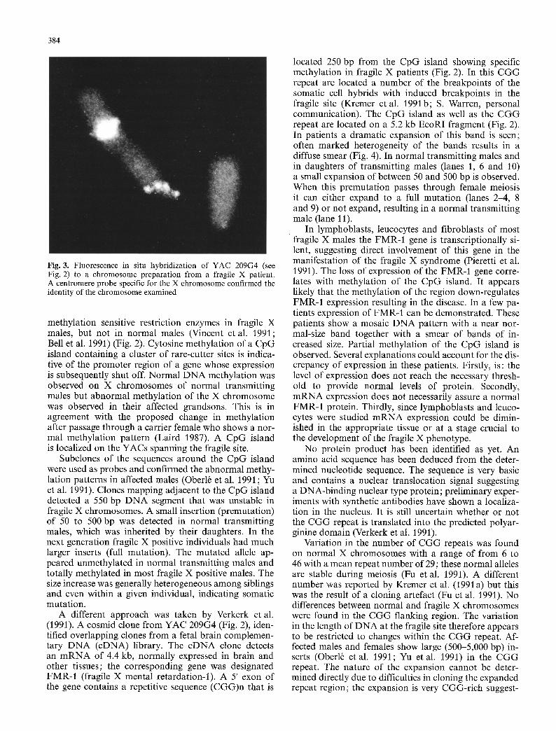
ments up to 1 Mb. YAC libraries became available from normal X chromosomes as well as from a fragile X chro- mosome. By using markers tightly linked to the fragile site YACs were isolated with insert sizes between 275 and 520 kb (Heitz et al. 1991; Verkerk et al. 1991; Kremer et al. 1991 a; Hirst et al. 1991 b). The YACs con- tained breakpoints from the cell hybrids isolated by War- ren (1987), with breakpoints in the fragile region. Fluo- rescence in situ hybridization with these YAC clones was performed on metaphase chromosomes displaying the fragile site. Fluorescent signals were scored on the proximal and on the distal side of the (fragile) gap (Heitz et al. 1991; Verkerk et al. 1991; Kremer et al. 1991a; Hirst et al. 1991b; Verkerk et al. 1992) indicating that the YAC clones indeed span the fragile site (Fig. 3). Hy- bridization of a subclone of YAC 209G4 to a fragile X chromosome is shown on the cover. The fluorescent signal falls on the fragile site. The size of the fragile site has been suggested to be limited to less than 20 kb (Kremer et al. 1991 b; Verkerk et al. 1992). The presence of well-separated signals on both sides of the fragile site indicates that the D N A is locally highly unfolded, An- other explanation could be that a break has occurred in one of the chromatids.
The FMR-1 gene and the nature of the fragile X mutation
Physical studies of the distal region of the long arm of the X chromosome using pulsed field gel electrophore- sis (PFGE) disclosed that the fragile site is located ap- proximately 9 Mb from the telomere (Poustka et al. 1991). Detailed P F G E mapping of the fragile X region showed that rare-cutter restriction sites (rich in CpG dinucleotide sequences) were resistant to digestion by
384
Fig. 3. Fluorescence in situ hybridization of YAC 209G4 (see Fig. 2) to a chromosome preparation from a fragile X patient. A centromere probe specific for the X chromosome confirmed the identity of the chromosome examined
methylation sensitive restriction enzymes in fragile X males, but not in normal males (Vincent et al. 1991; Bell et al. 1991) (Fig. 2). Cytosine methylation of a CpG island containing a cluster of rare-cutter sites is indica- tive of the promoter region of a gene whose expression is subsequently shut off. Normal DNA methylation was observed on X chromosomes of normal transmitting males but abnormal methylation of the X chromosome was observed in their affected grandsons. This is in agreement with the proposed change in methylation after passage through a carrier female who shows a nor- mal methylation pattern (Laird 1987). A CpG island is localized on the YACs spanning the fragile site.
Subclones of the sequences around the CpG island were used as probes and confirmed the abnormal methy- lation patterns in affected males (Oberl6 et al. 1991 ; Yu et al. 1991). Clones mapping adjacent to the CpG island detected a 550 bp DNA segment that was unstable in fragile X chromosomes. A small insertion (premutation) of 50 to 500 bp was detected in normal transmitting males, which was inherited by their daughters. In the next generation fragile X positive individuals had much larger inserts (full mutation). The mutated allele ap- peared unmethylated in normal transmitting males and totally methylated in most fragile X positive males. The size increase was generally heterogeneous among siblings and even within a given individual, indicating somatic mutation.
A different approach was taken by Verkerk et al. (1991). A cosmid clone from YAC 209G4 (Fig. 2), iden- tified overlapping clones from a fetal brain complemen- tary DNA (cDNA) library. The cDNA clone detects an mRNA of 4.4 kb, normally expressed in brain and other tissues; the corresponding gene was designated FMR-1 (fragile X mental retardation-l). A 5' exon of the gene contains a repetitive sequence (CGG)n that is
located 250 bp from the CpG island showing specific methylation in fragile X patients (Fig. 2). In this CGG repeat are located a number of the breakpoints of the somatic cell hybrids with induced breakpoints in the fragile site (Kremer et al. 1991b; S. Warren, personal communication). The CpG island as well as the CGG repeat are located on a 5.2 kb EcoRI fragment (Fig. 2). In patients a dramatic expansion of this band is seen; often marked heterogeneity of the bands results in a diffuse smear (Fig. 4). In normal transmitting males and in daughters of transmitting males (lanes 1, 6 and 10) a small expansion of between 50 and 500 bp is observed. When this premutation passes through female meiosis it can either expand to a full mutation (lanes 2-4, 8 and 9) or not expand, resulting in a normal transmitting male (lane 11).
In lymphoblasts, leucocytes and fibroblasts of most fragile X males the FMR-1 gene is transcriptionally si- lent, suggesting direct involvement of this gene in the manifestation of the fragile X syndrome (Pieretti et al. 1991). The loss of expression of the FMR-I gene corre- lates with methylation of the CpG island. It appears likely that the methylation of the region down-regulates FMR-1 expression resulting in the disease. In a few pa- tients expression of FMR-1 can be demonstrated. These patients show a mosaic DNA pattern with a near nor- mal-size band together with a smear of bands of in- creased size. Partial methylation of the CpG island is observed. Several explanations could account for the dis- crepancy of expression in these patients. Firstly, is: the level of expression does not reach the necessary thresh- old to provide normal levels of protein. Secondly, mRNA expression does not necessarily assure a normal FMR-1 protein. Thirdly, since lymphoblasts and leuco- cytes were studied mRNA expression could be dimin- ished in the appropriate tissue or at a stage crucial to the development of the fragile X phenotype.
No protein product has been identified as yet. An amino acid sequence has been deduced from the deter- mined nucleotide sequence. The sequence is very basic and contains a nuclear translocation signal suggesting a DNA-binding nuclear type protein; preliminary exper- iments with synthetic antibodies have shown a localiza- tion in the nucleus. It is still uncertain whether or not the CGG repeat is translated into the predicted polyar- ginine domain (Verkerk et al. 1991).
Variation in the number of CGG repeats was found on normal X chromosomes with a range of from 6 to 46 with a mean repeat number of 29; these normal alleles are stable during meiosis (Fu et al. 1991). A different number was reported by Kremer et al. (1991 a) but this was the result of a cloning artefact (Fu et al. 1991). No differences between normal and fragile X chromosomes were found in the CGG flanking region. The variation in the length of DNA at the fragile site therefore appears to be restricted to changes within the CGG repeat. Af- fected males and females show large (500-5,000 bp) in- serts (Oberl6 et al. 1991; Yu et al. 1991) in the CGG repeat. The nature of the expansion cannot be deter- mined directly due to difficulties in cloning the expanded repeat region; the expansion is very CGG-rich suggest-

ing that the insert is built up of C G G repeats. Normal transmitting males and their daughters show a premuta- tion with a small expansion. Premutation alleles range in size from 52 to 193 CGGs (Fu et al. 1991). The premu- tation alleles, in contrast to normal alleles, change in size with each meiotic transmission. It will be necessary to analyse a considerable number of alleles to establish whether the boundary between stable alleles (the largest allele 46) and unstable alleles (the smallest premutation allele 52) is really the cut-off point between normal and premutation.
The risk of expansion of the premutation allele is dependent on the size of the allele (Fu et al. 1991). The risk for a female of having a mentally retarded son de- pends on the size of her C G G repeat. I f the repeat number is low ( 5~70 CGGs) the risk of a large expan- sion is low; if the repeat number is high (above 90 CGGs) the risk of expansion to a full mutation is 100%. This can explain the Sherman paradox: the risk that grandsons of normal transmitting males with be mental- ly retarded is 40% and not 50%. The mother of a normal transmitting male (with a low repeat number) has only a 9% risk of having a mentally retarded son.
Diagnostics
Scoring for the expansion has already proved to be a simple diagnostic test for the fragile X syndrome (Oberl~ et al. 1991 ; Yu et al. 1991 ; Rousseau et al. 1991b; Fu et al. 1991). Patients with a full mutat ion can be diag- nosed by Southern blot analysis (see Fig. 4). The insert size is always above 500 bp (Oberl6 et al. 1991 ; Fu et al. 1991). In males a band of near normal size is sometimes
385
seen in addition to the diffuse band; the subject is then mentally retarded. An accurate prediction of mental re- tardation in female carriers is not possible.
Premutation alleles can be studied by analysing the number of C G G repeats. The insert size ranges from 50-500 bp. D N A analysis has not detected an expansion in a number of cases diagnosed previously as fragile X positive; this is most likely the result of misdiagnosis or the existence of a different fragile site (Rousseau et al. 1991 b; Pieretti et al. 1991).
The D N A test for the unstable region is very useful in testing carriers and for prenatal diagnosis (Hirst et al. 1991c; Sutherland et al. 1991). If the prenatal test is carried out on chorionic villi special care has to be taken. In D N A of villi in contrast to fetal D N A no methylation of the CpG island in front of the C G G repeat was ob- served. Thus methylation cannot be used as a diagnostic test in villi. In patients a heterogeneous D NA pattern is often observed indicating a considerable somatic mu- tation rate. If the prenatal test is carried out on villi in a male pregnancy of a female carrier and a premuta- tion is detected this is not sufficient p roof that the status of the mutat ion in the D N A of the villi is indicative of the mutat ion status in the fetus. More data from (re- trospective) tests are needed to solve this problem.
The D N A diagnostic test can be used to screen sys- tematically population of men and women with (unex- plained) mental retardation. Before population screen- ing for carriers can be set up the cut-off points that distinguish between the repeat size of the normal allele, the premutation and the full mutat ion have to be deter- mined in more detail. The ethical implications of such a diagnosis should also be examined.
$
a m n m
2 3 4
cW 8 9 / I f 1 1 12
$
, , . a m n
Fig. 4. Southern blot analysis of EcoRI digested DNA of a frag- ile X family. Squares and circles represent male and female sub- jects, respectively. Open symbols indicate normal subjects; N no cytogenetic fragile X expression. Filled symbols represent subjects that are mentally retarded and show fragile X expression. Half- filled symbols indicate a subject who is normal but shows fragile X expression. Hybridization was performed with probe pP2 (Fig. 2). n normal 5,2 kb band; a small insert of between 50 and 500 bp; s smear of bands

386
Mechanism of mutation
Since fully penetrant males rarely reproduce, a very high mutation rate (1/3,000) has been suggested to maintain the frequency of the disease (Brown 1990). The number of C G G repeats is highly polymorphic suggesting a high mutation rate. Direct testing of the D N A mutation has not revealed any new mutations. This is probably caused by a bias in selecting families with an affected member. This suggests that the transition of a (we)mutat ion to a full mutation most likely takes several generations. The transmission of a premutation over five generations has been noted (A. Smits, personal communication). A high mutation rate is seen in the premutation allele (Fu et al. 1991). Other high mutation rates have been re- ported in small repeats (Jeffreys et al. 1988). An explana- tion could be a pause or stop occurring during D N A replication at the C G G repeat, with subsequent slippage of the polymerase resulting in a change in the number of CGGs. Another mechanism is termination of replica- tion followed by reinitiation at an earlier C G G repeat. Oberl6 et al. (1991) have suggested that the mutation could be due to unequal sister chromatid exchange or to unequal meiotic crossing over.
Normal transmitting males with an expanded C G G repeat never show methylation of the CpG island. Thus, the region is unstable before methylation takes place. It has been suggested that X inactivation (Laird 1987) is involved since the expansion of premutation to full mutation is never observed in daughters of transmitting males. I f this chromosome is inactivated in the daughter as part of the process of dosage compensation, the reac- tivation of this chromosome is blocked in the fragile X region, Sutherland et al. (1991) have observed an ex- panded C G G repeat of the same full mutation size in both chorionic villi and fetal cells, while the methylation was limited to the fetal DNA. This suggests that first expansion has taken place and that the FMR-1 methyla- tion in the fragile X chromosome is acquired during fetal developmental in response to the expansion of the C G G repeat.
Conclusion
Rapid progress has been made in the analysis of the fragile X syndrome during 1991. Different groups have discovered that fragile X chromosomes are preferentially methylated. In these X chromosomes an insertion has been found in the methylated region.
The FMR-1 gene, the transcription of which is shut off in patients, has been isolated. The expansion found in fragile X chromosomes is localized in the coding re- gion of the FMR-1 gene. The fragile X syndrome results from mutations in a (CGG)n repeat found in the coding region of the F M R - I gene. It will be crucial to determine the FMR-1 protein product in order to learn more about the function of the gene.
Diagnosis of the unstable region by D N A analysis is now available as an efficient and reliable test for the diagnosis of carriers, as well as for prenatal diagnosis.
Acknowledgements. We are indebted to Bert Eussen and Jan van Hemel for performing the fluorescence in situ hybridization and to Cathy Bakker for helpful comments on the manuscript.
References
Bell MV, Hirst MC, Nakahori Y, MacKinnon RN, Roche A, Flint TJ, Tommerup N, Tranebjaerg L, Froster-Iskenius U, Kerr B, Turner G, Lindebaum D, Winter R, Pembrey M, Thibodeau S, Davies KE (1991) Physical mapping across the fragile X: Hypermethylation and clinical expression of the fragile X syn- drome. Cell 64:861 866
Brown WT (1990) The fragile X: progress towards solving the puzzle. Am J Hum Genet 47:175-180
Brown WT, Gross AC, Chan C, Jenkins EC (1985) Genetic linkage heterogeneity in the fragile X syndrome. Hum Genet 78:71:11- 18
Brown WT, Gross AC, Chan C, Jenkins EC, Mandel J-L, Oberl~ I, Arveiler B, Novelli G, Thibodeau S, Hagerman R, Summers K, Turner G, White BN, Mulligan L, Forster-Gibson C, Holden JJA, Zoll B, Krawzak M, Gonnewardena P, Gustavson KH, Petterson U, Holmgren C, Schwartz C, Howard-Peebles PN, Murphy P, Breg WR, Veenema H, Carperter NJ (1988) Multilo- cus analysis of the fragile X syndrome. Hum Genet 78:201-205
Camerino G, Mattei MG, Mattei JF, Jaye M, Mandel JL (1983) Close mapping of fragile X mental retardation syndrome to haemophilia B and transmission through a normal male. Na- ture 306 : 701-704
Dahl N, Goonewardena P, Malmgren H, Gustavson KH, Holm- gren G, Seemanowa E, Anneren G, Flood A, Pettersson U (1989) Linkage analysis of families with fragile X mental retar- dation using a novel RFLP marker (DXS304). Am J Hum Gen- et 45 : 304-309
Djabali M, Nguyen C, Biunno I, Oostra BA, Mattei M-G, Ikeda J-E, Jordan BR (1991) Laser microdissection of the fragile X region: identification of cosmid clones and of conserved se- quences in this region. Genomics 10:1053-1060
Fish GS, Arinami T, Froster-Iskenius U, Fryns JP, Curfs LM, Borggraef M, Howard-Peebles PN, Schwartz CE, Simensen R J, Shapiro LR (1991) Relationship between age and IQ among fragile X males: a multicenter study. Am J Med Genet 38:481- 487
Fu Y-H, Kuhl DPA, Pizzuti A, Pieretti M, Sutcliffe J, Richards S, Verkerk AJMH, Holden JJA, Fenwick RG, Warren ST, Oos- tra BA, Nelson DL, Caskey CT (1991) Variation of the CGG repeat at the fragile X site results in genetic instability: resolu- tion of the Sherman paradox. Cell 67:1047-1058
Gustavson KH, Blomquist H, Hotmgren G (1986) Prevalence of fragile X syndrome in mentally retarded children in a Swedish county. Am J Hum Genet 23:581-588
Heitz D, Rousseau F, Devys D, Saccone S, Abderrahim H, Le Paslier D, Cohen D, Vincent A, Toniolo D, Della Valle G, Johson S, Schlessinger D, Oberl~ I, Mandel JL (1991) Isolation of sequences that span the fragile X and identification of a fragile X-related CpG island. Science 251 :I 236-1239
Hirst HC, Roche A, Flint TJ, Mackinnon RN, Bassett JHD, Naka- hori Y, Watson JE, Bell MV, Patterson MN, Boyd Y, Thomas NST, Knight SJL, Warren ST, Hors-Cayla M, Schmidt M, Da- vies KE (1991a) Linear order of new established markers around the fragile site at Xq27.3. Genomics 10:243-249
Hirst MC, Rack K, Makahori Y, Roche A, Bell MV, Flynn G, Christadoulou Z, Mackinnon RN, Francis M, Littler AJ, An- and R, Poustka A-M, Lehrach H, Schlessinger D, D'Urso M, Buckle VJ, Davies KE (1991 b) A YAC contig across the fragile X site defines the region of fragility. Nucleic Acids Res 19: 3283-3288
Hirst MC, Knight S, Davies K, Cross G, Ocraft K, Raeburn S, Heeger S, Eunpu D, Jenkins EC, Lindenbaum R (1991 c) Prena- tal diagnosis of fragile X syndrome. Lancet 338 : 956-957

387
Hulsebos ThJM, Oostra BA, Broersen S, van Oost BA, Westerveld A (1991) New distal marker closely linked to the fragile X locus. Hum Genet 87:369-372
Jacky PB, Ahuja YR, Anyane-Yeboa K, Breg WR, Carpenter NJ, Froster-Iskenius UG, Fryns JP, Glover TW, Gustavson KH, Hoegerman SF, Holmgren G, Howard-Peebles PN, Jenkins EC, Krawczun M, Neri G, Pettigrew A, Schaap T, Schonber SA, Shapiro LR, Spinner N, Steinbach P, Vianna-Morgante AM, Watson MS, Wilmot PL (1991) Guidelines for the preparation and analysis of the fragile X chromosome in lymphocytes. Am J Med Genet 38:400-403
Jeffreys AJ, Roule NJ, Wilson V, Wong Z (1988) Spontaneous mutation rates to new length alleles at tandem-repetitive hyper- variable loci in human DNA. Nature 332:278 281
Kremer E J, Yu S, Pritchard M, Nagaraja R, Heitz D, Lynch M, Baker E, Hyland VJ, Little RD, Wada M, Toniolo D, Vincent A, Rousseau F, Schlessinger D, Sutherland G, Richards RI (1991 a) Isolation of a human DNA sequence which spans the fragile X. Am J Hum Genet 49:65(~661
Kremer EJ, Pritchard M, Lynch M, Yu S, Holman K, Baker E, Warren ST, Schlessinger D, Sutherland GR, Richards RI (1991 b) Mapping of DNA instability at the fragile X to a trin- ucleotide repeat sequence p(CCG)n. Science 252:1711-1714
Laird CD (1987) Proposed mechanism of inheritance and expres- sion of the human fragile-X syndrome of mental retardation. Genetics 117 : 582599
Lubs HA (1969) A marker X-chromosome. Am J Hum Genet 21 : 231-244
Mackinnon RN, Hirst MC, Bell MV, Watson JEV, Claussen U, Ludecke HJ, Senger G, Hortshemke B, Davies KE (1990) Mi- crodissection of the fragile X region. Am J Hum Genet 47 : 181- 187
Martin JP, Bell J (1943) A pedigree of mental defect showing sex- linkage. J Neurol Psych 6:154-157
Nielsen KB, Tommerup N, Poulsen H, Jacobsen P, Beck B, Mikkel- sen M (1983) Carrier detection and X inactivation studies in the fragile X syndrome. Cytogenetic studies in 63 obligate and potential carriers of the fragile X. Hum Genet 64: 240-245
Oberl6 I, Rousseau F, Heitz D, Kretz C, Devys D, Hanauer A, Bou6 J, Bertheas MF, Mandel JF (1991) Instability of a 550- base pair DNA segment and abnormal methylation in fragile X syndrome. Science 252:1097-1102
Oostra BA, Hupkes PE, Perdon LF, van Bennekom CA, Bakker E, Halley DJJ, Schmidt M, Du Sart D, Smits A, Wieringa B, van Oost BA (1990) New polymorphic DNA marker close to the fragile site FRAXA. Genomics 6:129-132
Opitz JM, Sutherland GR (1984) Conference report: International workshop on the fragile X and X-linked mental retardation. Am J Med Genet 17 : 5 94
Pembrey ME, Winter RM, Davies KE (1985) A premutation that generates a defect at crossing over explains the inheritance of Fragile X mental retardation. Am J Med Genet 21:709-717
Pieretti M, Zhang F, Fu Y-H, Warren ST, Oostra BA, Caskey CT, Nelson DL (1991) Absence of expression of the FMR-1 gene in fi'agile X syndrome. Cell 66:817-822
Poustka A, Dietrich A, Langenstein G, Toniolo D, Warren ST, Lehrach H ( 1991) Physical map of human Xq27-qter: localizing the region of the fragile X mutation. Proc Natl Acad Sci USA 88 : 830~8306
Riggins GJ, Sherman SL, Oostra BA, Feitell D, Sutcliffe JS, Nelson DL, van Oost BA, Smits APT, Kuhl D, Caskey CT, Warren ST (1992) Characterization of a highly polymorphic dinucleo- tide repeat 150 kb proximal to the fragile site. Am J Med Genet, in press
Rousseau F, Vincent A, Rivella S, Heitz D, Tribioli C, Maestrini E, Warren ST, Suthers GK, Goodfellow P, Mandel JL, Toniolo D, Oberl6 I (1991a) Four chromosomal breakpoints and four
new probes mark out a 10cM region encompassing the FRAXA locus. Am J Hum Genet 48:108 116
Rousseau F, Heitz D, Biancalana V, Blumenfeld S, Kretz C, Bouh J, Tommerup N, van der Hagen C, DeLozier-Blanchet C, Cro- quette M-F, Gilgenkrantz S, Jalbert P, Voelckel M-A, Oberl6 I, Mandel J-L (199b) Direct diagnosis by DNA analysis of the fragile X syndrome of mental retardation. New Engl J Med 325 : 1673-1681
Sherman SL, Jacobs PA, Morton NE, Froster-Iskenius U, Howard- Peebles PN, Nielsen KB, Partington NW, Sutherland GR, Turner G, Watson M (1985) Further segregation of the fragile X syndrome with special reference to transmitting males. Hum Genet 69 : 3289-3299
Sutherland GR (1977) Fragile sites on human chromosome: dem- onstration of their dependence on the type of tissue culture medium. Science J97:265-266
Sutherland GR, Baker E (1986) Effects of nudeotides on expres- sion of the folate sensitive fragile site. Am J Med Genet 23:409- 417
Sutherland GR, Hecht F (1985) Fragile sites on human chromo- somes. Oxford University Press, New York, pp 113-131
Sutherland GR, Ledbetter DH (1989) Report of the committee on cytogenetic markers. Cytogenet Cell Genet 51:452~458
Sutherland GR, Gedeon A, Kornman L, Donnelly A, Byard RW, Mulley JC, Kremer E, Lynch M, Pritchard M, Yu S, Richards RI (1991) Prenatal diagnosis of fragile X syndrome by direct detection of the unstable DNA sequence. N Engl J Med 325:1720-1722
Suthers GK, Callen CF, Hyland VJ, Kozman HM, Baker E, Eyre H, Harper PS, Roberts SH, Hors-Cayla MC, Davies KE, Bell MV, Sutherland GR (1989) A new DNA marker tightly linked to the fi'agile X locus (FRAXA). Science 246:1298-1300
Suthers GK, Mulley JC, Voelckel MA, Dahl N, Vaisanen ML, Steinbach P, Glass IA, Schwartz CE, van Oost BA, Thibodeau SN, Haites NE, Oostra BA, Schinzel A, Carballo M, Morris CP, Hopwood JJ, Sutherland GF (1991 a) Linkage homogeneity near the fragile X locus in normal and fragile X families. Geno- mics 10:576582
Suthers GK, Mulley JC, Voelckel MA, Dahl N, Vaisanen ML, Steinbach P, Glass IA, Schwartz CE, van Oost BA, Thibodeau SN, Haites NE, Oostra BA, Gin+ R, Carballo M, Morris CP, Hopwood JJ, Sutherland GR (1991 b) Genetic mapping of new DNA probes at Xq27 defines a strategy for DNA studies in the fragile X syndrome. Am J Hum Genet 48 : 460-467
Verkerk AJMH, Pieretti M, Sutcliffe JS, Fu Y, Kuhl DPA, Pizzuti A, Reiner O, Richards S, Victoria MF, Zhang F, Eussen BE, van Ommen G-JB, Blonden LAJ, Riggins G J, Chastain JL, Kunst CB, Galjaard H, Caskey CT, Nelson DL, Oostra BA, Warren ST (1991) Identification of a gene (FMR-I) containing a CGG repeat coincident with a fragile X breakpoint duster region exhibiting length variation in fragile X syndrome. Cell 65 : 905-914
Verkerk AJHM, Eussen EHJ, van Hemel JO, Oostra BA (1992) The limited size of the fragile site shown by fluorescence in situ hybridization. Am J Med Genet (in press)
Vincent A, Heitz D, Petit C, Kretz C, Oberl6 I, Mandel JF (1991) Abnormal pattern detected in fragile X patients by pulse-field gel electrophoresis. Nature 349:624-626
Warren ST, Zhang F, Licamelli GR, Peters JF (1987) The fragile X site in somatic cell hybrids: an approach for molecular clon- ing of fragile sites. Science 237:420-423
Webb TP, Bundey SE, Thake AI, Todd J (1986) Population inci- dence and segregation ratios in the Martin-Bell syndrome. Am J Med Genet 23 : 573-580
Yu S, Pritchard M, Kremer E, Lynch M, Nancarrow J, Baker E, Holman K, Mulley JC, Warren ST, Schlessinger D, Suther- land GR, Richards RI (1991) Fragile X genotype characterized by an unstable region of DNA. Science 252:1179-1181