Review The evolution of irradiance detection: melanopsin
17
Review The evolution of irradiance detection: melanopsin and the non-visual opsins Stuart N. Peirson, Stephanie Halford and Russell G. Foster* Nuffield Laboratory of Ophthalmology, University of Oxford, Level 5 and 6 West Wing, The John Radcliffe Hospital, Headley Way, Headington, Oxford OX3 9DU, UK Circadian rhythms are endogenous 24 h cycles that persist in the absence of external time cues. These rhythms provide an internal representation of day length and optimize physiology and behav- iour to the varying demands of the solar cycle. These clocks require daily adjustment to local time and the primary time cue (zeitgeber) used by most vertebrates is the daily change in the amount of environmental light (irradiance) at dawn and dusk, a process termed photoentrainment. Attempts to understand the photoreceptor mechanisms mediating non-image-forming responses to light, such as photoentrainment, have resulted in the discovery of a remarkable array of different photo- receptors and photopigment families, all of which appear to use a basic opsin/vitamin A-based photopigment biochemistry. In non-mammalian vertebrates, specialized photoreceptors are located within the pineal complex, deep brain and dermal melanophores. There is also strong evidence in fish and amphibians for the direct photic regulation of circadian clocks in multiple tissues. By con- trast, mammals possess only ocular photoreceptors. However, in addition to the image-forming rods and cones of the retina, there exists a third photoreceptor system based on a subset of melanopsin- expressing photosensitive retinal ganglion cells (pRGCs). In this review, we discuss the range of vertebrate photoreceptors and their opsin photopigments, describe the melanopsin/pRGC system in some detail and then finally consider the molecular evolution and sensory ecology of these non-image-forming photoreceptor systems. Keywords: circadian; melanopsin; photopigment; photoreceptor 1. INTRODUCTION The 24 h cycle of light and dark caused by the rotation of the Earth produces dramatic but predictable changes in the light environment. Instead of passively responding to these changes, organisms have evolved an endogenous representation of the 24 h day—a cir- cadian timing system. These circadian clocks set the time or phase at which physiological and behavioural events occur with respect to the external 24 h environ- mental cycle. In this way, change can be anticipated, and physiology is optimized to the varying demands of night and day. This allows the organism to exploit the changed conditions as soon as they take place; avoiding the time lost in physiological and behavioural adjustments. However, the circadian system will only provide a selective advantage if biological time remains synchronized (entrained) to environmental time. Thus, the circadian oscillator requires a daily synchro- nization with the external environment via time cues termed zeitgebers (time givers) (Aschoff 1984; Pittendrigh 1993). The systematic daily change in the gross amount of environmental light (irradiance) at dawn or dusk provides the primary indicator of the time of day. As a result, most organisms have evolved to use the twilight transition as their main zeitgeber to adjust circadian time to local time. This process is termed photoentrainment (Roenneberg & Foster 1997). The study of the photoreceptors mediating irradi- ance-detection tasks such as photoentrainment has led to the identification of a range of vertebrate opsins, and perhaps most remarkably, the identifi- cation of a novel photoreceptor system within the mammalian retina, a subset of retinal ganglion cells (RGCs) that express the photopigment melanopsin (Opn4). In this review, we will consider the range of vertebrate photoreceptors and their opsin photo- pigments and then provide an overview of the melanopsin/photosensitive retinal ganglion cell (pRGC) system in detail. Finally, we will discuss the molecular evolution of the opsin photopigments and consider the different selective pressures acting upon visual pigments and non-image-forming photopigments in general. 2. VERTEBRATE PHOTORECEPTORS In mammals, both visual and non-visual photorecep- tion is ocular, and enucleation abolishes all responses to light (Nelson & Zucker 1981; Foster et al. 1991). By contrast, non-mammalian vertebrates possess a wide range of photoreceptive sites, including the pineal complex, deep-brain photoreceptors and dermal photoreceptors (Shand & Foster 1999). As * Author for correspondence ([email protected]). One contribution of 13 to a Theme Issue ‘The evolution of phototransduction and eyes’. Phil. Trans. R. Soc. B (2009) 364, 2849–2865 doi:10.1098/rstb.2009.0050 2849 This journal is q 2009 The Royal Society
Transcript of Review The evolution of irradiance detection: melanopsin
Review
The evolution of irradiance detection:melanopsin and the non-visual opsins
Stuart N. Peirson, Stephanie Halford and Russell G. Foster*
Nuffield Laboratory of Ophthalmology, University of Oxford, Level 5 and 6 West Wing,The John Radcliffe Hospital, Headley Way, Headington, Oxford OX3 9DU, UK
Circadian rhythms are endogenous 24 h cycles that persist in the absence of external time cues.These rhythms provide an internal representation of day length and optimize physiology and behav-iour to the varying demands of the solar cycle. These clocks require daily adjustment to local timeand the primary time cue (zeitgeber) used by most vertebrates is the daily change in the amount ofenvironmental light (irradiance) at dawn and dusk, a process termed photoentrainment. Attemptsto understand the photoreceptor mechanisms mediating non-image-forming responses to light,such as photoentrainment, have resulted in the discovery of a remarkable array of different photo-receptors and photopigment families, all of which appear to use a basic opsin/vitamin A-basedphotopigment biochemistry. In non-mammalian vertebrates, specialized photoreceptors are locatedwithin the pineal complex, deep brain and dermal melanophores. There is also strong evidence infish and amphibians for the direct photic regulation of circadian clocks in multiple tissues. By con-trast, mammals possess only ocular photoreceptors. However, in addition to the image-forming rodsand cones of the retina, there exists a third photoreceptor system based on a subset of melanopsin-expressing photosensitive retinal ganglion cells (pRGCs). In this review, we discuss the range ofvertebrate photoreceptors and their opsin photopigments, describe the melanopsin/pRGC systemin some detail and then finally consider the molecular evolution and sensory ecology of thesenon-image-forming photoreceptor systems.
Keywords: circadian; melanopsin; photopigment; photoreceptor
1. INTRODUCTIONThe 24 h cycle of light and dark caused by the rotationof the Earth produces dramatic but predictablechanges in the light environment. Instead of passivelyresponding to these changes, organisms have evolvedan endogenous representation of the 24 h day—a cir-cadian timing system. These circadian clocks set thetime or phase at which physiological and behaviouralevents occur with respect to the external 24 h environ-mental cycle. In this way, change can be anticipated,and physiology is optimized to the varying demandsof night and day. This allows the organism to exploitthe changed conditions as soon as they take place;avoiding the time lost in physiological and behaviouraladjustments. However, the circadian system will onlyprovide a selective advantage if biological time remainssynchronized (entrained) to environmental time.Thus, the circadian oscillator requires a daily synchro-nization with the external environment via time cuestermed zeitgebers (time givers) (Aschoff 1984;Pittendrigh 1993). The systematic daily change inthe gross amount of environmental light (irradiance)at dawn or dusk provides the primary indicator ofthe time of day. As a result, most organisms have
evolved to use the twilight transition as their mainzeitgeber to adjust circadian time to local time. Thisprocess is termed photoentrainment (Roenneberg &Foster 1997).
The study of the photoreceptors mediating irradi-ance-detection tasks such as photoentrainment hasled to the identification of a range of vertebrateopsins, and perhaps most remarkably, the identifi-cation of a novel photoreceptor system within themammalian retina, a subset of retinal ganglion cells(RGCs) that express the photopigment melanopsin(Opn4). In this review, we will consider the range ofvertebrate photoreceptors and their opsin photo-pigments and then provide an overview of themelanopsin/photosensitive retinal ganglion cell (pRGC)system in detail. Finally, we will discuss the molecularevolution of the opsin photopigments and consider thedifferent selective pressures acting upon visual pigmentsand non-image-forming photopigments in general.
2. VERTEBRATE PHOTORECEPTORSIn mammals, both visual and non-visual photorecep-tion is ocular, and enucleation abolishes all responsesto light (Nelson & Zucker 1981; Foster et al. 1991).By contrast, non-mammalian vertebrates possess awide range of photoreceptive sites, including thepineal complex, deep-brain photoreceptors anddermal photoreceptors (Shand & Foster 1999). As
* Author for correspondence ([email protected]).
One contribution of 13 to a Theme Issue ‘The evolution ofphototransduction and eyes’.
Phil. Trans. R. Soc. B (2009) 364, 2849–2865
doi:10.1098/rstb.2009.0050
2849 This journal is q 2009 The Royal Society

well as being anatomically diverse (figure 1), thesephotoreceptors mediate many different aspects ofphysiology and behaviour. Identifying the extraretinalopsin photopigments that underlie these responses innon-mammalian photoreceptors has a long and fairlycomplex history. Many immunocytochemical studieswere undertaken on these tissues, using a wide rangeof antibodies raised to different retinal or visual pig-ment preparations (Shand & Foster 1999). However,as the epitopes/specificities of many of these antibodieswere not known, it was difficult to make any definitiveconclusions regarding the molecular identity of themolecules labelled. The ambiguous terms ‘rod-like’or ‘cone-like’ were frequently used to describe suchimmunolabelling. The molecular characterization ofthe extraretinal photopigments became further blurredwith the discovery of multiple new opsin families, quitedifferent from the rod and cone opsins. Parallel studieson mammals also produced results that were initiallydifficult to interpret. Although it was clear that mam-mals lack extraocular photoreceptors (Nelson &Zucker 1981; Foster et al. 1991), mice lacking rodand cone photoreceptors could still regulate multiplephysiological responses to light (Freedman et al.1999; Lucas et al. 1999). Further, these responseswere clearly being mediated by an opsin/vitamin A-based photopigment system (Lucas et al. 2001). Yetuntil recently the molecular identity of this photo-receptor system remained unknown. In the pastdecade much new information has emerged regardingthe location and function of the vertebrate non-rod,non-cone photoreceptor systems. Here in §2, we take
the opportunity to summarize some of these findings,and in §3 we discuss the photopigment biochemistry indetail.
(a) Lateral eyesThe lateral eyes are the most familiar photoreceptivesite in vertebrates. The classical photoreceptors ofthe vertebrate retina consist of the rods and cones.Rods mediate scotopic (dim light) vision, providinglow-resolution but high sensitivity, whereas cones areinvolved in photopic (bright light) vision, and enablehigh-resolution colour vision/contrast detection. Rodand cone light detection is characterized by rapidand transient electrical responses. The graded poten-tials from these receptors are processed by inner retinalneurons prior to advanced visual processing in thebrain. Light information reaches the visual centres ofthe brain via topographically mapped axons of theRGCs that form the optic nerve (Rodieck 1998). Butin addition to the ‘classical’ photoreceptors of theouter retina, other retinal cells are now also knownto be capable of responding to light. A subset ofRGCs (approx. 1% in the mouse) expresses the photo-pigment melanopsin (Opn4; Hattar et al. 2002) and arecapable of responding to light directly (Berson et al.2002; Sekaran et al. 2003). The identification ofthese pRGCs is discussed in more detail in §4.
In addition to melanopsin-based pRGCs, the teleostretina (and perhaps other non-mammalian retinae)possesses photosensitive horizontal cells. In the cyprinidretina of the roach (Rutilus rutilus), a subtype of the
OT Tel Cer
pineal peripheral oscillators
Hyp deep brain
brain
eye
retinadermal iridophore/melanophore
iris
Figure 1. Photoreceptive sites in the vertebrates. As well as the classical photoreceptors within the retina of the lateral eye,direct photoreception in the isolated iris has also been described. In non-mammalian species the pineal complex also containsphotoreceptors, and deep brain photoreceptors may also occur. Dermal photoreception has been described in amphibians andfish. Finally, in the zebrafish peripheral tissues have been shown to be able to entrain their molecular oscillators directly to light.See text for further details.
2850 S. N. Peirson et al. Review. Melanopsin and the non-visual opsins
Phil. Trans. R. Soc. B (2009)

horizontal cell, termed HC-RSD, expresses bothmelanopsin and vertebrate ancient (VA) opsin andshows depolarizing responses to light that are maximallysensitive to approximately 477 nm. These cells havelonger integration times than rods or cones andmaintain their responses when classical photoreceptorinputs are saturated by background light (Jenkinset al. 2003). These cells may signal environmentalirradiance as well as modulating rod and cone outputs.
(b) PinealPerhaps the best known photoreceptive site outside theretina is the pineal organ (epiphysis cerebri). Here wewill use the term pineal complex to refer to theintracranial pineal proper as well as the parapinealand the extracranial ‘third’ eyes found in tuatara(Sphenadon punctatus, Rhynchocephalia), some lizards(Squamata) and frogs (Anura). The intracranial para-pineal organ only occurs in some species of fish, andremarkably little is known about the physiologicalfunctions of this enigmatic organ (Vollrath 1981;Shand & Foster 1999). The extracranial third eyescan be further subdivided into the frontal organs (orStirnorgan) of anuran amphibians and the parietaleyes found in lizards. The parietal eye shows remark-ably structural similarity to the lateral eyes, with atransparent cornea and lens (Shand & Foster 1999).
Embryologically, the pineal complex is derived froman evagination of the dorsal diencephalon, similar tothe retina, and in non-mammalian vertebrates islocated near the surface of the brain (Vollrath 1981).In teleost and cyclostome species there is often atranslucent window or area of reduced pigmentationoverlying the pineal, allowing approximately 10 percent of the incident light to reach the pineal. In amphi-bians, reptiles and birds, such a pineal window is lessapparent or absent. Despite this, a considerableamount of light is still able to penetrate the overlyingtissues, amounting to 0.1 per cent20.3 per cent ofthe incident light (Dodt & Meissl 1982). In allnon-mammalian vertebrates, the pineal complexis photoreceptive, and the predominant cell type isphotoreceptor-like in appearance. In mammals, thepineal organ expresses many elements of the photo-transduction cascade (Korf et al. 1985a,b), but lacksphotosensitivity and appears exclusively secretory(Foster et al. 1989, 2003). The pineal organ is the pri-mary source of the neurohormone melatonin, which issynthesized in the dark phase of the light/dark cycle,and acts as a signal of darkness to regulate circadianrhythms and photoperiodic responses (Arendt 1998;Korf et al. 1998). Melatonin synthesis is locallyregulated by light at the level of the pineal in non-mammalian vertebrates, but in mammals photicinformation reaches the pineal via a multisynapticpathway via the retinohypothalamic tract (RHT) andthe superior cervical ganglion of the sympatheticnervous system (Korf & Moller 1984; Meissl 1997).
A range of opsins has been detected in the pinealcomplex of vertebrates (Shand & Foster 1999). Oneof the first extraretinal opsins to be identified, Pinopsin(P-opsin), was isolated from the avian pineal (Okanoet al. 1994; Max et al. 1995). In the teleost pineal, a
range of rod and cone opsins along with VA-opsinare expressed (Philp et al. 2000b). However, it appearsthat the predominant opsin in the fish pineal is a rod-like opsin (exo-rhodopsin/extraretinal rod-like opsin)which differs from that found in the lateral eyes(Mano et al. 1999; Philp et al. 2000a). Despite thelack of information about the structure and functionof the parapineal organ a novel opsin photopigment,parapinopsin, has been isolated specifically from theparapineal of the channel catfish (Ictalarus punctatus)(Blackshaw & Snyder 1997) as well as from the lam-prey pineal (Koyanagi et al. 2004). Most recently,studies on the parietal eye have identified the expressionof two opsins within the same cell, a blue-sensitivepinopsin and a novel green-sensitive opsin named parie-topsin (Su et al. 2006).
(c) Deep brainDeep-brain photoreceptors were first described follow-ing studies by Karl von Frisch in 1911 on blinded andpinealectomized European minnows (Phoxinus phoxi-nus). These fish still demonstrated colour changesin response to light, leading to the suggestion of‘deep-diencephalic photoreceptors’ (von Frisch1911). Similarly, studies in blinded pinealectomizedEuropean eels (Anguilla anguilla) by van Veen et al.(1976) showed that deep-brain photoreceptors med-iate photoentrainment as well as negative phototaxis.The photoperiodic response in birds, whereby gonadalgrowth is regulated by day length, is also mediated by adeep-brain photoreceptor rather than by the pinealcomplex or lateral eyes (Benoit 1964). Action spec-troscopy provided a clue as to the molecular identityof these photoreceptors. An absorption correctedaction spectrum for photoperiodic induction in theJapanese quail described an opsin/vitamin A-basedphotopigment with a lmax at 492 nm. A recent reana-lysis of the original data suggests that the lmax may becloser to 483 nm (S. N. Peirson & R. G. Foster 2008,unpublished data). Although this action spectruminferred the biochemistry of the photopigment, the pre-cise molecular identity still remains unresolved. Attemptsto characterize these photoreceptors have involved theuse of immunocytochemical techniques employing anti-bodies raised against rod and cone photopigment opsinsor other elements of the phototransduction cascade.Such approaches either failed to localize opsins withinthe avian hypothalamus or produced ambiguous resultsowing to the use of unknown epitopes (Silver et al.1988).
(d) IrisLight striking the isolated iris has been reported toproduce rapid sphincter pupillae constriction in severalnon-mammalian species of vertebrate. For example,an opsin/vitamin A type action spectrum has beendescribed from the isolated frog iris (Barr & Alpern1963) and eel iris (Selinger 1962). In the isolatedchick iris, there are marked and rapid responses tolight (Tu et al. 2004). In some mammalian species,there have also been reports of an extremely gradualpupil constriction (occurring over at least 20 s to verybright stimuli) that survives both isolation of the iris
Review. Melanopsin and the non-visual opsins S. N. Peirson et al. 2851
Phil. Trans. R. Soc. B (2009)

from the eye and application of atropine (Bito &Turansky 1975; Lau et al. 1992). In all cases, adefinitive photopigment characterization for irisphotosensitivity is lacking, although in Xenopus laevismelanopsin has been implicated (Provencio et al.1998).
(e) DermalPhotoreception by dermal cells mediates colourchanges in chromatophores and iridophores. Dermalphotoreception has also been linked to the triggeringof locomotor activity (Wolken & Mogus 1979;Shand & Foster 1999). Dermal chromatophores arephotosensitive in many vertebrates, regulating theaggregation and dispersal of pigment granules withinthese cells (Weber 1983). Melanopsin, the photopig-ment of retinal pRGCs, was first isolated from Xenopuslaevis melanophores (Provencio et al. 1998) andappears to be the photopigment mediating the pig-ment dispersal responses within dermal melanophores(Isoldi et al. 2005).
(f) Tissue photoreceptionPerhaps the most surprising result in recent years hasbeen the discovery that the circadian clocks locatedwithin the peripheral organs (such as the heart and
kidney) of zebrafish can be entrained to light afterbeing isolated and maintained in vitro (Whitmoreet al. 2000). The photopigment(s) mediating theseresponses remain poorly understood, although thewidespread expression pattern of teleost multipletissue (TMT) opsin (see below) makes this opsin astrong candidate.
3. OPSIN PHOTOPIGMENTSAll vertebrate photoreceptors identified to date use anopsin/vitamin A-based photopigment. These photo-pigments consist of an opsin protein bound to avitamin-A chromophore. In most terrestrial and marinevertebrates, this chromophore is 11-cis-retinaldehyde(A1), while in many freshwater vertebrates the chromo-phore is based upon 11-cis-3-dehydroretinal (A2)(Knowles & Dartnall 1977). The first stage of lightdetection involves the absorption of a photon by the reti-nal chromophore and the photoisomerization of thismolecule to the all-trans state (figure 2a). The confor-mational change of the chromophore allows the opsinto interact with a G-protein (transducin) and triggersthe phototransduction cascade, ultimately giving rise toa change in receptor membrane potential (Hargrave &McDowell 1992; Okada et al. 2001; Pepe 2001;Shichida & Matsuyama 2009). All vitamin A-based
K
0.0001
0.0010
0.0100
0.1000
1.0000
10.0000
300 400 500 600 700 800
C-terminus
N-terminus
cellmembrane
intracellular
extracellular
O
O
(a) (b)
(c)
hv
wavelength
abso
rptio
n
Figure 2. Structure and function of vertebrate photopigments. Vertebrate photopigments consist of an isomer of vitamin A,retinaldehyde, bound to an opsin protein. (a) The primary step in phototransduction is the absorption of a photon of light(hv) by the 11-cis isomer of retinal resulting in isomerization to the all-trans form. (b) All vitamin A/opsin-based photopigmentshave a characteristic absorption spectrum which can be used as a ‘spectral fingerprint’ to determine the photopigment mediat-ing a given biological response. (c) Opsins consist of a single polypeptide chain forming seven a-helical transmembrane regionsconnected by cytoplasmic and extracellular loops. The intracellular domains mediate G-protein interactions. The retinalbinding site (K) is indicated in the 7th transmembrane domain. Structure based on that of Palczewski et al. 2000.
2852 S. N. Peirson et al. Review. Melanopsin and the non-visual opsins
Phil. Trans. R. Soc. B (2009)
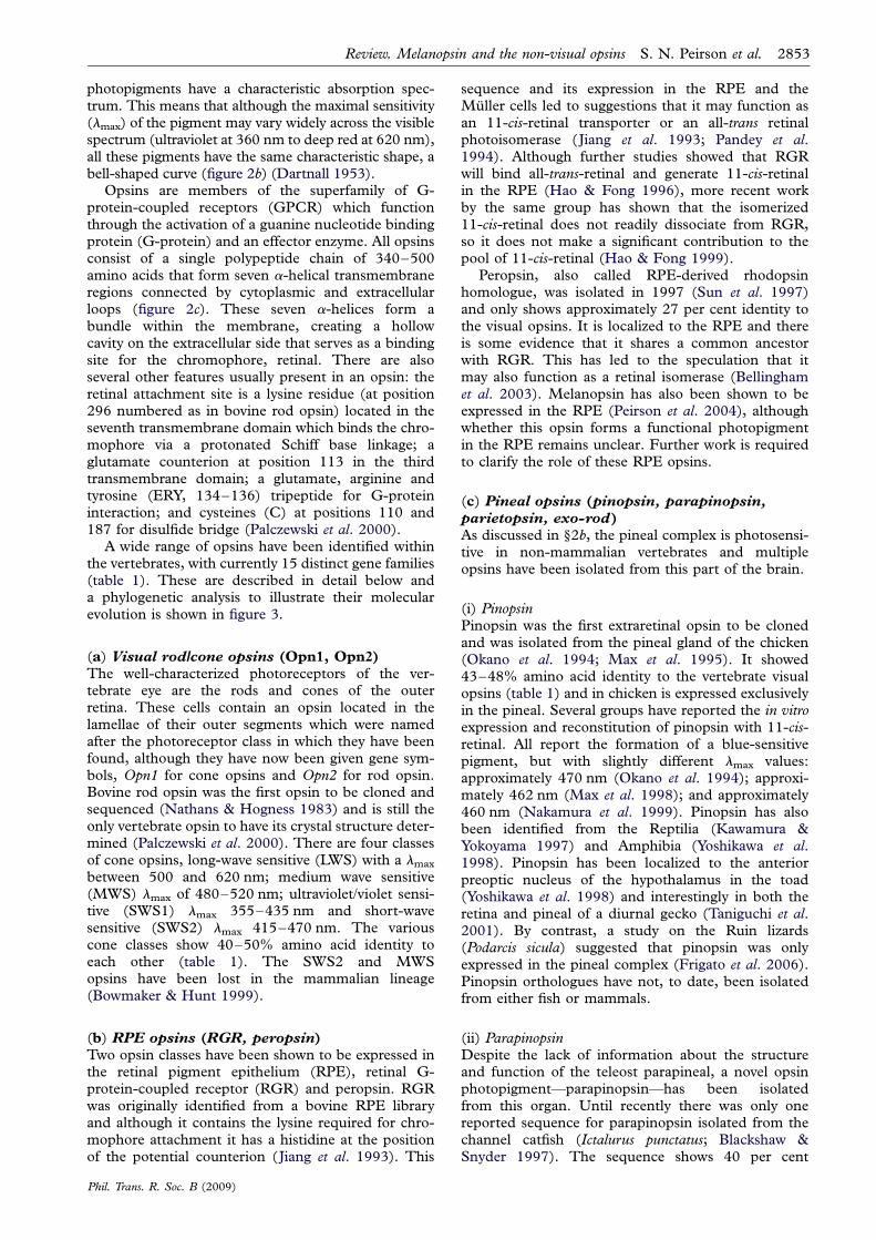
photopigments have a characteristic absorption spec-trum. This means that although the maximal sensitivity(lmax) of the pigment may vary widely across the visiblespectrum (ultraviolet at 360 nm to deep red at 620 nm),all these pigments have the same characteristic shape, abell-shaped curve (figure 2b) (Dartnall 1953).
Opsins are members of the superfamily of G-protein-coupled receptors (GPCR) which functionthrough the activation of a guanine nucleotide bindingprotein (G-protein) and an effector enzyme. All opsinsconsist of a single polypeptide chain of 340–500amino acids that form seven a-helical transmembraneregions connected by cytoplasmic and extracellularloops (figure 2c). These seven a-helices form abundle within the membrane, creating a hollowcavity on the extracellular side that serves as a bindingsite for the chromophore, retinal. There are alsoseveral other features usually present in an opsin: theretinal attachment site is a lysine residue (at position296 numbered as in bovine rod opsin) located in theseventh transmembrane domain which binds the chro-mophore via a protonated Schiff base linkage; aglutamate counterion at position 113 in the thirdtransmembrane domain; a glutamate, arginine andtyrosine (ERY, 134–136) tripeptide for G-proteininteraction; and cysteines (C) at positions 110 and187 for disulfide bridge (Palczewski et al. 2000).
A wide range of opsins have been identified withinthe vertebrates, with currently 15 distinct gene families(table 1). These are described in detail below anda phylogenetic analysis to illustrate their molecularevolution is shown in figure 3.
(a) Visual rod/cone opsins (Opn1, Opn2)The well-characterized photoreceptors of the ver-tebrate eye are the rods and cones of the outerretina. These cells contain an opsin located in thelamellae of their outer segments which were namedafter the photoreceptor class in which they have beenfound, although they have now been given gene sym-bols, Opn1 for cone opsins and Opn2 for rod opsin.Bovine rod opsin was the first opsin to be cloned andsequenced (Nathans & Hogness 1983) and is still theonly vertebrate opsin to have its crystal structure deter-mined (Palczewski et al. 2000). There are four classesof cone opsins, long-wave sensitive (LWS) with a lmax
between 500 and 620 nm; medium wave sensitive(MWS) lmax of 480–520 nm; ultraviolet/violet sensi-tive (SWS1) lmax 355–435 nm and short-wavesensitive (SWS2) lmax 415–470 nm. The variouscone classes show 40–50% amino acid identity toeach other (table 1). The SWS2 and MWSopsins have been lost in the mammalian lineage(Bowmaker & Hunt 1999).
(b) RPE opsins (RGR, peropsin)Two opsin classes have been shown to be expressed inthe retinal pigment epithelium (RPE), retinal G-protein-coupled receptor (RGR) and peropsin. RGRwas originally identified from a bovine RPE libraryand although it contains the lysine required for chro-mophore attachment it has a histidine at the positionof the potential counterion (Jiang et al. 1993). This
sequence and its expression in the RPE and theMuller cells led to suggestions that it may function asan 11-cis-retinal transporter or an all-trans retinalphotoisomerase (Jiang et al. 1993; Pandey et al.1994). Although further studies showed that RGRwill bind all-trans-retinal and generate 11-cis-retinalin the RPE (Hao & Fong 1996), more recent workby the same group has shown that the isomerized11-cis-retinal does not readily dissociate from RGR,so it does not make a significant contribution to thepool of 11-cis-retinal (Hao & Fong 1999).
Peropsin, also called RPE-derived rhodopsinhomologue, was isolated in 1997 (Sun et al. 1997)and only shows approximately 27 per cent identity tothe visual opsins. It is localized to the RPE and thereis some evidence that it shares a common ancestorwith RGR. This has led to the speculation that itmay also function as a retinal isomerase (Bellinghamet al. 2003). Melanopsin has also been shown to beexpressed in the RPE (Peirson et al. 2004), althoughwhether this opsin forms a functional photopigmentin the RPE remains unclear. Further work is requiredto clarify the role of these RPE opsins.
(c) Pineal opsins (pinopsin, parapinopsin,parietopsin, exo-rod)As discussed in §2b, the pineal complex is photosensi-tive in non-mammalian vertebrates and multipleopsins have been isolated from this part of the brain.
(i) PinopsinPinopsin was the first extraretinal opsin to be clonedand was isolated from the pineal gland of the chicken(Okano et al. 1994; Max et al. 1995). It showed43–48% amino acid identity to the vertebrate visualopsins (table 1) and in chicken is expressed exclusivelyin the pineal. Several groups have reported the in vitroexpression and reconstitution of pinopsin with 11-cis-retinal. All report the formation of a blue-sensitivepigment, but with slightly different lmax values:approximately 470 nm (Okano et al. 1994); approxi-mately 462 nm (Max et al. 1998); and approximately460 nm (Nakamura et al. 1999). Pinopsin has alsobeen identified from the Reptilia (Kawamura &Yokoyama 1997) and Amphibia (Yoshikawa et al.1998). Pinopsin has been localized to the anteriorpreoptic nucleus of the hypothalamus in the toad(Yoshikawa et al. 1998) and interestingly in both theretina and pineal of a diurnal gecko (Taniguchi et al.2001). By contrast, a study on the Ruin lizards(Podarcis sicula) suggested that pinopsin was onlyexpressed in the pineal complex (Frigato et al. 2006).Pinopsin orthologues have not, to date, been isolatedfrom either fish or mammals.
(ii) ParapinopsinDespite the lack of information about the structureand function of the teleost parapineal, a novel opsinphotopigment—parapinopsin—has been isolatedfrom this organ. Until recently there was only onereported sequence for parapinopsin isolated from thechannel catfish (Ictalurus punctatus; Blackshaw &Snyder 1997). The sequence shows 40 per cent
Review. Melanopsin and the non-visual opsins S. N. Peirson et al. 2853
Phil. Trans. R. Soc. B (2009)

Tab
le1.Vertebrate
opsinsubgroups.
Tab
leshowingtheam
inoacid
iden
tity
(%)across
the‘core’region
ofrepresentativesofthevariousverteb
rate
opsinclasses.
Theco
reregion
isdefi
ned
byresidues
34–306ofthebovinerod
opsinmodel
ofPalczew
skiet
al.,(2000).
Thech
ickensequen
ceswereused
whereavailable,other
speciesarestated
below.Accession
numbers:
rod
opsin
D00702;LWS
M62903;M
WS
M92038;SWS
M92037;versusM
92039;RGR
AY339627;peropsin
AY339626;pinopsin
U15762;Opn4m
AY036061;Opn4x
AY882944;Opn5XM
_001130743;catfish
parap
inopsinAF028014;lizard
parietopsin,DQ100320;zebrafish
VA1AB035276;zebrafish
VA2,AY996588;pufferfish
exo-rodAF201472;
human
OPN3NM
_014322;pufferfish
TM
TAF402774.Abbreviationsas
follows:exo,exo-rod;per,peropsin;P,pinopsin;PP,
parap
inopsin;par,parietopsin.
opsin
rod
LWS
MWS
SWS1
SWS2
RGR
per
PPP
par
VAa
VAb
exo
Opn3
TM
TOpn4m
Opn4x
Opn5
rodopsin
—LW
Sopsin
41
—M
WSopsin
69
42
—SWS1opsin
45
40
49
—SWS2opsin
51
41
53
50
—RGR
28
22
21
21
23
—peropsin
27
24
23
21
24
24
—pinopsin
46
49
48
47
51
24
26
—parap
inopsin
40
40
40
40
41
24
28
48
—parietopsin
36
35
35
30
37
23
27
42
42
—VAa
38
42
40
41
43
22
27
43
43
41
—VAb
37
40
40
41
41
20
26
45
46
40
80
—exo-rod
78
42
66
46
49
27
27
48
40
35
39
36
—Opn3
30
26
31
27
32
25
29
30
29
29
31
30
30
—TM
T33
34
34
35
36
22
29
38
39
37
36
34
30
41
—Opn4m
30
30
30
32
27
26
27
28
29
31
29
29
30
27
34
—Opn4x
29
33
29
28
26
27
29
29
28
30
28
28
32
31
34
59
—Opn5
25
24
26
20
25
26
30
26
25
26
26
24
25
28
29
33
29
—
2854 S. N. Peirson et al. Review. Melanopsin and the non-visual opsins
Phil. Trans. R. Soc. B (2009)

identity to other vertebrate opsins (table 1) and isexpressed in a majority of parapinealocytes and asubset of pineal cells. However, a homologue of para-pinopsin was isolated from the lamprey pineal complexand appears to form a bi-stable photopigment(Koyanagi et al. 2004). In situ hybridization showedthat lamprey parapinopsin is expressed in the photo-receptor cells located in the dorsal region of thepineal and parapineal organs. The authors alsodemonstrated that lamprey parapinopsin photopig-ment has a lmax at 370 nm and that UV light causescis– trans isomerization of its retinal chromophore,forming a stable photoproduct with lmax at 515 nm(Koyanagi et al. 2004). The authors of this paperalso report the isolation of parapinopsin sequencesfrom the rainbow trout and the clawed frog whichexhibit 61 and 71 per cent amino acid identity,respectively, to the lamprey sequence (Koyanagi et al.2004).
(iii) ParietopsinMost recently, studies on another photoreceptive struc-ture, the parietal eye of a lizard, have identifiedexpression of two opsins within the same photoreceptor,a blue-sensitive pinopsin and a novel green-sensitiveopsin named parietopsin (Su et al. 2006). These findingsare consistent with the observation that the parietal eyephotoreceptors have two antagonistic light signallingpathways, a hyperpolarizing pathwaymaximally sensitiveto blue light and a depolarizing pathway maximallysensitive to green light (Solessio & Engbretson 1993).Parietopsin showed the highest degree of amino acididentity (approx. 40%) to parapinopsin (table 1) (Suet al. 2006).
(iv) Exo-rod opsinVigh-Teichmann and colleagues first reported thepresence of opsin immunoreactivity in the teleostpineal in the early 1980s (Vigh-Teichmann et al.1982, 1983). However, it was not until the indepen-dent isolation of a rod-like opsin from the pineal ofthe zebrafish (Mano et al. 1999) and from the puffer-fish and Atlantic salmon (Philp et al. 2000a) that themolecular identity of this opsin was elucidated. Exo-rod opsins are 74 per cent identical to the retinal rodopsin from the same species suggesting that theydiverged early in the teleost lineage (Philp et al.2000a). Their expression is restricted to the pinealgland and their exact function remains unknown.
(d) VA opsinVA opsin was first described in the Atlantic salmon(Salmo salar) (Soni & Foster 1997) and wassubsequently isolated from several other teleost fish:zebrafish (Danio rerio) (Kojima et al. 2000), thecommon carp (Cyprinus carpio) (Moutsaki et al.2000), a smelt fish (Plecoglossus altivelis) (Minamoto &Shimizu 2002) and roach (Rutilus rutilus) (Jenkinset al. 2003). VA opsins show 37–41% identity withthe rod and cone opsins and approximately 43 percent identity to other non-visual opsins such as pinop-sin (table 1). Phylogenetic analysis suggests that theVA opsins diverged from a common ancestor before
the other known opsin families (Soni & Foster1997). Lamprey (Petromyzon marinus) pinopsinoriginally described by Yokoyama and Zhang(Yokoyama & Zhang 1997) is now considered to be amember of the VA family (Moutsaki et al. 2000;Bellingham & Foster 2002).
Functional studies demonstrated that salmon VAopsin can form a photopigment with a lmax between460 and 480 nm when expressed in vitro andreconstituted with 11-cis-retinal (Soni et al. 1998).Significantly VA opsin was shown to be expressed ina subset of horizontal cells and retinal ganglion cells(Soni et al. 1998). Subsequently VA opsin was shownto be expressed within the pineal organ and epithala-mic/hypothalamic regions of the teleost brain (Philpet al. 2000b), sites strongly implicated as photo-receptive in fish. Similar findings were reported inthe zebrafish (Kojima et al. 2000).
Two VA opsin isoforms were isolated in zebrafish, along (VAL) and short (VAS) form, which vary in thelength of their C-terminal tails (74 and seven aminoacids, respectively). Both isoforms were functionallyexpressed in human embryonic kidney cells 293S butonly VAL appeared capable of forming a photopig-ment when reconstituted with 11-cis-retinal (Kojimaet al. 2000). Studies of several other teleosts, such ascarp (Cyprinus carpio) (Moutsaki et al. 2000), smelt(Plecoglossus altivelis) (Minamoto & Shimizu 2002)and roach (Rutilus rutilus) (Jenkins et al. 2003) haveconfirmed the existence of different isoforms of VAopsins. In all cases, the shorter isoforms appear to begenerated by intron retention at a splice site. A com-parison of the known teleost sequences indicates thatthey fall into two groups, one consisting of zebrafish,roach and carp, the other of smelt and salmon. Thissplit might be explained by the identification of asecond VA gene in zebrafish, named VAL-opsin B bythe authors (Kojima et al. 2008) (table 1). Thenewly isolated gene clades with the smelt and salmonsequences. The functional significance of this geneduplication in the teleost genome remains unclear.
These results in teleost fish prompted the search fororthologues of VA opsin in other vertebrate classes,but until recently attempts have met with failure.This restricted taxonomic distribution of the VAopsins was puzzling as most other opsins classes spanmultiple vertebrate taxa. Recent and unpublishedstudies have led to the isolation of the full-lengthsequence of chicken VA. The gene contains an openreading frame of 972 base pairs and encodes a pre-dicted protein of 323 amino acids. Further studieshave also identified VA-like genes in the Amphibia(Xenopus tropicalis), Reptilia (Anolis carolinensis)and the Elasmobranchii (Callorhinchus milii), buthave failed to find any VA homologues within themammalian lineage (S. Halford & R. G. Foster 2008,unpublished data). This surprising finding raises impor-tant questions as to the possible function of this opsinwithin vertebrate taxa.
(e) Encephalopsin/panopsin (Opn3)Opn3 was originally termed encephalopsin, andreported to be an extra-ocular opsin with strong
Review. Melanopsin and the non-visual opsins S. N. Peirson et al. 2855
Phil. Trans. R. Soc. B (2009)

chicken rod
human rod
zebrafish rod
pufferfish exo-rod
zebrafish exo-rod100
95
91
100
100
rod opsins
chicken MWS
zebrafish MWS1
zebrafish MWS2
zebrafish MWS3
zebrafish MWS4
chicken SWS
100
95
99
10080
90
MWS opsins
SWS2 opsins zebrafish SWS2
zebrafish SWS1
human SWS
chicken VS
chicken pinopsin
toad pinopsin
zebrafishLWS199
100
100
99
100
46
70
SWS1 opsins
pinopsins
zebrafish LWS2
chicken LWS
human red
human green
zebrafish VAa
roach VA
99
99
89
55
10053
47
LWS opsins
VAzebrafish VAb
salmon VA
catfish parapinopsin
lamprey parapinopsin
lizard parietopsin
mouse Opn3
human OPN3
100
66
100
100
99
94
VA opsins
parapinopsins
Opn3
parietopsin
pufferfish TMT
zebrafish TMT
chicken peropsin
human peropsin
chicken Opn5
human OPN5100
100
100
8928
20
TMT opsins
peropsins
Opn5
zebrafish Opn4m1
chicken Opn4m
human OPN4m
zebrafish Opn4m2
chicken Opn4x
cod Opn4x2
codOpn4x1
90
95
95
100
100
100
84
melanopsins
squid opsin
human RGR
chicken RGR
human beta1 adrenergic receptor
100
95
64
0.1
RGR
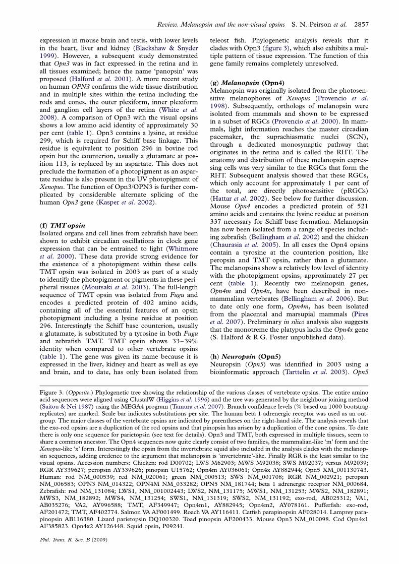
Figure 3. (Caption opposite.)
2856 S. N. Peirson et al. Review. Melanopsin and the non-visual opsins
Phil. Trans. R. Soc. B (2009)
expression in mouse brain and testis, with lower levelsin the heart, liver and kidney (Blackshaw & Snyder1999). However, a subsequent study demonstratedthat Opn3 was in fact expressed in the retina and inall tissues examined; hence the name ‘panopsin’ wasproposed (Halford et al. 2001). A more recent studyon human OPN3 confirms the wide tissue distributionand in multiple sites within the retina including therods and cones, the outer plexiform, inner plexiformand ganglion cell layers of the retina (White et al.2008). A comparison of Opn3 with the visual opsinsshows a low amino acid identity of approximately 30per cent (table 1). Opn3 contains a lysine, at residue299, which is required for Schiff base linkage. Thisresidue is equivalent to position 296 in bovine rodopsin but the counterion, usually a glutamate at pos-ition 113, is replaced by an aspartate. This does notpreclude the formation of a photopigment as an aspar-tate residue is also present in the UV photopigment ofXenopus. The function of Opn3/OPN3 is further com-plicated by considerable alternate splicing of thehuman Opn3 gene (Kasper et al. 2002).
(f) TMTopsinIsolated organs and cell lines from zebrafish have beenshown to exhibit circadian oscillations in clock geneexpression that can be entrained to light (Whitmoreet al. 2000). These data provide strong evidence forthe existence of a photopigment within these cells.TMT opsin was isolated in 2003 as part of a studyto identify the photopigment or pigments in these peri-pheral tissues (Moutsaki et al. 2003). The full-lengthsequence of TMT opsin was isolated from Fugu andencodes a predicted protein of 402 amino acids,containing all of the essential features of an opsinphotopigment including a lysine residue at position296. Interestingly the Schiff base counterion, usuallya glutamate, is substituted by a tyrosine in both Fuguand zebrafish TMT. TMT opsin shows 33–39%identity when compared to other vertebrate opsins(table 1). The gene was given its name because it isexpressed in the liver, kidney and heart as well as eyeand brain, and to date, has only been isolated from
teleost fish. Phylogenetic analysis reveals that itclades with Opn3 (figure 3), which also exhibits a mul-tiple pattern of tissue expression. The function of thisgene family remains completely unresolved.
(g) Melanopsin (Opn4)Melanopsin was originally isolated from the photosen-sitive melanophores of Xenopus (Provencio et al.1998). Subsequently, orthologs of melanopsin wereisolated from mammals and shown to be expressedin a subset of RGCs (Provencio et al. 2000). In mam-mals, light information reaches the master circadianpacemaker, the suprachiasmatic nuclei (SCN),through a dedicated monosynaptic pathway thatoriginates in the retina and is called the RHT. Theanatomy and distribution of these melanopsin expres-sing cells was very similar to the RGCs that form theRHT. Subsequent analysis showed that these RGCs,which only account for approximately 1 per cent ofthe total, are directly photosensitive (pRGCs)(Hattar et al. 2002). See below for further discussion.Mouse Opn4 encodes a predicted protein of 521amino acids and contains the lysine residue at position337 necessary for Schiff base formation. Melanopsinhas now been isolated from a range of species includ-ing zebrafish (Bellingham et al. 2002) and the chicken(Chaurasia et al. 2005). In all cases the Opn4 opsinscontain a tyrosine at the counterion position, likeperopsin and TMT opsin, rather than a glutamate.The melanopsins show a relatively low level of identitywith the photopigment opsins, approximately 27 percent (table 1). Recently two melanopsin genes,Opn4m and Opn4x, have been described in non-mammalian vertebrates (Bellingham et al. 2006). Butto date only one form, Opn4m, has been isolatedfrom the placental and marsupial mammals (Pireset al. 2007). Preliminary in silico analysis also suggeststhat the monotreme the platypus lacks the Opn4x gene(S. Halford & R.G. Foster unpublished data).
(h) Neuropsin (Opn5)Neuropsin (Opn5) was identified in 2003 using abioinformatic approach (Tarttelin et al. 2003). Opn5
Figure 3. (Opposite.) Phylogenetic tree showing the relationship of the various classes of vertebrate opsins. The entire aminoacid sequences were aligned using ClustalW (Higgins et al. 1996) and the tree was generated by the neighbour joining method(Saitou & Nei 1987) using the MEGA4 program (Tamura et al. 2007). Branch confidence levels (% based on 1000 bootstrapreplicates) are marked. Scale bar indicates substitutions per site. The human beta 1 adrenergic receptor was used as an out-group. The major classes of the vertebrate opsins are indicated by parentheses on the right-hand side. The analysis reveals thatthe exo-rod opsins are a duplication of the rod opsins and that pinopsin has arisen by a duplication of the cone opsins. To datethere is only one sequence for parietopsin (see text for details). Opn3 and TMT, both expressed in multiple tissues, seem toshare a common ancestor. The Opn4 sequences now quite clearly consist of two families, the mammalian-like ‘m’ form and theXenopus-like ‘x’ form. Interestingly the opsin from the invertebrate squid also included in the analysis clades with the melanop-sin sequences, adding credence to the argument that melanopsin is ‘invertebrate’-like. Finally RGR is the least similar to thevisual opsins. Accession numbers: Chicken: rod D00702; LWS M62903; MWS M92038; SWS M92037; versus M92039;RGR AY339627; peropsin AY339626; pinopsin U15762; Opn4m AY036061; Opn4x AY882944; Opn5 XM_001130743.Human: rod NM_000539; red NM_020061; green NM_000513; SWS NM_001708; RGR NM_002921; peropsinNM_006583; OPN3 NM_014322; OPN4M NM_033282; OPN5 NM_181744; beta 1 adrenergic receptor NM_000684.Zebrafish: rod NM_131084; LWS1, NM_001002443; LWS2, NM_131175; MWS1, NM_131253; MWS2, NM_182891;MWS3, NM_182892; MWS4, NM_131254; SWS1, NM_131319; SWS2, NM_131192; exo-rod, AB025312; VA1,AB035276; VA2, AY996588; TMT, AF349947; Opn4m1, AY882945; Opn4m2, AY078161. Pufferfish: exo-rod,AF201472; TMT, AF402774. Salmon VA AF001499. Roach VA AY116411. Catfish parapinopsin AF028014. Lamprey para-pinopsin AB116380. Lizard parietopsin DQ100320. Toad pinopsin AF200433. Mouse Opn3 NM_010098. Cod Opn4x1AF385823. Opn4x2 AY126448. Squid opsin, P09241.
Review. Melanopsin and the non-visual opsins S. N. Peirson et al. 2857
Phil. Trans. R. Soc. B (2009)

encodes a predicted protein of 377 amino acids in themouse and 354 amino acids in human, with the mousehaving a longer C-terminal tail. All of the expected fea-tures of an opsin are conserved, but Opn5 shows only25–30% identity to the vertebrate members of theopsin superfamily (table 1). RT-PCR suggests thatOpn5 is expressed in mouse testis, brain and eye andin human retina and brain. Further work is necessaryto establish both the type of retinal cells that expressOpn5/OPN5 and whether it can function as aphotopigment.
4. A NOVEL RETINAL PHOTORECEPTORThe identification of multiple photoreceptors, and thenon-rod, non-cone opsin-based photopigments acrossthe vertebrates were not predicted. Indeed, the assump-tion until the early 1990s was that some form ofconventional rod- or cone-like opsin would mediateall forms of vertebrate photoreception, both visualand extraocular. But perhaps the least expectedresult to emerge has been the discovery of anotherclass of photoreceptor within the eye, quite distinctfrom the rods and cones. Until recently it was incon-ceivable to most vision biologists that there could bean unrecognized class of photoreceptor within the ver-tebrate eye. After all, the eye was the best understoodpart of the central nervous system. One hundred andfifty years of research had explained how we see: pho-tons are detected by the rods and cones and theirgraded potentials are assembled into an image byinner retinal neurons, followed by advanced visual pro-cessing in the brain. But image detection is very differ-ent from the demands of irradiance detection. Rodsand cones are highly sensitive radiance detectors,which rapidly adapt and can only integrate signals ofa short duration. By contrast, the circadian system isrelatively insensitive to light, requiring high intensityand long duration stimuli to bring about photoentrain-ment. The appreciation that the mammalian eye has toperform two quite radically different sensory taskstriggered a line of enquiry that ultimately led to thediscovery of a population of pRGC which use thephotopigment melanopsin. The key findings thathave led to this discovery are outlined next.
(a) Retinal mutant studiesInitial studies to determine which retinal photo-receptors mediate circadian photoentrainment tookadvantage of a naturally occurring mutation in mice,termed retinal degeneration (rd/rd). These animalslack rods, and show a greatly reduced number ofcones. As might be expected, rd/rd mice fail to showany classical visual responses to light. Studies werethen undertaken to determine whether the circadiansystem of rd/rd mice was similarly impaired. When amouse is housed under a light–dark cycle, its circadianwheel running behaviour is entrained. As would beexpected of a nocturnal species, mice are largelyactive during the dark and inactive in the light.Under conditions of constant darkness, entrainmentis lost and circadian behaviour drifts or freeruns witha recurring period approximately 23.5 h, starting itsactivity cycle approximately 0.5 h earlier every day. If
the mouse is then exposed to a pulse of light shortlyafter activity onset it will delay the onset of activitythe following day. The magnitude of this phase delay(Df) is intensity dependent and can be used to deter-mine the sensitivity of circadian responses to light.Both the wavelength and intensity of the light pulsecan be systematically varied and the effect of thesetreatments on delaying wheel running behaviour canbe assessed in a dose-dependent manner to producea series of irradiance response curves.
Initial studies on photoentrainment in rd/rd mice(C3H) and wild-type controls (C57) demonstratedthat these mice could still entrain, although thethreshold for entrainment in rd/rd animals wasapproximately 2 log units higher (Ebihara & Tsuji1980). The difference in the genetic background inthese animals (C3H versus C57) appears to accountfor this difference in sensitivity (Foster & Helfrich-Forster 2001). Remarkably, in mice of the samegenetic background, the massive loss of classicalphotoreceptors in the rd/rd mutants had little or noeffect on the ability of the mice to either entrain to alight–dark cycle or phase-shift circadian rhythms inwheel running behaviour (Foster et al. 1991). Theirirradiance response curves were indistinguishablefrom congenic wild-type controls, while eye loss com-pletely blocked all effects of light on the clock (Fosteret al. 1991). These studies demonstrated that the pro-cessing of light for circadian and visual responses mustbe different and hinted at the fact that there may beanother class of ocular photoreceptor. Such sugges-tions were met with considerable scepticism and thefavoured explanation was that as approximately 5 percent of the cones survive in the retina of rd/rd mice(Carter-Dawson et al. 1978), it is probable that onlya small number of photoreceptors are necessary forphotoentrainment. Action spectrum studies on thespectral sensitivity of phase-shifting responses in rd/rdmice were subsequently conducted, suggesting amaximum sensitivity at either 511 or 480 nm, butthese studies again failed to exclude the possibility ofa residual cone contribution to these responses(Provencio & Foster 1995; Yoshimura & Ebihara1996). The development of a mouse model lackingall rods and cones, the rd/rd cl, finally resolved theseissues, and demonstrated that both phase-shiftingresponses and pineal melatonin suppression inresponse to light were apparently normal even whenrod or cone opsins were undetectable (Freedmanet al. 1999; Lucas et al. 1999).
The results from rd/rd cl mice provided the conclus-ive evidence that an additional photoreceptor existswithin the mammalian eye and the conceptual frame-work for a host of further studies, including the findingthat non-rod, non-cone photoreceptors do more thanregulate the circadian system. Two examples arelisted here: (i) In mammals, light-induced pupil con-striction is regulated by both rods and cones, but stilloccurs in animals showing profound damage to thesephotoreceptors. Not unreasonably, it was assumedthat the residual pupil light response was owing tothe survival of a few rod and/or cone photoreceptors.Pupil measurements were undertaken in rd/rd cl miceand showed that these animals maintained a pupillary
2858 S. N. Peirson et al. Review. Melanopsin and the non-visual opsins
Phil. Trans. R. Soc. B (2009)

light response. Although less sensitive than congenicwild-type animals, rd/rd cl mice retained the ability tofully constrict their pupils (Lucas et al. 2001). (ii) Noc-turnal rodents will inhibit their general activity whenexposed to light during the night. This response,called masking, is thought to complement circadianentrainment by ensuring that activity is restricted tothe hours of darkness or near darkness. Masking maybe particularly important in environments where daylength changes rapidly, and circadian behaviour mayhave difficulty keeping up with the expanding photo-period (Mrosovsky 1999). Masking experiments wereundertaken in rd/rd cl mice and demonstrated thatthere is marked inhibition of activity upon exposureto light presented two hours after normal lights off(Mrosovsky et al. 2001). Thus phase shifting, melato-nin suppression, pupil constriction, masking and anumber of other responses to light, such as sleepregulation (Lupi et al. 2008) are either intact orretained at some degree in mice lacking all their rodsand cones.
The first action spectrum to be published on rd/rd clmice was for pupil constriction, and the resultsdescribed an opsin/vitamin A-based photopigmentwith a lmax in the blue part of the spectrum near480 nm (opsin photopigment/OP480). The knownvisual pigments of the mouse have lmax values of360, 508 and 498 nm for the ultraviolet-sensitivecone, long-wavelength sensitive cone and rodpigments, respectively. None of these classical photo-receptors could account for the pupillary responsesto light (Lucas et al. 2001). Since 2001, a plethora ofaction spectra from mice to man have been deducedfor a range of irradiance responses to light. Theseinclude the light responses of pRGCs in mice(Hattar et al. 2003), rats (Berson et al. 2002) and pri-mates (Dacey et al. 2005) spanning pupil constriction,phase shifting circadian rhythms, plasma melatoninsuppression, together with irradiance dependentregulation of human retinal cone function (Hankins &Lucas 2002). All these action spectra point to the exist-ence of a single novel opsin photopigment with a lmax
of around 480 nm. A single invariant spectral sensitivityfor the pRGCs is in marked contrast to the cone pig-ments, which are highly divergent and appear spectrallytuned in a species-specific manner. It remains unclearwhat ecological advantage this wavelength mightconfer on such diverse species. One possibility is thatthe pRGCs are tuned to the dominant wavelength oflight at twilight. When the sun is close to the horizonthere is relative enrichment of ‘blue’ light in the domeof the sky because of the preferential scattering ofshort wavelengths of light passing obliquely throughthe atmosphere.
(b) Photosensitive retinal ganglion cellsThe identification of the cells mediating non-rod, non-cone responses to light was provided by two sets ofexperiments. Studies on the rat used retrograde tracersinjected into the SCN coupled with single cell record-ings on isolated retina in which rod and cone responseswere pharmacologically blocked (Berson et al. 2002).Parallel studies were undertaken using calcium
imaging on the rd/rd cl retina (Sekaran et al. 2003).Both approaches identified a population of RGCswhich responded directly to light. Significantly thesepRGCs expressed the photopigment melanopsin(Hattar et al. 2002).
(c) Melanopsin knockout studiesThe essential data that melanopsin plays a critical rolein the transduction of light information in pRGCscame from gene ablation studies. Melanopsin knockoutmice (Opn42/2) exhibited attenuated phase-shiftingand pupillary responses to light, as well as reducedperiod lengthening in constant light (LL) (Panda et al.2002; Ruby et al. 2002; Lucas et al. 2003). However,the critical involvement of melanopsin in photorecep-tion came from triple-knockout studies, lacking rods,cones and melanopsin. These animals were totally unre-sponsive to light, demonstrating that melanopsin is insome way essential for pRGC photosensitivity (Hattaret al. 2003), but precisely what function melanopsinwas playing was only finally resolved by using functionalexpression studies.
(d) Melanopsin expression studiesThe first investigation of the biochemistry of melanop-sin involved expression of melanopsin in COS cellsand reconstitution with 11-cis-retinal, an approachwhich has been particularly successful with visualpigments. This study produced a functional photo-pigment that was capable of activating transducinwith a lmax between 420 and 440 nm, an absorptionmaxima considerably shifted away from OP480
(Newman et al. 2003). The discrepancy in lmax
between spectroscopy and action spectra, coupledwith low pigment yields, prompted other researchersto investigate whether expression of melanopsinalone was enough to confer photosensitivity. Quiteindependently, three groups combined the expressionof melanopsin protein with physiological assays of cel-lular photosensitivity. All three studies showed thatmelanopsin transfection can confer photosensitivity tonon-photosensitive cell types (Neuro-2a; HEK293-TRPC3; Xenopus oocyte) (Melyan et al. 2005; Pandaet al. 2005; Qiu et al. 2005). In addition, thesegroups were able to show that specific forms of retinal(especially 11-cis-retinal) are needed for theseresponses to light, that light will ultimately triggerthe release of intracellular calcium, and that this mayinvolve a Gq-type G-protein rather than transducin.Furthermore, melanopsin acts as a bistable pigmentable to regenerate (recycle) its chromophore (11-cis-retinal) using all-trans-retinal and long-wavelengthlight in a manner reminiscent of the invertebratephotopigments (Melyan et al. 2005). In this regardmelanopsin may be unique among mammalianphotopigments in forming a stable association withall-trans-retinal.
Expression studies on human melanopsin suggestthat the lmax of light responses is close to 420–430 nm, and in this regard the findings were similarto those obtained by Newman and colleagues(Melyan et al. 2005). The studies on murine melanop-sin, however, showed an action spectrum for light
Review. Melanopsin and the non-visual opsins S. N. Peirson et al. 2859
Phil. Trans. R. Soc. B (2009)

responses that exhibited a lmax very close to 480 nm(Qiu et al. 2005). The current consensus from thevarious groups is that something about the localenvironment in which melanopsin is reconstituted isimportant in determining its lmax.
(e) Melanopsin phototransductionMost recently, research has turned to the phototrans-duction cascade used by melanopsin. Rod and coneopsins mediate a phototransduction cascade thatinvolves the activation of transducin (a member ofthe Gi/G0 class of G-proteins), phosphodiesteraseand closure of cyclic nucleotide gated channels and ahyperpolarizing membrane potential. By contrast,invertebrate phototransduction, most extensivelycharacterized in Drosophila, involves activation of aGq/G11-type G-protein, activation of phospholipaseC (PLC), gating of transient receptor potential(TRP) channels and the depolarization of membranepotential (Hardie & Raghu 2001). Interestingly, themelanopsins appear to share some of the key charac-teristics of an invertebrate-like signal transductionpathway. Both pRGCs and cells transfected withmelanopsin show depolarizing responses to light and,as discussed in §4d, melanopsin displays chromophorebistability, another feature of the invertebrate photo-pigments. Largely by analogy, it was proposed thatmelanopsin could be coupled to a G-protein of theGq/G11 class (for review see Peirson & Foster 2006).While not conclusive, there is support for this fromthe expression studies. For example, melanopsinresponses are greatly attenuated (although notblocked) by antibodies against Gq/G11 G-proteins(but not by antibodies to Gi/G0) (Panda et al. 2003).In Neuro-2a cells, the use of Gi/G0 blockers fails toinhibit melanopsin-dependent light responses(Melyan et al. 2005), while Gq/G11 agonists fullyblocked the melanopsin-dependent light responses inHEK293-TRPC3 cells (Qiu et al. 2005). Collectivelythese initial results suggest that the Gq/G11 G-proteinscould be activated by melanopsin-dependent photo-transduction. It is important to stress, however, thatthe coupling potential in non-native host environmentsmight not reflect the native pRGCs. Downstream ofthe G-protein, melanopsin-dependent light responsesare greatly attenuated or blocked in Xenopus oocytesand HEK293-TRPC3 cells by PLC inhibitors(Panda et al. 2005; Qiu et al. 2005). Furthermore,co-expression of melanopsin with TRPC3 in Xenopusoocytes (similar to the Drosophila TRP channels)shows that TRPC3 channels can generate a light-activated photocurrent in the presence of melanopsin(Panda et al. 2005; Qiu et al. 2005). Collectively,a partial model of the phototransduction cascade hasemerged, suggesting that light activated melanopsinmay interact with Gq/G11 that in turn activates aPLC-b. PLC-b generates inositol triphosphate (IP3)and diacylglycerol (DAG), which may ultimatelymodulate a TRPC channel, possibly via a proteinkinase C (PKC). Most recently combined pharmacol-gical and anatomical approaches have suggestedTRPC7 as the channel (Sekaran et al. 2007). Inaddition, a microarray-based approach has been usedto investigate the transcriptional realignment that
occurs in the rd/rd cl mouse eye following a light pulse.This approach identified a number of candidate genes/proteins that might be associated with the melanopsincascade. Among these was the atypical protein kinaseC zeta (Prkcz). Remarkably the genetic ablation ofPrkcz mimics precisely the melanopsin knock-outphenotype in a battery of behavioural and pupillometrictests (Peirson et al. 2007). Why an ‘invertebrate-like’signalling pathway, rather than a more conventionalvertebrate phototransduction pathway, is employed bythe pRGCs remains an intriguing sensory questionand may be relevant to understanding the evolutionaryorigins of the melanopsin/pRGCs photoreceptorsystem (Arendt 2003).
5. EVOLUTIONARY CONSIDERATIONS OFNON-IMAGE-FORMING PHOTORECEPTIONGiven the multiplicity of photoreceptive tissues inthe non-mammalian vertebrates, why have thesebeen lost in the mammalian lineage? One possibleexplanation may be related to the early evolutionaryhistory of the mammals and their passage through a‘nocturnal bottleneck’. Modern mammals seem tohave been derived from nocturnal insectivorous oromnivorous animals about 100 million years ago(Young 1962). Pineal and deep-brain photoreceptorswould have been perfectly adequate for monitoringchanges in diurnal light conditions but may not havebeen sufficiently sensitive to discriminate twilightchanges in mammals living in burrows or otherwiseconcealed during the day. The occupation of the noc-turnal realm may have led to the loss of the extraocularphotoreceptors and the exclusive reliance on irradi-ance detection by the pRGCs (Foster & Menaker1993; Menaker et al. 1997). But of course this expla-nation does not explain why the vertebrates evolvedso many photosensitive tissues in the first place. Inthis context it is worth emphasizing that the sensorytask of reliable irradiance detection is not trivial, andextracting time-of-day information from environ-mental irradiance is even more complex. For example,during twilight, the quality of light changes in threeimportant respects: (i) the amount of light; (ii) thespectral composition of light; (iii) and the source oflight (i.e. the position of the sun). These parametersall change in a systematic way and could be used bythe circadian system to detect the phase of twilightand hence time of day (Roenneberg & Foster 1997).However, each of these parameters is subject to con-siderable sensory ‘noise’. The sources of this noiseare summarized in table 2. Clearly, the impact of thisnoise will depend upon the organism and the environ-ment it inhabits. Integrating the information from amultiplicity of photoreceptors, which collect lightfrom different regions of the environment, with differ-ing integration times, and tuned to different spectralchannels will act to reduce signal noise and henceprovide a more reliable measure of environmentalirradiance. The non-mammalian vertebrates mightintegrate light information from the pineal, deepbrain and eyes for reliable time-of-day detection, andthere is good evidence for this in birds (Menaker &Underwood 1976). In mammals, twilight detection is
2860 S. N. Peirson et al. Review. Melanopsin and the non-visual opsins
Phil. Trans. R. Soc. B (2009)

either less precise, because of the reliance on a singlephotoreceptor type, or the pRGCs themselves showheterogeneity in their responses to light, for whichthere is also good evidence (Sekaran et al. 2005).
Opsins in general have evolved to mediate specificphotoreceptive tasks in different light environments(Lythgoe 1979). For example, in environmentswhere the spectral composition of the light isrestricted, such as in deep water, the lmax of photopig-ments is spectrally tuned to match the maximumavailable photon flux around 480 nm (Douglas &Partridge 1997; Hope et al. 1997; Hunt et al. 2001).Whether similar spectral tuning arguments can beused to understand the lmax of the non-image-formingphotopigments remains an intriguing question. Manyphotoreceptors involved in non-image-forming tasksappear to peak close to 480 nm, with a spread rangingfrom 460 to 530 nm (Shand & Foster 1999). In pinealand deep-brain photoreceptors the light available willbe dominated by the transmission of the overlyingtissues. This is primarily influenced by two factors.Firstly, short wavelength light is scattered more thanlonger wavelength light, resulting in relatively morelight of long wavelengths penetrating to reach intracra-nial photoreceptors. Secondly, light will be modifiedby light-absorbing pigments before reaching thesephotoreceptors. The most important such pigment ishaemoglobin. Haemoglobin has a transmissionwindow between 460 and 540 nm, peaking around490 nm (Hartwig & van Veen 1979; Foster & Follett1985). This transmission window may have exerted astrong selection pressure on the spectral tuning ofdeep brain and pineal photoreceptors. But thiscannot be the entire explanation as many non-image-forming photoreceptors are directly exposed toenvironmental light, such as the pRGCs in the eye ordermal photoreceptors, and these have lmax around480 nm.
Changes in the amount and spectral composition ofenvironmental irradiance occur throughout the diur-nal cycle. As well as the obvious gross changes inirradiance at twilight (approx. 6 log units), changes
in the spectral composition of light also occur andare known as the Chappuis effect (Lythgoe 1979).As the sun’s rays must pass through a thicker layer ofthe atmosphere when the sun is lower in the sky, theabsorption of light by ozone (500–650 nm) results ina relative enrichment of shorter wavelength light(,500 nm) at twilight (Munz & McFarland 1977).As changes in the light environment at twilight arecritical for photoentrainment (Roenneberg & Foster1997), ‘twilight detectors’ spectrally tuned to theblue part of the spectrum could allow increasedphoton capture and hence an increase in signal-to-noise detection. Perhaps, however, it is not simplythe amount of light that is being detected at twilightbut rather its change in spectral quality. Evidence forspectral discrimination, a chromatic response, wasfirst shown in the pineal organ of fish (Meissl &Yanez 1994) and more recently in the parietal eye oflizards (Su et al. 2006). These chromatic responsescould arise from an interaction between differentphotopigments with differing lmax or a single bistablephotopigment. Significantly, melanopsin appears toact as a bi-stable pigment, able to regenerate its chro-mophore using all-trans-retinal and long-wavelengthlight (Melyan et al. 2005). This photoreversal capacityof melanopsin has also been observed with spectro-scopic approaches in the case of Amphioxus melanop-sin (Koyanagi et al. 2004). If the two stable states ofmelanopsin are capable of interacting with differentdownstream signalling transduction pathways, thismay provide an alternative means of attaining spectraldiscrimination.
The spectral tuning of vertebrate opsins will also beinfluenced by their evolutionary history (Goldsmith1990). For example, key amino acid residues influen-cing spectral tuning sites may provide structural orfunctional properties, such that any mutation ofthese residues will be deleterious to protein function.Additionally, there will be trade-offs between structureand function that will influence spectral tuning.Scotopic vision is limited by dark noise produced byspontaneous thermal isomerizations of the retinalchromophore (Barlow et al. 1993). Long wavelengthsensitive photopigments have been suggested to bemore prone to dark noise owing to their lowerexcitation energy (Barlow 1957). Thus the spectraltuning of the non-visual opsins, like the visualopsins, will always be a compromise between func-tional constraints and the photon flux of the lightenvironment (Lythgoe 1984; Goldsmith 1990;Barlow et al. 1993).
6. CONCLUSIONSConsiderable progress has been made in the lastdecade in characterizing the photoreceptors andphotopigments mediating non-image-formingresponses to light, such as photoentrainment. Whileover a dozen different opsin photopigments havebeen identified in recent years (table 1), we are onlyjust beginning to understand what roles these proteinsplay in the signalling of light information. Perhaps thegreatest single advance has been the identification of athird photoreceptive system in the vertebrate eye, the
Table 2. The major sources of noise associated with thedetection of environmental irradiance. The main sources ofsignal noise for irradiance detection are listed withexamples. In each case the impact of this noise will dependupon the organism, its developmental state and theenvironment that it inhabits. Integrating the informationfrom multiple photoreceptors, which collect light fromdifferent regions of the environment, having differingintegration times, and tuned to different spectral channelswill act to reduce signal noise.
source of signal noise examples
fluctuation in the lightsignal
cloud cover, day-length
extraneous lightsignals
starlight, moonlight, lightning
receptor noise variation in external temperaturesensory adaptation receptor habituationbehavioural noise emergence from burrow, place of
rest, feeding etc.
Review. Melanopsin and the non-visual opsins S. N. Peirson et al. 2861
Phil. Trans. R. Soc. B (2009)
Ianhopp
Ianhopp
Ianhopp
Ianhopp
Ianhopp
Ianhopp
Ianhopp

melanopsin-expressing pRGCs, which mediate a rangeof irradiance detection tasks ranging from photoen-trainment, pineal melatonin suppression, pupilconstriction and the modulation of arousal states andsleep induction (Altimus et al. 2008; Lupi et al.2008). By contrast, the photopigments responsiblefor non-image-forming responses to light in manynon-mammalian species remain poorly characterized.Molecular studies are required to determine whetherthese opsins can form photopigments or whetherthey act as photoisomerases or retinal carrier proteins(Foster & Bellingham 2002). However a functionalanalysis of these opsins requires more than biochemis-try. If we are to place these remarkable photoreceptorsinto any sort of evolutionary context we will need amuch better understanding of their sensory ecology.We now appreciate that these photoreceptors domore than act as simple photon counters—butbeyond this—any detailed understanding is lacking.
REFERENCESAltimus, C. M., Guler, A. D., Villa, K. L., McNeill, D. S.,
Legates, T. A. & Hattar, S. 2008 Rods–cones andmelanopsin detect light and dark to modulate sleep inde-pendent of image formation. Proc. Natl Acad. Sci. USA105, 19 998–20 003. (doi:10.1073/pnas.0808312105)
Arendt, J. 1998 Melatonin and the pineal gland: influence onmammalian seasonal and circadian physiology. Rev.Reprod. 3, 13–22. (doi:10.1530/ror.0.0030013)
Arendt, D. 2003 Evolution of eyes and photoreceptor celltypes. Int. J. Dev. Biol. 47, 563–571.
Aschoff, J. 1984 Circadian timing. Ann. N. Y. Acad. Sci. 423,442–468. (doi:10.1111/j.1749-6632.1984.tb23452.x)
Barlow, H. B. 1957 Purkinje shift and retinal noise. Nature179, 255–256. (doi:10.1038/179255b0)
Barlow, R. B., Birge, R. R., Kaplan, E. & Tallent, J. R. 1993On the molecular origin of photoreceptor noise. Nature366, 64–66. (doi:10.1038/366064a0)
Barr, L. & Alpern, M. 1963 Photosensitivity of the frog iris.J. Gen. Physiol. 46, 1249–1265. (doi:10.1085/jgp.46.6.1249)
Bellingham, J. & Foster, R. G. 2002 Opsins and mammalianphotoentrainment. Cell Tissue Res. 309, 57–71. (doi:10.1007/s00441-002-0573-4)
Bellingham, J., Whitmore, D., Philp, A. R., Wells, D. J. &Foster, R. G. 2002 Zebrafish melanopsin: isolation,tissue localisation and phylogenetic position. Brain Res.Mol. Brain Res. 107, 128–136. (doi:10.1016/S0169-328X(02)00454-0)
Bellingham, J., Wells, D. J. & Foster, R. G. 2003 In silicocharacterisation and chromosomal localisation of humanRRH (peropsin)—implications for opsin evolution.BMC Genomics 4, 3. (doi:10.1186/1471-2164-4-3)
Bellingham, J. et al. 2006 Evolution of melanopsinphotoreceptors: discovery and characterization of a newmelanopsin in nonmammalian vertebrates. PLoS Biol. 4,e254. (doi:10.1371/journal.pbio.0040254)
Benoit, J. 1964 The role of the eyes and of the hypothalamusin the photostimulation of gonads in the duck. Ann. N. Y.Acad. Sci. 117, 204–215. (doi:10.1111/j.1749-6632.1964.tb48175.x)
Berson, D. M., Dunn, F. A. & Takao, M. 2002 Phototrans-duction by retinal ganglion cells that set the circadianclock. Science 295, 1070–1073. (doi:10.1126/science.1067262)
Bito, L. Z. & Turansky, D. G. 1975 Photoactivation of pupil-lary constriction in the isolated in vitro iris of mammal
(Mesocricetus auratus). Comp. Biochem. Physiol. A 50,407–413. (doi:10.1016/0300-9629(75)90034-1)
Blackshaw, S. & Snyder, S. H. 1997 Parapinopsin, a novelcatfish opsin localized to the parapineal organ, defines anew gene family. J. Neurosci. 17, 8083–8092.
Blackshaw, S. & Snyder, S. H. 1999 Encephalopsin: a novelmammalian extraretinal opsin discretely localized in thebrain. J Neurosci. 19, 3681–3690.
Bowmaker, J. & Hunt, D. M. 1999 Molecular biologyof photoreceptor spectral sensitivity. In Adaptivemechanisms in the ecology of vision (eds S. N. Archer,M. B. A. Djamgoz, E. R. Loew, J. C. Partridge & S.Vallerga), pp. 439–462. Dordrecht, The Netherlands:Kluwer Academic Publishers.
Carter-Dawson, L. D., LaVail, M. M. & Sidman, R. L. 1978Differential effect of the rd mutation on rods and conesin the mouse retina. Invest Ophthalmol. Vis. Sci. 17,489–498.
Chaurasia, S. S. et al. 2005 Molecular cloning, localizationand circadian expression of chicken melanopsin (Opn4):differential regulation of expression in pineal and retinalcell types. J. Neurochem. 92, 158–170. (doi:10.1111/j.1471-4159.2004.02874.x)
Dacey, D. M., Liao, H. W., Peterson, B. B., Robinson, F. R.,Smith, V. C., Pokorny, J., Yau, K. W. & Gamlin, P. D.2005 Melanopsin-expressing ganglion cells in primateretina signal colour and irradiance and project to theLGN. Nature 433, 749–754. (doi:10.1038/nature03387)
Dartnall, H. 1953 The interpretation of spectral sensitivitycurves. Br. Med. Bull. 9, 24–30.
Dodt, E. & Meissl, H. 1982 The pineal and parietal organsof lower vertebrates. Experientia 38, 996–1000. (doi:10.1007/BF01955342)
Douglas, R. H. & Partridge, J. C. 1997 On the visual pig-ments of deep-sea fish. J. Fish Biol. 50, 68–85. (doi:10.1111/j.1095-8649.1997.tb01340.x)
Ebihara, S. & Tsuji, K. 1980 Entrainment of the circadianactivity rhythm to the light cycle: effective light intensityfor a Zeitgeber in the retinal degenerate C3H mouseand the normal C57BL mouse. Physiol. Behav. 24,523–527. (doi:10.1016/0031-9384(80)90246-2)
Foster, R. & Bellingham, J. 2002 Opsins and melanopsins.Curr. Biol. 12, R543–R544. (doi:10.1016/S0960-9822(02)01047-3)
Foster, R. G. & Follett, B. K. 1985 The involvement of arhodopsin-like photopigment in the photoperiodicresponse of the Japanese quail. J. Comp. Physiol. 157A,519–528. (doi:10.1007/BF00615153)
Foster, R. G. & Helfrich-Forster, C. 2001 The regulation ofcircadian clocks by light in fruitflies and mice. Phil.Trans. R. Soc. B 356, 1779–1789. (doi:10.1098/rstb.2001.0962)
Foster, R. G. & Menaker, M. 1993 Circadian photorecep-tion in mammals and other vertebrates. In Light andbiological rhythms in man (ed. L. Wetterberg), pp. 73–91.Oxford, UK and New York: Pergamon.
Foster, R. G., Timmers, A. M., Schalken, J. J. & De Grip,W. J. 1989 A comparison of some photoreceptor charac-teristics in the pineal and retina. II. The Djungarianhamster (Phodopus sungorus). J. Comp. Physiol. 165A,565–572. (doi:10.1007/BF00611242)
Foster, R. G., Provencio, I., Hudson, D., Fiske, S., De Grip,W. & Menaker, M. 1991 Circadian photoreception in theretinally degenerate mouse (rd/rd). J. Comp. Physiol.169A, 39–50. (doi:10.1007/BF00198171)
Foster, R. G., Provencio, I., Bovee-Geurts, P. H. & DeGrip,W. J. 2003 The photoreceptive capacity of the developingpineal gland and eye of the golden hamster (Mesocricetusauratus). J. Neuroendocrinol. 15, 355–363. (doi:10.1046/j.1365-2826.2003.01004.x)
2862 S. N. Peirson et al. Review. Melanopsin and the non-visual opsins
Phil. Trans. R. Soc. B (2009)

Freedman, M. S., Lucas, R. J., Soni, B., von Schantz, M.,Munoz, M., David-Gray, Z. & Foster, R. 1999 Regu-lation of mammalian circadian behavior by non-rod,non-cone, ocular photoreceptors. Science 284, 502–504.(doi:10.1126/science.284.5413.502)
Frigato, E., Vallone, D., Bertolucci, C. & Foulkes, N. S.2006 Isolation and characterization of melanopsin andpinopsin expression within photoreceptive sites of rep-tiles. Naturwissenschaften 93, 379–385. (doi:10.1007/s00114-006-0119-9)
Goldsmith, T. H. 1990 Optimization, constraint, and historyin the evolution of eyes. Q. Rev. Biol. 65, 281–322.(doi:10.1086/416840)
Halford, S., Freedman, M., Bellingham, J., Inglis, S.,Poopalasundaram, S., Soni, B., Foster, R. & Hunt, D.2001 Characterization of a novel human opsin genewith wide tissue expression and identification ofembedded and flanking genes on chromosome 1q43.Genomics 72, 203–208. (doi:10.1006/geno.2001.6469)
Hankins, M. W. & Lucas, R. J. 2002 The primary visualpathway in humans is regulated according to long-termlight exposure through the action of a nonclassicalphotopigment. Curr. Biol. 12, 191–198. (doi:10.1016/S0960-9822(02)00659-0)
Hao, W. & Fong, H. K. 1996 Blue and ultraviolet light-absorbing opsin from the retinal pigment epithelium.Biochemistry 35, 6251–6256. (doi:10.1021/bi952420k)
Hao, W. & Fong, H. K. 1999 The endogenous chromophoreof retinal G protein-coupled receptor opsin from thepigment epithelium. J. Biol. Chem. 274, 6085–6090.(doi:10.1074/jbc.274.10.6085)
Hardie, R. C. & Raghu, P. 2001 Visual transductionin Drosophila. Nature 413, 186–193. (doi:10.1038/35093002)
Hargrave, P. A. & McDowell, J. H. 1992 Rhodopsin andphototransduction: a model system for G protein-linkedreceptors. FASEB J. 6, 2323–2331.
Hartwig, H.-G. & van Veen, T. 1979 Spectral characteristicsof visible radiations penetrating into the brain and stimu-lating extra-retinal photoreceptors. J. Comp. Physiol. A120, 277–282. (doi:10.1007/BF00614615)
Hattar, S., Liao, H. W., Takao, M., Berson, D. M. & Yau,K. W. 2002 Melanopsin-containing retinal ganglioncells: architecture, projections, and intrinsic photosensi-tivity. Science 295, 1065–1070. (doi:10.1126/science.1069609)
Hattar, S. et al. 2003 Melanopsin and rod–cone photorecep-tive systems account for all major accessory visualfunctions in mice. Nature 424, 75–81. (doi:10.1038/nature01761)
Higgins, D. G., Thompson, J. D. & Gibson, T. J. 1996 UsingCLUSTAL for multiple sequence alignments. MethodsEnzymol. 266, 383–402. (doi:10.1016/S0076-6879(96)66024-8)
Hope, A. J., Partridge, J. C., Dulai, K. S. & Hunt, D. M.1997 Mechanisms of wavelength tuning in the rodopsins of deep-sea fishes. Proc. R. Soc. Lond. B 264,155–163. (doi:10.1098/rspb.1997.0023)
Hunt, D. M., Dulai, K. S., Partridge, J. C., Cottrill, P. &Bowmaker, J. K. 2001 The molecular basis for spectraltuning of rod visual pigments in deep-sea fish. J. Exp.Biol. 204, 3333–3344.
Isoldi, M. C., Rollag, M. D., Castrucci, A. M. & Provencio, I.2005 Rhabdomeric phototransduction initiated by thevertebrate photopigment melanopsin. Proc. Natl Acad.Sci. USA 102, 1217–1221. (doi:10.1073/pnas.0409252102)
Jenkins, A., Munoz, M., Tarttelin, E. E., Bellingham, J.,Foster, R. G. & Hankins, M. W. 2003 VA opsin, melanop-sin, and an inherent light response within retinal
interneurons. Curr. Biol. 13, 1269–1278. (doi:10.1016/S0960-9822(03)00509-8)
Jiang, M., Pandey, S. & Fong, H. K. 1993 An opsin homol-ogue in the retina and pigment epithelium. Invest.Ophthalmol. Vis. Sci. 34, 3669–3679.
Kasper, G. et al. 2002 Different structural organization ofthe encephalopsin gene in man and mouse. Gene 295,27–32. (doi:10.1016/S0378-1119(02)00799-0)
Kawamura, S. & Yokoyama, S. 1997 Expression of visualand nonvisual opsins in American chameleon. Vision Res.37, 1867–1871. (doi:10.1016/S0042-6989(96)00309-4)
Knowles, A. & Dartnall, H. 1977 In The photobiology ofvision. New York, NY: Academic Press.
Kojima, D., Mano, H. & Fukada, Y. 2000 Vertebrateancient-long opsin: a green-sensitive photoreceptivemolecule present in zebrafish deep brain and retinal hori-zontal cells. J. Neurosci. 20, 2845–2851.
Kojima, D., Torii, M., Fukada, Y. & Dowling, J. E. 2008Differential expression of duplicated VAL-opsin genes inthe developing zebrafish. J. Neurochem. 104, 1364–1371. (doi:10.1111/j.1471-4159.2007.05093.x)
Korf, H.-W. & Moller, M. 1984 The innervation of themammalian pineal gland with special reference to centralpinealopetal projections. Pineal Res. Rev. 2, 41–86.
Korf, H. W., Foster, R. G., Ekstrom, P. & Schalken, J. J.1985a Opsin-like immunoreaction in the retinae andpineal organs of four mammalian species. Cell TissueRes. 242, 645–648. (doi:10.1007/BF00225432)
Korf, H. W., Moller, M., Gery, I., Zigler, J. S. & Klein, D. C.1985b Immunocytochemical demonstration of retinalS-antigen in the pineal organ of four mammalian species.Cell Tissue Res. 239, 81–85. (doi:10.1007/BF00214906)
Korf, H. W., Schomerus, C. & Stehle, J. H. 1998 The pinealorgan, its hormone melatonin, and the photoneuroendo-crine system. Adv. Anat. Embryol. Cell Biol. 146, 1–100.
Koyanagi, M., Kawano, E., Kinugawa, Y., Oishi, T.,Shichida, Y., Tamotsu, S. & Terakita, A. 2004 BistableUV pigment in the lamprey pineal. Proc. Natl Acad. Sci.USA 101, 6687–6691. (doi:10.1073/pnas.0400819101)
Lau, K. C., So, K. F., Campbell, G. & Lieberman, A. R.1992 Pupillary constriction in response to light inrodents, which does not depend on central neuralpathways. J. Neurol. Sci. 113, 70–79. (doi:10.1016/0022-510X(92)90267-O)
Lucas, R. J., Freedman, M. S., Munoz, M., Garcia-Fernandez, J. M. & Foster, R. G. 1999 Regulation ofthe mammalian pineal by non-rod, non-cone, ocularphotoreceptors. Science 284, 505–507. (doi:10.1126/science.284.5413.505)
Lucas, R. J., Douglas, R. H. & Foster, R. G. 2001 Character-ization of an ocular photopigment capable of drivingpupillary constriction in mice. Nat. Neurosci. 4, 621–626. (doi:10.1038/88443)
Lucas, R. J., Hattar, S., Takao, M., Berson, D. M., Foster,R. G. & Yau, K. W. 2003 Diminished pupillary lightreflex at high irradiances in melanopsin-knockout mice.Science 299, 245–247. (doi:10.1126/science.1077293)
Lupi, D., Oster, H., Thompson, S. & Foster, R. G. 2008The acute light-induction of sleep is mediated byOPN4-based photoreception. Nat. Neurosci. 11, 1068–1073.
Lythgoe, J. 1979 The ecology of vision. Oxford, UK: OxfordUniversity Press.
Lythgoe, J. N. 1984 Visual pigments and environmentallight. Vision Res. 24, 1539–1550. (doi:10.1016/S0042-6989(84)80003-6)
Mano, H., Kojima, D. & Fukada, Y. 1999 Exo-rhodopsin: anovel rhodopsin expressed in the zebrafish pineal gland.Brain Res. Mol. Brain Res. 73, 110–118. (doi:10.1016/S0169-328X(99)00242-9)
Review. Melanopsin and the non-visual opsins S. N. Peirson et al. 2863
Phil. Trans. R. Soc. B (2009)

Max, M., McKinnon, P. J., Seidenman, K. J., Barrett, R. K.,Applebury, M. L., Takahashi, J. S. & Margolskee, R. F.1995 Pineal opsin: a nonvisual opsin expressed in chickpineal. Science 267, 1502–1506. (doi:10.1126/science.7878470)
Max, M., Surya, A., Takahashi, J. S., Margolskee, R. F. &Knox, B. E. 1998 Light-dependent activation ofrod transducin by pineal opsin. J. Biol. Chem. 273,26 820–26 826. (doi:10.1074/jbc.273.41.26820)
Meissl, H. 1997 Photic regulation of pineal function. Analo-gies between retinal and pineal photoreception. Biol. Cell89, 549–554. (doi:10.1016/S0248-4900(98)80158-5)
Meissl, H. & Yanez, J. 1994 Pineal photosensitivity: a com-parison with retinal photoreception. Acta Neurobiol. Exp.54, 19–29.
Melyan, Z., Tarttelin, E. E., Bellingham, J., Lucas, R. J. &Hankins, M. W. 2005 Addition of human melanopsinrenders mammalian cells photoresponsive. Nature 433,741–745. (doi:10.1038/nature03344)
Menaker, M. & Underwood, H. 1976 Extraretinal photo-reception in birds. Photochem. Photobiol. 23, 299–306.(doi:10.1111/j.1751-1097.1976.tb07251.x)
Menaker, M., Moreira, L. F. & Tosini, G. 1997 Evolutionof circadian organization in vertebrates. Braz. J.Med. Biol. Res. 30, 305–313. (doi./10.1590/S0100-879X1997000300003)
Minamoto, T. & Shimizu, I. 2002 A novel isoform of ver-tebrate ancient opsin in a smelt fish Plecoglossus altivelis.Biochem. Biophys. Res. Commun. 290, 280–286. (doi:10.1006/bbrc.2001.6186)
Moutsaki, P., Bellingham, J., Soni, B. G., David-Gray, Z. K. &Foster, R. G. 2000 Sequence, genomic structure and tissueexpression of carp (Cyprinus carpio L.) vertebrate ancient(VA) opsin. FEBS Lett. 473, 316–322. (doi:10.1016/S0014-5793(00)01550-7)
Moutsaki, P., Whitmore, D., Bellingham, J., Sakamoto, K.,David-Gray, Z. K. & Foster, R. G. 2003 Teleost multipletissue (tmt) opsin: a candidate photopigment regulatingthe peripheral clocks of zebrafish? Brain Res. Mol. BrainRes. 112, 135–145. (doi:10.1016/S0169-328X(03)00059-7)
Mrosovsky, N. 1999 Masking: history, definitions, andmeasurement. Chronobiol. Int. 16, 415–429. (doi:10.3109/07420529908998717)
Mrosovsky, N., Lucas, R. & Foster, R. 2001 Persistence ofmasking responses to light in mice lacking rods andcones. J. Biol. Rhythms 16, 585–587. (doi:10.1177/074873001129002277)
Munz, F. W. & McFarland, W. N. 1977 Evolutionary adap-tations of fishes to the photic environment. In Handbookof sensory physiology (ed. F. Crescitelli), pp. 193–274.Berlin, Germany: Springer.
Nakamura, A., Kojima, D., Imai, H., Terakita, A., Okano,T., Shichida, Y. & Fukada, Y. 1999 Chimeric natureof pinopsin between rod and cone visual pigments.Biochemistry 38, 14 738–14 745. (doi:10.1021/bi9913496)
Nathans, J. & Hogness, D. S. 1983 Isolation, sequenceanalysis, and intron–exon arrangement of the geneencoding bovine rhodopsin. Cell 34, 807–814. (doi:10.1016/0092-8674(83)90537-8)
Nelson, R. J. & Zucker, I. 1981 Absence of extra-ocularphotoreception in diurnal and nocturnal rodents exposedto direct sunlight. Comp. Biochem. Physiol. 69A, 145–148.(doi:10.1016/0300-9629(81)90651-4)
Newman, L. A., Walker, M. T., Brown, R. L., Cronin, T. W. &Robinson, P. R. 2003Melanopsin forms a functional short-wavelengthphotopigment.Biochemistry 42, 12 734–12 738.(doi:10.1021/bi035418z)
Okada, T., Ernst, O. P., Palczewski, K. & Hofmann, K. P.2001 Activation of rhodopsin: new insights from
structural and biochemical studies. Trends Biochem. Sci.26, 318–324. (doi:10.1016/S0968-0004(01)01799-6)
Okano, T., Yoshizawa, T. & Fukada, Y. 1994 Pinopsin is achicken pineal photoreceptive molecule. Nature 372,94–97. (doi:10.1038/372094a0)
Palczewski, K. et al. 2000 Crystal structure of rhodopsin: aG protein-coupled receptor. Science 289, 739–745.(doi:10.1126/science.289.5480.739)
Panda, S., Sato, T. K., Castrucci, A. M., Rollag, M. D.,DeGrip, W. J., Hogenesch, J. B., Provencio, I. & Kay, S.A. 2002 Melanopsin (Opn4) requirement for normallight-induced circadian phase shifting. Science 298,2213–2216. (doi:10.1126/science.1076848)
Panda, S. et al. 2003 Melanopsin is required for non-image-forming photic responses in blind mice. Science301, 525–527. (doi:10.1126/science.1086179)
Panda, S., Nayak, S. K., Campo, B., Walker, J. R.,Hogenesch, J. B. & Jegla, T. 2005 Illumination of themelanopsin signaling pathway. Science 307, 600–604.(doi:10.1126/science.1105121)
Pandey, S., Blanks, J. C., Spee, C., Jiang, M. & Fong, H. K.1994 Cytoplasmic retinal localization of an evolutionaryhomolog of the visual pigments. Exp. Eye Res. 58, 605–613. (doi:10.1006/exer.1994.1055)
Peirson, S. & Foster, R. G. 2006 Melanopsin: another wayof signaling light. Neuron 49, 331–339. (doi:10.1016/j.neuron.2006.01.006)
Peirson, S. N., Bovee-Geurts, P. H., Lupi, D., Jeffery, G.,DeGrip, W. J. & Foster, R. G. 2004 Expression of thecandidate circadian photopigment melanopsin (Opn4) inthe mouse retinal pigment epithelium. Brain Res. Mol.Brain Res. 123, 132–135. (doi:10.1016/j.molbrainres.2004.01.007)
Peirson, S. N., Oster, H., Jones, S. L., Leitges, M.,Hankins, M. W. & Foster, R. G. 2007 Microarray analy-sis and functional genomics identify novel componentsof melanopsin signaling. Curr. Biol. 17, 1363–1372.(doi:10.1016/j.cub.2007.07.045)
Pepe, I. M. 2001 Recent advances in our understanding ofrhodopsin and phototransduction. Prog. Retin Eye Res.20, 733–759. (doi:10.1016/S1350-9462(01)00013-1)
Philp, A. R., Bellingham, J., Garcia-Fernandez, J. & Foster,R. G. 2000a A novel rod-like opsin isolated from theextra-retinal photoreceptors of teleost fish. FEBS Lett.468, 181–188. (doi:10.1016/S0014-5793(00)01217-5)
Philp, A. R., Garcia-Fernandez, J. M., Soni, B. G., Lucas, R.J., Bellingham, J. & Foster, R. G. 2000b Vertebrateancient (VA) opsin and extraretinal photoreception inthe Atlantic salmon (Salmo salar). J. Exp. Biol. 203,1925–1936.
Pires, S. S., Shand, J., Bellingham, J., Arrese, C., Turton,M., Peirson, S., Foster, R. G. & Halford, S. 2007Isolation and characterization of melanopsin (Opn4)from the Australian marsupial Sminthopsis crassicaudata(fat-tailed dunnart). Proc. Biol. Sci. 274, 2791–2799.(doi:10.1098/rspb.2007.0976)
Pittendrigh, C. S. 1993 Temporal organisation: reflections ofa Darwinian clock-watcher. Annu. Rev. Physiol. 55, 17–54. (doi:10.1146/annurev.ph.55.030193.000313)
Provencio, I. & Foster, R. G. 1995 Circadian rhythms inmice can be regulated by photoreceptors with cone-likecharacteristics. Brain Res. 694, 183–190. (doi:10.1016/0006-8993(95)00694-L)
Provencio, I., Jiang, G., De Grip, W. J., Hayes, W. P. &Rollag, M. D. 1998 Melanopsin: an opsin in melano-phores, brain, and eye. Proc. Natl Acad. Sci. USA 95,340–345. (doi:10.1073/pnas.95.1.340)
Provencio, I., Rodriguez, I. R., Jiang, G., Hayes, W. P.,Moreira, E. F. & Rollag, M. D. 2000 A novel humanopsin in the inner retina. J. Neurosci. 20, 600–605.
2864 S. N. Peirson et al. Review. Melanopsin and the non-visual opsins
Phil. Trans. R. Soc. B (2009)

Qiu, X., Kumbalasiri, T., Carlson, S. M., Wong, K. Y.,Krishna, V., Provencio, I. & Berson, D. M. 2005 Induc-tion of photosensitivity by heterologous expression ofmelanopsin. Nature 433, 745–749. (doi:10.1038/nature03345)
Rodieck, R. W. 1998 In The first steps in seeing. Sunderland,MA: Sinauer Associates.
Roenneberg, T. & Foster, R. G. 1997 Twilight times: lightand the circadian system. Photochem. Photobiol. 66,549–561. (doi:10.1111/j.1751-1097.1997.tb03188.x)
Ruby, N. F., Brennan, T. J., Xie, X., Cao, V., Franken, P.,Heller, H. C. & O’Hara, B. F. 2002 Role of melanopsinin circadian responses to light. Science 298, 2211–2213.(doi:10.1126/science.1076701)
Saitou, N. & Nei, M. 1987 The neighbor-joining method:a new method for reconstructing phylogenetic trees.Mol. Biol. Evol. 4, 406–425.
Sekaran, S., Foster, R. G., Lucas, R. J. & Hankins, M. W.2003 Calcium imaging reveals a network of intrinsicallylight-sensitive inner-retinal neurons. Curr. Biol. 13,1290–1298. (doi:10.1016/S0960-9822(03)00510-4)
Sekaran, S., Lupi, D., Jones, S. L., Sheely, C. J., Hattar, S.,Yau, K. W., Lucas, R. J., Foster, R. G. & Hankins, M. W.2005 Melanopsin-dependent photoreception providesearliest light detection in the mammalian retina. Curr.Biol. 15, 1099–1107. (doi:10.1016/j.cub.2005.05.053)
Sekaran, S., Lall, G. S., Ralphs, K. L., Wolstenholme, A. J.,Lucas, R. J., Foster, R. G. & Hankins, M. W. 20072-Aminoethoxydiphenylborane is an acute inhibitor ofdirectly photosensitive retinal ganglion cell activity invitro and in vivo. J. Neurosci. 27, 3981–3986. (doi:10.1523/JNEUROSCI.4716-06.2007)
Selinger, H. H. 1962 Direct action of light in naturally pig-mented muscle fibres. I. Action spectrum for contractionin eel iris sphincter. J. Gen. Physiol. 46, 277–283.
Shand, J. & Foster, R. G. 1999 The extraretinal photo-receptors of non-mammalian vertebrates. In AdaptiveMechanisms in the Ecology of Vision (eds S. N. Archer,M. B. A. Djamgoz, E. R. Loew, J. C. Partridge &S. Vallerga), pp. 197–222. Dordrecht, The Netherlands:Kluwer Academic Publishers.
Shichida, Y. & Matsuyama, T. 2009 Evolution of opsins andphototransduction. Phil. Trans. R. Soc. B 364, 2881–2895. (doi:10.1098/rstb.2009.0051)
Silver, R., Witkovsky, P., Horvath, P., Alones, V., Barnstable,C. J. & Lehman, M. N. 1988 Coexpression of opsin- andVIP-like-immunoreactivity in CSF-contacting neurons ofthe avian brain. Cell Tissue. Res. 253, 189–198. (doi:10.1007/BF00221754)
Solessio, E. & Engbretson, G. A. 1993 Antagonistic chro-matic mechanisms in photoreceptors of the parietal eyeof lizards. Nature 364, 442–445. (doi:10.1038/364442a0)
Soni, B. G. & Foster, R. G. 1997 A novel and ancient ver-tebrate opsin. FEBS Lett. 406, 279–283. (doi:10.1016/S0014-5793(97)00287-1)
Soni, B., Philp, A., Knox, B. & Foster, R. 1998 Novelretinal photoreceptors. Nature 394, 27–28. (doi:10.1038/27794)
Su, C. Y., Luo, D. G., Terakita, A., Shichida, Y., Liao, H. W.,Kazmi, M. A., Sakmar, T. P. & Yau, K. W. 2006 Parietal-eye phototransduction components and their potentialevolutionary implications. Science 311, 1617–1621.(doi:10.1126/science.1123802)
Sun, H., Gilbert, D. J., Copeland, N. G., Jenkins, N. A. &Nathans, J. 1997 Peropsin, a novel visual pigment-likeprotein located in the apical microvilli of the retinal
pigment epithelium. Proc. Natl Acad. Sci. USA 94,9893–9898. (doi:10.1073/pnas.94.18.9893)
Tamura, K., Dudley, J., Nei, M. & Kumar, S. 2007MEGA4:Molecular Evolutionary Genetics Analysis (MEGA)software version 4.0. Mol. Biol. Evol. 24, 1596–1599.(doi:10.1093/molbev/msm092)
Taniguchi, Y., Hisatomi, O., Yoshida, M. & Tokunaga, F.2001 Pinopsin expressed in the retinal photoreceptors ofa diurnal gecko. FEBS Lett. 496, 69–74. (doi:10.1016/S0014-5793(01)02395-X)
Tarttelin, E. E., Bellingham, J., Hankins, M. W., Foster,R. G. & Lucas, R. J. 2003 Neuropsin (Opn5): a novelopsin identified in mammalian neural tissue. FEBS Lett.554, 410–416. (doi:10.1016/S0014-5793(03)01212-2)
Tu, D. C., Batten, M. L., Palczewski, K. & Van Gelder,R. N. 2004 Nonvisual photoreception in the chick iris.Science 306, 129–131. (doi:10.1126/science.1101484)
van Veen, T., Hartwig, H. G. & Muller, K. 1976 Lightdependent motor activity and photonegative behavior inthe eel (Anguilla anguilla L.). J. Comp. Physiol. A 111,209–219. (doi:10.1007/BF00605532)
Vigh-Teichmann, I., Korf, H. W., Oksche, A. & Vigh, B.1982 Opsin-immunoreactive outer segments andacetylcholinesterase-positive neurons in the pineal com-plex of Phoxinus phoxinus (Teleostei, Cyprinidae). CellTissue Res. 227, 351–369. (doi:10.1007/BF00210891)
Vigh-Teichmann, I., Korf, H. W., Nurnberger, F., Oksche,A., Vigh, B. & Olsson, R. 1983 Opsin-immunoreactiveouter segments in the pineal and parapineal organs ofthe lamprey (Lampetra fluviatilis), the eel (Anguilla angu-illa), and the rainbow trout (Salmo gairdneri). Cell TissueRes. 230, 289–307.
Vollrath, L. 1981 In The pineal organ. Heidelberg, Germany:Springer.
von Frisch, K. 1911 Beitrage zur Physiologie der Pigment-zellen in der Fischhaut. Pfluger’s Archv. Gesamte Physiol.Menschen Tiere 138, 319–387. (doi:10.1007/BF01680752)
Weber, W. 1983 Photosensitivity of chromatophores. Am.Zool. 23, 495–506.
White, J. H. et al. 2008 Identification of a novel asthma sus-ceptibility gene on chromosome 1qter and its functionalevaluation. Hum. Mol. Genet. 17, 1890–1903. (doi:10.1093/hmg/ddn087)
Whitmore, D., Foulkes, N. S. & Sassone-Corsi, P. 2000Light acts directly on organs and cells in culture to setthe vertebrate circadian clock. Nature 404, 87–91.(doi:10.1038/35003589)
Wolken, J. J. & Mogus, M. A. 1979 Extra-ocular photo-sensitivity. Photochem. Photobiol. 29, 189–196. (doi:10.1111/j.1751-1097.1979.tb09281.x)
Yokoyama, S. & Zhang, H. 1997 Cloning and characteriz-ation of the pineal gland-specific opsin gene of marinelamprey (Petromyzon marinus). Gene 202, 89–93.(doi:10.1016/S0378-1119(97)00458-7)
Yoshikawa, T., Okano, T., Oishi, T. & Fukada, Y. 1998 Adeep brain photoreceptive molecule in the toadhypothalamus. FEBS Lett. 424, 69–72. (doi:10.1016/S0014-5793(98)00139-2)
Yoshimura, T. & Ebihara, S. 1996 Spectral sensitivity ofphotoreceptors mediating phase-shifts of circadianrhythms in retinally degenerate CBA/J (rd/rd) andnormal CBA/N (þ/þ) mice. J. Comp. Physiol. 178A,797–802. (doi:10.1007/BF00225828)
Young, J. Z. 1962 In The life of the vertebrates. Oxford, UK:The Clarendon Press.
Review. Melanopsin and the non-visual opsins S. N. Peirson et al. 2865
Phil. Trans. R. Soc. B (2009)