Review Studies on erythrocyte glycolipidsglycolipids will probably increase as further studies...
12
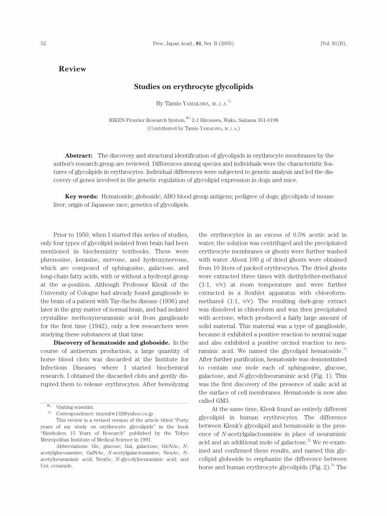
52 [Vol. 81(B), Prior to 1950, when I started this series of studies, only four types of glycolipid isolated from brain had been mentioned in biochemistry textbooks. These were phrenosine, kerasine, nervone, and hydroxynervone, which are composed of sphingosine, galactose, and long-chain fatty acids, with or without a hydroxyl group at the α -position. Although Professor Klenk of the University of Cologne had already found ganglioside in the brain of a patient with Tay-Sachs disease (1936) and later in the gray matter of normal brain, and had isolated crystalline methoxyneuraminic acid from ganglioside for the first time (1942), only a few researchers were studying these substances at that time. Discovery of hematoside and globoside. In the course of antiserum production, a large quantity of horse blood clots was discarded at the Institute for Infectious Diseases where I started biochemical research. I obtained the discarded clots and gently dis- rupted them to release erythrocytes. After hemolyzing the erythrocytes in an excess of 0.5% acetic acid in water, the solution was centrifuged and the precipitated erythrocyte membranes or ghosts were further washed with water. About 100 g of dried ghosts were obtained from 10 liters of packed erythrocytes. The dried ghosts were extracted three times with diethylether-methanol (1:1, v/v) at room temperature and were further extracted in a Soxhlet apparatus with chloroform- methanol (1:1, v/v). The resulting dark-gray extract was dissolved in chloroform and was then precipitated with acetone, which produced a fairly large amount of solid material. This material was a type of ganglioside, because it exhibited a positive reaction to neutral sugar and also exhibited a positive orcinol reaction to neu- raminic acid. We named the glycolipid hematoside. 1) After further purification, hematoside was demonstrated to contain one mole each of sphingosine, glucose, galactose, and N-glycolylneuraminic acid (Fig. 1). This was the first discovery of the presence of sialic acid at the surface of cell membranes. Hematoside is now also called GM3. At the same time, Klenk found an entirely different glycolipid in human erythrocytes. The difference between Klenk’s glycolipid and hematoside is the pres- ence of N-acetylgalactosamine in place of neuraminic acid and an additional mole of galactose. 2) We re-exam- ined and confirmed these results, and named this gly- colipid globoside to emphasize the difference between horse and human erythrocyte glycolipids (Fig. 2). 3) The Proc. Japan Acad., 81, Ser. B (2005) Studies on erythrocyte glycolipids By Tamio YAMAKAWA, M. J. A. †) RIKEN Frontier Research System, * ) 2-1 Hirosawa, Wako, Saitama 351-0198 (Contributed by Tamio YAMAKAWA, M. J. A.) Abstract: The discovery and structural identification of glycolipids in erythrocyte membranes by the author’s research group are reviewed. Differences among species and individuals were the characteristic fea- tures of glycolipids in erythrocytes. Individual differences were subjected to genetic analysis and led the dis- covery of genes involved in the genetic regulation of glycolipid expression in dogs and mice. Key words: Hematoside; globoside; ABO blood group antigens; pedigree of dogs; glycolipids of mouse liver; origin of Japanese race; genetics of glycolipids. * ) Visiting scientist. †) Correspondence: [email protected] This review is a revised version of the article titled “Forty years of my study on erythrocyte glycolipids” in the book “Rinshoken 15 Years of Research” published by the Tokyo Metropolitan Institute of Medical Science in 1991. Abbreviations: Glc, glucose; Gal, galactose; GlcNAc, N- acetylglucosamine; GalNAc, N-acetylgalactosamine; NeuAc, N- acetylneuraminic acid; NeuGc, N-glycolylneuraminic acid; and Cer, ceramide. Review
Transcript of Review Studies on erythrocyte glycolipidsglycolipids will probably increase as further studies...
52 [Vol. 81(B),
Prior to 1950, when I started this series of studies,only four types of glycolipid isolated from brain had beenmentioned in biochemistry textbooks. These werephrenosine, kerasine, nervone, and hydroxynervone,which are composed of sphingosine, galactose, andlong-chain fatty acids, with or without a hydroxyl groupat the α -position. Although Professor Klenk of theUniversity of Cologne had already found ganglioside inthe brain of a patient with Tay-Sachs disease (1936) andlater in the gray matter of normal brain, and had isolatedcrystalline methoxyneuraminic acid from gangliosidefor the first time (1942), only a few researchers werestudying these substances at that time.
Discovery of hematoside and globoside. In thecourse of antiserum production, a large quantity ofhorse blood clots was discarded at the Institute forInfectious Diseases where I started biochemicalresearch. I obtained the discarded clots and gently dis-rupted them to release erythrocytes. After hemolyzing
the erythrocytes in an excess of 0.5% acetic acid inwater, the solution was centrifuged and the precipitatederythrocyte membranes or ghosts were further washedwith water. About 100 g of dried ghosts were obtainedfrom 10 liters of packed erythrocytes. The dried ghostswere extracted three times with diethylether-methanol(1:1, v/v) at room temperature and were furtherextracted in a Soxhlet apparatus with chloroform-methanol (1:1, v/v). The resulting dark-gray extractwas dissolved in chloroform and was then precipitatedwith acetone, which produced a fairly large amount ofsolid material. This material was a type of ganglioside,because it exhibited a positive reaction to neutral sugarand also exhibited a positive orcinol reaction to neu-raminic acid. We named the glycolipid hematoside.1)
After further purification, hematoside was demonstratedto contain one mole each of sphingosine, glucose,galactose, and N-glycolylneuraminic acid (Fig. 1). Thiswas the first discovery of the presence of sialic acid atthe surface of cell membranes. Hematoside is now alsocalled GM3.
At the same time, Klenk found an entirely differentglycolipid in human erythrocytes. The differencebetween Klenk’s glycolipid and hematoside is the pres-ence of N-acetylgalactosamine in place of neuraminicacid and an additional mole of galactose.2) We re-exam-ined and confirmed these results, and named this gly-colipid globoside to emphasize the difference betweenhorse and human erythrocyte glycolipids (Fig. 2).3) The
Proc. Japan Acad., 81, Ser. B (2005)
Studies on erythrocyte glycolipids
By Tamio YAMAKAWA, M. J. A.†)
RIKEN Frontier Research System,*)2-1 Hirosawa, Wako, Saitama 351-0198
(Contributed by Tamio YAMAKAWA, M. J. A.)
Abstract: The discovery and structural identification of glycolipids in erythrocyte membranes by theauthor’s research group are reviewed. Differences among species and individuals were the characteristic fea-tures of glycolipids in erythrocytes. Individual differences were subjected to genetic analysis and led the dis-covery of genes involved in the genetic regulation of glycolipid expression in dogs and mice.
Key words: Hematoside; globoside; ABO blood group antigens; pedigree of dogs; glycolipids of mouseliver; origin of Japanese race; genetics of glycolipids.
*) Visiting scientist.†) Correspondence: [email protected]
This review is a revised version of the article titled “Fortyyears of my study on erythrocyte glycolipids” in the book“Rinshoken 15 Years of Research” published by the TokyoMetropolitan Institute of Medical Science in 1991.
Abbreviations: Glc, glucose; Gal, galactose; GlcNAc, N-acetylglucosamine; GalNAc, N-acetylgalactosamine; NeuAc, N-acetylneuraminic acid; NeuGc, N-glycolylneuraminic acid; andCer, ceramide.
Review

aforementioned difference in carbohydrate composi-tion prompted us to study the glycolipids of othermammalian erythrocytes, because we thought that thiscould be a chemical basis for species-specific differencesamong mammalian erythrocytes. With respect to themajor glycolipids, mammals have been classified intothree groups (Table I). The first group is the globoside-type, which refers to glycolipids with hexosamine but nosialic acid, which are found in humans, hogs, guinea pigs,sheep, and goats. The second group is the hematoside-type, which refers to glycolipids that contain sialic acidbut no hexosamine, which are found in dogs, cats, andhorses. The third group is the ganglioside-type, whichrefers to glycolipids that contain both sialic acid and hex-osamine, which are found in some individual cattle.4)
Characterization of Forssman antigen was an attractiveresearch subject in the 1960s, and this molecule waspurified from sheep erythrocytes, and subsequentlyclassified as a globoside-type glycolipid, GalNAcα1-3GalNAcβ1-3Galα1-4Galβ1-4Glcβ1-Ceramide.5),6)
The glycolipid content of human erythrocytes is rel-atively low, as compared to other lipids such as phos-pholipids and cholesterol (Fig. 3). Glycolipids compriseonly 5% of total lipids in humans. Investigation of theminor glycolipids in a large amount of human erythro-cytes led to the identification of more than twelve typesof glycolipid (Table II).7)
In 1953, we found ABO blood group activity in acrude globoside preparation.8) This was the first discov-ery that glycolipids were involved in cell recognition,which is now an accepted biological activity of glyco-lipids. After using column chromatography to purifyglycolipids, the active portions were separated from themajor globoside molecule (globoside I) and were furtherdivided into two fractions, namely globoside II and III.9)
Further studies indicated that these blood group-active glycolipids were ceramide-hexasaccharide and -octasaccharide, and the structures of these moleculeswere elucidated by Hakomori and coworkers.10) Thechemistry of blood group substances had already been
Studies on erythrocyte glycolipidsNo. 2] 53
Fig. 1. Structure of hematoside, the major glycolipid isolated from horse ery-throcytes. R, a fatty acid.
Fig. 2. Structure of globoside I, the major glycolipid isolated from human erythrocytes.
Table I. Nature of the hexosamine and sialic acids in the major glycolipidsof erythrocytes from various animals
Animal Glucosamine Galactosamine Sialic acids
Men – ++ –Hog – ++ –Guinea pig – ++ –Sheep – ++ –Goat – ++ –Rabbit ++ – –Cattle ++ – ++ or –Horse – – ++Dog – – ++Cat – – ++

elucidated by Morgan and coworkers in England, and byKabat and associates in the U.S.A., using active materialfrom body fluids or secretions. These blood group sub-stances were polysaccharide-protein complexes, butwe found that the antigenic material on the erythrocytesurface was glycolipid in nature with the same antigenicdeterminant groups. Nevertheless, some investigatorsstill insisted that the blood group material on erythrocytemembranes was glycoprotein,10),11) whereas othersbelieved that it must be a glycolipid with more thantwenty carbohydrate chains (i.e., a macroglyco-lipid).12),13) However, I believe that the major activity isattributable to glycolipids with relatively short carbohy-drate chains of 6 to 8.15) The number of different minorglycolipids will probably increase as further studiesproceed, although the total amounts of these moleculesare very small compared to the major glycolipid, globo-side I. These glycolipids are believed to be located on the
surface of the erythrocyte membrane, with the carbohy-drate moiety projecting from the cells and this charac-teristic feature facilitates the importance of these gly-colipids in cell-to-cell and cell-to-macromoleculerecognition.
The precise chemical structures of glycolipidspurified from various sources have been determinedusing chemical and biochemical techniques. For exam-ple, the structure of globoside I was elucidated by gaschromatography of partially methylated sugars aftermethanolysis of permethylated glycolipids, and by isola-tion of the oligosaccharides after hydrolysis with mildacid and specific hydrolases. Recently, direct massspectrometry and high-resolution nuclear magneticresonance spectroscopy have been useful in elucidatingthe structure of minute amounts of these com-pounds.16),17) Although the erythrocyte membranes of dif-ferent animals have several glycolipids, the dominant gly-
T. YAMAKAWA54 [Vol. 81(B),
Fig. 3. Lipid composition of human erythrocyte membranes. PC, phosphatidylcholine; PE,phosphatidylethanolamine; SM, sphingomyelin; PS, phosphatidylserine.
Table II. Structures and amounts of various glycolipids in human erythrocytes
Structure NameAmount
(µg/100 ml)
Glc-Cer Ceramide monohexoside 279Gal-Glc-Cer Ceramide dihexoside 2,211
Gal-Gal-Glc-Cer Ceramide trihexoside 1,021GlcNAc-Gal-Glc-Cer Amino-ceramide trihexoside 47
GalNAc-Gal-Gal-Glc-Cer Globoside I 9,600GalNAc-GalNAc-Gal-Gal-Glc-Cer Para-Forssman antigen 7
Gal-GlcNAc-Gal-Glc-Cer Paragloboside 553Gal-Gal-GlcNAc-Gal-Glc-Cer P1 antigen 5-10
GalNAc-Gal(Fuc)-GlcNAc-Gal-Glc-Cer A antigen 124or Gal-Gal(Fuc)-GlcNAc-Gal-Glc-Cer B antigen 120
NeuAc-Gal-Glc-Cer Hematoside 355NeuAc-Gal-GlcNAc-Gal-Glc-Cer Sialosylparagloboside 426

colipids are species-specific. The chemical structures ofthese major glycolipids have been elucidated for differentanimal species (Table III), and different species of animalcan be distinguished by the chemical structures of ery-throcyte glycolipids; thus, glycolipids can be used tocharacterize erythrocytes and species.
Individual differences of erythrocyte glyco-
lipids. One characteristic feature of erythrocyte glyco-lipids is that there are individual differences, like theABO blood group antigens in human erythrocytes. In1964, we reported that the erythrocyte glycolipid in dogis the hematoside-type glycolipid, sialyl-lactosylceramide, and that sialic acid comprises 73% of N-acetylneuraminic acid (NeuAc) and 27% of N-glycolyl-
Studies on erythrocyte glycolipidsNo. 2] 55
Table III. Predominant glycolipids in the erythrocytes of various animals
Globoside typeMan GalNAcβ1-3Galα1-4Galβ1-4Glcβ1-CerHog GalNAcβ1-3Galα1-4Galβ1-4Glcβ1-CerGuinea pig GalNAcβ1-4Galβ1-4Glcβ1-CerSheep (Forssman antigen) GalNAcα1-3GalNAcβ1-3Galα1-4Galβ1-4Glcβ1-CerGoat (Forssman antigen) GalNAcα1-3GalNAcβ1-3Galα1-4Galβ1-4Glcβ1-CerJapanese serow GalNAcα1-3GalNAcβ1-3Galα1-4Galβ1-4Glcβ1-CerRabbit Galα1-3Galβ1-3 or 4GlcNAcβ1-3Galβ1-4Glcβ1-CerCattle Galα1-3Galβ1-4GlcNAcβ1-3Galβ1-4Glcβ1-Cer
Ganglioside typeCattle NeuGcα2-3Galβ1-4GlcNAcβ1-3Galβ1-4Glcβ1-CerMouse (C3H/He, DBA/2) GalNAcβ1-4Gal(3-2αNeuGc)β1-4Glcβ1-Cer
Hematoside typeHorse, Mouse (WHT/Ht) NeuGcα2-3Galβ1-4Glcβ1-CerHorse 4-O-Ac-NeuGcα2-3Galβ1-4Glcβ1-CerShika deer NeuAc(and NeuGc)α2-3Galβ1-4Glcβ1-CerDog(European), Jackal, Dingo(A-type) NeuAcα2-3Galβ1-4Glcβ1-CerDog(some oriental), (G-type) NeuGcα2-3Galβ1-4Glcβ1-CerRacoon dog, Giant panda NeuGcα2-3Galβ1-4Glcβ1-CerCat, Lion, Hyena, Giant panda NeuGcα2-8NeuGcα2-3Galβ1-4Glcβ1-CerCat(some Persian cats) NeuAcα2-8NeuAcα2-3Galβ1-4Glcβ1-CerMouse(C3H/He) NeuAcα2-3Galβ1-Cer
Fig. 4. The structural difference between N-acetylneuraminic acid(NeuAc) and N-glycolylneuraminic acid (NeuGc).
Fig. 5. Thin-layer chromatogram of erythrocyteglycolipids from individual dogs. A: hematosidewith N-acetylneuraminic acid. B: hematosidewith N-glycolylneuraminic acid. Numbers 1-5correspond to erythrocyte glycolipids from 5individual dogs.

neuraminic acid (NeuGc) (Fig. 4).18) By contrast, whenKlenk and Heuer examined the structure of dog ery-throcyte glycolipid in 1960, they found that the neu-raminic acid was exclusively N-acetylated.19) Thisapparent discrepancy puzzled us for a long time.
In 1978, a graduate student in my laboratory re-examined erythrocyte glycolipids from individual dogsand found that some dogs had the N-glycolyl type ofhematoside (G type hematoside), while others had theN-acetyl type (A type); no mixed type existed (Fig. 5).20)
Whether a hematoside contains NeuAc or NeuGc can bedetermined readily by thin-layer chromatography, andthe sialic acid species can be determined by gas chro-matography with trimethylsilylation after mild acidhydrolysis. Because our previous study was carried outwith a pool of blood from several dogs, the sialic acidmight not have been structurally uniform. In addition,because the dogs that we examined in the aforemen-tioned study were mongrels, we tried to determine
which breeds of dog had the A or G type hematoside.We found only the A type (not the G type) in the
erythrocytes of all of the European dog breeds that weexamined (Table IV).20),21) On further investigation, wefound the G type in some oriental dogs, namely the Shibadog, Kai dog, Kishu dog, Chin, and Pekinese. Akita dogsand Hokkaido dogs had the A type, even though thesedogs originated from Japan (Table V).20),21) Shiba dogsoriginate from Japan and are rather small in size (~9 kgin weight). The population of Shiba dogs in Japan isdecreasing, so the Society for Preservation of the Shibadog was established to preserve this breed. By courtesyof this Society, we had an opportunity to examine thesialic acid species of hematoside in a family of Shiba dogsthat had been guaranteed to be purebred. As indicated inthe pedigree in Fig. 6, both the G and A types occurredin males and females in every generation. The female ( ) indicated by an arrow in Fig. 6 had the A type,whereas both her parents (★) had the G type. The same★
T. YAMAKAWA56 [Vol. 81(B),
Table V. Types of hematosides in erythrocytes from oriental dogs
BreedPhenotypes
NeuAc NeuGc
Japanese dogsHokkaido-dog 102 0Akita-dog 160 35Kai-dog 47 53Kishu-dog 39 33Shiba-dog
(Shinshu) 103 51(San’in) 23 8(Mino) 42 28
Shikoku-dog(Okayama) 17 66(Chyoshun) 0 51
Tosa-dog 8 1Nihon Spitz 5 3
Subtotal 546 329
Chinese origin dogs(north)Chow Chow 5 0Pug 2 0Shih Tzu 6 0
Subtotal 13 0
Chinese origin dogs(south)Chin 5 5Pekinese 0 3
Subtotal 5 8
Korean native dogs(Jindo) 50 151Taiwanese native dogs 97 11Eskimo dogs 18 1
Total 729 500
Table IV. Types of hematosides in erythrocytes from European dogs
BreedPhenotypes
NeuAc NeuGc
Afghan Hound 6 0American Cocker Spaniel 3 0Beagle 371 0Bulldog 3 0Borzoi 4 0Boxer 8 0Cairn Terrier 2 0Chihuahua 2 0Collie 10 0Dachshund 6 0Dalmatian 5 0Doberman Pinscher 8 0Fox Terrier 3 0French Poodle 1 0Great Dane 4 0Laika 1 0Maltese 43 0Mastiff 1 0Miniature Poodle 1 0Old English Sheepdog 2 0Pointer 38 0Pomeranian 11 0Poodle 10 0Setter 5 0Shepherd 41 0Shetland Sheepdog 9 0Siberian Husky 5 0St. Bernard 5 0Yorkshire Terrier 22 0Weimaraner 1 0West Highland 1 0
Total 632 0

female had two pups (○) with the G type by a dog (X)that had the G type. This indicates that the expression ofthe G type in erythrocytes is inherited as an autosomaldominant trait. Furthermore, in the case of the pedigreeof a family of beagles (a European breed), the genotypeis a recessive homozygote, and all dogs examinedexpressed only the A type hematoside (Fig. 7).
The difference between the A and G types ofhematoside lies only in the existence of one additionaloxygen atom per molecule in the latter (Fig. 4). As
reported by Shaw and Schauer 22) and Bouhours andBouhours,23) the conversion of NeuAc to NeuGc is cat-alyzed by a monooxygenase that acts preferentially at thestep of CMP-N-acetylneuraminic acid rather than atestablished glycoconjugates or free sialic acid. My col-leagues determined that the expression of NeuGc is reg-ulated by the expression of CMP-N-acetylneuraminicacid hydroxylase together with cytochrome b5 andcytochrome b5 reductase in the presence of nicotin-amide adenine dinucleotide (NADH).24)-27) The molecular
Studies on erythrocyte glycolipidsNo. 2] 57
Fig. 6. Pedigree of pure-bred Shiba dogs. □, male not examined; ○,female not examined; =, consanguinity (when consanguinity is obvi-ous, this symbol is omitted). Dogs in generation B had 7 differentmothers that were all related to the same father (a dog in generationA; not indicated in the figure).
Fig. 7. Pedigree of the beagles. ■, male with NeuAc-hematoside; ●, female with NeuAc-hematoside; =,consanguinity.
Fig. 8. Geographical distribution of dogs and related animals indicating the possible origins ofJapanese breeds of dog.

mechanisms that are involved in the regulation of theexpression of the G type hematoside are the subject offurther studies, and might include a mutation of the genethat controls erythrocyte-specific expression of thehydroxylase.
The origin of Japanese breeds of dog. The ori-gin of Japanese breeds of dog is controversial. Someclaim that Japanese breeds arrived in Japan fromEurope via a northern route through Siberia, whereasothers are of the opinion that they migrated from SouthAsia via a southern route. We believe that Akita andHokkaido dogs in Japan originated in Europe, becausethe erythrocytes of these breeds have the A typehematoside. Dogs that are indigenous to Japan andhave the G type hematoside are thought to have comefrom another area, possibly from South Asia (Fig. 8).20),21)
In the course of this work, we examined blood fromvarious animals obtained through the courtesy of manyfriends. We examined the erythrocytes of jackals and din-goes (of the genus Canis) and also examined raccoondogs (a member of the dog family, Canidae). In addition,we had the opportunity to study the erythrocyte glyco-lipid of the giant panda. Raccoon dogs and giant pandaslive in the Far East and the erythrocytes of thesespecies were found to contain the G type hematoside.This is interesting in light of the fact that the presence ofthe G type is limited to the erythrocytes of some orientaldogs. Both dingoes and jackals have the A type hemato-
side. Dingoes are wild dogs that are indigenous toAustralia; these animals have not been domesticated.Dingoes are thought to be the ancestor of moderndomestic dogs (Canis familiaris). At first, we suspect-ed that some Japanese breeds of dog might have origi-nated from dingoes. If this was the case, the erythrocytesof dingoes would be expected to be the G type hemato-side. However, erythrocytes from five dingoes werefound to have the A type, as do most European dogs.Therefore, it is unlikely that dingoes are the ancestors ofJapanese breeds, although this possibility cannot beexcluded.
Historically, dogs are thought to have been accom-panied by humans. Therefore, an investigation of the ori-gin of Japanese breeds of dog requires a study of the ori-gin of the Japanese race. From an archaeologicalaspect, there are two hypotheses concerning the origin ofthe Japanese. One holds that more than 2,300 years ago,an ancestral people migrated from the south of mainlandChina to the Kyushu district, where they developed anagricultural culture. The other hypothesis is that theancestors arrived at the southern part of Japan via theKorean Peninsula. The distribution of the A and G typehematoside in dogs does not allow us to favor one of theaforementioned hypotheses over the other, but it doessupport the view that the development of the modernJapanese people has been influenced strongly byChinese and Korean people (Fig. 8).
We also examined the erythrocytes of cats.28) Cat
T. YAMAKAWA58 [Vol. 81(B),
Fig. 9. Structures, biosynthetic pathways, and genes that regulate the expression of glycolipids.

erythrocyte glycolipid has two moles of NeuGc per mol-ecule, which was characterized as GD3, NeuGcα2-8NeuGcα2-3Galβ1-4Glcβ1-Ceramide (Fig. 9). Of 41samples of blood obtained from individual cats of variousbreeds, only two Persian cats were found to contain GD3with two moles of NeuAc. GD3 with two moles ofNeuGc occurred in the siblings of a family of Persiancats, but because relatively few of the family memberswere analyzed, no definitive conclusion about geneticregulation could be drawn.
The erythrocytes of lions (Felidae) and hyenas (aclose relative of felids) were found to contain GD3 withtwo moles of NeuGc, which is similar to the majority ofcats; GD3 with NeuAc, which occurred in some Persiancats, was not found in these species.
Polymorphism of mouse erythrocyte glyco-
lipids. We found polymorphic expression of erythrocyteglycolipids among various strains of inbred mice.29) Asrevealed by thin-layer chromatography (Fig. 10),C3H/He and BALB/c mice have mostly GM4 or sialylgalactosylceramide, as well as a small amount of GM2,GalNAcβ1-4(NeuGcα2-3)Galβ1-4Glcβ1-Ceramide (Fig.9). Other strains, such as DBA/2, DDD, SS, NC, SII,C57BL/6, BDF1 (C57BL/6 × DBA/2), FM, and SIII, havemore GM2 than GM4. Strains NZB and 129/J have a near-ly equal amount of GM4 and GM2. Only one strain(WHT/Ht) has only GM3 (NeuGcα2-3Galβ1-4Glcβ1-
Ceramide). Structural characterization of GM4 andGM2 purified from erythrocytes of C3H/He and CDF1
(C3H/He × DBA/2) mice indicated that the sialic acid ofGM4 is exclusively NeuAc, whereas that of GM2 isNeuGc (Fig. 9).30)
The ganglioside content of mouse erythrocytes isvery low: lipid-bound sialic acid comprised only 1 µg/mlof packed erythrocytes. Therefore, mouse erythrocytesappeared to be an unsuitable model to study the controlmechanisms that underlie genetic regulation of glycolipidexpression, because erythrocyte gangliosides of individ-ual mice must be analyzed. We happened subsequentlyto examine liver gangliosides of various inbred strains ofmice. We found that the liver ganglioside compositionresembled that of erythrocytes, but that the lipid-bound sialic acid content of the liver was almost 100times that of erythrocytes. These results prompted us toanalyze polymorphic variation in liver gangliosides.31),32)
We carried out a genetic study of the expression ofGM2 that contained NeuGc, GM2(NeuGc). By matingWHT/Ht mice (which lack GM2(NeuGc) but expressGM3(NeuGc) in liver) with BALB/c mice (whichexpress GM2(NeuGc) in liver), we were able to demon-strate that the expression of GM2(NeuGc) is a dominantphenotype over the expression of GM3(NeuGc).Backcrossed mice (F1 × WHT/Ht) segregated into twophenotypes that were GM2(NeuGc)-positive and -nega-
Studies on erythrocyte glycolipidsNo. 2] 59
Fig. 10. Thin-layer chromatogram of gangliosidesfrom erythrocytes of inbred mice. Sialic acid-containing glycolipids were visualized withresorcinol reagent.
Fig. 11. The activity of UDP-GalNAc:GM3(NeuAc) N-acetyl-galactosaminyltransferase in the liver of WHT/Ht mice,BALB/c mice, and their progeny. Each dot represents thetransferase activity in the liver of individual mice. In thebackcross of F1 and WHT/Ht, GM2(+) represents a group ofmice that expressed GM2(NeuGc), while GM2(-) indicatesmice that lacked GM2(NeuGc).
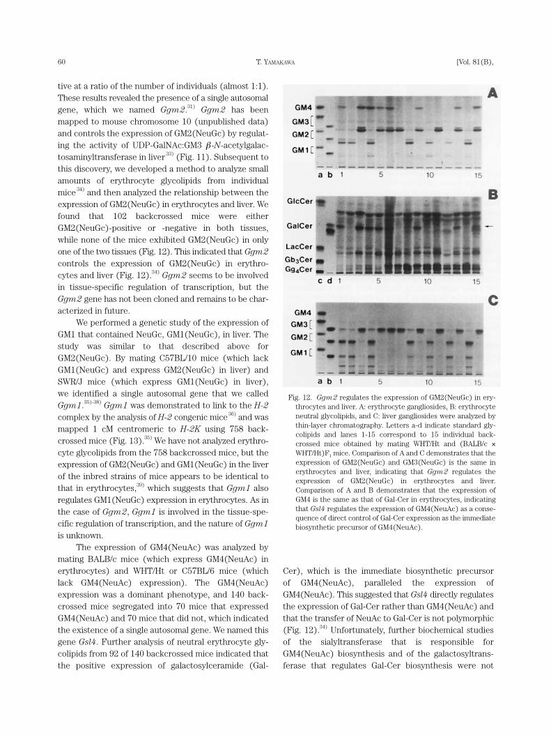
tive at a ratio of the number of individuals (almost 1:1).These results revealed the presence of a single autosomalgene, which we named Ggm2.31) Ggm2 has beenmapped to mouse chromosome 10 (unpublished data)and controls the expression of GM2(NeuGc) by regulat-ing the activity of UDP-GalNAc:GM3 β -N-acetylgalac-tosaminyltransferase in liver33) (Fig. 11). Subsequent tothis discovery, we developed a method to analyze smallamounts of erythrocyte glycolipids from individualmice 34) and then analyzed the relationship between theexpression of GM2(NeuGc) in erythrocytes and liver. Wefound that 102 backcrossed mice were eitherGM2(NeuGc)-positive or -negative in both tissues,while none of the mice exhibited GM2(NeuGc) in onlyone of the two tissues (Fig. 12). This indicated that Ggm2controls the expression of GM2(NeuGc) in erythro-cytes and liver (Fig. 12).34) Ggm2 seems to be involvedin tissue-specific regulation of transcription, but theGgm2 gene has not been cloned and remains to be char-acterized in future.
We performed a genetic study of the expression ofGM1 that contained NeuGc, GM1(NeuGc), in liver. Thestudy was similar to that described above forGM2(NeuGc). By mating C57BL/10 mice (which lackGM1(NeuGc) and express GM2(NeuGc) in liver) andSWR/J mice (which express GM1(NeuGc) in liver), we identified a single autosomal gene that we calledGgm1.35)-38) Ggm1 was demonstrated to link to the H-2complex by the analysis of H-2 congenic mice36) and wasmapped 1 cM centromeric to H-2K using 758 back-crossed mice (Fig. 13).35) We have not analyzed erythro-cyte glycolipids from the 758 backcrossed mice, but theexpression of GM2(NeuGc) and GM1(NeuGc) in the liverof the inbred strains of mice appears to be identical tothat in erythrocytes,39) which suggests that Ggm1 alsoregulates GM1(NeuGc) expression in erythrocytes. As inthe case of Ggm2, Ggm1 is involved in the tissue-spe-cific regulation of transcription, and the nature of Ggm1is unknown.
The expression of GM4(NeuAc) was analyzed bymating BALB/c mice (which express GM4(NeuAc) inerythrocytes) and WHT/Ht or C57BL/6 mice (whichlack GM4(NeuAc) expression). The GM4(NeuAc)expression was a dominant phenotype, and 140 back-crossed mice segregated into 70 mice that expressedGM4(NeuAc) and 70 mice that did not, which indicatedthe existence of a single autosomal gene. We named thisgene Gsl4. Further analysis of neutral erythrocyte gly-colipids from 92 of 140 backcrossed mice indicated thatthe positive expression of galactosylceramide (Gal-
Cer), which is the immediate biosynthetic precursor of GM4(NeuAc), paralleled the expression ofGM4(NeuAc). This suggested that Gsl4 directly regulatesthe expression of Gal-Cer rather than GM4(NeuAc) andthat the transfer of NeuAc to Gal-Cer is not polymorphic(Fig. 12).34) Unfortunately, further biochemical studies of the sialyltransferase that is responsible forGM4(NeuAc) biosynthesis and of the galactosyltrans-ferase that regulates Gal-Cer biosynthesis were not
T. YAMAKAWA60 [Vol. 81(B),
Fig. 12. Ggm2 regulates the expression of GM2(NeuGc) in ery-throcytes and liver. A: erythrocyte gangliosides, B: erythrocyteneutral glycolipids, and C: liver gangliosides were analyzed bythin-layer chromatography. Letters a-d indicate standard gly-colipids and lanes 1-15 correspond to 15 individual back-crossed mice obtained by mating WHT/Ht and (BALB/c ×WHT/Ht)F1 mice. Comparison of A and C demonstrates that theexpression of GM2(NeuGc) and GM3(NeuGc) is the same inerythrocytes and liver, indicating that Ggm2 regulates theexpression of GM2(NeuGc) in erythrocytes and liver.Comparison of A and B demonstrates that the expression ofGM4 is the same as that of Gal-Cer in erythrocytes, indicatingthat Gsl4 regulates the expression of GM4(NeuAc) as a conse-quence of direct control of Gal-Cer expression as the immediatebiosynthetic precursor of GM4(NeuAc).
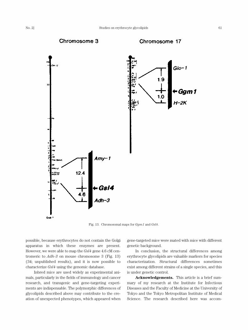
possible, because erythrocytes do not contain the Golgiapparatus in which these enzymes are present.However, we were able to map the Gsl4 gene 4.6 cM cen-tromeric to Adh-3 on mouse chromosome 3 (Fig. 13)(34; unpublished results), and it is now possible tocharacterize Gsl4 using the genomic database.
Inbred mice are used widely as experimental ani-mals, particularly in the fields of immunology and cancerresearch, and transgenic and gene-targeting experi-ments are indispensable. The polymorphic differences ofglycolipids described above may contribute to the cre-ation of unexpected phenotypes, which appeared when
gene-targeted mice were mated with mice with differentgenetic background.
In conclusion, the structural differences amongerythrocyte glycolipids are valuable markers for speciescharacterization. Structural differences sometimesexist among different strains of a single species, and thisis under genetic control.
Acknowledgements. This article is a brief sum-mary of my research at the Institute for InfectiousDiseases and the Faculty of Medicine at the University ofTokyo and the Tokyo Metropolitan Institute of MedicalScience. The research described here was accom-
Studies on erythrocyte glycolipidsNo. 2] 61
Fig. 13. Chromosomal maps for Ggm1 and Gsl4.

plished only with the efforts of many collaborators.
References
1) Yamakawa, T., and Suzuki, S. (1951) The chemistry of the
lipids of posthemolytic residue or stroma of erythrocytes. I.
Concerning the ether-insoluble lipids of lyophilized horse
blood stroma. J. Biochem. 38, 199-212.
2) Klenk, E., and Lauenstein, K. (1951) Ueber die zuckerhaltigen
Lipoide der Formbestandteile des menschlichen Blutes. Z.
Physiol. Chem. 288, 220-228.
3) Yamakawa, T., and Suzuki, S. (1952) The chemistry of the
lipids of posthemolytic residue or stroma of erythrocytes. III.
Globoside, the suger-containing lipid of human blood stro-
ma. J. Biochem. 39, 220-228.
4) Yamakawa, T. (1966) Glycolipids of mammalian red blood
cells. In Lipoid (16. Colloquium der Gesellschaft fuer
Physiologische Chemie) (ed. Schuette, E.). Springer-
Verlag, Berlin, Heidelberg and New York, pp. 87-111.
5) Ando, S., and Yamakawa, T. (1970) On the oligosaccharide of
Forssman-active sheep red cell glycolipid. Chem. Phys.
Lipids 5, 91-95.
6) Siddiqui, B., and Hakomori, S. (1971) A revised structure for
the Forssman glycolipid hapten. J. Biol. Chem. 246,
5766-5769.
7) Yamakawa, T., and Nagai, Y. (1978) Glycolipids at the cell sur-
face and their biological functions. Trends Biochem. Sci. 3,
128-131.
8) Yamakawa, T., and Iida, T. (1953) Immunological study of the
red blood cells. I. Globoside, as the agglutinogen of the ABO
system on erythrocytes. Jpn. J. Exp. Med. 23, 327-331.
9) Yamakawa, T., Nishimura, S., and Kamimura, M. (1965) The
chemistry of the lipids of posthemolytic residue or stroma of
erythrocytes. XIII. Further studies on human red cell gly-
colipids. Jpn. J. Exp. Med. 35, 201-207.
10) Marchesi, V. T., and Andrews, E. P. (1971) Glycoproteins:
Isolation from cell membranes with lithium diiodosalicylate.
Science 174, 1247-1248.
11) Finne, J., Krusius, T., Rauvala, H., Kekomaki, R., and Myllyla,
G. (1978) Alkali-stable blood group A- and B-active poly
(glycosyl) peptides from human erythrocyte membrane.
FEBS Lett. 89, 111-115.
12) Hakomori, S., Stellner, K., and Watanabe, K. (1972) Four anti-
genic variants of blood group A glycolipid: Examples of high-
ly complex, branched chain glycolipid of animal cell mem-
brane. Biochem. Biophys. Res. Commun. 49, 1061-1068.
13) Gardas, A., and Koscielak, J. (1973) New form of A-, B-, and
H-blood group-active substances extracted from erythrocyte
membranes. Eur. J. Biochem. 32, 178-187.
14) Dejter-Juszynski, M., Harpaz, N., Flowers, H. M., and Sharon,
N. (1978) Blood-group ABH-specific macroglycolipids of
human erythrocytes: Isolation in high yield from a crude
membrane glycoprotein fraction. Eur. J. Biochem. 83,
363-373.
15) Yamato, K., Handa, S., and Yamakawa, T. (1975) Blood group
A activities of glycoprotein and glycolipid from human ery-
throcyte membranes. J. Biochem. 78, 1207-1214.
16) Sekine, M., Suzuki, M., Inagaki, F., Suzuki, A., and
Yamakawa, T. (1987) A new extended globoglycolipid car-
rying the stage specific embryonic antigen-1(SSEA-1)
determinant in mouse kidney. J. Biochem. 101, 553-562.
17) Nakamura, K., Suzuki, M., Inagaki, F., Yamakawa. T., and
Suzuki, A. (1987) A new ganglioside showing cholera-
genoid-binding activity in mouse spleen. J. Biochem. 101,
825-835.
18) Handa, S., and Yamakawa, T. (1964) Chemistry of lipids of
posthemolytic residue or stroma of erythrocytes. XII.
Chemical structure and chromatographic behavior of
hematosides obtained from equine and dog erythrocytes.
Jpn. J. Exp. Med. 34, 293-304.
19) Klenk, E., and Heuer, K. (1960) Ueber die Ganglioside der
Hundererythrozyten. Dtsch. Z. Verdauungs-u. Stoffwechselkr.
20, 180-183.
20) Yasue, S., Handa, S., Miyagawa, S., Inoue, J., Hasegawa, A., and
Yamakawa, T. (1978) Difference in form of sialic acid in red
blood cell glycolipids of different breeds of dogs. J.
Biochem. 83, 1101-1107.
21) Hashimoto, Y., Yamakawa, T., and Tanabe, Y. (1984) Further
studies on the red cell glycolipids of various breeds of dogs.
A possible assumption about the origin of Japanese dogs. J.
Biochem. 96, 1777-1782.
22) Shaw, L., and Schauer, R. (1988) The biosynthesis of N-gly-
colylneuraminic acid occurs by hydroxylation of the CMP-
glycoside of N-acetylneuraminic acid. Biol. Chem. Hoppe-
Seyler. 369, 477-486.
23) Bouhours, J.-F., and Bouhours, D. (1989) Hydroxylation of
CMP-NeuAc controls the expression of N-glycolylneur-
aminic acid in GM3 ganglioside of the small intestine of
inbred rats. J. Biol. Chem. 264, 16992-16999.
24) Kozutsumi, Y., Kawano, T., Yamakawa, T., and Suzuki, A.
(1990) Participation of cytochrome b5 in CMP-N-acetyl-
neuraminic acid hydroxylation in mouse liver cytosol. J.
Biochem. 108, 704-706.
25) Kawano, T., Kozutsumi, Y., Kawasaki, T., and Suzuki, A.
(1994) Biosynthesis of N-glycolylneuraminic acid-containing
glycoconjugates. Purification and characterization of the key
enzyme of the cytidine monophospho-N-acetylneuraminic
acid hydroxylation system. J. Biol. Chem. 269, 9024-9029.
26) Kawano, T., Koyama, S., Takematsu, H., Kozutsumi, Y.,
Kawasaki, H., Kawashima, S., Kawasaki, T., and Suzuki, A.
(1995) Molecular cloning of cytidine monophospho-N-
acetylneuraminic acid hydroxylase. Regulation of species-
and tissue-specific expression of N-glycolylneuraminic
acid. J. Biol. Chem. 270, 16458-16463.
27) Irie, A., Koyama, S., Kozutsumi, Y., Kawasaki, T., and Suzuki,
A. (1998) The molecular basis for the absence of N-gly-
colylneuraminic acid in humans. J. Biol. Chem. 273,
15866-15871.
28) Hamanaka, S., Handa, S., Inoue, J., Hasegawa, A., and
Yamakawa, T. (1979) Occurrence of hematoside with two
T. YAMAKAWA62 [Vol. 81(B),

moles of N-acetylneuraminic acid in a certain breed of
Persian cat. J. Biochem. 86, 695-698.
29) Hamanaka, S., Handa, S., and Yamakawa, T. (1979)
Ganglioside compositions of erythrocytes from various
strains of inbred mice. Occurrence of sialosylgalactosyl-
ceramide in red blood cells of inbred mice. J. Biochem. 86,
1623-1626.
30) Hashimoto, Y., Otsuka, H., and Yamakawa, T. (1982) The
occurrence of GM4 and GM2 in erythrocytes from inbred
strains of mice. J. Biochem. 91, 1039-1046.
31) Hashimoto, Y., Otsuka, H., Sudo, K., Suzuki, K., Suzuki, A., and
Yamakawa, T. (1983) Genetic regulation of GM2 expression
in liver of mouse. J. Biochem. 93, 895-901.
32) Yamakawa, T., Suzuki, A., and Hashimoto, Y. (1986) Genetic
control of glycolipid expression. Chem. Phys. Lipids 42,
75-90.
33) Hashimoto, Y., Abe, M., Kiuchi, Y., Suzuki, A., and Yamakawa,
T. (1984) Genetically regulated expression of UDP-N-
acetylgalactosamine: GM3(NeuGc) N-acetylgalactosaminyl-
transferase [EC 2.4.1.92] activity in mouse liver. J.
Biochem. 95, 1543-1549.
34) Nakamura, K., Hashimoto, Y., Moriwaki, K., Yamakawa, T., and
Suzuki, A. (1990) Genetic regulation of GM4(NeuAc)
expression in mouse erythrocytes. J. Biochem. 107, 3-7.
35) Sakaizumi, M., Hashimoto, Y., Suzuki, A., Yamakawa, T.,
Kiuchi, Y., and Moriwaki, K. (1988) The locus controlling
liver GM1(NeuGc) expression is mapped 1 cM centromeric
to H-2K. Immunogenetics 27, 57-60.
36) Hashimoto, Y., Suzuki, A., Yamakawa, T., Miyashita, N., and
Moriwaki, K. (1983) Expression of GM1 and GD1a in
mouse liver is linked to the H-2 complex on chromosome 17.
J. Biochem. 94, 2043-2048.
37) Hashimoto, Y., Suzuki, A., Yamakawa, T., Wang, C. H.,
Bonhomme, F., Miyashita, N., and Moriwaki, K. (1984)
Expression of GM1 and GD1a in liver of wild mice. J.
Biochem. 95, 7-12.
38) Hashimoto, Y., Abe, M., Suzuki, A., Iwasaki, K., and
Yamakawa, T. (1985) A locus controlling the activity of UDP-
galactose: GM2(NeuGc) galactosyltransferase in mouse
liver is linked to the H-2 complex. Glycoconjugate J. 2,
255-265.
39) Nakamura, K., Hashimoto, Y., Yamakawa, T., and Suzuki, A.
(1988) Genetic polymorphism of ganglioside expression in
mouse organs. J. Biochem. 103, 201-208.
(Received Jan. 24, 2005; accepted Feb. 14, 2005)
Studies on erythrocyte glycolipidsNo. 2] 63
Profile
Tamio Yamakawa was born in 1921 and started his research career in1945 with studies on the metabolic fate of branched-chain fatty acids in theanimal body at the Institute for Infectious Diseases, after graduating from theFaculty of Medicine at the University of Tokyo. He performed pioneeringwork on the isolation and structural characterization of a ganglioside from themembrane of horse erythrocytes in 1950; this was the first demonstration ofthe occurrence of glycosphingolipids and sialic acid in the plasma membrane,and this study led to subsequent extensive studies that demonstratedspecies-specific carbohydrate structures of glycolipids in erythrocyte mem-branes. He was promoted to Professor at the University of Tokyo in 1959 andbecame Head of the Department of Chemistry at the Institute for Infectious Diseases. He moved to theDepartment of Biochemistry, Faculty of Medicine, at the University of Tokyo in 1966, where he educatedmany students in the field of biochemistry. He was awarded the Asahi Culture Award in 1975, the JapanAcademy Prize in 1976, and the Sphinx Prize by the International Glycolipid Research Associates for his pio-neering studies. Between 1982 and 1991, he was Director of the Tokyo Metropolitan Institute of MedicalScience. He was elected a member of the Japan Academy in 1988 and is an honorary member of theAmerican Society for Biochemistry and Molecular Biology, the Japanese Biochemical Society, and thePharmaceutical Society of Japan. Between 1991 and 1994, he was President of the Tokyo College ofPharmacy.