REVIEW Open Access The significance of macrophage ...
13
REVIEW Open Access The significance of macrophage phenotype in cancer and biomaterials Hannah C Bygd 1 , Kiva D Forsmark 2 and Kaitlin M Bratlie 1,2,3* Abstract Macrophages have long been known to exhibit heterogeneous and plastic phenotypes. They show functional diversity with roles in homeostasis, tissue repair, immunity and disease. There exists a spectrum of macrophage phenotypes with varied effector functions, molecular determinants, cytokine and chemokine profiles, as well as receptor expression. In tumor microenvironments, the subset of macrophages known as tumor-associated macrophages generates byproducts that enhance tumor growth and angiogenesis, making them attractive targets for anti-cancer therapeutics. With respect to wound healing and the foreign body response, there is a necessity for balance between pro-inflammatory, wound healing, and regulatory macrophages in order to achieve successful implantation of a scaffold for tissue engineering. In this review, we discuss the multitude of ways macrophages are known to be important in cancer therapies and implanted biomaterials. Keywords: Macrophage reprogramming; Cancer; Biomaterials; Anti-angiogenic therapy; Recruitment inhibition; Tissue engineering; Foreign body response Introduction Heterogeneity of Macrophages Macrophages are considered to be functionally hetero- geneous cells with different phenotypes representing distinct sublineages [1,2]. The heterogeneity of these cells is attributed to their location in the tissue, due to microenvironmental signals that control the functional phenotype [1,3-5]. In the presence of specific microen- viromental signals, macrophages are able to switch from one phenotype to another, indicating that these cells have a degree of plasticity in addition to heterogeneity [3,6]. In general, heterogeneity of macrophages can be described as a spectrum of phenotypes [1-3,6-10]. One end represents classical macrophages activated with interferon (IFN)-γ ,M (IFN-γ), and at the other end alternative macrophages activated by interleukin (IL)-4, M(IL-4) [7,8,11-15]. This new nomenclature, recently proposed by Murray et al., more accurately reflects the individual phenotypes and po- larizations of these cells. Other variations of macrophages that lie along this spectrum include: M(Ic), activated by immune complexes (Ic); M(IL-10); those stimulated by glucocorticoids (GC) and transforming growth factor (TGF)-β, M(GC + TGF-β); M(GC); M(LPS), activated by lipopolysaccharides; and M(LPS + IFN-γ) [3,6,8-10]. Each of these phenotypes varies in their effector functions, mo- lecular determinants, cytokine and chemokine profiles, as well as receptor expression. Overall, classically activated, formerly referred to as M1, macrophages are known to be pro-inflammatory and cyto- toxic. Macrophages are skewed towards this phenotype when IFNs and toll-like receptor (TLR) signaling activate IFN regulatory factor/signal transducers and activators of transcription (IRF/STAT) signaling pathways via STAT1 [7,10,15-18]. This transcription factor then causes macro- phages to upregulate IRF5, which is essential for pro- duction of large amounts of pro-inflammatory cytokines [16], including tumor necrosis factor (TNF)-α, IL-1β, IL-1, IL-6, IL-8, IL-12, IL-15, IL-18, and IL-23 that elicit both T-helper (Th)1 and Th17 responses [9,16,18-20]. TLR stimulation can also activate nuclear factor (NF)-κB, such that p65/p50 heterodimers are formed and lead to the production of hypoxia-inducible factor (HIF)-1α [15,21,22]. This protein, found in the presence of low oxygen concen- trations, regulates the NOS2 gene to increase the secretion of inducible nitric oxide synthase (iNOS) [21], toxic nitric * Correspondence: [email protected] 1 Department of Materials Science & Engineering, Iowa State University, Ames, Iowa 50011, USA 2 Department of Chemical & Biological Engineering, Iowa State University, Ames, Iowa 50011, USA Full list of author information is available at the end of the article © 2014 Bygd et al.; licensee Springer. This is an Open Access article distributed under the terms of the Creative Commons Attribution License (http://creativecommons.org/licenses/by/4.0), which permits unrestricted use, distribution, and reproduction in any medium, provided the original work is properly credited. Bygd et al. Clinical and Translational Medicine 2014, 3:41 http://www.clintransmed.com/content/3/1/41
Transcript of REVIEW Open Access The significance of macrophage ...
Bygd et al. Clinical and Translational Medicine 2014, 3:41http://www.clintransmed.com/content/3/1/41
REVIEW Open Access
The significance of macrophage phenotype incancer and biomaterialsHannah C Bygd1, Kiva D Forsmark2 and Kaitlin M Bratlie1,2,3*
Abstract
Macrophages have long been known to exhibit heterogeneous and plastic phenotypes. They show functional diversitywith roles in homeostasis, tissue repair, immunity and disease. There exists a spectrum of macrophage phenotypes withvaried effector functions, molecular determinants, cytokine and chemokine profiles, as well as receptor expression. Intumor microenvironments, the subset of macrophages known as tumor-associated macrophages generates byproductsthat enhance tumor growth and angiogenesis, making them attractive targets for anti-cancer therapeutics. With respectto wound healing and the foreign body response, there is a necessity for balance between pro-inflammatory, woundhealing, and regulatory macrophages in order to achieve successful implantation of a scaffold for tissue engineering. Inthis review, we discuss the multitude of ways macrophages are known to be important in cancer therapies andimplanted biomaterials.
Keywords: Macrophage reprogramming; Cancer; Biomaterials; Anti-angiogenic therapy; Recruitment inhibition;Tissue engineering; Foreign body response
IntroductionHeterogeneity of MacrophagesMacrophages are considered to be functionally hetero-geneous cells with different phenotypes representingdistinct sublineages [1,2]. The heterogeneity of thesecells is attributed to their location in the tissue, due tomicroenvironmental signals that control the functionalphenotype [1,3-5]. In the presence of specific microen-viromental signals, macrophages are able to switch fromone phenotype to another, indicating that these cells havea degree of plasticity in addition to heterogeneity [3,6]. Ingeneral, heterogeneity of macrophages can be described asa spectrum of phenotypes [1-3,6-10]. One end representsclassical macrophages activated with interferon (IFN)-γ, M(IFN-γ), and at the other end alternative macrophagesactivated by interleukin (IL)-4, M(IL-4) [7,8,11-15]. Thisnew nomenclature, recently proposed by Murray et al.,more accurately reflects the individual phenotypes and po-larizations of these cells. Other variations of macrophagesthat lie along this spectrum include: M(Ic), activated by
* Correspondence: [email protected] of Materials Science & Engineering, Iowa State University, Ames,Iowa 50011, USA2Department of Chemical & Biological Engineering, Iowa State University,Ames, Iowa 50011, USAFull list of author information is available at the end of the article
© 2014 Bygd et al.; licensee Springer. This is anAttribution License (http://creativecommons.orin any medium, provided the original work is p
immune complexes (Ic); M(IL-10); those stimulated byglucocorticoids (GC) and transforming growth factor(TGF)-β, M(GC + TGF-β); M(GC); M(LPS), activated bylipopolysaccharides; and M(LPS + IFN-γ) [3,6,8-10]. Eachof these phenotypes varies in their effector functions, mo-lecular determinants, cytokine and chemokine profiles, aswell as receptor expression.Overall, classically activated, formerly referred to as M1,
macrophages are known to be pro-inflammatory and cyto-toxic. Macrophages are skewed towards this phenotypewhen IFNs and toll-like receptor (TLR) signaling activateIFN regulatory factor/signal transducers and activators oftranscription (IRF/STAT) signaling pathways via STAT1[7,10,15-18]. This transcription factor then causes macro-phages to upregulate IRF5, which is essential for pro-duction of large amounts of pro-inflammatory cytokines[16], including tumor necrosis factor (TNF)-α, IL-1β, IL-1,IL-6, IL-8, IL-12, IL-15, IL-18, and IL-23 that elicit bothT-helper (Th)1 and Th17 responses [9,16,18-20]. TLRstimulation can also activate nuclear factor (NF)-κB,such that p65/p50 heterodimers are formed and lead to theproduction of hypoxia-inducible factor (HIF)-1α [15,21,22].This protein, found in the presence of low oxygen concen-trations, regulates the NOS2 gene to increase the secretionof inducible nitric oxide synthase (iNOS) [21], toxic nitric
Open Access article distributed under the terms of the Creative Commonsg/licenses/by/4.0), which permits unrestricted use, distribution, and reproductionroperly credited.
Bygd et al. Clinical and Translational Medicine 2014, 3:41 Page 2 of 13http://www.clintransmed.com/content/3/1/41
oxide (NO), and reactive oxygen intermediates (ROI) [19]. Achemokine profile for classically activated macrophages mayinclude HCC-2 (CCL15), macrophage inflammatory protein(MIP)-3α (CCL20), and B cell attracting chemokine-1(CXCL13), as well as IFN-γ-inducible chemokines such as,monocyte chemotactic protein (MCP)-1 (CCL2), interferon-inducible T cell alpha chemoattractant (I-TAC) (CXCL11),interferon gamma-induced protein 10 (IP-10) (CXCL10)and monokine induced by gamma interferon (MIG)(CXCL9) [7,18,20-22]. Production of these chemokinescan be a result of previously mentioned transcription fac-tors STAT1 or NF- κB [16,18]. These chemokines alsocoordinate natural killer (NK) and Th1 cell responses,integrating classically activated macrophages into the amp-lification and regulation of polarized T cell responses[20,21]. Surface molecules expressed by classically acti-vated macrophages include elevated amounts of MHCclass II receptors; costimulatory molecules CD80 andCD86; IL-2Ra, IL-15Ra and IL-7R; and low levels of man-nose receptor C type 1 (MRC1) and Fcγ RII [17,18,20].Each of these characteristics allow classically activatedmacrophages to be potent effector cells that mediate re-sistance against bacterial, viral, and fungal infections aswell as tumor cells [18,19]. They are also important in theinflammatory stages of wound healing and the foreignbody response (FBR) to biomaterials [23-25].Alternatively activated, previously known as M2 mac-
rophages, are said to be pro-angiogenic, promoting tis-sue remodeling and repair. This phenotype arises whenIL-4 activates the IRF/STAT signaling pathway via STAT6[7,10,15-18]. IL-10, on the other hand, activates STAT3-mediated alternative activation and gene expression[7,15-18]. This STAT-mediated activation of macrophagesis regulated by the suppressor of cytokine signaling (SOCS)family: where IL-4 can upregulate SOCS1, inhibiting theaction of STAT1, but IFN-γ and TLR stimulation causeSOCS3 to be upregulated to prevent the activity of STAT3[16,26]. The transcription factors STAT3 and STAT6 allowfor high-level production of the cytokines IL-10, IL-1 re-ceptor antagonist (IL-1Rα), IL-4Rα, TGF-β, and the type IIIL-1 decoy receptor [16,18,20,21]. Other genes activated bySTAT6 include mannose receptor (Mrc1), resistin-likeα (Retnla/Fizz1), and chitinase 3-like 3 (Chi3l3/Ym1).For STAT3, some of the genes expressed are Il10, Tgfb1,and Mrc1 [16]. STAT6 also coordinates with peroxisomeproliferator-activated receptors PPARγ and PPARδ, as wellas Krüppel-like factor (KLF)-4, to induce some alternativegenes (Arg-1, Mrc1, Fizz1, PPARγ) while inhibiting genesassociated with classical activation (TNFa, Cox-2, CCL5,iNOS) by preventing NF-κB activation [16]. However, NF-κB activation and the formation of p50 homodimers arealso important in alternative activation and resolution ofinflammation [15,21,22]. Chemokines induced by IL-4 orIL-13 alternative activation include monocyte chemotactic
protein (MCP)-4 (CCL13), MCP-2 (CCL8), MCP-1 (CCL2),macrophage-derived chemokine (MDC) (CCL22), alternativemacrophage activation-associated chemokine (AMAC)-1(CCL18) and eotaxin-3 (CCL26) [7,18,20-22]. CCL22specifically attracts Th2 and Treg cells, showing thatalternative macrophages are also involved in the polarizationof T cell responses [21]. Macrophages activated by IL-10, TGF-β, and GC produce the chemokines eotaxin-2(CCL24), IP-10 (CXCL10), I-TAC (CXCL11), and regulatedon activation, normal T cell expressed and secreted(RANTES) (CCL5) [20,21]. Other factors produced includevascular endothelial growth factor (VEGF), matrix metallo-proteinases (MMPs); and HIF-2α to regulate ARG1 and thearginase pathway to produce ornithine and polyamines[18]. The exception to alternative activation is the pheno-type of macrophages induced by Ic; they retain the abilityto produce high levels of pro-inflammatory cytokines [18].Overall, alternatively activated macrophages are efficientphagocytic cells with the expression of mannose and galact-ose receptors; CD163, TLR8, TLR1, and IL21a; and MRC1and scavenger receptor type 1 (SR-A1) [17,18,20]. They areinvolved in parasite containment, tumor progression, andfunction to dampen immune responses [12]. In the reso-lution stages of the FBR, alternative macrophages drive thewound healing response, often leading to fibrotic encapsu-lation and failure of implanted devices and scaffolds [23,25].
ReviewMacrophages as cancer therapeutic targetsTumor-associated macrophagesTumor-associated macrophages (TAMs) have propertiesconsistent with alternatively activated macrophages [27].They produce cytokines like IL-10 and TGF-β [21]. Thepolarization of macrophages recruited to a tumor site,or any other tissue, is highly dependent on the cytokinespresent. The production of both IL-10 and TGF-β sup-presses anti-tumor activities of the immune system allow-ing tumor cells to avoid destruction by immune cells [28].TAMs have been found to be poor producers of NO andROIs, which are typically products of classically activatedmacrophages [29]. In addition, TAMs express low levels ofcytokines such as IL-12, TNF-α, and IL-6 [29]. Lastly,TAMs have been found to be poor antigen-presentingcells indicating that they do not have the potent effectorcell functions attributed to classically activated macro-phages [19]. This information establishes that TAMs rep-resent a subset of alternatively activated macrophages, andthat many of their byproducts enhance tumor growth andangiogenesis.While angiogenesis plays a central role in the progres-
sion of tumors from benign to malignant, there are manyother factors involved. MMPs contribute to tumor inva-sion through matrix remodeling where they are capable ofcleaving extracellular matrix (ECM) proteins [29], which

Bygd et al. Clinical and Translational Medicine 2014, 3:41 Page 3 of 13http://www.clintransmed.com/content/3/1/41
normally provide a barrier for tumor growth. These MMPsalong with other proteases such as plasmin and urokinase-type plasminogen activator (uPA) are all produced byTAMs [21,29]. The continued proliferation and growthaided by TAMs can lead to metastasis of tumor cells. Inmetastasis, it is suggested that primary tumors are ableto release factors that increase a metastatic outcome atother sites. These sites are referred to as premetastaticniches where the factors secreted by primary tumorscause the accumulation of myeloid progenitor cells [30]. Arecent study has shown that TAMs play an important rolein controlling the survival, migration and growth of meta-static cells to these niches [31]. TAMs were also found toenhance tumor cell extravasation, establishment and sub-sequent growth in surrounding tissue. The involvement ofTAMs in tumor angiogenesis, growth, progression andmetastasis makes them attractive targets for anti-cancertherapeutics. Therapeutic strategies directed at TAMsfall into four categories: reduction of effector function,limiting recruitment, prevention of pro-tumor polarization,and macrophage reprogramming [32,33]; the benefitsand drawbacks of which are outlined in Table 1 [33-42].
Anti-angiogenic therapyAngiogenesis must occur in tumors for them to groweven small amounts [35]. This process can be influencedvia a multitude of factors that are induced in hypoxicregions including VEGF, placental growth factor (PlGF),angiopoietins (ANGs), colony stimulating factor (CSF)-1, and CCL2/MCP-1 [35]. Anti-angiogenic therapy viathe VEGF pathway, the primary angiogenic pathway ofmacrophages, is ineffective, as tumor cells are able to acti-vate other pro-angiogenic pathways [36]. However, macro-phage angiogenic abilities can be indirectly prohibitedthrough the use of other factors. When a tumor developsregions of inadequate oxygen supply, HIF1-α subunits arestabilized, recruiting bone marrow (BM)-derived cells in-cluding macrophages that up regulate angiogenesis. Theelimination of HIF1-α from the tumor environment pro-vides a potential anti-angiogenic cancer therapy pathwayby inhibiting the recruitment of macrophages and otherpro-angiogenic cells [43]. HIF1-α knockout mice (HIFko)with glioblastoma (GBM) tumors, show a decrease inangiogenesis when compared to HIF functional mice withtumors [43].
Table 1 Advantages and disadvantages of anti-cancer therap
Approach Advantages
Anti-angiogenic therapy Inhibit tumor growth and prevent metastasimproves efficacy of chemotherapeutics [35
Recruitment inhibition Prevent macrophages from entering tumorbecoming TAMs [38,39]
Macrophage reprogramming Macrophages secrete tumoricidal molecule
ANG2 is produced by endothelial cells in hypoxic envi-ronments and would typically recruit pro-angiogenic cells,however binding of ANG2 with a monoclonal antibodyinhibited angiogenesis by blocking the interaction ofANG2 with TIE2-expressing monocytes [44,45]. TIE2-expressing monocytes are a subpopulation of TAMs thathave the greatest role in tumor angiogenesis [44]; prevent-ing activation of these cells can halt their angiogenic activ-ity and disable further recruitment of pro-angiogenic cells.Blocking of ANG2 with a monoclonal antibody inhibitstumor growth; causes regression of tumor vasculature byinducing apoptosis in some pro-angiogenic cells; andhinders progression of some late stage cancers (Figure 1)[45]. While the anti-angiogenic treatments mentioned herehave not been shown to be extremely efficient alone, theymay be used in combination with other chemotherapeuticsto improve the outlook for patients [34,37,42].
Recruitment inhibitionAnother option for targeting TAMs is to inhibit the recruit-ment of monocytes to the primary tumor site [38,39].CXCL12 is a chemokine that is thought to regulate themigration of BM-derived cells, facilitating their transmi-gration through endothelial cell barriers into the tumormicroenvironment [46]. Also, secretion of CXCL12 bystromal cells outside of the tumor microenvironment at-tracts cancer cells via their upregulated CXCR4 receptor[46]. Thus, several CXCR4 antagonists are being studiedas additive cancer therapeutics to reduce tumor infiltra-tion by BM-derived cells and prevent further metastaticspread [38]. One antagonist of interest is CTCE-9908,which is a chemokine-based therapy [47-49]. In prostatecancer cell lines (PC-3-Neo and PC-3-Bcl-2 transfectedwith Bcl-2), treatment with CTCE-9908 reduces VEG-FR1and CD11b expressing cells [49]. Both VEGFR1 andCD11b are expressed on tumor-infiltrating cells that pro-mote angiogenesis [15,35,36,50]. Phase II clinical trials inhepatocellular carcinoma using CTCE-9908 have alsobeen initiated [51].CCL2 is a chemokine that has been heavily investigated
in prostate, ovarian and breast cancers because CCL2 reg-ulates the recruitment of monocytes and macrophages totumors and other sites of inflammation [38,52]. In recentglioma therapy studies, a mAB-based CCL2 blockade re-duced the percentage of CD11b+CD45+ TAMs by about
ies targeting macrophage behaviors
Disadvantages
is [33,34],]
Must be used in combination with chemotherapeutics [36];systemic effects [36,37]
, Systemic effects [38]
s [40,41] Local delivery necessary to avoid altering systemic Th1/Th2paradigm [42]

Figure 1 Masson’s trichrome staining of orthotopic, late-stage MMTV-PyMT mammary tumors treated according to an extended (9 weeks)treatment schedule. Collagen’s blue staining demonstrates abundant fibrotic tissue and scant tumor cells in 3.19.3-treated tumors (day 78). Leftpanels show tumor periphery. Scale bars, 600 mm (left panels) and 300 mm (right panels). Images are representative of five 3.19.3-treated (day 78)and three control IgG-treated (day 48) tumors. Reproduced with permission [45].
Bygd et al. Clinical and Translational Medicine 2014, 3:41 Page 4 of 13http://www.clintransmed.com/content/3/1/41
50% and decreased the total number of these cells five-fold (Figure 2) [53]. In a previous study, the use of anti-CCL2 decreased the overall burden of prostate tumorsin vivo by 96% after 5 weeks [54]. Combining this therapywith the already in use, anti-mitotic chemotherapy medi-cation, Docetaxel, further improved the results [54].Since then, more work has been done to examine thesynergy of these two treatments in preventing metastasisof primary prostate cancer to bone [55,56].
Figure 2 C57BL/6 mice bearing GL261 glioma received 2 mg/kg/doseIgG twice weekly by i.p. injections starting on day 7 after tumor cell iisolated BILs were pooled from all mice in the same treatment group, andCD45 (A). Absolute numbers of CD11b + CD45 + (p = 0.0008) (B). Reproduc
As CD11b is a macrophage receptor that is import-ant in recruitment to tumor sites, a CD11b antibodyprovides another treatment option for TAM targetedcancer therapy [50]. The use of a monoclonal CD11bantibody both enhances tumor response to radiationand reduces infiltration of myeloid cells [50]. Based onthese examples, the targeting of chemokines andchemokine receptors has resulted in an effective en-hancement of anti-cancer therapies by showing both
(approximately 40 μg/mouse) anti-mouse CCL2 mAb or controlnoculation (n = 5/group). On day 24, mice were euthanized andevaluated by flow cytometry for surface expression of CD11b anded with permission [53].
Bygd et al. Clinical and Translational Medicine 2014, 3:41 Page 5 of 13http://www.clintransmed.com/content/3/1/41
decreased tumor size and prevention of tumor metas-tasis [38,39].
Macrophage reprogrammingMacrophage plasticity has led to the idea of utilizingmacrophage reprogramming to synergistically act withchemotherapeutics. Many of the ways in which TAMscontribute to tumor development and survival are specificto the alternatively activated phenotype. Thus, being ableto prevent TAMs from alternatively differentiating or pro-moting reprogramming of TAMs to classical macrophageswill prevent tumor growth.Several mechanisms of M2 macrophage polarization
have been studied, and these pathways may also proveto be viable targets in cancer therapeutics. Jumonji do-main containing-3 (Jmjd3) is a histone 3 Lys27 (H3k27)demethylase that has been implicated in regulating M2macrophage polarization [57]. A deficiency of Jmjd3 re-sults in trimethylation of H3k27 on the gene Irf4, whichencodes a key transcription factor M2 activation [57]. Re-active oxygen species (ROS) production has also beenfound to play a critical role in macrophage differentiation[58]. Specifically, inhibition of superoxide (O2−) productionprevents M2 macrophage polarization but does not hinderthe M1 phenotype [58]. Thus, blocking of the Jmjd3-Irf4axis or ROS production may be potentially effectivemethods for added tumor inhibition.The differentiation of infiltrating monocytes into TAMs
also results from cytokines like IL-4, IL-10, and IL-13. Theuse of IL-3 has been successful at inhibiting IL-4 producedby basophils, resulting in macrophages skewed towards a
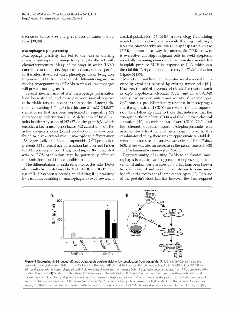
Figure 3 Repressing IL-3-induced M2 macrophages through inhibiting ILproduction of more IL-4 from SHIP−/− than SHIP+/+ Lin- BM cells. SHIP+/+ an24 h and supernatants were subjected to IL-4 ELISAs. Data shown are theunstimulated cells. (B) Model of IL-3-induced M2 skewing and the role thadifferentiation of both basophil precursors and monocyte/macrophage pand basophil progenitors in a STAT5-dependent manner. SHIP within theskews, via STAT6, the maturing and mature MΦs to an M2 phenotype. Co
classical polarization [59]. SHIP (src-homology 2-containinginositol 5' phosphatase) is a molecule that negatively regu-lates the phosphatidylinositol-4,5-bisphosphate 3-kinase(PI3K) apoptotic pathway. In cancers, the PI3K pathwayis overactive, allowing malignant cells to avoid apoptosis,essentially becoming immortal. It has been determined thatbasophils produce SHIP in response to IL-3, which canthen inhibit IL-4 production necessary for TAM activation(Figure 3) [59].Many tumor-infiltrating monocytes are alternatively acti-
vated by cytokines released by existing tumor cells [41].However, the added presence of classical activators suchas CpG oligodeoxynucleotides (CpG) and an anti-CD40agonist can increase anti-tumor activity of macrophages.CpG causes a pro-inflammatory response in macrophagesand the agonistic anti-CD40 can reverse immune suppres-sion. As a follow up study to those that indicated that thesynergistic effects of anti-CD40 and CpG increase classicalactivation [40], a combination of anti-CD40, CpG, andthe chemotherapeutic agent cyclophosphamide wasused to study treatment of melanoma in vivo. In thiscombinatorial study, there was an approximate ten-fold de-crease in tumor size and survival was extended by ~12 days[60]. There was also an increase in the percentage of F4/80+Gr1+ inflammatory monocytes [60,61].Reprogramming of existing TAMs to be classical mac-
rophages is another valid approach to improve upon con-ventional anticancer therapies. IFN-α has long been knownto be tumoricidal and was the first cytokine to show somebenefit in the treatment of some cancer types [62]. Becauseof the protein’s short half-life, however, the dose required
-4 production from basophils. (A) IL-3 and GM-CSF stimulate thed SHIP−/− Lin- BM cells were cultured with M-CSF, IL-3, or GM-CSF formeans ± SEM of duplicate determinations. *, p < 0.05 compared witht SHIP plays in this process. IL-3 stimulates the proliferation androgenitors. IL-3 also stimulates the production of IL-4 from basophilsbasophils represses this IL-4 production. The secreted IL-4, in turn,pyright 2009. The American Association of Immunologists, Inc. [59].

Bygd et al. Clinical and Translational Medicine 2014, 3:41 Page 6 of 13http://www.clintransmed.com/content/3/1/41
for efficacy becomes toxic to healthy tissue and the tumoris only exposed to short bursts of therapy [62]. This is whythe use of TIE2-expressing monocytes, which are regularlyrecruited to tumor sites, to selectively deliver IFN-α, caninhibit angiogenesis and skew macrophage polarizationto the classical end of the spectrum [62]. This is shownby the presence of cells expressing Iba1, a monocyte/macrophage/microglia protein, in and around the tumorsite.Histidine-rich glycoprotein (HRG), a host produced anti-
angiogenic and immunomodulatory factor to promoteTAM reprogramming is another viable target [63]. HRGhas been studied to identify mechanisms by which it medi-ates anti-tumor effects; and the results revealed thatTAMs activated by HRG down regulated expression ofpro-angiogenic cytokines and upregulated that of angio-static cytokines. At the same time, HRG activated TAMsshowed improved quality of existing vasculature causingan increase in the effectiveness of other chemotherapeutics[63]. Another target for reprogramming TAMS is the NF-κB signaling pathway [64]. Inhibition of NF-κB signalingwas found with IκB kinase (IKK)β reduction, stimulatingTAMs to become cytotoxic through recruitment of NKcells with the production of IL-12 [64]. These three exam-ples, along with many more, provide proof-of-conceptdata for the reprogramming of macrophages in cancertherapeutics.
Macrophages and scaffolds for tissue engineeringMacrophages are involved in ECM remodeling, prolifer-ation of epithelial cells, development of vasculature andthe organization of tissues during development [65]. Thesefunctional capacities of macrophages extend into thewound healing response and the FBR to biomaterials.Macrophage phenotype is dynamic throughout the courseof these processes, and the balance between phenotypes isinstrumental in the timely progression of these responsesfrom injury to successful healing. As with TAMs, macro-phages involved in healing retain their plasticity and altertheir phenotype in response to a variable cytokine micro-environment in the progression of these processes [6].
Overview of the foreign body response to implantedscaffoldsSurgical implantation or injection of a biomaterial-basedconstruct injures the tissue, resulting in an influx of bloodand cell death. Dying cells release danger signals (dangerassociated molecular patterns, DAMPs) that induce localinflammation [66] and activate resident macrophages[67,68]. These DAMPs include HMGB1, histones, anduric acid [66,67,69,70]. Blood proteins such as albumin,fibrinogen, fibronectin, immunoglobulin G (IgG), and vari-ous complement proteins adsorb to the surface of the bio-material [71]. Activation of the complement cascade results
in opsonization of the biomaterial surface with C3b andinduces inflammation through the anaphylatoxins C3aand C5a [72]. These anaphylatoxins recruit leukocytesto the site of inflammation, cause histamine release frommast cells, and induce oxidative bursts in neutrophils [73].Release of histamine from mast cells attracts neutrophilsand monocytes [74,75]. Neutrophils are the first immunecells to arrive at the implant site [76] and, along withmast cells, secrete IL-4 and IL-13 early in innate immuneresponses [9].Monocytes are the next immune cells to extravasate
into the tissue where they differentiate into tissue macro-phages [77]. These macrophages are classically activatedupon the adsorbed protein layer [78,79]. Proteins, such asfibrinogen, C3, and C3b on the surface of the biomaterialare bound by the integrin αMβ2 (CD11b:CD18), alsoknown as complement receptor 3 (CR3), on the surfaceof macrophages [77,80-82]. Activated macrophages se-crete TNF-α, IL-6, IL-8, MCP-1, RANTES, ROS, iNOS,IL-1β, and MMPs [83-85]. The chemokines MIP-1α,IL-8, and MCP-1 attract additional monocytes [83]. Thesebiomaterial-activated macrophages are also characterizedby an increased phagocytic capacity [86]. Continued pres-ence of pro-inflammatory macrophages causes acute in-flammation to morph into chronic inflammation [87].Attempted phagocytosis of biomaterials leads to the
fusion of adherent classically activated macrophages intoforeign body giant cells (FBGCs) [88]. IL-4 and IL-13 in-duce the fusion of adherent macrophages [88]. β1 and β2integrins are involved in the fusion of these macrophages[89], and CCL2 guides the chemotaxis of adherent macro-phages towards each other [90]. FBGCs have a cytokineprofile more characteristic of alternatively activated mac-rophages that includes TGF-β, platelet derived growth fac-tor (PDGF), IL-1rα, and IL-10 [9,77,84,91]. FBGCs secreteprotons, ROS, and MMPs in an attempt to eradicate theforeign body [92,93]. Like M1 macrophages, FBGCs se-crete pro-inflammatory RANTES and the chemoattractantMCP-1 [84]. ECM breakdown by MMPs leads to increasedDAMPs in the microenvironment and further macrophageactivation [94].The resolution stage of the FBR is dominated by alter-
natively activated macrophages. A profibrotic, alternativelyactivated, wound healing macrophage phenotype resultsfrom macrophage phagocytosis of dying cells, stimulationby IL-4 or by IL-13 [12,95]. These dying cells includeepithelial and endothelial cells that are damaged by pro-inflammatory cytokines, such as TNF-α, and short-livedneutrophils [25,96]. Alternatively activated macrophagessecrete profibrotic mediators such as TGF-β, IL-4, IL-13,IL-10, arginase, and ECM components [9,97]. Thesemacrophages drive the wound healing response by acti-vating mesenchymal cells that participate in the woundhealing process [98,99]. TGF-β can also induce an M2-

Bygd et al. Clinical and Translational Medicine 2014, 3:41 Page 7 of 13http://www.clintransmed.com/content/3/1/41
like phenotype in macrophages [100]. These M2 macro-phages are profibrotic, but are still unable to reduce thepro-inflammatory response. Reduction of chronic in-flammation requires IL-10-induced activation of regula-tory M2-like macrophages [9,101]. These macrophagessecrete high levels of the same protein that activates them[9]. IL-10 prevents the translation of pro-inflammatorycytokines by macrophages through STAT3 [102,103].As in the immune response to parasitic infections, the
early phase of wound healing and the FBR is characterizedby M1-like macrophages and the late phases are controlledby M2-like macrophages [25,91,104-106]. In the healing ofaseptic wounds regulatory M2 (IL-10 stimulated) macro-phages rapidly downregulate the inflammatory response topromote tissue repair [9,107-110]. Conversely, in the FBR,further activation of macrophages will occur, resultingin continued chronic inflammation (pro-inflammatorymacrophages and FBGCs) and continued wound healing(wound healing macrophages).It has long been hypothesized that chronic inflammation
is present until an extensive fibrous capsule surrounds thebiomaterial [76]. Resident fibroblasts, fibrocytes, and mac-rophages are activated by TGF-β, and become myofibro-blasts [111-115]. Myofibroblasts secrete high amounts ofcollagen I, collagen III, and fibronectin [110,116]. The ex-pression of α-smooth muscle actin (α-SMA) permits myo-fibroblasts to contract collagen networks in a processknown as contractile scarring [117,118]. Incessant acti-vation of myofibroblasts results in continued secretionand contraction of ECM components. This eventuallyresults in excessive scarring, and fibrous encapsulation.The fibrous capsule is a dense, hypocellular, avascularcollagenous network that reduces the diffusion of allmolecules, and results in the failure of scaffolds forapplications in tissue engineering [119,120]. The entireprocess leading up to fibrous encapsulation is illustratedin Figure 4.
Scaffolds to instruct phenotypic macrophage responsesDepending on biomaterial properties and the cytokinessecreted by inflammatory cells in the biomaterial micro-environment, macrophages adopt either an M1- or M2-like state [23]. As macrophages are plastic, they can existon a spectrum between these two states. This leads tothe hypothesis that surface chemistry and physical prop-erties of scaffolds can be used to polarize macrophagestowards a specific phenotype, or away from another. Inparticular, some scaffolds have been engineered to reduceprolonged activation of M1-like macrophages, so that cell-laden scaffolds maintain cell viability [121,122]. Additionalscaffolds have been engineered to reduce excessive fibrosisand decrease time to incorporation of the implant [123]. Abalance in macrophage phenotype must be achieved forscaffold vascularization.
Varied scaffold chemistries suggest the ability to decreasethe expression of M1 macrophages. Microgel conform-ational coatings formed from poly(N-isopropylacrylamide)(pNIPAm) and poly(ethylene glycol) diacrylate (PEGDA)reduce fibrinogen adsorption, macrophage adhesion,macrophage spreading, and secretion of inflammatorycytokines [124]. Zwitterionic hydrogels are able to re-duce protein adsorption and are characterized by anti-inflammatory, pro-healing macrophages that promoteangiogenesis and show no evidence of a collagenouscapsule for longer than three months [125]. The abilityof macrophages to induce positive tissue remodelingon fourteen different biologically-derived surgical mesheswas investigated, and suggested that a predominance ofM2 macrophages could potentially lead to more construct-ive tissue remodeling after two weeks [23]. Sugisis andMatristem scaffolds – derived from porcine small intestinalsubmucosa, and urinary bladder, respectively – appearedto increase macrophage infiltration; whereas the other scaf-folds, derived from human and porcine dermis, appearedto prolong the healing response and exhibited an increasein M1-like macrophages [23].In addition to chemical properties, physical proper-
ties of scaffolds can significantly influence macrophagephenotype. Controlling the pore size of scaffolds isone technique that shows promise in decreasing pro-inflammatory macrophage presence and improving thehealing outcome. A pore size of 30–40 μm within poroustemplate scaffolds formed of five different synthetic poly-mers and one natural polymer appeared to increase infil-tration of macrophages and vascular density, suggestingthat these materials induce regenerative macrophages[126-128]. It is generally thought that geometric restric-tion of macrophages within these pores prevents themfrom spreading out into their phagocytic, inflammatoryphenotype [24,129,130]. Vascular density is suggested topeak at pores size of 35 μm [126,131]. The degree ofporosity in a material can also influence macrophagephenotypes, with more porous materials leading to de-creased healing time of implants and, therefore, a reducedfibrous capsule thickness. For example, even though por-ous polytetrafluoroethylene (PTFE) surfaces seemed to in-duce inflammatory cytokine secretion by macrophages,a thinner fibrous capsule was formed on porous versusnonporous PTFE [132]. BMDMs cultured on electrospunpolydioxanone (PDO) of larger fiber length and pore sizeshowed increased arginase, TGF-β, VEGF, and basic fibro-blast growth factor expression, characteristic of alterna-tively activated macrophages, than those cells cultured onscaffolds with smaller fiber length and pore size [123].Substrate morphology and surface topography repre-
sent two other physical properties of scaffolds that arethought to influence macrophage phenotype and thusthe foreign body response and material biocompatibility.

Figure 4 Macrophage phenotype in the wound healing and foreign body responses.
Bygd et al. Clinical and Translational Medicine 2014, 3:41 Page 8 of 13http://www.clintransmed.com/content/3/1/41
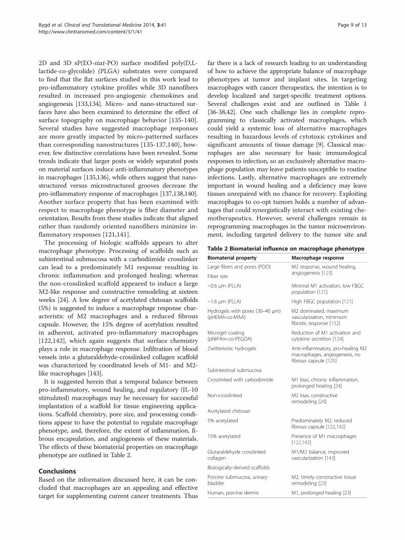
Table 2 Biomaterial influence on macrophage phenotype
Biomaterial property Macrophage response
Large fibers and pores (PDO) M2 response, wound healing,angiogenesis [123]
Fiber size
~0.6 μm (PLLA) Minimal M1 activation, low FBGCpopulation [121]
~1.6 μm (PLLA) High FBGC population [121]
Hydrogels with pores (30–40 μm)(pHEMA-co-MAA)
M2 dominated, maximumvascularization, minimumfibrotic response [132]
Microgel coating(pNIPAm-co-PEGDA)
Reduction of M1 activation andcytokine secretion [124]
Zwitterionic hydrogels Anti-inflammatory, pro-healing M2macrophages, angiogenesis, nofibrous capsule [125]
Subintestinal submucosa
Crosslinked with carbodiimide M1 bias, chronic inflammation,prolonged healing [24]
Non-crosslinked M2 bias, constructiveremodeling [24]
Acetylated chitosan
5% acetylated Predominately M2, reducedfibrous capsule [122,142]
15% acetylated Presence of M1 macrophages[122,142]
Glutaraldehyde crosslinkedcollagen
M1/M2 balance, improvedvascularization [143]
Biologically-derived scaffolds
Porcine submucosa, urinarybladder
M2, timely constructive tissueremodeling [23]
Human, porcine dermis M1, prolonged healing [23]
Bygd et al. Clinical and Translational Medicine 2014, 3:41 Page 9 of 13http://www.clintransmed.com/content/3/1/41
2D and 3D sP(EO-stat-PO) surface modified poly(D,L-lactide-co-glycolide) (PLGA) substrates were comparedto find that the flat surfaces studied in this work lead topro-inflammatory cytokine profiles while 3D nanofibersresulted in increased pro-angiogenic chemokines andangiogenesis [133,134]. Micro- and nano-structured sur-faces have also been examined to determine the effect ofsurface topography on macrophage behavior [135-140].Several studies have suggested macrophage responsesare more greatly impacted by micro-patterned surfacesthan corresponding nanostructures [135-137,140], how-ever, few distinctive correlations have been revealed. Sometrends indicate that larger posts or widely separated postson material surfaces induce anti-inflammatory phenotypesin macrophages [135,136], while others suggest that nano-structured versus microstructured grooves decrease thepro-inflammatory response of macrophages [137,138,140].Another surface property that has been examined withrespect to macrophage phenotype is fiber diameter andorientation. Results from these studies indicate that alignedrather than randomly oriented nanofibers minimize in-flammatory responses [121,141].The processing of biologic scaffolds appears to alter
macrophage phenotype. Processing of scaffolds such assubintestinal submucosa with a carbodiimide crosslinkercan lead to a predominately M1 response resulting inchronic inflammation and prolonged healing; whereasthe non-crosslinked scaffold appeared to induce a largeM2-like response and constructive remodeling at sixteenweeks [24]. A low degree of acetylated chitosan scaffolds(5%) is suggested to induce a macrophage response char-acteristic of M2 macrophages and a reduced fibrouscapsule. However, the 15% degree of acetylation resultedin adherent, activated pro-inflammatory macrophages[122,142], which again suggests that surface chemistryplays a role in macrophage response. Infiltration of bloodvessels into a glutaraldehyde-crosslinked collagen scaffoldwas characterized by coordinated levels of M1- and M2-like macrophages [143].It is suggested herein that a temporal balance between
pro-inflammatory, wound healing, and regulatory (IL-10stimulated) macrophages may be necessary for successfulimplantation of a scaffold for tissue engineering applica-tions. Scaffold chemistry, pore size, and processing condi-tions appear to have the potential to regulate macrophagephenotype, and, therefore, the extent of inflammation, fi-brous encapsulation, and angiogenesis of these materials.The effects of these biomaterial properties on macrophagephenotype are outlined in Table 2.
ConclusionsBased on the information discussed here, it can be con-cluded that macrophages are an appealing and effectivetarget for supplementing current cancer treatments. Thus
far there is a lack of research leading to an understandingof how to achieve the appropriate balance of macrophagephenotypes at tumor and implant sites. In targetingmacrophages with cancer therapeutics, the intention is todevelop localized and target-specific treatment options.Several challenges exist and are outlined in Table 1[36-38,42]. One such challenge lies in complete repro-gramming to classically activated macrophages, whichcould yield a systemic loss of alternative macrophagesresulting in hazardous levels of cytotoxic cytokines andsignificant amounts of tissue damage [9]. Classical mac-rophages are also necessary for basic immunologicalresponses to infection, so an exclusively alternative macro-phage population may leave patients susceptible to routineinfections. Lastly, alternative macrophages are extremelyimportant in wound healing and a deficiency may leavetissues unrepaired with no chance for recovery. Exploitingmacrophages to co-opt tumors holds a number of advan-tages that could synergistically interact with existing che-motherapeutics. However, several challenges remain inreprogramming macrophages in the tumor microenviron-ment, including targeted delivery to the tumor site and

Bygd et al. Clinical and Translational Medicine 2014, 3:41 Page 10 of 13http://www.clintransmed.com/content/3/1/41
selective delivery to alternatively activated TAM popula-tions. Immunomodulation of macrophages may also resultin improved success of implants for tissue engineering. Inthe FBR, the complete absence of M1 macrophages isdetrimental in the progression of the response. Specific-ally for successful implantation of a scaffold for tissueengineering, M1 macrophages are necessary to instigatethe inflammatory response and initiate the M2-coordinated wound healing process. Timely resolution ofthe response requires the presence of IL-10high M2 macro-phages. Though many of the materials mentioned herelead to a timely resolution of the FBR and successfulvascularization, these materials cannot be used for all tis-sue engineering applications. Therefore, strategies tomodulate macrophages in the tumor and biomaterialmicroenvironment require consideration of the desiredend result.
AbbreviationsIFN: Interferon; IL: Interleukin; Ic: Immune complexes; GC: Glucocorticoids;TGF: Transforming growth factor; LPS: Lipopolysaccharides; M1: Classicallyactivated, pro-inflammatory cytotoxic macrophages; TLR: Toll-like receptor;IRF: Interferon regulator factor; STAT: Signal transducers and activators oftranscription; TNF: Tumor necrosis factor; Th: T-helper; NF: Nuclear factor;HIF: Hypoxia-inducible factor; iNOS: Inducible nitric oxide synthase; NO: Nitricoxide; ROI: Reactive oxygen intermediates; MIP: Macrophage inflammatoryprotein; MCP: Monocyte chemotactic protein; I-TAC: Interferon-inducible Tcell alpha chemoattractant; IP: Interferon gamma-induced protein;MIG: Monokine induced by gamma interferon; NK: Natural killer;MRC1: Mannose receptor C type 1; FBR: Foreign body response;M2: Alternatively activated, pro-angiogenic, wound healing macrophages;SOCS: Suppressor of cytokine signaling; IL-1Rα: Interleukin-1 receptor agonist;Mrc1: Mannose receptor gene; Retnla/Fizz1: Resistin-like α gene; Chi3l3/Ym1: Chitinase 3-like 3 gene; PPARγ: Peroxisome proliferator-activatedreceptors; KLF: Krüppel-like factor; MDC: Macrophage-derived chemokine;AMAC: Alternative macrophage activation-associated chemokine;Treg: Regulatory T cell; VEGF: Vascular endothelial growth factor;MMPs: Matrix metalloproteinases; SR-A1: Scavenger receptor type 1;TAMs: Tumor-associated macrophages; ECM: Extracellular matrix;uPA: Urokinase-type plasminogen activator; PlGF: Placental growth factor;ANGs: Angiopoietins; CSF: Colony stimulating factor; BM: Bone marrow;HIFko: Hypoxia-inducible factor-α knockout mice; GBM: Glioblastoma;TIE: Tyrosine kinase with immunoglobulin-like and EGF-like domains;mAB: Monoclonal antibody; i.p.: Intraperitoneal; Jmjd3: Jumonji domaincontaining-3; H3k27: Histone 3 Lys27; ROS: Reactive oxygen species;SHIP: Src-homology 2-containing inositol 5’ phosphatase;PI3K: Phosphatidylinositol-4,5-bisphosphate 3-kinase; GM-CSF: Granulocytemacrophage colony-stimulating factor; SEM: Standard error of the mean;CpG: CpG oligodeoxynucleotides; HRG: Histidine-rich glycoprotein; IKK: IκBkinase; DAMPs: Danger associated molecular patterns; HMGB1: High-mobilitygroup protein B1; IgG: Immunoglobulin G; C: Complement protein;CR3: Complement receptor 3; ENA-78: CXCL5; FBGCs: Foreign body giantcells; PDGF: Platelet derived growth factor; α-SMA: α-smooth muscle actin;pNIPAm: Poly(N-isopropylacrylamide); PEGDA: Poly(ethylene glycol)diacrylate; PTFE: Polytetrafluoroethylene; BMDMs: Bone-marrow derivedmacrophages; PDO: Polydioxanone; PLGA: Poly(D,L-lactide-co-glycolide;PLLA: Poly-L-lactic acid; pHEMA-co-MAA: Poly(2-hydroxyethyl methacrylate-comethyl methacrylate); RANTES: Regulated on activation, normal T cellexpressed and secreted.
Competing interestsThe authors declare that they have no competing interests.
Authors’ contributionsHCB, KDF, and KMB drafted the manuscript. All authors read and approvedthe final manuscript.
AcknowledgementsThis work was supported by the National Science Foundation under GrantNo. CBET 1227867 and the Roy J. Carver Charitable Trust Grant No. 13–4265.
Author details1Department of Materials Science & Engineering, Iowa State University, Ames,Iowa 50011, USA. 2Department of Chemical & Biological Engineering, IowaState University, Ames, Iowa 50011, USA. 3Ames National Laboratory, Ames,Iowa 50011, USA.
Received: 19 September 2014 Accepted: 10 November 2014
References1. Stout RD, Watkins SK, Suttles J: Functional plasticity of macrophages: in
situ reprogramming of tumor-associated macrophages. J Leukoc Biol2009, 86:1105–1109.
2. Gordon S, Taylor PR: Monocyte and macrophage heterogeneity. Nat RevImmunol 2005, 5:953–964.
3. Stout RD, Suttles J: Functional plasticity of macrophages: reversibleadaptation to changing microenvironments. J Leukoc Biol 2004, 76:509–513.
4. Akilbekova D, Philiph R, Graham A, Bratlie KM: Macrophage reprogramming:influence of latex beads with various functional groups on macrophagephenotype and phagocytic uptake in vitro. J Biomed Mater Res Part A 2014,doi: 10.1002/jbm.a.35169.
5. McWhorter FY, Wang T, Nguyen P, Chung T, Liu WF: Modulation ofmacrophage phenotype by cell shape. Proc Natl Acad Sci U S A 2013,110:17253–17258.
6. Stout RD, Jiang C, Matta B, Tietzel I, Watkins SK, Suttles J: Macrophagessequentially change their functional phenotype in response to changesin microenvironmental influences. J Immunol 2005, 175:342–349.
7. Martinez FO, Gordon S: The M1 and M2 paradigm of macrophageactivation: time for reassessment. F1000Prime Rep 2014, 6:1–13.
8. Murray PJ, Allen JE, Biswas SK, Fisher EA, Gilroy DW, Goerdt S, Gordon S,Hamilton JA, Ivashkiv LB, Lawrence T, Locati M, Mantovani A, Martinez FO,Mege J-L, Mosser DM, Natoli G, Saeij JP, Schultze JL, Shirey KA, Sica A,Suttles J, Udalova I, van Ginderachter JA, Vogel SN, Wynn TA: Macrophageactivation and polarization: nomenclature and experimental guidelines.Immunity 2014, 41:14–20.
9. Mosser DM, Edwards JP: Exploring the full spectrum of macrophageactivation. Nat Rev Immunol 2008, 8:958–969.
10. Xue J, Schmidt SV, Sander J, Draffehn A, Krebs W, Quester I, De Nardo D,Gohel TD, Emde M, Schmidleithner L, Ganesan H, Nino-Castro A, Mallmann MR,Labzin L, Theis H, Kraut M, Beyer M, Latz E, Freeman TC, Ulas T, Schultze JL:Transcriptome-based network analysis reveals a spectrum model of humanmacrophage activation. Immunity 2014, 40:274–288.
11. Gordon S, Martinez FO: Alternative activation of macrophages:mechanism and functions. Immunity 2010, 32:593–604.
12. Gordon S: Alternative activation of macrophages. Nat Rev Immunol 2003,3:23–35.
13. Lawrence T, Natoli G: Transcriptional regulation of macrophage polarization:enabling diversity with identity. Nat Rev Immunol 2011, 11:750–761.
14. Taub DD, Cox GW: Murine Th1 and Th2 cell clones differentially regulatemacrophage nitric oxide production. J Leukoc Biol 1995, 58:80–89.
15. Wynn TA, Chawla A, Pollard JW: Macrophage biology in development,homeostasis and disease. Nature 2013, 496:445–455.
16. Sica A, Mantovani A: Macrophage plasticity and polarization : in vivoveritas. J Clin Invest 2012, 122:787–795.
17. Wolfs IMJ, Donners MMPC, de Winther MPJ: Differentiation factors andcytokines in the atherosclerotic plaque micro-environment as a triggerfor macrophage polarisation. Thromb Haemost 2011, 106:763–771.
18. Mantovani A, Sica A, Sozzani S, Allavena P, Vecchi A, Locati M: Thechemokine system in diverse forms of macrophage activation andpolarization. Trends Immunol 2004, 25:677–686.
19. Sica A, Schioppa T, Mantovani A, Allavena P: Tumour-associatedmacrophages are a distinct M2 polarised population promoting tumourprogression: potential targets of anti-cancer therapy. Eur J Cancer 2006,42:717–727.
20. Martinez FO, Sica A, Mantovani A, Locati M: Macrophage activation andpolarization. Front Biosci 2008, 13:453–461.

Bygd et al. Clinical and Translational Medicine 2014, 3:41 Page 11 of 13http://www.clintransmed.com/content/3/1/41
21. Mantovani A, Sozzani S, Locati M, Allavena P, Sica A: Macrophagepolarization: tumor-associated macrophages as a paradigm for polarizedM2 mononuclear phagocytes. Trends Immunol 2002, 23:549–555.
22. Biswas SK, Chittezhath M, Shalova IN, Lim J-Y: Macrophage polarizationand plasticity in health and disease. Immunol Res 2012, 53:11–24.
23. Brown BN, Londono R, Tottey S, Zhang L, Kukla KA, Wolf MT, Daly KA, Reing JE,Badylak SF: Macrophage phenotype as a predictor of constructiveremodeling following the implantation of biologically derived surgicalmesh materials. Acta Biomater 2012, 8:978–987.
24. Badylak SF, Valentin JE, Ravindra AK, McCabe GP, Stewart-Akers AM:Macrophage phenotype as a determinant of biologic scaffold remodeling.Tissue Eng Part A 2008, 14:1835–1842.
25. Mantovani A, Biswas SK, Galdiero MR, Sica A, Locati M: Macrophageplasticity and polarization in tissue repair and remodelling. J Pathol 2013,229:176–185.
26. Mantovani A, Locati M: Tumor-associated macrophages as a paradigm ofmacrophage plasticity, diversity, and polarization: lessons and openquestions. Arterioscler Thromb Vasc Biol 2013, 33:1478–1483.
27. Kawamura K, Komohara Y, Takaishi K, Katabuchi H, Takeya M: Detectionof M2 macrophages and colony-stimulating factor 1 expression inserous and mucinous ovarian epithelial tumors. Pathol Int 2009,59:300–305.
28. Solinas G, Germano G, Mantovani A, Allavena P: Tumor-associatedmacrophages (TAM) as major players of the cancer-related inflammation.J Leukoc Biol 2009, 86:1065–1073.
29. Allavena P, Sica A, Solinas G, Porta C, Mantovani A: The inflammatorymicro-environment in tumor progression: the role of tumor-associatedmacrophages. Crit Rev Oncol Hematol 2008, 66:1–9.
30. Qian B-Z, Pollard JW: Macrophage diversity enhances tumor progressionand metastasis. Cell 2010, 141:39–51.
31. Qian B, Deng Y, Im JH, Muschel RJ, Zou Y, Li J, Lang RA, Pollard JW: Adistinct macrophage population mediates metastatic breast cancer cellextravasation, establishment and growth. PLoS One 2009, 4:e6562.
32. Ruffell B, Affara NI, Coussens LM: Differential macrophage programming inthe tumor microenvironment. Trends Immunol 2012, 33:119–126.
33. De Palma M, Lewis CE: Macrophage regulation of tumor responses toanticancer therapies. Cancer Cell 2013, 23:277–286.
34. Zhang W, Zhu X-D, Sun H-C, Xiong Y-Q, Zhuang P-Y, Xu H-X, Kong L-Q,Wang L, Wu W-Z, Tang Z-Y: Depletion of tumor-associated macrophagesenhances the effect of sorafenib in metastatic liver cancer models byantimetastatic and antiangiogenic effects. Clin Cancer Res 2010,16:3420–3430.
35. Squadrito ML, De Palma M: Macrophage regulation of tumorangiogenesis: implications for cancer therapy. Mol Aspects Med 2011,32:123–145.
36. Bergers G, Hanahan D: Modes of resistance to anti-angiogenic therapy.Nat Rev Cancer 2008, 8:592–603.
37. Jain RK: Antiangiogenic therapy for cancer: current and emergingconcepts. Oncology 2005, 19:7–16.
38. Garber K: First results for agents targeting cancer-related inflammation.J Natl Cancer Inst 2009, 101:1110–1112.
39. Mantovani A, Sica A: Macrophages, innate immunity and cancer: balance,tolerance, and diversity. Curr Opin Immunol 2010, 22:231–237.
40. Buhtoiarov IN, Lum HD, Berke G, Sondel PM, Rakhmilevich AL: Synergisticactivation of macrophages via CD40 and TLR9 results in T cellindependent antitumor effects. J Immunol 2005, 176:309–318.
41. Guiducci C, Vicari AP, Sangaletti S, Trinchieri G: Redirecting in vivo elicitedtumor infiltrating macrophages and dendritic cells towards tumorrejection. Cancer Res 2005, 65:3437–3446.
42. Zeisberger SM, Odermatt B, Marty C, Zehnder-Fjällman AHM, Ballmer-Hofer K,Schwendener RA: Clodronate-liposome-mediated depletion of tumour-associated macrophages: a new and highly effective antiangiogenictherapy approach. Br J Cancer 2006, 95:272–281.
43. Du R, Lu KV, Petritsch C, Liu P, Ganss R, Passegué E, Song H, Vandenberg S,Johnson RS, Werb Z, Bergers G: HIF1alpha induces the recruitment ofbone marrow-derived vascular modulatory cells to regulate tumorangiogenesis and invasion. Cancer Cell 2008, 13:206–220.
44. Coffelt SB, Tal AO, Scholz A, De Palma M, Patel S, Urbich C, Biswas SK,Murdoch C, Plate KH, Reiss Y, Lewis CE: Angiopoietin-2 regulates geneexpression in TIE2-expressing monocytes and augments their inherentproangiogenic functions. Cancer Res 2010, 70:5270–5280.
45. Mazzieri R, Pucci F, Moi D, Zonari E, Ranghetti A, Berti A, Politi LS, Gentner B,Brown JL, Naldini L, De Palma M: Targeting the ANG2/TIE2 axis inhibitstumor growth and metastasis by impairing angiogenesis and disablingrebounds of proangiogenic myeloid cells. Cancer Cell 2011, 19:512–526.
46. Sun X, Cheng G, Hao M, Zheng J, Zhou X, Zhang J, Taichman RS, Pienta KJ,Wang J: CXCL12 / CXCR4 / CXCR7 chemokine axis and cancerprogression. Cancer Metastasis Rev 2010, 29:709–722.
47. Kim SY, Lee CH, Midura BV, Yeung C, Mendoza A, Hong SH, Ren L, Wong D,Korz W, Merzouk A, Salari H, Zhang H, Hwang ST, Khanna C, Helman LJ:Inhibition of the CXCR4/CXCL12 chemokine pathway reduces thedevelopment of murine pulmonary metastases. Clin Exp Metastasis 2008,25:201–211.
48. Richert MM, Vaidya KS, Mills CN, Wong D, Korz W, Hurst DR, Welch DR:Inhibition of CXCR4 by CTCE-9908 inhibits breast cancer metastasis tolung and bone. Oncol Rep 2009, 21:761–767.
49. Porvasnik S, Sakamoto N, Kusmartsev S, Eruslanov E, Kim W-J, Cao W,Urbanek C, Wong D, Goodison S, Rosser CJ: Effects of CXCR4 antagonistCTCE-9908 on prostate tumor growth. Prostate 2009, 69:1460–1469.
50. Ahn G-O, Tseng D, Liao C-H, Dorie MJ, Czechowicz A, Brown JM: Inhibitionof Mac-1 (CD11b/CD18) enhances tumor response to radiation by reducingmyeloid cell recruitment. Proc Natl Acad Sci U S A 2010, 107:8363–8368.
51. Wong D, Korz W: Translating an antagonist of chemokine receptorCXCR4: from bench to bedside. Clin Cancer Res 2008, 14:7975–7980.
52. Zhang J, Patel L, Pienta KJ: CC chemokine ligand 2 (CCL2) promotesprostate cancer tumorigenesis and metastasis. Cytokine Growth Factor Rev2010, 21:41–48.
53. Zhu X, Fujita M, Snyder LA, Okada H: Systemic delivery of neutralizingantibody targeting CCL2 for glioma therapy. J Neurooncology 2011,104:83–92.
54. Loberg RD, Ying C, Craig M, Day LL, Sargent E, Neeley C, Wojno K, SnyderLA, Yan L, Pienta KJ: Targeting CCL2 with systemic delivery of neutralizingantibodies induces prostate cancer tumor regression in vivo. Cancer Res2007, 67:9417–9424.
55. Rozel S, Galbán CJ, Nicolay K, Lee KC, Sud S, Neeley C, Snyder LA, Chenevert TL,Rehemtulla A, Ross BD, Pienta KJ: Synergy between anti-CCL2 and docetaxelas determined by DW-MRI in a metastatic bone cancer model. J CellBiochem 2009, 107:58–64.
56. Kirk PS, Koreckij T, Nguyen HM, Brown LG, Snyder LA, Vessella RL, Corey E:Inhibition of CCL2 Signaling in Combination with Docetaxel TreatmentHas Profound Inhibitory Effects on Prostate Cancer Growth in Bone.Int J Mol Sci 2013, 14:10483–10496.
57. Satoh T, Takeuchi O, Vandenbon A, Yasuda K, Tanaka Y, Kumagai Y, Miyake T,Matsushita K, Okazaki T, Saitoh T, Honma K, Matsuyama T, Yui K, Tsujimura T,Standley DM, Nakanishi K, Nakai K, Akira S: The Jmjd3-Irf4 axis regulates M2macrophage polarization and host responses against helminth infection.Nat Immunol 2010, 11:936–944.
58. Zhang Y, Choksi S, Chen K, Pobezinskaya Y, Linnoila I, Liu Z-G: ROS play acritical role in the differentiation of alternatively activated macrophagesand the occurrence of tumor-associated macrophages. Cell Res 2013,23:898–914.
59. Kuroda E, Ho V, Ruschmann J, Antignano F, Hamilton M, Rauh MJ, Antov A,Flavell RA, Sly LM, Krystal G: SHIP represses the generation of IL-3-inducedM2 macrophages by inhibiting IL-4 production from basophils.J Immunol 2009, 183:3652–3660.
60. Johnson EE, Buhtoiarov IN, Baldeshwiler MJ, Felder MA, Van Rooijen N,Sondel PM, Rakhmilevich AL: Enhanced T cell-independent antitumoreffect of cyclophospamide combined with anti-CD40 mAb and CpG inmice. J Immunother 2011, 34:76–84.
61. Arora M, Poe SL, Ray A, Ray P: LPS-induced CD11b + Gr1(int)F4/80+regulatory myeloid cells suppress allergen-induced airway inflammation.Int Immunopharmacol 2011, 11:827–832.
62. De Palma M, Mazzieri R, Politi LS, Pucci F, Zonari E, Sitia G, MazzoleniS, Moi D, Venneri MA, Indraccolo S, Falini A, Guidotti LG, Galli R,Naldini L: Tumor-targeted interferon-alpha delivery by Tie2-expressing monocytes inhibits tumor growth and metastasis. CancerCell 2008, 14:299–311.
63. Rolny C, Mazzone M, Tugues S, Laoui D, Johansson I, Coulon C, Squadrito ML,Segura I, Li X, Knevels E, Costa S, Vinckier S, Dresselaer T, Åkerud P, De Mol M,Salomäki H, Phillipson M, Wyns S, Larsson E, Buysschaert I, Botling J,Himmelreich U, Van Ginderachter JA, De Palma M, Dewerchin M,Claesson-Welsh L, Carmeliet P: HRG inhibits tumor growth and metastasis

Bygd et al. Clinical and Translational Medicine 2014, 3:41 Page 12 of 13http://www.clintransmed.com/content/3/1/41
by inducing macrophage polarization and vessel normalization throughdownregulation of PlGF. Cancer Cell 2011, 19:31–44.
64. Hagemann T, Lawrence T, McNeish I, Charles KA, Kulbe H, Thompson RG,Robinson SC, Balkwill FR: “Re-educating” tumor-associated macrophagesby targeting NF-kappaB. J Exp Med 2008, 205:1261–1268.
65. Pollard JW: Trophic macrophages in development and disease.Nat Rev Immunol 2009, 9:259–270.
66. Klune JR, Dhupar R, Cardinal J, Billiar TR, Tsung A: HMGB1: endogenousdanger signaling. Mol Med 2008, 14:476–484.
67. Bianchi ME: DAMPs, PAMPs and alarmins: all we need to know aboutdanger. J Leukoc Biol 2007, 81:1–5.
68. Osterloh A, Kalinke U, Weiss S, Fleischer B, Breloer M: Synergistic anddifferential modulation of immune responses by Hsp60 andlipopolysaccharide. J Biol Chem 2007, 282:4669–4680.
69. Huang H, Evankovich J, Yan W, Nace G, Zhang L, Ross M, Liao X, Billiar T, Xu J,Esmon CT, Tsung A: Endogenous histones function as alarmins in sterileinflammatory liver injury through Toll-like receptor 9 in mice. Hepatology2011, 54:999–1008.
70. Shi Y, Evans JE, Rock KL: Molecular identification of a danger signal thatalerts the immune system to dying cells. Nature 2003, 425:516–521.
71. Sperling C, Fischer M, Maitz MF, Werner C: Blood coagulation onbiomaterials requires the combination of distinct activation processes.Biomaterials 2009, 30:4447–4456.
72. Nilsson B, Ekdahl KN, Mollnes TE, Lambris JD: The role of complement inbiomaterial-induced inflammation. Mol Immunol 2007, 44:82–94.
73. Sarma JV, Ward PA: The complement system. Cell Tissue Res 2011,343:227–235.
74. Zdolsek J, Eaton JW, Tang L: Histamine release and fibrinogen adsorptionmediate acute inflammatory responses to biomaterial implants inhumans. J Transl Med 2007, 5:31–36.
75. Abraham SN, St John AL: Mast cell-orchestrated immunity to pathogens.Nat Rev Immunol 2010, 10:440–452.
76. Anderson JM: Biological responses to materials. Annu Rev Mater Res 2001,31:81–110.
77. Higgins DM, Basaraba RJ, Hohnbaum AC, Lee EJ, Grainger DW, Gonzalez-Juarrero M: Localized immunosuppressive environment in the foreignbody response to implanted biomaterials. Am J Pathol 2009, 175:161–170.
78. McNally AK, Anderson JM: Complement C3 participation in monocyteadhesion to different surfaces. Proc Natl Acad Sci U S A 1994,91:10119–10123.
79. Yang D, Jones KS: Effect of alginate on innate immune activation ofmacrophages. J Biomed Mater Res A 2009, 90:411–418.
80. Brodbeck WG, Colton E, Anderson JM: Effects of adsorbed heat labileserum proteins and fibrinogen on adhesion and apoptosis ofmonocytes/macrophages on biomaterials. J Mater Sci Mater Med 2003,14:671–675.
81. Love RJ, Jones KS: The recognition of biomaterials: pattern recognition ofmedical polymers and their adsorbed biomolecules. J Biomed Mater Res A2013, 101:2740–2752.
82. Hamad OA, Ekdahl KN, Nilsson B: Non-proteolytically activated C3 promotesbinding of activated platelets and platelet-derived microparticles toleukocytes via CD11b/CD18. Immunology 2012, 217:1191–1191.
83. Rodriguez A, Meyerson H, Anderson JM: Quantitative in vivo cytokineanalysis at synthetic biomaterial implant sites. J Biomed Mater Res A 2009,89:152–159.
84. Jones JA, Chang DT, Meyerson H, Colton E, Kwon IK, Matsuda T, Anderson JM:Proteomic analysis and quantification of cytokines and chemokines frombiomaterial surface-adherent macrophages and foreign body giant cells.2007, 83:585–596.
85. Mesure L, De Visscher G, Vranken I, Lebacq A, Flameng W: Gene expressionstudy of monocytes/macrophages during early foreign body reactionand identification of potential precursors of myofibroblasts. PLoS One2010, 5:e12949.
86. Xia Z, Triffitt JT: A review on macrophage responses to biomaterials.Biomed Mater 2006, 1:R1–R9.
87. Sindrilaru A, Peters T, Wieschalka S, Baican C, Baican A, Peter H, Hainzl A, Schatz S,Qi Y, Schlecht A, Weiss JM, Wlaschek M, Sunderkötter C, Scharffetter-Kochanek K:An unrestrained proinflammatory M1 macrophage population induced by ironimpairs wound healing in humans and mice. J Clin Invest 2011, 121:985–997.
88. MacLauchlan S, Skokos EA, Meznarich N, Zhu DH, Raoof S, Shipley JM,Senior RM, Bornstein P, Kyriakides TR: Macrophage fusion, giant cell
formation, and the foreign body response require matrixmetalloproteinase 9. J Leukoc Biol 2009, 85:617–626.
89. McNally AK, Anderson JM: Beta1 and beta2 integrins mediate adhesionduring macrophage fusion and multinucleated foreign body giant cellformation. Am J Pathol 2002, 160:621–630.
90. Helming L, Gordon S: Molecular mediators of macrophage fusion.Trends Cell Biol 2009, 19:514–522.
91. Gretzer C, Emanuelsson L, Liljensten E, Thomsen P: The inflammatory cellinflux and cytokines changes during transition from acute inflammationto fibrous repair around implanted materials. J Biomater Sci Polym Ed2006, 17:669–687.
92. Lynn AD, Kyriakides TR, Bryant SJ: Characterization of the in vitromacrophage response and in vivo host response to poly(ethyleneglycol)-based hydrogels. J Biomed Mater Res A 2010, 93:941–953.
93. Santerre JP, Woodhouse K, Laroche G, Labow RS: Understanding thebiodegradation of polyurethanes: from classical implants to tissueengineering materials. Biomaterials 2005, 26:7457–7470.
94. Sorokin L: The impact of the extracellular matrix on inflammation.Nat Rev Immunol 2010, 10:712–723.
95. Savill J, Gregory C, Haslett C: Eat Me or Die. 2003, 302(November):1516–1517.96. Lech M, Anders H-J: Macrophages and fibrosis: How resident and infiltrating
mononuclear phagocytes orchestrate all phases of tissue injury and repair.Biochim Biophys Acta 1832, 2013:989–997.
97. Pesce JT, Ramalingam TR, Mentink-Kane MM, Wilson MS, El Kasmi KC, SmithAM, Thompson RW, Cheever AW, Murray PJ, Wynn TA: Arginase-1-expressingmacrophages suppress Th2 cytokine-driven inflammation and fibrosis. PLoSPathog 2009, 5:e1000371.
98. Diegelmann RF, Evans MC: Wound healing: an overview of acute, fibroticand delayed healing. Front Biosci 2004, 9:283–289.
99. Hamilton JA: Nondisposable materials, chronic inflammation, andadjuvant action. 2003, 73:702–712.
100. Vidal B, Serrano AL, Tjwa M, Suelves M, Ardite E, De Mori R, Baeza-Raja B,Martínez de Lagrán M, Lafuste P, Ruiz-Bonilla V, Jardí M, Gherardi R,Christov C, Dierssen M, Carmeliet P, Degen JL, Dewerchin M, Muñoz-CánovesP: Fibrinogen drives dystrophic muscle fibrosis via a TGFbeta/alternativemacrophage activation pathway. Genes Dev 2008, 22:1747–1752..
101. Wang Y, Wang YP, Zheng G, Lee VWS, Ouyang L, Chang DHH, Mahajan D,Coombs J, Wang YM, Alexander SI, Harris DCH: Ex vivo programmedmacrophages ameliorate experimental chronic inflammatory renaldisease. Kidney Int 2007, 72:290–299.
102. Lang R, Patel D, Morris JJ, Rutschman RL, Murray PJ: Shaping geneexpression in activated and resting primary macrophages by IL-10.J Immunol 2002, 169:2253–2263.
103. Saraiva M, O’Garra A: The regulation of IL-10 production by immune cells.Nat Rev Immunol 2010, 10:170–181.
104. Murray PJ, Wynn TA: Obstacles and opportunities for understandingmacrophage polarization. J Leukoc Biol 2011, 89:557–563.
105. Brys L, Beschin A, Raes G, Ghassabeh GH, Noel W, Brandt J, Brombacher F,Baetselier PD: Reactive oxygen species and 12/15-lipoxygenase contributeto the antiproliferative capacity of alternatively activated myeloid cellselicited during helminth infection. J Immunol 2005, 174:6095–6104.
106. Le SJ, Gongora M, Zhang B, Grimmond S, Campbell GR, Campbell JH, Rolfe BE:Gene expression profile of the fibrotic response in the peritoneal cavity.Differentiation 2010, 79:232–243.
107. Gurtner GC, Werner S, Barrandon Y, Longaker MT: Wound repair andregeneration. Nature 2008, 453:314–321.
108. Martin P, Leibovich SJ: Inflammatory cells during wound repair: the good,the bad and the ugly. Trends Cell Biol 2005, 15:599–607.
109. Eming SA, Krieg T, Davidson JM: Inflammation in wound repair: molecularand cellular mechanisms. J Invest Dermatol 2007, 127:514–525.
110. Sarrazy V, Billet F, Micallef L, Coulomb B, Desmoulière A: Mechanisms ofpathological scarring: role of myofibroblasts and current developments.Wound repair Regen 2011, 19:S10–S155.
111. Hinz B: Formation and function of the myofibroblast during tissue repair.J Invest Dermatol 2007, 127:526–537.
112. Wynn TA: Cellular and molecular mechanisms of fibrosis. J Pathol 2008,214:199–210.
113. Ninomiya K, Takahashi A, Fujioka Y, Ishikawa Y, Yokoyama M:Transforming growth factor-beta signaling enhances transdifferentiationof macrophages into smooth muscle-like cells. Hypertens Res 2006,29:269–276.

Bygd et al. Clinical and Translational Medicine 2014, 3:41 Page 13 of 13http://www.clintransmed.com/content/3/1/41
114. Jabs A, Moncada GA, Nichols CE, Waller EK, Wilcox JN: Peripheral bloodmononuclear cells acquire myofibroblast characteristics in granulationtissue. J Vasc Res 2005, 42:174–180.
115. Mooney JE, Rolfe BE, Osborne GW, Sester DP, van Rooijen N, Campbell GR,Hume DA, Campbell JH: Cellular plasticity of inflammatory myeloid cellsin the peritoneal foreign body response. Am J Pathol 2010, 176:369–380.
116. Hutchison N, Fligny C, Duffield JS: Resident mesenchymal cells andfibrosis. Biochim Biophys Acta 1832, 2013:962–971.
117. Tomasek JJ, Gabbiani G, Hinz B, Chaponnier C, Brown RA: Myofibroblastsand mechano-regulation of connective tissue remodelling. Nat Rev MolCell Biol 2002, 3:349–363.
118. Hinz B, Celetta G, Tomasek JJ, Gabbiani G, Chaponnier C: Alpha-smoothmuscle actin expression upregulates fibroblast contractile activity.Mol Biol Cell 2001, 12:2730–2741.
119. Kenneth Ward W: A review of the foreign-body response to subcutaneously-implanted devices: the role of macrophages and cytokines in biofoulingand fibrosis. J Diabetes Sci Technol 2008, 2:768–777.
120. Sharkawy AA, Klitzman B, Truskey GA, Reichert WM: Engineering the tissuewhich encapsulates subcutaneous implants. I. Diffusion properties.J Biomed Mater Res 1997, 37:401–412.
121. Saino E, Focarete ML, Gualandi C, Emanuele E, Cornaglia AI, Imbriani M, Visai L:Effect of electrospun fiber diameter and alignment on macrophageactivation and secretion of proinflammatory cytokines and chemokines.Biomacromolecules 2011, 12:1900–1911.
122. Vasconcelos DP, Fonseca AC, Costa M, Amaral IF, Barbosa MA, Aguas AP,Barbosa JN: Macrophage polarization following chitosan implantation.Biomaterials 2013, 34:9952–9959.
123. Garg K, Pullen NA, Oskeritzian CA, Ryan JJ, Bowlin GL: Macrophagefunctional polarization (M1/M2) in response to varying fiber and poredimensions of electrospun scaffolds. Biomaterials 2013, 34:4439–4451.
124. Bridges AW, Singh N, Burns KL, Babensee JE, Andrew Lyon L, García AJ:Reduced acute inflammatory responses to microgel conformal coatings.Biomaterials 2008, 29:4605–4615.
125. Zhang L, Cao Z, Bai T, Carr L, Ella-Menye J-R, Irvin C, Ratner BD, Jiang S:Zwitterionic hydrogels implanted in mice resist the foreign-bodyreaction. Nat Biotechnol 2013, 31:553–556.
126. Bryers JD, Giachelli CM, Ratner BD: Engineering biomaterials to integrateand heal: the biocompatibility paradigm shifts. Biotechnol Bioeng 2012,109:1898–1911.
127. Beckstead BL, Tung JC, Liang KJ, Tavakkol Z, Usui ML, Olerud JE, Giachelli CM:Methods to promote Notch signaling at the biomaterial interface andevaluation in a rafted organ culture model. J Biomed Mater Res A 2009,91:436–446.
128. Linnes MP, Ratner BD, Giachelli CM: A fibrinogen-based precision microporousscaffold for tissue engineering. Biomaterials 2007, 28:5298–5306.
129. Ratner BD: The biocompatibility manifesto: biocompatibility for thetwenty-first century. J Cardiovasc Transl Res 2011, 4:523–527.
130. Mantovani A: Macrophage diversity and polarization: in vivo veritas.Blood 2006, 108:408–409.
131. Fukano Y, Usui ML, Underwood RA, Isenhath S, Marshall AJ, Hauch KD,Ratner BD, Olerud JE, Fleckman P: Epidermal and dermal integration intosphere-templated porous poly(2-hydroxyethyl methacrylate) implants inmice. J Biomed Mater Res A 2010, 94:1172–1186.
132. Bota PCS, Collie AMB, Puolakkainen P, Vernon RB, Sage EH, Ratner BD,Stayton PS: Biomaterial topography alters healing in vivo and monocyte/macrophage activation in vitro. J Biomed Mater Res A 2010, 95:649–657.
133. Bartneck M, Heffels K-H, Pan Y, Bovi M, Zwadlo-Klarwasser G, Groll J: Inducinghealing-like human primary macrophage phenotypes by 3D hydrogelcoated nanofibres. Biomaterials 2012, 33:4136–4146.
134. Bartneck M, Heffels K-H, Bovi M, Groll J, Zwadlo-Klarwasser G: The role ofsubstrate morphology for the cytokine release profile of immature humanprimary macrophages. Mater Sci Eng C Mater Biol Appl 2013, 33:5109–5114.
135. Bartneck M, Schulte VA, Paul NE, Diez M, Lensen MC, Zwadlo-Klarwasser G:Induction of specific macrophage subtypes by defined micro-patternedstructures. Acta Biomater 2010, 6:3864–3872.
136. Paul NE, Skazik C, Harwardt M, Bartneck M, Denecke B, Klee D, Salber J,Zwadlo-Klarwasser G: Topographical control of human macrophages by aregularly microstructured polyvinylidene fluoride surface. Biomaterials2008, 29:4056–4064.
137. Martínez E, Engel E, Planell JA, Samitier J: Effects of artificial micro- andnano-structured surfaces on cell behaviour. Ann Anat 2009, 191:126–135.
138. Yim EKF, Leong KW: Significance of synthetic nanostructures in dictatingcellular response. Nanomedicine 2005, 1:10–21.
139. Lord MS, Foss M, Besenbacher F: Influence of nanoscale surfacetopography on protein adsorption and cellular response. Nano Today2010, 5:66–78.
140. Chen S, Jones JA, Xu Y, Low H-Y, Anderson JM, Leong KW: Characterizationof topographical effects on macrophage behavior in a foreign body responsemodel. Biomaterials 2010, 31:3479–3491.
141. Cao H, McHugh K, Chew SY, Anderson JM: The topographical effect ofelectrospun nanofibrous scaffolds on the in vivo and in vitro foreignbody reaction. J Biomed Mater Res A 2010, 93:1151–1159.
142. Barbosa JN, Amaral IF, Aguas AP, Barbosa MA: Evaluation of the effect ofthe degree of acetylation on the inflammatory response to 3D porouschitosan scaffolds. J Biomed Mater Res A 2010, 93:20–28.
143. Spiller KL, Anfang RR, Spiller KJ, Ng J, Nakazawa KR, Daulton JW, Vunjak-Novakovic G: The role of macrophage phenotype in vascularization oftissue engineering scaffolds. Biomaterials 2014, 35:4477–4488.
doi:10.1186/s40169-014-0041-2Cite this article as: Bygd et al.: The significance of macrophagephenotype in cancer and biomaterials. Clinical and TranslationalMedicine 2014 3:41.
Submit your manuscript to a journal and benefi t from:
7 Convenient online submission
7 Rigorous peer review
7 Immediate publication on acceptance
7 Open access: articles freely available online
7 High visibility within the fi eld
7 Retaining the copyright to your article
Submit your next manuscript at 7 springeropen.com