Review of RyR1 pathway and associated pathomechanisms | Acta ...
20
REVIEW Open Access Review of RyR1 pathway and associated pathomechanisms Jessica W. Witherspoon * and Katherine G. Meilleur Abstract Ryanodine receptor isoform-1 (RyR1) is a major calcium channel in skeletal muscle important for excitation-contraction coupling. Mutations in the RYR1 gene yield RyR1 protein dysfunction that manifests clinically as RYR1-related congenital myopathies (RYR1-RM) and/or malignant hyperthermia susceptibility (MHS). Individuals with RYR1-RM and/or MHS exhibit varying symptoms and severity. The symptoms impair quality of life and put patients at risk for early mortality, yet the cause of varying severity is not well understood. Currently, there is no Food and Drug Administration (FDA) approved treatment for RYR1-RM. Discovery of effective treatments is therefore critical, requiring knowledge of the RyR1 pathway. The purpose of this review is to compile work published to date on the RyR1 pathway and to implicate potential regions as targets for treatment. The RyR1 pathway is comprised of protein-protein interactions, protein-ligand interactions, and post-translational modifications, creating an activation/regulatory macromolecular complex. Given the complexity of this pathway, we divided these interactions and modifications into six regulatory groups. Three of several RyR1 interacting proteins, FK506-binding protein 12 (FKBP12), triadin, and calmodulin, were identified as playing important roles across all groups and may serve as promising target sites for treatment. Also, variability in disease severity may be influenced by prolongation or hyperactivity of post- translational modifications resulting from RyR1 dysfunction. Keywords: RyR1, Myopathies, Skeletal, Muscle, Oxidative, Stress, Excitation-contraction, Pathomechanism, Treatment, Mitochondria, Post-translational modifications Introduction Ryanodine receptor 1-related myopathies (RyR1-RM) comprise the most common non-dystrophic congenital myopathy, with a prevalence of approximately 1/90,000 in the United States [2, 83]. Causative mutations in the gene (RYR1), which encode the major sarcoplasmic reticulum calcium release channel of skeletal muscle (RyR1), have been found in several myopathy subtypes. Although dominantly inherited central core disease (CCD, MIM# 11700) and recessively inherited multi- minicore disease (MmD) are the most commonly associ- ated myopathies caused by mutations in RYR1, mutations have also been identified in cases of centronuclear myop- athy (CNM), congenital fiber-type disproportion (CFTD), and King Denborough syndrome [41, 47, 85] These mutations result in constant calcium leak at rest, de- fective excitation-contraction coupling, and increased mitochondrial oxidative stress [48, 83]. Malignant hyperthermia susceptibility (MHS) trait, a dominantly inherited, severe pharmacogenetic reaction to volatile anesthetics and muscle relaxants, is an allelic condition (MIM# 145600) [85]. The clinical spectrum of RYR1-RM is quite broad. Even within CCD, symptoms may range from very mild to severe. Individuals with CCD typically present with proximal muscle weakness of the hip girdle, hypotonia, mild facial weakness, joint laxity and/or mild contrac- tures, and/or orthopedic complications such as scoliosis [22, 83, 85]. Both hip girdle weakness due to fetal hypo- tonia and acetabular dysplasia contribute to congenital hip dislocation, which is also observed in CCD [22, 40, 45]. A more severe form of CCD is CCD-associated fetal akin- esia. Related symptoms include severe hypotonia, arthro- gryposis, skeletal deformities, kyphoscoliosis, and failure to thrive. In some cases, individuals survive birth, and present with strabismus and bilateral ptosis [118]. Individ- uals with MmD present with moderate to severe * Correspondence: [email protected] National Institute of Nursing Research/Tissue Injury Branch/Neuromuscular Symptoms Unit, National Institutes of Health, Bethesda, MD 20814, USA © The Author(s). 2016 Open Access This article is distributed under the terms of the Creative Commons Attribution 4.0 International License (http://creativecommons.org/licenses/by/4.0/), which permits unrestricted use, distribution, and reproduction in any medium, provided you give appropriate credit to the original author(s) and the source, provide a link to the Creative Commons license, and indicate if changes were made. The Creative Commons Public Domain Dedication waiver (http://creativecommons.org/publicdomain/zero/1.0/) applies to the data made available in this article, unless otherwise stated. Witherspoon and Meilleur Acta Neuropathologica Communications (2016) 4:121 DOI 10.1186/s40478-016-0392-6
Transcript of Review of RyR1 pathway and associated pathomechanisms | Acta ...
REVIEW Open Access
Review of RyR1 pathway and associatedpathomechanismsJessica W. Witherspoon* and Katherine G. Meilleur
Abstract
Ryanodine receptor isoform-1 (RyR1) is a major calcium channel in skeletal muscle important for excitation-contractioncoupling. Mutations in the RYR1 gene yield RyR1 protein dysfunction that manifests clinically as RYR1-related congenitalmyopathies (RYR1-RM) and/or malignant hyperthermia susceptibility (MHS). Individuals with RYR1-RM and/or MHSexhibit varying symptoms and severity. The symptoms impair quality of life and put patients at risk for early mortality,yet the cause of varying severity is not well understood. Currently, there is no Food and Drug Administration (FDA)approved treatment for RYR1-RM. Discovery of effective treatments is therefore critical, requiring knowledge of theRyR1 pathway. The purpose of this review is to compile work published to date on the RyR1 pathway and toimplicate potential regions as targets for treatment. The RyR1 pathway is comprised of protein-proteininteractions, protein-ligand interactions, and post-translational modifications, creating an activation/regulatorymacromolecular complex. Given the complexity of this pathway, we divided these interactions and modificationsinto six regulatory groups. Three of several RyR1 interacting proteins, FK506-binding protein 12 (FKBP12), triadin,and calmodulin, were identified as playing important roles across all groups and may serve as promising targetsites for treatment. Also, variability in disease severity may be influenced by prolongation or hyperactivity of post-translational modifications resulting from RyR1 dysfunction.
Keywords: RyR1, Myopathies, Skeletal, Muscle, Oxidative, Stress, Excitation-contraction, Pathomechanism,Treatment, Mitochondria, Post-translational modifications
IntroductionRyanodine receptor 1-related myopathies (RyR1-RM)comprise the most common non-dystrophic congenitalmyopathy, with a prevalence of approximately 1/90,000in the United States [2, 83]. Causative mutations in thegene (RYR1), which encode the major sarcoplasmicreticulum calcium release channel of skeletal muscle(RyR1), have been found in several myopathy subtypes.Although dominantly inherited central core disease(CCD, MIM# 11700) and recessively inherited multi-minicore disease (MmD) are the most commonly associ-ated myopathies caused by mutations in RYR1, mutationshave also been identified in cases of centronuclear myop-athy (CNM), congenital fiber-type disproportion (CFTD),and King Denborough syndrome [41, 47, 85] Thesemutations result in constant calcium leak at rest, de-fective excitation-contraction coupling, and increased
mitochondrial oxidative stress [48, 83]. Malignanthyperthermia susceptibility (MHS) trait, a dominantlyinherited, severe pharmacogenetic reaction to volatileanesthetics and muscle relaxants, is an allelic condition(MIM# 145600) [85].The clinical spectrum of RYR1-RM is quite broad.
Even within CCD, symptoms may range from very mildto severe. Individuals with CCD typically present withproximal muscle weakness of the hip girdle, hypotonia,mild facial weakness, joint laxity and/or mild contrac-tures, and/or orthopedic complications such as scoliosis[22, 83, 85]. Both hip girdle weakness due to fetal hypo-tonia and acetabular dysplasia contribute to congenital hipdislocation, which is also observed in CCD [22, 40, 45]. Amore severe form of CCD is CCD-associated fetal akin-esia. Related symptoms include severe hypotonia, arthro-gryposis, skeletal deformities, kyphoscoliosis, and failureto thrive. In some cases, individuals survive birth, andpresent with strabismus and bilateral ptosis [118]. Individ-uals with MmD present with moderate to severe
* Correspondence: [email protected] Institute of Nursing Research/Tissue Injury Branch/NeuromuscularSymptoms Unit, National Institutes of Health, Bethesda, MD 20814, USA
© The Author(s). 2016 Open Access This article is distributed under the terms of the Creative Commons Attribution 4.0International License (http://creativecommons.org/licenses/by/4.0/), which permits unrestricted use, distribution, andreproduction in any medium, provided you give appropriate credit to the original author(s) and the source, provide a link tothe Creative Commons license, and indicate if changes were made. The Creative Commons Public Domain Dedication waiver(http://creativecommons.org/publicdomain/zero/1.0/) applies to the data made available in this article, unless otherwise stated.
Witherspoon and Meilleur Acta Neuropathologica Communications (2016) 4:121 DOI 10.1186/s40478-016-0392-6

symptoms, such as proximal and distal weakness, hypo-tonia, a combination of contractures and/or laxity, pro-gressive scoliosis, and, in some cases, bulbar weaknessand/or external ophthalmoplegia [158]. Individuals withCNM are similar to MmD, but have more severe symp-toms initially with improvement overtime. They oftenpresent with proximal muscle weakness, ophthalmoplegia,facial weakness, and respiratory impairment [82]. Symp-toms associated with CFTD may include skeletal musclewasting and weakness, hypotonia, ophthalmoplegia, pto-sis, respiratory impairment, congenital hip dislocations,joint contractures, foot deformities, and kyphoscoliosis[29, 37]. In rare recessive cases, congenital onset is verysevere with respiratory failure requiring ventilation [21,22, 83, 85, 158]. The phenotype is complicated by sev-eral symptoms and may include myalgia, axial weakness,and fatigability [46, 80, 83]. Severity may vary within thefamily [83] with some individuals presenting with myop-athy and MHS and others presenting with only MHS [85].The defining histopathological feature on muscle bi-
opsy is the presence of a single, central amorphous coreextending longitudinally along the muscle fiber in thecase of CCD or multiple smaller cores in one fiber(MmD), which in both cases are likely due to reducedmitochondrial oxidative enzyme activity as a result ofmitochondrial deficiency or depletion [83, 85]. Fiber typepredominance is another histological finding in RYR-RM[81, 158]. Fiber type transformation has been shown toresult from mitochondrial activity relative to nitric oxide.As mentioned below in the nitrosylation section ofgroup 5, nitric oxide binds to cytochrome C oxidase ofthe electron transport chain and inhibits mitochondrialrespiration. This phenomenon has been shown in adi-pose tissue and skeletal muscle and is dependent onperoxisome proliferator-activated receptor gamma coac-tivator 1-alpha (PGC-1α). PGC-1α regulates muscle fibertype generation, favoring type I fibers. Additionally,PGC-1α overexpression in mouse skeletal muscle resultsin increased β-oxidation of fatty acids, increased muscleglucose uptake, and overexpression of proteins involvedin fat oxidation and glucose transport [134]. We maybetter understand type I fiber predominance, and pos-sibly uniformity, by studying PGC-1α in RYR1-RM skel-etal muscle. PGC-1α levels may be affected greatly dueto skeletal muscle fatty infiltration and mitochondrialdysfunction in RYR1-RM.Type I fiber predominance and hypotrophy are identi-
fied in most cases [81, 85, 154, 158] In some cases(namely C-terminal RYR1 mutations), there is also type1 fiber uniformity without structural changes in over99 % of the type 1 muscle fibers. This is very similar tocongenital neuromuscular disease with uniform type 1fiber (CNMU1), and, in this disease, ophthalmoplegia isconsidered to be an important clinical manifestation
[125, 126]. This may also be true in RYR1-RM wheretype 1 fibers are uniform in cases with ophthalmoplegia.On the other hand, cores may be absent in CNM andCFTD, where the predominant features are central nu-cleation and fiber-type disproportion where type 2 fibersare at least 25 % larger than type 1 fibers [154], as theirnames suggest. Fatty replacement, fibrosis, and/or nuclearinternalization may also be present [22, 83].In some cases, nemaline rods and cores coexist with
myofibrillar disorganization. Patients with rods andcores are considered to have central core/rod disease(CCRD). Interestingly, instead of leaky RyR1 channels asnoted in CCD, individuals with CCRD present with ex-cess ryanodine receptors in the central cores [127]. Inthe recessive form of CCD, MmD, there is a depletion ofthe RyR1 protein [158].Although RyR1 is a simple transmembrane protein
(homotetrameric), the variation in symptomology inRYR1-RM suggests there is more to this protein and itsfunction, including modifying factors [83, 158]. Whenscanning the literature, each article unveils small piecesto a bigger puzzle. This review combines several piecesto gain a more complete understanding of RyR1. Under-standing RyR1 is critical for treatment development inRYR1-RM, especially given the lack of FDA approvedtreatment to date. Combined, the literature elucidatesRyR1 as a simple protein with a complex pathway due toits tight regulation of ortho- and retrograde calcium fluxby several factors including proteins, post-translationalmodifications, and ligands. Additionally, this paper high-lights what is known about RyR1 mutations, the affectedinteraction sites, possible regulatory functions disrupted,and translation into the diseased state. These compiledresults suggest target sites and regulatory complexes forpotential therapies.
RyR1 StructureRyR1 is a major Ca++ ion channel in skeletal muscle. Itis a six transmembrane (S1–S6) homotetrameric proteinlocated in the sarcoplasmic reticulum and functions torelease Ca++ from the SR to produce skeletal musclecontraction. The 3-dimensional structure of RyR1 wasrecently unveiled by Zalk et al. (2015). The transmem-brane region of RyR1 is comprised of two domainsincluding the pseudo voltage sensor domain and thepore-forming domain. S1–S4 helices form the pseudovoltage sensor domains interface with the pore-formingdomain of the adjacent RyR1 subunit. S5–S6 helices andthe p-segment create the pore-forming domain of RyR1.Similar to other six transmembrane ion channels, RyR1also has a conserved glycine (aa4934). This region in theother ion channels serves as glycine hinges, allowing forthe reorientation of the pore-forming regions in the ionchannels. The same may hold true for RyR1. The P-
Witherspoon and Meilleur Acta Neuropathologica Communications (2016) 4:121 Page 2 of 20
segment, an extended peptide, is thought to contribute tothe high conductance of RyR1 as it has an acidic predom-inance due to anionic amino acid residues. This is also thecase for the cytosolic region of the S6 helix [156].
RyR1 PathwayThere are several mutations in various RyR1-proteininteraction and post-translational modification sites thatresult in autosomal dominant and recessive myopathies.RYR1 mutations for CCD and MH are primarily locatedin the hot spots of RyR1. The hotspots, also referred toas domains 1–3 (D1, D2, and D3), include N-terminalresidues 1–614 (sarcoplasm), central domain residues2163–2458 (sarcoplasm), and C-terminal residues 4136–4973 (Pore-forming, SR lumen, and membrane) [155]. MH,however, does not have corresponding mutations in thepore-forming regions [146]. CCD and MH mutations resultin leaky RyR1 channels. MmD, CNM, and CFTD mutationsresult in reduced RyR1 protein expression [10, 82]. CCRD,though uncommon, results in excess RyR1 protein [127].The corresponding mutations for the recessive RYR1-RMare located across the gene [130, 144, 159].The RyR1 pathway is comprised of several RyR1
protein-protein interactions, protein-ligand interactions,and post-translational modifications that comprise anactivation/regulatory macromolecular complex. Giventhe complexity of this pathway, we have divided theseinteractions and modifications into six regulatorygroups. Namely, group 1 responds to action potentials(initiation of Ca++ release) and changes in sarcoplasmicand sarcoplasmic reticulum [Ca++]. Groups 2 and 3 re-spond to changes in SR [Ca++]. Group 4 responds tochanges in cAMP (elevated due to ACh release), Group5 responds to changes in muscle O2 and glutathione ra-tio (GSH/GSSG), and group 6 seems to respond tosarcoplasmic [Ca++]. Each group functions to open andclose the RyR1 channel and will be discussed in detail.Disease causing mutations are outlined at the end ofeach applicable group.Review of 6 Regulatory Groups
� Group 1 contributes to orthograde signalingwhere EC coupling is initiated in response toneuromuscular stimulation.
� Group 2 includes RyR1 interdomain interactionsthat contribute to the opening and closing of thechannel externally (group 1) and internally (group 3).
� Group 3 regulates retrograde signaling depending onSR [Ca++] and calsequestrin (CSQ) phosphorylation/dephosphorylation states.
� Group 4, like group 1, is activated based onneuromuscular stimulation with the exceptionthat group 1 responds to a resulting action potential,and group 4 responds to resulting cAMP production.
� Group 5 is comprised of post-translational modifica-tions (nitrosylation, oxidation, glutathionylation,and palmitoylation) of which all act on RyR1cysteine residues regulating the key proteins in theother groups.
� Group 6 includes extracellular ligands that whenbound stabilize the closed-state of non-voltageactivated RyR1 channels until the RYR1 openstate is activated by PKA-dependent phosphorylationof RyR1 (group 4). Ca++ has both activation andinhibitory sites on RyR1 affecting Calmodulin(CaM)-binding (group 2).
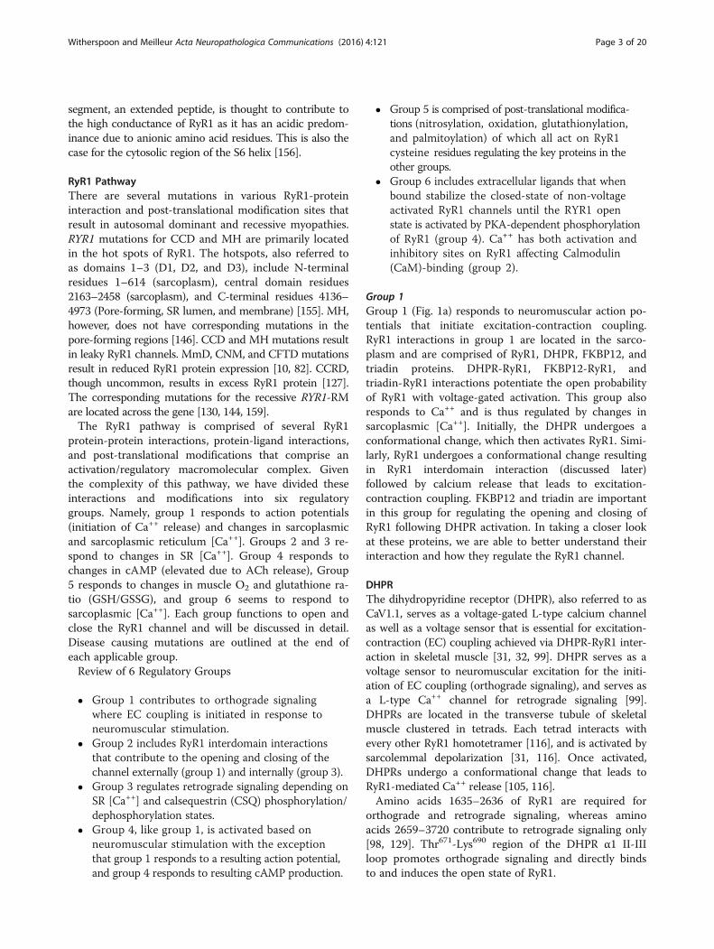
Group 1Group 1 (Fig. 1a) responds to neuromuscular action po-tentials that initiate excitation-contraction coupling.RyR1 interactions in group 1 are located in the sarco-plasm and are comprised of RyR1, DHPR, FKBP12, andtriadin proteins. DHPR-RyR1, FKBP12-RyR1, andtriadin-RyR1 interactions potentiate the open probabilityof RyR1 with voltage-gated activation. This group alsoresponds to Ca++ and is thus regulated by changes insarcoplasmic [Ca++]. Initially, the DHPR undergoes aconformational change, which then activates RyR1. Simi-larly, RyR1 undergoes a conformational change resultingin RyR1 interdomain interaction (discussed later)followed by calcium release that leads to excitation-contraction coupling. FKBP12 and triadin are importantin this group for regulating the opening and closing ofRyR1 following DHPR activation. In taking a closer lookat these proteins, we are able to better understand theirinteraction and how they regulate the RyR1 channel.
DHPRThe dihydropyridine receptor (DHPR), also referred to asCaV1.1, serves as a voltage-gated L-type calcium channelas well as a voltage sensor that is essential for excitation-contraction (EC) coupling achieved via DHPR-RyR1 inter-action in skeletal muscle [31, 32, 99]. DHPR serves as avoltage sensor to neuromuscular excitation for the initi-ation of EC coupling (orthograde signaling), and serves asa L-type Ca++ channel for retrograde signaling [99].DHPRs are located in the transverse tubule of skeletalmuscle clustered in tetrads. Each tetrad interacts withevery other RyR1 homotetramer [116], and is activated bysarcolemmal depolarization [31, 116]. Once activated,DHPRs undergo a conformational change that leads toRyR1-mediated Ca++ release [105, 116].Amino acids 1635–2636 of RyR1 are required for
orthograde and retrograde signaling, whereas aminoacids 2659–3720 contribute to retrograde signaling only[98, 129]. Thr671-Lys690 region of the DHPR α1 II-IIIloop promotes orthograde signaling and directly bindsto and induces the open state of RyR1.
Witherspoon and Meilleur Acta Neuropathologica Communications (2016) 4:121 Page 3 of 20

Fig. 1 (See legend on next page.)
Witherspoon and Meilleur Acta Neuropathologica Communications (2016) 4:121 Page 4 of 20

DHPRs are comprised of five subunits including α1,α2, β, γ, and δ [32, 107] where the β and α1 subunitshave been shown to be key players in excitation-contraction coupling [105, 112]. β and α1 subunits dir-ectly interact with RyR1 [35, 105, 112].The β subunit guanylate kinase domain binds to the
II-III loop of the α1 subunit and its C-terminus binds toRyR1 residues K3494-R3502 [35, 112]. Rebbeck [112] the βsubunit functions to ensure correct positioning of theDHPR to allow α1 subunit coupling to RyR1 and main-tains the open state of RyR1 in the presence of Ca++ andATP. However, physiologic levels of Mg2+ inhibit β sub-unit activity [112], but the effect of Mg2+ is relieved byactivation of the RyR1 voltage sensor, DHPR, wheremagnesium is disassociated from the inhibitory site onRyR1 [86, 124]. It has been shown that Ca++ and Mg++compete for the same binding site on RyR1. Unlike Mg2+, Ca++ reinforces the open state of RyR1 [86].The II-III loop of the α1 subunit is critical for ortho-
grade signaling to RyR1 in response to DHPR voltage-gated activation [98] and also enhances DHPR functionvia retrograde signaling from RyR1 to the DHPR [97, 104].Orthograde signaling results in the release of Ca++ from thesarcoplasmic reticulum through the RyR1 ion channel. Incontrast, RyR1-DHPR retrograde signaling has been sug-gested to promote inactivation of the DHPR “to limit SRCa++ release and store depletion.” This hypothesis is basedon the Y522S mutation, in myotubes of RyR1 knock-inmice, altering voltage-dependent inactivation of the DHPRwhere the voltage-dependent inactivation of DHPR-triggered Ca++ release was shifted to more negative holdingpotentials. In doing so, the voltage threshold for Ca++ re-lease is lowered, limiting Ca++ release. Heat also results inthis shift. Given the reduced Ca++ release, it is proposedthat steady state DHPR-inactivation may be a compensa-tory mechanism used to counteract excessive Ca++ leak andSR Ca++ store depletion. In cases where this compensatory
mechanism did not exist, the mutant (Y522S) myotubes ex-hibited Ca++ leak and SR Ca++ store depletion [6]. Recently,DHPR-inactivation, due to prolonged depolarization, as theprimary cause of limiting excessive Ca++ leak and SR Ca++
store depletion has been challenged, and instead is believedto primarily be due to SR Ca++ store depletion triggeringsteady-state DHPR-inactivation [93]. Although controver-sial, investigating these mechanisms in greater detail toidentify future paths toward treatment is essential.
FKBP12FK506-binding protein 12 (FKBP12) is encoded by thecalstabin-1 gene and is located in the sarcoplasm ofskeletal muscle. There are four FKBP12 subunits thatbind to the homotetrameric RyR1 protein in a 1:1manner [151]. Until recently, FKBP12 has been shownto bind to RyR1 at aa sites 2461 and 2462 [11, 61, 65]. Fur-ther exploration of the FKBP binding sites on RyR1 re-vealed that FKBP interacts with the N-terminal (76–619)and central (2157–2777) domains of RyR1 [65]. Morespecifically, FKBP binds at aa sites 619, 2157, 2341,and 2502.The interaction between FKBP12 and RyR1 alters
DHPR-RyR1 functional interaction [11]. FKBP12 pre-vents leaky RYR1 signaling under sub-optimal ligandconcentratations, and therefore serves as a molecular“gradient reader.” (Uniprot) Therefore, FKBPs are sug-gested to have “a stabilizing effect on RyR channel functionby lowering open probability and preventing subconduc-tance state gating,” which leads to “fewer leaky RyR chan-nels and fewer aberrant Ca++ release events (Venturi et al.2014).” Originally, FKBP12 was thought to stabilize RyR1in skeletal muscle whereas FKBP12.6 stabilized RyR2 incardiac cells [62]. Additional studies showed RyR2 likelyundergoes dual modulation by FKBP12 and 12.6, such thatFKBP12.6 acts as an FKBP12 antagonist indirectly redu-cing RyR2 open probability and SR Ca++ release. Since
(See figure on previous page.)Fig. 1 a Group 1. 1) RyR1 open (voltage-gated): RyR1 activation in response to DHPR activation by acetylcholine (ACh) release at theneuromuscular junction in skeletal muscle 2) RyR1 closed: no neuromuscular-stimulated ACh release. b Group 2. 1) RyR1 open: No interdomaininteraction (unzipped) with bound ApoCaM in response to RyR1 activation preceded by DHPR activation resulting in high sarcoplasmic Ca2+
2) RyR1 closed: bound Ca2+-Cam at high [Ca2+] resulting in interdomain interaction (zipped) and high sarcoplasmic reticulum (SR) Ca2+. cGroup 3. 1) RyR1 open: RyR1-triadin interaction due to triadin-calsequestrin (CSQ) interaction at high Ca2+ bound CSQ resulting in highsarcoplasmic Ca2+ 2) RyR1 closed: RyR1-junctin interaction due to junctin-CSQ interaction at low Ca2+ bound CSQ in phosphorylated state resultingin high SR Ca2+. d Group 4. 1) RyR1 open: RyR1 and Gm phosphorylation by PKA due to high [cAMP] resulting in high sarcoplasmic Ca2+ 2) RyR1closed: RyR1 dephosphorylation by PP1 due to low [cAMP] resulting in high SR Ca2+. e Group 5. S-nitrosylation. 1) RyR1 open: nitrosylation of RyR1cysteine residues by nitric oxide-glutathione reaction in response to physiologic p(O2) resulting in high sarcoplasmic Ca2+2) RyR1 open: nitrosylationof RyR1 by nitric oxide overrides Ca-CaM inhibitory effect resulting in high sarcoplasmic Ca2+ S-oxidation. 1) RyR1 open: oxidation of RyR1 cysteineresidues by reactive oxygen species reaction in response to increased physiologic p(O2) resulting in high sarcoplasmic Ca2+ 2) RyR1 closed: no RyR1oxidation at low physiologic p(O2) with bound Ca2+-CaM resulting in high SR Ca2+ S-glutathionylation. 1) RyR1 open: initial increase in reducedglutathione (GSH) stimulating glutathionylation of RyR1 cysteine residues by GSH, in turn, decreasing the GSH:GSSG and reducing RyR1 sensitivity toMg2+ inhibition resulting in high sarcoplasmic Ca2+ 2) RyR1 closed: no glutathionylation until GSH:GSSG ratio is restored resulting in high SRCa2+S-palmitoylation. 1) RyR1 open: palmitoylation of RyR1 cysteine residues by the fatty acid palmitoyl-CoA resulting in high sarcoplasmic Ca2+2)RyR1 closed: no palmitoylation resulting in high SR Ca2+. f Group 6. 1) RyR1 open: RyR1-ligand (Ca2+and ATP) binding activation resulting in highsarcoplasmic Ca2+ 2) RyR1 closed: RyR1-ligand (Ca2+ and Mg2+) binding deactivation resulting in high SR Ca2+
Witherspoon and Meilleur Acta Neuropathologica Communications (2016) 4:121 Page 5 of 20

FKBP12.6 affects skeletal muscle function, dual modula-tion of RyR1 is also thought to occur [147].The role of FKBP12 in the RyR1 EC-coupling pathway
is controversial (Avila et al. 2003). Researchers initiallydemonstrated FKBP12 functions to close the RyR1 chan-nel, but later showed FKBP12 also potentiates the RyR1open state. According to Gaburjakova et al. (2001), theincreased RyR1 gating frequency in the absence ofFKBP12 suggests that FKBP12 functions to stabilizeRyR1 in its open and closed states.In group 1, the interaction between DHPR and RyR1
is modulated by FKBP12 [98] such that FKBP12 stronglypotentiates the open state of RyR1 when RyR1 is boundto the Thr671-Lys690 region of the DHPR II-III loop[100]. In support, when FKBP12 was depleted, this re-sulted in obliteration of the open state of RyR1 followingDHPR activation [100]. Not only does FKBP12 directlyinteract with RyR1 in the presence of DHPR activation,but it has also been identified in a group including RyR1,protein kinase A (PKA), phosphodiesterase 4D3 (PDE4D3),and protein phosphatase 1 (PP1). Within this group,FKBP12 functions to close the RyR1 channel, which isdiscussed in more detail later.Given that FKBP12 plays a role in RyR1 open and
closed states, future studies are required to determine ifFKBP12 function changes depending on the RyR1 aaposition to which FKBP12 binds. It may also be import-ant for treatment purposes. For example, rycals, drugsthat enhance FKBP12 binding to RyR1 [20], may be aneffective treatment in the RYR1-RM population withmutations that negatively affect FKBP12 interaction withRyR1 and result in RYR1-RM. For this reason, we arecurrently performing a pre-clinical study testing the ef-fect of Rycals on RyR1 function in muscle fibers biopsiedfrom patients with RYR1-RM in collaboration withMarks and colleagues (unpublished data).
TriadinFKBP12 modulation of RyR1 activity is proposed to me-diate the regulatory role of triadin on RyR1 activity [24].Triadin is a junctional SR membrane glycoprotein thathas been shown to interact with DHPR and RyR1 inthe sarcoplasm [58, 68] supporting triadin playing arole in orthograde coupling. Disruption in RyR1 andtriadin binding reduces orthograde signaling. However,it does not affect retrograde signaling between RyR1and DHPR [67].When the interaction between sarcoplasmic RyR1 and
triadin is disrupted, this results in RyR1 channel inhib-ition. Amino acids 18–46 of triadin interact with RyR1in the sarcoplasm at low Ca++ levels, whereas this bind-ing is inhibited at high Ca++ levels. The prevention ofamino acids 2–17 of triadin from binding to RyR1 byuse of antibodies does affect RyR1 channel function,
thereby resulting in a reduced rate of SR Ca++ releaseand decreased open probability of RyR1 [68].Although triadin is not well understood, it is thought
to regulate RyR1-DHPR interaction, and in turn, modu-late EC coupling [53]. A number of studies have identi-fied that triadin is “primarily a negative regulator ofRyR1 [52]” [68, 70, 103, 131]. One study showed thatamino acids 664 to 799 of DHPR alpha 1 subunit bindto triadin primarily at amino acids 68–278 [58]. Al-though triadin interacts with DHPR in the sarcoplasm,EC coupling and RyR1 channel regulation were not pre-vented in triadin null mice compared to wild type [131],yet, a significant reduction in muscle strength wasshown in triadin null mice [101]. Interestingly, Shen etal. (2007) demonstrated little to no difference in forcegeneration between wild type and triadin null mice inresponse to electrical stimulation. It is important to notethat both Shen et al. (2007) and Oddoux et al. (2009)used triadin null mice, but electrically stimulated differ-ent muscles including the lumbricals and flexor digi-torum of the hindlimb, respectively. These muscles maybe affected differently in triadin null mice, and soaffected muscle groups should be determined.Despite no change in force generation, Shen et al.
(2007) demonstrated electrical stimulation still resultedin a lower magnitude of Ca++ transients. Similar findingswere shown in isolated myotubes from the same mice aswell as a significant increase in resting myoplasmic Ca++
[131]. In support, Eltit et al. (2010) demonstrated chron-ically elevated resting myoplasmic Ca++ due to FKBP12-RyR1 dysfunction and SR store-operated Ca++ entry[52, 53], suggesting increased basal RyR1 activity intriadin null myotubes isolated from skeletal muscle ofmice. Additionally, Oddoux et al. (2009) revealed areduction of SR Ca++. Together, the results reveal thattriadin ablation affects resting Ca++ levels such that thereis an increase in myoplasmic Ca++ and a reduction in SRCa++, supporting a lower magnitude of Ca++ transients inresponse to electrical stimulation. Eltit et al. (2011) alsoshowed that triadin null mice result in no significant dis-ruption of EC coupling. However, further kinetic analysis,in isolated myotubes, revealed that voltage-gated activa-tion time for Ca++ release was slowed [53].Given that triadin null mice presented with what ap-
pears to be a “normal” phenotype, it was suggested byShen et al. (2007) that the role triadin plays in musclefunction is minor or replaced with a compensatorymechanism [131] Conversely, according to Oddoux et al.(2009), a decrease in muscle strength in triadin nullmice, suggests triadin dysfunction may lead to the devel-opment of a myopathy and is therefore essential forskeletal muscle function. Since both authors make validpoints, whether or not the absence of triadin results in aRyR1 myopathy has yet to be determined.
Witherspoon and Meilleur Acta Neuropathologica Communications (2016) 4:121 Page 6 of 20

Group 1 Pathomechanisms:
CCD/MH: Mutations in the RyR1 N-terminal and centraldomains that disrupt FKBP12-RyR1 interaction areproposed to result in MH [65, 104] or CCD/MH[104]. V2461G and V2461I are RyR1 mutations thatdisrupt FKBP12 binding. In skeletal myotubes expressingthese mutations, there was approximately a 50 %reduction in voltage-gated Ca + release due to theV2461G mutation compared to wild type. Myotubesexpressing the V2461I mutation resulted in the bindingof FKBP12.6 as opposed to FKBP12. In response, therewas a reduction in SR Ca++ release [11]. A studyperformed by Galfre et al. (2013) identified threeFKBP12 sites that interact with RyR1. Mutationsintroduced at any of these sites changed the functionof FKBP12 such that mutant FKBP12 (at sites Glu31,Asp32, orTrp59) functioned as FKBP12.6, thereby ac-tivating the RyR1 channel and resulting in Ca++re-lease. Based upon the above results, FKBP12 shouldbe studied in more detail under different conditions.CCRD: Similar to I4898T, a well known mutationmentioned in group 3, the Y4796C mutation in RyR1 isalso suspected to interfere with the interaction betweenRyR1 and triadin, but rather in the myoplasmic domaininstead of the SR luminal region. Patients with theY4796C mutation present with cores and rods onmuscle biopsy, and therefore have CCRD.Consequently, there is increased rate of calcium leakagefrom the SR [95]. Y4637A and Y4637I mutations, likeY4796, also result in CCRD [90]. Amino acid 4637 islocated in the membranous region of the RyR1 C-terminus. Similar to characteristics of the I4898Tmutation, resting calcium levels associated with T4637Asignificantly increase and SR luminal Ca++ decreases.However, instead of leaky RyR1 channels as noted inCCD, individuals with the T4637A mutation presentwith excess ryanodine receptors in the central cores[127]. The T4637 pathomechanism may be the same forthe Y4796C mutation rather than an increased rate ofcalcium leakage, but further research is needed.CFTD: Mutations associated with autosomal recessivemyopathies often include a missense mutation alongwith a null mutation, and sometimes a homozygousmissense mutation [10]. In a study with six patientsdiagnosed with CFTD, each patient exhibited aheterozygous missense mutation in addition a nullmutation [37]. RYR1 mutations resulting in CFTDare linked to the RyR1-DHPR (α and β) binding sites.
Group 2Group 2 (Fig. 1b) is located in the pore-forming regionof RyR1 and is comprised of RyR1 interdomain inter-action and calmodulin (CaM). This group responds to
the activation of RyR1 by DHPR and is regulated by SRCa++. It has been suggested that RyR1 conformationalchange in response to DHPR activation leads to RyR1 in-trinsic modulation of the opening/closing of the RyR1ion channel. This intrinsic modulation is based on inter-domain interaction where the RyR1 central domain(Leu2442-Pro2477) interacts with the RyR1 N-terminal do-main [104]. This interdomain interaction is regulated byCaM and Ca++ levels regulate the function of CaM.
RyR1 Interdomain interactionBannister et al. (2007) proposed the “domain switch” hy-pothesis that reflects the structure-function relationshipbetween the interdomain interaction and RyR1 function.The hypothesis states that “In the non-activated state,the N-terminal and central domain make close contactthrough several sub-domains: this ‘zipped’ state stabilizesthe closed state of the channel. Under normal stimulat-ing conditions, the inter-domain contact is weakenedleading to an ‘unzipped’ state, which is recognized bythe channel as an activation signal.” Interestingly, MHmutations have been shown to result in a partial un-zipped state leading to “hyperactivation/hypersensitiza-tion” of RyR1 [12].Domain peptide 4 (DP4) is a synthetic peptide that
corresponds to Leu2442-Pro2477 of RyR1. When DP4 wasbound to the N-terminus of RyR1, this interaction re-sulted in an “unzipped” state that led to activation ofryanodine binding and SR Ca++ release [12]. Olojo et al.(2011) determined how the interdomain interaction in-fluences orthograde and retrograde signaling by usingDP4. The results showed enhanced RyR1 orthograde Ca++ release without affecting the DHPR voltage sensorand mediated retrograde signaling that results in a RyR1open state [104]. In summary, the RyR1 conformationalchange in response to DHPR activation results in the“unzipped” state where the interdomain interaction isweakened and is recognized by RyR1 as an activationsignal leading to the release of Ca++ [12]. Under normalconditions, the central domain and N-terminus makeclose contact maintaining the “zipped” state of RyR1thereby stabilizing the RyR1 closed state [12].
CalmodulinUnder normal conditions, CaM disrupts the interdo-main interaction [77]. CaM exists in two forms, with-out Ca++ (apocalmodulin, apoCaM) and Ca++ bound(Ca++-CaM). Both forms bind to RyR1 with Ca++-CaMhaving a greater binding affinity [157]. ApoCaMserves as an agonist resulting in the release of Ca++
at low sarcoplasmic [Ca++], whereas Ca++-CaM main-tains the closed state of RyR1 at high sarcoplasmic[Ca++] [64, 73, 77, 92, 157]. CaM levels increase assarcoplasmic Ca++ levels increase. More recently,
Witherspoon and Meilleur Acta Neuropathologica Communications (2016) 4:121 Page 7 of 20

researchers showed that activation of CaM results inCaM Kinase II (CaMKII) activation, which, in turn,phosphorylates RyR1, affecting skeletal muscle con-tractility. In summary, Ca++-CaM binds to RyR1 onthe sarcoplasmic side inhibiting SR Ca++ release, and,while bound, CAMKII phosphorylates RyR1 [64].CAMKII, and as discussed later, PKA, both phosphor-
ylate RyR1 and are thus considered modulators of RyR1activity. It is important to note that hyperphosphoryla-tion of RyR1 by CAMKII or PKA results in FKBP12disassociation, and consequently, a higher open prob-ability of RyR1. Along this continuum, a higher RyR1open probability due to hyperphosphorylation may affectskeletal muscle contractility under resting conditionswhere skeletal muscle contractility is decreased [64].In 2002, O’Connell et al. demonstrated that the intro-
duction of CaM binding sites (3624 and 3620) in dyspedicmyotubes primarily regulates L-type channel currents forretrograde signaling compared with EC coupling fororthograde signaling [99]. The structure of CaM is com-prised of two lobes, a N- and C-lobe where the C-lobe ofCa++-CaM binds at RyR1 sites 3614–3643 [77] and the N-lobe to 1975–1999. Both of these undergo interdomaininteraction [157]. Specifically, the interdomain interactionincludes disulfide bonds formed between cysteine residuesthat include 3635, 2000, and 2401 [157] of adjacent RyR1subunits within a tetramer. The ApoCaM-binding domainof RyR1 (Lys3614-Asn3643) also interacts with RyR1 sitesCys4114-Asn4142. When bound, this leads to Ca++ release[63]. ApoCaM not only binds to aa 3614–3643, but alsoaa 3625–3644 [117].Although ApoCaM and Ca++-CaM have opposing func-
tions, both prevent oxidation-induced intersubunit cross-linking where disulfide bonds are formed between eachRyR1 subunit leading to Ca++ release [72, 110]. Post-translational modifications of RyR1, group 5, are discussedlater. It is postulated that CaM protects RyR1 from oxida-tive stress associated with strenuous exercise [28, 73].Conversely, oxidation of RyR1 prevents the binding of
CaM (both forms) to RyR1 at low [Ca++]. Nitric oxide(NO), which plays a role in redox reactions involvingRyR1, not only blocks intersubunit disulfide bondsformed by oxidation but also prevents the binding ofApoCaM [72, 110]. These data suggest that NO regu-lates oxidation and ApoCaM activity, both of which pro-mote the RyR1 open state. The unaffected Ca++-CaM byNO, when bound to RyR1, results in the RyR1 closedstate. The redox reactions are discussed later.In the nitrosylation subsection, the literature demon-
strates NO has a high affinity for CaM such that CaM isrequired for nitrosylation to occur. Ca++-CaM boundRyR1 is unaffected at most sites, thereby protectingRyR1 from oxidation. Further research is necessary todetermine what occurs in a hypernitrosylated state or
when mutations are present in the Ca++-CaM bindingsite on RyR1. If such changes result in a myopathy ormalignant hyperthermia phenotype, this research wouldopen the door to potential treatments. Additionally,given that CaM is not only required for nitrosylation,but also activates downstream phosphorylation of RyR1,it is important to determine whether hyperphosphoryla-tion and hypernitrosylation occur simultaneously andpossibly contribute to disease severity.Interestingly, increased levels of CaM not only activate
CAMKII, but also calcineurin. Calcineurin is a phos-phatase responsible for skeletal muscle satellite celldifferentiation, which is important for skeletal musclefiber regeneration after injury and skeletal muscle hyper-trophy [64, 145]. Activation of calcineurin primarilyinfluences slow twitch fiber hypertrophy. In mice, inhib-ition of calcineurin resulted in marked inflammation,fiber atrophy, presence of immature myotubes, and cal-cification in regenerating muscle compared with controls[122, 123]. Further research is required to understandthe role of calcinuerin in RYR1-RMs. Targeting calcine-urin may be a potential therapeutic treatment.Group 2 Pathomechanisms:
MH: When DP4 was isolated in skinned skeletal musclefibers, it enhanced ryanodine binding and sensitized therelease of SR Ca++ similar to what has been shown inMH pathology. It is believed that MH results from thedisrupted interdomain interactions between DP4 andthe N-terminus of RyR1 that result in destabilization ofthe RyR1 closed state [87].MmD: Mutations in RyR1 that manifest as MmDare dispersed throughout RyR1 primarily outside thehot spot regions. RyR1 mutations P3527S and V4849Icause an increase in sarcoplasmic resting Ca++ withoutdepleting SR Ca++ stores [143, 144, 159]. V4849I is aninteresting mutation linked to autosomal recessiveCCD, which presents as MmD [59].The aforementioned mutations are located in theS100A1 and CaM binding sites. Researchers arecontinously learning more about S100A1, but it isbelieved that this S100A1 is responsible for linkingRyR1 subunits. S100A1 is considered one of the mostimportant ligands in cardiac muscle, possibly skeletalmuscle, and is also responsible for Ca++ release at low[Ca++]. A single site on RyR1 binds both S100A1 andCa++-Cam. The release of Ca++ at low [Ca++] contributesto muscle twitches, however, the same site is critical forinhibiting Ca++ release during “repeated or sustainedactivation by binding Ca++-CaM at higher [Ca++].In this way, Ca++ is able to slow energy expenditurelater in contraction [92, 148]. Other RyR1 mutations,R109W (also P109W) and M485W, occur simultaneouslyand some are intronic variants such as homozygous
Witherspoon and Meilleur Acta Neuropathologica Communications (2016) 4:121 Page 8 of 20

14646 splicing variant resulting in a reduced numberof RyR1 [143, 144, 159]. MmD patients with thesemutations clinically present with ophthalmoplegiaand muscle weakness.Ophthalmoplegia, in this patient population, is thoughtto be due to the absence of RyR3 compensation [159].In support, results reported by Perez et al. (2005)suggest RyR1 and RyR3 together regulate skeletalmuscle Ca++. Ophthalmoplegia is also suspected tobe mutation specific or caused by mitochondrialdysfunction [128, 130]. Causal RYR1 mutations arelocated outside the hotspot regions or include amalignant hyperthermia causing mutation accompaniedby another mutation outside the hotspot regions. Twoknown mutation combinations include R3772W +E989G and R3772W +H283R. A previously reportedR3772Q mutation caused a more severe phenotypeincluding ptosis, facial weakness, myopathy, and MHS.MRI pathophysiological findings potentially responsiblefor ophthalmoplegia, ptosis, and facial weakness includethin hypoplastic intraorbital motor cranial nerves inaddition to hypoplasia of the extraocular muscles.Interestingly, the optic nerve remained healthy andintact [130]. Given the eye is a high-energy demandorgan, the extraocular muscles are comprised of severalmitochondria. However, chronic oxidative damage resultsin mitochondrial instability yielding mitochondrialdamage. Mitochondrial dysfunction, increased oxidativestress, and increased apoptosis are common causes ofophthalmologic disorders in the aging population [128].Although mitochondrial-related extraocular muscledysfunction has not been shown in RYR1-RM, thispathomechanism may be worth assessing in thispatient population.CNM: RyR1 mutations that manifest as CNM occur inDHPR, CaM, and sometimes the triadin binding sitesdisrupting interdomain interaction [3]. RyR1-CaMinteraction can be disrupted in an environment withhigh oxidant concentrations [110]. Associated mutationsinclude Glu1909GlyfsX39, Met3081Thr, Val4842Met,10348-6C >G (intronic), Ser1342Gly, Thr2787Ser, and3381 + 1 G > A (intronic). To achieve protein reduction,it is suggested that the aforementioned mutations coexistwith the intronic mutation 10348-6C >G, which furtherresults in the production of another mutation,His3449ins33fsX54 [82, 154].
Group 3Group 3 (Fig. 1c) is located in the SR and is com-prised of RyR1, CSQ, triadin, and junctin. This groupresponds to the RyR1 interdomain interaction and isregulated by SR Ca++. In group 3, CSQ seems to bethe primary protein of interest for RyR1 channel ac-tivity because it indirectly regulates RyR1 open and
closed states depending on SR [Ca++]. CSQ, in itsphosphorylated and dephosphorylated states, regulatesRyR1 channel activity through its interaction withjunctin and triadin. The phosphorylated and dephos-phorylated states of CSQ seem to be a regulatorymechanism of the CSQ/junctin/triadin complex, andthe CSQ/junctin/triadin complex regulates RyR1 activ-ity from the SR.Triadin seems to be the key communicator between
orthograde and retrograde signaling following voltage-gated activation of RyR1. Like FKBP12, it functions topotentiate both the open and closed states of RyR1. Triadinmay also be a target for potential treatment. In group 1,FKBP12.6 restored resting Ca++ levels by acting directly onRyR1. Boncompagni showed that the SR luminal contentand cisternae volume were significantly altered in triadinnull mice [24]. How FKBP12.6 affects SR content while re-storing resting levels is still to be determined. More studiesare required to focus on the mechanisms of action betweengroups 1 and 3 as well as the pathomechanisms that resultfrom RyR1 mutations that interfere with sarcoplasmic andSR luminal triadin binding sites.
CSQCSQ is a Ca++ storage glycoprotein located in the lumenof the SR, which functions to lower the amount of freeCa++ in the SR [79, 132, 142]. More recently, studieshave shown that CSQ is not only a storage protein. CSQalso modulates RyR1 channel activity [132] and is pri-marily located in close proximity to RyR1 [142]. Previousresearch has shown that the amount of Ca++ releasedfrom the SR is dependent on the amount of Ca++ boundto CSQ [79]. When CSQ is partially bound, smallamounts of Ca++ are released at a high rate constant,whereas when fully bound, Ca++ is released at a slowrate constant [79]. In support, CSQ has been shown tohave a controlled inhibitory effect on RyR. In theabsence of CSQ, Ca++ release increased by 10 fold. Thiseffect was reversed after reintroducing CSQ [15]. Specific-ally, the intraluminal phosphorylation/dephosphoryla-tion of CSQ controlled RyR1 channel activity in thepresence of Ca++. When CSQ is dephosphorylated, Ca++is released from the SR, but when phosphorylated,Ca++-bound CSQ has no effect on RyR1 [142].It is important to note that CSQ does not directly
interact with RyR1. Rather, it interacts indirectly throughjunctin and triadin [17]. Using a DCAM probe and elec-tron microscopy, Ikemoto et al. (1989) showed Ca++
bound CSQ undergoes a conformational change, subse-quently binding to junctional face membrane (jfm) pro-teins later identified as junctin and triadin [17, 103]. Itwas also demonstrated that conformational changes inCSQ were coupled to conformational changes in RyR1; a
Witherspoon and Meilleur Acta Neuropathologica Communications (2016) 4:121 Page 9 of 20

conformational change in one was transmitted to theother [142].Junctin and triadin are transmembrane anchoring pro-
teins that form a stable quaternary group, includingRyR1, junctin, triadin, and CSQ. CSQ binds to junctinand triadin under low Ca++ concentrations resulting inthe closed state of RyR1 [152]. Beard et al. (2008) dem-onstrated that when CSQ is phosphorylated under lowSR luminal Ca++ concentrations, CSQ binds to junctinonly. This phosphorylated state of CSQ does not disruptthe ability of CSQ to maintain the closed state of RyR1[16]. Rather, it enhances the Ca++ binding affinity toCSQ [17]. These results suggest that the inhibitory effectof CSQ on RyR1 activity is mediated by junctin whenCSQ is in its phosphorylated state under low Ca++ con-centrations [17]. Under physiological conditions of Ca++,neither the phosphorylated nor the dephosphorylatedstate affects the coupling of CSQ, junctin, and triadin[16]. CSQ, in its dephosphorylated state under low SRCa++ conditions, binds only to triadin, in turn activatingryanodine receptors. High luminal Ca++, on the otherhand, results in dissociation of the CSQ, triadin, andjunctin group [16]. Whether CSQ has an inhibitory [13,14, 150] or activation [84, 102] effect on RyR1 has beencontroversial.In summary, CSQ and RyR1 have an indirect relation-
ship by way of triadin and junctin. This relationshipappears to depend on SR luminal [Ca++] as well as phos-phorylation/dephosphorylation mechanisms. Low SRluminal Ca++ promotes the binding of CSQ to junctinand triadin. This results in the RyR1 closed state. Thebinding of CSQ to junctin and triadin changes whenphosphorylation and dephosphorylation occur. Low SRluminal Ca++ with CSQ phosphorylation still results inthe binding of CSQ to junctin only with no RyR1 activ-ity. However, in its dephosphorylated state, CSQ bindsto triadin only and this interaction leads to RyR1 activation(Fig. 1c). This RyR1 activation is inhibited by ryanodinebinding and cannot be reversed with dephosphorylatedCSQ. Since CSQ does not bind to triadin and junctin whenSR luminal Ca++ is high, based on current knowledge,CSQ only communicates with RyR1 when SR luminalCa++ levels are low.
TriadinIn group 1, triadin was shown to bind to RyR1 andDHPR on the sarcoplasmic side potentiating voltage-gated RyR1 Ca++ release. Triadin also interacts withRyR1 and CSQ in the SR lumen (group 3) in a Ca++
dependent manner serving as a linker protein betweenRyR1 and CSQ [69, 119]. The sarcoplasmic region ofRyR1 and triadin become disassociated when the SRluminal binding of these proteins are disrupted, but donot affect RyR1 channel function [67]. Beard [18] based
on these results, SR Ca++ and group 3 seem to regulatethe function of triadin in group 1.Three regions of triadin are responsible for its
localization at the membrane. These regions include thetargeting region (TR) 1 (18–47, sarcoplasmic), TR2(106–214), and TR3 (233–440, 441–729). At least two ofthese three regions are required for correct localizationin the membrane. Binding regions for RyR1 have beenidentified in TR3, and the same is true for CSQ [30].Specifically, triadin binds to the SR luminal side of RyR1at amino acids D4907, E4908, and D4878 [89]. CSQ ap-pears to be associated with triadin stabilization (reducedmobility) in the SR membrane, and more importantly, akey component for the formation of a stable groupbetween triadin and RyR1 [120].Unlike the effect of triadin on RyR1 in group 1, triadin
in group 3 functions to close the RyR1 channel while en-hancing the binding affinity of ryanodine to RyR1 [67].Ryanodine binding is used to study Ca++ binding affinitybecause ryanodine binding is Ca++ dependent. “Low af-finity Ca++ binding sites resulted in inhibition of ryano-dine binding and Ca++ release from isolated SR vesicles.”[71] Ohkura et al. (1998) showed that depletion oftriadin increases ryanodine binding, but when available,triadin functions to inhibit ryanodine binding to the SRand maintain the closed state of RYR1. The effect oftriadin on ryanodine binding is the same even whenryanodine binding is potentiated by CSQ [103].Wei et al. [153] demonstrated that when triadin and
junctin are exposed to RyR1 independently, the openstate of RyR1 is enhanced. Once CSQ was added to eachsolution, only the RyR1/junctin interaction led to areduction in RyR1 activity when the SR luminal Ca++
was lowered [18].
JunctinJunctin, like triadin, is a transmembrane protein thatbinds to RyR1 and CSQ. Unlike triadin, junctin onlybinds to the luminal domain of RyR1. Junctin is believedto play a more critical role in maintaining the SR Ca++
store and CSQ/RyR1 signaling in myotubes (Boncompagniet al. 2012-refs 38 and 39). Boncompagni et al. (2012)studied the function of triadin and junctin in Ca++ homeo-stasis using hind legs from mice (triadin null, junctin null,triadin/junctin null). Their results showed reduced coup-ling in triadin-null mice, whereas junctin null mice dem-onstrated minimal to no changes in functional activity.Based on these results, the interaction between triadinand CSQ has a major impact on the SR architecture andmyoplasmic Ca++ [24] as previously noted in group 1.More specifically, the SR luminal content and volume ofSR cisternae are significantly altered in triadin null andtriadin/junctin null mice. CSQ is also less defined. Thefindings from Boncompagni et al. (2012) further support
Witherspoon and Meilleur Acta Neuropathologica Communications (2016) 4:121 Page 10 of 20

triadin as an important factor in skeletal muscle functionas suggested in group 1.Group 3 Pathomechanisms:The triadin binding-domain in RyR1 is located within
the hotspot 3 region of which gives rise to CCD andCCD/MH mutations [78]. Several RYR1 mutations,resulting in central core disease, lead to amino acidchanges in the SR luminal side of RyR1 that disruptRyR1-triadin interaction as well as influence voltage-gated Ca++ release [11, 67]. I4898T is a very commonRyR1 mutation within this SR luminal region that resultsin severe CCD and is proposed to possibly disrupt theinteraction between RyR1 and triadin [95].
Group 4Group 4, represented in Fig. 1d, responds to changes inadenosine 3’, 5’ cyclic monophosphate (cAMP), which iselevated due to acetylcholine (ACh) release. Group 1responds to an action potential resulting from AChrelease, whereas group 4 responds to elevated cAMPlevels resulting from ACh release. Therefore, thesegroups may be activated simultaneously. Group 4 in-cludes FKBP12, protein kinase A (PKA), phosphodiester-ase 4D3 (PDE4D3), and protein phosphatase 1 (PP1).RyR1 undergoes phosphorylation and dephosphorylation[56] within the group.cAMP levels are increased in response to acetylcholine
release. As a result, PKA is activated and is anchored toRyR1 by way of A-kinase anchoring proteins of the skel-etal muscle (mAKAP). PKA then phosphorylates RyR1(S2483) preventing the binding of Mg2+ to RyR1, result-ing in RyR1 open probability. PKA not only phosphory-lates RyR1, but also phosphorylates the targeting subunitGm, which results in the dissociation of PP1 from Gmand the SR. PP1 dissociation from Gm prevents PP1from dephosphorylating RyR1. The above pathway re-sults in RyR1 open probability. However, when cAMPlevels are lowered by PDE4D3, PP1 is not dissociatedfrom the Gm subunit nor is the Gm subunit separatedfrom RyR1. PP1 is then able to dephosphorylate RyR1resulting in FKBP12 binding, thus a RyR1 closed state.To better understand this portion of the pathway, theinvolved proteins of this group are further discussed.Group 4 has several regulatory components and ap-
pears to be the only group that not only affects voltage-activated RyR1, but also adjacent non voltage-activatedRyR1. Because group 4 affects both voltage-activatedand non voltage-activated RyR1, this raises the questionof whether mutations in RyR1 affecting this group influ-ence clinical severity. Several of the aforementionedstudies focused on a single component of the group,therefore, studies are needed that demonstrate pathome-chanisms related to all components making up thisgroup and their associated phenotype. For example,
FKBP12 is responsible for synchronizing gating mecha-nisms between adjacent RyR1 proteins. Will a mutationaffecting FKBP12 binding affinity to RyR1 and its abilityto synchronize adjacent RyR1 influence severity? Add-itionally, as mentioned above, PKA phosphorylation andS-nitrosylation both dissociate FKBP12 from RyR1.What happens in skeletal muscle if the phosphorylationsite on RyR1 is changed due to a mutation or hyperni-trosylation? Lastly, in group 1, rycals were mentioned asa drug that enhances the binding affinity of FKBP12 toRyR1. Could this be a potential treatment that resolvesany issue resulting from RyR1 mutations in this group?Further research is needed regarding group 4 and itsrelated pathomechanisms.
FKBP12FKBP12 is not only a component of group 1, but also acomponent of group 4. In group 4, FKBP12 is an im-portant regulatory protein. When bound to RyR1, it sta-bilizes the RyR1 closed state and synchronizes the gatingbetween neighboring RyRs [39, 43, 147]. NeighboringRyR1 channels are very close to each other and are mod-ulated by extracellular ligands, including Ca++, Mg2+,and ATP. Non-voltage activated neighboring RyR1 chan-nels are activated via RyR1-RyR1 physical interaction,and are stabilized by luminal Ca++ and cytosolic ATP/Mg2+ [109].FKBP12 is suggested to coordinate this multiprotein
group formation [114] such that bound FKBP12 doesnot promote RyR1 activity. However, FKBP12 dissociatesfrom RyR1 due to PKA phosphorylation at RyR1 sitesS2843 in humans [19, 114] and S2844 in mice [19, 114],yet becomes bound again due to PP1 activity. PKA andPP1 functions are discussed below.Similar to PKA phosphorylation, S-nitrosylation
(group 5) of RyR1 also reduces the binding affinity ofFKBP12 to RyR1; specifically, S-nitrosylation of cysteineresidues at positions 3635 and 2327 [7, 8, 19, 136]. Un-like PKA, S-nitrosylation does not respond to cAMP asdiscussed later under group 5.
PKAPKA is a holoenzyme with a tetrameric group consistingof two catalytic (C) subunits and a regulatory subunitdimer. Adrenaline, a hormone that acts on the skeletalmuscle in response to neural ACh release [91], elevatescAMP levels resulting in PKA activation, which in turninduces Gm phosphorylation [149].cAMP is required for PKA phosphorylation of the
RyR1 channel, otherwise referred to as cAMP-inducedPKA phosphorylation [114]. When cAMP levels are low,the C subunit binds to the regulatory subunit makingPKA inactive. On the other hand, in the presence ofhigh levels of cAMP, cAMP binds to the regulatory C
Witherspoon and Meilleur Acta Neuropathologica Communications (2016) 4:121 Page 11 of 20

subunit. In turn, the affinity of the regulatory subunit forthe C subunit is reduced thus freeing the C subunits andactivating PKA [34]. mAKAPs co-localize with RyR1 andfunction to anchor PKA to RyR1 in the presence ofelevated cAMP levels [121]. In response, PKA phosphory-lates serine residue, S2843 [19, 114], on RyR1 subunits inthe sarcoplasm leading to a skeletal muscle contractionand greater muscle force generation [5]. PKA-dependentphosphorylation prevents the binding of Mg2+ to theRyR1 channel thereby increasing RyR1 open probabil-ity [121].Additionally, glutathionylation regulates PKA activity,
which regulates RyR1 activity in the presence of cAMP.PKA cannot be glutathionylated in the absence of cAMPand is therefore protected from oxidation. In the presenceof cAMP, PKA becomes active. Once active, glutathionyla-tion makes PKA more susceptible to dephosphorylation,and thus its’ inhibition. “PKA deglutathionylation leads toPKA reactivation” [106].
PDE4D3Like PKA, PDE4D3 is targeted to RyR1 by way ofmAKAP, which is an anchoring protein [19]. PDE4D3 isspecific for cAMP [28]. Phosphodiesterases regulatecAMP levels by binding and degrading cAMP. PDE4D3,specifically, functions to control cAMP concentration bydegradation when co-localized with RyR1 [19].
PP1PP1 dephosphorylates RyR1 [114] resulting in the bind-ing of FKBP12 to RyR1. PP1 is a serine/threonine kinasewith a catalytic subunit and several targeting subunits.Specifically, the Gm targeting subunit of PP1 binds anddirects PP1 to glycogen particles and the SR. PP1 bindsto the Gm N-terminus and the SR to its C-terminus.However, phosphorylation of Gm at Ser67, by PKA, dis-sociates PP1 from the Gm binding domain subsequentlyreleasing Gm from both glycogen and the SR [149].
Group 5Group 5, shown in Fig. 1e, responds to changes inmuscle O2 and glutathione ratio (GSH/GSSG). Thisgroup encompasses protein post-translational modifi-cations including S-nitrosylation, S-oxidation, S-glutathionylation, and S-palmitoylation [56] and themolecules nitric oxide (NO), S-nitrosoglutathione(GSNO), reduced glutathione (GSH), oxidized gluta-thione (GSSG), hydrogen peroxide (H2O2).Within this group, RyR1 serves as a redox sensor
where certain cysteine residues undergo redox reactionsby way of post-translational modifications [7]. RyR1, as aredox sensor, is enhanced by the aforementioned mole-cules [9]. Each of these post-translational modifications
occur based on O2 levels, which change depending onoxygen demand of the active muscle [139].Post-translational modifications serve as on/off switches
of protein function [96]. S-nitrosylation, S-oxidation (di-sulfide oxidation), and S-glutathionylation each activateRyR1 by way of different mechanisms [9]. However, to-gether, they regulate RyR1 channel activity over a range ofskeletal muscle oxygen tension (pO2) [141].
S-nitrosylationPhysiological pO2 levels (~4–20 mm Hg, 0.5-2.5 %) con-trol the redox state of thiols in the RyR1 subunits main-taining the ready state of RyR1. NO, at physiological tissuepO2 (~10 mm Hg), activates RyR1 by S-nitrosylation ofRyR1 cysteine residues. Both reactive oxygen and nitrogenspecies modify RyR1 thiols altering RyR1 channel function[56, 57]. Oxidation and nitrosylation enhance Ca++ releasefrom the SR via the RyR1 channel [141].In skeletal muscle, NO is derived from neuronal NO
synthase (nNOS) and functions to S-nitrosylate proteinsforming S-nitrosothiols [139]. S-nitrosothiols are com-pounds that S-nitrosate a specific protein cys thiol [25].S-nitrosoglutathione (GSNO), formed by NO and GSHinteraction, is an example of a nitrosothiol [25]. GSNO,under atmospheric pO2, GSNO can nitrosylate and glu-tathionylate RyR1 cysteine residues [9]. Specifically, invitro GSNO treatment resulted in nitrosylation of RyR1 aaresidues 1–1509 while decreasing S-nitrosylation atresidues 3120–4475 and 3631–4475. Glutathionylationoccurred at the same residues, further including aa 1396–2401. Although GSNO is able to both nitrosylate and glu-tathionylate RyR1, glutathionylation seems to be preferred[9]. GSNO is the S-nitrosated derivative of glutathioneand is considered to be a pertinent mediator of NO. It isthe intermediate in the formation and degradation of S-nitrosothiols, and for this reason, it is considered to be po-tentially therapeutic [25]. GSNO not only activates RyR1by nitrosylation, but also oxidation (C. Hidalgo 2005).It is important to note that NO only nitrosylates RyR1
cysteine residues in the presence of CaM and at lowmuscle pO2 levels [136, 139]. Specifically, in the skeletalmuscle, 6–8 RyR1 thiols are S-nitrosylated [36, 135].Cys3635 is one of the 6–8 residues identified that isnitrosylated at low pO2, but not at high pO2 [141]. Con-sequently, RyR1 changes conformation to the open statepromoting Ca++ release and muscle force production[135]. Interestingly, Cys3635 is one cysteine residue that isunaffected by GSNO for Cys3635 does not discriminatebetween O2 levels [75].Eu et al. (2000) determined that the effect of NO
on RyR activity is dependent on the RyR1 redox stateas well as CaM. Brookes et al. (2004) believe mito-chondria may serve as a “redox signaling box” byconverting the NO signal into an ROS signal [26, 27].
Witherspoon and Meilleur Acta Neuropathologica Communications (2016) 4:121 Page 12 of 20

This phenomenon only occurs at physiological levelsof NO. In skeletal muscle, NO is produced by nitricoxide synthases (NOS) in the sarcolemma and muscu-lar endplate [60] to maintain skeletal muscle responseto increased exercise. It is important to note thatNOS activity inhibits mitochondrial respiration [136].More specifically, NO in the presence of high [Ca++]inhibits mitochondrial respiration [27, 133]. However,pathological levels of NO disrupt this process, affect-ing mitochondrial function, and, in turn, ATP synthe-sis and cell function [27, 133].It has been shown that NO in skeletal muscle is pro-
duced at rest and in greater concentrations with increasedexercise. In addition to the increase in NO, there is alsoan increase in reactive oxygen and nitrogen species due toincreased muscle contractile activity. In response to exer-cise, NOS binds CaM, which enhances NOS activity. CaMserves as a molecular switch activating the transfer ofelectrons that results in NO production [138]. Al-though CaM plays such a major role in S-nitrosylation,RyR1-bound Ca++-CaM is left unaffected during theprocess [56, 75]. However, Ca++-CaM bound RyR1 atsite Cys3635 (to date, it is the only site known to date)is affected by S-nitrosylation, reversing its inhibitoryeffect and resulting in RyR1 activation [75, 141].In summary, NO seems to regulate oxidative and
glycolytic activity in skeletal muscle, which is furtherdiscussed in the “oxidative stress” section below. NOfunctions to activate RyR1 in the presence of CaM andlow O2 levels. It also functions to inhibit mitochondrialrespiration in the presence of high [Ca++]. On the otherhand, at physiological NO, mitochondria convert theNO signal into a redox signal yielding reactive nitrogenand oxygen species. The dominant form of RyR1 myop-athies manifests clinically due to a leaky RyR1 channel,which results in excessive skeletal muscle Ca++. Futureresearch in RyR1 myopathies should not only observeCa++ regulation with different RyR1 mutations but alsoNOS activity and localization, NO levels, CaM levelsand CaM-bound NOS together. Excessive Ca++, theoret-ically, would deplete NO, and in turn reduce thefrequency of inhibition of mitochondrial respiration.Consequently, this could result in excessive productionof RNS and ROS via mitochondrial respiration. If thisis the case, treatment targeting NO signaling may bebeneficial in this patient population.
S-oxidationS-oxidation is coupled to S-nitrosylation. As muscle O2
levels change, there is a transition from nitrosylation tooxidation and visa versa. “O2 based signaling is mediatedby reversible RyR1 channel oxidation/reduction coupledto H2O2 production by SR-resident NADPH oxidase 4(Nox4) that results in channel activation/deactivation
[140, 141].” Nox4 is considered an O2 sensor in skeletalmuscle. (Sun et al. 2011) Reactive oxygen species (ROS,superoxide anions and H2O2) are oxidizing moleculesproduced by Nox4 [76]. They are generated in proportionto pO2 in the SR [141].More specifically, S-oxidation of RyR1 is determined
by muscle pO2 where there is an O2-dependent produc-tion of H2O2 by Nox4, and so oxidation primarily occursat high O2 concentrations [141]. H2O2 are reactive oxy-gen species that oxidize the RyR1 cys-thiols. Like NO,ROS activate the RyR1 channel; releasing Ca++ fromthe SR.Oxidants activate RyR1 by producing inter-subunit di-
sulfide linkages [72, 110] whereas CaM-bound RyR1(ApoCaM and Ca++-CaM) prevents the formation of theintersubunit disulfide linkages. Conversely, CaM inter-action with RyR1 is inhibited by oxidation [110].Hamilton [72] cys3635 has been identified as an inter-subunit contact site and is located within the CaM bind-ing region [72]. Moore et al. (1999) identified one CaMbinding site per RyR1 subunit at high or low Ca++ levels.Interestingly, a mutation at Cys3635 does not interferewith RyR1 activation by the oxidizing molecule H2O2, asit is not required for RyR1 to serve as a redox sensor [9].Essentially, CaM protects RyR1 from oxidation. How-ever, high concentrations of oxidants (oxidative stress)may result in the loss of RyR1 bound CaM during whicha person may experience fatigue [110].Cysteine residues that are coupled to muscle oxygen
tension are located in the cytoplasmic domain of RyR1and regulate RyR1 interaction with DHPR and FKBP12.Other cysteine residues are located in hotspot regionsthat correspond to different diseased states including MHand CCD. The residues within the hotspot regionsundergo oxidation, but not glutathionylation. Yet, glu-tathionylation is a reversible oxidative modification [141].
S-glutathionylationDuring physical exercise, endogenous glutathione ismodulated with high oxygen consumption and ROS gen-eration [111]. Although not well understood, the gluta-thione ratio dictates cellular redox potential [42, 94].Physiologically, the sarcoplasm is a reducing environ-ment in which the protein redox state is dependent onthe GSH/GSSG ratio, and a high GSH/GSSG ratio in thecytosol creates a redox buffer [42]. A GSH/GSSG ratioabove 100 promotes s-glutathionylation, as does the oxida-tion of small amounts of GSH [42, 94]. S-glutathionylationof RyR1 functions to decrease RyR1 sensitivity to Mg2+-inhibition maintaining RyR1 open probability [7, 8].To date, researchers have identified the superoxide
anion, H2O2, as a primary oxidizing molecule for RyR1 glu-tathionylation even though oxidized glutathione (GSSG)and O2 have also been shown to enhance RyR1 channel
Witherspoon and Meilleur Acta Neuropathologica Communications (2016) 4:121 Page 13 of 20
activity. H2O2, in the presence of reduced glutathione(GSH), reacts with redox sensitive RyR1 for glutathionyla-tion of RyR1 as well as enhances the process [7, 75].Specifically, redox-sensitive proteins, like RyR1, have
cysteine residues that exist as thiolate anions at neutralpH, unlike proteins that are not redox-sensitive. Proteinsthat are not redox sensitive have cysteine residues thatremain protonated. The difference in charge betweencysteine residues that are thiolate anions and cysteineresidues that are protonated, within a redox-sensitiveprotein, make the thiolate anions “active cysteines” thatare vulnerable to oxidation. H2O2 oxidizes the proteinthiols creating an unstable protein sulfenic acid thatserves as an intermediate. The sulfenic acid then undergoesglutathionylation during which they become thiolatedforming a disulfide bond with GSH [42, 106].In the presence of oxidative stress, proteins are targeted
for s-glutathionylation. GSH then becomes depleted andthere is an increase in the oxidized derivatives (GS-,GSNO, and GSSG) resulting in a decreased GSH/GSSGratio (Mieyal et al. 2008). It is important to note that bothGSNO and GSSG, in addition to GSH, are responsible forprotein glutathionylation. Consequently, under stressfulconditions, these factors may be responsible for the devel-opment of pathological states through the stimulation ofuncontrolled calcium-induced calcium release [8]. Uponrestoration of the GSH/GSSG ratio, S-glutathionylation isreversed [42, 49]. Durham et al. (2008) inhibited NOS inmutant mice, and in doing so, restored the GSH/GSSGratio. T-tubule NOS has been shown to promote RyR1glutathionylation [106]. Under physiological conditions,NO levels derived from nNOS are lower than the GSH/GSSG ratio; however, when nNOS-related NO productionincreases, the enzymes responsible for glutathione synthe-sis are inhibited. The Durham results suggest that nitrosa-tive stress mediates oxidative stress and that GSH/GSSGratio is decreased in Y522S mouse models [50, 137].N-acetylcysteine is a precursor of glutathione andsuccessfully restored GSH/GSSG ratio in this model.Hind limb muscle force in the mouse also improvedwith NAC [50].
S-palmitoylationPalmitoylation is a reversible process where the 16-carbon saturated fatty acid palmitate forms a thioesterlink to cysteine thiols creating an acyl chain [23]. Re-moving palmitate from RyR1 diminishes RyR1 Ca++ re-lease. S-palmitoylation includes the modification of atleast 18 RyR1 cys residues. These residues have beenidentified in protein interaction regions for DHPR, CaM,and FKBP12. They are also located in RyR1 hot spotregions that correspond to malignant hyperthermia andcentral core disease. Eight of the 18 residues are cys
residues that are also subject to S-nitrosylation and S-glutathionylation [33].Palmitoylation is one of the least studied processes in
the RyR1 pathway, yet may be a potential treatment. Pal-mitoylation removes palmitate from two binding sites(CaM and FKBP12), of which may function to close theRyR1 channel. It would be interesting to study the differ-ences in palmitate levels in RyR1 myopathic musclecompared with “normal” tissue.Group 5 Pathomechanisms:
CCD/MH: Cysteine residues at sites 36, 253, and 315are located in hot spot 1 of RyR1 whereas Cys residuesbetween 2326 and 2363 are located in hot spot 2.Mutations at these sites interfere with regulation ofRyR1 resulting in MH [9].Hyper-S-nitrosylation of RyR1 results in FKBP12depletion thus leaky channels in muscular dystrophinmice. Together, hyper-S-nitrosylation and FKBP12depletion are suggested to contribute to muscleweakness in muscular dystrophy [20, 74]. This mayalso be the case for muscle weakness observed inindividuals with RyR1 myopathies as discussed aboveunder CCD and S-nitrosylation.The Y522S mutation in RyR1, although not located inthe RyR1-DHPR binding site, alters DHPR inactivationduring retrograde signaling where there is an increasein Ca++ release [6]. This mutation is located in hotspot1 and results in CCD/MH.Durham et al. (2008) studied S-nitrosylation in RyR1mice with a point mutation (Y522S) associated withMH, and in humans, central core disease. The Y522Smutation resulted in RyR1 Ca++ leak that led toincreased production of reactive nitrogen species (RNS).S-nitrosylation, following excessive RNS production,results in “increased temperature sensitivity for RyR1activation, producing muscle contractures upon exposureto elevated temperatures.” Additionally, the mitochondriaare abnormally shaped, there is increased mitochondriallipid peroxidation, and decreased muscle force production[50]. In Y522S knock-in mice, there were elevated ROS,leaky channels, and damaged enlarged mitochondria[51]. Following N-acetylcysteine administration, themitochondria and muscles were protected againstoxidative damage and reduced force production,respectively [50].Similar to Y522S, R163C knock-in mice also presentedwith greater sarcoplasmic [Ca++] and ROS, but rather adifferent pathomechanism and manifests as MH only.In myotubes of R163C knock-in mice, the Ca++ decayrate is slowed such that the RyR1 retrograde signal isaltered thus resulting in delayed DHPR inactivation. Itis important to note that MH due to the R163Cmutant does not result in SR Ca++ depletion or RyR1
Witherspoon and Meilleur Acta Neuropathologica Communications (2016) 4:121 Page 14 of 20
inactivation suggesting no leaky channels [55]. Anotherstudy of the R163C mutant mouse, showed increasedmitochondrial Ca++ and ROS as well as reduced oxidativephosphorylation and lower mitochondrial proteinexpression. Ultimately, the R163C mutation resultsin elevated sarcoplasmic resting Ca++ levels [51, 66]Abnormal oxidation of RyR1 cys thiols may be connectedto dysregulation of S-nitrosylation, of which leads to anRyR1 Ca++ leak resulting in various muscle pathologies,including exercise-induced fatigue, CCD, and MH[9, 140]. RyR1 activation due to oxidation is preventedby NO, of which nitrosylation is CAM dependent[1, 113]. Conversely, RyR1 is activated by oxidationof RyR1 cys thiols at high PO2 concentration andthis oxidation prevents S-nitrosylation of a separatecys thiol that activates RyR1 at low PO2 [56, 57, 140].CCD or MH: S-palmitoylation targets cysteine residuesin the hot spot regions that are linked to MH and CCDand are interaction sites for DHPR, CaM, and FKBP12[33]. The severity of one’s condition or onset of MHmay be due to post-translational modifications atCCD/MH mutation sites.Exercise Intolerance: During exercise, RyR1 isprogressively PKA-hyperphosphorylated, S-nitrosylated,and depleted of PDE4D3 and FKBP12 ultimatelyresulting in “leaky” channels thus decreased exercisetolerance. S107, a type of Rycal, prevents the depletionof FKBP12, and in turn improves force generation andexercise capacity [19]. FKBP12 dissociates from RyR1due to PKA phosphorylation at RyR1 sites S2843 inhumans [114] and S2844 in mice [19, 114]. However, amutation (S2843A and S2844A) at these sites does notallow for PKA phosphorylation of the RyR1 channeland thus a decrease in the RyR1 open probability.S2843D and S2844D mutations, on the other hand,mimic PKA phosphorylation otherwise referred toas hyperphosphorylation, thus increasing RyR1 openprobability [19, 114].
Group 6Group 6 seems to respond to [Ca++]. This group includesthree different ligands including calcium (Ca++), adenosinetriphosphate (ATP), and magnesium (Mg2+), which areextracellular ligands that regulate RyR1 activity [56]. RyR1includes two types of sites for ligand binding, activationand inhibitory sites. The activation sites are referred to asA-sites, whereas the inhibitory sites are called I-sites [92].Ca++ and ATP bind to the RyR1 A-sites increasing RyR1open probability, whereas Ca++ and Mg2+ bind to the I-sites promoting the RyR1 closed state [88, 92] I-sites aredivalent, nonspecific cation sites to which both Ca++ andMg2+ bind. The binding affinity of these cations to the I-sites are unaffected by ATP unlike the binding of activatingCa++ to the A-sites [88]. Figure 1f outlines the group 6.
Ca++
Recently, RyR1 has been shown to have an alpha solen-oid scaffold in the cytosolic region. In the core solenoid(starting at aa3679) of this region, there are calmodulin(CaM)-like binding domains referred to as the putativeCa++ binding domain and suggested to serve as Ca++
sensors. “Six of the eight residues that coordinate Ca++
in CaM are conserved in the putative Ca++-binding do-main of RyR1. Since the S2 and S3 helices (aa4675-4790)are located close to putative Ca++ binding domains andthe C-terminal, it is thought that they contribute to trans-mitting Ca++-mediated RyR1 conformational changes tothe cytosolic formation of the pore [156].Ca++ has both an activating and inhibitory effect on
RyR1 when bound [88]. Physiologic Ca++ levels yield anun-stimulatory effect on RyR1 [54]. Upon skeletalmuscle stimulation, Ca++ is released from the SR intothe sarcoplasm triggering several downstream events[54]. As evidenced by RyR1 truncation experiments, Ca++
activation sites are suggested to be located betweenaa4007-5037, within the pore-forming region of the RyR1cytoplasmic subunits. Ca++ activation sites on ryanodinereceptors are located in the cytoplasmic region of ryano-dine receptors and are referred to as A-sites. Specifically,aa4032 is a part of the “A-site gating-mechanism” as evi-denced by the introduction of the E4032A mutation,which led to a significant decrease in sarcoplasmic Ca++
activation [88]. Amino acids 1873–1903, 1641–2437, and615 have been implicated as the inhibitory sites.
ATPWhen bound to ATP at one or more of its ATP bindingsites, RyR1 is activated. However, the interaction betweenATP and RyR1 is affected by Ca++, Mg2+, and pharmaco-logic agents including dantrolene [44]. In the presence ofactivating Ca++, the ATP binding affinity decreases,whereas it increases in the presence of inhibitory Ca++. Al-though the ATP binding affinity increases in the presenceof inhibitory Ca++, the number of ATP accessible sites de-creases [44]. The different sites at which ATP binds hasyet to be determined. Popova et al. (2012) identified fourpotential ATP binding sites on RyR1 including aa 699–704, 701–706, 1081–1084, and 1195–1200. These sites arelocated next to the RyR1 hotspot regions [108].
Mg2+
In contrast to ATP, Mg2+ inhibits RyR1 activity. Mg2+
binds to both high affinity Ca++ activation sites as wellMg2+ inhibitory sites [38]. Although Mg2+ binds to the A-site, it does not activate the RyR1 channel [92]. Instead, ityields an inhibitory effect by reducing RyR1 sensitivity toCa++ [54]. Under normal physiological conditions, Mg2+
remains bound to the RyR1 I-sites inhibiting the activationeffect of both Ca++ and ATP [92].
Witherspoon and Meilleur Acta Neuropathologica Communications (2016) 4:121 Page 15 of 20

ConclusionIn this review, several studies were compiled to outlinethe RyR1 pathway and RYR1-RM-related pathomechan-isms in an effort to highlight potential target sites fortreatment as well as areas that require further explor-ation. Given the complexity of the pathway, we dividedits interactions and modifications into six regulatorygroups. FKBP12, Triadin, and CaM were the only identifiedinteracting proteins that function across all six groups. Notonly do they function across groups, but pathogenic muta-tions located at their RyR1 interaction sites also clinicallypresent as CCD, MmD, and CNM [3, 65, 78, 104]. Add-itionally, post-translational modifications in the presence ofmutations related to FKBP12 and CaM binding sitescontribute to CCD/MH [19, 33, 50, 114]. Based on thisinformation, these interaction sites serve as potential sitesof treatment targets.Evidently, post-translational modifications also play a
major role in regulating the RyR1 channel. For example,nitric oxide nitrosylates RyR1 but also affects mitochon-drial activity [50]. Additionally, nitrosylation in the pres-ence certain RyR1 mutations, such as Y522S, increasestemperature sensitivity of RyR1 activation to elevatedtemperatures. Thus further research related to hyper-nitrosylation and its role in disease severity is needed. Oxi-dation is coupled to nitrosylation such that both processesmediate the other [1, 9, 113, 140]. Phosphorylation affectsFKBP12 in the sarcoplasm, CSQ activity in the SR, andPP1 activity that influences RyR1 function [19, 114]. Pal-mitoylation occurs at binding sites for FKBP12, CaM, andDHPR as well as at sites that are subject to nitrosylationand glutathionylation [33]. The interplay between differentpost-translational modifications may contribute to diseaseseverity in the presence of pathogenic mutations.Although much remains to be learned about the RyR1
pathway, the potential for effective treatments exists basedon what is currently known. As mentioned previously, weare currently performing the first randomized, placebo-controlled, double-blinded drug trial in patients withRYR1-RM using N-acetylcysteine to target mitochondrialoxidative stress [48]. Additionally, pre-clinical studies areongoing using rycals, which target FKBP12 binding affinity[4, 20], one of the three interacting proteins that functionsacross groups. Gene replacement therapy has also beenconsidered for RYR1-RM but proves difficult due to thelarge size of the RYR1 gene and the challenge of insertingthe full gene into a delivery vector [115]. On the otherhand, CRISPR/Cas9 technology offers promise by correct-ing the specific mutation in each patient and is currentlyin beginning stages in mouse models of RYR1-RM. AsRyR1 plays a major role in skeletal muscle calcium regula-tion in general, components of these potential therapiesmay apply to RYR1-RM plus other conditions related tocalcium dysregulation in the future.
AcknowledgementsWe thank Carmel Nichols and Alan Hoofring for sketching initial drawingsof groups and adding revisions to manuscript. Joan K. Austin, PhD forcontributing critical revisions to manuscript. Gaetano Santulli, MD, PhD forsuggesting further articles to add to strengthen the manuscript. This workwas supported by the National Institutes of Health, National Institute ofNursing Research, and Division of Intramural Research.
Authors’ contributionsJWW reviewed literature, prepared manuscript, designed and developedfigures of RyR1 groups, and performed revisions to manuscript and aminoacid table. KGM prepared introduction and amino acid table, and performedrevisions to manuscript and figures of RyR1 groups. Both authors read andapproved the final manuscript.
Competing interestsThe authors declare that they have no competing interests.
Received: 8 September 2016 Accepted: 2 November 2016
References1. Aghdasi B, Reid MB, Hamilton SL (1997) Nitric oxide protects the skeletal
muscle Ca2+ release channel from oxidation induced activation. J BiolChem 272:25462–25467
2. Amburgey K, McNamara N, Bennett LR, McCormick ME, Acsadi G, DowlingJJ (2011) Prevalence of congenital myopathies in a representative pediatricunited states population. Ann Neurol 70:662–665. doi:10.1002/ana.22510
3. Amburgey K, Bailey A, Hwang JH, Tarnopolsky MA, Bonnemann CG, MedneL, Mathews KD, Collins J, Daube JR, Wellman GP et al (2013) Genotype-phenotype correlations in recessive RYR1-related myopathies. Orphanet JRare Dis 8:117. doi:10.1186/1750-1172-8-117
4. Andersson DC, Betzenhauser MJ, Reiken S, Meli AC, Umanskaya A, Xie W,Shiomi T, Zalk R, Lacampagne A, Marks AR (2011) Ryanodine receptoroxidation causes intracellular calcium leak and muscle weakness in aging.Cell Metab 14:196–207. doi:10.1016/j.cmet.2011.05.014
5. Andersson DC, Meli AC, Reiken S, Betzenhauser MJ, Umanskaya A, Shiomi T,D’Armiento J, Marks AR (2012) Leaky ryanodine receptors in beta-sarcoglycandeficient mice: a potential common defect in muscular dystrophy. SkeletMuscle 2:9. doi:10.1186/2044-5040-2-9
6. Andronache Z, Hamilton SL, Dirksen RT, Melzer W (2009) A retrograde signalfrom RyR1 alters DHP receptor inactivation and limits window Ca2+ releasein muscle fibers of Y522S RyR1 knock-in mice. Proc Natl Acad Sci U S A 106:4531–4536. doi:10.1073/pnas.0812661106
7. Aracena P, Tang W, Hamilton SL, Hidalgo C (2005) Effects of S-glutathionylationand S-nitrosylation on calmodulin binding to triads and FKBP12 bindingto type 1 calcium release channels. Antioxid Redox Signal 7:870–881.doi:10.1089/ars.2005.7.870
8. Aracena-Parks P, Sanchez G, Donoso P, Hamilton SL, Hidalgo C (2003) S-glutathionylation decreases Mg2+ inhibition and s-nitrosylation enhancesCa2+ activation of RyR1 channels. J Biol Chem 278:42927
9. Aracena-Parks P, Goonasekera SA, Gilman CP, Dirksen RT, Hidalgo C,Hamilton SL (2006) Identification of cysteines involved in S-nitrosylation, S-glutathionylation, and oxidation to disulfides in ryanodine receptor type 1.J Biol Chem 281:40354–40368. doi:10.1074/jbc.M600876200
10. Attali R, Aharoni S, Treves S, Rokach O, Becker Cohen M, Fellig Y, Straussberg R,Dor T, Daana M, Mitrani-Rosenbaum S et al (2013) Variable myopathicpresentation in a single family with novel skeletal RYR1 mutation. PLoSOne 8:e69296. doi:10.1371/journal.pone.0069296
11. Avila G, Lee EH, Perez CF, Allen PD, Dirksen RT (2003) FKBP12 binding to RyR1modulates excitation-contraction coupling in mouse skeletal myotubes. J BiolChem 278:22600–22608. doi:10.1074/jbc.M205866200
12. Bannister ML, Hamada T, Murayama T, Harvey PJ, Casarotto MG, Dulhunty AF,Ikemoto N (2007) Malignant hyperthermia mutation sites in the Leu2442-Pro2477 (DP4) region of RyR1 (ryanodine receptor 1) are clustered ina structurally and functionally definable area. Biochem J 401:333–339.doi:10.1042/BJ20060902
13. Beard NA, Laver DR, Dulhunty AF (1999) Regulation of skeletal muscleryanodine receptors by calsequestrin. Proceedings of the AustralianPhysiological and Pharmacological Society, City, p 43P
Witherspoon and Meilleur Acta Neuropathologica Communications (2016) 4:121 Page 16 of 20

14. Beard NA, Dulhunty AF, Laver DR (2000) The effect of increasing luminalcalcium on skeletal muscle calsequestrin. Proceedings of the AustralianPhysiological and Pharmacalogical Society, City, p 43P
15. Beard NA, Sakowska MM, Dulhunty AF, Laver DR (2002) Calsequestrin is aninhibitor of skeletal muscle ryanodine receptor calcium release channels.Biophys J 82:310–320. doi:10.1016/S0006-3495(02)75396-4
16. Beard NA, Casarotto MG, Wei L, Varsanyi M, Laver DR, Dulhunty AF (2005)Regulation of ryanodine receptors by calsequestrin: effect of high luminal Ca2+and phosphorylation. Biophys J 88:3444–3454. doi:10.1529/biophysj.104.051441
17. Beard NA, Wei L, Cheung SN, Kimura T, Varsanyi M, Dulhunty AF (2008)Phosphorylation of skeletal muscle calsequestrin enhances its Ca2+ bindingcapacity and promotes its association with junctin. Cell Calcium 44:363–373
18. Beard NA, Wei L, Dulhunty AF (2009) Ca(2+) signaling in striated muscle: theelusive roles of triadin, junctin, and calsequestrin. Eur Biophys J 39:27–36.doi:10.1007/s00249-009-0449-6
19. Bellinger AM, Reiken S, Dura M, Murphy PW, Deng SX, Landry DW, Nieman D,Lehnart SE, Samaru M, LaCampagne A et al (2008) Remodeling of ryanodinereceptor complex causes “leaky” channels: a molecular mechanism fordecreased exercise capacity. Proc Natl Acad Sci U S A 105:2198–2202.doi:10.1073/pnas.0711074105
20. Bellinger AM, Reiken S, Carlson C, Mongillo M, Liu X, Rothman L, Matecki S,Lacampagne A, Marks AR (2009) Hypernitrosylated ryanodine receptor calciumrelease channels are leaky in dystrophic muscle. Nat Med 15:325–330.doi:10.1038/nm.1916
21. Bevilacqua JA, Monnier N, Bitoun M, Eymard B, Ferreiro A, Monges S,Lubieniecki F, Taratuto AL, Laquerriere A, Claeys KG et al (2011) RecessiveRYR1 mutations cause unusual congenital myopathy with prominentnuclear internalization and large areas of myofibrillar disorganization.Neuropathol Appl Neurobiol 37:271–284. doi:10.1111/j.1365-2990.2010.01149.x
22. Bharucha-Goebel DX, Santi M, Medne L, Zukosky K, Dastgir J, Shieh PB, WinderT, Tennekoon G, Finkel RS, Dowling JJ et al (2013) Severe congenital RYR1-associated myopathy: the expanding clinicopathologic and genetic spectrum.Neurology 80:1584–1589. doi:10.1212/WNL.0b013e3182900380
23. Blaskovic S, Blanc M, van der Goot FG (2013) What does S-palmitoylationdo to membrane proteins? FEBS J 280:2766–2774. doi:10.1111/febs.12263
24. Boncompagni S, Thomas M, Lopez JR, Allen PD, Yuan Q, Kranias EG,Franzini-Armstrong C, Perez CF (2012) Triadin/Junctin double null mousereveals a differential role for Triadin and Junctin in anchoring CASQ to thejSR and regulating Ca(2+) homeostasis. PLoS One 7:e39962.doi:10.1371/journal.pone.0039962
25. Broniowska KA, Diers AR, Hogg N (2013) S-nitrosoglutathione. BiochimBiophys Acta 1830:3173–3181. doi:10.1016/j.bbagen.2013.02.004
26. Brookes P, Darley-Usmar VM (2002) Hypothesis: the mitochondrial NO(*)signaling pathway, and the transduction of nitrosative to oxidative cellsignals: an alternative function for cytochrome C oxidase. Free Radic BiolMed 32:370–374
27. Brookes PS, Yoon Y, Robotham JL, Anders MW, Sheu SS (2004) Calcium, ATP,and ROS: a mitochondrial love-hate triangle. Am J Physiol Cell Physiol 287:C817–C833. doi:10.1152/ajpcell.00139.2004
28. Capes EM, Loaiza R, Valdivia HH (2011) Ryanodine receptors. Skelet Muscle1:18. doi:10.1186/2044-5040-1-18
29. Carsana A (2014) RYR1-Related myopathies and anesthesiological implications.Int J Clin Anesthesiol 2:1037–1041
30. Caswell AH, Motoike HK, Fan H, Brandt NR (1999) Location of ryanodinereceptor binding site on skeletal muscle triadin. Biochemistry 38:90–97.doi:10.1021/bi981306+
31. Catterall WA (2000) Structure and regulation of voltage-gated Ca2+ channels.Annu Rev Cell Dev Biol 16:521–555. doi:10.1146/annurev.cellbio.16.1.521
32. Catterall WA (2011) Voltage-gated calcium channels. Cold Spring HarbPerspect Biol 3:a003947. doi:10.1101/cshperspect.a003947
33. Chaube R, Hess DT, Wang YJ, Plummer B, Sun QA, Laurita K, Stamler JS (2014)Regulation of the skeletal muscle ryanodine receptor/Ca2 + −release channelRyR1 by S-palmitoylation. J Biol Chem 289:8612–8619. doi:10.1074/jbc.M114.548925
34. Cheng X, Ma Y, Moore M, Hemmings BA, Taylor SS (1998) Phosphorylationand activation of cAMP-dependent protein kinase by phosphoinositide-dependent protein kinase. Proc Natl Acad Sci U S A 95:9849–9854
35. Cheng W, Altafaj X, Ronjat M, Coronado R (2005) Interaction between thedihydropyridine receptor Ca2+ channel beta-subunit and ryanodine receptortype 1 strengthens excitation-contraction coupling. Proc Natl Acad Sci U S A102:19225–19230. doi:10.1073/pnas.0504334102
36. Cheong E, Tumbev V, Stoyanovsky D, Salama G (2005) Effects of pO2 on theactivation of skeletal muscle ryanodine receptors by NO: a cautionary note.Cell Calcium 38:481–488. doi:10.1016/j.ceca.2005.07.001
37. Clarke NF, Waddell LB, Cooper ST, Perry M, Smith RL, Kornberg AJ, MuntoniF, Lillis S, Straub V, Bushby K et al (2010) Recessive mutations in RYR1 area common cause of congenital fiber type disproportion. Hum Mutat 31:E1544–E1550. doi:10.1002/humu.21278
38. Copello JA, Barg S, Sonnleitner A, Porta M, Diaz-Sylvester P, Fill M, Schindler H,Fleischer S (2002) Differential activation by Ca2+, ATP and caffeine of cardiacand skeletal muscle ryanodine receptors after block by Mg2+. J Membr Biol187:51–64. doi:10.1007/s00232-001-0150-x
39. Corona BT, Rouviere C, Hamilton SL (1985) Ingalls CP (2008) FKBP12 deficiencyreduces strength deficits after eccentric contraction-induced muscle injury.J Appl Physiol 105:527–537. doi:10.1152/japplphysiol.01145.2007
40. Cuperman T, Fernandes SA, Lourenco NC, Yamamoto LU, Silva HC,Pavanello RC, Yamamoto GL, Zatz M, Oliveira AS, Vainzof M (2014) Silentpolymorphisms in the RYR1 gene do not modify the phenotype of the p.4898 I > T pathogenic mutation in central core disease: a case report. BMCRes Notes 7:487. doi:10.1186/1756-0500-7-487
41. D’Arcy CE, Bjorksten A, Yiu EM, Bankier A, Gillies R, McLean CA, Shield LK,Ryan MM (2008) King-denborough syndrome caused by a novel mutationin the ryanodine receptor gene. Neurology 71:776–777. doi:10.1212/01.wnl.0000324929.33780.2f
42. Dalle-Donne I, Rossi R, Giustarini D, Colombo R, Milzani A (2007) S-glutathionylation in protein redox regulation. Free Radic Biol Med 43:883–898. doi:10.1016/j.freeradbiomed.2007.06.014
43. Danila CI, Hamilton SL (2004) Phosphorylation of ryanodine receptors. BiolRes 37:521–525
44. Dias JM, Vogel PD (2009) Effects of small molecule modulators on ATPbinding to skeletal ryanodine receptor. Protein J 28:240–246. doi:10.1007/s10930-009-9189-9
45. Disease NOfR (2007) Central core disease https://rarediseases.org/rare-diseases/central-core-disease/. Accessed 18 Oct 2016.
46. Donkervoort S, Bonnemann CG, Loeys B, Jungbluth H, Voermans NC (2015)The neuromuscular differential diagnosis of joint hypermobility. Am J MedGenet C Semin Med Genet 169C:23–42. doi:10.1002/ajmg.c.31433
47. Dowling JJ, Lillis S, Amburgey K, Zhou H, Al-Sarraj S, Buk SJ, Wraige E, ChowG, Abbs S, Leber S et al (2011) King-Denborough syndrome with andwithout mutations in the skeletal muscle ryanodine receptor (RYR1) gene.Neuromuscul Disord 21:420–427. doi:10.1016/j.nmd.2011.03.006
48. Dowling JJ, Arbogast S, Hur J, Nelson DD, McEvoy A, Waugh T, Marty I,Lunardi J, Brooks SV, Kuwada JY et al (2012) Oxidative stress and successfulantioxidant treatment in models of RYR1-related myopathy. Brain 135:1115–1127.doi:10.1093/brain/aws036
49. Dulce RA, Schulman IH, Hare JM (2011) S-glutathionylation: a redox-sensitiveswitch participating in nitroso-redox balance. Circ Res 108:531–533. doi:10.1161/RES.0b013e3182147d74
50. Durham WJ, Aracena-Parks P, Long C, Rossi AE, Goonasekera SA, BoncompagniS, Galvan DL, Gilman CP, Baker MR, Shirokova N et al (2008) RyR1 S-nitrosylationunderlies environmental heat stroke and sudden death in Y522S RyR1 knockinmice. Cell 133:53–65. doi:10.1016/j.cell.2008.02.042
51. Eisner V, Lenaers G, Hajnoczky G (2014) Mitochondrial fusion is frequent inskeletal muscle and supports excitation-contraction coupling. J Cell Biol 205:179–195. doi:10.1083/jcb.201312066
52. Eltit JM, Feng W, Lopez JR, Padilla IT, Pessah IN, Molinski TF, Fruen BR, AllenPD, Perez CF (2010) Ablation of skeletal muscle triadin impairs FKBP12/RyR1channel interactions essential for maintaining resting cytoplasmic Ca2+.J Biol Chem 285:38453–38462. doi:10.1074/jbc.M110.164525
53. Eltit JM, Li H, Ward CW, Molinski T, Pessah IN, Allen PD, Lopez JR (2011)Orthograde dihydropyridine receptor signal regulates ryanodine receptor passiveleak. Proc Natl Acad Sci U S A 108:7046–7051. doi:10.1073/pnas.1018380108
54. Endo M (2009) Calcium-induced calcium release in skeletal muscle. PhysiolRev 89:1153–1176. doi:10.1152/physrev.00040.2008
55. Esteve E, Eltit JM, Bannister RA, Liu K, Pessah IN, Beam KG, Allen PD, LopezJR (2010) A malignant hyperthermia-inducing mutation in RYR1 (R163C):alterations in Ca2+ entry, release, and retrograde signaling to the DHPR.J Gen Physiol 135:619–628. doi:10.1085/jgp.200910328
56. Eu JP, Sun J, Xu L, Stamler JS, Meissner G (2000) The skeletal muscle calciumrelease channel: coupled O2 sensor and NO signaling functions. Cell 102:499–509
57. Eu JP, Hare JM, Hess DT, Skaf M, Sun J, Cardenas-Navina I, Sun QA, DewhirstM, Meissner G, Stamler JS (2003) Concerted regulation of skeletal muscle
Witherspoon and Meilleur Acta Neuropathologica Communications (2016) 4:121 Page 17 of 20

contractility by oxygen tension and endogenous nitric oxide. Proc NatlAcad Sci U S A 100:15229–15234. doi:10.1073/pnas.2433468100
58. Fan H, Brandt NR, Peng M, Schwartz A, Caswell AH (1995) Binding sites ofmonoclonal antibodies and dihydropyridine receptor alpha 1 subunitcytoplasmic II-III loop on skeletal muscle triadin fusion peptides. Biochemistry34:14893–14901
59. Ferreiro A, Monnier N, Romero NB, Leroy JP, Bonnemann C, Haenggeli CA,Straub V, Voss WD, Nivoche Y, Jungbluth H et al (2002) A recessive form ofcentral core disease, transiently presenting as multi-minicore disease, isassociated with a homozygous mutation in the ryanodine receptor type 1gene. Ann Neurol 51:750–759. doi:10.1002/ana.10231
60. Fujii Y, Guo Y, Hussain SN (1998) Regulation of nitric oxide production inresponse to skeletal muscle activation. J Appl Physiol (1985) 85:2330–2336
61. Gaburjakova M, Gaburjakova J, Reiken S, Huang F, Marx SO, Rosemblit N,Marks AR (2001) FKBP12 binding modulates ryanodine receptor channelgating. J Biol Chem 276:16931–16935. doi:10.1074/jbc.M100856200
62. Galfre E, Venturi E, Pitt SJ, Bellamy S, Sessions RB, Sitsapesan R (2013) Investigatingthe Relationship between FKBP Structure and the Ability to Activate RyRChannels. Biophys J 104:443a, http://dx.doi.org/10.1016/j.bpj.2012.11.2459
63. Gangopadhyay JP, Ikemoto N (2008) Interaction of the Lys(3614)-Asn(3643)calmodulin-binding domain with the Cys(4114)-Asn(4142) region of the type 1ryanodine receptor is involved in the mechanism of Ca2+/agonist-inducedchannel activation. Biochem J 411:415–423. doi:10.1042/BJ20071375
64. Gehlert S, Bloch W, Suhr F (2015) Ca2 + −dependent regulations andsignaling in skeletal muscle: from electro-mechanical coupling toadaptation. Int J Mol Sci 16:1066–1095. doi:10.3390/ijms16011066
65. Girgenrath T, Mahalingam M, Svensson B, Nitu FR, Cornea RL, Fessenden JD(2013) N-terminal and central segments of the type 1 ryanodine receptormediate its interaction with FK506-binding proteins. J Biol Chem 288:16073–16084. doi:10.1074/jbc.M113.463299
66. Giulivi C, Ross-Inta C, Omanska-Klusek A, Napoli E, Sakaguchi D, Barrientos G,Allen PD, Pessah IN (2011) Basal bioenergetic abnormalities in skeletalmuscle from ryanodine receptor malignant hyperthermia-susceptible R163Cknock-in mice. J Biol Chem 286:99–113. doi:10.1074/jbc.M110.153247
67. Goonasekera SA, Beard NA, Groom L, Kimura T, Lyfenko AD, Rosenfeld A,Marty I, Dulhunty AF, Dirksen RT (2007) Triadin binding to the C-terminalluminal loop of the ryanodine receptor is important for skeletal muscleexcitation contraction coupling. J Gen Physiol 130:365–378. doi:10.1085/jgp.200709790
68. Groh S, Marty I, Ottolia M, Prestipino G, Chapel A, Villaz M, Ronjat M (1999)Functional interaction of the cytoplasmic domain of triadin with the skeletalryanodine receptor. J Biol Chem 274:12278–12283
69. Guo W, Campbell KP (1995) Association of triadin with the ryanodine receptorand calsequestrin in the lumen of the sarcoplasmic reticulum. J Biol Chem 270:9027–9030
70. Guo W, Jorgensen AO, Campbell KP (1996) Triadin, a linker for calsequestrinand the ryanodine receptor. Soc Gen Physiol Ser 51:19–28
71. Hadad N, Zable AC, Abramson JJ, Shoshan-Barmatz V (1994) Ca2+ bindingsites of the ryanodine receptor/Ca2+ release channel of sarcoplasmic reticulum.Low affinity binding site(s) as probed by terbium fluorescence. J Biol Chem 269:24864–24869
72. Hamilton SL, Reid MB (2000) RyR1 modulation by oxidation and calmodulin.Antioxid Redox Signal 2:41–45
73. Hamilton SL, Serysheva I, Strasburg GM (2000) Calmodulin and Excitation-Contraction Coupling. News Physiol Sci 15:281–284
74. Hess DT, Stamler JS (2012) Regulation by S-nitrosylation of protein post-translational modification. J Biol Chem 287:4411–4418. doi:10.1074/jbc.R111.285742
75. Hidalgo C (2005) Cross talk between Ca2+ and redox signalling cascadesin muscle and neurons through the combined activation of ryanodinereceptors/Ca2+ release channels. Philos Trans R Soc Lond B Biol Sci 360:2237–2246. doi:10.1098/rstb.2005.1759
76. Hidalgo C, Sanchez G, Barrientos G, Aracena-Parks P (2006) A transversetubule NADPH oxidase activity stimulates calcium release from isolatedtriads via ryanodine receptor type 1 S -glutathionylation. J Biol Chem 281:26473–26482. doi:10.1074/jbc.M600451200
77. Huang X, Fruen B, Farrington DT, Wagenknecht T, Liu Z (2012) Calmodulin-binding locations on the skeletal and cardiac ryanodine receptors. J BiolChem 287:30328–30335. doi:10.1074/jbc.M112.383109
78. Hwang JH, Zorzato F, Clarke NF, Treves S (2012) Mapping domains andmutations on the skeletal muscle ryanodine receptor channel. Trends MolMed 18:644–657. doi:10.1016/j.molmed.2012.09.006
79. Ikemoto N, Ronjat M, Meszaros LG, Koshita M (1989) Postulated role ofcalsequestrin in the regulation of calcium release from sarcoplasmicreticulum. Biochemistry 28:6764–6771
80. Illingworth MA, Main M, Pitt M, Feng L, Sewry CA, Gunny R, VorstmanE, Beeson D, Manzur A, Muntoni F et al (2014) RYR1-related congenitalmyopathy with fatigable weakness, responding to pyridostigimine.Neuromuscul Disord 24:707–712. doi:10.1016/j.nmd.2014.05.003
81. Jungbluth H (2007) Multi-minicore Disease. Orphanet J Rare Dis 2:31.doi:10.1186/1750-1172-2-31
82. Jungbluth H, Gautel M (2014) Pathogenic mechanisms in centronuclearmyopathies. Front Aging Neurosci 6:339. doi:10.3389/fnagi.2014.00339
83. Jungbluth H, Sewry CA, Muntoni F (2011) Core myopathies. Semin PediatrNeurol 18:239–249. doi:10.1016/j.spen.2011.10.005
84. Kawasaki T, Kasai M (1994) Regulation of calcium channel in sarcoplasmicreticulum by calsequestrin. Biochem Biophys Res Commun 199:1120–1127.doi:10.1006/bbrc.1994.1347
85. Klein A, Lillis S, Munteanu I, Scoto M, Zhou H, Quinlivan R, Straub V, ManzurAY, Roper H, Jeannet PY et al (2012) Clinical and genetic findings in a largecohort of patients with ryanodine receptor 1 gene-associated myopathies.Hum Mutat 33:981–988. doi:10.1002/humu.22056
86. Lamb GD, Stephenson DG (1991) Effect of Mg2+ on the control of Ca2+release in skeletal muscle fibres of the toad. J Physiol 434:507–528
87. Lamb GD, Cellini MA, Stephenson DG (2001) Different Ca2+ releasing actionof caffeine and depolarisation in skeletal muscle fibres of the rat. J Physiol531:715–728
88. Laver DR (2007) Ca2+ stores regulate ryanodine receptor Ca2+ releasechannels via luminal and cytosolic Ca2+ sites. Clin Exp Pharmacol Physiol34:889–896. doi:10.1111/j.1440-1681.2007.04708.x
89. Lee JM, Rho SH, Shin DW, Cho C, Park WJ, Eom SH, Ma J, Kim DH (2004)Negatively charged amino acids within the intraluminal loop of ryanodinereceptor are involved in the interaction with triadin. J Biol Chem 279:6994–7000. doi:10.1074/jbc.M312446200
90. Lueck J, Goonasekera SA, Dirksen R (2004) Ryanodinopathies: muscle disorderslinked to mutations in ryanodine receptors. Basic Appl Myol 14:345–358
91. Lynch GS, Ryall JG (2008) Role of beta-adrenoceptor signaling in skeletalmuscle: implications for muscle wasting and disease. Physiol Rev 88:729–767.doi:10.1152/physrev.00028.2007
92. MacIntosh BR, Holash RJ, Renaud JM (2012) Skeletal muscle fatigue–regulationof excitation-contraction coupling to avoid metabolic catastrophe. J Cell Sci125:2105–2114. doi:10.1242/jcs.093674
93. Melzer W (2013) Skeletal muscle fibers: Inactivated or depleted after longdepolarizations? J Gen Physiol 141:517–520. doi:10.1085/jgp.201310997
94. Mieyal JJ, Gallogly MM, Qanungo S, Sabens EA, Shelton MD (2008)Molecular mechanisms and clinical implications of reversible proteinS-glutathionylation. Antioxid Redox Signal 10:1941–1988. doi:10.1089/ars.2008.2089
95. Monnier N, Romero NB, Lerale J, Nivoche Y, Qi D, MacLennan DH, FardeauM, Lunardi J (2000) An autosomal dominant congenital myopathy withcores and rods is associated with a neomutation in the RYR1 geneencoding the skeletal muscle ryanodine receptor. Hum Mol Genet9:2599–2608
96. Munday AD, Lopez JA (2007) Posttranslational protein palmitoylation:promoting platelet purpose. Arterioscler Thromb Vasc Biol 27:1496–1499.doi:10.1161/ATVBAHA.106.136226
97. Nakai J, Dirksen RT, Nguyen HT, Pessah IN, Beam KG, Allen PD (1996)Enhanced dihydropyridine receptor channel activity in the presence ofryanodine receptor. Nature 380:72–75. doi:10.1038/380072a0
98. Nakai J, Sekiguchi N, Rando TA, Allen PD, Beam KG (1998) Two regions ofthe ryanodine receptor involved in coupling with L-type Ca2+ channels.J Biol Chem 273:13403–13406
99. O’Connell KM, Yamaguchi N, Meissner G, Dirksen RT (2002) Calmodulinbinding to the 3614–3643 region of RyR1 is not essential for excitation-contraction coupling in skeletal myotubes. J Gen Physiol 120:337–347
100. O’Reilly FM, Robert M, Jona I, Szegedi C, Albrieux M, Geib S, De Waard M,Villaz M, Ronjat M (2002) FKBP12 modulation of the binding of the skeletalryanodine receptor onto the II-III loop of the dihydropyridine receptor.Biophys J 82:145–155. doi:10.1016/S0006-3495(02)75381-2
101. Oddoux S, Brocard J, Schweitzer A, Szentesi P, Giannesini B, Brocard J,Faure J, Pernet-Gallay K, Bendahan D, Lunardi J et al (2009) Triadindeletion induces impaired skeletal muscle function. J Biol Chem 284:34918–34929. doi:10.1074/jbc.M109.022442
Witherspoon and Meilleur Acta Neuropathologica Communications (2016) 4:121 Page 18 of 20

102. Ohkura M, Ide T, Furukawa K, Kawasaki T, Kasai M, Ohizumi Y (1995)Calsequestrin is essential for the Ca2+ release induced by myotoxin alpha inskeletal muscle sarcoplasmic reticulum. Can J Physiol Pharmacol 73:1181–1185
103. Ohkura M, Furukawa K, Fujimori H, Kuruma A, Kawano S, Hiraoka M, KuniyasuA, Nakayama H, Ohizumi Y (1998) Dual regulation of the skeletal muscleryanodine receptor by triadin and calsequestrin. Biochemistry 37:12987–12993.doi:10.1021/bi972803d
104. Olojo RO, Hernandez-Ochoa EO, Ikemoto N, Schneider MF (2011) Effectsof conformational peptide probe DP4 on bidirectional signaling betweenDHPR and RyR1 calcium channels in voltage-clamped skeletal muscle fibers.Biophys J 100:2367–2377. doi:10.1016/j.bpj.2011.04.012
105. Pallone TL, Khurana S, Cao C (2012) Voltage-Gated Calcium Channels: Structureand Function (CACNA). Springer Science, City
106. Pastore A, Piemonte F (2012) S-Glutathionylation signaling in cell biology:progress and prospects. Eur J Pharm Sci 46:279–292. doi:10.1016/j.ejps.2012.03.010
107. Pietri-Rouxel F, Gentil C, Vassilopoulos S, Baas D, Mouisel E, Ferry A, VignaudA, Hourde C, Marty I, Schaeffer L et al (2010) DHPR alpha1S subunit controlsskeletal muscle mass and morphogenesis. EMBO J 29:643–654. doi:10.1038/emboj.2009.366
108. Popova OB, Baker MR, Tran TP, Le T, Serysheva II (2012) Identification ofATP-binding regions in the RyR1 Ca(2)(+) release channel. PLoS One 7:e48725. doi:10.1371/journal.pone.0048725
109. Porta M, Diaz-Sylvester PL, Neumann JT, Escobar AL, Fleischer S, Copello JA(2012) Coupled gating of skeletal muscle ryanodine receptors is modulatedby Ca2+, Mg2+, and ATP. Am J Physiol Cell Physiol 303:C682–C697.doi:10.1152/ajpcell.00150.2012
110. Porter Moore C, Zhang JZ, Hamilton SL (1999) A role for cysteine 3635 of RYR1in redox modulation and calmodulin binding. J Biol Chem 274:36831–36834
111. Radak Z, Zhao Z, Koltai E, Ohno H, Atalay M (2013) Oxygen consumptionand usage during physical exercise: the balance between oxidative stressand ROS-dependent adaptive signaling. Antioxid Redox Signal 18:1208–1246.doi:10.1089/ars.2011.4498
112. Rebbeck RT, Karunasekara Y, Gallant EM, Board PG, Beard NA, Casarotto MG,Dulhunty AF (2011) The beta(1a) subunit of the skeletal DHPR binds toskeletal RyR1 and activates the channel via its 35-residue C-terminal tail.Biophys J 100:922–930. doi:10.1016/j.bpj.2011.01.022
113. Reid MB (2001) Nitric oxide, reactive oxygen species, and skeletal musclecontraction. Med Sci Sports Exerc 33:371–376
114. Reiken S, Lacampagne A, Zhou H, Kherani A, Lehnart SE, Ward C, Huang F,Gaburjakova M, Gaburjakova J, Rosemblit N et al (2003) PKA phosphorylationactivates the calcium release channel (ryanodine receptor) in skeletal muscle:defective regulation in heart failure. J Cell Biol 160:919–928. doi:10.1083/jcb.200211012
115. Rendu J, Brocard J, Denarier E, Monnier N, Pietri-Rouxel F, Beley C, Roux-BuissonN, Gilbert-Dussardier B, Perez MJ, Romero N et al (2013) Exon skippingas a therapeutic strategy applied to an RYR1 mutation with pseudo-exon inclusion causing a severe core myopathy. Hum Gene Ther 24:702–713. doi:10.1089/hum.2013.052
116. Robin G, Allard B (2012) Dihydropyridine receptors actively control gating ofryanodine receptors in resting mouse skeletal muscle fibres. J Physiol 590:6027–6036. doi:10.1113/jphysiol.2012.237321
117. Rodney GG, Moore CP, Williams BY, Zhang JZ, Krol J, Pedersen SE, HamiltonSL (2001) Calcium binding to calmodulin leads to an N-terminal shift inits binding site on the ryanodine Receptor. J Biol Chem 276:2069–2074.doi:10.1074/jbc.M008891200
118. Romero NB, Monnier N, Viollet L, Cortey A, Chevallay M, Leroy JP, Lunardi J,Fardeau M (2003) Dominant and recessive central core disease associatedwith RYR1 mutations and fetal akinesia. Brain 126:2341–2349. doi:10.1093/brain/awg244
119. Rossi AE, Boncompagni S, Dirksen RT (2009) Sarcoplasmic reticulum-mitochondrial symbiosis: bidirectional signaling in skeletal muscle. ExercSport Sci Rev 37:29–35. doi:10.1097/JES.0b013e3181911fa4
120. Rossi D, Bencini C, Maritati M, Benini F, Lorenzini S, Pierantozzi E, ScarcellaAM, Paolini C, Protasi F, Sorrentino V (2014) Distinct regions of triadin arerequired for targeting and retention at the junctional domain of thesarcoplasmic reticulum. Biochem J 458:407–417. doi:10.1042/BJ20130719
121. Ruehr ML, Russell MA, Ferguson DG, Bhat M, Ma J, Damron DS, Scott JD, Bond M(2003) Targeting of protein kinase A by muscle A kinase-anchoring protein(mAKAP) regulates phosphorylation and function of the skeletal muscleryanodine receptor. J Biol Chem 278:24831–24836. doi:10.1074/jbc.M213279200
122. Sakuma K, Yamaguchi A (2010) The functional role of calcineurin inhypertrophy, regeneration, and disorders of skeletal muscle. J BiomedBiotechnol 2010:721219. doi:10.1155/2010/721219
123. Sakuma K, Nishikawa J, Nakao R, Watanabe K, Totsuka T, Nakano H, Sano M,Yasuhara M (2003) Calcineurin is a potent regulator for skeletal muscleregeneration by association with NFATc1 and GATA-2. Acta Neuropathol105:271–280. doi:10.1007/s00401-002-0647-0
124. Samso M, Feng W, Pessah IN, Allen PD (2009) Coordinated movement ofcytoplasmic and transmembrane domains of RyR1 upon gating. PLoS Biol 7:e85. doi:10.1371/journal.pbio.1000085
125. Sanmaneechai O, Likasitwattanakul S, Sangruchi T, Nishino I (2015)Ophthalmoplegia in congenital neuromuscular disease with uniformtype 1 fiber. Brain Dev 37:459–462. doi:10.1016/j.braindev.2014.07.009
126. Sato I, Wu S, Ibarra MC, Hayashi YK, Fujita H, Tojo M, Oh SJ, Nonaka I,Noguchi S, Nishino I (2008) Congenital neuromuscular disease with uniformtype 1 fiber and RYR1 mutation. Neurology 70:114–122. doi:10.1212/01.wnl.0000269792.63927.86
127. Scacheri PC, Hoffman EP, Fratkin JD, Semino-Mora C, Senchak A, Davis MR,Laing NG, Vedanarayanan V, Subramony SH (2000) A novel ryanodine receptorgene mutation causing both cores and rods in congenital myopathy. Neurology55:1689–1696
128. Schrier SA, Falk MJ (2011) Mitochondrial disorders and the eye. Curr OpinOphthalmol 22:325–331. doi:10.1097/ICU.0b013e328349419d
129. Sencer S, Papineni RV, Halling DB, Pate P, Krol J, Zhang JZ, Hamilton SL (2001)Coupling of RYR1 and L-type calcium channels via calmodulin bindingdomains. J Biol Chem 276:38237–38241. doi:10.1074/jbc.C100416200
130. Shaaban S, Ramos-Platt L, Gilles FH, Chan WM, Andrews C, De Girolami U,Demer J, Engle EC (2013) RYR1 mutations as a cause of ophthalmoplegia,facial weakness, and malignant hyperthermia. JAMA Ophthalmol 131:1532–1540. doi:10.1001/jamaophthalmol.2013.4392
131. Shen X, Franzini-Armstrong C, Lopez JR, Jones LR, Kobayashi YM, Wang Y,Kerrick WG, Caswell AH, Potter JD, Miller T et al (2007) Triadins modulateintracellular Ca(2+) homeostasis but are not essential for excitation-contraction coupling in skeletal muscle. J Biol Chem 282:37864–37874.doi:10.1074/jbc.M705702200
132. Shin DW, Pan Z, Kim EK, Lee JM, Bhat MB, Parness J, Kim DH, Ma J (2003) Aretrograde signal from calsequestrin for the regulation of store-operated Ca2+entry in skeletal muscle. J Biol Chem 278:3286–3292. doi:10.1074/jbc.M209045200
133. Shiva S, Darley-Usmar VM (2003) Control of the nitric oxide-cytochrome coxidase signaling pathway under pathological and physiological conditions.IUBMB Life 55:585–590. doi:10.1080/152165430310001640489
134. Sivitz WI, Yorek MA (2010) Mitochondrial dysfunction in diabetes: from molecularmechanisms to functional significance and therapeutic opportunities. AntioxidRedox Signal 12:537–577. doi:10.1089/ars.2009.2531
135. Stamler JS, Hess DT (2010) Nascent nitrosylases. Nat Cell Biol 12:1024–1026.doi:10.1038/ncb1110-1024
136. Stamler JS, Meissner G (2001) Physiology of nitric oxide in skeletal muscle.Physiol Rev 81:209–237
137. Stamler JS, Sun QA, Hess DT (2008) A SNO storm in skeletal muscle.Cell 133:33–35. doi:10.1016/j.cell.2008.03.013
138. Su Z, Blazing MA, Fan D, George SE (1995) The calmodulin-nitric oxide synthaseinteraction. Critical role of the calmodulin latch domain in enzyme activation.J Biol Chem 270:29117–29122
139. Suhr F, Gehlert S, Grau M, Bloch W (2013) Skeletal muscle function duringexercise-fine-tuning of diverse subsystems by nitric oxide. Int J Mol Sci 14:7109–7139. doi:10.3390/ijms14047109
140. Sun QA, Hess DT, Nogueira L, Yong S, Bowles DE, Eu J, Laurita KR, Meissner G,Stamler JS (2011) Oxygen-coupled redox regulation of the skeletal muscleryanodine receptor-Ca2+ release channel by NADPH oxidase 4. Proc Natl AcadSci U S A 108:16098–16103. doi:10.1073/pnas.1109546108
141. Sun QA, Wang B, Miyagi M, Hess DT, Stamler JS (2013) Oxygen-coupledredox regulation of the skeletal muscle ryanodine receptor/Ca2+ releasechannel (RyR1): sites and nature of oxidative modification. J Biol Chem 288:22961–22971. doi:10.1074/jbc.M113.480228
142. Szegedi C, Sarkozi S, Herzog A, Jona I, Varsanyi M (1999) Calsequestrin: morethan ‘only’ a luminal Ca2+ buffer inside the sarcoplasmic reticulum. BiochemJ 337(Pt 1):19–22
143. Treves S, Anderson AA, Ducreux S, Divet A, Bleunven C, Grasso C, Paesante S,Zorzato F (2005) Ryanodine receptor 1 mutations, dysregulation of calciumhomeostasis and neuromuscular disorders. Neuromuscul Disord 15:577–587.doi:10.1016/j.nmd.2005.06.008
Witherspoon and Meilleur Acta Neuropathologica Communications (2016) 4:121 Page 19 of 20

144. Treves S, Jungbluth H, Muntoni F, Zorzato F (2008) Congenital muscledisorders with cores: the ryanodine receptor calcium channel paradigm.Curr Opin Pharmacol 8:319–326. doi:10.1016/j.coph.2008.01.005
145. Uezumi A, Ikemoto-Uezumi M, Tsuchida K (2014) Roles of nonmyogenicmesenchymal progenitors in pathogenesis and regeneration of skeletalmuscle. Front Physiol 5:68. doi:10.3389/fphys.2014.00068
146. Vega AV, Ramos-Mondragon R, Calderon-Rivera A, Zarain-Herzberg A, Avila G(2011) Calcitonin gene-related peptide restores disrupted excitation-contractioncoupling in myotubes expressing central core disease mutations in RyR1.J Physiol 589:4649–4669. doi:10.1113/jphysiol.2011.210765
147. Venturi E, Galfre E, O’Brien F, Pitt SJ, Bellamy S, Sessions RB, Sitsapesan R (2014)FKBP12.6 activates RyR1: investigating the amino acid residues critical forchannel modulation. Biophys J 106:824–833. doi:10.1016/j.bpj.2013.12.041
148. Volkers M, Rohde D, Goodman C, Most P (2010) S100A1: a regulator of striatedmuscle sarcoplasmic reticulum Ca2+ handling, sarcomeric, and mitochondrialfunction. J Biomed Biotechnol 2010:178614. doi:10.1155/2010/178614
149. Walker KS, Watt PW, Cohen P (2000) Phosphorylation of the skeletal muscleglycogen-targetting subunit of protein phosphatase 1 in response to adrenalinein vivo. FEBS Lett 466:121–124
150. Wang J, Maertz NA, Lokua AJ, Kranias EG, Valdivia HH (2001) Regulation ofcardiac ryanodine receptors activity by calsequestrin. Biophys J 80
151. Wehrens XH, Lehnart SE, Reiken S, van der Nagel R, Morales R, Sun J, ChengZ, Deng SX, de Windt LJ, Landry DW et al (2005) Enhancing calstabinbinding to ryanodine receptors improves cardiac and skeletal musclefunction in heart failure. Proc Natl Acad Sci U S A 102:9607–9612.doi:10.1073/pnas.0500353102
152. Wei L, Varsanyi M, Dulhunty AF, Beard NA (2006) The conformation ofcalsequestrin determines its ability to regulate skeletal ryanodine receptors.Biophys J 91:1288–1301. doi:10.1529/biophysj.106.082610
153. Wei L, Gallant EM, Dulhunty AF, Beard NA (2009) Junctin and triadin eachactivate skeletal ryanodine receptors but junctin alone mediates functionalinteractions with calsequestrin. Int J Biochem Cell Biol 41:2214–2224.doi:10.1016/j.biocel.2009.04.017
154. Wilmshurst JM, Lillis S, Zhou H, Pillay K, Henderson H, Kress W, Muller CR,Ndondo A, Cloke V, Cullup T et al (2010) RYR1 mutations are a commoncause of congenital myopathies with central nuclei. Ann Neurol 68:717–726.doi:10.1002/ana.22119
155. Wu S, Ibarra MC, Malicdan MC, Murayama K, Ichihara Y, Kikuchi H, Nonaka I,Noguchi S, Hayashi YK, Nishino I (2006) Central core disease is due toRYR1 mutations in more than 90 % of patients. Brain 129:1470–1480.doi:10.1093/brain/awl077
156. Zalk R, Clarke OB, des Georges A, Grassucci RA, Reiken S, Mancia F, HendricksonWA, Frank J, Marks AR (2015) Structure of a mammalian ryanodine receptor.Nature 517:44–49. doi:10.1038/nature13950
157. Zhang H, Zhang JZ, Danila CI, Hamilton SL (2003) A noncontiguous, intersubunitbinding site for calmodulin on the skeletal muscle Ca2+ release channel. J BiolChem 278:8348–8355. doi:10.1074/jbc.M209565200
158. Zhou H, Jungbluth H, Sewry CA, Feng L, Bertini E, Bushby K, Straub V, Roper H,Rose MR, Brockington M et al (2007) Molecular mechanisms and phenotypicvariation in RYR1-related congenital myopathies. Brain 130:2024–2036.doi:10.1093/brain/awm096
159. Zorzato F, Jungbluth H, Zhou H, Muntoni F, Treves S (2007) Functionaleffects of mutations identified in patients with multiminicore disease.IUBMB Life 59:14–20. doi:10.1080/15216540601187803
• We accept pre-submission inquiries
• Our selector tool helps you to find the most relevant journal
• We provide round the clock customer support
• Convenient online submission
• Thorough peer review
• Inclusion in PubMed and all major indexing services
• Maximum visibility for your research
Submit your manuscript atwww.biomedcentral.com/submit
Submit your next manuscript to BioMed Central and we will help you at every step:
Witherspoon and Meilleur Acta Neuropathologica Communications (2016) 4:121 Page 20 of 20