Review Diagnosis and management of Duchenne … · Published online November 30, 2009...
30
www.thelancet.com/neurology Published online November 30, 2009 DOI:10.1016/S1474-4422(09)70271-6 1 Review Diagnosis and management of Duchenne muscular dystrophy, part 1: diagnosis, and pharmacological and psychosocial management Katharine Bushby, Richard Finkel, David J Birnkrant, Laura E Case, Paula R Clemens, Linda Cripe, Ajay Kaul, Kathi Kinnett, Craig McDonald, Shree Pandya, James Poysky, Frederic Shapiro, Jean Tomezsko, Carolyn Constantin, for the DMD Care Considerations Working Group* Duchenne muscular dystrophy (DMD) is a severe, progressive disease that affects 1 in 3600–6000 live male births. Although guidelines are available for various aspects of DMD, comprehensive clinical care recommendations do not exist. The US Centers for Disease Control and Prevention selected 84 clinicians to develop care recommendations using the RAND Corporation–University of California Los Angeles Appropriateness Method. The DMD Care Considerations Working Group evaluated assessments and interventions used in the management of diagnostics, gastroenterology and nutrition, rehabilitation, and neuromuscular, psychosocial, cardiovascular, respiratory, orthopaedic, and surgical aspects of DMD. These recommendations, presented in two parts, are intended for the wide range of practitioners who care for individuals with DMD. They provide a framework for recognising the multisystem primary manifestations and secondary complications of DMD and for providing coordinated multidisciplinary care. In part 1 of this Review, we describe the methods used to generate the recommendations, and the overall perspective on care, pharmacological treatment, and psychosocial management. Introduction Duchenne muscular dystrophy (DMD; Online Mendelian Inheritance in Man [OMIM] reference 310200) is an X-linked disease that affects 1 in 3600–6000 live male births. 1–3 Affected individuals can have mildly delayed motor milestones and most are unable to run and jump properly due to proximal muscle weakness, which also results in the use of the classic Gowers’ manoeuvre when arising from the floor. Most patients are diagnosed at approximately 5 years of age, when their physical ability diverges markedly from that of their peers. 4 Untreated, muscle strength deteriorates, and boys require the use of a wheelchair before their teens. Respiratory, orthopaedic, and cardiac complications emerge, and without intervention, the mean age at death is around 19 years. Non-progressive cognitive dysfunction might also be present. 5 DMD occurs as a result of mutations (mainly deletions) in the dystrophin gene (DMD; locus Xp21.2). Mutations lead to an absence of or defect in the protein dystrophin, which results in progressive muscle degeneration leading to loss of independent ambulation by the age of 13 years. 6 Variable phenotypic expression relates mainly to the type of mutation and its effect on the production of dystrophin. Milder allelic forms of the disease also exist, including intermediate muscular dystrophy and Becker muscular dystrophy, which cause loss of ambulation at 13–16 years or over 16 years, respectively. With the use of corticosteroids to prolong ambulation, these boundaries are less distinct. However, that these phenotypes exist is important, and if progression is milder than expected for DMD, assessment for these alternative forms should be done. Some patients with dystrophin mutations also have an isolated cardiac phenotype. 7–12 Approximately 10% of female carriers show some disease manifestations that might include or even exclusively affect cognitive and/or cardiac function. 13–15 Although the disorder in affected girls is usually much milder than in boys, a few cases do have disease severity similar to that seen in affected boys. 13–15 Apart from a few cases associated with chromosomal rearrangements, most girls are assumed to be affected as a result of skewed X inactivation. The molecular basis of DMD has been known for over 20 years. 16,17 Many promising therapeutic strategies have since been developed in animal models. 18 Human trials of these strategies have started, leading to the hope of definitive treatments for this currently incurable disease. 18 Although specific treatments for DMD have not yet reached the clinic, the natural history of the disease can be changed by the targeting of interventions to known manifestations and complications. Diagnosis can be swiftly reached; the family and child can be well supported, and individuals who have DMD can reach their full potential in education and employment. Corticosteroid, respiratory, cardiac, orthopaedic, and rehabilitative interventions have led to improvements in function, quality of life, health, and longevity, with children who are diagnosed today having the possibility of a life expectancy into their fourth decade. 19–32 Advocacy organisations report variable and inconsis- tent health care for individuals with DMD. Although anticipatory and preventive clinical management of DMD is essential, recommendations exist in only a few areas. Addressing the many complications of DMD in a comprehensive and consistent way is crucial for planning multicentre trials, as well as for improving care worldwide. The development and implementation of standardised care recommendations were initially emphasised by stakeholders in the DMD community, including government agencies, clinicians, scientists, volunteer Published Online November 30, 2009 DOI:10.1016/S1474- 4422(09)70271-6 See Online/Review DOI:10.1016/S1474- 4422(09)70272-8 *Members listed at end of paper Institute of Human Genetics, Newcastle University, Newcastle upon Tyne, UK (K Bushby MD); Division of Neurology (R Finkel MD) and Divisions of Pulmonary Medicine and Gastroenterology, Hepatology, and Nutrition (J Tomezsko PhD), Children’s Hospital of Philadelphia, Philadelphia, PA, USA; Division of Pediatric Pulmonary Medicine, MetroHealth Medical Center, Case Western Reserve University, Cleveland, OH, USA (D J Birnkrant MD); Division of Physical Therapy, Department of Community and Family Medicine, Duke University, Durham, NC, USA (L E Case DPT); Department of Neurology, Molecular Genetics and Biochemistry, University of Pittsburgh, and Department of Veteran Affairs Medical Center, Pittsburgh, PA, USA (P R Clemens MD); Division of Cardiology (L Cripe MD, K Kinnett MSN) and Division of Pediatric Gastroenterology, Hepatology, and Nutrition (A Kaul MD), Cincinnati Children’s Hospital Medical Center, Cincinnati, OH, USA; Department of Physical Medicine and Rehabilitation, University of California, Davis, CA, USA (C McDonald MD); Department of Neurology, University of Rochester, Rochester, NY, USA (S Pandya PT); School of Allied Health Sciences, Baylor College of Medicine, Houston, TX, USA (J Poysky PhD); Department of Orthopaedic Surgery, Children’s Hospital Boston, Boston, MA, USA (F Shapiro MD); National Center on Birth Defects and
Transcript of Review Diagnosis and management of Duchenne … · Published online November 30, 2009...
www.thelancet.com/neurology Published online November 30, 2009 DOI:10.1016/S1474-4422(09)70271-6 1
Review
Diagnosis and management of Duchenne muscular dystrophy, part 1: diagnosis, and pharmacological and psychosocial managementKatharine Bushby, Richard Finkel, David J Birnkrant, Laura E Case, Paula R Clemens, Linda Cripe, Ajay Kaul, Kathi Kinnett, Craig McDonald, Shree Pandya, James Poysky, Frederic Shapiro, Jean Tomezsko, Carolyn Constantin, for the DMD Care Considerations Working Group*
Duchenne muscular dystrophy (DMD) is a severe, progressive disease that aff ects 1 in 3600–6000 live male births. Although guidelines are available for various aspects of DMD, comprehensive clinical care recommendations do not exist. The US Centers for Disease Control and Prevention selected 84 clinicians to develop care recommendations using the RAND Corporation–University of California Los Angeles Appropriateness Method. The DMD Care Considerations Working Group evaluated assessments and interventions used in the management of diagnostics, gastroenterology and nutrition, rehabilitation, and neuromuscular, psychosocial, cardiovascular, respiratory, orthopaedic, and surgical aspects of DMD. These recommendations, presented in two parts, are intended for the wide range of practitioners who care for individuals with DMD. They provide a framework for recognising the multisystem primary manifestations and secondary complications of DMD and for providing coordinated multidisciplinary care. In part 1 of this Review, we describe the methods used to generate the recommendations, and the overall perspective on care, pharmacological treatment, and psychosocial management.
IntroductionDuchenne muscular dystrophy (DMD; Online Mendelian Inheritance in Man [OMIM] reference 310200) is an X-linked disease that aff ects 1 in 3600–6000 live male births.1 –3 Aff ected individuals can have mildly delayed motor milestones and most are unable to run and jump properly due to proximal muscle weakness, which also results in the use of the classic Gowers’ manoeuvre when arising from the fl oor. Most patients are diagnosed at approximately 5 years of age, when their physical ability diverges markedly from that of their peers.4 Untreated, muscle strength deteriorates, and boys require the use of a wheelchair before their teens. Respiratory, orthopaedic, and cardiac complications emerge, and without intervention, the mean age at death is around 19 years. Non-progressive cognitive dysfunction might also be present.5
DMD occurs as a result of mutations (mainly deletions) in the dystrophin gene (DMD; locus Xp21.2). Mutations lead to an absence of or defect in the protein dystrophin, which results in progressive muscle degeneration leading to loss of independent ambulation by the age of 13 years.6 Variable phenotypic expression relates mainly to the type of mutation and its eff ect on the production of dystrophin. Milder allelic forms of the disease also exist, including intermediate muscular dystrophy and Becker muscular dystrophy, which cause loss of ambulation at 13–16 years or over 16 years, respectively. With the use of corticosteroids to prolong ambulation, these boundaries are less distinct. However, that these phenotypes exist is important, and if progression is milder than expected for DMD, assessment for these alternative forms should be done. Some patients with dystrophin mutations also have an isolated cardiac phenotype. 7–12 Approximately 10% of female carriers show some disease manifestations that might include
or even exclusively aff ect cognitive and/or cardiac function.13–15 Although the disorder in aff ected girls is usually much milder than in boys, a few cases do have disease severity similar to that seen in aff ected boys.13–15 Apart from a few cases associated with chromosomal rearrangements, most girls are assumed to be aff ected as a result of skewed X inactivation.
The molecular basis of DMD has been known for over 20 years.16,17 Many promising therapeutic strategies have since been developed in animal models.18 Human trials of these strategies have started, leading to the hope of defi nitive treatments for this currently incurable disease.18 Although specifi c treatments for DMD have not yet reached the clinic, the natural history of the disease can be changed by the targeting of interventions to known manifestations and complications. Diagnosis can be swiftly reached; the family and child can be well supported, and individuals who have DMD can reach their full potential in education and employment. Corticosteroid, respiratory, cardiac, orthopaedic, and rehabilitative interventions have led to improvements in function, quality of life, health, and longevity, with children who are diagnosed today having the possibility of a life expectancy into their fourth decade.19–32
Advocacy organisations report variable and inconsis-tent health care for individuals with DMD. Although anticipatory and preventive clinical management of DMD is essential, recommendations exist in only a few areas. Addressing the many complications of DMD in a comprehensive and consistent way is crucial for planning multicentre trials, as well as for improving care worldwide.
The development and implementation of standardised care recommendations were initially emphasised by stakeholders in the DMD community, including government agencies, clinicians, scientists, volunteer
Published Online November 30, 2009DOI:10.1016/S1474-4422(09)70271-6
See Online/ReviewDOI:10.1016/S1474-4422(09)70272-8
*Members listed at end of paper
Institute of Human Genetics, Newcastle University, Newcastle upon Tyne, UK (K Bushby MD); Division of Neurology (R Finkel MD) and Divisions of Pulmonary Medicine and Gastroenterology, Hepatology, and Nutrition (J Tomezsko PhD), Children’s Hospital of Philadelphia, Philadelphia, PA, USA; Division of Pediatric Pulmonary Medicine, MetroHealth Medical Center, Case Western Reserve University, Cleveland, OH, USA (D J Birnkrant MD); Division of Physical Therapy, Department of Community and Family Medicine, Duke University, Durham, NC, USA (L E Case DPT); Department of Neurology, Molecular Genetics and Biochemistry, University of Pittsburgh, and Department of Veteran Aff airs Medical Center, Pittsburgh, PA, USA (P R Clemens MD); Division of Cardiology (L Cripe MD, K Kinnett MSN) and Division of Pediatric Gastroenterology, Hepatology, and Nutrition (A Kaul MD), Cincinnati Children’s Hospital Medical Center, Cincinnati, OH, USA; Department of Physical Medicine and Rehabilitation, University of California, Davis, CA, USA (C McDonald MD); Department of Neurology, University of Rochester, Rochester, NY, USA (S Pandya PT); School of Allied Health Sciences, Baylor College of Medicine, Houston, TX, USA (J Poysky PhD); Department of Orthopaedic Surgery, Children’s Hospital Boston, Boston, MA, USA (F Shapiro MD); National Center on Birth Defects and

2 www.thelancet.com/neurology Published online November 30, 2009 DOI:10.1016/S1474-4422(09)70271-6
Review
health agencies, and advocacy organisations such as the Muscular Dystrophy Association and Parent Project Muscular Dystrophy. In the USA, the Muscular Dystrophy Community Assistance, Research, and Education Amendments of 2001 directed increased research and public health initiatives towards the muscular dystrophies.33 Development of these care recommendations are part of these activities. In Europe, a European Union-funded Network of Excellence (EC036825), TREAT-NMD, received funding to advance the treatment and care for neuromuscular diseases, with standardisation of care in DMD as one of their priorities. The US Centers for Disease Control and Prevention (CDC) has facilitated the development of these care recommendations as a collaborative eff ort among these stakeholders.
The aim of this Review is to present recommendations for DMD management based on analysis of independent expert ratings of assessments and interventions. These recommendations focus attention on the many positive areas promoting effi cient diagnosis and eff ective management in DMD. They are intended for the wide range of health-care providers who work with individuals who have DMD and their families, from primary care to the multidisciplinary team. The purpose of these recommendations is to provide a framework for recognising the primary manifestations and possible complications and for planning optimum treatment across diff erent specialties with a coordinated multidisciplinary team. In the fi rst part of this Review, we describe the methods used, and provide recommendations for diagnosis, pharmacological treatment, and psychosocial management. In the second part,34 we will discuss the implementation of multidisciplinary care.
MethodsVery few large-scale randomised controlled trials (RCTs) have been done in DMD. In areas in which such trials exist (eg, for the use of corticosteroids), the evidence that can be derived from these studies has been emphasised. For most of the other recommendations, the CDC chose the RAND Corporation–University of California Los Angeles Appropriateness Method (RAM) to guide their development.35 RAM combines scientifi c evidence with the collective judgment of experts to determine the appropriateness and necessity of clinical assessments and interventions. Unlike consensus-driven methods, RAM preserves the integrity of individual expert opinion through anonymous and independent ratings, allowing areas of agreement, as well as areas of disagreement and uncertainty, to be revealed.35
An international coalition of 84 experienced practitioners, who represent the specialties involved in the delivery of DMD care, were nominated by their peers, and selected by the CDC and steering committee to serve on one or more panels. Experts independently rated interventions and assessments used in DMD manage-
ment for appropriateness and necessity based on clinical scenarios presented in a matrix format. The matrices were developed from an extensive literature review for articles pertaining to interventions and assessments for DMD, augmented by expert opinion. Of the 1981 articles reviewed, the CDC used 489 articles in its fi nal literature review. On completion of the literature review, the CDC and the expert panellists identifi ed signs and symptoms that trigger the use of an assessment tool or intervention, and any clinical factors that should be taken into account. On the basis of expert input, the CDC organised the clinical factors and signs or symptoms into a matrix format. Each matrix addressed a particular assessment or intervention and included a clinical question, objective, or major presenting symptom (see webappendix for clinical scenarios reviewed).
The experts then rated the matrices in three rounds of ratings: two for appropriateness and one for necessity. In round 1, each expert anonymously rated the appropriateness of using a particular assessment tool or intervention in specifi c clinical scenarios on an ordinal scale of 1–9. An intervention or assessment tool was designated as “appropriate” if the expected health benefi t outweighs the anticipated risk, irrespective of fi nancial implications.35 The CDC tabulated and analysed median ratings for each scenario according to RAM guidelines. During in-person meetings, the expert panels discussed the results and edited the matrices for round 2 for appropriateness. After round 2, the CDC categorised the assessments and interventions as “appropriate”, “inappropriate”, or “uncertain”, and identifi ed any disagreement among the experts.
In round 3, the experts rated the assessments and interventions deemed appropriate without panel disagreement in round 2 for necessity on a similar 1–9 scale. Experts could rate an intervention or assess-ment tool as “necessary” if it met the following four criteria: (1) intervention or assessment tool was rated “appropriate” without disagreement; (2) it would be improper not to off er the intervention or assessment tool under the clinical scenario proposed; (3) there is a reasonable chance that the intervention or assessment tool will benefi t the patient; and (4) the magnitude of the expected benefi t is not small.35 See webappendix for examples of matrices, analyses, and results. After three rounds of independent ratings, the expert panellists reviewed and interpreted the data to develop the recommendations into a clinically relevant document.
This two-part Review concentrates on those assess-ments and interventions that were found to be “necessary”, “appropriate”, and “inappropriate”, as defi ned by RAM. Areas of disagreement or uncertainty are underscored if particularly pertinent to practice. These recommendations are therefore based on the RAM results except in cases in which clinical trial evidence exists, in particular RCT data. We have noted the rare instances in which there is RCT evidence to
Developmental Disabilities, Centers for Disease Control and
Prevention, Atlanta, GA, USA (C Constantin PhD)
Correspondence to: Katharine Bushby, Newcastle
University, Institute of Human Genetics, International Centre
for Life, Centre Parkway, Newcastle upon Tyne NE1 3BZ,
For OMIM see http://www.ncbi.nlm.nih.gov/omim/
For the Muscular Dystrophy Association see http://www.
mda.org/
For Parent Project Muscular Dystrophy see http://www.
parentprojectmd.org/
For TREAT-NMD see http://www.treat-nmd.eu/
See Online for webappendix
www.thelancet.com/neurology Published online November 30, 2009 DOI:10.1016/S1474-4422(09)70271-6 3
Review
support the recommendations. During the development of the recommendations, the expert panels identifi ed clinical questions not covered in the original matrices. If indicated, RAM results were supplemented by literature and expert opinion to provide a comprehensive picture of recommended care for DMD.
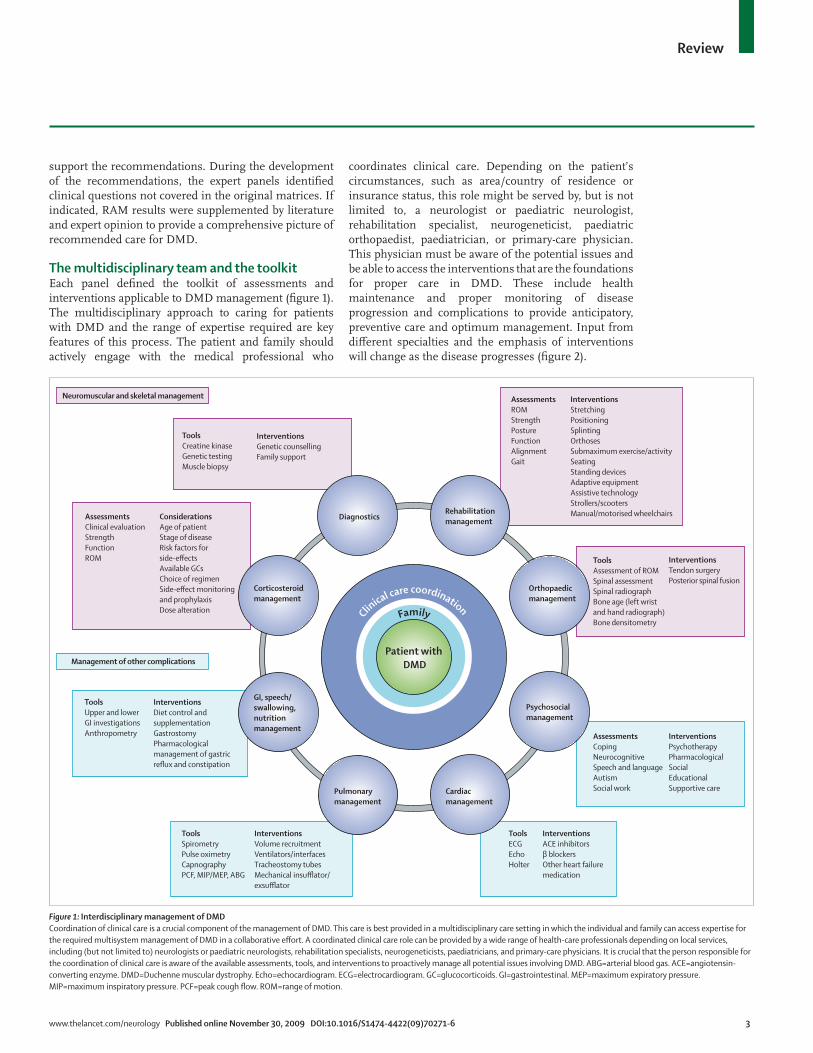
The multidisciplinary team and the toolkitEach panel defi ned the toolkit of assessments and interventions applicable to DMD management (fi gure 1). The multidisciplinary approach to caring for patients with DMD and the range of expertise required are key features of this process. The patient and family should actively engage with the medical professional who
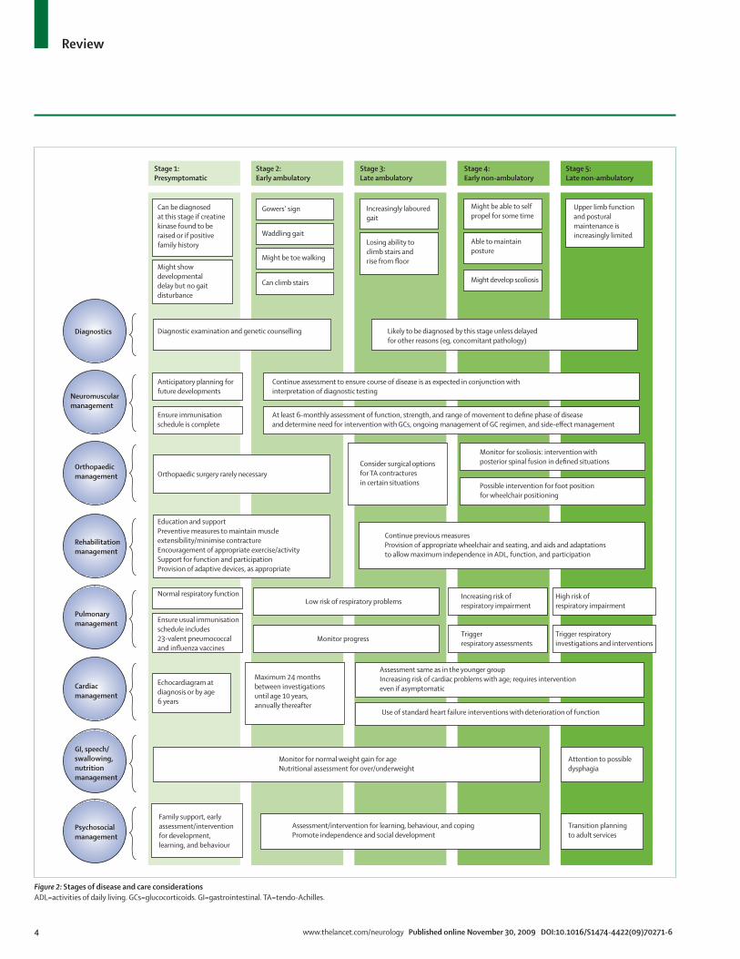
coordinates clinical care. Depending on the patient’s circumstances, such as area/country of residence or insurance status, this role might be served by, but is not limited to, a neurologist or paediatric neurologist, rehabilitation specialist, neurogeneticist, paediatric orthopaedist, paediatrician, or primary-care physician. This physician must be aware of the potential issues and be able to access the interventions that are the foundations for proper care in DMD. These include health maintenance and proper monitoring of disease progression and complications to provide anticipatory, preventive care and optimum management. Input from diff erent specialties and the emphasis of interventions will change as the disease progresses (fi gure 2).
InterventionsPsychotherapyPharmacologicalSocialEducationalSupportive care
Neuromuscular and skeletal management
Management of other complications
ToolsCreatine kinaseGenetic testingMuscle biopsy
AssessmentsClinical evaluationStrengthFunctionROM
InterventionsDiet control andsupplementationGastrostomyPharmacological management of gastric reflux and constipation
ToolsSpirometryPulse oximetryCapnographyPCF, MIP/MEP, ABG
InterventionsVolume recruitmentVentilators/interfacesTracheostomy tubesMechanical insufflator/exsufflator
ToolsECGEchoHolter
AssessmentsCopingNeurocognitiveSpeech and languageAutismSocial work
InterventionsACE inhibitorsβ blockersOther heart failuremedication
ConsiderationsAge of patientStage of diseaseRisk factors forside-effectsAvailable GCsChoice of regimenSide-effect monitoringand prophylaxisDose alteration
InterventionsGenetic counsellingFamily support
AssessmentsROMStrengthPostureFunctionAlignmentGait
ToolsAssessment of ROMSpinal assessmentSpinal radiographBone age (left wristand hand radiograph)Bone densitometry
InterventionsTendon surgeryPosterior spinal fusion
InterventionsStretchingPositioningSplintingOrthosesSubmaximum exercise/activitySeatingStanding devicesAdaptive equipmentAssistive technologyStrollers/scootersManual/motorised wheelchairs
ToolsUpper and lowerGI investigationsAnthropometry
Clinical care coordinationFamily
Patient with DMD
DiagnosticsRehabilitationmanagement
Orthopaedicmanagement
Psychosocialmanagement
Cardiacmanagement
Pulmonarymanagement
GI, speech/swallowing,nutritionmanagement
Corticosteroidmanagement
Figure 1: Interdisciplinary management of DMDCoordination of clinical care is a crucial component of the management of DMD. This care is best provided in a multidisciplinary care setting in which the individual and family can access expertise for the required multisystem management of DMD in a collaborative eff ort. A coordinated clinical care role can be provided by a wide range of health-care professionals depending on local services, including (but not limited to) neurologists or paediatric neurologists, rehabilitation specialists, neurogeneticists, paediatricians, and primary-care physicians. It is crucial that the person responsible for the coordination of clinical care is aware of the available assessments, tools, and interventions to proactively manage all potential issues involving DMD. ABG=arterial blood gas. ACE=angiotensin-converting enzyme. DMD=Duchenne muscular dystrophy. Echo=echocardiogram. ECG=electrocardiogram. GC=glucocorticoids. GI=gastrointestinal. MEP=maximum expiratory pressure. MIP=maximum inspiratory pressure. PCF=peak cough fl ow. ROM=range of motion.
4 www.thelancet.com/neurology Published online November 30, 2009 DOI:10.1016/S1474-4422(09)70271-6
Review
Diagnostics
Stage 1:Presymptomatic
Stage 2:Early ambulatory
Stage 3:Late ambulatory
Stage 4:Early non-ambulatory
Stage 5:Late non-ambulatory
Rehabilitationmanagement
Orthopaedicmanagement
Psychosocialmanagement
Cardiacmanagement
Pulmonarymanagement
GI, speech/swallowing,nutritionmanagement
Neuromuscularmanagement
Can be diagnosedat this stage if creatinekinase found to beraised or if positivefamily history
Upper limb functionand posturalmaintenance isincreasingly limited
Gowers’ sign Increasingly labouredgait
Might be able to selfpropel for some time
Able to maintainposture
Might develop scoliosis
Losing ability toclimb stairs andrise from floor
Waddling gait
Might be toe walking
Can climb stairs
Might showdevelopmentaldelay but no gaitdisturbance
Orthopaedic surgery rarely necessary
Family support, earlyassessment/interventionfor development,learning, and behaviour
Transition planningto adult services
Assessment/intervention for learning, behaviour, and copingPromote independence and social development
Monitor for normal weight gain for ageNutritional assessment for over/underweight
Attention to possibledysphagia
Echocardiagram atdiagnosis or by age6 years
Maximum 24 monthsbetween investigationsuntil age 10 years,annually thereafter
Assessment same as in the younger groupIncreasing risk of cardiac problems with age; requires intervention even if asymptomatic
Use of standard heart failure interventions with deterioration of function
Normal respiratory functionLow risk of respiratory problems
Increasing risk ofrespiratory impairment
Triggerrespiratory assessments
Trigger respiratoryinvestigations and interventions
High risk ofrespiratory impairment
Monitor progress
Ensure usual immunisationschedule includes23-valent pneumococcaland influenza vaccines
Education and supportPreventive measures to maintain muscleextensibility/minimise contractureEncouragement of appropriate exercise/activitySupport for function and participationProvision of adaptive devices, as appropriate
Continue previous measuresProvision of appropriate wheelchair and seating, and aids and adaptationsto allow maximum independence in ADL, function, and participation
Monitor for scoliosis: intervention withposterior spinal fusion in defined situations
Possible intervention for foot positionfor wheelchair positioning
Consider surgical options for TA contracturesin certain situations
Continue assessment to ensure course of disease is as expected in conjunction withinterpretation of diagnostic testing
Diagnostic examination and genetic counselling
Anticipatory planning forfuture developments
Ensure immunisationschedule is complete
At least 6-monthly assessment of function, strength, and range of movement to define phase of diseaseand determine need for intervention with GCs, ongoing management of GC regimen, and side-effect management
Likely to be diagnosed by this stage unless delayedfor other reasons (eg, concomitant pathology)
Figure 2: Stages of disease and care considerationsADL=activities of daily living. GCs=glucocorticoids. GI=gastrointestinal. TA=tendo-Achilles.
www.thelancet.com/neurology Published online November 30, 2009 DOI:10.1016/S1474-4422(09)70271-6 5
Review
At a practical level, management of the patient with DMD in the clinic requires a physically accessible environment and parking structure, with proper equipment (eg, mechanical hoist or sliding board) and trained personnel available for the safe transfer of the non-ambulatory patient. The expertise and means to obtain accurate measures of weight, height, and vital signs with appropriately trained staff are essential. Special weight scales that accommodate wheelchairs are available. Height measurements in patients with severe scoliosis are not accurate and can be replaced by arm-span measurements.
Diagnosis of DMDThe aim of care around diagnosis is to provide an accurate and prompt diagnosis, allowing initiation of appropriate interventions, continuing support and education, and minimising the length and impact of a potentially protracted diagnostic process. Diagnosis should be done by a neuromuscular specialist who can assess the child clinically and can rapidly access and interpret appropriate investigations in the context of the clinical presentation. Family follow-up and support after diagnosis will often
be augmented by support from geneticists and genetic counsellors.
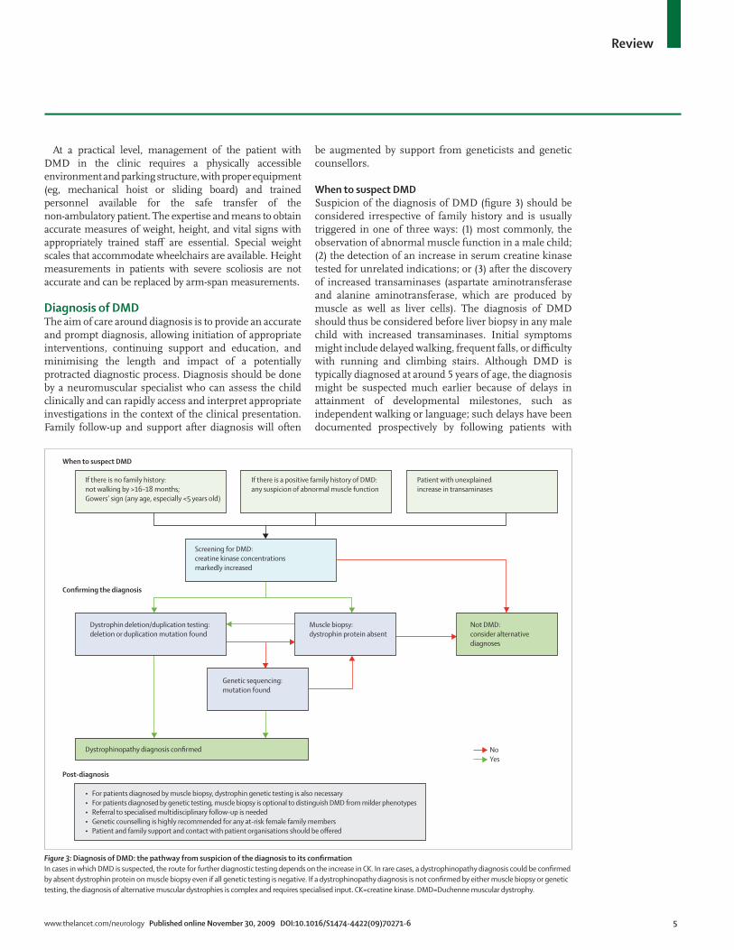
When to suspect DMDSuspicion of the diagnosis of DMD (fi gure 3) should be considered irrespective of family history and is usually triggered in one of three ways: (1) most commonly, the observation of abnormal muscle function in a male child; (2) the detection of an increase in serum creatine kinase tested for unrelated indications; or (3) after the discovery of increased transaminases (aspartate aminotransferase and alanine aminotransferase, which are produced by muscle as well as liver cells). The diagnosis of DMD should thus be considered before liver biopsy in any male child with increased transaminases. Initial symptoms might include delayed walking, frequent falls, or diffi culty with running and climbing stairs. Although DMD is typically diagnosed at around 5 years of age, the diagnosis might be suspected much earlier because of delays in attainment of developmental milestones, such as independent walking or language; such delays have been documented prospectively by following patients with
When to suspect DMD
Confirming the diagnosis
Dystrophinopathy diagnosis confirmed
• For patients diagnosed by muscle biopsy, dystrophin genetic testing is also necessary• For patients diagnosed by genetic testing, muscle biopsy is optional to distinguish DMD from milder phenotypes• Referral to specialised multidisciplinary follow-up is needed• Genetic counselling is highly recommended for any at-risk female family members• Patient and family support and contact with patient organisations should be offered
Muscle biopsy:dystrophin protein absent
Not DMD:consider alternativediagnoses
Dystrophin deletion/duplication testing:deletion or duplication mutation found
If there is no family history:not walking by >16–18 months;Gowers’ sign (any age, especially <5 years old)
Screening for DMD:creatine kinase concentrationsmarkedly increased
Genetic sequencing:mutation found
If there is a positive family history of DMD:any suspicion of abnormal muscle function
Patient with unexplainedincrease in transaminases
Post-diagnosis
NoYes
Figure 3: Diagnosis of DMD: the pathway from suspicion of the diagnosis to its confi rmationIn cases in which DMD is suspected, the route for further diagnostic testing depends on the increase in CK. In rare cases, a dystrophinopathy diagnosis could be confi rmed by absent dystrophin protein on muscle biopsy even if all genetic testing is negative. If a dystrophinopathy diagnosis is not confi rmed by either muscle biopsy or genetic testing, the diagnosis of alternative muscular dystrophies is complex and requires specialised input. CK=creatine kinase. DMD=Duchenne muscular dystrophy.
6 www.thelancet.com/neurology Published online November 30, 2009 DOI:10.1016/S1474-4422(09)70271-6
Review
DMD identifi ed by newborn screening.36 The presence of Gowers’ sign in a male child should trigger the diagnostic investigation of DMD, especially if the child also has a waddling gait. Toe walking might be present but is not additionally helpful in deciding whether to suspect DMD. In the presence of a positive family history of DMD, there should be a low threshold for testing creatine kinase, although this will be infl uenced by the age of the child. In a child less than 5 years of age, suspicion of DMD probably cannot be excluded completely by a normal muscle examination. However, with increasing age, a normal muscle examination renders the chance of a child having DMD progressively less likely. A boy older than 10 years of age with normal muscle function is thus highly unlikely to have DMD.
Confi rmation of the diagnosisThe route to confi rming the diagnosis (fi gure 3) depends on local availability of rapid and reliable testing, which must be interpreted alongside the clinical presentation owing to the range of severity possible with dystrophin mutations. Testing for a DMD mutation in a blood sample is always necessary even if DMD is fi rst con fi rmed by the absence of dystrophin protein expression on muscle biopsy. The results of genetic testing provide the clinical information required for genetic counselling, prenatal diagnosis, and consideration for future mutation-specifi c therapies. Diff erent types of mutations in DMD can be the genetic basis for DMD.12 The genetic tests commonly used to identify dystrophin mutations are multiplex PCR,37 multiplex ligation-dependent probe amplifi cation,38 single-condition amplifi cation/internal primer,39,40 and multiplex amplifi able probe hybridisation.40 Multiplex PCR is widely available and the least expensive, but only detects deletions and does not cover the whole gene, so that a deletion might not always be fully characterised. Multiplex ligation-dependent probe amplifi cation and amplifi able probe hybridisation will detect deletions and duplications and cover all exons, and single-condition amplifi cation/internal primer will detect deletions and provide sequence data. None of these techniques is universally available.
If analysis by one or more of these techniques leads to the identifi cation and full characterisation of a dystrophin mutation, then no further testing is required. If deletion/duplication testing is negative, then dystrophin gene sequencing should be done to look for point mutations or small deletions/insertions.39,40 Full characterisation of the mutation (deletion endpoints or exact position of any point mutation) is required to allow correlation of the predicted eff ect of the mutation on the reading frame of the gene, which is the major determinant of the phenotypic variability seen in dystrophinopathy,19,21,22 as well as to determine eligibility for the mutation-specifi c treatments currently in trials.41–43
A muscle biopsy could be done, depending on the clinical situation, availability of genetic testing, and the facilities in
the centre where the patient is seen.44 An open muscle biopsy is necessary if the diff erential diagnosis includes DMD among other diagnostic possibilities, such as other types of muscular dystrophy, so that adequate amounts of tissue will be available for further analysis. A needle biopsy might be appropriate if testing is only for DMD or if the clinician is skilled in taking multiple cores of tissue from paediatric patients.45,46 In those centres where it is done, the conchotome technique has the advantage of providing a larger sample than a single-core needle biopsy, and does not require an open surgical procedure.47,48
The key tests done on the muscle biopsy for DMD are immunocytochemistry and immunoblotting for dys-trophin, and should be interpreted by an experienced neuromuscular pathologist.7–9 A muscle biopsy can provide information on the amount and molecular size of dystrophin, as long as the protein is present.7–9,12,44 Diff erentiating total and partial absence of dystrophin can help to distinguish DMD from a milder dys-trophinopathy phenotype.7–9,12,44 Electron microscopy is not required to confi rm DMD.
Genetic testing after a positive biopsy diagnosis of DMD is mandatory. A muscle biopsy is not necessary if a genetic diagnosis is secured fi rst, particularly as some families might view the procedure as traumatic. However, if genetic testing has been done and no mutation identifi ed, but creatine kinase concentrations are increased and signs or symptoms consistent with DMD are present, then the next necessary diagnostic step is to do a muscle biopsy. This is also the case if there is a family history of DMD and a suspicion of the diagnosis, but no family mutation is known.
Whereas electromyography and nerve-conduction studies have been a traditional part of the assessment of a child with a suspected neuromuscular disorder, these tests are not believed by the expert panels to be now indicated or necessary for the specifi c assessment of DMD.
Neuromuscular and skeletal assessmentsClinical assessment in DMD includes taking a standard medical and family history and undertaking a physical examination, with a focus on the musculoskeletal system and related functional impairments. The neuromuscular specialist should be experienced in the expected disease course for DMD to understand the implications of a deviation from this course (eg, the possibility that a milder course might indicate a less severe dystrophinopathy or that more severe disease might suggest concomitant morbidity). This judgment will be informed by the results of regular assessments of disease progression (ie, strength, range of motion, posture, gait, timed testing),49 monitoring of ability to cope with activities of daily living, and application of motor function scales. These assessments, which are also used to inform decisions about therapeutic interventions and monitor response to therapy, are
www.thelancet.com/neurology Published online November 30, 2009 DOI:10.1016/S1474-4422(09)70271-6 7
Review
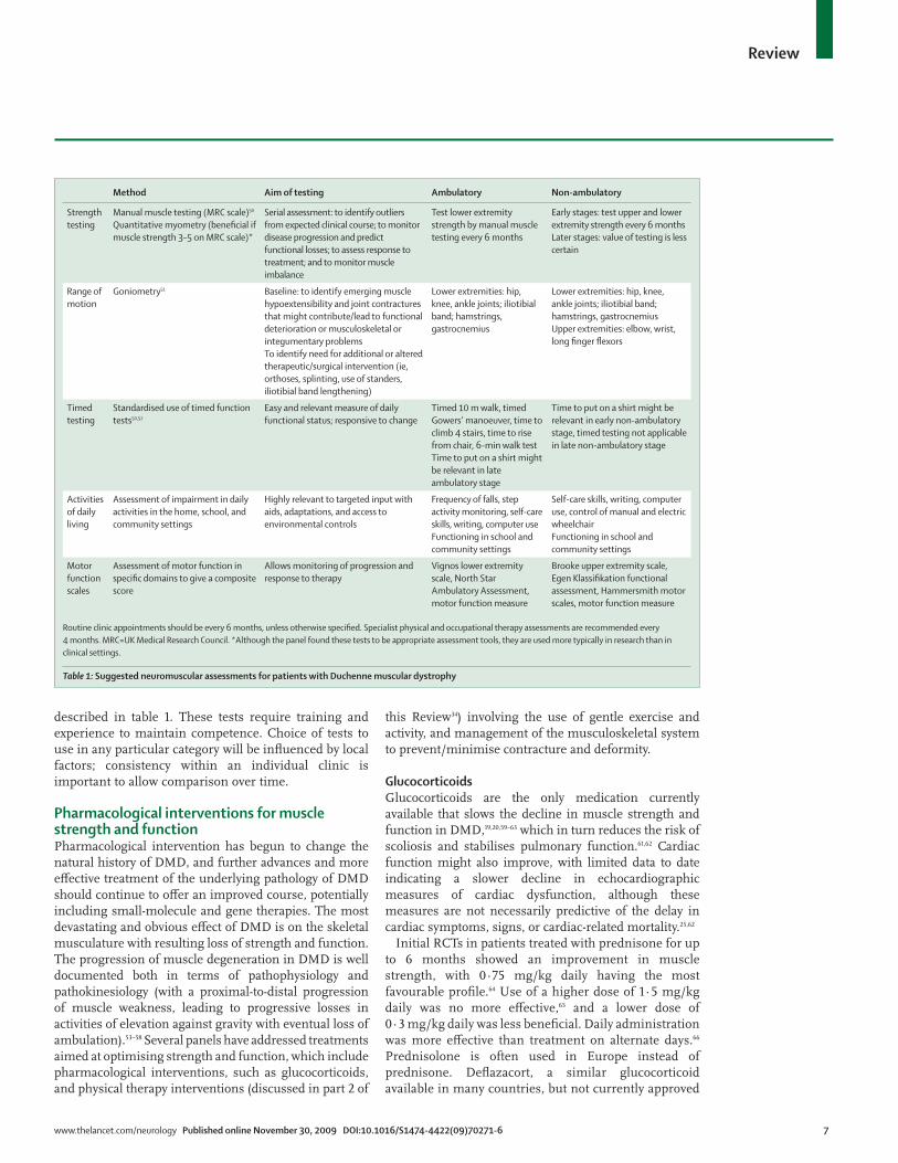
described in table 1. These tests require training and experience to maintain competence. Choice of tests to use in any particular category will be infl uenced by local factors; consistency within an individual clinic is important to allow comparison over time.
Pharmacological interventions for muscle strength and functionPharmacological intervention has begun to change the natural history of DMD, and further advances and more eff ective treatment of the underlying pathology of DMD should continue to off er an improved course, potentially including small-molecule and gene therapies. The most devastating and obvious eff ect of DMD is on the skeletal musculature with resulting loss of strength and function. The progression of muscle degeneration in DMD is well documented both in terms of pathophysiology and pathokinesiology (with a proximal-to-distal progression of muscle weakness, leading to progressive losses in activities of elevation against gravity with eventual loss of ambulation).53–58 Several panels have addressed treatments aimed at optimising strength and function, which include pharmacological interventions, such as glucocorticoids, and physical therapy interventions (discussed in part 2 of
this Review34) involving the use of gentle exercise and activity, and management of the musculoskeletal system to prevent/minimise contracture and deformity.
GlucocorticoidsGlucocorticoids are the only medication currently available that slows the decline in muscle strength and function in DMD,19,20,59–63 which in turn reduces the risk of scoliosis and stabilises pulmonary function.61,62 Cardiac function might also improve, with limited data to date indicating a slower decline in echocardiographic measures of cardiac dysfunction, although these measures are not necessarily predictive of the delay in cardiac symptoms, signs, or cardiac-related mortality.25,62
Initial RCTs in patients treated with prednisone for up to 6 months showed an improvement in muscle strength, with 0·75 mg/kg daily having the most favourable profi le.64 Use of a higher dose of 1·5 mg/kg daily was no more eff ective,65 and a lower dose of 0·3 mg/kg daily was less benefi cial. Daily administration was more eff ective than treatment on alternate days.66 Prednisolone is often used in Europe instead of prednisone. Defl azacort, a similar glucocorticoid available in many countries, but not currently approved
Method Aim of testing Ambulatory Non-ambulatory
Strength testing
Manual muscle testing (MRC scale)50
Quantitative myometry (benefi cial if muscle strength 3–5 on MRC scale)*
Serial assessment: to identify outliers from expected clinical course; to monitor disease progression and predict functional losses; to assess response to treatment; and to monitor muscle imbalance
Test lower extremity strength by manual muscle testing every 6 months
Early stages: test upper and lower extremity strength every 6 monthsLater stages: value of testing is less certain
Range of motion
Goniometry51 Baseline: to identify emerging muscle hypoextensibility and joint contractures that might contribute/lead to functional deterioration or musculoskeletal or integumentary problems To identify need for additional or altered therapeutic/surgical intervention (ie, orthoses, splinting, use of standers, iliotibial band lengthening)
Lower extremities: hip, knee, ankle joints; iliotibial band; hamstrings, gastrocnemius
Lower extremities: hip, knee, ankle joints; iliotibial band; hamstrings, gastrocnemius Upper extremities: elbow, wrist, long fi nger fl exors
Timed testing
Standardised use of timed function tests50,52
Easy and relevant measure of daily functional status; responsive to change
Timed 10 m walk, timed Gowers’ manoeuver, time to climb 4 stairs, time to rise from chair, 6-min walk testTime to put on a shirt might be relevant in late ambulatory stage
Time to put on a shirt might be relevant in early non-ambulatory stage, timed testing not applicable in late non-ambulatory stage
Activities of daily living
Assessment of impairment in daily activities in the home, school, and community settings
Highly relevant to targeted input with aids, adaptations, and access to environmental controls
Frequency of falls, step activity monitoring, self-care skills, writing, computer use Functioning in school and community settings
Self-care skills, writing, computer use, control of manual and electric wheelchairFunctioning in school and community settings
Motor function scales
Assessment of motor function in specifi c domains to give a composite score
Allows monitoring of progression and response to therapy
Vignos lower extremity scale, North Star Ambulatory Assessment, motor function measure
Brooke upper extremity scale, Egen Klassifi kation functional assessment, Hammersmith motor scales, motor function measure
Routine clinic appointments should be every 6 months, unless otherwise specifi ed. Specialist physical and occupational therapy assessments are recommended every 4 months. MRC=UK Medical Research Council. *Although the panel found these tests to be appropriate assessment tools, they are used more typically in research than in clinical settings.
Table 1: Suggested neuromuscular assessments for patients with Duchenne muscular dystrophy
8 www.thelancet.com/neurology Published online November 30, 2009 DOI:10.1016/S1474-4422(09)70271-6
Review
for use by the US Food and Drug Administration or the CDC in the USA, has been shown to have a similar effi cacy at a daily dose of 0·9 mg/kg and has a slightly diff erent chronic risk profi le.67,68
Subsequent longer term studies on the use of prednisone/prednisolone and defl azacort have focused more on their eff ect in prolonging ambulation than on the short-term improvement in strength (ie, decline in motor function still occurs, but more slowly).69,70 More recently, continued treatment after the patient becomes non-ambulatory has also shown reduction in the risk of progressive scoliosis and stabilisation of pulmonary function test variables.61,62
On the basis of this convincing literature, practice parameter guidelines, and personal experience, the panel strongly urges consideration of glucocorticoid therapy in all patients who have DMD.19,20 The rest of this section provides guidance on what clinical information is necessary to determine when to start glucocorticoid medication and how to monitor and manage side-eff ects.
The goal of the use of glucocorticoids in the ambulatory child is the preservation of ambulation and the mini-misation of later respiratory, cardiac, and orthopaedic complications, taking into account the well-described risks associated with chronic glucocorticoid admin-istration. If such issues are pre-existing, the risk of side-eff ects might be increased (table 2). Particular care needs to be taken with such patients in deciding which glucocorticoid to choose, when to initiate treatment, and how best to monitor the child for any problems. A high index of suspicion for steroid-related side-eff ects needs to be maintained at all times. Prevention and management of side-eff ects needs to be proactive.59 Families should be provided with a steroid card or similar notifi cation that the child is on steroids, listing emergency-care con-siderations in the setting of acute medical presentation, fracture, serious infection, need for surgery, or general anaesthesia, to alert any medical professional with whom the child might come into contact.
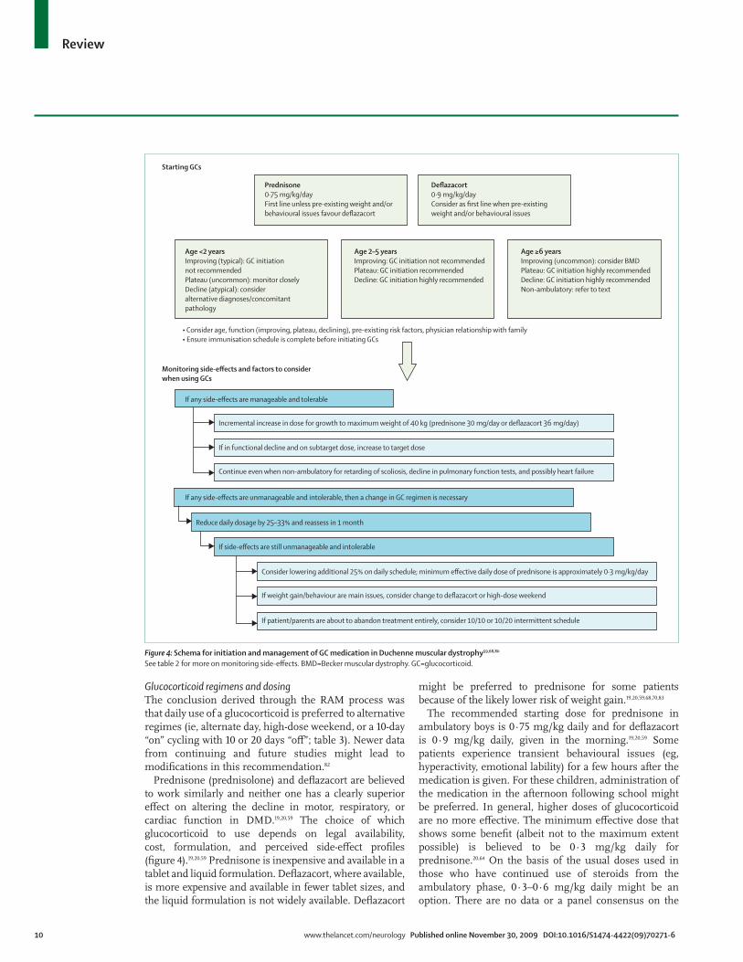
Initiation of glucocorticoid therapyNo generally accepted guidelines exist in the literature about the best time to initiate glucocorticoid therapy in an ambulatory boy with DMD. The panel’s opinion, derived through the RAM process, is that the timing of initiation of glucocorticoid therapy must be an individual decision, based on functional state and also considering age and pre-existing risk factors for adverse side-eff ects. Recognition of the three phases of motor function in DMD (making progress, plateau, and decline) helps the clinician to make this decision (fi gure 4). In all cases, the recommended national immunisation schedule should be complete and varicella immunity should be established before steroids are started.
Initiation of glucocorticoid treatment is not recommended for a child who is still gaining motor skills, especially when he is under 2 years of age. The
typical boy with DMD continues to make progress in motor skills until approximately age 4–6 years, albeit at a slower rate than his peers.81 The eventual use of glucocorticoids should be discussed with caregivers at this stage, in anticipation of the plateau in motor skills and subsequent decline. The plateau phase, which might last only a few months, can be identifi ed when there is no longer progress in motor skills, but prior to decline, as determined by history and timed testing (table 1). The child who takes longer in timed testing, loses a skill (such as climbing stairs), shows less endurance, or has more falls, is in a decline phase. Once the plateau phase has been clearly identifi ed, usually at age 4–8 years, the clinician should propose initiation of glucocorticoids unless there are substantial reasons (such as major pre-existing risk factors for side-eff ects) to wait until the decline phase. Starting steroids when in the full decline phase or when ambulation is more marginal is still recommended, but might be of more limited benefi t.
These recommendations for when to initiate glucocorticoid treatment should be interpreted as a minimum threshold. Some practitioners favour a more aggressive approach with earlier initiation of treatment when clinical symptoms fi rst appear, although there are no published data to support this, so the panel did not believe it appropriate to endorse earlier glucocorticoid treatment.
Because the decision to initiate glucocorticoids is based on serial assessment as well as parental report, additional care is required in initiating glucocorticoid therapy at an initial visit or at a second-opinion consultation. The assessment of the child’s course of motor function (making progress, plateau, and decline) is based purely on the caregiver’s history at a fi rst visit, so care should be exercised in making such conclusions in a child aged under 6 years. If glucocorticoids are initiated at a fi rst visit, we suggest that a physician be identifi ed at that time who will be in charge of monitoring the child, particularly if the physician making the recommendation cannot fulfi l this role.
Long-term use of glucocorticoids requires much commitment on the part of the family. Essential issues for discussions should include potential side-eff ects, the obligation to closely monitor and manage any adverse issues that might arise, and the requirement to have the child followed closely by their primary-care physician and specialty health-care team.
Use of glucocorticoids after loss of ambulationIn patients who have used glucocorticoids while ambulatory, many experts continue medication after loss of ambulation,62 with the goal of preserving upper limb strength, reducing progression of scoliosis, and delaying decline in respiratory and cardiac function.19,61,62
Indications for initiation of glucocorticoids in non-ambulatory patients are more relative than absolute. The eff ectiveness of glucocorticoid treatment in
www.thelancet.com/neurology Published online November 30, 2009 DOI:10.1016/S1474-4422(09)70271-6 9
Review
preventing scoliosis or in stabilising cardiac or respiratory function in this setting is not known; this issue thus warrants further study. However, limited data from trials suggest short-term stabilisation of pulmonary function in the early non-ambulatory patient.65 If the patient and caregiver request the initiation of steroids, daily dosing is indicated if there is a stable functional course. A daily dose is also appropriate in the presence of declining function. However, there is greater need in this group to consider the eff ect of pre-existing risk factors, such as
behavioural issues, fracture risk, or obesity; side-eff ects require close monitoring. Whether patients with more limited arm function and advanced pulmonary disease (such as those who already require nocturnal bi-level positive airway pressure assistance) can benefi t from glucocorticoid therapy is uncertain. The presence of an abnormal echocardiogram or symptoms of heart failure are not contraindications to glucocorticoid therapy, but use of glucocorticoids if advanced cardiomyopathy is present might carry higher risk of side-eff ects.
Recommended monitoring Intervention
Constitutional and cosmetic
Cushingoid features,19
obesity70,71
Particular vigilance needed if patient, parents, or siblings are obese Dietary advice to be reinforced before starting steroids; warn about increased appetite
Implement proactive dietary management for the entire family, not just the patient Consider change from prednisone to defl azacort Select an alternative regimen
Hirsutism19 Forewarn parents Does not usually occur to an extent that warrants a change in medication
Acne, tinea, warts More notable in teenagers Use ancillary treatment measures (topical prescription) and do not rush to change the GC regimen unless the boy is emotionally distressed
Growth retardation72,73 Monitor height at least every 6 months as part of general care (stature tends to be small in DMD even without steroid treatment61)
Consider endocrine evaluation if growth plateaus
Delayed puberty Monitor Tanner stage Identify any family history of delayed sexual maturation
Consider endocrine assessment if notably delayed or patient is upset by the delay
Adverse behavioural changes19,74–76
Identify any baseline mood, temperament, ADHD issues, and advise parents that these often transiently worsen in the initial 6 weeks on GC therapy
Decide whether baseline issues should be treated before starting GC therapy (eg, ADHD counselling or prescription) Consider changing timing of GC medication to later in the day Consider behavioural health referral
Immune/adrenal suppression77
Advise parents of risk of serious infection and need to promptly address minor infection Advise parents to inform all medical personnel that their child is on steroids and carry steroid alert card Ensure that the GC is not stopped abruptly
Obtain varicella immunisation before starting GC therapy; confi rm with protective serum titre Engage in tuberculosis surveillance Obtain infectious diseases consultation if serious infection occurs Substitute prednisone equivalent if defl azacort is temporarily unavailable Implement intravenous stress-dose hydrocortisone or methylprednisolone coverage for surgery or major illness (no accepted treatment strategy; anaesthesia or endocrine consultation recommended) Give intravenous coverage if nothing by mouth
Hypertension76 Monitor blood pressure as percentile for height and sex at each clinic visit
If blood pressure >99%, reduce salt intake, weight reduction If ineff ective, refer for possible ACE inhibitor or β blocker medication
Glucose intolerance Urine dipstick for glucose at clinic visits Enquire about polyuria, polydipsia
If urine is glucose-positive, then try fasting or post-prandial blood glucose, and if abnormal, then seek an endocrine consultation
GERD Enquire about GERD symptoms (heartburn) Advise parents to report symptoms
Avoid NSAIDs Prescribe ranitidine or proton-pump inhibitor and antacid if symptomatic
Peptic ulcer disease78 Advise parents of risk and to report symptoms History of gastritis, GERD, abdominal pain, or faecal bloodTest stool for blood if anaemic or suggestive history
Avoid NSAIDs Prescribe ranitidine or proton-pump inhibitor and antacid if symptomatic Seek gastrointestinal consultation
Cataracts Annual ophthalmological examination Consider switching from defl azacort to prednisone if cataracts evolve that aff ect vision Seek ophthalmology consultation
Bone demineralisation and increased fracture risk*76,79
Take careful fracture history Annual DEXA to monitor bone density Annual monitoring of 25-hydroxy vitamin D blood concentration (ideally late winter in seasonal climates) and supplement with vitamin D3 if level is <32 nmol/L Dietitian should assess calcium and vitamin D intake
For 25-hydroxy vitamin D concentration 20–31 nmol/L, give 1000 IU orally twice daily, for <20 nmol/L, give 2000 IU orally twice daily Recheck serum 25-hydroxy vitamin D concentration again after 3 months on therapy Encourage weight-bearing activities Take multivitamin supplements with vitamin D3 Consider bisphosphonates, such as pamidronate
Myoglobinuria80 Enquire about abnormal coloration of urine after exercise, urine testing
Advise avoidance of excessive eccentric (eg, descending stairs, squatting down, trampolining) and resistive exercise Commence renal investigations if persistent
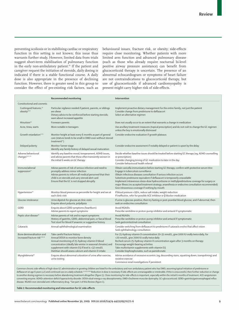
Common chronic side-eff ects of high-dose GC administration in growing children are listed for the ambulatory and non-ambulatory patient who has DMD, assuming typical initiation of prednisone or defl azacort at age 6 years (±2) and continued use on a daily schedule.19,20,59,78,80 Reduction in dose is necessary if side-eff ects are unmanageable or intolerable. If this is unsuccessful, then further reduction or change to another dosing regimen is necessary before abandoning treatment altogether (fi gure 5). Close monitoring for side-eff ects is important, especially within the initial 6 months of treatment. ACE=angiotensin converting enzyme. ADHD=attention-defi cit hyperactivity disorder. DEXA=dual-energy x-ray absorptiometry. DMD=Duchenne muscular dystrophy. GC=glucocorticoid. GERD=gastritis/gastroesophageal refl ux disease. NSAID=non-steroidal anti-infl ammatory drug. *See part 2 of this Review (fi gure 1).
Table 2: Recommended monitoring and intervention for GC side-eff ects
10 www.thelancet.com/neurology Published online November 30, 2009 DOI:10.1016/S1474-4422(09)70271-6
Review
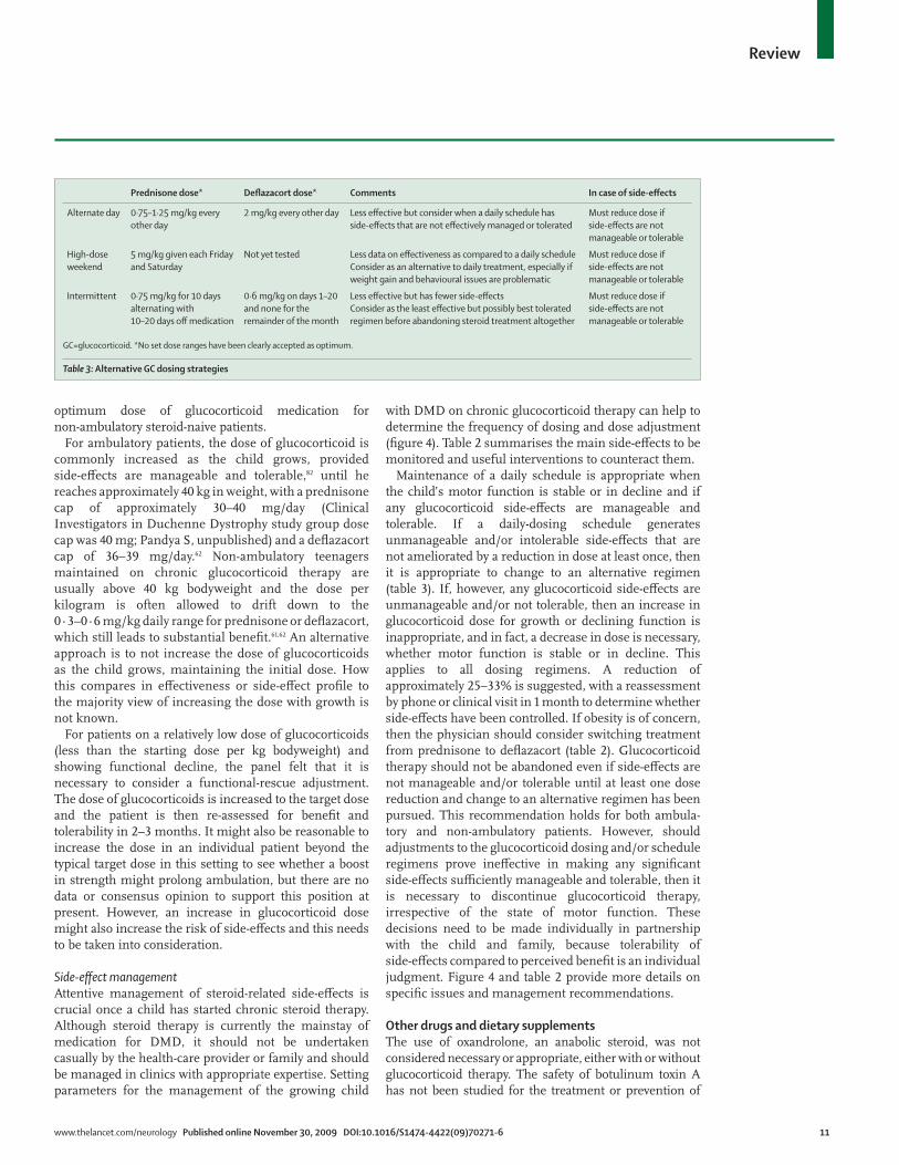
Glucocorticoid regimens and dosingThe conclusion derived through the RAM process was that daily use of a glucocorticoid is preferred to alternative regimes (ie, alternate day, high-dose weekend, or a 10-day “on” cycling with 10 or 20 days “off ”; table 3). Newer data from continuing and future studies might lead to modifi cations in this recommendation.82
Prednisone (prednisolone) and defl azacort are believed to work similarly and neither one has a clearly superior eff ect on altering the decline in motor, respiratory, or cardiac function in DMD.19,20,59 The choice of which glucocorticoid to use depends on legal availability, cost, formulation, and perceived side-eff ect profi les (fi gure 4).19,20,59 Prednisone is inexpensive and available in a tablet and liquid formulation. Defl azacort, where available, is more expensive and available in fewer tablet sizes, and the liquid formulation is not widely available. Defl azacort
might be preferred to prednisone for some patients because of the likely lower risk of weight gain.19,20,59,68,70,83
The recommended starting dose for prednisone in ambulatory boys is 0·75 mg/kg daily and for defl azacort is 0·9 mg/kg daily, given in the morning.19,20,59 Some patients experience transient behavioural issues (eg, hyperactivity, emotional lability) for a few hours after the medication is given. For these children, administration of the medication in the afternoon following school might be preferred. In general, higher doses of glucocorticoid are no more eff ective. The minimum eff ective dose that shows some benefi t (albeit not to the maximum extent possible) is believed to be 0·3 mg/kg daily for prednisone.20,64 On the basis of the usual doses used in those who have continued use of steroids from the ambulatory phase, 0·3–0·6 mg/kg daily might be an option. There are no data or a panel consensus on the
Starting GCs
Monitoring side-effects and factors to consider when using GCs
Prednisone0·75 mg/kg/dayFirst line unless pre-existing weight and/orbehavioural issues favour deflazacort
Deflazacort0·9 mg/kg/dayConsider as first line when pre-existingweight and/or behavioural issues
Age <2 yearsImproving (typical): GC initiationnot recommendedPlateau (uncommon): monitor closelyDecline (atypical): consideralternative diagnoses/concomitantpathology
Age 2–5 yearsImproving: GC initiation not recommendedPlateau: GC initiation recommendedDecline: GC initiation highly recommended
Age ≥6 yearsImproving (uncommon): consider BMDPlateau: GC initiation highly recommendedDecline: GC initiation highly recommendedNon-ambulatory: refer to text
• Consider age, function (improving, plateau, declining), pre-existing risk factors, physician relationship with family• Ensure immunisation schedule is complete before initiating GCs
If any side-effects are manageable and tolerable
Incremental increase in dose for growth to maximum weight of 40 kg (prednisone 30 mg/day or deflazacort 36 mg/day)
If in functional decline and on subtarget dose, increase to target dose
Continue even when non-ambulatory for retarding of scoliosis, decline in pulmonary function tests, and possibly heart failure
If any side-effects are unmanageable and intolerable, then a change in GC regimen is necessary
Reduce daily dosage by 25–33% and reassess in 1 month
If side-effects are still unmanageable and intolerable
Consider lowering additional 25% on daily schedule; minimum effective daily dose of prednisone is approximately 0·3 mg/kg/day
If weight gain/behaviour are main issues, consider change to deflazacort or high-dose weekend
If patient/parents are about to abandon treatment entirely, consider 10/10 or 10/20 intermittent schedule
Figure 4: Schema for initiation and management of GC medication in Duchenne muscular dystrophy59,68,80
See table 2 for more on monitoring side-eff ects. BMD=Becker muscular dystrophy. GC=glucocorticoid.
www.thelancet.com/neurology Published online November 30, 2009 DOI:10.1016/S1474-4422(09)70271-6 11
Review
optimum dose of glucocorticoid medication for non-ambulatory steroid-naive patients.
For ambulatory patients, the dose of glucocorticoid is commonly increased as the child grows, provided side-eff ects are manageable and tolerable,82 until he reaches approximately 40 kg in weight, with a prednisone cap of approximately 30–40 mg/day (Clinical Investigators in Duchenne Dystrophy study group dose cap was 40 mg; Pandya S, unpublished) and a defl azacort cap of 36–39 mg/day.62 Non-ambulatory teenagers maintained on chronic glucocorticoid therapy are usually above 40 kg bodyweight and the dose per kilogram is often allowed to drift down to the 0·3–0·6 mg/kg daily range for prednisone or defl azacort, which still leads to substantial benefi t.61,62 An alternative approach is to not increase the dose of glucocorticoids as the child grows, maintaining the initial dose. How this compares in eff ectiveness or side-eff ect profi le to the majority view of increasing the dose with growth is not known.
For patients on a relatively low dose of glucocorticoids (less than the starting dose per kg bodyweight) and showing functional decline, the panel felt that it is necessary to consider a functional-rescue adjustment. The dose of glucocorticoids is increased to the target dose and the patient is then re-assessed for benefi t and tolerability in 2–3 months. It might also be reasonable to increase the dose in an individual patient beyond the typical target dose in this setting to see whether a boost in strength might prolong ambulation, but there are no data or consensus opinion to support this position at present. However, an increase in glucocorticoid dose might also increase the risk of side-eff ects and this needs to be taken into consideration.
Side-eff ect managementAttentive management of steroid-related side-eff ects is crucial once a child has started chronic steroid therapy. Although steroid therapy is currently the mainstay of medication for DMD, it should not be undertaken casually by the health-care provider or family and should be managed in clinics with appropriate expertise. Setting parameters for the management of the growing child
with DMD on chronic glucocorticoid therapy can help to determine the frequency of dosing and dose adjustment (fi gure 4). Table 2 summarises the main side-eff ects to be monitored and useful interventions to counteract them.
Maintenance of a daily schedule is appropriate when the child’s motor function is stable or in decline and if any glucocorticoid side-eff ects are manageable and tolerable. If a daily-dosing schedule generates unmanageable and/or intolerable side-eff ects that are not ameliorated by a reduction in dose at least once, then it is appropriate to change to an alternative regimen (table 3). If, however, any glucocorticoid side-eff ects are unmanageable and/or not tolerable, then an increase in glucocorticoid dose for growth or declining function is inappropriate, and in fact, a decrease in dose is necessary, whether motor function is stable or in decline. This applies to all dosing regimens. A reduction of approximately 25–33% is suggested, with a reassessment by phone or clinical visit in 1 month to determine whether side-eff ects have been controlled. If obesity is of concern, then the physician should consider switching treatment from prednisone to defl azacort (table 2). Glucocorticoid therapy should not be abandoned even if side-eff ects are not manageable and/or tolerable until at least one dose reduction and change to an alternative regimen has been pursued. This recommendation holds for both ambula-tory and non-ambulatory patients. However, should adjustments to the glucocorticoid dosing and/or schedule regimens prove ineff ective in making any signifi cant side-eff ects suffi ciently manageable and tolerable, then it is necessary to discontinue glucocorticoid therapy, irrespective of the state of motor function. These decisions need to be made individually in partnership with the child and family, because tolerability of side-eff ects compared to perceived benefi t is an individual judgment. Figure 4 and table 2 provide more details on specifi c issues and management recommendations.
Other drugs and dietary supplementsThe use of oxandrolone, an anabolic steroid, was not considered necessary or appropriate, either with or without glucocorticoid therapy. The safety of botulinum toxin A has not been studied for the treatment or prevention of
Prednisone dose* Defl azacort dose* Comments In case of side-eff ects
Alternate day 0·75–1·25 mg/kg every other day
2 mg/kg every other day Less eff ective but consider when a daily schedule has side-eff ects that are not eff ectively managed or tolerated
Must reduce dose if side-eff ects are not manageable or tolerable
High-dose weekend
5 mg/kg given each Friday and Saturday
Not yet tested Less data on eff ectiveness as compared to a daily scheduleConsider as an alternative to daily treatment, especially if weight gain and behavioural issues are problematic
Must reduce dose if side-eff ects are not manageable or tolerable
Intermittent 0·75 mg/kg for 10 days alternating with 10–20 days off medication
0·6 mg/kg on days 1–20 and none for the remainder of the month
Less eff ective but has fewer side-eff ectsConsider as the least eff ective but possibly best tolerated regimen before abandoning steroid treatment altogether
Must reduce dose if side-eff ects are not manageable or tolerable
GC=glucocorticoid. *No set dose ranges have been clearly accepted as optimum.
Table 3: Alternative GC dosing strategies
12 www.thelancet.com/neurology Published online November 30, 2009 DOI:10.1016/S1474-4422(09)70271-6
Review
contractures in individuals with DMD and is thought to be inappropriate. No recommendations for the use of creatine were established. An RCT of creatine in DMD failed to show a clear benefi t.84 If a patient is taking creatine and has evidence of renal dysfunction, it is necessary to discontinue this supplement.
Supplements, such as coenzyme Q10, carnitine, aminoacids (glutamine, arginine), anti-infl ammatories/anti-oxidants (fi sh oil, vitamin E, green-tea extract), and others, are being used by some parents and are endorsed by some practitioners. In the absence of supportive data from the literature or expert opinion consensus from these panels, we make no recommendations for the use of supplements. The expert panels also did not rate the value of potential disease-modifying drugs, such as pentoxifylline or various herbal or botanical agents. This was identifi ed as an area for which additional research is needed. Active
involvement of families in activities that help with the advancement of knowledge about DMD, such as patient registries and clinical trials, was encouraged.
Psychosocial managementThe medical care of a patient who has DMD and his family is not complete without support for their psychosocial wellbeing.85,86 For many parents, the stress caused by the psychosocial problems of their child exceeds the stress associated with the physical aspects of the disease.87 Needs vary with the age of the patient and stage of disease (fi gure 2), but several general statements are valid.
DMD is a multilevel/multisystem disease. Biological factors (including the lack of dystrophin and/or its isoforms and the subsequent eff ect on brain development and functioning),88 social and emotional factors, and treatment factors (eg, glucocorticoids) can all play a part in psychosocial health.5 Although most psychosocial issues are not unique to DMD, patients with DMD are at increased risk for problems in these areas. The psychosocial diffi culties that are observed in DMD should be treated with the same eff ective, evidence-based interventions that are used in the general population,89 with a strong emphasis on prevention and early intervention, because this will maximise potential outcome.
In general, psychosocial adjustment of boys with DMD is similar to that for other chronic medical conditions.90 However, some specifi c areas of risk are of particular concern. Diffi culties in social functioning might be due to biologically based defi cits in specifi c cognitive skills, such as social reciprocity, social judgment, perspective taking, and aff ective discrimination, whereas the consequences of DMD (ie, physical limitations) might result in social isolation, social withdrawal, and reduced access to social activities. The pattern of speech and language defi cits, including those in language development, short-term verbal memory, and phonological processing, as well as cognitive delays, including impaired intelligence and specifi c learning disorders, are well documented.91–94 There is also increased risk for neurobehavioural and neuro-developmental disorders, including autism spectrum disorders, attention-defi cit hyperactivity disorder, and obsessive-compulsive disorder.95 Problems might be encountered with emotional adjustment and depression.5 Anxiety might also be an issue and can be exacerbated by cognitive defi cits in mental fl exibility and adaptability (ie, overly-rigid thought processes). Similarly, defi cits in mental fl exibility and emotional regulation can result in oppositional/argumentative behaviour and explosive temper problems. Increased rates of depression in parents of children who have DMD underscore the need for assessment and support of the entire family.96
AssessmentsCrucial times to consider assessments include the time around diagnosis (for some families, a 6–12-month window will be needed for some assessments to allow for
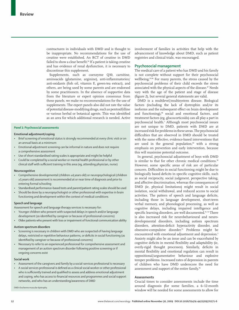
Panel 1: Psychosocial assessments
Emotional adjustment/coping• Brief screening of emotional status is strongly recommended at every clinic visit or on
an annual basis at a minimum• Emotional adjustment screening can be informal in nature and does not require
a comprehensive assessment• Use of short standardised rating scales is appropriate and might be helpful• Could be completed by a social worker or mental health professional or by other
clinical staff with suffi cient training in this area (eg, attending physician, nurse)
Neurocognitive• Comprehensive developmental (children ≤4 years old) or neuropsychological (children
≥5 years old) assessment is recommended at or near time of diagnosis and prior to entering formal schooling
• Standardised performance-based tests and parent/patient rating scales should be used• Should be done by a neuropsychologist or other professional with expertise in brain
functioning and development within the context of medical conditions
Speech and languageAssessment for speech and language therapy services is necessary for: • Younger children who present with suspected delays in speech and/or language
development (as identifi ed by caregiver or because of professional concerns)• Older patients who present with loss or impairment of functional communication ability
Autism spectrum disorders• Screening is necessary in children with DMD who are suspected of having language
delays, restricted or repetitive behaviour patterns, or defi cits in social functioning (as identifi ed by caregiver or because of professional concerns)
• Necessary to refer to an experienced professional for comprehensive assessment and management of an autism spectrum disorder following positive screening or if ongoing concerns exist
Social work • Assessment of the caregivers and family by a social-services professional is necessary• A social services professional is defi ned as a clinical social worker or other professional
who is suffi ciently trained and qualifi ed to assess and address emotional adjustment and coping, who has access to fi nancial resources and programmes and social support networks, and who has an understanding/awareness of DMD
DMD=Duchenne muscular dystrophy.
www.thelancet.com/neurology Published online November 30, 2009 DOI:10.1016/S1474-4422(09)70271-6 13
Review
adjustment after diagnosis), before entering school, and after a change in function. Although not every clinic will have direct access to all assessments and interventions listed (panels 1 and 2), we hope that these recommendations can serve as a guide to fi lling gaps in clinical staff and directing referrals, where appropriate. Assessments are targeted at the areas of emotional adjustment and coping, neurocognitive functioning, speech and language development, the possible presence of autism spectrum disorders, and social support. Routine screening of psychosocial wellbeing in the patient, parents, and siblings is necessary.
InterventionsInterventions will depend on the individual, but should be available to meet a broad spectrum of needs. Of crucial importance to patient/family psychosocial health is the designation of a care coordinator who can serve as a point of contact for families and who has suffi cient knowledge and background in neuromuscular disorders to be able to meet the family’s information needs.86 Proactive intervention to help families and patients avoid the social problems and social isolation that occur in the context of DMD is necessary (panel 2).
Development of an individual education plan for all children with DMD in collaboration with their parents and schools is necessary to address potential learning problems. In addition, this will help with modifi cation of activities that might otherwise prove harmful to the child’s muscles (eg, physical education) or might lead to reduced energy/fatigue (eg, walking long distances to and from lunch) or safety (eg, playground activities) and accessibility issues. Promoting patient independence and involvement in decision making (ie, as it relates to their medical care) is also necessary.
Psychopharmacological interventions should be considered for the treatment of moderate to severe psychiatric symptoms as part of a multimodal treatment plan that includes appropriate psychotherapies and educational interventions. Standard prescribing practices and guidelines apply, with additional considerations focused on the patient’s cardiac status and drug interactions and side-eff ects when combined with other medications (eg, weight gain and glucocorticoids), and the patient’s general medical condition. Close monitoring with systematic, routine follow-up is highly recommended, including consultation with the appropriate specialist if concerns arise.
Palliative care is appropriate to relieve or prevent suff ering and to improve quality of life in patients who have DMD, as needed. In addition to pain management, palliative care teams might also be able to provide emotional and spiritual support, assist families in clarifying treatment goals and making diffi cult medical decisions, facilitate communication between families and medical teams, and address issues related to grief, loss, and bereavement.
ConclusionsThe recommendations presented in the two parts of this Review represent the outcome of an international collaboration of clinical experts working to inform optimum care for DMD. Because of a paucity of data from RCTs for DMD (a common situation in rare disorders), a well-established method was chosen to generate statements about the appropriateness or
Panel 2: Psychosocial interventions
Psychotherapy• Parental management training: recommended for externalising behaviours (eg,
noncompliance/disruptive behaviour and parent–child confl ict)• Individual therapy: recommended for internalising behaviours (eg, low self-esteem
and depression, anxiety, and obsessive-compulsive disorder, adjustment and coping diffi culties)
• Group therapy: recommended for social skills defi cits• Family therapy: recommended for adjustment and coping diffi culties and parent–child
confl ict• Applied behaviour analysis: recommended for specifi c behaviours related to autism
Pharmacological interventions• Selective serotonin re-uptake inhibitors for depression, anxiety, obsessive-compulsive
disorder• Mood stabilisers for aggression, anger/emotional dysregulation• Stimulants for attention-defi cit hyperactivity disorder
Social interaction interventions • Increasing DMD awareness and knowledge among school personnel• Peer education about DMD• Social skills training (as needed to address defi cits in this area)• Modifi ed/adapted sports, summer camps, and youth groups/programmes• Art groups, equestrian, and aqua therapies, use of service dogs, nature programmes,
and internet/chat rooms, among others• Promoting patient independence and self-advocacy
Educational interventions• Neuropsychological assessment at diagnosis and before entering school• Individualised education programme on entering school• Measures to address defi cits as they are identifi ed
Care/support interventions• Care coordinator: serves as a point of contact for the family to meet family
information needs, schedule and coordinate appointments, and facilitate communication with clinicians, etc; should be a professional with a suffi cient level of training regarding clinical care for DMD
• Home health-care services: should be used if a patient’s health is at risk because suffi cient care cannot be provided in their current setting or circumstances; might also be appropriate in other situations when the current care providers cannot suffi ciently meet the patient’s care needs
• Transition planning: encouraging self-advocacy in medical care, facilitating transfer to a new medical care team, and developing educational and vocational opportunities
• Palliative care: appropriate for pain management, as needed; emotional and spiritual support; and guidance for treatment and medical decisions
• Hospice care: necessary for end-stage patients
DMD=Duchenne muscular dystrophy.
14 www.thelancet.com/neurology Published online November 30, 2009 DOI:10.1016/S1474-4422(09)70271-6
Review
inappropriateness and necessity of clinical interventions. RAM off ers several benefi ts compared with other consensus-based methods, including evidence-based generation of the questions to be addressed, independent appraisal of the options followed by group discussion, and several rounds of iteration.35 A further advantage was the systematic evaluation of distinct clinical scenarios, mimicking as much as possible the clinical decision-making process in all its complexity.
This fi rst part of the care recommendations generated by use of this method emphasises the overall ethos of multidisciplinary care for DMD and goes on to discuss the detail of diagnosis, pharmacological, and psychosocial management. Precise genetic diagnosis is now the gold standard for diagnosis of DMD, and here we recommend that it should be actively sought in all cases. The future possibility of mutation-specifi c therapies (currently in phase 1 and 2 clinical trials) adds a further urgency to the need for this kind of technology to be universally available.41 As genetic technologies change, in particular with the development of high-throughput diagnostics, this algorithm should become more straightforward.
The pharmacological mainstay of neuromuscular management in DMD is the use of glucocorticoids. Data from RCTs support their use,19 although treatment regimens are highly variable across diff erent countries and diff erent clinics. Further trials of glucocorticoids and management of their side-eff ects are likely to augment our knowledge of their optimum use.60,82 In the meantime, these guidelines provide a framework for glucocorticoid use that allows greater consistency—a point that is of importance not only for current patient care, but in the context of the planning of multicentre trials of other novel therapies, which are allowing the baseline use of steroids as part of the standard of care.
Despite many studies reporting that both behavioural and learning issues are important for patients with DMD and their families, few publications have provided pragmatic guidelines on psychosocial care in this condition. Providing support for these kinds of issues is
frequently a challenge within a medically orientated care structure, but these recommendations clearly put this element of care at the centre of management, with an emphasis on anticipatory interventions, and suggest that measurement of impact on these areas will be a signifi cant challenge as the fi eld moves towards clinical trials.
In the second part of this Review,34 the discussion focuses on the role of rehabilitation, cardiovascular, gastroenterology/nutrition, orthopaedic/surgical, and respiratory specialties in DMD, so that together the two Reviews can provide a comprehensive and current guide to management in this condition.ContributorsAll authors provided intellectual expertise in the study design, generation and interpretation of data, writing of the Review, and the decision to publish. KB, aided by RF, drafted and edited the Review, and approved the fi nal version. DJB, LEC, LC, SP, and CC were involved in the literature search.
DMD Care Considerations Working Group (CCWG) steering committeeT Abresch, C McDonald (University of California, Davis, CA, USA); L E Case (Duke University, Durham, NC, USA); D Atkins, K Siegel (US Agency for Healthcare Research and Quality, Rockville, MD, USA); L Cripe, B Wong (Cincinnati Children’s Hospital Medical Center, Cincinnati, OH, USA); V Cwik (Muscular Dystrophy Association, Tucson, AZ, USA); J Finder (Children’s Hospital of Pittsburgh, Pittsburgh, PA, USA); P Furlong (Parent Project Muscular Dystrophy, Fort Lee, NJ, USA); A Kenneson, A Vatave, C Constantin (CDC National Center on Birth Defects and Developmental Disabilities, Atlanta, GA, USA); S Pandya (University of Rochester, Rochester, NY, USA); J Porter (National Institute of Neurological Disorders and Stroke, US National Institutes of Health, Bethesda, MD, USA); M Sussman (Shriner’s Hospital for Children, Portland, OR, USA).
DMD-CCWG publication committeeK Bushby (managing editor; Newcastle University, Newcastle upon Tyne, UK), all expert panel chairs (see below), and the following members of the steering committee: T Abresch, C Constantin, V Cwik, J Finder, P Furlong, J Porter, K Siegel, M Sussman, B Wong.
DMD-CCWG expert panel membersCardiovascular management—L Cripe (chair); J Towbin (Cincinnati Children’s Hospital Medical Center, Cincinnati, OH, USA); J Bourke (Newcastle University, UK); D Connuck (Janet Weis Children’s Hospital, Danville, PA, USA); E Goldmuntz (Children’s Hospital of Philadelphia, Philadelphia, PA, USA); L Markham (Vanderbilt University, Nashville, TN, USA); K D Mathews (University of Iowa Children’s Hospital, Iowa City, IA, USA); E McNally (University of Chicago, Chicago, IL, USA); R Moxley (University of Rochester, Rochester, NY, USA); R Williams (University of Utah, Salt Lake City, UT, USA).Diagnostics—P R Clemens (chair; University of Pittsburgh and the Department of Veteran Aff airs Medical Center, Pittsburgh, PA, USA); A M Connolly (Washington University School of Medicine, St Louis, MO, USA); C Cunniff (University of Arizona College of Medicine, Tucson/Phoenix, AZ, USA); K Dent, K Flanigan (University of Utah, Salt Lake City, UT, USA); E Hoff man (Children’s National Medical Center, Washington, DC, USA); S Iannaccone (University of Texas Southwestern Medical Center, Dallas, TX, USA); N Johnson (Johns Hopkins School of Medicine, Baltimore, MD, USA); T Miller (University of Arizona Health Sciences Center, Tucson/Phoenix, AZ, USA); T Sejersen (Karolinska Institute, Stockholm, Sweden).Gastrointestinal and nutritional management—A Kaul (co-chair); J Tomezsko (co-chair [retired]; Children’s Hospital of Philadelphia, Philadelphia, PA, USA); S Casey (Seattle Children’s Hospital, Seattle, WA, USA); N Goemans (University Hospital Leuven, Leuven, Belgium); A Gulyas, K Swan (University of Medicine and Dentistry of New Jersey, Newark, NJ, USA); K Larson (Gillette Children’s Specialty Healthcare, St Paul, MN, USA); H Lipner (Hackensack University Medical Center, Hackensack, NJ, USA); M Mascarenhas (Children’s Hospital of
Search strategy and selection criteria
Peer-reviewed literature was searched using the key search terms of “Duchenne” or “muscular dystrophy”, or both, paired with one of 410 other search terms related to a comprehensive list of assessment tools and interventions used in DMD management. The full list of search terms is available on request. The databases used included Medline, Embase, Web of Science, and the Cochrane Library. Initial inclusion criteria consisted of available abstracts of human studies published in English between 1986 and 2006. Each working group also incorporated major articles from its discipline published before 1986 and from 2007 to mid-2009 in the process of discussions, fi nal assessments, and write-up of recommendations.
www.thelancet.com/neurology Published online November 30, 2009 DOI:10.1016/S1474-4422(09)70271-6 15
Review
Philadelphia, Philadelphia, PA, USA); K Rao (Cincinnati Children’s Hospital Medical Center, Cincinnati, OH, USA).Neuromuscular management—R Finkel (chair); C Bonnemann (Children’s Hospital of Philadelphia, Philadelphia, PA, USA); D Biggar (Bloorview Kids Rehab, Toronto, Canada); K Bushby (Newcastle University, Newcastle upon Tyne, UK); D Escolar (formerly with Children’s National Medical Center, Washington, DC, USA); E W Massey (Duke University School of Medicine, Durham, NC, USA); T Miller (University of Arizona Health Sciences Center, Tucson/Phoenix, AZ, USA); J Pascual (University of Texas Southwestern Medical Center, Dallas, TX, USA); J Sladky (Emory University, Atlanta, GA, USA); K Wagner (Johns Hopkins Hospital, Baltimore, MD, USA); B Wong (Cincinnati Children’s Hospital Medical Center, Cincinnati, OH, USA).Orthopaedic and surgical management—F Shapiro (chair; Children’s Hospital Boston, Boston, MA, USA); B Alman (Hospital for Sick Children, Toronto, Canada); J Bach, A Patel (University of Medicine and Dentistry of New Jersey, Newark, NJ, USA); D J Birnkrant (MetroHealth Medical Center/Case Western Reserve University, Cleveland, OH, USA); T Do, N Weidner (Cincinnati Children’s Hospital Medical Center, Cincinnati, OH, USA); W Mackenzie (Jeff erson Medical College, Thomas Jeff erson University, Philadelphia, PA, USA); C McDonald (University of California, Davis, CA, USA); H Saraste (Karolinska Institute, Stockholm, Sweden); M Sussman (Shriner’s Hospital for Children, Portland, OR, USA); E Vroom (Algra and Vroom Orthodontics Practice, Purmerend, Netherlands).Psychosocial management—K Kinnett (co-chair); J Poysky (co-chair), S Moreland (Baylor College of Medicine, Houston, TX, USA); S Cotton (University of Melbourne, Melbourne, Australia); J Hendriksen (Maastricht University Hospital, Maastricht, Netherlands); V Hinton (Columbia University, New York, NY, USA); J Kiefel (Children’s Healthcare of Atlanta, Atlanta, GA, USA); K McGuire (Muscular Dystrophy Association, Tucson, AZ, USA); M Ritzo (Children’s National Medical Center, Washington, DC, USA); C Trout (University of Iowa Children’s Hospital, Iowa City, IA, USA); N Weidner (Cincinnati Children’s Hospital Medical Center, Cincinnati, OH, USA).Rehabilitation management—L E Case (co-chair; Duke University, Durham, NC, USA); C McDonald (co-chair; University of California, Davis, CA, USA); S Pandya (co-chair; University of Rochester, Rochester, NY, USA); G Carter (University of Washington School of Medicine, Seattle, WA, USA); C Cronin, A Gulyas (University of Medicine and Dentistry of New Jersey, Newark, NJ, USA); M Eagle (Newcastle University, Newcastle upon Tyne, UK); J Engel (University of Washington, Seattle, WA, USA); S Evans (Children’s National Medical Center, Washington, DC, USA); H Lipner (Hackensack University Medical Center, Hackensack, NJ, USA); D Matthews (University of Colorado School of Medicine, Denver, CO, USA); L Morrison (University of New Mexico School of Medicine, Albuquerque, NM, USA); H Posselt (Montrose Access, Corinda, QLD, Australia); P Ryan, J Smith (New York Presbyterian Hospital, New York, NY, USA); N Strauss (Columbia University Medical Center, New York, NY, USA); C Trout (University of Iowa Children’s Hospital, Iowa City, IA, USA); A Wright (Carolinas Neuromuscular/ALS-MDA Center, Charlotte, NC, USA).Respiratory management—D J Birnkrant (chair; MetroHealth Medical Center, Case Western Reserve University, Cleveland, OH, USA); R Amin (Cincinnati Children’s Hospital Medical Center, Cincinnati, OH, USA); J Bach (University of Medicine and Dentistry of New Jersey, Newark, NJ, USA); J Benditt (University of Washington School of Medicine, Seattle, WA, USA); M Eagle (Newcastle University, Newcastle upon Tyne, UK); J Finder (Children’s Hospital of Pittsburgh, Pittsburgh, PA, USA); J T Kissel (Ohio State University Medical Center, Columbus, OH, USA); A Koumbourlis (Children’s National Medical Center, Albert Einstein College of Medicine, Bronx, NY, USA); R M Kravitz (Duke University, Durham, NC, USA).
Confl icts of interestKB is a consultant for Acceleron, AVI, Debiopharm, Prosensa, and Santhera. LEC has received honoraria from Genzyme Corporation, has participated in research supported by Genzyme Corporation, PTC Therapeutics, the Leal Foundation, and Families of Spinal Muscular Atrophy, has been awarded grant support from the National Skeletal Muscle Research Center, and is a member of the Pompe Registry Board of Advisors. All other authors have no confl icts of interest.
AcknowledgmentsWe thank P Eubanks, A Kenneson, A Vatave (CDC); A Cyrus and E Levy (Oak Ridge Institute for Science and Education); B Bradshaw, H Desai, P Haskell, E Hunt, A Marsden, C Muse, and L Yuson (Booz/Allen/Hamilton); and B Tseng (Massachusetts General Hospital, Harvard Medical School) for their contributions to this study and manuscript. We also thank the following organisations for their collaboration on this study: Muscular Dystrophy Association, National Institute on Disability and Rehabilitation Research, Parent Project Muscular Dystrophy, and TREAT-NMD (EC036825). In addition, we thank M Levine, M Mascarenhas, and M Thayu (Children’s Hospital of Philadelphia) for their participation in discussions about bone health; and G Carter, J Engel (University of Washington), H Posselt (Montrose Access, Australia), and C Trout (University of Iowa Children’s Hospital) for their advice on pain management. The CDC provided support for the project through funding, study design, collection, analysis, and interpretation of data and manuscript preparation. The fi ndings and conclusions in this report are those of the authors and do not necessarily represent the offi cial position of the CDC.
References1 Drousiotou A, Ioannou P, Georgiou T, et al. Neonatal screening for
Duchenne muscular dystrophy: a novel semiquantitative application of the bioluminescence test for creatine kinase in a pilot national program in Cyprus. Genet Test 1998; 2: 55–60.
2 Bradley D, Parsons E. Newborn screening for Duchenne muscular dystrophy. Semin Neonatol 1998; 3: 27–34.
3 Emery AE. Population frequencies of inherited neuromuscular diseases—a world survey. Neuromuscul Disord 1991; 1: 19–29.
4 Bushby KM, Hill A, Steele JG. Failure of early diagnosis in symptomatic Duchenne muscular dystrophy. Lancet 1999; 353: 55–78.
5 Poysky J, Behavior in DMD Study Group. Behavior patterns in Duchenne muscular dystrophy: report on the Parent Project Muscular Dystrophy behavior workshop 8–9 of December 2006, Philadelphia, USA. Neuromuscul Disord 2007; 17: 986–94.
6 Hoff man EP, Brown RH Jr, Kunkel LM. Dystrophin: the protein product of the Duchenne muscular dystrophy locus. Cell 1987; 51: 919–28.
7 Nicholson LV, Johnson MA, Bushby KM, Gardner-Medwin D, Curtis A, Ginjaar IB. Integrated study of 100 patients with Xp21 linked muscular dystrophy using clinical, genetic, immunochemical, and histopathological data. Part 1. Trends across the clinical groups. J Med Genet 1993; 30: 728–36.
8 Nicholson LV, Johnson MA, Bushby KM, Gardner-Medwin D, Curtis A, Ginjaar IB. Integrated study of 100 patients with Xp21 linked muscular dystrophy using clinical, genetic, immunochemical, and histopathological data. Part 2. Correlations within individual patients. J Med Genet 1993; 30: 737–44.
9 Nicholson LV, Johnson MA, Bushby KM, Gardner-Medwin D, Curtis A, Ginjaar IB. Integrated study of 100 patients with Xp21 linked muscular dystrophy using clinical, genetic, immunochemical, and histopathological data. Part 3. Diff erential diagnosis and prognosis. J Med Genet 1993; 30: 745–51.
10 Towbin, JA. The role of cytoskeletal proteins in cardiomyopathies. Curr Opin Cell Biol 1998; 10: 131–39.
11 Towbin, JA, Bowles, NE. Molecular genetics of left ventricular dysfunction. Curr Mol Med 2001; 1: 81–90.
12 Muntoni F, Torelli S, Ferlini A. Dystrophin and mutations: one gene, several proteins, multiple phenotypes. Lancet Neurol 2003; 2: 731–40.
13 Bushby KM, Goodship JA, Nicholson LV, et al. Variability in clinical, genetic and protein abnormalities in manifesting carriers of Duchenne and Becker muscular dystrophy. Neuromuscul Disord 1993; 3: 57–64.
14 Richards CS, Watkins SC, Hoff man EP, et al. Skewed X inactivation in a female MZ twin results in Duchenne muscular dystrophy. Am J Hum Genet 1990; 46: 672–81.
15 Hoff man EP, Arahata K, Minetti C, Bonilla E, Rowland LP. Dystrophinopathy in isolated cases of myopathy in females. Neurology 1992; 42: 967–75.
16 Koenig M, Hoff man EP, Bertelson CJ, Monaco AP, Feener C, Kunkel LM. Complete cloning of the Duchenne muscular dystrophy (DMD) cDNA and preliminary genomic organization of the DMD gene in normal and aff ected individuals. Cell 1987; 50: 509–17.
16 www.thelancet.com/neurology Published online November 30, 2009 DOI:10.1016/S1474-4422(09)70271-6
Review
17 Kunkel LM, Monaco AP, Hoff man E, Koenig M, Feener C, Bertelson C. Molecular studies of progressive muscular dystrophy (Duchenne). Enzyme 1987; 38: 72–75.
18 Muntoni F, Wells D. Genetic treatments in muscular dystrophies. Curr Opin Neurol 2007; 20: 590–94.
19 Manzur AY, Kuntzer T, Pike M, Swan A. Glucocorticoid corticosteroids for Duchenne muscular dystrophy. Cochrane Database Syst Rev 2008; 1: CD003725.
20 Moxley RT 3rd, Ashwal S, Pandya S, et al. Practice parameter: corticosteroid treatment of Duchenne dystrophy: report of the Quality Standards Subcommittee of the American Academy of Neurology and the Practice Committee of the Child Neurology Society. Neurology 2005; 64: 13–20.
21 Jeppesen J, Green A, Steff ensen BF, Rahbek J. The Duchenne muscular dystrophy population in Denmark, 1977–2001: prevalence, incidence and survival in relation to the introduction of ventilator use. Neuromuscul Disord 2003; 13: 804–12.
22 Yasuma F, Konagaya M, Sakai M, Kuru S, Kawamura T. A new lease on life for patients with Duchenne muscular dystrophy in Japan. Am J Med 2004; 117: 363.
23 Eagle M, Bourke J, Bullock R, et al. Managing Duchenne muscular dystrophy—the additive eff ect of spinal surgery and home nocturnal ventilation in improving survival. Neuromuscul Disord 2007; 17: 470–75.
24 Eagle M. Report on the muscular dystrophy campaign workshop: exercise in neuromuscular diseases Newcastle, January 2002. Neuromuscul Disord 2002; 12: 975–83.
25 Markham LW, Kinnett K, Wong BL, Woodrow Benson D, Cripe LH. Corticosteroid treatment retards development of ventricular dysfunction in Duchenne muscular dystrophy. Neuromuscul Disord 2008; 18: 365–70.
26 American Academy of Pediatrics Section on Cardiology and Cardiac Surgery. Cardiovascular health supervision for individuals aff ected by Duchenne or Becker muscular dystrophy. Pediatrics 2005; 116: 1569–73.
27 Bushby K, Muntoni F, Bourke JP. 107th ENMC International Workshop: the management of cardiac involvement in muscular dystrophy and myotonic dystrophy. 7th–9th June 2002, Naarden, the Netherlands. Neuromuscul Disord 2003; 13: 166–72.
28 Duboc D, Meune C, Lerebours G, Devaux JY, Vaksmann G, Bécane HM. Eff ect of perindopril on the onset and progression of left ventricular dysfunction in Duchenne muscular dystrophy. J Am Coll Cardiol 2005; 45: 855–57.
29 Brooke MH, Fenichel GM, Griggs RC, Mendell JR, Moxley RT, CIDD Group. Duchenne muscular dystrophy: patterns of clinical progression and eff ects of supportive therapy. Neurology 1989; 39: 475–81.
30 Vignos PJ, Wagner MB, Karlinchak B, Katirji B. Evaluation of a program for long-term treatment of Duchenne muscular dystrophy. Experience at the University Hospitals of Cleveland. J Bone Joint Surg Am 1996; 78: 1844–52.
31 Rahbek J, Werge B, Madsen A, Marquardt J, Steff ensen BF, Jeppesen J. Adult life with Duchenne muscular dystrophy: observations among an emerging and unforeseen patient population. Pediatr Rehabil 2005; 8: 17–28.
32 Tatara K, Shinno S. Management of mechanical ventilation and prognosis in duchenne muscular dystrophy. IRYO Jap J Natl Med Serv 2008; 62: 566–71.
33 Parent Project Muscular Dystrophy. MD-CARE Act. http://www.parentprojectmd.org/site/PageServer?pagename=ending_speakup_mdcareact (accessed Oct 7, 2009).
34 Bushby K, Finkel R, Birnkrant DJ, et al. Diagnosis and management of Duchenne muscular dystrophy, part 2: implemenetation of multidisciplinary care. Lancet Neurol 2009; published online Nov 30. DOI:10.1016/S1474-4422(09)70272-8.
35 Fitch K, Bernstein SJ, Aguilar MS, et al. The RAND/UCLA appropriateness method user’s manual. Santa Monica, CA: RAND Corporation, 2001.
36 Parsons, EP, Clark AJ, Bradley DM. Developmental progress in Duchenne muscular dystrophy: lessons for earlier detection. Eur J Paediatr Neurol 2004; 8: 145–53.
37 Prior TW, Bridgeman SJ. Experience and strategy for the molecular testing of Duchenne muscular dystrophy. J Mol Diagn 2005; 7: 317–26.
38 Lalic T, Vossen RH, Coff a J, et al. Deletion and duplication screening in the DMD gene using MLPA. Eur J Hum Genet 2005; 13: 1231–34.
39 Flanigan KM, von Niederhausern A, Dunn DM, Alder J, Mendell JR, Weiss RB. Rapid direct sequence analysis of the dystrophin gene. Am J Hum Genet 2003; 72: 931–39.
40 Dent KM, Dunn DM, von Niederhausern AC, et al. Improved molecular diagnosis of dystrophinopathies in an unselected clinical cohort. Am J Med Genet A 2005; 134: 295–98.
41 Lim LE, Rando TA. Technology insight: therapy for Duchenne muscular dystrophy—an opportunity for personalized medicine? Nat Clin Pract Neurol 2008; 4: 149–58.
42 van Ommen GJ, van Deutekom J, Aartsma-Rus A. The therapeutic potential of antisense-mediated exon skipping. Curr Opin Mol Ther 2008; 10: 140–49.
43 van Deutekom JC, Janson AA, Ginjaar IB, et al. Local dystrophin restoration with antisense oligonucleotide PRO051. N Engl J Med 2007; 357: 2677–86.
44 Muntoni F. Is a muscle biopsy in Duchenne dystrophy really necessary? Neurology 2001; 57: 574–75.
45 Lacomis D. The use of percutaneous needle muscle biopsy in the diagnosis of myopathy. Curr Rheumatol Rep 2000; 2: 225–29.
46 Lacomis D. The utility of muscle biopsy. Curr Neurol Neurosci Rep 2004; 4: 81–86.
47 Henriksson KG. Semi-open muscle biopsy technique. A simple outpatient procedure. Acta Neurol Scand 1979; 59: 317–23.
48 Dorph C, Nennesmo I, Lundberg IE. Percutaneous conchotome muscle biopsy. A useful diagnostic and assessment tool. J Rheumatol 2001; 28: 1591–99.
49 Beenakker EA, Maurits NM, Fock JM, Brouwer OF, van der Hoeven JH. Functional ability and muscle force in healthy children and ambulant Duchenne muscular dystrophy patients. Eur J Paediatr Neurol 2005; 9: 387–93.
50 Florence JM, Pandya S, King WM, et al. Clinical trials in Duchenne dystrophy. Standardization and reliability of evaluation procedures. Phys Ther 1984; 64: 41–45.
51 Pandya S, Florence JM, King WM, Robinson J, Signore LC, CIDD Group. Reliability of goniometric measurements in patients with Duchenne muscular dystrophy. Phys Ther 1985; 65: 1339–42.
52 Brooke MH, Griggs RC, Mendell JR, Fenichel GM, Shumate JB, Pellegrino RJ. Clinical trial in Duchenne dystrophy. 1. The design of the protocol. Muscle Nerve 1981; 4: 186–97.
53 Gorospe JR, Hoff man EP. Duchenne muscular dystrophy. Curr Opin Rheumatol 1992; 4: 794–800.
54 Matsumura K, Ohlendieck K, Ionasescu VV, et al. The role of the dystrophin-glycoprotein complex in the molecular pathogenesis of muscular dystrophies. Neuromuscul Disord 1993; 3: 533–35.
55 Johnson EW, Walter J. Zeiter Lecture: pathokinesiology of Duchenne muscular dystrophy: implications for management. Arch Phys Med Rehabil 1977; 58: 4–7.
56 McDonald CM, Abresch RT, Carter GT, et al. Profi les of neuromuscular diseases. Duchenne muscular dystrophy. Am J Phys Med Rehabil 1995; 74 (suppl): S70–92.
57 Siegel IM. Pathomechanics of stance in Duchenne muscular dystrophy. Arch Phys Med Rehabil 1972; 53: 403–06.
58 Sutherland DH, Olshen R, Cooper L, et al. The pathomechanics of gait in Duchenne muscular dystrophy. Dev Med Child Neurol 1981; 23: 3–22.
59 Angelini C. The role of corticosteroids in muscular dystrophy: a critical appraisal. Muscle Nerve 2007; 36: 424–35.
60 Bushby K, Muntoni F, Urtizberea A, Hughes R, Griggs R. Report on the 124th ENMC International Workshop. Treatment of Duchenne muscular dystrophy; defi ning the gold standards of management in the use of corticosteroids. 2–4 April 2004, Naarden, The Netherlands. Neuromuscul Disord 2004; 14: 526–34.
61 King WM, Ruttencutter R, Nagaraja HN, et al. Orthopedic outcomes of long-term daily corticosteroid treatment in Duchenne muscular dystrophy. Neurology 2007; 68: 1607–13.
62 Biggar WD, Harris VA, Eliasoph L, Alman B. Long-term benefi ts of defl azacort treatment for boys with Duchenne muscular dystrophy in their second decade. Neuromuscul Disord 2006; 16: 249–55.
63 Houde S, Filiatrault M, Fournier A, et al. Defl azacort use in Duchenne muscular dystrophy: an 8-year follow-up. Pediatr Neurol 2008; 38: 200–06.
www.thelancet.com/neurology Published online November 30, 2009 DOI:10.1016/S1474-4422(09)70271-6 17
Review
64 Griggs RC, Moxley RT 3rd, Mendell JR, et al. Prednisone in Duchenne dystrophy. A randomized, controlled trial defi ning the time course and dose response. Arch Neurol 1991; 48: 383–88.
65 Mendell JR, Moxley, RT, Griggs RC, et al. Randomized, double-blind six-month trial of prednisone in Duchenne’s muscular dystrophy. N Engl J Med 1989; 320: 1592–97.
66 Fenichel GM, Mendell JR, Moxley RT, et al. A comparison of daily and alternate-day prednisone therapy in the treatment of Duchenne muscular dystrophy. Arch Neurol 1991; 48: 575–79.
67 Mesa LE, Dubrovsky AL, Corderi J, Marco P, Flores D. Steroids in Duchenne muscular dystrophy—defl azacort trial. Neuromuscul Disord 1991; 1: 261–66.
68 Biggar WD, Politano L, Harris VA, et al. Defl azacort in Duchenne muscular dystrophy: a comparison of two diff erent protocols. Neuromuscul Disord 2004; 14: 476–82.
69 Fenichel GM, Florence JM, Pestronk A, et al. Long-term benefi t from prednisone therapy in Duchenne muscular dystrophy. Neurology 1991; 41: 1874–77.
70 Bonifati MD, Ruzza G, Bonometto P, et al. A multicenter, double-blind, randomized trial of defl azacort versus prednisone in Duchenne muscular dystrophy. Muscle Nerve 2000; 23: 1344–47.
71 American Academy of Pediatrics Committee, Kleinman RE, eds. Pediatric nutrition handbook (6th edn). Elk Grove Village, IL: American Academy of Pediatrics Press, 2009.
72 Daley-Yates PT, Richards DH. Relationship between systemic corticosteroid exposure and growth velocity: development and validation of a pharmacokinetic/pharmacodynamic model. Clin Ther 2004; 26: 1905–19.
73 Foster BJ, Shults J, Zemel BS, Leonard MB. Interactions between growth and body composition in children treated with high-dose chronic glucocorticoids. Am J Clin Nutr 2004; 80: 1334–41.
74 Stuart FA, Segal TY, Keady S. Adverse psychological eff ects of corticosteroids in children and adolescents. Arch Dis Child 2005; 90: 500–06.
75 Bender BG, Ikle DN, DuHamel T, Tinkelman D. Neuropsychological and behavioral changes in asthmatic children treated with beclomethasone dipropionate versus theophylline. Pediatrics 1998; 101: 355–60.
76 Balaban B, Matthews DJ, Clayton GH, Carry T. Corticosteroid treatment and functional improvement in Duchenne muscular dystrophy: long-term eff ect. Am J Phys Med Rehabil 2005; 84: 843–50.
77 Mortimer KJ, Tata LJ, Smith CJ, et al. Oral and inhaled corticosteroids and adrenal insuffi ciency: a case-control study. Thorax 2006; 61: 405–08.
78 Brunton LL, Lazo JS, Parker KL, eds. Goodman and Gilman’s the pharmacological basis of therapeutics (11th edn). New York: McGraw-Hill Medical, 2005.
79 Brown JJ, Zacharin MR. Proposals for prevention and management of steroid-induced osteoporosis in children and adolescents. J Paediatr Child Health 2005; 41: 553–57.
80 Garrood P, Eagle M, Jardine PE, Bushby K, Straub V. Myoglobinuria in boys with Duchenne muscular dystrophy on corticosteroid therapy. Neuromuscul Disord 2008; 18: 71–73.
81 Brooke MH, Fenichel GM, Griggs RC, et al. Clinical investigation in Duchenne dystrophy: 2. Determination of the power of therapeutic trials based on the natural history. Muscle Nerve 1983; 6: 91–103.
82 Bushby K, Griggs R, MSG/ENMC FOR DMD trial study group. 145th ENMC International Workshop: planning for an international trial of steroid dosage regimes in DMD (FOR DMD), 22–24th October 2006, Naarden, The Netherlands. Neuromuscul Disord 2007; 17: 423–28.
83 Biggar WD, Gingras M, Fehlings DL, Harris VA, Steele CA. Defl azacort treatment of Duchenne muscular dystrophy. J Pediatr 2001; 138: 45–50.
84 Escolar DM, Buyse G, Henricson E, et al. CINRG randomized controlled trial of creatine and glutamine in Duchenne muscular dystrophy. Ann Neurol 2005; 58: 151–55.
85 Cyrulnik SE, Fee RJ, Batchelder A, Kiefel J, Goldstein E, Hinton VJ. Cognitive and adaptive defi cits in young children with Duchenne muscular dystrophy (DMD). J Int Neuropsychol Soc 2008; 14: 853–61.
86 Green JM, Murton FE. Diagnosis of Duchenne muscular dystrophy: parents’ experiences and satisfaction. Child Care Health Dev 1996; 22: 113–28.
87 Nereo NE, Fee RJ, Hinton VJ. Parental stress in mothers of boys with Duchenne muscular dystrophy. J Pediatr Psychol 2003; 28: 473–84.
88 Anderson JL, Head SI, Rae C, Morley JW. Brain function in Duchenne muscular dystrophy. Brain 2002; 125: 4–13.
89 Kazdin AE, Weisz JR. Evidence-based psychotherapies for children and adolescents. New York: Guilford Press, 2003.
90 Hendriksen JG, Poysky JT, Schrans DG, Schouten EG, Aldenkamp AP, Vles JS. Psychosocial adjustment in males with Duchenne muscular dystrophy: psychometric properties and clinical utility of a parent-report questionnaire. J Pediatr Psychol 2009; 34: 69–78.
91 Hinton VJ, De Vivo DC, Nereo NE, Goldstein E, Stern Y. Selective defi cits in verbal working memory associated with a known genetic etiology: the neuropsychological profi le of Duchenne muscular dystrophy. J Int Neuropsychol Soc 2001; 7: 45–54.
92 Cyrulnik SE, Fee RJ, De Vivo DC, Goldstein E, Hinton VJ. Delayed developmental language milestones in children with Duchenne’s muscular dystrophy. J Pediatr 2007; 150: 474–78.
93 Cotton SM, Voudouris NJ, Greenwood KM. Association between intellectual functioning and age in children and young adults with Duchenne muscular dystrophy: further results from a meta-analysis. Dev Med Child Neurol 2005; 47: 257–65.
94 Hendriksen JG, Vles JS. Are males with Duchenne muscular dystrophy at risk for reading disabilities? Pediatr Neurol 2006; 34: 296–300.
95 Hendriksen JG, Vles JS. Neuropsychiatric disorders in males with duchenne muscular dystrophy: frequency rate of attention-defi cit hyperactivity disorder (ADHD), autism spectrum disorder, and obsessive-compulsive disorder. J Child Neurol 2008; 23: 477–81.
96 Abi Daoud MS, Dooley JM, Gordon KE. Depression in parents of children with Duchenne muscular dystrophy. Pediatr Neurol 2004; 31: 16–19.
www.thelancet.com/neurology Vol 9 February 2010 177
Review
Diagnosis and management of Duchenne muscular dystrophy, part 2: implementation of multidisciplinary careKatharine Bushby, Richard Finkel, David J Birnkrant, Laura E Case, Paula R Clemens, Linda Cripe, Ajay Kaul, Kathi Kinnett, Craig McDonald, Shree Pandya, James Poysky, Frederic Shapiro, Jean Tomezsko, Carolyn Constantin, for the DMD Care Considerations Working Group*
Optimum management of Duchenne muscular dystrophy (DMD) requires a multidisciplinary approach that focuses on anticipatory and preventive measures as well as active interventions to address the primary and secondary aspects of the disorder. Implementing comprehensive management strategies can favourably alter the natural history of the disease and improve function, quality of life, and longevity. Standardised care can also facilitate planning for multicentre trials and help with the identification of areas in which care can be improved. Here, we present a comprehensive set of DMD care recommendations for management of rehabilitation, orthopaedic, respiratory, cardiovascular, gastroenterology/nutrition, and pain issues, as well as general surgical and emergency-room precautions. Together with part 1 of this Review, which focuses on diagnosis, pharmacological treatment, and psychosocial care, these recommendations allow diagnosis and management to occur in a coordinated multidisciplinary fashion.
IntroductionIn part 1 of this Review, the importance of multidisciplin-ary care was underscored in the context of diagnosis and pharmacological and psychosocial management of Duchenne muscular dystrophy (DMD), emphasising that no one aspect of the care of this disease can be taken in isolation.1 This model of care emphasises the value of multidisciplinary involvement to anticipate early changes in many systems and to manage the wide spectrum of complications that can be predicted in DMD. We applied this model of care to the patient and family across the different stages of the disease. The optimum delivery of care by rehabilitation, cardiovascular, gastroenterology/nutrition, orthopaedic/surgical, and respiratory specialties is presented in this second part of the Review. As before, the RAND Corporation–University of California Los Angeles Appropriateness Method was used2 (full details of the methods are described in part 1 of this Review1).
Management of muscle extensibility and joint contracturesDecreased muscle extensibility and joint contractures in DMD occur as a result of various factors, including loss of ability to actively move a joint through its full range of motion, static positioning in a position of flexion, muscle imbalance about a joint, and fibrotic changes in muscle tissue.3–8 The maintenance of good ranges of movement and bilateral symmetry are important to allow optimum movement and functional positioning, to maintain ambulation, prevent development of fixed deformities, and maintain skin integrity.9–14
The management of joint contractures requires input from neuromuscular specialists, physical therapists, rehabilitation physicians, and orthopaedic surgeons.15,16 Programmes to prevent contractures are usually mon-itored and implemented by a physical therapist and tailored to individual needs, stage of the disease, response to therapy, and tolerance. Local care needs to be augmented by guidance from a specialist every 4 months.
Physical therapy interventionsStretching and positioningEffective stretching of the musculotendinous unit requires a combination of interventions, including active stretching, active-assisted stretching, passive stretching, and prolonged elongation using positioning, splinting, orthoses, and standing devices.9,10,12,17–20 As standing and walking become more difficult, standing programmes are recommended.
Active, active-assisted, and/or passive stretching to prevent or minimise contractures should be done a minimum of 4–6 days per week for any specific joint or muscle group. Stretching should be done at home and/or school, as well as in the clinic.
During both the ambulatory and non-ambulatory phases, regular stretching at the ankle, knee, and hip is necessary. During the non-ambulatory phase, regular stretching of the upper extremities, including the long finger flexors and wrist, elbow, and shoulder joints, also becomes necessary. Additional areas that require stretching can be identified by individual examination.
Assistive devices for musculoskeletal management OrthosesPrevention of contractures also relies on resting orthoses, joint positioning, and standing programmes. Resting ankle–foot orthoses (AFOs) used at night can help to prevent or minimise progressive equinus contractures and are appropriate throughout life.6,17–19,21,22 AFOs should be custom-moulded and fabricated for comfort and optimum foot and ankle alignment. Knee–ankle–foot orthoses (KAFOs; eg, long leg braces or callipers) for prevention of contracture and deformity can be of value in the late ambulatory and early non-ambulatory stages to allow standing and limited ambulation for therapeutic purposes,23 but might not be well tolerated at night.6 Use of AFOs during the daytime can be appropriate for full-time wheelchair users. Resting hand splints for patients with tight long finger flexors are appropriate.
Lancet Neurol 2010; 9: 177–89
Published Online November 30, 2009 DOI:10.1016/S1474-4422(09)70272-8
See Lancet Neurol 2010; 9: 77–93
*Members listed at end of part 1 of this Review
Institute of Human Genetics, Newcastle University, Newcastle upon Tyne, UK (K Bushby MD); Division of Neurology (R Finkel MD) and Divisions of Pulmonary Medicine and Gastroenterology, Hepatology, and Nutrition (J Tomezsko PhD), Children’s Hospital of Philadelphia, Philadelphia, PA, USA; Division of Pediatric Pulmonary Medicine, MetroHealth Medical Center, Case Western Reserve University, Cleveland, OH, USA (D J Birnkrant MD); Division of Physical Therapy, Department of Community and Family Medicine, Duke University, Durham, NC, USA (L E Case DPT); Department of Neurology, Molecular Genetics and Biochemistry, University of Pittsburgh, and Department of Veteran Affairs Medical Center, Pittsburgh, PA, USA (P R Clemens MD); Division of Cardiology (L Cripe MD, K Kinnett MSN) and Division of Pediatric Gastroenterology, Hepatology, and Nutrition (A Kaul MD), Cincinnati Children’s Hospital Medical Center, Cincinnati, OH, USA; Department of Physical Medicine and Rehabilitation, University of California, Davis, CA, USA (C McDonald MD); Department of Neurology, University of Rochester, Rochester, NY, USA (S Pandya PT); School of Allied Health Sciences, Baylor College of Medicine, Houston, TX, USA (J Poysky PhD); Department of Orthopaedic Surgery, Children’s Hospital Boston, Boston, MA, USA (F Shapiro MD); and National
178 www.thelancet.com/neurology Vol 9 February 2010
Review
Center on Birth Defects and Developmental Disabilities, Centers for Disease Control
and Prevention, Atlanta, GA, USA (C Constantin PhD)
Correspondence to: Katharine Bushby, Newcastle
University, Institute of Human Genetics, International Centre
for Life, Centre Parkway, Newcastle upon Tyne NE1 3BZ,
Standing devicesA passive standing device for patients with either no or mild hip, knee, or ankle contractures is necessary for late ambulatory and early non-ambulatory stages. Many advocate continued use of passive standing devices or a power standing wheelchair into the late non-ambulatory stage if contractures are not too severe to restrict positioning and if devices are tolerable.24
Surgical intervention for lower-limb contracturesNo unequivocal situations exist in which lower-limb contracture surgery is invariably indicated. If lower-limb contractures are present despite range-of-motion exercises and splinting, there are certain scenarios in which surgery can be considered.15,25–32 In such cases, the approach must be strictly individualised.
Joints most amenable to surgical correction, and even subsequent bracing, are the ankles, and to a slightly lesser extent, the knees. The hip responds poorly to surgery for fixed flexion contractures and cannot be effectively braced. Surgical release or lengthening of the iliopsoas muscle and other hip flexors might further weaken these muscles and make the patient unable to walk, even with contracture correction. In ambulant patients, hip deformity often corrects itself if knees and ankles are straightened because hip flexion and lumbar lordosis might be compensatory and not fixed.
Various surgical options exist, none of which could be recommended above any other. Options for surgery will depend on individual circumstances, but there can be a role for surgery in the ambulatory and non-ambulatory phases.
Early ambulatory phaseProcedures for early contractures including heel-cord (tendo-Achillis) lengthenings for equinus contractures, hamstring tendon lengthenings for knee-flexion contractures, anterior hip-muscle releases for hip-flexion contractures, and even excision of the iliotibial band for hip-abduction contractures have been performed in patients as young as 4–7 years.25,26 Some clinics even recommend that the procedures are done before contractures develop.25,26 However, this approach, developed 20–25 years ago in an attempt to balance musculature when muscle strength is good,25 is not widely practised today but does still have some proponents.
Middle ambulatory phaseInterventions in this phase are designed to prolong ambulation because a contracted joint can limit walking, even if overall limb musculature has sufficient strength. There is some evidence to suggest that walking can be prolonged by surgical intervention for 1–3 years,15,25–27,30–32 but consensus on surgical correction of contractures for prolonging ambulation is difficult because it is hard to assess objectively the results reported. Non-operated patients who are not on steroids lose ambulation over a
wide range of ages. Consequently, use of mean age as a comparator for a particular intervention is not statistically relevant if small numbers are compared. We found that few studies have addressed the fact that, rather than a sudden loss of ambulation, walking ability gradually decreases over a 1–2-year period. This makes it difficult to assess prolongation of walking with specific interventions. Prolonging ambulation by use of steroids has, for the moment, further increased uncertainty of the value of contracture corrective surgery. Bearing these considerations in mind, certain recommendations can be offered to prolong the period of walking, irrespective of steroid status. Muscle strength and range of motion around individual joints should be considered before deciding on surgery.
Approaches to lower-extremity surgery to maintain walking include bilateral multi-level (hip–knee–ankle or knee–ankle) procedures, bilateral single-level (ankle) procedures, and, rarely, unilateral single-level (ankle) procedures for asymmetric involvement.15,25–32 The surgeries involve tendon lengthening, tendon transfer, tenotomy (cutting the tendon), along with release of fibrotic joint contractures (ankle) or removal of tight fibrous bands (iliotibial band at lateral thigh from hip to knee). Single-level surgery (eg, correction of ankle equinus deformity >20°) is not indicated if there are knee flexion contractures of 10° or greater and quadriceps strength of grade 3/5 or less. Equinus foot deformity (toe-walking) and varus foot deformities (severe inversion) can be corrected by heel-cord lengthening and tibialis posterior tendon transfer through the interosseous membrane onto the dorsolateral aspect of the foot to change plantar flexion–inversion activity of the tibialis posterior to dorsiflexion–eversion.15,27–29,32 Hamstring lengthening behind the knee is generally needed if there is a knee-flexion contracture of more than 15°.
After tendon lengthening and tendon transfer, postoperative bracing might be needed, which should be discussed preoperatively. Following tenotomy, bracing is always needed. When surgery is performed to maintain walking, the patient must be mobilised using a walker or crutches on the first or second postoperative day to prevent further disuse atrophy of lower-extremity muscles. Post-surgery walking must continue throughout limb immobilisation and post-cast rehabilitation. An experienced team with close coordination between the orthopaedic surgeon, physical therapist, and orthotist is required.
Late ambulatory phaseDespite promising early results,30–32 surgery in the late ambulatory phase has generally been ineffective and served to obscure the benefits gained by more timely and earlier interventions.
Early non-ambulatory phaseIn the early non-ambulatory phase, some clinics perform extensive lower-extremity surgery and bracing to regain
www.thelancet.com/neurology Vol 9 February 2010 179
Review
ambulation within 3–6 months after walking ability is lost. However, this is generally ineffective and not currently considered appropriate.
Late non-ambulatory phaseSevere equinus foot deformities of more than 30° can be corrected with heel-cord lengthening or tenotomy and varus deformities (if present) with tibialis posterior tendon transfer, lengthening, or tenotomy. This is done for specific symptomatic problems, generally to alleviate pain and pressure, to allow the patient to wear shoes, and to correctly place the feet on wheelchair footrests.27,28 This approach is not recommended as routine.
Assistive/adaptive devices for functionAFOs are not indicated for use during ambulation because they typically limit compensatory movements needed for efficient ambulation, add weight that can compromise ambulation, and make it difficult to rise from the floor. During the late ambulatory stage, a KAFO with locked knee might prolong ambulation but is not essential.
During the early ambulatory stage, a lightweight manual mobility device is appropriate to allow the child to be pushed on occasions when long-distance mobility demands exceed endurance. In the late ambulatory stage, an ultra lightweight manual wheelchair with solid seat and back, seating to support spinal symmetry and neutral lower extremity alignment, and swing-away footrests is necessary. In the early non-ambulatory stage, a manual wheelchair with custom seating and recline features might serve as a necessary back-up to a powered wheelchair.
As functional community ambulation declines, a powered wheelchair is advocated. Increasingly, rehabili-tation providers recommend custom seating and power-positioning components for the initial powered wheelchair, to include a headrest, solid seat and back, lateral trunk supports, power tilt and recline, power-adjustable seat height, and power-elevating leg rests (with swing-away or flip-up footrests to facilitate transfers). Some recommend power standing chairs. Additional custom seating modifications could include a pressure-relieving cushion, hip guides, and flip-down knee adductors.
As upper-extremity strength declines, referral to a specialist in rehabilitation assistive technology is necessary for evaluation of alternative computer or environmental control access, such as a tongue-touch control system, switch scanning, infrared pointing, or eye-gaze selection.33–35
Other adaptations in the late ambulatory and non-ambulatory stages could include an elevated lap tray, with adaptive straw, hands-free water pouch, and/or turntable (indicated if the hand cannot be brought to the mouth or if biceps strength is grade 2/5), power adjustable bed with pressure relief cushion or mattress, bathing and bathroom equipment, and transfer devices, including a hydraulic patient lift, ceiling lift (hoist), slide sheets, and environmental control options.
Recommendations for exerciseLimited research has been carried out on the type, frequency, and intensity of exercise that is optimum in DMD.36–48 Many recommendations are made on the basis of the known pathophysiology and animal studies showing contraction-induced muscle injury in dys-trophinopathy.49
Submaximum, aerobic exercise/activity is recommended by some clinicians, especially early in the course of the disease when residual strength is higher, whereas others emphasise avoidance of overexertion and overwork weakness.44 High-resistance strength training and eccentric exercise are inappropriate across the lifespan owing to concerns about contraction-induced muscle-fibre injury. To avoid disuse atrophy and other secondary complications of inactivity, it is necessary that all boys who are ambulatory or in the early non-ambulatory stage participate in regular submaximum (gentle) functional strengthening/activity, including a combination of swimming-pool exercises and recreation-based exercises in the community. Swimming, which might have benefits for aerobic conditioning and respiratory exercise, is highly recommended from the early ambulatory to early non-ambulatory phases and could be continued in the non-ambulatory phase as long as it is medically safe. Additional benefits might be provided by low-resistance strength training and optimisation of upper body function. Significant muscle pain or myoglobinuria in the 24-h period after a specific activity is a sign of overexertion and contraction-induced injury, and if this occurs the activity should be modified.50
Skeletal managementSpinal managementPatients not treated with glucocorticoids have a 90% chance of developing significant progressive scoliosis28,51 and a small chance of developing vertebral compression fractures due to osteoporosis. Daily glucocorticoid treatment has been shown to reduce the risk of scoliosis;52,53 however, risk of vertebral fracture is increased.54,55 Whether glucocorticoids reduce the risk of scoliosis in the long term or simply delay its onset is, as yet, unclear. Spinal care should involve an experienced spinal surgeon, and comprises scoliosis monitoring, support of spinal/pelvic symmetry and spinal extension by the wheelchair seating system, and (in patients using glucocorticoids, in particular) monitoring for painful vertebral body fractures.
Monitoring for scoliosis should be by clinical observation through the ambulatory phase, with spinal radiography warranted only if scoliosis is observed. In the non-ambulatory phase, clinical assessment for scoliosis is essential at each visit. Spinal radiography is indicated as a baseline assessment for all patients around the time that wheelchair dependency begins with a sitting anteroposterior full-spine radiograph and lateral projection film. An anteroposterior spinal radiograph is warranted annually for curves of less than 15–20° and every 6 months for curves of more than 20°, irrespective
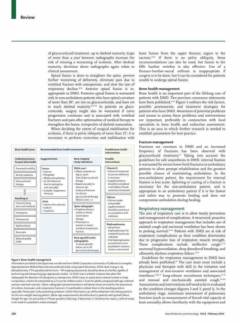
180 www.thelancet.com/neurology Vol 9 February 2010
Review
of glucocorticoid treatment, up to skeletal maturity. Gaps of more than a year between radiographs increase the risk of missing a worsening of scoliosis. After skeletal maturity, decisions about radiographs again relate to clinical assessment.
Spinal fusion is done to straighten the spine, prevent further worsening of deformity, eliminate pain due to vertebral fracture with osteoporosis, and slow the rate of respiratory decline.28,56 Anterior spinal fusion is in-appropriate in DMD. Posterior spinal fusion is warranted only in non-ambulatory patients who have spinal curvature of more than 20°, are not on glucocorticoids, and have yet to reach skeletal maturity.28,57,58 In patients on gluco-corticoids, surgery might also be warranted if curve progression continues and is associated with vertebral fractures and pain after optimisation of medical therapy to strengthen the bones, irrespective of skeletal maturation.
When deciding the extent of surgical stabilisation for scoliosis, if there is pelvic obliquity of more than 15°, it is necessary to perform correction and stabilisation with
bone fusion from the upper thoracic region to the sacrum.59,60 If there is no pelvic obliquity, these recommendations can also be used, but fusion to the fifth lumbar vertebra is also effective. Use of a thoraco–lumbar–sacral orthosis is inappropriate if surgery is to be done, but it can be considered for patients unable to undergo spinal fusion.
Bone-health management Bone health is an important part of the lifelong care of patients with DMD. Two previous consensus statements have been published.61,62 Figure 1 outlines the risk factors, possible assessments, and treatment strategies for patients who have DMD. Awareness of potential problems and means to assess these problems and interventions are important, preferably in conjunction with local specialists in bone health and endocrine assessment. This is an area in which further research is needed to establish parameters for best practice.
Fracture managementFractures are common in DMD and an increased frequency of fractures has been observed with glucocorticoid treatment.63 Taking into account the guidelines for safe anaesthesia in DMD, internal fixation is warranted for severe lower-limb fractures in ambulatory patients to allow prompt rehabilitation and the greatest possible chance of maintaining ambulation. In the non-ambulatory patient, the requirement for internal fixation is less acute. Splinting or casting of a fracture is necessary for the non-ambulatory patient, and is appropriate in an ambulatory patient if it is the fastest and safest way to promote healing and does not compromise ambulation during healing.
Respiratory managementThe aim of respiratory care is to allow timely prevention and management of complications. A structured, proactive approach to respiratory management that includes use of assisted cough and nocturnal ventilation has been shown to prolong survival.64–66 Patients with DMD are at risk of respiratory complications as their condition deteriorates due to progressive loss of respiratory muscle strength. These complications include ineffective cough,67–75 nocturnal hypoventilation, sleep disordered breathing, and ultimately daytime respiratory failure.76–84
Guidelines for respiratory management in DMD have already been published.85 The care team must include a physician and therapist with skill in the initiation and management of non-invasive ventilation and associated interfaces,36,86–91 lung-volume recruitment techniques,92–94 and manual and mechanically assisted cough.95–102 Assessments and interventions will need to be re-evaluated as the condition changes (figures 2 and 3, panel 1). In the ambulatory stage, minimum assessment of pulmonary function (such as measurement of forced vital capacity at least annually) allows familiarity with the equipment and
Recommended bone-health assessments
Suggested tests
Urine• Calcium (for calciuria)• Sodium• Creatinine
Serum• Calcium• Phosphate• Alkaline phosphatase• 25-OH vitamin D
level (in springtimeor bi-annually)
• Consider: magnesium,PTH level
Possible bone-healthinterventions
Possibleinterventions
Vitamin D• Vitamin D treatment
for proven deficiency is necessary
• Supplementation should be consideredin all children if levels cannot be maintained
Calcium• Calcium intake
and possible supplementationshould be carried out in consultation with a dietitian
Bisphosphonates• Intravenous
bisphosphonates forvertebral fracture areindicated
• Oral bisphosphonatesas treatment or as aprophylactic measureremain controversial
Bone imaging*study indications
DEXA scan†• Obtain a baseline at:
Age 3+ yearsStart of glucocorticoidtherapy
• Repeat annually forthose at risk:History of fracturesOn chronic glucocorticoid therapyDEXA Z score <–2
Spine radiographॠIf kyphoscoliosis is
noted on clinicalexaminationtherapy
• If back pain ispresent, to assessvertebral compressionfracture
Bone age (left wrist)radiography§
To assess growthfailure (on or offglucocorticoid therapy)
Bone-health issues
Underlying factorsfor poor bone health
Decreased mobility
Muscle weakness
Glucocorticoidtherapy
Resulting in:
Fractures (long bone and vertebral)
Osteopenia
Osteoporosis
Kyphoscoliosis
Bone pain
Reduced quality of life
Figure 1: Bone-health managementInformation provided in this figure was not derived from RAND Corporation–University of California Los Angeles Appropriateness Method data and was produced solely using expert discussion. DEXA=dual-energy x-ray absorptiometry. PTH=parathyroid hormone. *All imaging assessments should be done at a facility capable of performing and interpreting age-appropriate studies. †A DEXA scan is a better measure than plain film radiographs for detection of osteopenia or osteoporosis. DEXA scans, to assess bone mineral content or body composition, need to be interpreted as a Z score for children and a T score for adults (compared with age-matched and sex-matched controls). ‡Spine radiographs (posterior/anterior and lateral views) are used for the assessment of scoliosis, bone pain, and compression fractures. It is preferable to obtain them in the standing position, especially if bone pain is the presenting symptom. Useful information can still be obtained in the sitting position for the non-weight-bearing patient. §Bone-age measurements should be done in patients with growth failure (height for age <5% percentile or if linear growth is faltering). If abnormal (>2 SD below the mean), a referral needs to be made to a paediatric endocrinologist.
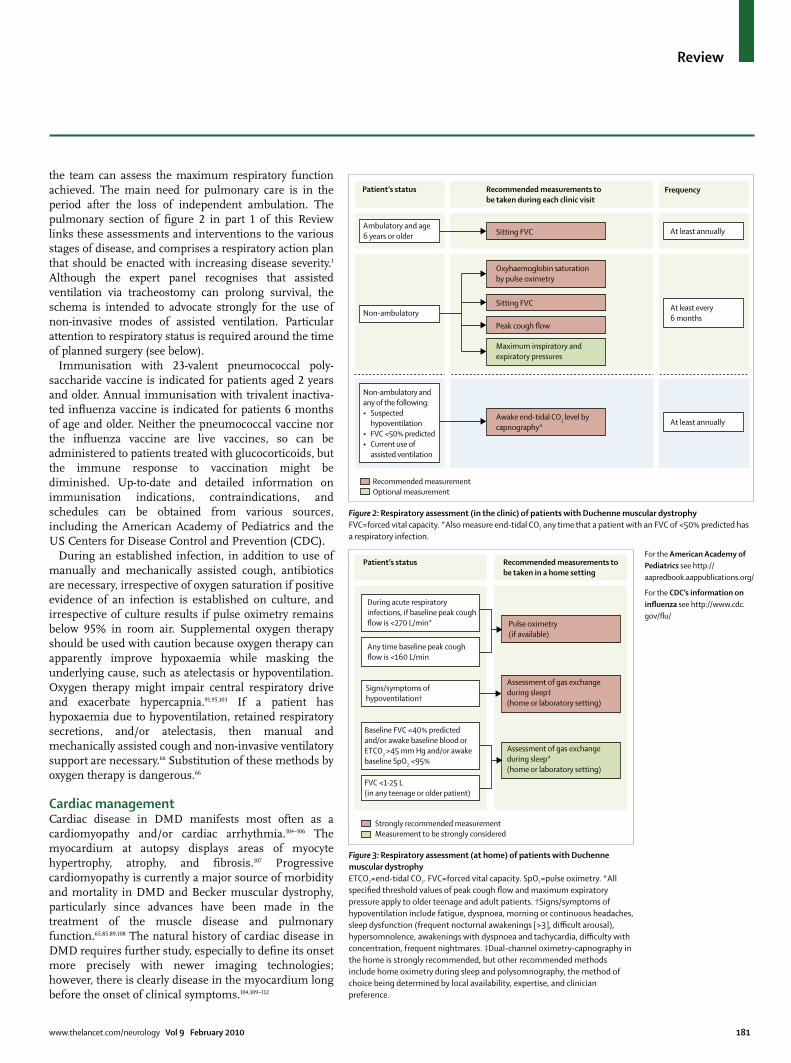
www.thelancet.com/neurology Vol 9 February 2010 181
Review
the team can assess the maximum respiratory function achieved. The main need for pulmonary care is in the period after the loss of independent ambulation. The pulmonary section of figure 2 in part 1 of this Review links these assessments and interventions to the various stages of disease, and comprises a respiratory action plan that should be enacted with increasing disease severity.1 Although the expert panel recognises that assisted ventilation via tracheostomy can prolong survival, the schema is intended to advocate strongly for the use of non-invasive modes of assisted ventilation. Particular attention to respiratory status is required around the time of planned surgery (see below).
Immunisation with 23-valent pneumococcal poly-saccharide vaccine is indicated for patients aged 2 years and older. Annual immunisation with trivalent inactiva-ted influenza vaccine is indicated for patients 6 months of age and older. Neither the pneumococcal vaccine nor the influenza vaccine are live vaccines, so can be administered to patients treated with glucocorticoids, but the immune response to vaccination might be diminished. Up-to-date and detailed information on immunisation indications, contraindications, and schedules can be obtained from various sources, including the American Academy of Pediatrics and the US Centers for Disease Control and Prevention (CDC).
During an established infection, in addition to use of manually and mechanically assisted cough, antibiotics are necessary, irrespective of oxygen saturation if positive evidence of an infection is established on culture, and irrespective of culture results if pulse oximetry remains below 95% in room air. Supplemental oxygen therapy should be used with caution because oxygen therapy can apparently improve hypoxaemia while masking the underlying cause, such as atelectasis or hypoventilation. Oxygen therapy might impair central respiratory drive and exacerbate hypercapnia.91,95,103 If a patient has hypoxaemia due to hypoventilation, retained respiratory secretions, and/or atelectasis, then manual and mechanically assisted cough and non-invasive ventilatory support are necessary.66 Substitution of these methods by oxygen therapy is dangerous.66
Cardiac managementCardiac disease in DMD manifests most often as a cardiomyopathy and/or cardiac arrhythmia.104–106 The myocardium at autopsy displays areas of myocyte hypertrophy, atrophy, and fibrosis.107 Progressive cardiomyopathy is currently a major source of morbidity and mortality in DMD and Becker muscular dystrophy, particularly since advances have been made in the treatment of the muscle disease and pulmonary function.65,85,89,108 The natural history of cardiac disease in DMD requires further study, especially to define its onset more precisely with newer imaging technologies; however, there is clearly disease in the myocardium long before the onset of clinical symptoms.104,109–112
Recommended measurementOptional measurement
Sitting FVC
Sitting FVC
Peak cough flow
Maximum inspiratory andexpiratory pressures
Awake end-tidal CO2 level bycapnography*
Oxyhaemoglobin saturationby pulse oximetry
At least every 6 months
Patient’s status Recommended measurements to be taken during each clinic visit
Frequency
Ambulatory and age6 years or older At least annually
At least annually
Non-ambulatory
Non-ambulatory andany of the following:• Suspected hypoventilation• FVC <50% predicted• Current use of assisted ventilation
Figure 2: Respiratory assessment (in the clinic) of patients with Duchenne muscular dystrophy FVC=forced vital capacity. *Also measure end-tidal CO2 any time that a patient with an FVC of <50% predicted has a respiratory infection.
Pulse oximetry(if available)
Assessment of gas exchangeduring sleep‡(home or laboratory setting)
Assessment of gas exchangeduring sleep*(home or laboratory setting)
Signs/symptoms ofhypoventilation†
Baseline FVC <40% predictedand/or awake baseline blood orETCO2 >45 mm Hg and/or awakebaseline SpO2 <95%
FVC <1·25 L(in any teenage or older patient)
During acute respiratoryinfections, if baseline peak coughflow is <270 L/min*
Any time baseline peak coughflow is <160 L/min
Strongly recommended measurementMeasurement to be strongly considered
Patient’s status Recommended measurements to be taken in a home setting
Figure 3: Respiratory assessment (at home) of patients with Duchenne muscular dystrophyETCO2=end-tidal CO2. FVC=forced vital capacity. SpO2=pulse oximetry. *All specified threshold values of peak cough flow and maximum expiratory pressure apply to older teenage and adult patients. †Signs/symptoms of hypoventilation include fatigue, dyspnoea, morning or continuous headaches, sleep dysfunction (frequent nocturnal awakenings [>3], difficult arousal), hypersomnolence, awakenings with dyspnoea and tachycardia, difficulty with concentration, frequent nightmares. ‡Dual-channel oximetry-capnography in the home is strongly recommended, but other recommended methods include home oximetry during sleep and polysomnography, the method of choice being determined by local availability, expertise, and clinician preference.
For the American Academy of Pediatrics see http://aapredbook.aappublications.org/
For the CDC’s information on influenza see http://www.cdc.gov/flu/
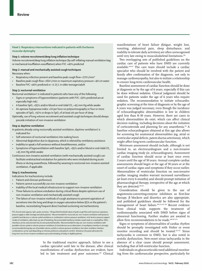
182 www.thelancet.com/neurology Vol 9 February 2010
Review
In the traditional reactive approach, failure to see a cardiac specialist until late in the disease, after clinical manifestations of cardiac dysfunction are evident, have led to late treatment and poor outcomes.104 Clinical
manifestations of heart failure (fatigue, weight loss, vomiting, abdominal pain, sleep disturbance, and inability to tolerate daily activities) are often unrecognised until very late owing to musculoskeletal limitations.104
Two overlapping sets of published guidelines on the cardiac care of patients who have DMD are currently available.104,113 The care team should include a cardiac specialist who should be involved with the patient and family after confirmation of the diagnosis, not only to manage cardiomyopathy, but also to initiate a relationship to ensure long-term cardiovascular health.
Baseline assessment of cardiac function should be done at diagnosis or by the age of 6 years, especially if this can be done without sedation. Clinical judgment should be used for patients under the age of 6 years who require sedation. The recommendation to initiate echocardio-graphic screening at the time of diagnosis or by the age of 6 years was judged necessary, even though the incidence of echocardiographic abnormalities is low in children aged less than 8–10 years. However, there are cases in which abnormalities do exist, which can affect clinical decision making, including decisions about the initiation of corticosteroids and planning for any anaesthesia.114 A baseline echocardiogram obtained at this age also allows for screening for anatomical abnormalities (eg, atrial or ventricular septal defects, patent ductus arteriosus), which might affect long-term cardiovascular function.
Minimum assessment should include, although is not limited to, an electrocardiogram and a non-invasive cardiac imaging study (ie, echocardiogram). Assessment of cardiac function should occur at least once every 2 years until the age of 10 years. Annual complete cardiac assessments should begin at the age of 10 years or at the onset of cardiac signs and symptoms if they occur earlier. Abnormalities of ventricular function on non-invasive cardiac imaging studies warrant increased surveillance (at least every 6 months) and should prompt initiation of pharmacological therapy, irrespective of the age at which they are detected.104,113
Consideration should be given to the use of angiotensin-converting-enzyme inhibitors as first-line therapy. β blockers and diuretics are also appropriate, and published guidelines should be followed for the management of heart failure.104,113,115–118 Recent evidence from clinical trials supports the treatment of cardiomyopathy associated with DMD before signs of abnormal functioning. Further studies are awaited to allow firm recommendations to be made.108,119–123
Signs or symptoms of abnormalities of cardiac rhythm should be promptly investigated with Holter or event monitor recording and should be treated.124–127 Sinus tachycardia is common in DMD, but is also noted in systolic dysfunction. New-onset sinus tachycardia in the absence of a clear cause should prompt assessment, including that of left-ventricular function.
Individuals on glucocorticoids need additional monitor-ing from the cardiovascular perspective, particularly for
Panel 1: Respiratory interventions indicated in patients with Duchenne muscular dystrophy
Step 1: volume recruitment/deep lung inflation technique Volume recruitment/deep lung inflation technique (by self-inflating manual ventilation bag or mechanical insufflation–exsufflation) when FVC <40% predicted
Step 2: manual and mechanically assisted cough techniques Necessary when: • Respiratory infection present and baseline peak cough flow <270 L/min* • Baseline peak cough flow <160 L/min or maximum expiratory pressure <40 cm water • Baseline FVC <40% predicted or <1·25 L in older teenager/adult
Step 3: nocturnal ventilation Nocturnal ventilation† is indicated in patients who have any of the following: • Signs or symptoms of hypoventilation (patients with FVC <30% predicted are at
especially high risk)• A baseline SpO2 <95% and/or blood or end-tidal CO2 >45 mm Hg while awake• An apnoea–hypopnoea index >10 per hour on polysomnography or four or more
episodes of SpO2 <92% or drops in SpO2 of at least 4% per hour of sleepOptimally, use of lung volume recruitment and assisted cough techniques should always
precede initiation of non-invasive ventilation
Step 4: daytime ventilation In patients already using nocturnally assisted ventilation, daytime ventilation‡ is
indicated for:• Self extension of nocturnal ventilation into waking hours• Abnormal deglutition due to dyspnoea, which is relieved by ventilatory assistance• Inability to speak a full sentence without breathlessness, and/or • Symptoms of hypoventilation with baseline SpO2 <95% and/or blood or end-tidal CO2
>45 mm Hg while awakeContinuous non-invasive assisted ventilation (with mechanically assisted cough) can
facilitate endotracheal extubation for patients who were intubated during acute illness or during anaesthesia, followed by weaning to nocturnal non-invasive assisted ventilation, if applicable
Step 5: tracheostomy Indications for tracheostomy include:• Patient and clinician preference§ • Patient cannot successfully use non-invasive ventilation• Inability of the local medical infrastructure to support non-invasive ventilation• Three failures to achieve extubation during critical illness despite optimum use of
non-invasive ventilation and mechanically assisted cough• The failure of non-invasive methods of cough assistance to prevent aspiration of
secretions into the lung and drops in oxygen saturation below 95% or the patient’s baseline, necessitating frequent direct tracheal suctioning via tracheostomy
FVC=forced vital capacity. SpO2=pulse oximetry. *All specified threshold values of peak cough flow and maximum expiratory pressure apply to older teenage and adult patients. †Recommended for nocturnal use: non-invasive ventilation with pressure cycled bi-level devices or volume cycled ventilators or combination volume-pressure ventilators. In bi-level or pressure support modes of ventilation, add a back-up rate of breathing. Recommended interfaces include a nasal mask or a nasal pillow. Other interfaces can be used and each has its own potential benefits. ‡Recommended for day use: non-invasive ventilation with portable volume cycled or volume-pressure ventilators; bi-level devices are an alternative. A mouthpiece interface is strongly recommended during day use of portable volume-cycled or volume-pressure ventilators, but other ventilator-interface combinations can be used depending on clinician preference and patient comfort. §However, the panel advocates the long-term use of non-invasive ventilation up to and including 24 h/day in eligible patients.

www.thelancet.com/neurology Vol 9 February 2010 183
Review
hypertension, which might necessitate adjustment in the glucocorticoid dose (table 2 in part 1 of this Review).1 Systemic arterial hypertension should be treated.
Prevention of systemic thromboembolic events by anticoagulation therapy can be considered in severe cardiac dysfunction, but is inappropriate in earlier cardiac dysfunction. The usefulness of an internal cardiac defibrillator has not been established and needs further research.
Because of the morbidity and mortality associated with cardiomyopathy, additional research is clearly needed, not only to define the natural history of the disease process, but also to establish treatments specific for the dystrophin-deficient myocardium. Further studies of pharmacological approaches aimed at early intervention are needed to delay the underlying disease process. With generally improved fitness of patients who have DMD, the option of cardiac transplant might need to be addressed in the future.
Nutritional, swallowing, gastrointestinal, and speech and language managementPatients might be at risk of both undernutrition/malnutrition and being overweight/obese at different ages and under different circumstances, in addition to deficiencies in calorie, protein, vitamin, mineral, and fluid intake. In later stages, pharyngeal weakness leads to dysphagia, further accentuating nutritional issues and gradual loss of respiratory muscle strength, combined with poor oral intake, and can result in severe weight loss and the need to consider tube feeding. Constipation might also be seen, typically in older patients and after surgery. With increasing survival, other complications are being reported, including gastric and intestinal dilatation related to air swallowing due to ventilator use, or more rarely to delayed gastric emptying and ileus. As the condition progresses, access to a dietitian or nutritionist, a swallowing/speech and language therapist, and a gastroenterologist is needed for the following reasons: (1) to guide the patient to maintain good nutritional status to prevent both undernutrition/malnutrition and being overweight/obese, and to provide a well-balanced, nutrient-complete diet (adding tube feeding, if necessary); (2) to monitor and treat swallowing problems (dysphagia) to prevent aspiration and weight loss, and to assess and treat delayed speech and language problems; and (3) to treat the common problems of constipation and gastro-oesophageal reflux with both medication and non-medication therapies.
Nutritional managementMaintaining good nutritional status, defined as weight for age or body-mass index for age from the 10th to 85th percentiles on national percentile charts, is essential. Poor nutrition can potentially have a negative effect on almost every organ system. Anticipatory guidance and prevention of undernutrition/malnutrition and being
overweight/obese should be goals from diagnosis throughout life. The monitoring and triggers for referral to an expert dietitian/nutritionist in DMD are described in panel 2.128–132 Diet should be assessed for energy, protein, fluid, calcium, vitamin D, and other nutrients. We recommend that each patient should receive a daily multivitamin supplement with vitamin D and minerals. If this is not general practice, a computer nutrient analysis of the patient’s diet can provide evidence for the possible need for specific foods or nutrient supplements. If there is a suspicion of undernutrition/malnutrition and poor intake, serum vitamin concentrations can be obtained and supplements could be recommended. Nutritional recommendations with regard to bone health are shown in figure 1.
Swallowing managementClinical swallowing examination is indicated if there is unintentional weight loss of 10% or more or a decline in the expected age-related weight gain. Prolonged meal times (>30 min) or meal times accompanied by fatigue, excessive spilling, drooling, pocketing, or any other clinical indicators of dysphagia make referral necessary, as do persistent coughing, choking, gagging, or wet vocal quality during eating or drinking.133 An episode of aspiration pneumonia, unexplained decline in pulmonary function, or fever of unknown origin might be signs of unsafe swallowing, necessitating assessment. There might be contributory factors for weight loss due to complications in other systems, such as cardiac or respiratory compromise.
A videofluoroscopic study of swallowing (also referred to as a modified barium swallow) is necessary for patients with clinical indicators of possible aspiration and pharyngeal dysmotility.134 Swallowing interventions and compensatory strategies are appropriate for patients with dysphagia. These should be delivered by a speech and language pathologist, with training and expertise in the treatment of oral-pharyngeal dysphagia, who can assess the likely appropriateness of interventions and deliver an individualised dysphagia treatment plan with the aim of preserving optimum swallowing function.
As the disease progresses, most patients begin to experience increasing difficulty with chewing and subsequently exhibit pharyngeal-phase swallowing deficits in young adulthood.135–140 The onset of dysphagia symptoms can be gradual and the impact of oral-pharyngeal dysphagia might be under-recognised and under-reported by patients.140 This leads to risk of complications, such as aspiration and inability to take in enough fluids and food energy to maintain weight.135–139 Weight problems can also be due to an inability to meet the increased effort of breathing.135–139
When it is no longer possible to maintain weight and hydration by oral means, gastric-tube placement should be offered. Discussions between other specialists and the family should involve explanations of the potential risks
For the US national percentile charts see http://www.cdc.gov/growthcharts/

184 www.thelancet.com/neurology Vol 9 February 2010
Review
and benefits of the procedure. A gastrostomy can be placed endoscopically or via open surgery, taking into account anaesthetic and ethical considerations and family and personal preference.141
Gastrointestinal managementConstipation and gastro-oesophageal reflux are the two most common gastrointestinal conditions seen in children with DMD in clinical practice.133,142,143 Stool softeners, laxatives, and stimulants are necessary if the patient has acute constipation or faecal impaction, and use of enemas might be needed occasionally. Daily use of laxatives, such as milk of magnesia, lactulose, or polyethylene glycol, is necessary if symptoms persist. In the case of persistent constipation, adequacy of free fluid intake should be determined and addressed. In cases of faecal impaction, manual/digital disimpaction under sedation or general anaesthesia is of uncertain benefit. Enemas, stimulant laxatives, such as dulcolax and senna, and stool softeners can be tried before considering manual disimpaction. Milk and molasses enemas are not recommended for paediatric patients. Supplementation with dietary fibre for chronic or severe constipation might worsen symptoms, particularly if fluid intake is not increased.
Gastro-oesophageal reflux is typically treated with proton-pump inhibitors or H2 receptor antagonists, with prokinetics, sucralfate, and neutralising antacids as adjunctive therapies. Common practice is to prescribe
acid blockers in children on corticosteroid therapy or oral bisphosphonates to avoid complications such as gastritis and to prevent reflux oesophagitis. It is necessary to recommend nutritional interventions for a patient who has symptoms suggestive of reflux.
Speech and language managementDelayed acquisition of early language milestones is common in boys who have DMD, with differences in language acquisition and language-skill deficits persisting throughout childhood.144 Referral to a speech and language pathologist for assessment and treatment is necessary on suspicion of difficulties with speech acquisition or with continuing deficits in language comprehension or oral expression. Oral motor exercises and articulation therapy are necessary for young boys with DMD with hypotonia and in older patients who have deteriorating oral muscle strength and/or impaired speech intelligibility. For older patients, compensatory strategies, voice exercises, and speech amplifications are appropriate if intelligibility deteriorates due to problems with respiratory support for speech and vocal intensity. Voice output communication aid assessment could be appropriate at all ages if speech output is limited.
Pain managementPain of varying intensity occurs in DMD.145,146 Effective pain management requires accurate determination of the cause. Interventions to address pain include physical therapy, postural correction, appropriate and individualised orthoses, wheelchair and bed enhancements, and pharmacological approaches (eg, muscle relaxants and anti-inflammatory medications). Pharmacological inter-ventions must take into account possible interactions with other medications (eg, steroids and non-steroidal anti-inflammatory drugs) and their side-effects, particularly those that might negatively affect cardiac or respiratory function. Rarely, orthopaedic intervention might be indicated for intractable pain that is amenable to surgery. Back pain, particularly in the context of glucocorticoid treatment, is an indication that a careful search for vertebral fractures is needed; such fractures respond well to bisphosphonate treatment and/or calcitonin.147,148 Research on effective pain interventions across the lifespan of individuals with DMD is warranted.146,149,150
Surgical considerationsVarious situations, related (muscle biopsy, joint contracture surgery, spinal surgery, and gastrostomy) and unrelated (intercurrent acute surgical events) to DMD, might require the use of general anaesthesia. There are several condition-specific issues that need to be taken into account for the planning of safe surgery. Surgery in a patient who has DMD should be done in a full-service hospital that has experience of patients with DMD. In addition, as with any situation in which
Panel 2: Improving underweight and overweight status
Monitor regularly for:• Weight*• Linear height in ambulatory patients (measured every
6 months)• Arm span/segmental length in non-ambulatory patients†
Refer for a nutritional/dietetic assessment:• At diagnosis• At initiation of glucocorticoids• If the patient is underweight (<10th age percentile)‡• If the patient is at risk of becoming overweight
(85–95th age percentile)‡• If the patient is overweight (>95th age percentile)‡• If there has been unintentional weight loss or gain• If there has been poor weight gain• If major surgery is planned• If the patient is chronically constipated• If dysphagia is present
National guidelines and recommendations for diets for underweight and overweight individuals can be found in Kleinman,128 and are often available from charities/associations for cardiac disorders and diabetes. *In non-ambulatory patients, wheelchair weight should be obtained first, then patient and wheelchair weight, or caregiver weight should be obtained first, then the weight of the patient held by the caregiver. †If the patient has scoliosis, the arm span should be measured if possible. ‡Overweight/underweight status should be judged on the basis of local body-mass index percentiles (weight for age is a possible alternative if height is unavailable). Body composition is altered in Duchenne muscular dystrophy (DMD) owing to the relatively low lean body mass seen in DMD with a relatively higher fat body mass.129
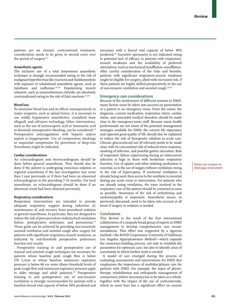
www.thelancet.com/neurology Vol 9 February 2010 185
Review
patients are on chronic corticosteroid treatment, consideration needs to be given to steroid cover over the period of surgery.151
Anaesthetic agentsThe exclusive use of a total intravenous anaesthetic technique is strongly recommended owing to the risk of malignant hyperthermia-like reactions and rhabdomyolysis with exposure to inhalational anaesthetic agents, such as halothane and isoflurane.152,153 Depolarising muscle relaxants, such as suxamethonium chloride, are absolutely contraindicated owing to the risk of fatal reactions.152,153
Blood lossTo minimise blood loss and its effects intraoperatively in major surgeries, such as spinal fusion, it is necessary to use mildly hypotensive anaesthetics, crystalloid bone allograft, and cell-saver technology. Other interventions, such as the use of aminocaproic acid or tranexamic acid to diminish intraoperative bleeding, can be considered.154 Postoperative anticoagulation with heparin and/or aspirin is inappropriate. Use of compression stockings or sequential compression for prevention of deep-vein thrombosis might be indicated.
Cardiac considerationsAn echocardiogram and electrocardiogram should be done before general anaesthesia. They should also be done if the patient is undergoing conscious sedation or regional anaesthesia if the last investigation was more than 1 year previously or if there had been an abnormal echocardiogram in the preceding 7–12 months. For local anaesthesia, an echocardiogram should be done if an abnormal result had been obtained previously.
Respiratory considerationsRespiratory interventions are intended to provide adequate respiratory support during induction of, maintenance of, and recovery from procedural sedation or general anaesthesia. In particular, they are designed to reduce the risk of post-procedure endotracheal extubation failure, postoperative atelectasis, and pneumonia.153 These goals can be achieved by providing non-invasively assisted ventilation and assisted cough after surgery for patients with significant respiratory-muscle weakness, as indicated by sub-threshold preoperative pulmonary function test results.
Preoperative training in and postoperative use of manual and assisted cough techniques are necessary for patients whose baseline peak cough flow is below 270 L/min or whose baseline maximum expiratory pressure is below 60 cm water (these threshold levels of peak cough flow and maximum expiratory pressure apply to older teenage and adult patients).155 Preoperative training in and postoperative use of non-invasive ventilation is strongly recommended for patients with a baseline forced vital capacity of below 50% predicted and
necessary with a forced vital capacity of below 30% predicted.155 Incentive spirometry is not indicated owing to potential lack of efficacy in patients with respiratory-muscle weakness and the availability of preferred alternatives, such as mechanical insufflation–exsufflation. After careful consideration of the risks and benefits, patients with significant respiratory-muscle weakness might be eligible for surgery, albeit with increased risk, if these patients are highly skilled preoperatively in the use of non-invasive ventilation and assisted cough.156,157
Emergency-care considerationsBecause of the involvement of different systems in DMD, many factors must be taken into account on presentation of a patient to an emergency room. From the outset, the diagnosis, current medication, respiratory status, cardiac status, and associated medical disorders should be made clear to the emergency-room staff. Because many health professionals are not aware of the potential management strategies available for DMD, the current life expectancy and expected good quality of life should also be explained to reduce the risk of therapeutic nihilism in acute care. Chronic glucocorticoid use (if relevant) needs to be made clear, with its concomitant risk of reduced stress response, masking of infection, and possible gastric ulceration. Risk of respiratory failure supervening during an intercurrent infection is high in those with borderline respiratory function. Use of opiates and other sedating medication is essential, as is the use of oxygen without ventilation owing to the risk of hypercapnia. If nocturnal ventilation is already being used, then access to the ventilator is essential during any acute event or intervention. For patients who are already using ventilation, the team involved in the respiratory care of the patient should be contacted as soon as possible. Awareness of the risk of arrhythmias and cardiomyopathy is important. Anaesthetic issues, as previously discussed, need to be taken into account at all times if surgery or sedation is needed.
ConclusionsThis Review is the result of the first international collaboration of a uniquely broad group of experts in DMD management to develop comprehensive care recom-mendations. This effort was supported by a rigorous method—the RAND Corporation–University of California Los Angeles Appropriateness Method2—which expands the consensus-building process, not only to establish the parameters for optimum care, but also to identify areas of uncertainty in which further work is needed.
A model of care emerged during the process of evaluating assessments and interventions for DMD that emphasises the importance of multidisciplinary care for patients with DMD. For example, the input of physio-therapy, rehabilitation and orthopaedic management of contractures (where necessary) has to be taken as a whole, together with the impact of the use of corticosteroids, which in most boys has a significant effect on muscle
Please see erratum on final page of document
186 www.thelancet.com/neurology Vol 9 February 2010
Review
strength and function. In this context, the various specialty reports are presented in this second part of the Review.
Clearly staged assessments and interventions have been described to address the cardiac and respiratory complications that are common in DMD and provide the framework for the safe management of these complications. Respiratory interventions, in particular the institution of nocturnal ventilation, have had a major effect on survival in DMD,65,66 and early indications are that prompt recognition and treatment of deterioration in cardiac status will also have a significant impact.119,121 Further trials are awaited to determine the optimum timing to start cardioactive treatment. In the meantime, the recommendations presented here are consistent with previously published guidelines,113,115 and most importantly reinforce the need for active engagement with a cardiologist at every stage of the condition.
In other areas, including management of complications of the gastrointestinal tract, less work had been done previously, but we conclude that proactive management in this area is important. An increasing awareness of the possibility of gastrointestinal complications in DMD is needed; this area has been relatively poorly studied until now and will merit further investigation in emerging adult populations with DMD to delineate the burden of disease in these patients and its optimum management.
We are in an unprecedented era of hope for therapies for DMD based on the underlying molecular basis of the disease. In the meantime, these care recommendations have been developed with the support of and input from stakeholders in the DMD community to be used as the current and future benchmark for anticipatory planning, appropriate surveillance, and interventions in all areas of this complex disease. It is hoped that they will provide a catalyst to improve care for patients with DMD worldwide.ContributorsAll authors provided intellectual expertise in the study design, generation and interpretation of data, writing of the Review, and the decision to publish. KB, aided by RF, drafted and edited the Review, and approved the final version. DJB, LEC, LC, SP, and CC were involved in the literature search.
Conflicts of interestKB is a consultant for Acceleron, AVI, Debiopharm, Prosensa, and Santhera. LEC has received honoraria from Genzyme Corporation, has participated in research supported by Genzyme Corporation, PTC Therapeutics, the Leal Foundation, and Families of Spinal Muscular Atrophy, has been awarded grant support from the National Skeletal Muscle Research Center, and is a member of the Pompe Registry Board of Advisors. All other authors have no conflicts of interest.
AcknowledgmentsWe thank P Eubanks, A Kenneson, A Vatave (CDC); A Cyrus and E Levy (Oak Ridge Institute for Science and Education); B Bradshaw, H Desai, P Haskell, E Hunt, A Marsden, C Muse, and L Yuson (Booz/Allen/Hamilton); and B Tseng (Massachusetts General Hospital, Harvard Medical School) for their contributions to this study and manuscript. We also thank the following organisations for their collaboration on this study: Muscular Dystrophy Association, National Institute on Disability and Rehabilitation Research, Parent Project Muscular Dystrophy, and TREAT-NMD (EC036825). In addition, we thank M Levine, M Mascarenhas, and M Thayu (Children’s Hospital of Philadelphia) for their participation in discussions about bone health; and G Carter, J Engel (University of Washington), H Posselt (Montrose Access, Australia), and C Trout (University of Iowa Children’s Hospital) for their advice on pain management. The CDC provided support for the project through funding, study design, collection, analysis, and interpretation of data and manuscript preparation. The findings and conclusions in this report are those of the authors and do not necessarily represent the official position of the CDC.
References1 Bushby K, Finkel R, Birnkrant DJ, et al. Diagnosis and management
of Duchenne muscular dystrophy, part 1: diagnosis, and pharmacological and psychosocial management. Lancet Neurol 2009; published online Nov 30. DOI:10.1016/S1474-4422(09)70271-6.
2 Fitch K, Bernstein SJ, Aguilar MS, et al. The RAND/UCLA appropriateness method user’s manual. Santa Monica, CA: RAND Corporation, 2001.
3 Brooke MH, Fenichel GM, Griggs RC, et al. Duchenne muscular dystrophy: patterns of clinical progression and effects of supportive therapy. Neurology 1989; 39: 475–81.
4 Johnson EW, Walter J. Zeiter Lecture: pathokinesiology of Duchenne muscular dystrophy: implications for management. Arch Phys Med Rehabil 1977; 58: 4–7.
5 Sutherland DH, Olshen R, Cooper L, et al. The pathomechanics of gait in Duchenne muscular dystrophy. Dev Med Child Neurol 1981; 23: 3–22.
6 Archibald KC, Vignos PJ Jr. A study of contractures in muscular dystrophy. Arch Phys Med Rehabil 1959; 40: 150–57.
7 Johnson ER, Fowler WM Jr, Lieberman JS. Contractures in neuromuscular disease. Arch Phys Med Rehabil 1992; 73: 807–10.
8 Hsu JD, Furumasu J. Gait and posture changes in the Duchenne muscular dystrophy child. Clin Orthop Relat Res 1993; 288: 122–25.
9 McDonald CM, Abresch RT, Carter GT, et al. Profiles of neuromuscular diseases. Duchenne muscular dystrophy. Am J Phys Med Rehabil 1995; 74 (suppl): S70–92.
10 Dubowitz V. Progressive muscular dystrophy: prevention of deformities. Clin Pediatr (Phila) 1964; 12: 323–28.
11 Dubowitz V. Prevention of deformities. Isr J Med Sci 1977; 13: 183–88.
12 Fowler WM Jr. Rehabilitation management of muscular dystrophy and related disorders: II. Comprehensive care. Arch Phys Med Rehabil 1982; 63: 322–28.
13 Vignos PJ Jr. Physical models of rehabilitation in neuromuscular disease. Muscle Nerve 1983; 6: 323–38.
14 Siegel IM, Weiss LA. Postural substitution in Duchenne’s muscular dystrophy. JAMA 1982; 247: 584.
15 Vignos PJ, Wagner MB, Karlinchak B, Katirji B. Evaluation of a program for long-term treatment of Duchenne muscular dystrophy. Experience at the University Hospitals of Cleveland. J Bone Joint Surg Am 1996; 78: 1844–52.
16 Vignos PJ Jr. Rehabilitation in progressive muscular dystrophy. In: Licht S, ed. Rehabilitation and medicine. New Haven, CT: Elizabeth Licht, 1968.
Search strategy and selection criteria
Peer-reviewed literature was searched using the key search terms of “Duchenne” or “muscular dystrophy”, or both, paired with one of 410 other search terms related to a comprehensive list of assessment tools and interventions used in DMD management. The full list of search terms is available on request. The databases used included Medline, Embase, Web of Science, and the Cochrane Library. Initial inclusion criteria consisted of available abstracts of human studies published in English between 1986 and 2006. Each working group also incorporated major articles from its discipline published before 1986 and from 2007 to mid-2009 in the process of discussions, final assessments, and write-up of recommendations.
www.thelancet.com/neurology Vol 9 February 2010 187
Review
17 Hyde SA, Fløytrup I, Glent S, et al. A randomized comparative study of two methods for controlling Tendo Achilles contracture in Duchenne muscular dystrophy. Neuromuscul Disord 2000; 10: 257–63.
18 Scott OM, Hyde SA, Goddard C, Dubowitz V. Prevention of deformity in Duchenne muscular dystrophy. A prospective study of passive stretching and splintage. Physiotherapy 1981; 67: 177–80.
19 McDonald CM. Limb contractures in progressive neuromuscular disease and the role of stretching, orthotics, and surgery. Phys Med Rehabil Clin N Am 1998; 9: 187–211.
20 Johnson EW, Kennedy JH. Comprehensive management of Duchenne muscular dystrophy. Arch Phys Med Rehabil 1971; 52: 110–14.
21 Siegel IM. Plastic-molded knee-ankle-foot orthoses in the treatment of Duchenne muscular dystrophy. Arch Phys Med Rehabil 1975; 56: 322.
22 Bakker JP, De Groot IJ, De Jong BA, Van Tol-De Jager MA, Lankhorst GJ. Prescription pattern for orthoses in the Netherlands: use and experience in the ambulatory phase of Duchenne muscular dystrophy. Disabil Rehabil 1997; 19: 318–25.
23 Bakker JP, de Groot IJ, Beckerman H, de Jong BA, Lankhorst GJ. The effects of knee-ankle-foot orthoses in the treatment of Duchenne muscular dystrophy: review of the literature. Clin Rehabil 2000; 14: 343–59.
24 Miller G, Dunn N. An outline of the management and prognosis of Duchenne muscular dystrophy in Western Australia. Aust Paediatr J 1982; 18: 277–82.
25 Rideau Y, Duport G, Delaubier A, Guillou C, Renardel-Irani A, Bach JR. Early treatment to preserve quality of locomotion for children with Duchenne muscular dystrophy. Semin Neurol 1995; 15: 9–17.
26 Forst J, Forst R. Lower limb surgery in Duchenne muscular dystrophy. Neuromuscul Disord 1999; 9: 176–81.
27 Scher DM, Mubarak SJ. Surgical prevention of foot deformity in patients with Duchenne muscular dystrophy. J Pediatr Orthop 2002; 22: 384–91.
28 Sussman M. Duchenne muscular dystrophy. J Am Acad Orthop Surg 2002; 10: 138–51.
29 Shapiro F. Orthopedic treatment. In: Jones HR, De Vivo DC, Darras BT, eds. Neuromuscular disorders of infancy, childhood, and adolescence: a clinician’s approach. Amsterdam/Boston: Butterworth-Heinemann, 2003: 1259–63.
30 Siegel IM, Miller JE, Ray RD. Subcutaneous lower limb tenotomy is the treatment of pseudohypertrophic muscular dystrophy: description of technique and presentation of twenty-one cases. J Bone Joint Surg Am 1968; 50: 1437–43.
31 Smith SE, Green NE, Cole RJ, Robison JD, Fenichel GM. Prolongation of ambulation in children with Duchenne muscular dystrophy by subcutaneous lower limb tenotomy. J Pediatr Orthop 1993; 13: 336–40.
32 Miller GM, Hsu JD, Hoffer MM, Rentfro R. Posterior tibial tendon transfer: a review of the literature and analysis of 74 procedures. J Pediatr Orthop 1982; 2: 363–70.
33 Wagner MB, Vignos PJ Jr, Carlozzi C, Hull AL. Assessment of hand function in Duchenne muscular dystrophy. Arch Phys Med Rehabil 1993; 74: 801–80.
34 Wagner MB, Vignos PJ Jr, Carlozzi C. Duchenne muscular dystrophy: a study of wrist and hand function. Muscle Nerve 1989; 12: 236–44.
35 Pellegrini N, Guillon B, Prigent H, et al. Optimization of power wheelchair control for patients with severe Duchenne muscular dystrophy. Neuromuscul Disord 2004; 14: 297–300.
36 Eagle M, Bourke J, Bullock R, et al. Managing Duchenne muscular dystrophy—the additive effect of spinal surgery and home nocturnal ventilation in improving survival. Neuromuscul Disord 2007; 17: 470–75.
37 Eagle M. Report on the muscular dystrophy campaign workshop: exercise in neuromuscular diseases Newcastle, January 2002. Neuromuscul Disord 2002; 12: 975–83.
38 Vignos PJ Jr, Watkins MP. The effect of exercise in muscular dystrophy. JAMA 1966; 197: 843–48.
39 Scott OM, Hyde SA, Goddard C, Jones R, Dubowitz V. Effect of exercise in Duchenne muscular dystrophy. Physiotherapy 1981; 67: 174–76.
40 de Lateur BJ, Giaconi RM. Effect on maximal strength of submaximal exercise in Duchenne muscular dystrophy. Am J Phys Med 1979; 58: 26–36.
41 Fowler WM Jr. Importance of overwork weakness. Muscle Nerve 1984; 7: 496–99.
42 Armstrong RB, Warren GL, Warren JA. Mechanisms of exercise-induced muscle fibre injury. Sports Med 1991; 12: 184–207.
43 Fowler WM Jr, Taylor M, Rehabilitation management of muscular dystrophy and related disorders: I. The role of exercise. Arch Phys Med Rehabil 1982; 63: 319–21.
44 Fowler WM Jr. Role of physical activity and exercise training in neuromuscular diseases. Am J Phys Med Rehabil 2002; 81 (suppl): S187–95.
45 McDonald CM. Physical activity, health impairments, and disability in neuromuscular disease. Am J Phys Med Rehabil 2002; 81 (suppl): S108–20.
46 Sockolov R, Irwin B, Dressendorfer RH, Bernauer EM. Exercise performance in 6-to-11-year-old boys with Duchenne muscular dystrophy. Arch Phys Med Rehabil 1977; 58: 195–201.
47 Petrof BJ. The molecular basis of activity-induced muscle injury in Duchenne muscular dystrophy. Mol Cell Biochem 1998; 179: 111–23.
48 Ansved T. Muscular dystrophies: influence of physical conditioning on the disease evolution. Curr Opin Clin Nutr Metab Care 2003; 6: 435–39.
49 Allen DG. Eccentric muscle damage: mechanisms of early reduction of force. Acta Physiol Scand 2001; 171: 311–19.
50 Garrood P, Eagle M, Jardine PE, Bushby K, Straub V. Myoglobinuria in boys with Duchenne muscular dystrophy on corticosteroid therapy. Neuromuscul Disord 2008; 18: 71–73.
51 Smith AD, Koreska J, Moseley CF. Progression of scoliosis in Duchenne muscular dystrophy. J Bone Joint Surg Am 1989; 71: 1066–74.
52 Alman BA, Raza SN, Biggar WD. Steroid treatment and the development of scoliosis in males with Duchenne muscular dystrophy. J Bone Joint Surg Am 2004; 86: 519–24.
53 Yilmaz O, Karaduman, Topaloglu H. Prednisolone therapy in Duchenne muscular dystrophy prolongs ambulation and prevents scoliosis. Eur J Neurol 2004; 11: 541–44.
54 Talim B, Malaguti C, Gnudi S, Politano L, Merlini L. Vertebral compression in Duchenne muscular dystrophy following deflazacort. Neuromuscul Disord 2002; 12: 294–95.
55 Bothwell JE, Gordon KE, Dooley JM, Mac Sween J, Cummings EA, Salisbury S. Vertebral fractures in boys with Duchenne muscular dystrophy. Clin Pediatr 2003; 42: 353–56.
56 Velasco MV, Colin AA, Zurakowski D, Darras BT, Shapiro F. Posterior spinal fusion for scoliosis in Duchenne muscular dystrophy diminishes the rate of respiratory decline. Spine 2007; 32: 459–65.
57 Shapiro F, Sethna N, Colan S, Wohl ME, Specht L. Spinal fusion in Duchenne muscular dystrophy: a multidisciplinary approach. Muscle Nerve 1992; 15: 604–14.
58 Heller KD, Wirtz DC, Siebert CH, Forst R. Spinal stabilization in Duchenne muscular dystrophy: principles of treatment and record of 31 operative treated cases. J Pediatr Orthop 2001; 10: 18–24.
59 Alman BA, Kim HKW. Pelvic obliquity after fusion of the spine in Duchenne muscular dystrophy. J Bone Joint Surg Br 1999; 81: 821–24.
60 Sengupta DK, Mehdian SH, McConnell JR, Eisenstein SM, Webb JK. Pelvic or lumbar fixation for the surgical management of scoliosis in Duchenne muscular dystrophy. Spine 2002; 27: 2072–79.
61 Quinlivan R, Roper H, Davie M, et al. Report of a Muscular Dystrophy Campaign funded workshop Birmingham, UK, January 16th 2004. Osteoporosis in Duchenne muscular dystrophy; its prevalence, treatment and prevention. Neuromuscul Disord 2005; 15: 72–79.
62 Biggar WD, Bachrach LK, Henderson RC, Kalkwarf H, Plotkin H, Wong BL. Bone health in Duchenne muscular dystrophy. Neuromuscul Disord 2005; 15: 80–85.
63 McDonald DGM, Kinali M, Gallagher AC, et al. Fracture prevalence in Duchenne muscular dystrophy. Dev Med Child Neurol 2002; 44: 695–98.
64 Phillips MF, Quinlivan CM, Edwards RH, Calverley PM. Changes in spirometry over time as a prognostic marker in patients with Duchenne muscular dystrophy. Am J Respir Crit Care Med 2001; 164: 2191–94.
65 Eagle M, Baudouin SV, Chandler C, Giddings DR, Bullock R, Bushby K. Survival in Duchenne muscular dystrophy: improvements in life expectancy since 1967 and the impact of home nocturnal ventilation. Neuromuscul Disord 2002; 12: 926–29.
188 www.thelancet.com/neurology Vol 9 February 2010
Review
66 Gomez-Merino E, Bach JR. Duchenne muscular dystrophy: prolongation of life by noninvasive ventilation and mechanically assisted coughing. Am J Phys Med Rehabil 2002; 81: 411–15.
67 Bach JR, Saporito LR. Criteria for extubation and tracheostomy tube removal for patients with ventilatory failure: a different approach to weaning. Chest 1996; 110: 1566–71.
68 Dohna-Schwake C, Ragette R, Teschler H, Voit T, Mellies U. Predictors of severe chest infections in pediatric neuromuscular disorders. Neuromuscul Disord 2006; 16: 325–28.
69 Bianchi C, Baiardi P. Cough peak flows: standard values for children and adolescents. Am J Phys Med Rehabil 2008; 87: 461–67.
70 Kang SW, Bach JR. Maximum insufflation capacity: vital capacity and cough flows in neuromuscular disease. Am J Phys Med Rehabil 2000; 79: 222–27.
71 Daftary AS, Crisanti M, Kalra M, Wong B, Amin R. Effect of long-term steroids on cough efficiency and respiratory muscle strength in patients with Duchenne muscular dystrophy. Pediatrics 2007; 117: e320–24.
72 Gauld LM, Boynton A. Relationship between peak cough flow and spirometry in Duchenne muscular dystrophy. Pediatr Pulmonol 2005; 39: 457–60.
73 Suarez AA, Pessolano FA, Monteiro SG, et al. Peak flow and peak cough flow in the evaluation of expiratory muscle weakness and bulbar impairment in patients with neuromuscular disease. Am J Phys Med Rehabil 2002; 81: 506–11.
74 Domenech-Clar R, Lopez-Andreu JA, Compte-Torrero L, et al. Maximal static respiratory pressures in children and adolescents. Pediatr Pulmonol 2003; 35: 126–32.
75 Szeinberg A, Tabachnik E, Rashed N, et al. Cough capacity in patients with muscular dystrophy. Chest 1988; 94: 1232–35.
76 Smith PE, Calverley PM, Edwards RH. Hypoxemia during sleep in Duchenne muscular dystrophy. Am Rev Respir Dis 1988; 137: 884–88.
77 Phillips MF, Smith PE, Carroll N, Edwards RH, Calverley PM. Nocturnal oxygenation and prognosis in Duchenne muscular dystrophy. Am J Respir Crit Care Med 1999; 160: 198–202.
78 Hukins CA, Hillman DR. Daytime predictors of sleep hypoventilation in Duchenne muscular dystrophy. Am J Repir Crit Care Med 2000; 161: 166–70.
79 Ragette R, Mellies U, Schwake C, Voit T, Teschler H. Patterns and predictors of sleep disordered breathing in primary myopathies. Thorax 2002; 57: 724–28.
80 Khan Y, Heckmatt JZ. Obstructive apnoeas in Duchenne muscular dystrophy. Thorax 1994; 49: 157–61.
81 Barbe F, Quera-Salva MA, McCann C, et al. Sleep-related respiratory disturbances in patients with Duchenne muscular dystrophy. Eur Respir J 1994; 7: 1403–08.
82 Uliel S, Tauman R, Greenfeld M, Sivan Y. Normal polysomnographic respiratory values in children and adolescents. Chest 2004; 125: 872–78.
83 Toussaint M, Steens M, Soudon P. Lung function accurately predicts hypercapnia in patients with Duchenne muscular dystrophy. Chest 2007; 131: 368–75.
84 Culebras A. Sleep-disordered breathing in neuromuscular disease. Sleep Med Clin 2008; 3: 377–86.
85 Finder JD, Birnkrant D, Carl J, et al. Respiratory care of the patient with Duchenne muscular dystrophy: an official ATS consensus statement. Am J Respir Crit Care Med 2004; 170: 456–65.
86 Ward S, Chatwin M, Heather S, Simonds AK. Randomised controlled trial of non-invasive ventilation (NIV) for nocturnal hypoventilation in neuromuscular and chest wall disease patients with daytime normocapnia. Thorax 2005; 60: 1019–24.
87 Bach JR, Alba AS. Management of chronic alveolar hypoventilation by nasal ventilation. Chest 1990; 97: 52–57.
88 Mellies U, Ragette R, Dohna Schwake C, Boehm H, Voit T, Teschler H. Long-term noninvasive ventilation in children and adolescents with neuromuscular disorders. Eur Respir J 2003; 22: 631–36.
89 Simonds AK, Muntoni F, Heather S, Fielding S. Impact of nasal ventilation on survival in hypercapnic Duchenne muscular dystrophy. Thorax 1998; 53: 949–52.
90 Piastra M, Antonelli M, Caresta E, Chiaretti A, Polidori G, Conti G. Noninvasive ventilation in childhood acute neuromuscular respiratory failure. Respiration 2006; 73: 791–98.
91 Niranjan V, Bach JR. Noninvasive management of pediatric neuromuscular respiratory failure. Crit Care Med 1998; 26: 2061–65.
92 Bach JR, Bianchi C, Vidigal-Lopes M, Turi S, Felisari G. Lung inflation by glossopharyngeal breathing and air stacking in Duchenne muscular dystrophy. Am J Phys Med Rehabil 2007; 86: 295–300.
93 Bach JR, Kang SW. Disorders of ventilation: weakness, stiffness and mobilization. Chest 2000; 117: 301–03.
94 Misuri G, Lanini B, Gigliotti F, et al. Mechanism of CO2 retention in patients with neuromuscular disease. Chest 2000; 117: 447–53.
95 Tzeng AC, Bach JR. Prevention of pulmonary morbidity for patients with neuromuscular disease. Chest 2000; 118: 1390–96.
96 Dohna-Schwake C, Ragette R, Teschler H, Voit T, Mellies U. IPPB-assisted coughing in neuromuscular disorders. Pediatr Pulmonol 2006; 41: 551–57.
97 Miske LJ, Hickey EM, Kolb SM, Weiner DJ, Panitch HB. Use of the mechanical in-exsufflator in pediatric patients with neuromuscular disease and impaired cough. Chest 2004; 125: 1406–12.
98 Boitano JL. Management of airway clearance in neuromuscular disease. Respir Care 2006; 51: 913–22.
99 Fauroux B, Guillemot N, Aubertin G, et al. Physiological benefits of mechanical insufflation-exsufflation in children with neuromuscular diseases. Chest 2008; 133: 161–68.
100 Chatwin M, Ross E, Hart N, Nickol AH, Polkey MI, Simonds AK. Cough augmentation with mechanical insufflation/exsufflation in patients with neuromuscular weakness. Eur Respir J 2003; 21: 502–08.
101 Winck JC, Goncalves MR, Lourenco C, Viana P, Almeida J, Bach JR. Effects of mechanical insufflation-exsufflation on respiratory parameters for patients with chronic airway secretion encumbrance. Chest 2004; 126: 774–80.
102 Homnick DN. Mechanical insufflation-exsufflation for airway mucus clearance. Respir Care 2007; 52: 1296–307.
103 Smith PE, Edwards RH, Calverley PM. Oxygen treatment of sleep hypoxaemia in Duchenne muscular dystrophy. Thorax 1989; 44: 997–1001.
104 American Academy of Pediatrics Section on Cardiology and Cardiac Surgery. Cardiovascular health supervision for individuals affected by Duchenne or Becker muscular dystrophy. Pediatrics 2005; 116: 1569–73.
105 Chenard AA, Becane HM, Tertrain F, de Kermadec JM, Weiss YA. Ventricular arrhythmia in Duchenne muscular dystrophy: prevalence, significance and prognosis. Neuromuscul Disord 1993; 3: 201–06.
106 de Kermadec JM, Bécane HM, Chénard A, Tertrain F, Weiss Y. Prevalence of left ventricular systolic dysfunction in Duchenne muscular dystrophy: an echocardiographic study. Am Heart J 1994; 127: 618–23.
107 Moriuchi T, Kagawa N, Mukoyama M, Hizawa K. Autopsy analyses of the muscular dystrophies. Tokushima J Exp Med 1993; 40: 83–93.
108 McNally E. New approaches in the therapy of cardiomyopathy in muscular dystrophy. Annu Rev Med 2007; 58: 75–88.
109 Giglio V, Pasceri V, Messano L, et al. Ultrasound tissue characterization detects preclinical myocardial structural changes in children affected by Duchenne muscular dystrophy. J Am Coll Cardiol 2003; 42: 309–16.
110 Sasaki K, Sakata K, Kachi E, Hirata S, Ishihara T, Ishikawa K. Sequential changes in cardiac structure and function in patients with Duchenne type muscular dystrophy: a two-dimensional echocardiographic study. Am Heart J 1998; 135: 937–44.
111 Takenaka A, Yokota M, Iwase M, Miyaguchi K, Hayashi H, Saito H. Discrepancy between systolic and diastolic dysfunction of the left ventricle in patients with Duchenne muscular dystrophy. Eur Heart J 1993; 14: 669–76.
112 Nigro G, Comi LI, Politano L, Nigro V. Dilated cardiomyopathy of muscular dystrophy: a multifaceted approach to management. Semin Neurol 1995; 15: 90–92.
113 Bushby K, Muntoni F, Bourke JP. 107th ENMC International Workshop: the management of cardiac involvement in muscular dystrophy and myotonic dystrophy. 7th–9th June 2002, Naarden, the Netherlands. Neuromuscul Disord 2003; 13: 166–72.
114 Vita GL, Kirk R, Lochmüller H, Bushby K, Straub V. Early cardiomyopathy in DMD [abstract GP.13.10]. Neuromuscul Disord 2009; 19: 642.

www.thelancet.com/neurology Vol 9 February 2010 189
Review
115 Hunt SA, Abraham WT, Chin MH, et al. ACC/AHA 2005 guideline update for the diagnosis and management of chronic heart failure in the adult: a report of the American College of Cardiology/American Heart Association Task Force on Practice Guidelines (Writing Committee to Update the 2001 Guidelines for the Evaluation and Management of Heart Failure). Circulation 2005; 112: 1825–52.
116 Saito T, Matsumura T, Miyai I, Nozaki S, Shinno S. Carvedilol effectiveness for left ventricular-insufficient patients with Duchenne muscular dystrophy. Rinsho Shinkeigaku 2001; 41: 691–94.
117 Ishikawa Y, Bach JR, Minami R. Cardioprotection for Duchenne’s muscular dystrophy. Am Heart J 1999; 137: 895–902.
118 Shaddy RE, Tani LY, Gidding SS, et al. Beta-blocker treatment of dilated cardiomyopathy with congestive heart failure in children: a multi-institutional experience. J Heart Lung Transplant 1999; 18: 269–74.
119 Duboc D, Meune C, Lerebours G, Devaux JY, Vaksmann G, Bécane HM. Effect of perindopril on the onset and progression of left ventricular dysfunction in Duchenne muscular dystrophy. J Am Coll Cardiol 2005; 45: 855–57.
120 Jefferies JL, Eidem BW, Belmont JW, et al. Genetic predictors and remodeling of dilated cardiomyopathy in muscular dystrophy. Circulation 2005; 112: 2799–804.
121 Duboc D, Meune C, Pierre B, et al. Perindopril preventive treatment on mortality in Duchenne muscular dystrophy: 10 years’ follow-up. Am Heart J 2007; 154: 596–602.
122 Meune C, Duboc D. How should physicians manage patients with Duchenne muscular dystrophy when experts’ recommendations are not unanimous? Dev Med Child Neurol 2006; 48: 863–64.
123 Bourke JP. Cardiac monitoring and treatment for children and adolescents with neuromuscular disorders. Dev Med Child Neurol 2006; 48: 164.
124 Yotsukura M, Fujii K, Katayama A, et al. Nine-year follow-up study of heart rate variability in patients with Duchenne-type progressive muscular dystrophy. Am Heart J 1998; 136: 289–96.
125 Yotsukura M, Sasaki K, Kachi E, Sasaki A, Ishihara T, Ishikawa K. Circadian rhythm and variability of heart rate in Duchenne-type progressive muscular dystrophy. Am J Cardiol 1995; 76: 947–51.
126 Lanza GA, Dello Russo A, Giglio V, et al. Impairment of cardiac autonomic function in patients with Duchenne muscular dystrophy: relationship to myocardial and respiratory function. Am Heart J 2001; 141: 808–12.
127 Corrado G, Lissoni A, Beretta S, et al. Prognostic value of electrocardiograms, ventricular late potentials, ventricular arrhythmias, and left ventricular systolic dysfunction in patients with Duchenne muscular dystrophy. Am J Cardiol 2002; 89: 838–41.
128 Kleinman RE, ed. Pediatric nutrition handbook (6th edn). Elk Grove Village, IL: Academy of Pediatrics Press, 2009.
129 Douvillez B, Braillon P, Hodgkinson I, Berard C. Pain, osteopenia and body composition of 22 patients with Duchenne muscular dystrophy: a descriptive study. Ann Readapt Med Phys 2005; 48: 616–22.
130 McCrory MA, Wright NC, Kilmer DD. Nutritional aspects of neuromuscular disease. Phys Med Rehab Clin North Am 1998; 9: 127.
131 Willig TN, Carlier L, Legrand M, Riviere H, Navarro J. Nutritional assessment in Duchenne muscular dystrophy. Dev Med Child Neurol 1993; 35: 1074.
132 Willig TN, Bach JR, Venance V, Navarro J. Nutritional rehabilitation in neuromuscular disorders. Semin Neurol 1995; 15: 18.
133 Jaffe KM, McDonald CM, Ingman E, Haas J. Symptoms of upper gastrointestinal dysfunction in Duchenne muscular dystrophy: case-control study. Arch Phys Med Rehabil 1990; 71: 742–44.
134 Logemann JA. Evaluation and treatment of swallowing disorders (2nd edn). Austin, TX: ProEd Inc., 1998.
135 Bach JR. Management of patients with neuromuscular disease. Philadelphia: Hanley & Belfus, 2004.
136 Pane M, Vasta I, Messina S, et al. Feeding problems and weight gain in Duchenne muscular dystrophy. Eur J Paediatr Neurol 2006; 10: 231–36.
137 Hanayama K, Liu M, Higuchi Y, et al. Dysphagia in patients with Duchenne muscular dystrophy evaluated with a questionnaire and videofluorography. Disabil Rehabil 2008; 30: 517–22.
138 Nozaki S, Umaki Y, Sugishita S, Tatara K, Adachi K, Shinno S. Videofluorographic assessment of swallowing function in patients with Duchenne muscular dystrophy. Rinsho Shinkeigaku 2007; 47: 407–12.
139 Aloysius A, Born P, Kinali M, Davis T, Pane M, Mercuri E. Swallowing difficulties in Duchenne muscular dystrophy: indications for feeding assessment and outcome of videofluroscopic swallowing studies. Eur J Paediatr Neurol 2008; 12: 239–45.
140 Shinonaga C, Fukuda M, Suzuki Y, et al. Evaluation of swallowing function in Duchenne muscular dystrophy. Dev Med Child Neurol 2008; 50: 478–80.
141 Zickler RW, Barbagiovanni JT, Swan KG. A simplified open gastrostomy under local anesthesia. Am Surg 2001; 67: 806–08.
142 Gottrand F, Guillonneau I, Carpentier A. Segmental colonic transit time in Duchenne muscular dystrophy. Arch Dis Child 1991; 66: 1262.
143 Tilton AH, Miller MD, Khoshoo V. Nutrition and swallowing in pediatric neuromuscular patients. Semin Pediatr Neurol 1998; 5: 106–15.
144 Cyrulnik SE, Fee RJ, De Vivo DC, Goldstein E, Hinton VJ. Delayed developmental language milestones in children with Duchenne’s muscular dystrophy. J Pediatr 2007; 150: 474–78.
145 Zebracki K, Drotar D. Pain and activity limitations in children with Duchenne or Becker muscular dystrophy. Dev Med Child Neurol 2008; 50: 546–52.
146 Engel JM, Kartin D, Jaffe KM. Exploring chronic pain in youths with Duchenne muscular dystrophy: a model for pediatric neuromuscular disease. Phys Med Rehabil Clin North Am 2005; 16: 1113–24, xii.
147 Knopp JA, Diner BM, Blitz M, Lyritis GP, Rowe BH. Calcitonin for treating acute pain of osteoporotic vertebral compression fractures: a systematic review of randomized, controlled trials. Osteoporos Int 2005; 16: 1281–90.
148 Armingeat T, Brondino R, Pham T, Legré V, Lafforgue P. Intravenous pamidronate for pain relief in recent osteoporotic vertebral compression fracture: a randomized double-blind controlled study. Osteoporos Int 2006; 17: 1659–65.
149 Hoffman AJ, Jensen MP, Abresch RT, Carter GT. Chronic pain in persons with neuromuscular disease. Phys Med Rehabil Clin North Am 2005; 16: 1099–112.
150 Jensen MP, Abresch RT, Carter GT, McDonald CM. Chronic pain in persons with neuromuscular disease. Arch Phys Med Rehabil 2005; 86: 1155–63.
151 Ames WA, Hayes JA, Crawford MW. The role of corticosteroids in Duchenne muscular dystrophy: a review for the anesthetist. Paediatr Anaesth 2005; 15: 3–8.
152 Hayes J, Veyckemans F, Bissonnette B. Duchenne muscular dystrophy: an old anesthesia problem revisited. Pediatr Anesth 2008; 18: 100–06.
153 Yemen TA, McClain C. Muscular dystrophy, anesthesia and the safety of inhalational agents revisited, again. Paediatr Anaesth 2006; 16: 105–08.
154 Shapiro F, Zurakowski D, Sethna NF. Tranexamic acid diminishes intraoperative blood loss and transfusion in spinal fusions for Duchenne muscular dystrophy scoliosis. Spine 2007; 32: 2278–83.
155 Birnkrant DJ, Panitch HB, Benditt JO, et al. American College of Chest Physicians consensus statement on the respiratory and related management of patients with Duchenne muscular dystrophy undergoing anesthesia or sedation. Chest 2007; 132: 1977–86.
156 Bach JR, Sabharwal S. High pulmonary risk scoliosis surgery: role of noninvasive ventilation and related techniques. J Spinal Disord Tech 2005; 18: 527–30.
157 Lumbierres M, Prats E, Farrero E, et al. Noninvasive positive pressure ventilation prevents postoperative pulmonary complications in chronic ventilator users. Respir Med 2007; 101: 62–68.
ErratumBushby K, Finkel R, Birnkrant DJ, et al. Diagnosis and management of Duchenne muscular dystrophy, part 2: implementation of multidisciplinary care. Lancet Neurol 2010; 9: 177–89. On page 185 of this Review (published Online First on Nov 30, 2009), in the paragraph entitled ‘Emergency-care considerations’ the sentence on the use of opiates should have read “Care in the use of opiates and other sedating medication is essential, as is care in the use of oxygen without ventilation owing to the risk of hypercapnia.”