Review - Circulation Researchcircres.ahajournals.org/content/circresaha/112/9/1272.full.pdf · he...
17
1272 T he formation of the circulatory system precedes that of all other organ systems during vertebrate development, born out of a need to ensure rapid delivery of nutrients and oxygen to tissues of the growing embryo. Thus, as the developing organism continues to increase in size and undergo cellular differentiation and morphogenesis, the vasculature likewise expands, remodels, and becomes specialized to adapt to the requirements of the tissues it supplies. Endothelial cells that line the entire circulatory system are thought to emerge de novo from the mesoderm to form a primary vascular plexus. Further specialization of the endothelium to arterial, venous, hemogenic, and lymphatic subtypes is necessary to fulfill diverse functions of the vasculature. Disrupting this normal program of vascular development often results in disease phenotypes or even embryonic lethality. This underscores the need to understand the mechanisms that govern normal vascular development because it would not only allow us to better treat vascular pathologies but also provide insights needed to direct the differentiation of pluripotent human stem cells for tissue engineering and regenerative medicine strategies. In this review, we will the discuss present understanding of the extrinsic and intrinsic signals that regulate endothelial cell differentiation from their mesodermal progenitors, and the establishment of arterial, venous, hemogenic, and lymphatic endothelial cell (LEC) identities. We discuss insights derived from in vivo mouse, zebrafish, and avian models, as well as in vitro–based systems, including differentiated embryonic stem (ES) cells, which recapitulate the earliest stages of mammalian lineage specification, events that have been difficult to study directly in the developing embryo. Vasculogenesis: Differentiation of Endothelial Cells and Vascular Plexus Formation Vasculogenesis, the de novo emergence of primordial endo- thelial cells and blood vessels, begins within the mammalian extraembryonic yolk sac soon after gastrulation, when signals from the visceral endoderm serve to pattern the underlying mesoderm. 1,2 Development of the circulatory system is there- fore dependent on these early events during which mesoder- mal precursors are specified toward an endothelial cell lineage (Figure 1). Signaling Pathways Fibroblast growth factor 2 (FGF2 or bFGF) and bone morpho- genetic protein 4 (BMP4) are 2 key signaling components that are not only important for specification of mesoderm 3–5 but also for its differentiation toward endothelial and hematopoi- etic cell fates. 6–8 BMP4 is sufficient to induce mesodermal dif- ferentiation in vitro, whereas its ablation results in a failure to generate mesoderm and leads to early embryonic lethality. 9–11 Embryos deficient for downstream effectors of BMP4 signal- ing, such as Smad5, lack an organized yolk sac vasculature similar to Bmp4 –/– mutant mice. 6 Smad4 null mice display sim- ilar phenotypes and are also remarkably smaller in size, owing Review © 2013 American Heart Association, Inc. Circulation Research is available at http://circres.ahajournals.org DOI: 10.1161/CIRCRESAHA.113.300506 Abstract: The circulatory system is the first organ system to develop in the vertebrate embryo and is critical throughout gestation for the delivery of oxygen and nutrients to, as well as removal of metabolic waste products from, growing tissues. Endothelial cells, which constitute the luminal layer of all blood and lymphatic vessels, emerge de novo from the mesoderm in a process known as vasculogenesis. The vascular plexus that is initially formed is then remodeled and refined via proliferation, migration, and sprouting of endothelial cells to form new vessels from preexisting ones during angiogenesis. Mural cells are also recruited by endothelial cells to form the surrounding vessel wall. During this vascular remodeling process, primordial endothelial cells are specialized to acquire arterial, venous, and blood-forming hemogenic phenotypes and functions. A subset of venous endothelium is also specialized to become lymphatic endothelium later in development. The specialization of all endothelial cell subtypes requires extrinsic signals and intrinsic regulatory events, which will be discussed in this review. (Circ Res. 2013;112:1272-1287.) Key Words: arterio-venous specification ■ endothelial cells ■ hemogenic endothelium ■ lymphatic endothelium Regulation of Endothelial Cell Differentiation and Specification Kathrina L. Marcelo, Lauren C. Goldie, Karen K. Hirschi Original received January 9, 2013; revision received March 15, 2013; accepted March 29, 2013. In February 2013, the average time from submission to first decision for all original research papers submitted to Circulation Research was 11.98 days. From the Interdepartmental Program in Developmental Biology (K.L.M.), Department of Pediatrics (L.C.G., K.K.H.), Center for Cell and Gene Therapy (K.L.M., L.C.G., K.K.H.), and USDA/ARS Children’s Nutrition Research Center (K.L.M., L.C.G., K.K.H.), Baylor College of Medicine, Houston, TX (K.L.M., L.C.G., K.K.H.); and Yale Cardiovascular Research Center, Yale University School of Medicine, New Haven, CT (K.K.H.). Correspondence to Karen K. Hirschi, PhD, Yale Cardiovascular Research Center and Yale Stem Cell Center, Yale University School of Medicine, 300 George St, New Haven, CT 06511. E-mail [email protected] by guest on June 18, 2018 http://circres.ahajournals.org/ Downloaded from
Transcript of Review - Circulation Researchcircres.ahajournals.org/content/circresaha/112/9/1272.full.pdf · he...
1272
The formation of the circulatory system precedes that of all other organ systems during vertebrate development, born
out of a need to ensure rapid delivery of nutrients and oxygen to tissues of the growing embryo. Thus, as the developing organism continues to increase in size and undergo cellular differentiation and morphogenesis, the vasculature likewise expands, remodels, and becomes specialized to adapt to the requirements of the tissues it supplies. Endothelial cells that line the entire circulatory system are thought to emerge de novo from the mesoderm to form a primary vascular plexus. Further specialization of the endothelium to arterial, venous, hemogenic, and lymphatic subtypes is necessary to fulfill diverse functions of the vasculature. Disrupting this normal program of vascular development often results in disease phenotypes or even embryonic lethality. This underscores the need to understand the mechanisms that govern normal vascular development because it would not only allow us to better treat vascular pathologies but also provide insights needed to direct the differentiation of pluripotent human stem cells for tissue engineering and regenerative medicine strategies. In this review, we will the discuss present understanding of the extrinsic and intrinsic signals that regulate endothelial cell differentiation from their mesodermal progenitors, and the establishment of arterial, venous, hemogenic, and lymphatic endothelial cell (LEC) identities. We discuss insights derived from in vivo mouse, zebrafish, and avian models, as well as in vitro–based systems, including differentiated embryonic stem
(ES) cells, which recapitulate the earliest stages of mammalian lineage specification, events that have been difficult to study directly in the developing embryo.
Vasculogenesis: Differentiation of Endothelial Cells and Vascular Plexus Formation
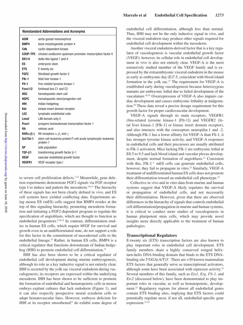
Vasculogenesis, the de novo emergence of primordial endo-thelial cells and blood vessels, begins within the mammalian extraembryonic yolk sac soon after gastrulation, when signals from the visceral endoderm serve to pattern the underlying mesoderm.1,2 Development of the circulatory system is there-fore dependent on these early events during which mesoder-mal precursors are specified toward an endothelial cell lineage (Figure 1).
Signaling PathwaysFibroblast growth factor 2 (FGF2 or bFGF) and bone morpho-genetic protein 4 (BMP4) are 2 key signaling components that are not only important for specification of mesoderm3–5 but also for its differentiation toward endothelial and hematopoi-etic cell fates.6–8 BMP4 is sufficient to induce mesodermal dif-ferentiation in vitro, whereas its ablation results in a failure to generate mesoderm and leads to early embryonic lethality.9–11 Embryos deficient for downstream effectors of BMP4 signal-ing, such as Smad5, lack an organized yolk sac vasculature similar to Bmp4–/– mutant mice.6 Smad4 null mice display sim-ilar phenotypes and are also remarkably smaller in size, owing
Review
© 2013 American Heart Association, Inc.
Circulation Research is available at http://circres.ahajournals.org DOI: 10.1161/CIRCRESAHA.113.300506
Abstract: The circulatory system is the first organ system to develop in the vertebrate embryo and is critical throughout gestation for the delivery of oxygen and nutrients to, as well as removal of metabolic waste products from, growing tissues. Endothelial cells, which constitute the luminal layer of all blood and lymphatic vessels, emerge de novo from the mesoderm in a process known as vasculogenesis. The vascular plexus that is initially formed is then remodeled and refined via proliferation, migration, and sprouting of endothelial cells to form new vessels from preexisting ones during angiogenesis. Mural cells are also recruited by endothelial cells to form the surrounding vessel wall. During this vascular remodeling process, primordial endothelial cells are specialized to acquire arterial, venous, and blood-forming hemogenic phenotypes and functions. A subset of venous endothelium is also specialized to become lymphatic endothelium later in development. The specialization of all endothelial cell subtypes requires extrinsic signals and intrinsic regulatory events, which will be discussed in this review. (Circ Res. 2013;112:1272-1287.)
Key Words: arterio-venous specification ■ endothelial cells ■ hemogenic endothelium ■ lymphatic endothelium
Regulation of Endothelial Cell Differentiation and Specification
Kathrina L. Marcelo, Lauren C. Goldie, Karen K. Hirschi
Original received January 9, 2013; revision received March 15, 2013; accepted March 29, 2013. In February 2013, the average time from submission to first decision for all original research papers submitted to Circulation Research was 11.98 days.
From the Interdepartmental Program in Developmental Biology (K.L.M.), Department of Pediatrics (L.C.G., K.K.H.), Center for Cell and Gene Therapy (K.L.M., L.C.G., K.K.H.), and USDA/ARS Children’s Nutrition Research Center (K.L.M., L.C.G., K.K.H.), Baylor College of Medicine, Houston, TX (K.L.M., L.C.G., K.K.H.); and Yale Cardiovascular Research Center, Yale University School of Medicine, New Haven, CT (K.K.H.).
Correspondence to Karen K. Hirschi, PhD, Yale Cardiovascular Research Center and Yale Stem Cell Center, Yale University School of Medicine, 300 George St, New Haven, CT 06511. E-mail [email protected]
by guest on June 18, 2018http://circres.ahajournals.org/
Dow
nloaded from
Marcelo et al Endothelial Cell Specification 1273
to severe cell proliferation defects.7,12 Meanwhile, gene dele-tion experiments demonstrate FGF2 signals via FGF receptor type I to induce and pattern the mesoderm.5,8,13 The hierarchy of these signals has not been clearly defined in vivo, and ES cell systems have yielded conflicting data. Experiments us-ing mouse ES (mES) cells suggest that BMP4 resides at the top of this signaling hierarchy, promoting mesoderm forma-tion and initiating a FGF2-dependent program to regulate the specification of angioblasts, which are thought to function as endothelial progenitors.9,10,14 In contrast, differentiation stud-ies in human ES cells, which require bFGF for survival and growth even in an undifferentiated state, do not support a role for this factor in the commitment of mesodermal cells to the endothelial lineage.15 Rather, in human ES cells, BMP4 is a critical regulator that functions downstream of Indian hedge-hog (IHH) to promote endothelial cell differentiation.15
IHH has also been shown to be a critical regulator of endothelial cell development during murine embryogenesis, although its role as a key inductive signal is not entirely clear. IHH is secreted by the yolk sac visceral endoderm during vas-culogenesis; its receptors are expressed within the underlying mesoderm. IHH has been shown to be sufficient to promote the formation of endothelial and hematopoietic cells in mouse embryo explant cultures that lack endoderm (Figure 1), and it can also respecify prospective neural ectoderm cells to adapt hematovascular fates. However, embryos deficient for IHH or its receptor smoothened16 do exhibit some degree of
endothelial cell differentiation, although less than normal. Thus, IHH may not be the only inductive signal in vivo, and the visceral endoderm may produce other signals required for endothelial cell development within the mesoderm.
Another visceral endoderm-derived factor that is a key regu-lator of vasculogenesis is vascular endothelial growth factor (VEGF); however, its cellular role in endothelial cell develop-ment in vivo is also not entirely clear. VEGF-A is the most extensively studied member of the VEGF family and is ex-pressed by the extraembryonic visceral endoderm in the mouse as early as embryonic day (E)7.5, coincident with blood island formation in the yolk sac.17 The requirement for VEGF-A is established early during vasculogenesis because heterozygous mutants are embryonic lethal due to failed development of the vasculature.18,19 Overexpression of VEGF-A also impairs car-diac development and causes embryonic lethality at midgesta-tion.20 These data reveal a precise dosage requirement for this growth factor for proper cardiovascular development.
VEGF-A signals through its main receptors, VEGFR1 (fms-related tyrosine kinase-1 [Flt-1]) and VEGFR2 (fe-tal liver kinase-1 [Flk-1] or kinase insert domain receptor), and also interacts with the coreceptors neuropilin-1 and -2. Although Flk-1 has a lower affinity for VEGF-A than Flt-1, it has stronger tyrosine kinase activity, and VEGF-A responses in endothelial cells and their precursors are usually attributed to Flk-1 activation. Mice lacking Flk-1 are embryonic lethal at E8.5 to 9.5 and lack blood island and vascular plexus develop-ment, despite normal formation of angioblasts.21 Consistent with this, Flk-1–/– mES cells can generate endothelial cells; however, they fail to propagate in vitro.22 Similarly, VEGF-A treatment of undifferentiated human ES cells does not promote their differentiation toward an endothelial cell phenotype.15
Collective in vivo and in vitro data from murine and human systems suggest that VEGF-A likely regulates the survival or propagation of endothelial cells, and not necessarily their differentiation. However, given that there are observed differences in the hierarchy of signals that controls endothelial cell differentiation/propagation in murine and human systems, it is critical to conduct more studies of vasculogenesis in human pluripotent stem cells, which may provide novel insights more directly applicable to the treatment of human pathologies.
Transcriptional RegulatorsE-twenty six (ETS) transcription factors are also known to play important roles in endothelial cell development. ETS family members share a highly conserved winged helix-turn-helix DNA-binding domain that binds to the ETS DNA-binding site 5′GGA(A/T)3′. There are ≈30 known mammalian ETS factors that generally serve as transcriptional activators, although some have been associated with repressor activity.23 Several members of this family, such as Ets1, Erg, Fli-1, and Etv2 (discussed below), have been demonstrated to play im-portant roles in vascular, as well as hematopoietic, develop-ment.23 Regulatory regions for almost all endothelial genes contain ETS binding sites, implying that ETS factors could potentially regulate most, if not all, endothelial-specific gene expression.23,24
Nonstandard Abbreviations and Acronyms
AGM aorta-gonad-mesonephros
BMP4 bone morphogenetic protein 4
Cdk cyclin-dependent kinase
COUP-TFII chicken ovalbumin upstream promoter–transcription factor II
Dll1/4 delta-like ligand 1 and 4
ES embryonic stem
ETS E-twenty six
FGF2 fibroblast growth factor 2
Flk-1 fetal liver kinase-1
Flt-1 fms-related tyrosine kinase-1
Foxc1/2 forkhead box C1 and C2
HSC hematopoietic stem cell
HSPC hematopoietic stem/progenitor cell
IHH Indian hedgehog
Kdr kinase insert domain receptor
LEC lymphatic endothelial cells
Lmo2 LIM domain only 2
Prox1 prospero homeodomain transcription factor 1
RA retinoic acid
RARα/β/γ RA receptors α, β, and γ
Scl/Tal-1 stem cell leukemia protein/T-cell acute lymphocytic leukemia protein-1
SP side population
TGF-β1 transforming growth factor β-1
VEGF vascular endothelial growth factor
VEGFR1 VEGF receptor type I
by guest on June 18, 2018http://circres.ahajournals.org/
Dow
nloaded from
1274 Circulation Research April 26, 2013
Although many ETS factors are thought to share redun-dant functions, recent studies have demonstrated a specific and critical role for Etv2(ER71/etsrp), or ETS variant 2, in the differentiation of mesodermal progenitors toward an endothelial cell fate. Etv2 expression is initially observed to be more widespread within the primitive streak meso-derm; however, it is soon restricted to developing vascular endothelial cells,25–28 and its critical function in their de-velopment is evolutionarily conserved.25–27,29–31 Morpholino knockdown25 of, or mutations30 within, the etsrp gene in zebrafish results in complete abrogation of circulation, to-gether with loss of expression of vascular endothelial cell markers, including VE-cadherin/Cdh5 and VEGFR2/Flk-1, in developing embryos.25,30 Likewise, Etv2–/– mice are em-bryonic lethal, and despite having intact mesoderm, they fail to generate angioblasts,25 a yolk sac primitive vascular plexus, dorsal aortae, or endocardium.26,32 Overexpression of Etv2 leads to ectopic expression of endothelial markers in multiple cell types in vivo,25,27,29,31 suggesting an expan-sion of the differentiation of endothelial cells. Thus, Etv2 seems to be unique among ETS domain transcription fac-tors in that it is both necessary and sufficient for in vivo vasculogenesis.25–27,30,32
Importantly, maintaining Etv2 expression within a narrow developmental window is critical for proper vasculogenesis. Transient Etv2 activation in the mesoderm is sufficient to initiate endothelial cell development,28,32,33 and Etv2 is rap-idly downregulated once endothelial cells begin to mature.28 Constitutive expression of Etv2 in endothelial cells under con-trol of the Tie2 promoter results in dilated yolk sac vessels and impaired remodeling of the extraembryonic vasculature, eventually leading to embryonic lethality.34 Interestingly, this
also results in perturbation of normal hematopoiesis attribut-able to endothelialization of hematopoietic progenitor cells that do not form properly.35
Molecular interactions between Etv2 and other signaling factors that regulate endothelial cell development are not yet clearly defined; however, Etv2 is closely associated with and known to precede Flk-1 expression during vasculogenesis.25,26,30 As previously mentioned, in vivo disruption of Etv2 or its orthologues results in loss of Flk-1–expressing cells,25,26,30,32 whereas forced expression of this ETS factor leads to ectopic Flk-1 expression.25,27,29,31 Flk-1:GFP transgenic zebrafish embryos injected with etsrp-directed morpholinos demonstrate loss of GFP+ cells in blood vessels.25 Etv2 expression is observed exclusively within Flk-1+ mesodermal cells in mES cell differentiation studies, and its induction within embryoid bodies results in de novo production of Flk-1+ cells.26 Thus, Etv2 seems to regulate expression of VEGF receptor Flk-1 within newly forming endothelial cells. Consistent with this, Etv2 also seems to be required for the generation of Flk-1 + platelet-derived growth factor receptor α–vascular progenitors from Flk-1 + platelet-derived growth factor receptor α + primitive mesoderm.36
Combining what is known about the role of ETS tran-scription factors and the VEGF-A/Flk-1 pathway, we can surmise that ETS transcriptional activity in mesodermal progenitors is necessary, if not sufficient, to induce Flk-1 expression, as well as to promote commitment to an en-dothelial cell fate. VEGF-A signaling via Flk-1 likely regulates the survival and propagation of newly forming en-dothelial cells, although it does not likely directly regulate their differentiation.
Mesoderm
RA
PrimordialEndothelial Cells
HemogenicEndothelial Cells
Arterial Endothelial Cells
VenousEndothelial Cells
Lymphatic Endothelial Cells
VASCULOGENESIS
DEFINITIVE HEMATOPOIESIS
Hematopoietic Progenitor Cells
ARTERIAL -VENOUS SPECIFICATION
Etv2
Runx1
Notch
COUP-TFII
Nrp1
ephrinB2
EphB4
COUP-TFII , Sox18
Prox1
LYMPHANGIOGENESIS
IHHBMP4FGF2
VEGFR2Figure 1. Major extrinsic and intrinsic factors that regulate endothelial cell specification throughout embryonic vascular development. BMP4 indicates bone morphogenetic protein 4; FGF2, fibroblast growth factor 2; IHH, Indian hedgehog; RA, retinoic acid; and VEGFR2, VEGF receptor type I.
by guest on June 18, 2018http://circres.ahajournals.org/
Dow
nloaded from

Marcelo et al Endothelial Cell Specification 1275
Control of Endothelial Cell Proliferation During Vascular Plexus Remodeling
Once endothelial cells are differentiated and coalesce into a vascular plexus, their proliferation must be tightly regulated to achieve proper remodeling of an expanding circulatory net-work. This regulation involves the coordination of multiple signaling pathways that either promote or inhibit endothelial cell cycle progression (summarized in Figure 2).
VEGF-AAs mentioned above, VEGF-A is required to stimulate en-dothelial cell proliferation and does so largely via signaling through Flk-1. Binding of VEGF-A to Flk-1 triggers its auto-phosphorylation, leading to complex formation with integrin αvβ3,35 an endothelial cell adhesion receptor, and αvβ3 auto-phosphorylation.37 Acting in a feedback loop, integrin αvβ3 is also capable of phosphorylating Flk-1, once it is bound by its ligand vitronectin.38 The cross-activation of these 2 receptors leads to the recruitment and activation of Src kinases,39 which mediate the mitogenic effects of VEGF-A40,41 by activating the mitogen-activated protein kinase intracellular signaling pathway.38
Flt-1/VEGFR1 also indirectly controls endothelial cell pro-liferation, by competing with Flk-1 for binding of VEGF-A. Flt-1–/– mutants42,43 and zebrafish flt-1 morphants44 exhibit en-dothelial cell hyperproliferation that leads to abnormal angio-genic development, suggesting that unlike Flk-1, Flt-1 plays a negative role in endothelial cell proliferation. Although VEGF-A has a higher affinity for Flt-1 than Flk-1, Flt-1 has a weaker kinase activity and is thought to act in part as a VEGF-A decoy receptor.45 In support of this idea, genetic studies have shown that the Flt-1 tyrosine kinase domain is dispensable for vasculogenesis,46 whereas the extracellular ligand–binding domain is essential to its ability to regulate proper VEGF-A signaling.45 Moreover, alternative splicing results in a soluble, catalytically inactive Flt-1 isoform that
acts as a molecular sink for VEGF-A and thereby limits Flk-1 ligation and activation.47,48
Retinoic AcidRetinoic acid (RA) signaling also controls endothelial cell proliferation as a negative regulator of cell cycle progression. RA is the biologically active derivative of dietary vitamin A or retinol. In circulation, retinol is complexed to retinoid-binding protein and can enter the cell via a specific receptor, stimulated by retinoic acid 6. Retinol is initially oxidized into retinaldehyde by either alcohol dehydrogenases or retinol dehydrogenases. Retinaldehyde dehydrogenases then catalyze the conversion of retinaldehyde to all-trans-RA, which is released and can be taken up by surrounding tissues. Cellular RA-binding proteins facilitate the uptake of RA in target cells, as well as its transport to the nucleus. RA then binds to nuclear RA receptors (RARα, β, and γ), which heterodimerize with retinoid X receptors (RXRα, β, and γ) to form active transcription factor complexes. These complexes occupy RA-response elements within target genes, altering the binding of transcriptional repressors and coactivators to modulate gene expression. In cells with inactive RA signaling, RA is oxidized for degradation by the cytochrome P450 enzyme, CYP26 (reviewed in Duester49).
In the murine and rat yolk sac, Raldh2 is expressed spe-cifically by the visceral endoderm at the onset of vasculogen-esis,50,51 whereas yolk sac endothelial cells within the adjacent mesoderm exclusively express RA receptors RARα1 and 2.51,52 Mice lacking the enzyme Raldh2 are RA-deficient and die in utero at approximately E10. One of the first morpohological defects observed in these mutants is lack of remodeling and blood vessel dilation within the yolk sac and embryo proper, as well as a marked decrease in blood cell production.51–53 Raldh2–/– mutant endothelial cells are hyperproliferative and exhibit reduced levels of p21(Cdkn1a) and p27(Cdkn1b), members of the Cip/Waf family of cyclin-dependent kinase
Figure 2. Retinoic acid (RA) is produced by the visceral endoderm (VE) and initiates a signaling cascade that regulates control of endothelial cell cycle proliferation. RA indicates retinoic acid; RAR, RA receptor; RBP, retinoid-binding protein; TGFβ, transforming growth factor β; and VEGF, vascular endothelial growth factor. (Illustration credit: Ben Smith).
by guest on June 18, 2018http://circres.ahajournals.org/
Dow
nloaded from
1276 Circulation Research April 26, 2013
(Cdk) inhibitors. In vivo and in vitro studies collectively sug-gest that RA regulates endothelial cell cycle progression by upregulating the expression of p21 and p27, resulting in G1 arrest of endothelial cells52 (Figure 2).
Transforming Growth Factor-β and Extracellular MatrixRA signaling also suppresses endothelial cell proliferation by upregulating transforming growth factor β-1 (TGF-β1). Signal transduction via TGF-β family members is facilitated by heteromeric type I and type II serine/threonine kinase re-ceptors. TGF-β first binds to the constitutively active type II receptor dimer, leading to recruitment, phosphorylation, and activation of type I receptor dimers. Type I receptors in turn phosphorylate receptor-regulated Smad proteins, which func-tion as effectors of TGF-β signaling by binding to common partner Smad proteins (co-Smads), such as Smad4. These TGF-β precomplexes then shuttle to the nucleus, where they associate with other transcription factors to regulate target gene expression (reviewed in Lee and Bae54).
TGF-β signaling contributes to the control of endothe-lial cell proliferation in 2 important ways; first, by inducing the production and deposition of fibronectin, an extracellu-lar matrix protein. Endothelial-derived fibronectin not only promotes visceral endoderm survival and function, which is necessary for VEGF-A production, but also binds to integrins α5β1 and αVβ3 expressed on endothelial cells to elicit oppos-ing effects on cell cycle progression. Fibronectin signaling via α5β1 inhibits proliferation, whereas binding to αVβ3 induces endothelial cell proliferation via the Flk-1/mitogen-activated protein kinase pathway51 (Figure 2). TGF-β signaling also controls endothelial cell cycle progression via the regulation of key cell cycle control genes. As previously discussed, pro-gression through G1 into S phase of the cell cycle is regu-lated by cyclin–cdk complexes, such as cyclin D–Cdk4/6 and cyclin E–Cdk2. Conversely, interaction between CdK inhibi-tors (such as RA-induced p21 and p27) and Cdks promotes cell cycle arrest. TGF-β signaling increases expression and stabilization of p15, a member of the INK4 family of CdK inhibitors.55,56 Increased p15 protein levels in the cytoplasm allow it to bind cyclin D–Cdk4/6 complexes more readily than p27, and active cyclin D–Cdk4/6-p27 complexes are re-placed by inactive cyclin D–Cdk4-p15 complexes. Displaced p27 molecules are shuttled toward and interact with cyclin E–Cdk2 complexes rendering them inactive because p27 bind-ing prevents Cdk2 activation that leads to G1 arrest.57 TGF-β signaling also contributes to stabilization of p27 by blocking its phosphorylation,58 a prerequisite for its export from the nucleus and subsequent degradation.59 Thus, regulation of en-dothelial cell cycle progression and proliferation during vas-cular plexus remodeling requires a balance between multiple signaling effectors and downstream intracellular pathways.
Angiogenic Sprouting: Selection of Tip Versus Stalk Endothelial Cells
Remodeling of the vascular plexus to form a fully differenti-ated circulatory network also involves the sprouting of new blood vessels from preexisting vessels, a process referred to as angiogenesis. Sprouting involves the loss of cell–cell junction
contacts between endothelial cells, degradation of the base-ment membrane by proteases, and migration of tip cells in re-sponse to angiogenic stimuli. Meanwhile, stalk cells trail tip cells and undergo extensive proliferation to expand the grow-ing endothelial tube.60,61 Not all endothelial cells exposed to proangiogenic signals become sprouting tip cells, and tight regulation of tip cell formation is necessary to ensure integrity and proper patterning of the developing vasculature.
Notch and VEGF SignalingInterplay between VEGF-A/Flk-1 and Notch pathways is pri-marily responsible for the specification of tip versus stalk cell identity during sprouting angiogenesis. There are multiple vertebrate Notch genes; humans and mice express ≥4 iso-forms (Notch1-4), and Notch1 and Notch4 are expressed by vascular endothelial cells. Evolutionarily conserved across di-verse species, Notch-mediated signaling occurs over short dis-tances and is commonly implicated in cell fate determination. Single-pass transmembrane Notch receptors are activated by engagement of ligands presented by neighboring cells and are cleaved by metalloprotease enzymes that remove most of the receptor extracellular domain. An enzymatic complex known as γ-secretase then cleaves the Notch transmembrane domain, releasing a Notch intracellular domain which is capable of translocating to the nucleus and forming complexes with other DNA-binding proteins (reviewed in Bigas and Espinosa62).
Expression of the Notch ligand, delta-like ligand 4 (Dll4) by tip cells of growing vascular sprouts is VEGF-dependent.63–65 Thus, VEGF-A provides an angiogenic stimulus that guides sprouting behavior and subsequent initiation of endothelial cell proliferation.65–68 Dll4 activates Notch signaling in adjacent endothelial cells, reducing their ability to respond to VEGF-A stimulation by downregulating Flk-1 expression.65,67 Dll4 thus functions to suppress tip cell fate by effectively conferring stalk cell identity on these neighboring cells.67,69,70 Impairment of Dll4-Notch signaling results in dysregulation of tip versus stalk cell specification. Dll4 heterozygous mutant mice and dll4 zebrafish morphants display increased angiogenic sprouting because of increased tip cell formation.63,67–69,71 Loss of Dll4 in mice also results in increased proliferation of endothelial cells.65,68,69 Similarly, inhibition of Notch signaling results in increased sprouting, increased expression of tip cell–associated genes,67 hyperproliferative endothelial cells, and a denser and more highly interconnected vasculature.67,69,70 Endothelial-specific deletion of Notch1 is sufficient to increase tip cell numbers.71 Interestingly, when both VEGF-A and Notch signaling are impaired, excessive sprouting and tip cell formation are not observed.63,67,69 VEGF and Notch regulation of angiogenic sprouting is therefore clearly complex; VEGF-A provides proangiogenic stimulus and induces Flk-1 expressing cells to sprout and form new vessels, while simultaneously activating Dll4-Notch pathway signaling to maintain the necessary balance between tip and stalk cell specification.
Interactions between the VEGF and Notch pathways are further complicated by the involvement of other members of these signaling pathways. For example, VEGFR-3 (or Flt-4) becomes ectopically expressed throughout remodeling vessels when Notch signaling is blocked.69 Endothelial cell–specific deletion of Flt-4 in mice leads to excessive angiogenic
by guest on June 18, 2018http://circres.ahajournals.org/
Dow
nloaded from

Marcelo et al Endothelial Cell Specification 1277
sprouting and branching in retinal vessels, resembling loss of Notch signaling. VEGF-C binds to Flt-4 in tip cells, promot-ing stalk cell specification.72 Flt-1 also acts as a negative regu-lator of tip cell differentiation in a Notch-dependent manner. Loss of flt-1 function in zebrafish results in a phenotype con-sistent with loss of Notch signaling, whereas overexpression of the notch1a intracellular domain in flt-1 morphants restores proper vessel branching and patterning.44
Other Notch ligands, including Dll1 and Jag1, are also in-volved in angiogenic sprouting with reported proangiogenic effects.73,74 Heterozygous deletion of Dll1 leads to reduced vessel branching, although it is not expressed by endothelial cells themselves. Dll1 is expressed by cells peripheral to the vasculature and acts as an extrinsic signal to activate Notch signaling in presumptive tip cells, which reinforces VEGF-A action by further inducing Dll4 expression to promote tip cell specification.73 Endothelial-specific deletion of Jag1 also results in reduced vessel sprouting. Stalk cells are enriched for Jag1, which antagonizes Dll4 signaling by competing for Notch receptor binding, and reduces Dll4-Notch activation in adjacent tip cells.74
Although Notch ligands can exert opposing effects on en-dothelial cells, proteasomal degradation of Notch signaling components positively regulates angiogenesis and tip cell specification. Endothelial cell–specific deletion or morpholi-no knockdown of Fbxw7, an E3 ubiquitin ligase, results in in-creased stability of Notch signaling components and impaired tip cell formation.75 B-cell chronic lymphocytic leukemia/lymphoma 6–associated zinc finger protein, a component of the cullin3-based E3 ligase complex, exerts its proangiogenic effect by promoting degradation of Notch signaling factor, C-promoter binding factor 1, thereby curtailing Notch signal-ing and lateral inhibition of tip cell phenotype.76
The specification of tip and stalk cell identities among en-dothelial cells is dynamic in both in vivo and in vitro sys-tems.77,78 Arima et al77 speculated that, in addition to other key functions, tip cells create a localized microenvironment that facilitates further elongation by the successive tip cell. Interestingly, among the genes enriched in tip cells are ex-tracellular matrix–degrading enzymes and secreted proteins nidogen-1 and -2, which interact with extracellular matrix proteins, such as laminin and collagen, to stabilize basement membranes.79 Laminin and laminin-binding integrins have also been found to stimulate Dll4-Notch signaling during tip versus stalk cell selection.80,81
MicroRNAMicroRNAs (miRs) are emerging as new players in the regu-lation of tip versus stalk cell specification of endothelial cells. Primary microRNA transcripts are transcribed from intergenic or intronic genomic sequences and then cleaved by the RNase III endonuclease Drosha, allowing export to the cytoplasm. Once there, miR transcripts are processed through the endo-nuclease Dicer to yield mature miRs, small RNA fragments 18 to 25 nucleotides long. These bind to the 3′-UTRs of their respective target mRNAs to mediate posttranscriptional gene silencing.82 In vivo silencing of either Dicer or Drosha results in reduced sprouting of endothelial cells, demonstrating an es-sential role for miRs in angiogenesis.83
miR-126 is known to elicit proangiogenic effects by re-pressing endothelial expression of Sprouty-related protein 1, a known negative regulator of VEGF-A signaling, thereby enhancing the angiogenic response of endothelial cells to VEGF-A signaling.84,85 Another proangiogenic microRNA is miR-221, which is also expressed by embryonic endothe-lial cells. Among its targets is p27,86 previously implicated in regulating endothelial cell proliferation in response to RA signaling.52 Notch signaling represses expression of miR-221, thereby allowing p27 expression and subsequent regulation of stalk cell proliferation.86 miR-27b also promotes endothelial cell tip specification, as well as increased branching of the vascular plexus, by downregulating Dll4 and Sprouty homo-logue 2 (Spry2) expression.87
Arterial-Venous SpecificationIn a properly remodeled blood circulatory system, the vascu-lature is subdivided into 2 interconnected, yet structurally and functionally distinct, networks of arteries and veins. Unlike veins, arteries have to withstand high blood pressure and are surrounded by several layers of smooth muscle cells and con-nective tissues. It was originally thought that endothelial cell identity is determined during development by hemodynamic forces. For example, Le Noble et al88 demonstrated that rerout-ing of blood flow in chick yolk sac arteries causes downregu-lation of arterial markers and conversion of endothelial cells to a venous phenotype. However, other studies suggest that arterial versus venous endothelial cell specification may also be genetically determined.
The ephrin family of receptor tyrosine kinases constitutes the largest family of growth factor receptors, which use membrane-tethered ephrins as their ligands. EphrinB2 marks arterial endothelial cells, whereas its receptor EphB4 is expressed by venous endothelium (Figure 1). Their complementary and mutually exclusive expression is observed during early stages of vascular plexus remodeling before the establishment of systemic blood circulation, which supports a genetic basis for the specification of arteries and veins.89 Furthermore, ephrinB2 knockout mice display severe angiogenic defects and a general lack of vessel remodeling, implying that reciprocal ephrinB2–EphB4 signaling may be crucial for the establishment of arteries and veins.89 In further support of a genetic program for arterio-venous specification, endothelial cells surrounding blood islands of the chick chorioallantoic membrane express both the arterial marker neuropilin-1 (Nrp-1) and venous marker Nrp-2 before the onset of blood flow.90 Hemodynamic forces may therefore not be required to induce the expression of genes that dictate arterial and venous endothelial cell phenotype and function but may instead be required for their maintenance, perhaps, via regulation of soluble signals that modulate their expression.
The roles of specific molecular signaling pathways in arterio-venous specification are just beginning to be defined (Figure 1). Thus far, it seems that VEGF-A binding to Flk 1 and its coreceptor Nrp-191 activates Notch signaling,92 pro-motes EphrinB2 expression, and suppresses EphB4,93 thereby establishing (or maintaining) arterial identity, while suppress-ing a venous fate in endothelial cells. Forkhead box C1 and C2 (Foxc1/2) is thought to act downstream of VEGF-A to induce
by guest on June 18, 2018http://circres.ahajournals.org/
Dow
nloaded from
1278 Circulation Research April 26, 2013
Dll494 and Hey295 expression by binding Foxc sites on their promoters.95 Mice deficient in either Foxc1 or Foxc2 present with an abnormal vasculature that lacks arteries but displays normal venous marker expression, suggesting a general fail-ure of arterialization.94 Consistent with this idea, inhibition of Notch signaling in vivo results in an arterial to venous fate switch.96 mES cell differentiation studies also demonstrate that high VEGF-A levels induce arterial endothelial cell phenotype, whereas low to intermediate concentrations are associated with a venous identity. Wnt and hedgehog signal-ing pathways are also involved in the specification of arte-rial endothelial cells. β-catenin, a transcriptional coactivator of Wnt signaling pathway, upregulates Dll4 expression and amplifies Notch signaling by forming a complex with Notch intracellular domain to induce arterial fate in vascular endo-thelial cells.97,98 In the mouse, Sonic hedgehog acts upstream of VEGF-A92 via the Smoothened receptor to promote arterial endothelial cell specification and repress venous fate. This is similarly observed in zebrafish smoothened mutants, which have disrupted hedgehog signaling and develop an expand-ed posterior cardinal vein at the expense of a dorsal aorta.99 Constitutive activation of hedgehog signaling in ptc1; ptc2 mutants also results in conversion of the posterior cardinal vein into an artery.100
Specification of venous endothelial cells is thought to be mediated primarily by chicken ovalbumin upstream promot-er–transcription factor II (COUP-TFII), a member of the orphan nuclear receptor superfamily (Figure 1). Endothelial cell–specific deletion of COUP-TFII results in arterialization of veins, whereas ectopic expression results in fusion of veins and arteries.101 COUP-TFII functions cell autonomously by suppressing Nrp-1 function and downstream Notch signal-ing to maintain vein identity.101 Thus, the establishment and maintenance of arterial and venous endothelial cell fates are likely to be dynamically regulated by a balance of signals within the milieu of the remodeling vasculature during de-velopment; the relevance of such signaling processes to the maintenance of the postnatal established vasculature is not yet clear.
Lymphangiogenesis: Specification of LECs From Venous Endothelial Cells
The lymphatic system is essential for returning extravasated proteins and cells to the bloodstream and for proper immune system surveillance. These roles are clearly distinct from those of blood vessels and require extensive specialization of LEC. LEC are derived from embryonic veins; thus, their develop-ment requires the further specification of a subpopulation of venous endothelial cells to a lymphatic fate via signals distinct from those that specify venous endothelium.
Prospero Homeodomain Transcription Factor 1Prospero homeodomain transcription factor (Prox1) is a ho-meobox transcription factor expressed by the subpopulation of venous endothelial cells that sprout and migrate to form the lymphatic system.102 A lineage-specific marker for LEC, Prox1, is essential for embryonic lymphatic development103,104 (Figure 1). Venous endothelial cells begin expressing Prox1 at E9.75,105 and by E11.5, clusters of Prox1-positive lymphatic
progenitor cells emerge along the anterioposterior axis of the cardinal vein.102,103,106 These clusters undergo ballooning to generate intermediate structures known as lymph sacs, from which lymphatic vessels emerge by a combination of sprout-ing102,106 and migration of streams of Nrp-2–positive LEC from the cardinal vein.106 Ablation of Prox1 leads to a general fail-ure of LEC specification and ultimately, a failure of lymphatic vascular development.103–105 Prox1-null endothelial cells are devoid of expression of LEC markers, such as lymphatic ves-sel hyaluronan receptor 1 and VEGFR-3, and instead express blood vessel endothelial cell markers.104 Conditional deletion of Prox1 in venous endothelium results in a severe reduc-tion in LEC numbers.102 Moreover, LEC were shown to de-differentiate into blood endothelial cells on postnatal Prox1 inactivation.105 Prox1 is, therefore, not only necessary for the specification of lymphatic lineage but also for maintenance of LEC identity.
Sox18Although Prox1 expression in venous endothelium is necessary for LEC specification, not all venous endothelial cells express Prox1; instead, its initial expression is clustered and polarized along the anterior-posterior axis of the cardinal vein,103,106 suggesting tight regulation of its expression. Both the SRY-related genes Sox18 and COUP-TFII, which is necessary for specifying venous endothelial cell fate, were shown to regulate Prox1 (Figure 1). Sox18 is expressed by cardinal vein endothelial cells that eventually express Prox1 and form the lymphatic vasculature. Ectopic Sox18 expression in blood endothelial cells induces Prox1 and LEC marker expression; conversely, Sox18-null embryos lack LEC specification from cardinal vein endothelium.107 Although Sox18 was demonstrated to induce Prox1 expression by direct binding to its promoter,107 it is not sufficient to control the distinct expression pattern exhibited by Prox1 because arteries also express Sox18.108
COUP-TFIICOUP-TFII potentially cooperates with Sox18 to tempo-rally and spatially regulate Prox1 expression. In addition to being expressed by venous endothelium, COUP-TFII is also expressed by LEC,109 and its conditional ablation in ve-nous endothelial cells either alone102 or ubiquitously during early development110 results in impaired lymphatic specifica-tion.102,110 Along with Sox18, COUP-TFII binds to the regula-tory regions of Prox1111 to cooperatively induce its expression in LEC progenitors. COUP-TFII also forms a stable complex with Prox1 itself109,111 to maintain Prox1 expression in LEC and regulate lineage-specific expression of markers, such as Flt4/VEGFR3109 and Nrp-2.110 Accordingly, siRNA-mediated knockdown experiments demonstrated a cell-autonomous re-quirement for COUP-TFII during maintenance of lymphatic identity and VEGFC-induced lymphangiogenesis.110
MicroRNAMicroRNAs have also been reported to regulate Prox1 expres-sion. For example, miR-181a binds to the 3′-untranslated re-gion of Prox1 mRNA to inhibit its expression, thereby acting as a negative regulator of lymphatic specification and develop-ment.112 Similarly, miR-31 is expressed in venous endothelial
by guest on June 18, 2018http://circres.ahajournals.org/
Dow
nloaded from

Marcelo et al Endothelial Cell Specification 1279
cells and inhibits lymphatic specification by modulating Prox1 transcript and protein levels.113
VEGF-CAfter specification of LEC via induction of Prox1 expres-sion in a subset of venous endothelium, LEC migrate out of the veins to form lymph sacs and then lymphatic vessels.106 VEGF-C is necessary for the initial steps of lymphangio-genic development, and, as discussed above, expression of its receptors Flt-4 and Nrp-2 by LEC enables VEGF-C re-sponsiveness.109,110 Homozygous and heterozygous deletion of VEGF-C in mouse embryos results in a failure of Prox1-positive LEC progenitors to sprout, suggesting that specifi-cation of lymphatic lineage can proceed without VEGF-C, but that subsequent morphogenesis requires precise dosage of this secreted factor. VEGF-C, but not VEGF-A, can res-cue the sprouting defect observed in these mutants, indicating specificity of Flt-4 function during lymphangiogenesis.114 In support of this, several studies have demonstrated a direct as-sociation between inhibition of Flt4 function and impairment of lymphatic system development.115–118 Several mechanisms for regulation of Flt-4 expression in LEC have been proposed. As previously mentioned, COUP-TFII induces expression of both Flt-4 and Nrp-2.109,110 Tbx1 binds to an enhancer el-ement in Flt-4 regulatory regions to induce VEGF-C recep-tor expression in LEC.119 Tbx1 itself is required for lymphatic vessel development.119 The ETS factor Ets-2 interacts with Prox1 to induce Flt-4 gene expression and effectively en-hance the lymphangiogenic response to VEGF-C.120 Finally, Notch has been shown to induce Flt-4 expression in endothe-lial cells by binding to its promoter in a complex consisting of C promoter–binding factor 1/suppressor of hairless/lag1, allowing these endothelial cells to increase their responsive-ness to VEGF-C.121 Interestingly, the Notch signaling pathway may also be active during the specification phase of lymphan-giogenic development. Knockdown of Dll4, or its receptors Notch1b or Notch6, in zebrafish severely impairs LEC sprout-ing and lymphatic system development.122
Hemogenic Endothelium: Specification of Hematopoietic Potential in Primordial
Endothelial CellsHemangioblast Versus Hemogenic Endothelium in Blood ProductionThe initial but transient phase of blood development known as primitive hematopoiesis involves the emergence of aggregates of mesoderm-derived cells at E7.0 to 7.5 within regions of the murine yolk sac referred to as blood islands (Figure 3A). Cells within these structures develop into nucleated primitive eryth-roblasts and megakaryocytes, whereas those along the border flatten and become endothelial cells.123,124 The close spatial and temporal development of blood and endothelial cells dur-ing this early phase of hematopoietic development led to the suggestion that a common bipotent progenitor known as the hemangioblast gives rise to both endothelial and hematopoi-etic cells. To date, however, the existence of hemangioblasts in any species and their relative contribution to hematopoietic development remain controversial.
A second wave of blood development known as definitive hematopoiesis involves the generation of all blood cell lineag-es. The extraembryonic yolk sac is the first site of definitive hematopoietic development (≈E8.25; Figure 3A); thereafter, multilineage hematopoietic stem/progenitor cells (HSPC) develop successively within the extraembryonic placenta (≈E9.5), as well as the embryonic aorta/gonad/mesonephros (AGM) region (≈E9.5–10.0), the fetal liver (E11.0–12.0), and the fetal bone marrow, where hematopoiesis persists postna-tally. It is during definitive hematopoiesis that a subset of the vascular endothelium, referred to as hemogenic endothelium, acquires blood-forming potential and gives rise to the first multilineage HSPC.
The first observations suggesting that endothelial cells give rise to blood cells came from studies of developing chick embryos, wherein endothelial cells of the aorta and yolk sac were found to reside in close proximity to hemoglobin-filled progeny, which seemed to project into the blood vessel lumen and bud off into the blood plasma.125,126 Many years later, in an attempt to prove this experimentally, the chick endothe-lium was labeled with Di-AcLDL before the onset of defini-tive hematopoiesis; dye-retaining hematopoietic clusters were demonstrated in direct physical association with the vascular endothelium of the aorta and within the blood circulation.127 Aortic endothelial-attached hematopoietic clusters were also noted by others in both mouse and human embryos, as well as in the lumens of murine yolk sac blood vessels.128–131 Phenotypic analysis of blood and endothelial cells revealed common expression of cell-surface markers during embryo-genesis,127,128,132 providing further evidence of their coordinat-ed development in vivo.
More recent lineage tracing and single cell–imaging studies have provided direct evidence that multipotent hematopoietic progenitors arise developmentally from vascular endothelium. Cells initially expressing the endothelial cell marker cluster of differentiation 31 (CD31) and then blood markers, CD41, stem cell antigen-1, and c-Kit, were shown to emerge from the ventral wall of explanted murine dorsal aortae.133 Endothelial lineage tracing also revealed hematopoietic cell clusters budding from reporter-labeled endothelium of the murine dorsal aorta, which were then found in the fetal liver and subsequently within the adult hematopoietic system.134 Similar studies in zebrafish demonstrated early hematopoietic cells emerging from the ventral wall of the dorsal aorta, expressing definitive hematopoietic genes and repopulating the adult hematopoietic organ.135,136 These studies provide direct evidence that specialized hemogenic endothelial cells generate definitive blood cells; however, we still know relatively little regarding mechanisms of their specification.
Phenotype of Hemogenic Endothelial CellsTo gain insight into the specialized functions of hemogenic endothelial cells and the molecular pathways that regulate their development, it is necessary to define their phenotype relative to non–blood-forming vascular endothelium. In vitro assays performed on sorted cell populations expressing both endothelial and hematopoietic markers have been used to try to define the phenotype of endothelial cells with hematopoietic potential. VE-cadherin+Ter119−CD45− endothelial cells that
by guest on June 18, 2018http://circres.ahajournals.org/
Dow
nloaded from

1280 Circulation Research April 26, 2013
coexpress CD31, CD34, and Flk-1 were isolated from mu-rine yolk sac and AGM and shown to exhibit lymphopoietic potential in colony formation assays.137 CD34+CD31+CD45− endothelial cells from human yolk sac and AGM also display lymphomyelopoietic potential when cultured with supporting stromal cells.130 In other studies, Flk-1+VE-cad+CD45− endo-thelial cells were shown to generate Flk-1−CD45+ hematopoi-etic colonies more readily than those that were VE-cad−.138 Although these studies suggest that expression of VE-cadherin is necessary for hematopoietic potential, it was not found to be required for hemogenic endothelial cell function in the murine yolk sac.139 However, Hoechst dye-efflux capacity, characteris-tic of side population (SP) cells,139,140 was found to be an essen-tial characteristic of all blood-forming cells in the murine yolk sac and AGM. Furthermore, it was found that among SP cells within these tissues, Flk-1+c-Kit+CD45− SP cells specifically represent hemogenic endothelial cells that exhibit multilineage hematopoietic potential on a clonal level139 (Figure 3B). These cells are distinguishable from non–blood-forming vascular endothelial cells, which do not exhibit an SP phenotype and do not express c-Kit. It was further established that Flk-1+c-Kit+CD45− SP cells give rise to Flk-1−c-Kit+CD45+ SP cells, which represent multilineage HSPC within these tissues.139 HSPC are distinguishable from mature blood cell types, which neither exhibit an SP phenotype nor express Flk-1 or c-Kit, al-though they retain expression of CD45. Thus, flow cytometric analysis enables identification and isolation of these cell types for further investigation of function and molecular regulation.
Specification of Hemogenic Endothelial CellsOnly a small subset of the primordial endothelium within the yolk sac and AGM acquires hematopoietic potential and gives rise to HSPC. Thus, they are present in small numbers in these tissues, which limits the study of their development and function. Consequently, little is known about the molecular mechanisms that regulate hemogenic endothelial cell specification and their generation of HSPC; nonetheless, we review what is known thus far about factors implicated in these processes and summarize the impact of their deletion on murine hematopoietic development in the Table.
Retinoic AcidRA signaling has been found to be essential for normal he-matopoiesis in the developing embryo and in the adult. Early studies using vitamin A (retinol)-deficient quail embryos dem-onstrated impaired proliferation and survival of yolk sac prim-itive erythrocytes, despite their normal formation.142 Genetic approaches also demonstrated a requirement for RA receptors in these processes. Mice deficient for the RXRα exhibit im-paired fetal liver erythropoiesis.143 Ablation of RARγ results in reduced generation of bone marrow hematopoietic stem cell (HSC), whereas its overexpression in HSPC promotes an undifferentiated phenotype, suggesting a role for RA sig-naling in maintaining the balance between HSC self-renewal and differentiation.144 Finally, chromosomal translocations involving the RARα gene result in acute promyelocytic leu-kemia, wherein the resulting oncogenic fusion proteins block
Figure 3. Parallel development of blood and blood vessels in early vertebrate embryos. A, In the extraembryonic yolk sac (YS), aggregates of mesodermal cells form blood islands composed of primitive erythroid cells and angioblasts during the late gastrulation phases (E7.0). A primitive vascular plexus is soon formed by coalescence of blood islands, and by the time definitive hematopoietic development is initiated (E8.25), the vascular plexus begins to be remodel to a vascular network composed of large vitelline veins, as well as smaller, pruned vessels. Definitive hematopoiesis is marked by the generation of multiple, if not all, blood cells lineages from hemogenic endothelial cells, which give rise to blood clusters initially observed in the yolk sac tissues and later intraembryonically, on the ventral side of the aorta-gonads-mesonephros region (AGM; as early as E9.5). B, Intrinsic and extrinsic factors that regulate hemogenic endothelium specification and hematopoietic stem/progenitor cell development. Phenotypic markers that define relevant cell subpopulations during definitive hematopoiesis are also indicated. RA indicates retinoic acid; and RAR, RA receptor. (Illustration credit: Ben Smith).
by guest on June 18, 2018http://circres.ahajournals.org/
Dow
nloaded from

Marcelo et al Endothelial Cell Specification 1281
terminal differentiation and increase self-renewal of leukemic progenitor cells.145
RA-deficient (Raldh2–/–) embryos also exhibit impaired blood cell production, coincident with previously discussed endothelial cell hyperproliferation.51,52 Although primitive he-matopoiesis is not impaired in Raldh2–/– embryos, they do not undergo hemogenic endothelial cell development and exhibit a concomitant decrease in HSPC production.139 The expres-sion of key hematopoietic regulators, including c-Kit, Runx1, and c-Myb, is also significantly downregulated in Raldh2–/– embryos. Restoration of RA signaling in Raldh2–/– mutants in utero, via maternal feeding of active RA during the critical
developmental period encompassing the onset of definitive hematopoiesis, rescues the observed defects in hemogenic endothelial cell development and definitive hematopoiesis. RA signaling is therefore required for proper specification of hemogenic endothelial cells from primordial endothelium and for subsequent formation of multilineage HSPC;139 thus, de-fining the direct targets of RA during these processes is criti-cally important.
c-KitEndothelial cells with hematopoietic potential uniquely express c-Kit, which is downregulated in the absence of RA signaling. c-Kit/CD117 belongs to a family of growth factor receptors with tyrosine kinase activity. Binding of its ligand, stem cell factor, results in dimerization or oligomerization of receptors and activation of tyrosine kinase activity, leading to rapid phosphorylation of its cellular targets. The result is the activation of intracellular signal transduction pathways, which depending on the cellular context, include the Src kinase, Ras/mitogen-activated protein kinase, and phospholipase C-γ pathways. c-Kit can also signal via phosphatidylinositol-3 kinase, which is usually associated with control of cell proliferation, migration, and adhesion. Activation of the Janus kinase/signal transducer and activator of transcription pathway by c-Kit is most commonly associated with maintenance and self-renewal of HSC (reviewed in Sattler and Salgia146). Deletion of c-Kit results in early embryonic lethality in murine embryos,147 whereas adult mice harboring mutant c-Kit alleles demonstrate reduced HSC numbers (reviewed in Sharma et al148). Fetal and adult tissues carrying mutations in the c-Kit kinase domain also display decreased colony-forming activity in vitro, suggesting that c-Kit expression is required for HSC expansion. c-Kit is also frequently coexpressed with Flk-1 in vascular endothelial cells that are associated with hematopoietic clusters in the murine yolk sac and dorsal aorta,129 suggesting that c-Kit may be involved in hemogenic endothelial cell formation and function.
Hematopoietic Stem/Progenitor Cell Development and Propagation
Although the signaling pathways that regulate the specifica-tion of hemogenic endothelial cells from primordial endothe-lial cells have not been clearly defined, more is known about regulators of hematopoietic stem and progenitor cell genera-tion and function.
Notch SignalingSeveral studies demonstrate the involvement of Notch signal-ing in multiple stages of blood cell development or function. For example, Notch1–/– mutant explants exhibit significantly impaired hematopoietic colony formation, and transplanta-tion studies indicate that Notch1 is required for HSC activ-ity and definitive hematopoiesis in both mouse yolk sac and AGM. Thus, Notch1 seems to be required for the generation of HSC from hemogenic endothelial cells,149 as well as for the establishment of long-term, self-renewing HSC.150 Ablation of recombining binding protein suppressor of hairless-jκ, a co-factor for Notch signaling, also leads to loss of HSPC and loss of expression of the GATA2 transcription factor,151,152 which
Table. Loss-of-Function Phenotypes of Genes Associated With Hemogenic Endothelial Cell and HSPC Development
Gene Loss-of-Function Phenotype Reference(s)
Hemogenic endothelial cell specification
Raldh2 Embryonic lethal at E9.5–10.0; severe anemia and cardiovascular defects; impaired hemogenic endothelial cell and HSPC development
53,139
c-Kit Early to midgestational embryonic lethality; severe anemia and failure of hematopoietic stem cell, erythroid, and mast cell development; mutant, viable alleles display pleiotropic hematopoietic defects
146,147
HSPC formation and function
Notch1 Embryonic lethal at E9.5–10.0; severe vascular defects and impaired HSPC maintenance and self-renewal
148,152
RBPjκ Embryonic lethal at E10.5; failure to undergo embryonic turning, severe defects in somite development; complete absence of intraembryonic (AGM) hematopoiesis
150,151
Runx1 Embryonic lethal at E11.5–12.5; severe hemorrhaging and defects in definitive hematopoiesis
155
Scl/Tal1 Embryonic lethal at E9.5; severe anemia, edema, and impaired definitive hematopoiesis
163
Lmo2 Embryonic lethal at E10.5; failure of yolk sac hematopoiesis (erythropoiesis)
164
GATA1 Embryonic lethal at E10.5–11.5; severe anemia and defects in erythropoiesis
141
GATA2 Embryonic lethal at E10.5; severe anemia and impaired proliferation and survival of hematopoietic progenitors
165
c-Myb Embryonic lethal at E15.5; failure of erythropoiesis and maintenance of hematopoietic progenitor cells
178
AGM indicates aorta-gonad-mesonephros; and HSPC, hematopoietic stem/progenitor cell.
by guest on June 18, 2018http://circres.ahajournals.org/
Dow
nloaded from
1282 Circulation Research April 26, 2013
is required for maintenance of HSC in an undifferentiated state. There may be earlier functions of Notch signaling in the specification of hemogenic endothelium because Notch1–/–
Notch4–/– mutants die at midgestation with defects in vascular remodeling and lack of establishment of specialized endothe-lial cell phenotypes.153
Runx1Also known as AML1, Runx1 has been shown to function downstream of Notch during developmental and adult hema-topoiesis.149,154,155 Runx1 encodes a transcription factor that het-erodimerizes with core binding factor β via its evolutionarily conserved Runt homology domain. Ablation of Runx1 results in embryonic lethality at E11.0 to E12.0 because of defects in definitive hematopoiesis.156 Runx1 expression by yolk sac and AGM endothelial cells is observed before the emergence of hematopoietic clusters131,157 and is necessary for HSPC for-mation.131,158,159 Once the endothelial-to-hematopoietic pro-genitor transition is completed, Runx1 function is no longer required.159 A novel role for Runx1 as a transcriptional repres-sor in hemogenic endothelial cells was suggested by mES cell differentiation studies, in which Flk-1–expressing cells were shown to initiate Runx1 expression, leading to silencing of the Flk-1 promoter and downregulation of receptor expression.160 This is consistent with a previous study demonstrating sup-pression of the Flk-1 regulatory region in Runx1-expressing hemogenic endothelial cells.161 Flk-1 gene silencing has been independently associated with the generation of HSPC from hemogenic endothelial cells.
Stem Cell Leukemia Protein/LIM Domain Only 2/Gata1/2 Transcription ComplexThe regulatory regions of hematopoietic genes, including the early hematopoietic marker CD41, are simultaneously bound by Runx1 and a transcription factor complex composed of stem cell leukemia protein (Scl)/T-cell acute lymphocytic leu-kemia protein-1 (Tal-1), GATA1, and GATA2.162,163 Mice de-ficient in Scl/Tal-1, LIM domain only 2 (Lmo2), or GATA1/2 all display midgestation lethality caused by severe hemato-poietic defects,164–167 suggesting that these transcription factors have closely related roles during embryonic hematopoiesis. Morpholino knockdown of scl and lmo2 in zebrafish results in similar phenotypes, including loss of intra-aortic hema-topoietic clusters and impaired expression of hematopoietic genes.168,169 ES cells deficient in Lmo2 also fail to contribute to blood cell production or to the endothelium of large ves-sels in adult chimeric mice.170,171 Both Scl/Tal-1 and Lmo2 are expressed by hematopoietic and vascular endothelial cells in midgestation embryos.169,170,172 Scl is necessary for the initial specification of hematopoietic lineage173,174 but dispensable for later fetal liver and adult HSC functions.173,175 Expression of GATA2, which promotes the expansion of hematopoietic progenitors, is enriched within blood islands during primi-tive hematopoiesis.166,176 This is followed by induction of GATA1 expression,176 which in turn represses GATA2, lead-ing to a developmental switch from proliferation to differ-entiation.177 Lmo2 acts as a bridging molecule between Scl/Tal-1 and GATA-binding proteins in a transcriptional trans-activating complex that has been shown to drive vertebrate
hematopoietic commitment.168,178 Although this complex is known to play a critical role in primitive hematopoiesis, it also likely regulates HSPC production and function during defini-tive hematopoiesis.
c-MybAnother transcriptional regulator thought to play a role in the regulation of HSPC self-renewal and propagation is c-Myb. Ablation of c-Myb results in embryonic lethality at E15.5 caused by failure of erythroid and myeloid development,179 whereas conditional deletion in adult bone marrow results in depletion and impaired self-renewal of the HSC pool.180 Loss of function studies in zebrafish also demonstrate impaired definitive hematopoietic development, despite intact primi-tive hematopoiesis.181 AGM explants from c-Myb–/– mutant mice also yield fewer HSPC,182 which usually express high c-Myb levels that are downregulated during differentiation. Consistent with this finding, overexpression of c-Myb results in inhibition of erythromyeloid differentiation.183
Blood FlowIn addition to soluble factors and transcriptional regulators, blood flow and hemodynamic forces also play a role in the regulation of HSC development. Endothelial cells lining blood vessels are known to respond to fluid forces through a complex of receptors capable of transducing biomechanical signals. Such signals initiate a cascade of downstream signal-ing events that precipitate changes in gene expression and cell behavior (reviewed in Li et al184). For example, shear stress regulates endothelium-dependent NO production, which is a known regulator of hematopoiesis. A decrease in hemato-poietic potential of the murine AGM is observed when the NO pathway is inhibited.185 In zebrafish, inhibition of NO synthase, the enzyme responsible for NO production, leads to a decrease in transplantable HSC; Nos3–/– knockout mice exhibit a similar phenotype.186 Recapitulating the hemody-namic environment of the murine AGM in an in vitro ES cell system significantly increases the generation of CD31-expressing endothelial cells, upregulates expression of HSC-associated genes, such as Runx1 and c-Myb, and increases the hematopoietic potential of differentiated CD41+c-Kit+ HSC.185 Zebrafish silent heart mutants, which lack a heartbeat and an established circulation, display impaired HSC generation.186 Ncx1–/–-null embryos, which also lack a heartbeat, properly initiate HSC development within the placenta, yet display fewer CD41-expressing hematopoietic cells than their wild-type counterparts.187 Collectively, these studies suggest that blood flow forces may not be required for hemogenic endothe-lial cell specification but may be necessary to promote HSC emergence or maintenance.
SummaryDuring vasculogenesis, multipotent mesodermal progenitors give rise to endothelial cells that form a primary vascular plexus. Tight control of endothelial cell proliferation and mi-gration, as well as proper specification of tip and stalk cells, enables endothelial cell sprouting and vascular plexus remod-eling. Endothelial cells become phenotypically and function-ally specialized because they acquire arterial, venous, and
by guest on June 18, 2018http://circres.ahajournals.org/
Dow
nloaded from

Marcelo et al Endothelial Cell Specification 1283
hemogenic fates. A subpopulation of venous endothelial cells later assumes a lymphatic endothelial identity. Precise regula-tion of these processes is not only necessary to ensure proper architecture and integrity of the circulatory system but also to guarantee that endothelial cells that line the blood and lym-phatic vessels are capable of performing their necessary func-tions. Perturbations of the molecular mechanisms that govern these events during development often results in embryonic lethality; defects in these processes postnatally contribute to the development of prevalent vascular, lymphatic, and hema-topoietic pathologies. An integrated understanding of how endothelial cells acquire distinct phenotypes, thus enabling specialized functions, not only extends our knowledge of the development of the circulatory system but also provides in-sights needed to optimize the treatment of prevalent diseases, as well as generate such cell types for tissue engineering and regenerative medicine applications.
Sources of FundingThese studies were supported by National Institutes of Health (NIH) grants R01-HL077675 and R01-HL096360 to K.K. Hirschi, and USDA/ARS CRIS 6250 51000 to K.K. Hirschi and L.C. Goldie.
DisclosuresNone.
References 1. Belaoussoff M, Farrington SM, Baron MH. Hematopoietic induction
and respecification of A-P identity by visceral endoderm signaling in the mouse embryo. Development. 1998;125:5009–5018.
2. Vokes SA, Krieg PA. Endoderm is required for vascular endothe-lial tube formation, but not for angioblast specification. Development. 2002;129:775–785.
3. Azar Y, Eyal-Giladi H. Marginal zone cells–the primitive streak-inducing component of the primary hypoblast in the chick. J Embryol Exp Morphol. 1979;52:79–88.
4. Marom K, Levy V, Pillemer G, Fainsod A. Temporal analysis of the early BMP functions identifies distinct anti-organizer and mesoderm patterning phases. Dev Biol. 2005;282:442–454.
5. Saxton TM, Pawson T. Morphogenetic movements at gastrulation re-quire the SH2 tyrosine phosphatase Shp2. Proc Natl Acad Sci U S A. 1999;96:3790–3795.
6. Chang H, Huylebroeck D, Verschueren K, Guo Q, Matzuk MM, Zwijsen A. Smad5 knockout mice die at mid-gestation due to multiple embryonic and extraembryonic defects. Development. 1999;126:1631–1642.
7. Sirard C, de la Pompa JL, Elia A, Itie A, Mirtsos C, Cheung A, Hahn S, Wakeham A, Schwartz L, Kern SE, Rossant J, Mak TW. The tumor sup-pressor gene Smad4/Dpc4 is required for gastrulation and later for anterior development of the mouse embryo. Genes Dev. 1998;12:107–119.
8. Yamaguchi TP, Harpal K, Henkemeyer M, Rossant J. FGFr-1 is required for embryonic growth and mesodermal patterning during mouse gastrula-tion. Genes Dev. 1994;8:3032–3044.
9. Park C, Afrikanova I, Chung YS, Zhang WJ, Arentson E, Fong Gh Gh, Rosendahl A, Choi K. A hierarchical order of factors in the generation of FLK1- and SCL-expressing hematopoietic and endothelial progenitors from embryonic stem cells. Development. 2004;131:2749–2762.
10. Pearson S, Sroczynska P, Lacaud G, Kouskoff V. The stepwise speci-fication of embryonic stem cells to hematopoietic fate is driven by se-quential exposure to Bmp4, activin A, bFGF and VEGF. Development. 2008;135:1525–1535.
11. Winnier G, Blessing M, Labosky PA, Hogan BL. Bone morphogenetic protein-4 is required for mesoderm formation and patterning in the mouse. Genes Dev. 1995;9:2105–2116.
12. Yang X, Li C, Xu X, Deng C. The tumor suppressor SMAD4/DPC4 is essential for epiblast proliferation and mesoderm induction in mice. Proc Natl Acad Sci U S A. 1998;95:3667–3672.
13. Flamme I, Risau W. Induction of vasculogenesis and hematopoiesis in vitro. Development. 1992;116:435–439.
14. Huber TL, Zhou Y, Mead PE, Zon LI. Cooperative effects of growth factors involved in the induction of hematopoietic mesoderm. Blood. 1998;92:4128–4137.
15. Kelly MA, Hirschi KK. Signaling hierarchy regulating human endothelial cell development. Arterioscler Thromb Vasc Biol. 2009;29:718–724.
16. Byrd N, Becker S, Maye P, Narasimhaiah R, St-Jacques B, Zhang X, McMahon J, McMahon A, Grabel L. Hedgehog is required for murine yolk sac angiogenesis. Development. 2002;129:361–372.
17. Breier G, Clauss M, Risau W. Coordinate expression of vascular en-dothelial growth factor receptor-1 (flt-1) and its ligand suggests a paracrine regulation of murine vascular development. Dev Dyn. 1995;204:228–239.
18. Ferrara N, Carver-Moore K, Chen H, Dowd M, Lu L, O’Shea KS, Powell-Braxton L, Hillan KJ, Moore MW. Heterozygous embryonic lethality induced by targeted inactivation of the VEGF gene. Nature. 1996;380:439–442.
19. Carmeliet P, Ferreira V, Breier G, et al. Abnormal blood vessel devel-opment and lethality in embryos lacking a single VEGF allele. Nature. 1996;380:435–439.
20. Miquerol L, Langille BL, Nagy A. Embryonic development is disrupted by modest increases in vascular endothelial growth factor gene expression. Development. 2000;127:3941–3946.
21. Shalaby F, Rossant J, Yamaguchi TP, Gertsenstein M, Wu XF, Breitman ML, Schuh AC. Failure of blood-island formation and vasculogenesis in Flk-1-deficient mice. Nature. 1995;376:62–66.
22. Schuh AC, Faloon P, Hu QL, Bhimani M, Choi K. In vitro hematopoietic and endothelial potential of flk-1(–/–) embryonic stem cells and embryos. Proc Natl Acad Sci U S A. 1999;96:2159–2164.
23. Dejana E, Taddei A, Randi AM. Foxs and Ets in the transcriptional regula-tion of endothelial cell differentiation and angiogenesis. Biochim Biophys Acta. 2007;1775:298–312.
24. De Val S, Black BL. Transcriptional control of endothelial cell develop-ment. Dev Cell. 2009;16:180–195.
25. Sumanas S, Lin S. Ets1-related protein is a key regulator of vasculogenesis in zebrafish. PLoS Biol. 2006;4:e10.
26. Lee D, Park C, Lee H, Lugus JJ, Kim SH, Arentson E, Chung YS, Gomez G, Kyba M, Lin S, Janknecht R, Lim DS, Choi K. ER71 acts downstream of BMP, Notch, and Wnt signaling in blood and vessel progenitor specifi-cation. Cell Stem Cell. 2008;2:497–507.
27. Salanga MC, Meadows SM, Myers CT, Krieg PA. ETS family protein ETV2 is required for initiation of the endothelial lineage but not the hema-topoietic lineage in the Xenopus embryo. Dev Dyn. 2010;239:1178–1187.
28. Kataoka H, Hayashi M, Nakagawa R, Tanaka Y, Izumi N, Nishikawa S, Jakt ML, Tarui H, Nishikawa S. Etv2/ER71 induces vascular mesoderm from Flk1+PDGFRα+ primitive mesoderm. Blood. 2011;118:6975–6986.
29. Sumanas S, Gomez G, Zhao Y, Park C, Choi K, Lin S. Interplay among Etsrp/ER71, Scl, and Alk8 signaling controls endothelial and myeloid cell formation. Blood. 2008;111:4500–4510.
30. Pham VN, Lawson ND, Mugford JW, Dye L, Castranova D, Lo B, Weinstein BM. Combinatorial function of ETS transcription factors in the developing vasculature. Dev Biol. 2007;303:772–783.
31. De Val S, Chi NC, Meadows SM, Minovitsky S, Anderson JP, Harris IS, Ehlers ML, Agarwal P, Visel A, Xu SM, Pennacchio LA, Dubchak I, Krieg PA, Stainier DY, Black BL. Combinatorial regulation of endo-thelial gene expression by ets and forkhead transcription factors. Cell. 2008;135:1053–1064.
32. Ferdous A, Caprioli A, Iacovino M, Martin CM, Morris J, Richardson JA, Latif S, Hammer RE, Harvey RP, Olson EN, Kyba M, Garry DJ. Nkx2-5 transactivates the Ets-related protein 71 gene and specifies an endothelial/endocardial fate in the developing embryo. Proc Natl Acad Sci U S A. 2009;106:814–819.
33. Wareing S, Mazan A, Pearson S, Göttgens B, Lacaud G, Kouskoff V. The Flk1-Cre-mediated deletion of ETV2 defines its narrow temporal requirement during embryonic hematopoietic development. Stem Cells. 2012;30:1521–1531.
34. Hayashi M, Pluchinotta M, Momiyama A, Tanaka Y, Nishikawa S, Kataoka H. Endothelialization and altered hematopoiesis by persistent Etv2 expression in mice. Exp Hematol. 2012;40:738–750.e11.
35. West XZ, Meller N, Malinin NL, Deshmukh L, Meller J, Mahabeleshwar GH, Weber ME, Kerr BA, Vinogradova O, Byzova TV. Integrin β3 cross-talk with VEGFR accommodating tyrosine phosphorylation as a regula-tory switch. PLoS One. 2012;7:e31071.
36. Sakurai H, Era T, Jakt LM, Okada M, Nakai S, Nishikawa S, Nishikawa S. In vitro modeling of paraxial and lateral mesoderm differentiation reveals early reversibility. Stem Cells. 2006;24:575–586.
by guest on June 18, 2018http://circres.ahajournals.org/
Dow
nloaded from

1284 Circulation Research April 26, 2013
37. Byzova TV, Goldman CK, Pampori N, Thomas KA, Bett A, Shattil SJ, Plow EF. A mechanism for modulation of cellular responses to VEGF: activation of the integrins. Mol Cell. 2000;6:851–860.
38. Soldi R, Mitola S, Strasly M, Defilippi P, Tarone G, Bussolino F. Role of alphavbeta3 integrin in the activation of vascular endothelial growth factor receptor-2. EMBO J. 1999;18:882–892.
39. Mahabeleshwar GH, Feng W, Reddy K, Plow EF, Byzova TV. Mechanisms of integrin-vascular endothelial growth factor receptor cross-activation in angiogenesis. Circ Res. 2007;101:570–580.
40. Werdich XQ, Penn JS. Src, Fyn and Yes play differential roles in VEGF-mediated endothelial cell events. Angiogenesis. 2005;8:315–326.
41. Eliceiri BP, Paul R, Schwartzberg PL, Hood JD, Leng J, Cheresh DA. Selective requirement for Src kinases during VEGF-induced angiogenesis and vascular permeability. Mol Cell. 1999;4:915–924.
42. Fong GH, Rossant J, Gertsenstein M, Breitman ML. Role of the Flt-1 receptor tyrosine kinase in regulating the assembly of vascular endothe-lium. Nature. 1995;376:66–70.
43. Fong GH, Zhang L, Bryce DM, Peng J. Increased hemangioblast commit-ment, not vascular disorganization, is the primary defect in flt-1 knock-out mice. Development. 1999;126:3015–3025.
44. Krueger J, Liu D, Scholz K, Zimmer A, Shi Y, Klein C, Siekmann A, Schulte-Merker S, Cudmore M, Ahmed A, le Noble F. Flt1 acts as a nega-tive regulator of tip cell formation and branching morphogenesis in the zebrafish embryo. Development. 2011;138:2111–2120.
45. Hiratsuka S, Nakao K, Nakamura K, Katsuki M, Maru Y, Shibuya M. Membrane fixation of vascular endothelial growth factor receptor 1 ligand-binding domain is important for vasculogenesis and angiogenesis in mice. Mol Cell Biol. 2005;25:346–354.
46. Hiratsuka S, Minowa O, Kuno J, Noda T, Shibuya M. Flt-1 lacking the tyrosine kinase domain is sufficient for normal development and angio-genesis in mice. Proc Natl Acad Sci U S A. 1998;95:9349–9354.
47. Kondo K, Hiratsuka S, Subbalakshmi E, Matsushime H, Shibuya M. Genomic organization of the flt-1 gene encoding for vascular endothelial growth factor (VEGF) receptor-1 suggests an intimate evolutionary rela-tionship between the 7-Ig and the 5-Ig tyrosine kinase receptors. Gene. 1998;208:297–305.
48. He Y, Smith SK, Day KA, Clark DE, Licence DR, Charnock-Jones DS. Alternative splicing of vascular endothelial growth factor (VEGF)-R1 (FLT-1) pre-mRNA is important for the regulation of VEGF activity. Mol Endocrinol. 1999;13:537–545.
49. Duester G. Retinoic acid synthesis and signaling during early organogen-esis. Cell. 2008;134:921–931.
50. Båvik C, Ward SJ, Ong DE. Identification of a mechanism to localize gen-eration of retinoic acid in rat embryos. Mech Dev. 1997;69:155–167.
51. Bohnsack BL, Lai L, Dolle P, Hirschi KK. Signaling hierarchy down-stream of retinoic acid that independently regulates vascular remodeling and endothelial cell proliferation. Genes Dev. 2004;18:1345–1358.
52. Lai L, Bohnsack BL, Niederreither K, Hirschi KK. Retinoic acid regu-lates endothelial cell proliferation during vasculogenesis. Development. 2003;130:6465–6474.
53. Niederreither K, Subbarayan V, Dollé P, Chambon P. Embryonic retinoic acid synthesis is essential for early mouse post-implantation development. Nat Genet. 1999;21:444–448.
54. Lee KY, Bae SC. TGF-beta-dependent cell growth arrest and apoptosis. J Biochem Mol Biol. 2002;35:47–53.
55. Hannon GJ, Beach D. p15INK4B is a potential effector of TGF-beta-induced cell cycle arrest. Nature. 1994;371:257–261.
56. Reynisdóttir I, Polyak K, Iavarone A, Massagué J. Kip/Cip and Ink4 Cdk inhibitors cooperate to induce cell cycle arrest in response to TGF-beta. Genes Dev. 1995;9:1831–1845.
57. Reynisdóttir I, Massagué J. The subcellular locations of p15(Ink4b) and p27(Kip1) coordinate their inhibitory interactions with cdk4 and cdk2. Genes Dev. 1997;11:492–503.
58. Polyak K, Kato JY, Solomon MJ, Sherr CJ, Massague J, Roberts JM, Koff A. p27Kip1, a cyclin-Cdk inhibitor, links transforming growth factor-beta and contact inhibition to cell cycle arrest. Genes Dev. 1994;8:9–22.
59. Kim TY, Kim WI, Smith RE, Kay ED. Role of p27(Kip1) in cAMP- and TGF-beta2-mediated antiproliferation in rabbit corneal endothelial cells. Invest Ophthalmol Vis Sci. 2001;42:3142–3149.
60. Carmeliet P, Jain RK. Molecular mechanisms and clinical applications of angiogenesis. Nature. 2011;473:298–307.
61. Adams RH, Alitalo K. Molecular regulation of angiogenesis and lymphan-giogenesis. Nat Rev Mol Cell Biol. 2007;8:464–478.
62. Bigas A, Espinosa L. Hematopoietic stem cells: to be or Notch to be. Blood. 2012;119:3226–3235.
63. Lobov IB, Renard RA, Papadopoulos N, Gale NW, Thurston G, Yancopoulos GD, Wiegand SJ. Delta-like ligand 4 (Dll4) is induced by VEGF as a negative regulator of angiogenic sprouting. Proc Natl Acad Sci U S A. 2007;104:3219–3224.
64. Noguera-Troise I, Daly C, Papadopoulos NJ, Coetzee S, Boland P, Gale NW, Lin HC, Yancopoulos GD, Thurston G. Blockade of Dll4 inhib-its tumour growth by promoting non-productive angiogenesis. Nature. 2006;444:1032–1037.
65. Ridgway J, Zhang G, Wu Y, et al. Inhibition of Dll4 signalling in-hibits tumour growth by deregulating angiogenesis. Nature. 2006;444:1083–1087.
66. Gerhardt H, Golding M, Fruttiger M, Ruhrberg C, Lundkvist A, Abramsson A, Jeltsch M, Mitchell C, Alitalo K, Shima D, Betsholtz C. VEGF guides angiogenic sprouting utilizing endothelial tip cell filopodia. J Cell Biol. 2003;161:1163–1177.
67. Suchting S, Freitas C, le Noble F, Benedito R, Bréant C, Duarte A, Eichmann A. The Notch ligand Delta-like 4 negatively regulates endo-thelial tip cell formation and vessel branching. Proc Natl Acad Sci U S A. 2007;104:3225–3230.
68. Leslie JD, Ariza-McNaughton L, Bermange AL, McAdow R, Johnson SL, Lewis J. Endothelial signalling by the Notch ligand Delta-like 4 restricts angiogenesis. Development. 2007;134:839–844.
69. Siekmann AF, Lawson ND. Notch signalling limits angiogenic cell behav-iour in developing zebrafish arteries. Nature. 2007;445:781–784.
70. Sainson RC, Aoto J, Nakatsu MN, Holderfield M, Conn E, Koller E, Hughes CC. Cell-autonomous notch signaling regulates endothelial cell branching and proliferation during vascular tubulogenesis. FASEB J. 2005;19:1027–1029.
71. Hellström M, Phng LK, Hofmann JJ, et al. Dll4 signalling through Notch1 regulates formation of tip cells during angiogenesis. Nature. 2007;445:776–780.
72. Tammela T, Zarkada G, Nurmi H, et al. VEGFR-3 controls tip to stalk conversion at vessel fusion sites by reinforcing Notch signalling. Nat Cell Biol. 2011;13:1202–1213.
73. Napp LC, Augustynik M, Paesler F, Krishnasamy K, Woiterski J, Limbourg A, Bauersachs J, Drexler H, Le Noble F, Limbourg FP. Extrinsic Notch ligand Delta-like 1 regulates tip cell selection and vascular branch-ing morphogenesis. Circ Res. 2012;110:530–535.
74. Benedito R, Roca C, Sörensen I, Adams S, Gossler A, Fruttiger M, Adams RH. The notch ligands Dll4 and Jagged1 have opposing effects on angiogenesis. Cell. 2009;137:1124–1135.
75. Izumi N, Helker C, Ehling M, Behrens A, Herzog W, Adams RH. Fbxw7 controls angiogenesis by regulating endothelial Notch activity. PLoS One. 2012;7:e41116.
76. Ohnuki H, Inoue H, Takemori N, Nakayama H, Sakaue T, Fukuda S, Miwa D, Nishiwaki E, Hatano M, Tokuhisa T, Endo Y, Nose M, Higashiyama S. BAZF, a novel component of cullin3-based E3 ligase complex, mediates VEGFR and Notch cross-signaling in angiogenesis. Blood. 2012;119:2688–2698.
77. Arima S, Nishiyama K, Ko T, Arima Y, Hakozaki Y, Sugihara K, Koseki H, Uchijima Y, Kurihara Y, Kurihara H. Angiogenic morphogenesis driven by dynamic and heterogeneous collective endothelial cell movement. Development. 2011;138:4763–4776.
78. Jakobsson L, Franco CA, Bentley K, Collins RT, Ponsioen B, Aspalter IM, Rosewell I, Busse M, Thurston G, Medvinsky A, Schulte-Merker S, Gerhardt H. Endothelial cells dynamically compete for the tip cell posi-tion during angiogenic sprouting. Nat Cell Biol. 2010;12:943–953.
79. del Toro R, Prahst C, Mathivet T, Siegfried G, Kaminker JS, Larrivee B, Breant C, Duarte A, Takakura N, Fukamizu A, Penninger J, Eichmann A. Identification and functional analysis of endothelial tip cell-enriched genes. Blood. 2010;116:4025–4033.
80. Estrach S, Cailleteau L, Franco CA, Gerhardt H, Stefani C, Lemichez E, Gagnoux-Palacios L, Meneguzzi G, Mettouchi A. Laminin-binding integ-rins induce Dll4 expression and Notch signaling in endothelial cells. Circ Res. 2011;109:172–182.
81. Stenzel D, Franco CA, Estrach S, Mettouchi A, Sauvaget D, Rosewell I, Schertel A, Armer H, Domogatskaya A, Rodin S, Tryggvason K, Collinson L, Sorokin L, Gerhardt H. Endothelial basement membrane limits tip cell formation by inducing Dll4/Notch signalling in vivo. EMBO Rep. 2011;12:1135–1143.
82. Anand S, Cheresh DA. Emerging role of micro-RNAs in the regulation of angiogenesis. Genes Cancer. 2011;2:1134–1138.
83. Kuehbacher A, Urbich C, Zeiher AM, Dimmeler S. Role of Dicer and Drosha for endothelial microRNA expression and angiogenesis. Circ Res. 2007;101:59–68.
by guest on June 18, 2018http://circres.ahajournals.org/
Dow
nloaded from
Marcelo et al Endothelial Cell Specification 1285
84. Fish JE, Santoro MM, Morton SU, Yu S, Yeh RF, Wythe JD, Ivey KN, Bruneau BG, Stainier DY, Srivastava D. miR-126 regulates angiogenic signaling and vascular integrity. Dev Cell. 2008;15:272–284.
85. Wang S, Aurora AB, Johnson BA, Qi X, McAnally J, Hill JA, Richardson JA, Bassel-Duby R, Olson EN. The endothelial-specific mi-croRNA miR-126 governs vascular integrity and angiogenesis. Dev Cell. 2008;15:261–271.
86. Nicoli S, Knyphausen CP, Zhu LJ, Lakshmanan A, Lawson ND. miR-221 is required for endothelial tip cell behaviors during vascular devel-opment. Dev Cell. 2012;22:418–429.
87. Biyashev D, Veliceasa D, Topczewski J, Topczewska JM, Mizgirev I, Vinokour E, Reddi AL, Licht JD, Revskoy SY, Volpert OV. miR-27b controls venous specification and tip cell fate. Blood. 2012;119:2679–2687.
88. Le Noble F, Moyon D, Pardanaud L, Yuan L, Djonov V, Matthijsen R, Bréant C, Fleury V, Eichmann A. Flow regulates arterial-venous differen-tiation in the chick embryo yolk sac. Development. 2004;131:361–375.
89. Wang HU, Chen ZF, Anderson DJ. Molecular distinction and angiogenic interaction between embryonic arteries and veins revealed by ephrin-B2 and its receptor Eph-B4. Cell. 1998;93:741–753.
90. Herzog Y, Guttmann-Raviv N, Neufeld G. Segregation of arterial and venous markers in subpopulations of blood islands before vessel forma-tion. Dev Dyn. 2005;232:1047–1055.
91. Gu C, Rodriguez ER, Reimert DV, Shu T, Fritzsch B, Richards LJ, Kolodkin AL, Ginty DD. Neuropilin-1 conveys semaphorin and VEGF signaling during neural and cardiovascular development. Dev Cell. 2003;5:45–57.
92. Lawson ND, Vogel AM, Weinstein BM. sonic hedgehog and vascular en-dothelial growth factor act upstream of the Notch pathway during arterial endothelial differentiation. Dev Cell. 2002;3:127–136.
93. Lawson ND, Scheer N, Pham VN, Kim CH, Chitnis AB, Campos-Ortega JA, Weinstein BM. Notch signaling is required for arterial-venous differentiation during embryonic vascular development. Development. 2001;128:3675–3683.
94. Seo S, Fujita H, Nakano A, Kang M, Duarte A, Kume T. The forkhead transcription factors, Foxc1 and Foxc2, are required for arterial specifi-cation and lymphatic sprouting during vascular development. Dev Biol. 2006;294:458–470.
95. Hayashi H, Kume T. Foxc transcription factors directly regulate Dll4 and Hey2 expression by interacting with the VEGF-Notch signaling path-ways in endothelial cells. PLoS One. 2008;3:e2401.
96. Lanner F, Sohl M, Farnebo F. Functional arterial and venous fate is determined by graded VEGF signaling and notch status during em-bryonic stem cell differentiation. Arterioscler Thromb Vasc Biol. 2007;27:487–493.
97. Yamamizu K, Matsunaga T, Uosaki H, Fukushima H, Katayama S, Hiraoka-Kanie M, Mitani K, Yamashita JK. Convergence of Notch and beta-catenin signaling induces arterial fate in vascular progenitors. J Cell Biol. 2010;189:325–338.
98. Corada M, Nyqvist D, Orsenigo F, Caprini A, Giampietro C, Taketo MM, Iruela-Arispe ML, Adams RH, Dejana E. The Wnt/beta-catenin pathway modulates vascular remodeling and specification by upregulat-ing Dll4/Notch signaling. Dev Cell. 2010;18:938–949.
99. Williams C, Kim SH, Ni TT, Mitchell L, Ro H, Penn JS, Baldwin SH, Solnica-Krezel L, Zhong TP. Hedgehog signaling induces arterial en-dothelial cell formation by repressing venous cell fate. Dev Biol. 2010;341:196–204.
100. Wilkinson RN, Koudijs MJ, Patient RK, Ingham PW, Schulte-Merker S, van Eeden FJ. Hedgehog signaling via a calcitonin receptor-like receptor can induce arterial differentiation independently of VEGF signaling in zebrafish. Blood. 2012;120:477–488.
101. You LR, Lin FJ, Lee CT, DeMayo FJ, Tsai MJ, Tsai SY. Suppression of Notch signalling by the COUP-TFII transcription factor regulates vein identity. Nature. 2005;435:98–104.
102. Srinivasan RS, Dillard ME, Lagutin OV, Lin FJ, Tsai S, Tsai MJ, Samokhvalov IM, Oliver G. Lineage tracing demonstrates the venous origin of the mammalian lymphatic vasculature. Genes Dev. 2007;21:2422–2432.
103. Wigle JT, Oliver G. Prox1 function is required for the development of the murine lymphatic system. Cell. 1999;98:769–778.
104. Wigle JT, Harvey N, Detmar M, Lagutina I, Grosveld G, Gunn MD, Jackson DG, Oliver G. An essential role for Prox1 in the in-duction of the lymphatic endothelial cell phenotype. EMBO J. 2002;21:1505–1513.
105. Johnson NC, Dillard ME, Baluk P, McDonald DM, Harvey NL, Frase SL, Oliver G. Lymphatic endothelial cell identity is reversible and its maintenance requires Prox1 activity. Genes Dev. 2008;22:3282–3291.
106. François M, Short K, Secker GA, Combes A, Schwarz Q, Davidson TL, Smyth I, Hong YK, Harvey NL, Koopman P. Segmental territories along the cardinal veins generate lymph sacs via a ballooning mechanism dur-ing embryonic lymphangiogenesis in mice. Dev Biol. 2012;364:89–98.
107. François M, Caprini A, Hosking B, et al. Sox18 induces development of the lymphatic vasculature in mice. Nature. 2008;456:643–647.
108. Pennisi D, Gardner J, Chambers D, Hosking B, Peters J, Muscat G, Abbott C, Koopman P. Mutations in Sox18 underlie cardiovascular and hair follicle defects in ragged mice. Nat Genet. 2000;24:434–437.
109. Lee S, Kang J, Yoo J, Ganesan SK, Cook SC, Aguilar B, Ramu S, Lee J, Hong YK. Prox1 physically and functionally interacts with COUP-TFII to specify lymphatic endothelial cell fate. Blood. 2009;113:1856–1859.
110. Lin FJ, Chen X, Qin J, Hong YK, Tsai MJ, Tsai SY. Direct transcrip-tional regulation of neuropilin-2 by COUP-TFII modulates mul-tiple steps in murine lymphatic vessel development. J Clin Invest. 2010;120:1694–1707.
111. Srinivasan RS, Geng X, Yang Y, Wang Y, Mukatira S, Studer M, Porto MP, Lagutin O, Oliver G. The nuclear hormone receptor Coup-TFII is required for the initiation and early maintenance of Prox1 expression in lymphatic endothelial cells. Genes Dev. 2010;24:696–707.
112. Kazenwadel J, Michael MZ, Harvey NL. Prox1 expression is negatively regulated by miR-181 in endothelial cells. Blood. 2010;116:2395–2401.
113. Pedrioli DM, Karpanen T, Dabouras V, Jurisic G, van de Hoek G, Shin JW, Marino D, Kälin RE, Leidel S, Cinelli P, Schulte-Merker S, Brändli AW, Detmar M. miR-31 functions as a negative regulator of lymphatic vascular lineage-specific differentiation in vitro and vascular develop-ment in vivo. Mol Cell Biol. 2010;30:3620–3634.
114. Karkkainen MJ, Haiko P, Sainio K, Partanen J, Taipale J, Petrova TV, Jeltsch M, Jackson DG, Talikka M, Rauvala H, Betsholtz C, Alitalo K. Vascular endothelial growth factor C is required for sprouting of the first lymphatic vessels from embryonic veins. Nat Immunol. 2004;5:74–80.
115. Ny A, Koch M, Vandevelde W, Schneider M, Fischer C, Diez-Juan A, Neven E, Geudens I, Maity S, Moons L, Plaisance S, Lambrechts D, Carmeliet P, Dewerchin M. Role of VEGF-D and VEGFR-3 in devel-opmental lymphangiogenesis, a chemicogenetic study in Xenopus tad-poles. Blood. 2008;112:1740–1749.
116. Karpanen T, Wirzenius M, Mäkinen T, Veikkola T, Haisma HJ, Achen MG, Stacker SA, Pytowski B, Ylä-Herttuala S, Alitalo K. Lymphangiogenic growth factor responsiveness is modulated by postna-tal lymphatic vessel maturation. Am J Pathol. 2006;169:708–718.
117. Mäkinen T, Jussila L, Veikkola T, Karpanen T, Kettunen MI, Pulkkanen KJ, Kauppinen R, Jackson DG, Kubo H, Nishikawa S, Ylä-Herttuala S, Alitalo K. Inhibition of lymphangiogenesis with resulting lymphede-ma in transgenic mice expressing soluble VEGF receptor-3. Nat Med. 2001;7:199–205.
118. Küchler AM, Gjini E, Peterson-Maduro J, Cancilla B, Wolburg H, Schulte-Merker S. Development of the zebrafish lymphatic system re-quires VEGFC signaling. Curr Biol. 2006;16:1244–1248.
119. Chen L, Mupo A, Huynh T, Cioffi S, Woods M, Jin C, McKeehan W, Thompson-Snipes L, Baldini A, Illingworth E. Tbx1 regulates Vegfr3 and is required for lymphatic vessel development. J Cell Biol. 2010;189:417–424.
120. Yoshimatsu Y, Yamazaki T, Mihira H, Itoh T, Suehiro J, Yuki K, Harada K, Morikawa M, Iwata C, Minami T, Morishita Y, Kodama T, Miyazono K, Watabe T. Ets family members induce lymphangiogen-esis through physical and functional interaction with Prox1. J Cell Sci. 2011;124:2753–2762.
121. Shawber CJ, Funahashi Y, Francisco E, Vorontchikhina M, Kitamura Y, Stowell SA, Borisenko V, Feirt N, Podgrabinska S, Shiraishi K, Chawengsaksophak K, Rossant J, Accili D, Skobe M, Kitajewski J. Notch alters VEGF responsiveness in human and murine endothe-lial cells by direct regulation of VEGFR-3 expression. J Clin Invest. 2007;117:3369–3382.
122. Geudens I, Herpers R, Hermans K, et al. Role of delta-like-4/Notch in the formation and wiring of the lymphatic network in zebrafish. Arterioscler Thromb Vasc Biol. 2010;30:1695–1702.
123. Palis J, Robertson S, Kennedy M, Wall C, Keller G. Development of erythroid and myeloid progenitors in the yolk sac and embryo proper of the mouse. Development. 1999;126:5073–5084.
124. Silver L, Palis J. Initiation of murine embryonic erythropoiesis: a spatial analysis. Blood. 1997;89:1154–1164.
by guest on June 18, 2018http://circres.ahajournals.org/
Dow
nloaded from

1286 Circulation Research April 26, 2013
125. Sabin FR. Studies on the origin of bloodvessels and of red blood- corpuscles as seen in the living blastoderm of chicks during the second day of incubation. Contrib Embryol. 1920;9:215–262
126. Murray PDF. The development in vitro of the blood of the early chick embryo. Proceedings of the Royal Society of London. Series B, Containing Papers of a Biological Character. 1932;111:497–521
127. Jaffredo T, Gautier R, Eichmann A, Dieterlen-Lièvre F. Intraaortic hemopoietic cells are derived from endothelial cells during ontogeny. Development. 1998;125:4575–4583.
128. North TE, de Bruijn MF, Stacy T, Talebian L, Lind E, Robin C, Binder M, Dzierzak E, Speck NA. Runx1 expression marks long-term repopu-lating hematopoietic stem cells in the midgestation mouse embryo. Immunity. 2002;16:661–672.
129. Yoshida H, Takakura N, Hirashima M, Kataoka H, Tsuchida K, Nishikawa S, Nishikawa S. Hematopoietic tissues, as a playground of receptor tyrosine kinases of the PDGF-receptor family. Dev Comp Immunol. 1998;22:321–332.
130. Oberlin E, Tavian M, Blazsek I, Péault B. Blood-forming poten-tial of vascular endothelium in the human embryo. Development. 2002;129:4147–4157.
131. Yokomizo T, Ogawa M, Osato M, Kanno T, Yoshida H, Fujimoto T, Fraser S, Nishikawa S, Okada H, Satake M, Noda T, Nishikawa S, Ito Y. Requirement of Runx1/AML1/PEBP2alphaB for the generation of hae-matopoietic cells from endothelial cells. Genes Cells. 2001;6:13–23.
132. Sugiyama D, Ogawa M, Hirose I, Jaffredo T, Arai K, Tsuji K. Erythropoiesis from acetyl LDL incorporating endothelial cells at the preliver stage. Blood. 2003;101:4733–4738.
133. Boisset JC, van Cappellen W, Andrieu-Soler C, Galjart N, Dzierzak E, Robin C. In vivo imaging of haematopoietic cells emerging from the mouse aortic endothelium. Nature. 2010;464:116–120.
134. Zovein AC, Hofmann JJ, Lynch M, French WJ, Turlo KA, Yang Y, Becker MS, Zanetta L, Dejana E, Gasson JC, Tallquist MD, Iruela-Arispe ML. Fate tracing reveals the endothelial origin of hematopoietic stem cells. Cell Stem Cell. 2008;3:625–636.
135. Bertrand JY, Chi NC, Santoso B, Teng S, Stainier DY, Traver D. Haematopoietic stem cells derive directly from aortic endothelium dur-ing development. Nature. 2010;464:108–111.
136. Kissa K, Herbomel P. Blood stem cells emerge from aortic endothelium by a novel type of cell transition. Nature. 2010;464:112–115.
137. Nishikawa SI, Nishikawa S, Kawamoto H, Yoshida H, Kizumoto M, Kataoka H, Katsura Y. In vitro generation of lymphohematopoietic cells from endothelial cells purified from murine embryos. Immunity. 1998;8:761–769.
138. Fraser ST, Ogawa M, Yu RT, Nishikawa S, Yoder MC, Nishikawa S. Definitive hematopoietic commitment within the embryonic vascular endothelial-cadherin(+) population. Exp Hematol. 2002;30:1070–1078.
139. Goldie LC, Lucitti JL, Dickinson ME, Hirschi KK. Cell signaling direct-ing the formation and function of hemogenic endothelium during murine embryogenesis. Blood. 2008;112:3194–3204.
140. Nadin BM, Goodell MA, Hirschi KK. Phenotype and hematopoietic potential of side population cells throughout embryonic development. Blood. 2003;102:2436–2443.
141. Fujiwara Y, Browne CP, Cunniff K, Goff SC, Orkin SH. Arrested development of embryonic red cell precursors in mouse embry-os lacking transcription factor GATA-1. Proc Natl Acad Sci U S A. 1996;93:12355–12358.
142. Ghatpande S, Ghatpande A, Sher J, Zile MH, Evans T. Retinoid signaling regulates primitive (yolk sac) hematopoiesis. Blood. 2002;99:2379–2386.
143. Makita T, Duncan SA, Sucov HM. Retinoic acid, hypoxia, and GATA factors cooperatively control the onset of fetal liver erythropoietin ex-pression and erythropoietic differentiation. Dev Biol. 2005;280:59–72.
144. Purton LE, Dworkin S, Olsen GH, Walkley CR, Fabb SA, Collins SJ, Chambon P. RARgamma is critical for maintaining a balance between hematopoietic stem cell self-renewal and differentiation. J Exp Med. 2006;203:1283–1293.
145. de Thé H, Le Bras M, Lallemand-Breitenbach V. Acute promyelo-cytic leukemia, arsenic, and pml bodies. The Journal of cell biology. 2012;198:11–21
146. Sattler M, Salgia R. Targeting c-Kit mutations: basic science to novel therapies. Leuk Res. 2004;28 suppl 1:S11–S20.
147. Chabot B, Stephenson DA, Chapman VM, Besmer P, Bernstein A. The proto-oncogene c-kit encoding a transmembrane tyrosine kinase recep-tor maps to the mouse W locus. Nature. 1988;335:88–89.
148. Sharma S, Gurudutta GU, Satija NK, Pati S, Afrin F, Gupta P, Verma YK, Singh VK, Tripathi RP. Stem cell c-KIT and HOXB4 genes: critical roles
and mechanisms in self-renewal, proliferation, and differentiation. Stem Cells Dev. 2006;15:755–778.
149. Kumano K, Chiba S, Kunisato A, Sata M, Saito T, Nakagami-Yamaguchi E, Yamaguchi T, Masuda S, Shimizu K, Takahashi T, Ogawa S, Hamada Y, Hirai H. Notch1 but not Notch2 is essential for generating hematopoi-etic stem cells from endothelial cells. Immunity. 2003;18:699–711.
150. Hadland BK, Huppert SS, Kanungo J, Xue Y, Jiang R, Gridley T, Conlon RA, Cheng AM, Kopan R, Longmore GD. A requirement for Notch1 distinguishes 2 phases of definitive hematopoiesis during development. Blood. 2004;104:3097–3105.
151. Robert-Moreno À, Espinosa L, de la Pompa JL, Bigas A. Rbpjκ-dependent notch function regulates gata2 and is essential for the formation of intra-embryonic hematopoietic cells. Development). 2005;132:1117–1126
152. Oka C, Nakano T, Wakeham A, de la Pompa JL, Mori C, Sakai T, Okazaki S, Kawaichi M, Shiota K, Mak TW, Honjo T. Disruption of the mouse RBP-J kappa gene results in early embryonic death. Development. 1995;121:3291–3301.
153. Krebs LT, Xue Y, Norton CR, Shutter JR, Maguire M, Sundberg JP, Gallahan D, Closson V, Kitajewski J, Callahan R, Smith GH, Stark KL, Gridley T. Notch signaling is essential for vascular morphogenesis in mice. Genes Dev. 2000;14:1343–1352.
154. Burns CE, Traver D, Mayhall E, Shepard JL, Zon LI. Hematopoietic stem cell fate is established by the Notch-Runx pathway. Genes Dev. 2005;19:2331–2342.
155. Guo Y, Maillard I, Chakraborti S, Rothenberg EV, Speck NA. Core binding factors are necessary for natural killer cell development and cooperate with Notch signaling during T-cell specification. Blood. 2008;112:480–492.
156. Wang Q, Stacy T, Binder M, Marin-Padilla M, Sharpe AH, Speck NA. Disruption of the Cbfa2 gene causes necrosis and hemorrhaging in the central nervous system and blocks definitive hematopoiesis. Proc Natl Acad Sci U S A. 1996;93:3444–3449.
157. North T, Gu TL, Stacy T, Wang Q, Howard L, Binder M, Marín-Padilla M, Speck NA. Cbfa2 is required for the formation of intra-aortic hema-topoietic clusters. Development. 1999;126:2563–2575.
158. Li Z, Chen MJ, Stacy T, Speck NA. Runx1 function in hematopoiesis is required in cells that express Tek. Blood. 2006;107:106–110.
159. Chen MJ, Yokomizo T, Zeigler BM, Dzierzak E, Speck NA. Runx1 is required for the endothelial to haematopoietic cell transition but not thereafter. Nature. 2009;457:887–891.
160. Hirai H, Samokhvalov IM, Fujimoto T, Nishikawa S, Imanishi J, Nishikawa S. Involvement of Runx1 in the down-regulation of fetal liver kinase-1 expression during transition of endothelial cells to hematopoi-etic cells. Blood. 2005;106:1948–1955.
161. Hirai H, Ogawa M, Suzuki N, Yamamoto M, Breier G, Mazda O, Imanishi J, Nishikawa S. Hemogenic and nonhemogenic endothelium can be distinguished by the activity of fetal liver kinase (Flk)-1 promoter/enhancer during mouse embryogenesis. Blood. 2003;101:886–893.
162. Tanaka Y, Joshi A, Wilson NK, Kinston S, Nishikawa S, Göttgens B. The transcriptional programme controlled by Runx1 during early embryonic blood development. Dev Biol. 2012;366:404–419.
163. Tijssen MR, Cvejic A, Joshi A, et al. Genome-wide analysis of simul-taneous GATA1/2, RUNX1, FLI1, and SCL binding in megakaryocytes identifies hematopoietic regulators. Dev Cell. 2011;20:597–609.
164. Robb L, Lyons I, Li R, Hartley L, Köntgen F, Harvey RP, Metcalf D, Begley CG. Absence of yolk sac hematopoiesis from mice with a targeted disruption of the scl gene. Proc Natl Acad Sci U S A. 1995;92:7075–7079.
165. Warren AJ, Colledge WH, Carlton MB, Evans MJ, Smith AJ, Rabbitts TH. The oncogenic cysteine-rich LIM domain protein rbtn2 is essential for erythroid development. Cell. 1994;78:45–57.
166. Tsai FY, Keller G, Kuo FC, Weiss M, Chen J, Rosenblatt M, Alt FW, Orkin SH. An early haematopoietic defect in mice lacking the transcrip-tion factor GATA-2. Nature. 1994;371:221–226.
167. Pevny L, Simon MC, Robertson E, Klein WH, Tsai SF, D’Agati V, Orkin SH, Costantini F. Erythroid differentiation in chimaeric mice blocked by a targeted mutation in the gene for transcription factor GATA-1. Nature. 1991;349:257–260.
168. Patterson LJ, Gering M, Eckfeldt CE, Green AR, Verfaillie CM, Ekker SC, Patient R. The transcription factors Scl and Lmo2 act to-gether during development of the hemangioblast in zebrafish. Blood. 2007;109:2389–2398.
169. Patterson LJ, Gering M, Patient R. Scl is required for dorsal aorta as well as blood formation in zebrafish embryos. Blood. 2005;105:3502–3511.
by guest on June 18, 2018http://circres.ahajournals.org/
Dow
nloaded from
Marcelo et al Endothelial Cell Specification 1287
170. Yamada Y, Pannell R, Forster A, Rabbitts TH. The oncogenic LIM-only transcription factor Lmo2 regulates angiogenesis but not vasculogenesis in mice. Proc Natl Acad Sci U S A. 2000;97:320–324.
171. Yamada Y, Warren AJ, Dobson C, Forster A, Pannell R, Rabbitts TH. The T cell leukemia LIM protein Lmo2 is necessary for adult mouse hematopoiesis. Proc Natl Acad Sci U S A. 1998;95:3890–3895.
172. Elefanty AG, Begley CG, Hartley L, Papaevangeliou B, Robb L. SCL expression in the mouse embryo detected with a targeted lacZ reporter gene demonstrates its localization to hematopoietic, vascular, and neural tissues. Blood. 1999;94:3754–3763.
173. Schlaeger TM, Mikkola HK, Gekas C, Helgadottir HB, Orkin SH. Tie2Cre-mediated gene ablation defines the stem-cell leukemia gene (SCL/tal1)-dependent window during hematopoietic stem-cell develop-ment. Blood. 2005;105:3871–3874.
174. Porcher C, Swat W, Rockwell K, Fujiwara Y, Alt FW, Orkin SH. The T cell leukemia oncoprotein SCL/tal-1 is essential for development of all hematopoietic lineages. Cell. 1996;86:47–57.
175. Mikkola HK, Klintman J, Yang H, Hock H, Schlaeger TM, Fujiwara Y, Orkin SH. Haematopoietic stem cells retain long-term repopulating ac-tivity and multipotency in the absence of stem-cell leukaemia SCL/tal-1 gene. Nature. 2003;421:547–551.
176. Minko K, Bollerot K, Drevon C, Hallais MF, Jaffredo T. From mesoderm to blood islands: patterns of key molecules during yolk sac erythropoi-esis. Gene Expr Patterns. 2003;3:261–272.
177. Grass JA, Boyer ME, Pal S, Wu J, Weiss MJ, Bresnick EH. GATA-1-dependent transcriptional repression of GATA-2 via disruption of posi-tive autoregulation and domain-wide chromatin remodeling. Proc Natl Acad Sci U S A. 2003;100:8811–8816.
178. Mead PE, Deconinck AE, Huber TL, Orkin SH, Zon LI. Primitive eryth-ropoiesis in the Xenopus embryo: the synergistic role of LMO-2, SCL and GATA-binding proteins. Development. 2001;128:2301–2308.
179. Mucenski ML, McLain K, Kier AB, Swerdlow SH, Schreiner CM, Miller TA, Pietryga DW, Scott WJ Jr, Potter SS. A functional c-myb gene is required for normal murine fetal hepatic hematopoiesis. Cell. 1991;65:677–689.
180. Lieu YK, Reddy EP. Conditional c-myb knockout in adult hemato-poietic stem cells leads to loss of self-renewal due to impaired pro-liferation and accelerated differentiation. Proc Natl Acad Sci U S A. 2009;106:21689–21694.
181. Soza-Ried C, Hess I, Netuschil N, Schorpp M, Boehm T. Essential role of c-myb in definitive hematopoiesis is evolutionarily conserved. Proc Natl Acad Sci U S A. 2010;107:17304–17308.
182. Takakura N, Watanabe T, Suenobu S, Yamada Y, Noda T, Ito Y, Satake M, Suda T. A role for hematopoietic stem cells in promoting angiogenesis. Cell. 2000;102:199–209.
183. Yanagisawa H, Nagasawa T, Kuramochi S, Abe T, Ikawa Y, Todokoro K. Constitutive expression of exogenous c-myb gene causes maturation block in monocyte-macrophage differentiation. Biochim Biophys Acta. 1991;1088:380–384.
184. Li YS, Haga JH, Chien S. Molecular basis of the effects of shear stress on vascular endothelial cells. J Biomech. 2005;38:1949–1971.
185. Adamo L, Naveiras O, Wenzel PL, McKinney-Freeman S, Mack PJ, Gracia-Sancho J, Suchy-Dicey A, Yoshimoto M, Lensch MW, Yoder MC, García-Cardeña G, Daley GQ. Biomechanical forces promote em-bryonic haematopoiesis. Nature. 2009;459:1131–1135.
186. North TE, Goessling W, Peeters M, Li P, Ceol C, Lord AM, Weber GJ, Harris J, Cutting CC, Huang P, Dzierzak E, Zon LI. Hematopoietic stem cell development is dependent on blood flow. Cell. 2009;137:736–748.
187. Rhodes KE, Gekas C, Wang Y, Lux CT, Francis CS, Chan DN, Conway S, Orkin SH, Yoder MC, Mikkola HK. The emergence of hematopoietic stem cells is initiated in the placental vasculature in the absence of circu-lation. Cell Stem Cell. 2008;2:252–263.
by guest on June 18, 2018http://circres.ahajournals.org/
Dow
nloaded from
Kathrina L. Marcelo, Lauren C. Goldie and Karen K. HirschiRegulation of Endothelial Cell Differentiation and Specification
Print ISSN: 0009-7330. Online ISSN: 1524-4571 Copyright © 2013 American Heart Association, Inc. All rights reserved.is published by the American Heart Association, 7272 Greenville Avenue, Dallas, TX 75231Circulation Research
doi: 10.1161/CIRCRESAHA.113.3005062013;112:1272-1287Circ Res.
http://circres.ahajournals.org/content/112/9/1272World Wide Web at:
The online version of this article, along with updated information and services, is located on the
http://circres.ahajournals.org//subscriptions/
is online at: Circulation Research Information about subscribing to Subscriptions:
http://www.lww.com/reprints Information about reprints can be found online at: Reprints:
document. Permissions and Rights Question and Answer about this process is available in the
located, click Request Permissions in the middle column of the Web page under Services. Further informationEditorial Office. Once the online version of the published article for which permission is being requested is
can be obtained via RightsLink, a service of the Copyright Clearance Center, not theCirculation Researchin Requests for permissions to reproduce figures, tables, or portions of articles originally publishedPermissions:
by guest on June 18, 2018http://circres.ahajournals.org/
Dow
nloaded from